behavioral/systems/cognitive ... · behavioral/systems/cognitive...
TRANSCRIPT

Behavioral/Systems/Cognitive
Genetics of Brain Fiber Architecture and IntellectualPerformance
Ming-Chang Chiang,1 Marina Barysheva,1 David W. Shattuck,1 Agatha D. Lee,1 Sarah K. Madsen,1 Christina Avedissian,1
Andrea D. Klunder,1 Arthur W. Toga,1 Katie L. McMahon,2 Greig I. de Zubicaray,2 Margaret J. Wright,3
Anuj Srivastava,4 Nikolay Balov,4 and Paul M. Thompson1
1Laboratory of Neuro Imaging, Department of Neurology, University of California, Los Angeles, School of Medicine, Los Angeles, California 90095-7334,2University of Queensland, Functional Magnetic Resonance Imaging Laboratory, Centre for Magnetic Resonance, Brisbane, Queensland 4072, Australia,3Queensland Institute of Medical Research, Brisbane, Queensland 4029, Australia, and 4Department of Statistics, Florida State University, Tallahassee,Florida 32306
The study is the first to analyze genetic and environmental factors that affect brain fiber architecture and its genetic linkage with cognitivefunction. We assessed white matter integrity voxelwise using diffusion tensor imaging at high magnetic field (4 Tesla), in 92 identical andfraternal twins. White matter integrity, quantified using fractional anisotropy (FA), was used to fit structural equation models (SEM) ateach point in the brain, generating three-dimensional maps of heritability. We visualized the anatomical profile of correlations betweenwhite matter integrity and full-scale, verbal, and performance intelligence quotients (FIQ, VIQ, and PIQ). White matter integrity (FA) wasunder strong genetic control and was highly heritable in bilateral frontal (a 2 � 0.55, p � 0.04, left; a 2 � 0.74, p � 0.006, right), bilateralparietal (a 2 � 0.85, p � 0.001, left; a 2 � 0.84, p � 0.001, right), and left occipital (a 2 � 0.76, p � 0.003) lobes, and was correlated with FIQand PIQ in the cingulum, optic radiations, superior fronto-occipital fasciculus, internal capsule, callosal isthmus, and the corona radiata( p � 0.04 for FIQ and p � 0.01 for PIQ, corrected for multiple comparisons). In a cross-trait mapping approach, common genetic factorsmediated the correlation between IQ and white matter integrity, suggesting a common physiological mechanism for both, and commongenetic determination. These genetic brain maps reveal heritable aspects of white matter integrity and should expedite the discovery ofsingle-nucleotide polymorphisms affecting fiber connectivity and cognition.
Key words: genetics; cognition; twins; white matter; diffusion imaging; structural equations
IntroductionIdentification of genes and environmental factors affecting brainwhite matter integrity is of fundamental importance in neuro-science. Fiber architecture develops according to an overall ge-netic program in utero, but neurochemical and environmentalcues affect the ultimate pattern of neuronal connectivity, withcritical periods for certain environmental inputs (Wiesel andHubel, 1963). Synaptic connectivity, dendritic complexity andmyelination vary dynamically throughout life, responding to sen-sory stimulation or deprivation, nutritional factors and rearing
environment. A key step in understanding the determinants ofwhite matter integrity is to find quantifiable measures of whitematter integrity in the brain that are related to cognition.
Diffusion tensor imaging (DTI) is a variant of magnetic reso-nance imaging that measures directional profiles of water diffu-sion at each point in the brain (Le Bihan et al., 2001). The frac-tional anisotropy (FA; or directional variability) of diffusion ishigher in heavily myelinated fiber tracts, and increases with pro-gressive myelination during development. Increases in myelina-tion and larger axonal diameter have been associated with in-creased neuronal conduction speed and may support bettercognitive function (Aboitiz, 1992; Jung and Haier, 2007). FA cor-relates with intellectual performance in normal subjects (Yu etal., 2008), and is reduced by degenerative processes that impairaxonal fiber integrity (Choi et al., 2005).
Several measures of brain morphometry are under strong ge-netic control, including regional gray and white matter volumes(Hulshoff Pol et al., 2006) and cortical thickness (Schmitt et al.,2008a,b; Lenroot et al., 2009). Total brain volume is correlatedwith intelligence quotient [IQ; r �0.33 in a meta-analysis of 1530subjects (McDaniel, 2005)] and the same set of genes influencesboth IQ and gray/white matter volumes (Posthuma et al., 2002).Only one diffusion imaging study has examined white matterintegrity in twins (Pfefferbaum et al., 2001); that study suggested
Received Sept. 2, 2008; revised Jan. 14, 2009; accepted Jan. 15, 2009.This work was supported by Grant RO1 HD050735 from the National Institute of Child Health and Human Devel-
opment and Project Grant 496682 from the National Health and Medical Research Council, Australia. The collectionof IQ data and zygosity typing was supported by the Australian Research Council (Grants A7960034, A79906588,A79801419, DP0212016). Additional support for algorithm development was provided by the National Institute onAging, National Institute of Biomedical Imaging and Bioengineering, and the National Center for Research Resources(Grants AG016570, EB01651, RR019771 to P.M.T.). We are also extremely grateful to the twins for their willingnessto participate in our studies, to the radiographer, Matt Meredith, Centre for Magnetic Resonance, University ofQueensland, for image acquisition, and research nurses, Marlene Grace and Ann Eldridge, Queensland Institute ofMedical Research, for twin recruitment.
Correspondence should be addressed to Dr. Paul M. Thompson, Laboratory of Neuro Imaging, Department ofNeurology, University of California, Los Angeles, School of Medicine, 635 Charles E. Young Drive South, Suite 225E,Los Angeles, CA 90095-7334. E-mail: [email protected].
DOI:10.1523/JNEUROSCI.4184-08.2009Copyright © 2009 Society for Neuroscience 0270-6474/09/292212-13$15.00/0
2212 • The Journal of Neuroscience, February 18, 2009 • 29(7):2212–2224
strong genetic influences, but regions of interest were limited tothe midsagittal corpus callosum.
In this study, we performed an observational exploratorystudy by scanning 92 genetically identical and fraternal twins (23pairs of each) using DTI, to estimate the relative contribution ofgenetics and environment to white matter integrity, and detectthe connection between the genetics of white matter integrity andintelligence. We applied an innovative information-theoreticmeasure, the symmetrized Kullback–Leibler divergence of thetwo tensor-valued images, to register DTI. We examined corre-lations between identical and fraternal twins, by fitting quantita-tive genetic models at each location in the brain (Neale et al.,1992), creating spatially detailed maps of genetic and environ-mental influences on white matter integrity. We found that brainarchitecture was under strong genetic control. White matter in-tegrity was linked with intellectual performance, especially withperformance IQ; this linkage was found to be primarily mediatedby common genetic influences.
Materials and MethodsParticipants. Twenty-three pairs of monozygotic twins (MZ; 11 malepairs/12 female pairs; age � 25.1 � 1.5 years, mean � SD) and 23 pairs ofdizygotic twins (DZ; all same-sex pairs; 10 male pairs/13 female pairs;age � 23.5 � 2.1 years, mean � SD) were recruited from different fam-ilies and received high-resolution brain magnetic resonance imaging(MRI) scans and neurocognitive evaluations as part of a 5-year researchproject evaluating healthy Australian twins and their nontwin siblings,with a projected sample size of �1150 at completion (for an overview, seede Zubicaray et al., 2008). Zygosity was established objectively by typingnine independent DNA microsatellite polymorphisms (polymorphisminformation content �0.7), using standard PCR methods and genotyp-ing. These results were cross-checked with blood group (ABO, MNS, andRh), and phenotypic data (hair, skin, and eye color), giving an overallprobability of correct zygosity assignment �99.99%. All twins werescreened to exclude cases of pathology known to affect brain structure.None of the twins reported a history of significant head injury, a neuro-
logical or psychiatric illness, substance abuse ordependence, or had a first-degree relative with apsychiatric disorder.
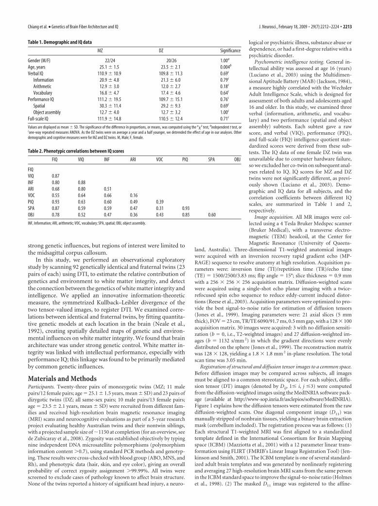
Psychometric intelligence testing. General in-tellectual ability was assessed at age 16 (years)(Luciano et al., 2003) using the Multidimen-sional Aptitude Battery (MAB) (Jackson, 1984),a measure highly correlated with the WechslerAdult Intelligence Scale, which is designed forassessment of both adults and adolescents aged16 and older. In this study, we examined threeverbal (information, arithmetic, and vocabu-lary) and two performance (spatial and objectassembly) subtests. Each subtest gave a rawscore, and verbal (VIQ), performance (PIQ),and full-scale (FIQ) intelligence quotient stan-dardized scores were derived from these sub-tests. The IQ data of one female DZ twin wasunavailable due to computer hardware failure,so we excluded her co-twin on subsequent anal-yses related to IQ. IQ scores for MZ and DZtwins were not significantly different, as previ-ously shown (Luciano et al., 2003). Demo-graphic and IQ data for all subjects, and thecorrelation coefficients between different IQscales, are summarized in Table 1 and 2,respectively.
Image acquisition. All MR images were col-lected using a 4 Tesla Bruker Medspec scanner(Bruker Medical), with a transverse electro-magnetic (TEM) headcoil, at the Center forMagnetic Resonance (University of Queens-
land, Australia). Three-dimensional T1-weighted anatomical imageswere acquired with an inversion recovery rapid gradient echo (MP-RAGE) sequence to resolve anatomy at high resolution. Acquisition pa-rameters were: inversion time (TI)/repetition time (TR)/echo time(TE) � 1500/2500/3.83 ms; flip angle � 15°; slice thickness � 0.9 mmwith a 256 � 256 � 256 acquisition matrix. Diffusion-weighted scanswere acquired using a single-shot echo planar imaging with a twice-refocused spin echo sequence to reduce eddy-current induced distor-tions (Reese et al., 2003). Acquisition parameters were optimized to pro-vide the best signal-to-noise ratio for estimation of diffusion tensors(Jones et al., 1999). Imaging parameters were: 21 axial slices (5 mmthick), FOV � 23 cm, TR/TE 6090/91.7 ms, 0.5 mm gap, with a 128 � 100acquisition matrix. 30 images were acquired: 3 with no diffusion sensiti-zation (b � 0, i.e., T2-weighted images) and 27 diffusion-weighted im-ages (b � 1132 s/mm 2) in which the gradient directions were evenlydistributed on the sphere (Jones et al., 1999). The reconstruction matrixwas 128 � 128, yielding a 1.8 � 1.8 mm 2 in-plane resolution. The totalscan time was 3.05 min.
Registration of structural and diffusion tensor images to a common space.Before diffusion images may be compared across subjects, all imagesmust be aligned to a common stereotaxic space. For each subject, diffu-sion tensor (DT) images (denoted by Dij, 1� i, j �3) were computedfrom the diffusion-weighted images using the MedINRIA software pack-age (available at http://www-sop.inria.fr/asclepios/software/MedINRIA).Figure 1 explains how the diffusion tensors were estimated from the rawdiffusion-weighted scans. One diagonal component image (D11) wasmanually stripped of nonbrain tissues, yielding a binary brain extractionmask (cerebellum included). The registration process was as follows: (1)Each structural T1-weighted MRI was first aligned to a standardizedtemplate defined in the International Consortium for Brain Mappingspace (ICBM) (Mazziotta et al., 2001) with a 12 parameter linear trans-formation using FLIRT (FMRIB’s Linear Image Registration Tool) (Jen-kinson and Smith, 2001). The ICBM template is one of several standard-ized adult brain templates and was generated by nonlinearly registeringand averaging 27 high-resolution brain MRI scans from the same personin the ICBM standard space to improve the signal-to-noise ratio (Holmeset al., 1998). (2) The masked D11 image was registered to the affine-
Table 1. Demographic and IQ data
MZ DZ Significance
Gender (M/F) 22/24 20/26 1.00a
Age, years 25.1 � 1.5 23.5 � 2.1 0.004b
Verbal IQ 110.9 � 10.9 109.8 � 11.3 0.69c
Information 20.9 � 4.8 21.3 � 6.0 0.79c
Arithmetic 12.9 � 3.0 12.0 � 2.7 0.18c
Vocabulary 16.8 � 4.7 17.4 � 4.6 0.64c
Performance IQ 111.2 � 19.5 109.7 � 15.1 0.76c
Spatial 30.3 � 11.4 29.2 � 9.3 0.69c
Object assembly 12.7 � 4.0 12.7 � 3.2 1.00c
Full-scale IQ 111.9 � 14.8 110.5 � 12.4 0.71c
Values are displayed as mean � SD. The significance of the difference in proportions, or means, was computed using the a�2 test, bindependent t test, orcone-way repeated measures ANOVA. As the DZ twins were on average a year and a half younger, we detrended the effect of age in our analyses. Otherdemographic and cognitive measures were for MZ and DZ twins. M, Male; F, female.
Table 2. Phenotypic correlations between IQ scores
FIQ VIQ INF ARI VOC PIQ SPA OBJ
FIQVIQ 0.87INF 0.80 0.88ARI 0.68 0.80 0.51VOC 0.55 0.64 0.66 0.16PIQ 0.93 0.63 0.60 0.49 0.39SPA 0.87 0.59 0.59 0.47 0.31 0.93OBJ 0.78 0.52 0.47 0.36 0.43 0.85 0.60
INF, Information; ARI, arithmetic; VOC, vocabulary; SPA, spatial; OBJ, object assembly.
Chiang et al. • Genetics of Brain Fiber Architecture and IQ J. Neurosci., February 18, 2009 • 29(7):2212–2224 • 2213

aligned T1-weighted MRI derived from part (1)with a 12-parameter linear transformation. Bycombining the transformations in (1) and (2), aconcatenated transformation was computed toalign the D11 image to the ICBM brain templateand also used to resample the D11 image to iso-tropic voxel resolution (dimension: 128 �128 � 93 voxels, resolution: 1.7 � 1.7 � 1.7mm 3). The resulting transformation parame-ters were used to rotationally reorient the tensorat each voxel (Alexander et al., 2001) and thenlinearly align the tensor-valued images based ontrilinear interpolation of the log-transformedtensors (Arsigny et al., 2005). The necessity ofprocesses involved in this step arises from thefact that the directional information at eachpoint in the diffusion images needs to be reori-ented when each subject’s data is aligned to astereotaxic space. (3) Since registration accu-racy in DTI may be improved by using the fulldiffusion tensor information in the cost func-tion that guides the registration, rather than itsscalar components (Park et al., 2003), we regis-tered all affine-registered DT images in (2) to arandomly selected subject’s DT image (a MZsubject), using an inverse-consistent fluid reg-istration algorithm that minimizes the symme-trized Kullback–Leibler divergence (sKL-divergence) of the two tensor-valued images(Chiang et al., 2008). Intuitively, this treats thelocal diffusion process in each subject as athree-dimensional (3D) field of probabilitydensity functions, and uses a fluid mapping,which is based on a standard measure from in-formation theory to quantify the discrepancybetween these probability density functions, todrive the tensor data into anatomical corre-spondence. Selection of an optimal registrationtarget in DTI studies is still an open question.Mori et al. (2008) generated the firstpopulation-averaged DTI atlas based on 81normal subjects linearly aligned to the ICBMspace. Since features in a single subject’s DTIare more similar to those in other individual DTimages than those in a multi-subject atlas, wechose a single subject’s DTI scan as the target,which may provide for enhanced registrationaccuracy (Christensen et al., 2006; Smith et al.,2006; Chiang et al., 2007).
For region-of-interest (ROI) analyses, masksof cerebral lobes, defined on the ICBM braintemplate according to a standardized anatomi-cal protocol (https://cms.loni.ucla.edu/NCRR/protocols.aspx), were mapped to the DT imagesby nonlinearly registering the ICBM template tosubjects’ T1-weighted MRI and were then trans-ferred from the T1-weighted MRI to the DT im-age using the affine transformation above. Thenonlinear registration was based on maximizingthe Jensen–Renyi divergence (JRD) of the jointintensity histogram, using a fully 3D fluid trans-formation which deforms the image according tothe laws of continuum mechanics (Chiang et al.,2007). All computational processes were executedusing a 306-node, dual-processor SUN Microsys-tems V20z cluster. Each compute node has a dual 64-bit 2.4 GHz AMDOpteron CPU. The registration processes were performed in parallel by sub-mitting one subject’s process to one compute node, and the average compu-tation time was �5.5 h by a single processor.
Definition of regions with high fractional anisotropy. A fractional anisot-ropy (FA) map was constructed from each fluidly registered DT image.FA is defined as the ratio of the SD to the root mean square of theeigenvalues of a diffusion tensor (Basser and Pierpaoli, 1996). Higher FAvalues indicate that water diffusion is more anisotropic, or more con-
Figure 1. a, In DTI, a set of diffusion-sensitized images is acquired, with diffusion-sensitizing magnetic field gradients of thesame strength, but oriented in different directions on an imaginary sphere. Each voxel in DTI contains multidimensional data (inour study, the dimension is 27). For example, b shows the raw diffusion data from a single voxel (the pink point in a) in alldiffusion-weighted images from one of the subjects. Water diffusion is antipodally symmetric along a specific direction, so onediffusion-sensitized image gives a pair of data points with identical values on the sphere. Each point on the sphere indicates theMR signal attenuation caused by water diffusion in the direction of the corresponding diffusion gradient, with greater signalattenuation (color-coded as red; magnitude of MR signal attenuation increases from yellow to red) indicating more rapid rates ofwater diffusion along that gradient direction. Water diffusion can be estimated by fitting the raw diffusion data in b to a diffusionellipsoid (c), whose axes correspond to the eigenvalue-eigenvector pairs of a diffusion tensor (Basser and Pierpaoli, 1996). Animage of the diffusion tensors estimated from all the voxels of the raw diffusion data is displayed in d (see Fig. 2 for details).Analysis of DTI scans is computationally expensive due to their high information content, especially for the covariance structureanalysis in twin studies, so it is more practical to first estimate anisotropy maps of scalar values, such as FA (e).
2214 • J. Neurosci., February 18, 2009 • 29(7):2212–2224 Chiang et al. • Genetics of Brain Fiber Architecture and IQ

strained by the ordered arrangement of myelinated fibers in the whitematter (Beaulieu, 2002). This is visualized in Figure 2, where the shape oftensor ellipsoids is more prolate in regions with higher FA, such as thegenu of the corpus callosum. FA also tends to be higher in regions that aremore heavily myelinated, and in regions where there is a dominant fiberdirection that constrains the directional profile of water diffusion. Weaveraged the FA images across all 92 subjects and restricted subsequentdata analysis to regions in which the average FA �0.3, as in Smith et al.(2006). This may sacrifice some of the information that DTI can provide.However, given the sample size in this pilot study (N � 92), we preferredto focus our regions of interest on major white matter fiber structures(Smith et al., 2006), as more highly anisotropic regions may have a bettersignal-to-noise ratio for measures derived from the diffusion images, andless residual misregistration error as a result of intersubject morpholog-ical variations. To reduce noise and improve the sensitivity of groupcomparisons (Leung et al., 2004; Smith et al., 2006), the FA map of eachsubject was smoothed using an isotropic Gaussian filter (FWHM � 12mm).
Univariate structural equation modeling. To estimate genetic and envi-ronmental twin correlations, and the relative contributions of additivegenetic (A), shared environmental (C), and unshared or unique environ-mental (E) components to the observed variable, y, we used structuralequation models (Fig. 3a) (SEM) (Neale et al., 1992; Schmitt et al., 2007).Shared (i.e., common) environment refers to aspects of the family rear-ing environment and other experiences shared by both siblings in a twinpair, while unshared environment refers to the unique experiences thateach twin does not share with their co-twin. For each twin, we modeledthe voxel value of FA as the sum of several latent factors, y � aA � cC �eE, where the variance of A, C, and E, respectively var(A) � a 2, var(C) �c 2, and var(E) � e 2, denotes their relative contribution to the variance ofthe observed variable, y, given that var( y) � a 2� c 2� e 2. The covarianceof trait y between the first and second twin in the same pair is a 2� c 2 forMZ twins, and (1/2)a 2� c 2 for DZ twins, because, on average, MZ twinsshare all and DZ twins share half of their genetic polymorphisms (theseare random variations in the DNA sequence that occur even across nor-mal individuals). Since A, C, and E are unobservable variables, the pathcoefficients � � (a, c, e) were estimated by comparing the variance-covariance matrix derived from the model above, and the sample covari-ance matrix from the observed values of y, using maximum-likelihoodfitting (Fornell and Larcker, 1981). Minus two times the log-likelihood
ratio, written as TML,�, follows a � 2 distributionwith p( p � 1) � t degrees of freedom, wherep � 2 is the number of observed variables, andt � 3 is the number of free model parameters.TML,� was estimated using the Broyden–Fletch-er–Goldfarb–Shanno (BFGS) method (Press etal., 2002). Acceptance of the null hypothesis( p � 0.05) indicates a good fit for the model.
We fitted the structural equation model tothe covariance of FA at each voxel to estimate ateach voxel the relative contributions of geneticand environmental factors to variance in whitematter integrity measures in the white matter.Since the MZ twins were slightly but signifi-cantly older than the DZ twins, the voxel FAvalue was adjusted for age and sex before modelfitting (for the univariate analysis here and alsothe cross-trait analysis below) (McGue andBouchard, 1984), to avoid inflation of thebetween-pair correlations and thus the esti-mated effects of shared environment due topossible associations between FA and age or sex.We also applied Levene’s test to each voxel(Brown and Forsythe, 1974) to test the assump-tion of homogeneity in variances across the MZand DZ twins, and found that there was no sig-nificant difference in variances between the twotypes of twins (FDR � 1.0; see below for detailsof FDR). It is standard in twin studies to exam-ine whether the observed measures are best
modeled using a combination of additive genetic, shared, and unsharedenvironmental factors, or whether only one or two of these factors issufficient to explain the observed pattern of inter-twin correlations. Westarted from the full model involving all 3 variance components, ACE: �� (a, c, e), and compared it with the more restricted models, AE: � � (a,e) and CE: � � (c, e). Under the rule of parsimony, AE or CE wereconsidered to fit the data better than ACE if their model-fitting p valuewas �0.05 (note that this differs from the more standard usage for pvalues in which values �0.05 are disregarded). In this case, we selectedAE or CE depending on which one had the smaller � 2 value, and theselected AE or CE model was further compared with E:� � (e) to deter-mine the best model. The significance of the genetic ( p(A)) and theshared environmental ( p(C)) factors was determined by the differencebetween the log-likelihood of the full (ACE) and the restricted (CE)model for p(A), and was determined by the difference between the log-likelihood of the full (ACE) and the restricted (AE) model for p(C). Thelog-likelihood for the full and the restricted models were denoted bylog(Lf) and log(Lr) respectively. Minus two times this difference, or�2{log(Lr) � log(Lf)}, is asymptotically distributed approximately as a� 2 distribution with one degree of freedom.
Linking diffusion anisotropy with intellectual performance. We usedrandom-effects regression models (RRM) (Hedeker et al., 1994) to mea-sure correlations between FA and IQ scores (including the sub-testscores). Ordinary regression methods are inappropriate here becauseobservations are correlated within twin pairs, violating the assumptionthat observations must be statistically independent. In RRM, this depen-dency is addressed by adding a random variable �i, to incorporate theclustering of the observed variables within the ith pair, into the ordinaryregression equations: yi � Xi � � 1i �i � �i.
Here, yi � the 2 � 1 vector of observed variables (FA) within the ithpair, � � a (q�1) � 1 vector of unknown regression coefficients, Xi � aknown 2 � (q � 1) covariate matrix, 1i � a 2 � 1 vector of ones, and �i
represents the 2 � 1 error vector. q was set to 3, with the subjects’ IQscore, age, and sex as the covariates. We estimated these unknown pa-rameters (� and �i) based on maximum marginal likelihood (MML)methods detailed in (Hedeker et al., 1994). The significance of the corre-lation between FA and IQ was determined by comparing the full (IQ, age,and sex) and the restricted (age and sex only) models, as described above.
Cross-trait cross-twin analysis of FA and intelligence. For IQ scores that
Figure 2. The image of diffusion tensors (a) selected from the brain region at the junction of the corpus callosum and thecorona radiata, shown as the yellow box in the corresponding FA image (b) of the same subject as in Figure 1. Diffusion tensors arevisualized as ellipsoids (which have been normalized to unit mass) that are color coded, as is conventional, to represent theorientation of the normalized principal eigenvector (dominant direction of water diffusion) relative to the medial–lateral axis(coded in red), anterior–posterior axis (coded in green), and superior–inferior axis (coded in blue) of the anatomical referenceframe. Glyphs of tensor ellipsoids were generated using the visualization software “BrainSuite” (http://www.loni.ucla.edu/Software/) (Shattuck et al., 2008).
Chiang et al. • Genetics of Brain Fiber Architecture and IQ J. Neurosci., February 18, 2009 • 29(7):2212–2224 • 2215

were significantly correlated with FA (with FDR �0.05), we performed a“cross-trait cross-twin” analysis to detect common genetic or environ-mental factors influencing both fiber architecture and intelligence.Cross-trait cross-twin analyses estimate the genetic and environmentalcontributions to the correlations between two phenotypes (FA and IQ inthis study) in the same set of subjects (Posthuma et al., 2002; Hulshoff Polet al., 2006). The path diagram is shown in Figure 3b. If the correlationbetween the voxel value of FA in one twin and the level of IQ in the othertwin (“cross-trait” and “cross-twin”) is greater in MZ pairs than in DZpairs, the excess in the MZ correlation over the DZ correlation is thenassumed to be attributable to common genetic factors that mediate bothwhite matter integrity and intelligence. Formally, we modeled the geneticand environmental contributions to FA and intellectual performance forsubject j ( j � 1 or 2) by defining xj � axAxj � cxCxj � exExj, and yj � ayAyj
� cyCy j� eyEyj, where x stands for the voxel value of FA, y for IQ, and A,C, and E respectively denote the additive genetic, shared and unsharedenvironmental components. Cross-trait correlations between FA and in-tellectual performance were then derived from the covariance matrix ofvector w � (x1, x2, y1, y2)T, where T denotes transpose, given by thefollowing:
covw � � �xx �xy
�xy �yy�,
where �xx and �yy are the covariance matrices for phenotype x or ybetween subject 1 and 2, as was described in the univariate SEM above.�xy is the cross-trait covariance matrix, composed of the covariancebetween the two traits within the same individual (cov(x1, y1) and cov(x2,y2)), and the cross-trait cross-twin covariance (cov(x1, y2) and cov(x2,y1)), as detailed below:
�xy � � covx1,y1 covx1,y2covx2,y2 covx2,y2
�� � raaxay � rccxcy � reexey raaxay � rccxcy
raaxay � rccxcy raaxay � rccxcy � reexey�,
where � 1 for MZ twins, and 1/2 for DZ twins. ra, rc, and re are thecross-trait correlation coefficients for Ax and Ay, Cx and Cy, and Ex andEy. A higher value of ra indicates that the two phenotypes are more likelymediated by a common set of genes (Lange and Boehnke, 1983; Seemanet al., 1996). The path coefficients were estimated by comparing thecovariance matrix implied by the model and the sample covariance ma-trix of the observed variables, using maximum-likelihood fitting to give a� 2 value. We started from the full set of path coefficients (ax, cx, ex, ay, cy,ey, ra, rc, re) and removed one of ax, cx, ay, and cy from the model step bystep. Removing ax or ay/cx or cy also removed ra/rc. e1, e2 and re werealways kept in the model to include random noise. Selection of submod-els was determined by the rule of parsimony, i.e., a model was considered“better” if the difference in � 2 values between it and the more compre-hensive model at the previous step was not significant. If two modelscontained the same number of parameters, the model with a smaller � 2
value was considered better. Model selection ended when the best modelwas achieved, i.e., when either (1) all possible more restricted modelswere not better than the current model or (2) the current model con-tained e1, e2 and re only. If ra or rc was included in the best model, thesignificance of ra or rc was then determined by comparing the � 2 values ofthe best model and its submodel where ra (or rc) � 0. The cross-traitmodel fitting was performed only at voxels where the FDR-adjusted pvalue (see below) of the correlation between FA and IQ was �0.05.
Correction for multiple comparisons. All statistical maps in this studywere further assessed using the false discovery rate method (FDR) (Ben-jamini and Hochberg, 2000; Storey and Tibshirani, 2003) to correct formultiple comparisons. FDR is defined as the expected proportion of falsepositive findings out of all rejected tests. For example, FDR � 0.05 meansthat 5% of the voxels that are identified to be significant are in fact falsepositive findings. Here, we consider the test statistic at a given voxel to besignificant when its p value is not greater than a primary threshold set to0.01. The 5% is an estimate that will be true on average when the methodis used, as the FDR method controls the expected false discovery rate, or
the expected proportion of false positives in the map. In other words, it isexpected that, on average using this method, 95% of the voxels labeled assignificant will be true positive findings. Mathematically, FDR() �m0�/S(), where S() is the number of voxels labeled as significant. m0 isthe total number of voxels where the null hypothesis is true, which isunknown and has to be estimated from the empirical distribution of thevoxel p values (Storey and Tibshirani, 2003). is the rejection rate for thenull hypotheses, so m0� equals the number of false positive voxels. As isconventional in brain mapping studies, statistical maps with an FDRvalue �0.05 were considered to reach overall significance. To visualizebrain regions where the test statistic is significant after correction formultiple comparisons, we constructed maps of FDR-adjusted p values, sothat if we consider voxels with the adjusted p value � � to be significant,then the FDR across the statistical map will be less than � (� was set to0.05 by convention). FDR-adjusted p values can be estimated as follows:Let p(1) � p(2) � … � p(m) be the rank-ordered p values of all m voxels,then the FDR-adjusted p value for voxel m is given by p (m) � FDR( p(m)),and p (i) � min(p(i�1), FDR( p(m))), for voxels i � m � 1, …, 1, sorted indescending order (Storey and Tibshirani, 2003).
ResultsMaps of genetic influences on white matter integrity (FA)Figure 4 reveals the profile of genetic versus environmentalinfluences on white matter integrity (as quantified by FA). Thefirst column shows the significance of the genetic effect, whilethe remaining 3 columns show the proportion of the variancein the 92 twins attributable to genetic, common and uniqueenvironmental factors. Clearly, white matter integrity is undersignificant genetic control in all posterior white matter regions(Fig. 4, first column), with high heritability values (a 2). Theheritability of white matter integrity may be regarded as theproportion of the observed variation in FA values that may beattributed to genetic differences among individuals. Geneticfactors explained between 75 and 90% of the observed vari-ance in white matter integrity in almost all white matter re-gions (Fig. 4, second column).
White matter integrity, as measured by FA, was under stronggenetic control in the genu and the splenium and part of body ofthe corpus callosum, the right cerebral peduncle, and right infe-rior longitudinal fasciculus (ILF)/inferior fronto-occipital fascic-ulus (IFO), the anterior limbs of the internal capsule bilaterally(which contain the corticobulbar tract and superior thalamic ra-diations). Other regions with strong genetic control of white mat-ter integrity included the left posterior thalamic radiation/opticradiation, the superior longitudinal fasciculus bilaterally, and thesuperior and posterior corona radiata (FDR for p(A) � 0.01 overthe whole brain, including only voxels with FA �0.3). The voxel-level maps p(A) were significant only in regions with extremelyhigh heritability values near 100% (this can be seen by comparingcolumns 2 and 1 of Fig. 4, which show heritability values through-out the white matter, and their significance). Contributions fromshared environmental factors were not detectable (FDR for p(C)
� 1.00).
Genetic influences in lobar subregionsBecause the pattern of genetic versus environmental influencesmay differ in different lobes of the brain, we also fitted geneticmodels to the regional average values of FA in regions of interestdefined by the ICBM atlas. As shown in Table 3 and Figure 5, theaverage FA in white matter (defined as the region with groupmean FA �0.3) served as the observed variable for the ACE struc-tural equation models. The highest heritability values were ob-served in the parietal lobes bilaterally, with a 2 reaching 84.8% onthe left and 84.3% on the right. Parietal fiber integrity is thereforeunder strong genetic control. When voxel FA data summarized in
2216 • J. Neurosci., February 18, 2009 • 29(7):2212–2224 Chiang et al. • Genetics of Brain Fiber Architecture and IQ

lobar regions were fitted for the full ACEmodel, the shared environmental effectswere detected but not significant at the lefttemporal lobe and the right occipital lobe,and the effects of unique environmentwere relatively small, except for the rightoccipital lobe where additive genetic,shared environmental, and unique envi-ronmental effects were approximatelyequal. Only the genetic and the unique en-vironmental effects were detected in thebest-fitting model. The genetic influencewas found to be significant in bilateral pa-rietal lobes (both with p(A) � 0.001), bilat-eral frontal lobes ( p(A) � 0.04, for the leftfrontal lobe; p(A) � 0.006, for the rightfrontal lobe), and for the left occipital( p(A) � 0.003) lobe (Table 3).
Mapping phenotypic and geneticcorrelations between FA and IQ scoresWhite matter integrity, measured by FA,was significantly correlated with fullscale IQ, and also with performance IQ,but not with verbal IQ. Among all IQscores tested using RRM, with age andsex controlled, the full-scale IQ (FIQ),performance IQ (PIQ) and one of itssub-tests, object assembly (OBJ), werefound to correlate significantly with FA(Table 4). Because correlations are plot-ted voxel by voxel across the brain, thesignificance of the overall pattern of cor-relations, adjusted for multiple compar-isons, is assessed using the false discov-ery rate value, with values �0.05 beingregarded as significant.
Figure 6 shows the pattern of correla-tions between each cognitive measure andwhite matter integrity. It is clear that thereis a pervasive correlation between whitematter integrity in almost all regions andintellectual performance. When the corre-lations are assessed within the corpus cal-losum at midline, there is a prominent re-gion of correlation (r � 0.3– 0.5 locally)between each cognitive score and whitematter integrity at the callosal isthmus. Such a correlation mightbe considered surprisingly high for an image-derived measure,and the commissural fibers carried in the callosal isthmus inner-vate the parietal cortices, which may be recruited in tasks such asobject assembly.
Specifically, FIQ, PIQ, and OBJ scores were positively corre-lated with white matter integrity, as measured by FA, in the isth-mus of the corpus callosum and the cingulum (r � 0.3– 0.4), inthe cerebral peduncles bilaterally (for PIQ and OBJ scores), ILF/IFO (for the OBJ score), bilateral posterior limbs of the internalcapsule (which contain the corticobulbar and corticospinal tractsand the superior thalamic radiations) and in the left posteriorthalamic radiation/optic radiation, right superior fronto-occipital fasciculus, and bilateral anterior, superior and posteriorcorona radiata (Fig. 6). The effect size for these correlations withwhite matter integrity was greatest for the object assembly sub-
scale, with highest effect sizes in the parietal lobe. Performance onthis task may benefit from increased myelination, which may en-hance physiological parameters such as axonal conduction speedand may also be reflected in diffusion-sensitive indices such as FA.
We further subdivided the cross-trait phenotypic correlationbetween FA and FIQ, PIQ, or OBJ into genetic (ra), and shared(rc) and unique (re) environmental components. As there is astrong genetic influence on fiber architecture throughout thebrain, perhaps most prominently in the parietal lobes, and giventhe correlation between IQ scores and white matter integrity inthe same region, it is plausible that overlapping sets of genes mayinfluence IQ measures and fiber architecture. One common wayto determine whether partially overlapping genes affect two dif-ferent traits (here FA and IQ) is to use a measure of one trait inone twin to predict the level of the other trait in the other twin. Ifsuch a prediction can be made with greater precision in identical
Figure 3. Path diagrams for (a) univariate (for FA) and (b) bivariate cross-trait (for FA and IQ) structural equation models (aand b are adapted from Neale et al., (1992) and Miles et al., (2002) respectively. a, Each twin’s phenotypic measure (e.g., FA in thisstudy) is assumed to be determined by additive genetic (A) and environment factors. Environmental factors are further subdividedinto those that the co-twins share (denoted by C, e.g., family rearing environment, and common developmental factors in utero),and those that are unique for each twin (denoted by E, e.g., they may be educated at different schools). Contributions of the ACEfactors to the phenotype are indicated by single arrows and assumed to be the same between co-twins (twin 1 and twin 2) forsimplification, with their weights, or path coefficients, denoted by a, c, and e, respectively. Random noise, or experimentalmeasurement error, is included in component E, and assumed to be independent between twin 1 and 2 (no correlation). Corre-lations in the genetic and shared environmental factors between co-twins are indicated by double arrows. For A1 and A2, thecorrelation coefficient is equal to 1 for MZ and 0.5 for DZ twin pairs. The correlation coefficient between C1 and C2 is always 1 fromthe definition of the shared environment. b, In bivariate cross-trait SEM, we assume that there are common genetic and environ-mental factors that affect various phenotypes within an individual. Here, we only consider factors that affect two phenotypes, suchas FA and IQ. The effects of these common factors are estimated by comparing the difference between MZ and DZ pairs, in thecorrelation between the two phenotypes within the same individual (cross-trait within-individual), and also in the correlationbetween one phenotype in one twin, and the other phenotype in the other twin (cross-trait cross-twin). The cross-trait within-individual correlation (the correlation between FA and IQ in twin 1 or in twin 2, shown as connected by gray arrows) is divided intoadditive genetic, and shared and unique environmental components (e.g., AFA, i, CFA, i, and EFA, i for FA, and AIQ, i, CIQ, i, and EIQ, i forIQ; i � 1 or 2 for twin 1 or 2), and the correlation coefficients between AFA, i and AIQ, i, CFA, i and CIQ, i, and EFA, i and EIQ, i, are denotedby ra, rc, and re, respectively. The cross-trait cross-twin correlation is shown as AFA, i and AIQ, j, and CFA, i and CIQ, j connected by blackarrows, for FA in twin i and IQ in twin j, where i, j � 1 or 2, and i j. There is no re term for EFA, i and EIQ, j, because the uniqueenvironmental factors between subjects are not correlated. The covariance across the two phenotypes within the same subject, orseparately in the two subjects, is then derived by multiplication of the path coefficients for the closed paths in the path diagram.For example, covariance between FA in twin 1 and IQ in twin 2 is equal to aFA�ra�aIQ�cFA�rc�cIQ for MZ twins, and aFA�1/2ra�aIQ�cFA�rc�cIQ for DZ twins. This implies that any excess in cross-trait cross-twin correlation in MZ twins over that in DZ twinsis attributed to common genetic factors that affect both FA and IQ. Correlations for the same phenotype (FA or IQ) betweenco-twins, as have already been described in a, are not shown for simplification.
Chiang et al. • Genetics of Brain Fiber Architecture and IQ J. Neurosci., February 18, 2009 • 29(7):2212–2224 • 2217

than fraternal twins, then a common set of genes must beinvolved.
Figure 7 shows these cross-trait maps, which suggest thatthe IQ of one twin may be predicted, with a high degree ofprecision, from the white matter integrity of the other twin.An overlapping set of genes therefore mediates this correla-tion. FA and FIQ, PIQ or OBJ scores were influenced by anoverlapping set of genes in the cingulum and isthmus of thecorpus callosum, the cerebral peduncles and ILF/IFO (for
OBJ), the posterior limbs of the internal capsule and the leftposterior thalamic radiation/optic radiation (for PIQ andOBJ), the right superior fronto-occipital fasciculus, and theanterior, superior and posterior (for PIQ and OBJ) coronaradiata bilaterally (Fig. 7) (FDR for p(ra � 0) � 0.015 for FIQ,0.027 for PIQ, and 0.016 for OBJ; negative ra was not signifi-cant). We did not find any significant effects for shared envi-ronmental factors between intellectual performance and whitematter integrity (rc).
Figure 4. Maps of genetic influences on white matter integrity. These maps show how different factors, genetic or environmental, explain different proportions of the observed variance in whitematter integrity across 92 subjects. The Montreal Neurological Institute (MNI) coordinate (which is also the coordinate used in the ICBM space; expressed in mm) of the slices is indicated at thebeginning of each row. The first column shows the statistical significance of the genetic influences ( p(A) , FDR-adjusted), and the proportional contributions to the overall variance in FA, from genetic(a 2), shared environmental (c 2) and unique environmental (e 2) factors (the second to the last columns, computed under the best-fitting statistical model). The genetic effect is detected in the genuand the splenium of the corpus callosum (x � 0), the right cerebral peduncle, and right ILF/IFO (z � �14), the anterior limbs of the internal capsule bilaterally, and the left posterior thalamicradiation/optic radiation (z � 5), the superior longitudinal fasciculus bilaterally (z � 22), and the superior and posterior corona radiata bilaterally (z � 35). A, Anterior; R, right.
2218 • J. Neurosci., February 18, 2009 • 29(7):2212–2224 Chiang et al. • Genetics of Brain Fiber Architecture and IQ

Comparisons of registration accuracy between MZ andDZ twinsIt is certainly possible in theory that the overall accuracy of cross-subject anatomical registration is better in the group of MZ twinsthan in the group of DZ twins. Even so, the smaller intra-pairmorphological variations in the MZ twins does not imply that thecross-subject variance is less within the group of MZ twins as awhole; in fact, they are likely to be as variable in anatomy acrosssubjects as randomly picked pairs of unrelated individuals, of thesame age and sex. It is worth noting that the twins’ images are notregistered by aligning one twin’s image directly to the other twin’s
image; instead, they are each individually aligned to a commontarget image from a randomly selected control subject who is nota member of the twin pair. As such, because the MZ twins vary asmuch, as a group, as the DZ group (and the general population),what matters most for registration is how much they deviate fromthe target image. As the target image was not the other member ofthe pair (except for the co-twin of the subject who served as thetarget), but an unrelated individual, a zygosity � registrationerror interaction is unlikely. Even so, there remains the empiricalpossibility that registration errors to the common target imagewere not the same for each twin group. To test whether this wasthe case in our subjects, we compared the registration accuracyfor the groups of MZ twins versus the group of DZ twins usingthree overlap measures:Total overlap:
Total overlap: TO �|Sr � Tr|
|Tr|,
Mean overlap:
Mean overlap: MO �2|Sr � Tr|
|Sr| � |Tr|,
Union overlap:
Union overlap: UO �|Sr � Tr|
|Sr � Tr|,
where Sr is the mask derived from regions where FA �0.3 in thesource DTI and then warped by the fluid deformation fields thatregistered the source DTI to the target DTI, and Tr is the maskderived from regions where FA �0.3 in the target DTI. |.| indi-cates the volume computed as the number of voxels. The mea-
Table 3. Twin correlations and variance components estimated from the values of the lobar FA
Twin correlationsPercentage of variance estimated under thebest model Percentage of variance estimated under the full model
p(A) p(C)MZ DZ a2 c2 e2 a2 c2 e2
Frontal-L 0.45* (0.07, 0.72) 0.01 (0, 0.28) 55.0 (15.3, 100.0) � 45.0 (24.7, 85.2) 55.0 (4.6, 100.0) 0.0 (0, 32.2) 45.0 (24.7, 85.2) 0.04 1.0Frontal-R 0.65* (0.34, 0.83) 0.05 (0, 0.43) 73.5 (37.0, 100.0) � 26.5 (14.7, 53.2) 73.5 (30.0, 100.0) 0.0 (0, 31.5) 26.5 (14.7, 53.2) 0.006 1.0Parietal-L 0.79* (0.58, 0.90) 0.02 (0, 0.41) 84.8 (52.1, 100.0) � 15.2 (8.5, 31.4) 84.8 (51.1, 100.0) 0.0 (0, 25.7) 15.2 (8.5, 31.4) < 0.001 1.0Parietal-R 0.80* (0.60, 0.91) 0.12 (0, 0.49) 84.3 (53.0, 100.0) � 15.7 (8.9, 31.2) 84.3 (48.2, 100.0) 0.0 (0, 33.4) 15.7 (8.9, 31.2) < 0.001 1.0Temporal-L 0.74* (0.49, 0.88) 0.56* (0.21, 0.78) 83.0 (54.7, 100.0) � 17.0 (10.2, 31.1) 43.9 (0, 100.0) 38.8 (0, 96.3) 17.3 (10.1, 33.4) 0.06 0.19Temporal-R 0.67* (0.37, 0.84) 0.19 (0, 0.54) 65.2 (34.8, 100.0) � 34.8 (21.1, 61.4) 65.2 (0, 100.0) 0.0 (0, 75.2) 34.8 (21.1, 62.7) 0.10 1.0Occipital-L 0.65* (0.35, 0.83) 0.03 (0, 0.42) 75.6 (39.6, 100.0) � 24.4 (13.5, 49.4) 75.6 (35.4, 100.0) 0.0 (0, 28.8) 24.4 (13.5, 49.4) 0.003 1.0Occipital-R 0.61* (0.28, 0.81) 0.42* (0.03, 0.70) 65.0 (35.2, 100.0) � 35.0 (21.4, 61.2) 33.6 (0, 100.0) 30.1 (0, 95.7) 36.3 (21.5, 65.1) 0.36 0.44
For each lobe, the average FA in white matter (defined as FA � 0.3) served as the observed variable for the ACE structural equation models. Values in parentheses indicate the 95% confidence interval, computed using the likelihood-basedmethod (Neale and Miller, 1997). Statistical significance of the relative contributions to the total variance of the lobar FA by the genetic (A) and the shared environmental (C) factors is denoted by p(A) and p(C), presented in bold font if p(A)
or p(C) � 0.05. Twin correlations were computed based on the analysis of variance for clustered measures, by comparing the between-group (each twin pair was treated as an individual group) variation and the total variation in values ofthe lobar FA (Ukoumunne, 2002). *p � 0.05. L, Left; R, right.
Figure 5. Proportions of variance in white matter integrity in different lobes of the brain(average lobar values of FA) due to genetic (a 2), shared environment (c 2) and unique environ-ment (e 2) components, under the full (a) and the best-fitting model (b). For each lobe, theaverage FA in white matter (defined as FA �0.3) served as the observed variable for the ACEstructural equation models. *p(A) � 0.04; **p(A) � 0.006, #p(A) � 0.001; ##p(A) � 0.001; §p(A)
� 0.003. p(A) in other lobes and p(C) are not significant. These data show the dominant effect ofgenes on white matter integrity and provide little evidence to support a strong environmentaleffect (that is independent of genetic variation). L, Left; R, right.
Table 4. Overall significance of correlations between FA and IQ scores
FDR valuesa
Verbal IQ 1.00Information 0.67Arithmetic 1.00Vocabulary 1.00
Performance IQ 0.01Spatial 0.13Object assembly 0.007
Full-scale IQ 0.04
FDR � 0.05 (presented in bold font) indicated that 5% of the voxels in which FA was found to be associated with theIQ score are in fact false positive findings. In other words, 95% of the associations labeled are true. The maps ofnegative correlations between each of these measures and FA were not significant.aFDR was computed only in regions where the individual IQ score correlated positively with FA.
Chiang et al. • Genetics of Brain Fiber Architecture and IQ J. Neurosci., February 18, 2009 • 29(7):2212–2224 • 2219

sures of overlap can be used to determine how well anatomy isaligned to the common template across a group of subjects. Im-portantly, there were no statistically significant differences be-tween the MZ group and DZ group for any of the overlap mea-sures (Mean � SD, MZ twins vs DZ twins: 0.527 � 0.059 vs0.512 � 0.047 for TO, 0.499 � 0.046 vs 0.495 � 0.042 for MO,0.334 � 0.038 vs 0.329 � 0.035 for UO). As such, we do notexpect the higher FA affinity among MZ pairs to be attributable tosystematic differences in cross-subject registration accuracy, atthe group level, between the two types of twins.
DiscussionThis study had three main findings. First, white matter integritywas under strong genetic control; genetic factors explained al-most 80% of the variance, with highest heritability in parietalbrain regions. Second, white matter integrity was linked withintellectual performance, with correlations as high as 0.3– 0.4 be-tween performance IQ and white matter integrity. Third, using across-trait mapping approach, we implicated the same genes asmediating the correlation between IQ and white matter integrity,suggesting a common physiological mechanism for both.
Fiber architecture in most major white matter structures washighly heritable, consistent with prior MRI-based reports thatbrain morphometry is also highly heritable (Toga and Thomp-
son, 2005). Over 50% of the variation in white matter integritywas due to genetic influences in the genu and the splenium of thecorpus callosum, cerebral peduncles, right inferior longitudinalfasciculus/inferior fronto-occipital fasciculus, anterior limbs ofthe internal capsule/corticobular tract/superior thalamic radia-tion, left posterior thalamic radiation/optic radiation, the supe-rior longitudinal fasciculus, and the corona radiata.
Our finding that FA is highly heritable in the genu and sple-nium of the corpus callosum is consistent with the only otherstudy using diffusion imaging in twins. Pfefferbaum et al. (2001)found that genetic contributions explained 67% and 49% of thetotal variance in FA in the splenium and genu, respectively. In ourstudy, variance in FA was heavily influenced by genetic factors (a 2
constituted �50 – 80% of total variation). FA may reflect under-lying levels of axonal myelination, which may account for differ-ences in reaction times, processing speed, and intellectual perfor-mance across subjects. As noted by Neale et al. (1992) and manyothers, a genetic effect on brain architecture does not imply thatenvironmental factors are unimportant for development; inmany cases beneficial genes and environmental factors are highlycorrelated; talented individuals, for example, may tend to seekout activities and environments that in turn promote improvedbrain function (see Gray and Thompson, 2004, for a discussion of
Figure 6. Association between FA and FIQ, PIQ and OBJ. Partial correlation coefficients are shown between white matter integrity (measured using FA) and the intellectual performance scores:FIQ, PIQ, and OBJ. The MNI coordinate (mm) of the slices is indicated at the beginning of each row. All statistics are controlled for age and sex, and are computed voxelwise for subjects randomlyselected, one from each pair (the left column in each group shows the correlation coefficient, and the right column shows its significance). Overall significance is measured by FDR-adjusted p valuesobtained from the random-effects regression model (RRM), as RRM also estimated the effect of twin pairing. Significant positive correlation between FA and FIQ, PIQ or OBJ was detected in theisthmus of the corpus callosum and the cingulum bundle (x � 3), the bilateral cerebral peduncles (for PIQ and OBJ) and ILF/IFO (for the object assembly test, OBJ) (z � �14), the posterior limbsof the internal capsule bilaterally and left posterior thalamic radiation/optic radiation (z � 3), the anterior corona radiata and right superior fronto-occipital fasciculus (z � 20), and the superior andposterior corona radiata bilaterally (z � 35). Negative correlations were not significant. Regions where FA is associated with OBJ are the most extensive and have the greatest effect sizes.
2220 • J. Neurosci., February 18, 2009 • 29(7):2212–2224 Chiang et al. • Genetics of Brain Fiber Architecture and IQ

gene � environment interactions, and IQ). Moreover, sharedenvironmental influences on FA may not be totally excluded,because the classical twin design may slightly overestimate heri-tability by attributing the excess in the MZ pair correlation overthe DZ pair correlation only to genetic effects (Grayson, 1989;Hopper and Visscher, 2005).
Imaging measures, such as FA, that are heritable and link withcognition may empower the search for specific genetic polymor-phisms (quantitative trait loci) that impact white matter struc-ture. A specific variant of the Neuregulin 1 (NRG1) gene has beenassociated with reduced white matter density and anisotropy(McIntosh et al., 2008), and in another study, hypomyelinationin the hippocampus and optic nerves was observed when theBace1 gene was knocked out (Hu et al., 2006). The associationbetween diffusion imaging measures and IQ may stem from thefact that both are sensitive to underlying levels of myelination,with common set of genes influencing them both.
Higher FA was linked with higher FIQ, PIQ or OBJ in theisthmus of the corpus callosum, the cingulum, cerebral pe-duncles, ILF/IFO, posterior limbs of the internal capsule bilater-ally, left posterior thalamic radiation/optic radiation, right SFO,and the anterior, superior and posterior corona radiata bilater-ally. The correlation coefficient was around 0.3– 0.4 in most re-
gions, close to the correlation coefficient observed between FIQ/PIQ and frontal gray matter volumes (Thompson et al., 2001),white matter density (Hulshoff Pol et al., 2006), and white matterFA (Schmithorst et al., 2005). Correlations between FA and FIQ,PIQ or OBJ in most of these regions were mediated by commongenetic factors. The fiber systems whose integrity was most tightlylinked with IQ include several with critical roles in visuospatialprocessing. The callosal isthmus interconnects bilateral primaryvisual and visual association areas of the parietal and occipitalcortex, and is involved in visuospatial memory (Begre et al.,2007). The cingulate gyrus, and the cingulum bundle that runswithin it, integrate information regarding motivation, error eval-uation, and emotion, and modulate cognitive, motor, endocrineand visceral responses (Bush et al., 2000). Gray matter density inthe anterior cingulate is associated with FIQ (Wilke et al., 2003;Frangou et al., 2004; Haier et al., 2004), whereas the mean diffu-sivity in the posterior cingulate is correlated with cognitive abilityin patients with Alzheimer’s disease (Yoshiura et al., 2002). Thesuperior and inferior fronto-occipital fasciculus, tracts whose in-tegrity is highly heritable, are part of a widely distributed visuo-spatial attention network. The superior fronto-occipital fascicu-lus innervates the dorsolateral prefrontal cortex, while theinferior fronto-occipital fasciculus innervates the ventrolateral
Figure 7. Cross-trait genetic correlations between FA and FIQ, PIQ and OBJ scores, with maps of correlation coefficients ra, corrected for age and sex (left), and the FDR-adjusted values for thesignificance of ra (right). The MNI coordinate (mm) of the slices is indicated at the beginning of each row. For each of these IQ scores, cross-trait analysis was limited to the brain regions wherephenotypic correlation between FA and that IQ score was significant. Common genetic factors affect both FA and FIQ, PIQ or OBJ in the cingulum and isthmus of the corpus callosum, a commissuralpathway innervating the parietal cortex, which is crucial for multi-modal sensory integration (x � 3), the cerebral peduncles (for OBJ) and ILF/IFO (right � left, for OBJ) (z � �14), the posteriorlimbs of the internal capsule and the left posterior thalamic radiation/optic radiation (for PIQ and OBJ) (z � 3), bilateral anterior corona radiata and right superior fronto-occipital fasciculus (z � 20),and bilateral superior and posterior corona radiata (for PIQ and OBJ) (z � 35). There were no negative genetic correlations between IQ scores and white matter integrity, i.e., negative values of ra
were not significant.
Chiang et al. • Genetics of Brain Fiber Architecture and IQ J. Neurosci., February 18, 2009 • 29(7):2212–2224 • 2221

prefrontal and medial orbitofrontal cortex (Wakana et al., 2004;Makris et al., 2007). Disconnection of inferior fronto-occipitalfasciculus can cause visual neglect (Urbanski et al., 2008).
Our findings are consistent with morphometric data by Lud-ers et al. (2007, 2008), who found that the midsagittal thickness atthe isthmus of the corpus callosum was positively correlated withFIQ, PIQ, and VIQ, with greatest effect sizes for FIQ and PIQ.Hines et al. (1992) also observed that the midsagittal surface areaof the genu and isthmus of the corpus callosum positively corre-lated with visuospatial ability. Moreover, Yu et al. (2008) foundthat higher values of FA in the optic radiation and pyramidaltract, were associated with higher PIQ. Our results also buildupon the voxel-based morphometry study by Hulshoff Pol et al.(2006), in which the white matter density, a measure derivedfrom structural MRI, in the superior fronto-occipital fasciculus,corpus callosum, and left optic radiation was correlated with PIQ;such correlations were attributable more to genetic influencesthan to environmental factors. However, we failed to find anysignificant correlation between our white matter measures andverbal IQ. It may be that performance IQ, rather than verbal IQ,is more tightly associated with physiological parameters such asnerve conduction velocity, which may be linked with diffusionanisotropy, and is sensitive to the level of axonal myelination(Arbuthnott et al., 1980; Beaulieu, 2002; Tyszka et al., 2006). Infuture, we will explore associations between white matter integ-rity and other cognitive measures, specifically processing speed,in a larger sample.
Certain disorders provide exceptions to the theory that highfiber anisotropy correlates with high IQ. Individuals with Wil-liams Syndrome, which is characterized by impaired visuospatialability but relatively intact language production, have profoundlylower PIQ than typically developing subjects, but FA in theirbilateral superior and inferior longitudinal fasciculus, and rightsuperior fronto-occipital fasciculus was significantly higher(Hoeft et al., 2007). The authors attributed this to increased my-elination and abnormal myelin packing in Williams syndrome.Consequently, there may be a complex relationship between fiberarchitecture and intelligence.
Several issues remain in our study. First, although the brainarchitecture of identical twins is more similar than the brain ar-chitecture of fraternal twins, we found, using overlap measures,that the overall registration accuracy was not statistically better inidentical than in fraternal twins. This is because the registrationaccuracy to a common template depends more on the groupvariance than the intra-pair differences, as all twins are registeredto a target image of an individual who is not related to them(except for the co-twin of the subject who served as the target).Even so, further empirical studies of zygosity � registration errorinteractions are of interest in heritability studies, because whenthey are significant, they could lead to inflated estimates of heri-tability. Second, the age range of our subjects is narrow and assuch we could not model the influence of age on heritability(Wallace et al., 2006; Lenroot et al., 2009). In a pediatric twinsample, the heritability of cortical thickness decreased with age inearly developing brain areas, such as primary motor and sensorycortices, but increased with age in later-maturing areas, e.g., theprefrontal cortex (Lenroot et al., 2009) (see also Gogtay et al.,2004, for maps of the cortical maturational sequence). This wasascribed to the effects of age-dependent gene expression and thetemporal variation in gene-environment correlations. Third, thecurrent study is based on voxel-by-voxel analysis of diffusionimages, in which regional characteristics of white matter fibersare summarized and assessed at a macroscopic scale (2 mm), but
no actual tracking of white matter fibers was performed. Futurestudies using tractography (Wakana et al., 2004; Deriche andDescoteaux, 2007; Makris et al., 2007; Frey et al., 2008; Lenglet etal., 2008) may be able to answer questions about genetic influ-ences on specific fiber pathways tracked in the images, such as thearcuate fasciculus or cingulum bundle, for example. Neverthe-less, tract labeling is somewhat labor intensive at present, and thespatial scope of the tractography analysis can be somewhat lim-ited, given the need to select fiber tracts of interest. Last, it may beoversimplified to use a scalar measure like FA to describe whitematter architecture, as the directional information of water dif-fusion is not included. Future studies may be able to fit full mul-tivariate structural equation models (Neale et al., 1992), to in-clude the full diffusivity information in the diffusion tensors.However, this is very computationally expensive as the number ofequations to solve is (2 � 6) � ((2 � 6) � 1) � 156 to fit the (a,c, e) parameters in the SEM for diffusion tensors.
In conclusion, we reported the first maps of genetic influenceson fiber architecture in the living brain, based on a populationstudy of diffusion tensor images. Major white matter fiber path-ways are highly genetically controlled, and higher diffusion an-isotropy was linked with superior intellectual performance inseveral key systems. These algorithms and results may help toguide future studies to detect individual genes contributing tofiber architecture, white matter integrity and cognition.
ReferencesAboitiz F (1992) The origin of the mammalian brain as a case of evolution-
ary irreversibility. Med Hypotheses 38:301–304.Alexander DC, Pierpaoli C, Basser PJ, Gee JC (2001) Spatial transforma-
tions of diffusion tensor magnetic resonance. IEEE Trans Med Imaging20:1131–1139.
Arbuthnott ER, Boyd IA, Kalu KU (1980) Ultrastructural dimensions ofmyelinated peripheral nerve fibres in the cat and their relation to conduc-tion velocity. J Physiol 308:125–157.
Arsigny V, Fillard P, Pennec X, Ayache N (2005) Fast and simple calculus ontensors in the log-Euclidean framework. Paper presented at the 8th Inter-national Conference on Medical Image Computing and Computer As-sisted Intervention (MICCAI), Palm Springs, California, October.
Basser PJ, Pierpaoli C (1996) Microstructural and physiological features oftissues elucidated by quantitative-diffusion-tensor MRI. J Magn Reson B111:209 –219.
Beaulieu C (2002) The basis of anisotropic water diffusion in the nervoussystem - a technical review. NMR Biomed 15:435– 455.
Begre S, Frommer A, von Kanel R, Kiefer C, Federspiel A (2007) Relation ofwhite matter anisotropy to visual memory in 17 healthy subjects. BrainRes 1168:60 – 66.
Benjamini Y, Hochberg Y (2000) On the adaptive control of the false dis-covery rate in multiple testing with independent statistics. J Educ BehavStat 25:60 – 83.
Brown MB, Forsythe AB (1974) Robust tests for the equality of variances.J Am Stat Assoc 69:364 –367.
Bush G, Luu P, Posner MI (2000) Cognitive and emotional influences inanterior cingulate cortex. Trends Cogn Sci 4:215–222.
Chiang MC, Dutton RA, Hayashi KM, Lopez OL, Aizenstein HJ, Toga AW,Becker JT, Thompson PM (2007) 3D pattern of brain atrophy in HIV/AIDS visualized using tensor-based morphometry. Neuroimage34:44 – 60.
Chiang MC, Leow AD, Klunder AD, Dutton RA, Barysheva M, Rose SE,McMahon KL, de Zubicaray GI, Toga AW, Thompson PM (2008) Fluidregistration of diffusion tensor images using information theory. IEEETrans Med Imaging 27:442– 456.
Choi SJ, Lim KO, Monteiro I, Reisberg B (2005) Diffusion tensor imaging offrontal white matter microstructure in early Alzheimer’s disease: a pre-liminary study. J Geriatr Psychiatry Neurol 18:12–19.
Christensen GE, Johnson HJ, Vannier MW (2006) Synthesizing average 3Danatomical shapes. Neuroimage 32:146 –158.
Deriche R, Descoteaux M (2007) Splitting tracking through crossing fibers:multidirectional Q-ball tracking. Paper presented at Fourth IEEE Inter-
2222 • J. Neurosci., February 18, 2009 • 29(7):2212–2224 Chiang et al. • Genetics of Brain Fiber Architecture and IQ

national Symposium on Biomedical Imaging: From Nano to Macro(ISBI), Washington DC, April.
de Zubicaray GI, Chiang MC, McMahon KL, Shattuck DW, Toga AW, MartinNG, Wright MJ, Thompson PM (2008) Meeting the challenges of neu-roimaging genetics. Brain Imaging Behav 2:258 –263.
Fornell C, Larcker DF (1981) Evaluating structural equation models withunobservable variables and measurement error. J Mark Res 18:39 –50.
Frangou S, Chitins X, Williams SC (2004) Mapping IQ and gray matterdensity in healthy young people. Neuroimage 23:800 – 805.
Frey S, Campbell JS, Pike GB, Petrides M (2008) Dissociating the humanlanguage pathways with high angular resolution diffusion fiber tractogra-phy. J Neurosci 28:11435–11444.
Gogtay N, Giedd JN, Lusk L, Hayashi KM, Greenstein D, Vaituzis AC, NugentTF 3rd, Herman DH, Clasen LS, Toga AW, Rapoport JL, Thompson PM(2004) Dynamic mapping of human cortical development during child-hood through early adulthood. Proc Natl Acad Sci U S A 101:8174 – 8179.
Gray JR, Thompson PM (2004) Neurobiology of intelligence: science andethics. Nat Rev Neurosci 5:471– 482.
Grayson DA (1989) Twins reared together: minimizing shared environ-mental effects. Behav Genet 19:593– 604.
Haier RJ, Jung RE, Yeo RA, Head K, Alkire MT (2004) Structural brainvariation and general intelligence. Neuroimage 23:425– 433.
Hedeker D, Gibbons RD, Flay BR (1994) Random-effects regression modelsfor clustered data with an example from smoking prevention research. JConsult Clin Psych 62:757–765.
Hines M, Chiu L, McAdams LA, Bentler PM, Lipcamon J (1992) Cognitionand the corpus callosum: verbal fluency, visuospatial ability, and languagelateralization related to midsagittal surface areas of callosal subregions.Behav Neurosci 106:3–14.
Hoeft F, Barnea-Goraly N, Haas BW, Golarai G, Ng D, Mills D, Korenberg J,Bellugi U, Galaburda A, Reiss AL (2007) More is not always better: in-creased fractional anisotropy of superior longitudinal fasciculus associ-ated with poor visuospatial abilities in Williams syndrome. J Neurosci27:11960 –11965.
Holmes CJ, Hoge R, Collins L, Woods R, Toga AW, Evans AC (1998) En-hancement of MR images using registration for signal averaging. J Com-put Assist Tomogr 22:324 –333.
Hopper JL, Visscher PM (2005) Variance component analysis. In: Encyclo-pedia of biostatistics, 2nd Edition (Armitage P, Colton T, eds). Hoboken,NJ: Wiley Interscience.
Hu X, Hicks CW, He W, Wong P, Macklin WB, Trapp BD, Yan R (2006)Bace1 modulates myelination in the central and peripheral nervous sys-tem. Nat Neurosci 9:1520 –1525.
Hulshoff Pol HE, Schnack HG, Posthuma D, Mandl RC, Baare WF, van Oel C,van Haren NE, Collins DL, Evans AC, Amunts K, Burgel U, Zilles K, deGeus E, Boomsma DI, Kahn RS (2006) Genetic contributions to humanbrain morphology and intelligence. J Neurosci 26:10235–10242.
Jackson DN (1984) MAB, multidimensional aptitude battery: manual. PortHurton, Michigan: Research Psychologists.
Jenkinson M, Smith S (2001) A global optimisation method for robust af-fine registration of brain images. Med Image Anal 5:143–156.
Jones DK, Horsfield MA, Simmons A (1999) Optimal strategies for measur-ing diffusion in anisotropic systems by magnetic resonance imaging.Magn Reson Med 42:515–525.
Jung RE, Haier RJ (2007) The parieto-frontal integration theory (P-FIT) ofintelligence: converging neuroimaging evidence. Behav Brain Sci 30:135–154; discussion 154 –187.
Lange K, Boehnke M (1983) Extensions to pedigree analysis. IV. Covariancecomponents models for multivariate traits. Am J Med Genet 14:513–524.
Le Bihan D, Mangin JF, Poupon C, Clark CA, Pappata S, Molko N, ChabriatH (2001) Diffusion tensor imaging: concepts and applications. J MagnReson Imaging 13:534 –546.
Lenglet C, Campbell J, Descoteaux M, Haro G, Savadjiev P, Wassermann D,Anwander A, Deriche R, Pike G, Sapiro G, Siddiqi K, Thompson P(2008) Mathematical methods for diffusion MRI processing. Neuro-image. Advance online publication. Retrieved January 29, 2009.doi:10.1016/j.neuroimage.2008.10.054.
Lenroot RK, Schmitt JE, Ordaz SJ, Wallace GL, Neale MC, Lerch JP, KendlerKS, Evans AC, Giedd JN (2009) Differences in genetic and environmen-tal influences on the human cerebral cortex associated with developmentduring childhood and adolescence. Hum Brain Mapp 30:163–174.
Leung LH, Ooi GC, Kwong DL, Chan GC, Cao G, Khong PL (2004) White-
matter diffusion anisotropy after chemo-irradiation: a statistical para-metric mapping study and histogram analysis. Neuroimage 21:261–268.
Luciano M, Wright MJ, Geffen GM, Geffen LB, Smith GA, Evans DM, MartinNG (2003) A genetic two-factor model of the covariation among a sub-set of Multidimensional Aptitude Battery and Wechsler Adult IntelligenceScale–Revised subtests. Intelligence 31:589 – 605.
Luders E, Narr KL, Bilder RM, Thompson PM, Szeszko PR, Hamilton L, TogaAW (2007) Positive correlations between corpus callosum thicknessand intelligence. Neuroimage 37:1457–1464.
Luders E, Narr K, Thompson P, Toga A (2008) Neuroanatomical correlatesof intelligence. Intelligence. Advance online publication. Retrieved Janu-ary 29, 2009. doi:10.1016/j.intell.2008.07.002.
Makris N, Papadimitriou GM, Sorg S, Kennedy DN, Caviness VS, Pandya DN(2007) The occipitofrontal fascicle in humans: a quantitative, in vivo,DT-MRI study. Neuroimage 37:1100 –1111.
Mazziotta J, Toga A, Evans A, Fox P, Lancaster J, Zilles K, Woods R, Paus T,Simpson G, Pike B, Holmes C, Collins L, Thompson P, MacDonald D,Iacoboni M, Schormann T, Amunts K, Palomero-Gallagher N, Geyer S,Parsons L, et al. (2001) A probabilistic atlas and reference system for thehuman brain: International Consortium for Brain Mapping (ICBM). Phi-los Trans R Soc Lond B Biol Sci 356:1293–1322.
McDaniel MA (2005) Big-brained people are smarter: a meta-analysis of therelationship between in vivo brain volume and intelligence. Intelligence33:337–346.
McGue M, Bouchard TJ Jr (1984) Adjustment of twin data for the effects ofage and sex. Behav Genet 14:325–343.
McIntosh AM, Moorhead TW, Job D, Lymer GK, Munoz Maniega S, McK-irdy J, Sussmann JE, Baig BJ, Bastin ME, Porteous D, Evans KL, JohnstoneEC, Lawrie SM, Hall J (2008) The effects of a neuregulin 1 variant onwhite matter density and integrity. Mol Psychiatry 13:1054 –1059.
Miles DR, van den Bree MB, Pickens RW (2002) Sex differences in sharedgenetic and environmental influences between conduct disorder symp-toms and marijuana use in adolescents. Am J Med Genet 114:159 –168.
Mori S, Oishi K, Jiang H, Jiang L, Li X, Akhter K, Hua K, Faria AV, MahmoodA, Woods R, Toga AW, Pike GB, Neto PR, Evans A, Zhang J, Huang H,Miller MI, van Zijl P, Mazziotta J (2008) Stereotaxic white matter atlasbased on diffusion tensor imaging in an ICBM template. Neuroimage40:570 –582.
Neale MC, Miller MB (1997) The use of likelihood-based confidence inter-vals in genetic models. Behav Genet 27:113–120.
Neale MC, Cardon LR, the NATO Scientific Affairs Division (1992) Meth-odology for genetic studies of twins and families. Dordrecht, the Nether-lands/Boston: Kluwer Academic.
Park HJ, Kubicki M, Shenton ME, Guimond A, McCarley RW, Maier SE,Kikinis R, Jolesz FA, Westin CF (2003) Spatial normalization of diffu-sion tensor MRI using multiple channels. Neuroimage 20:1995–2009.
Pfefferbaum A, Sullivan EV, Carmelli D (2001) Genetic regulation of re-gional microstructure of the corpus callosum in late life. Neuroreport12:1677–1681.
Posthuma D, De Geus EJ, Baare WF, Hulshoff Pol HE, Kahn RS, Boomsma DI(2002) The association between brain volume and intelligence is of ge-netic origin. Nat Neurosci 5:83– 84.
Press WH, Teukolsky SA, Vetterling WT, Flannery BP (2002) Numericalrecipes in C��, Ed 2. Cambridge: Cambridge UP.
Reese TG, Heid O, Weisskoff RM, Wedeen VJ (2003) Reduction of eddy-current-induced distortion in diffusion MRI using a twice-refocused spinecho. Magn Reson Med 49:177–182.
Schmithorst VJ, Wilke M, Dardzinski BJ, Holland SK (2005) Cognitivefunctions correlate with white matter architecture in a normal pediatricpopulation: a diffusion tensor MRI study. Hum Brain Mapp 26:139 –147.
Schmitt JE, Wallace GL, Rosenthal MA, Molloy EA, Ordaz S, Lenroot R,Clasen LS, Blumenthal JD, Kendler KS, Neale MC, Giedd JN (2007) Amultivariate analysis of neuroanatomic relationships in a genetically in-formative pediatric sample. Neuroimage 35:70 – 82.
Schmitt JE, Lenroot R, Ordaz SE, Wallace GL, Lerch JP, Evans AC, Prom EC,Kendler KS, Neale MC, Giedd JN (2008a) Variance decomposition ofMRI-based covariance maps using genetically informative samples andstructural equation modeling. Neuroimage, Advance online publication.Retreived January 29, 2009. doi:10.1016/jneuroimage.2008.06.039.
Schmitt JE, Lenroot RK, Wallace GL, Ordaz S, Taylor KN, Kabani N, Green-stein D, Lerch JP, Kendler KS, Neale MC, Giedd JN (2008b) Identifica-
Chiang et al. • Genetics of Brain Fiber Architecture and IQ J. Neurosci., February 18, 2009 • 29(7):2212–2224 • 2223

tion of genetically mediated cortical networks: a multivariate study of pedi-atric twins and siblings. Cereb Cortex 18:1737–1747.
Seeman E, Hopper JL, Young NR, Formica C, Goss P, Tsalamandris C (1996)Do genetic factors explain associations between muscle strength, leanmass, and bone density? A twin study. Am J Physiol 270:E320 –E327.
Shattuck DW, Chiang M-C, Barysheva M, McMahon KL, de Zubicaray GI,Meredith M, Wright MJ, Toga AW, Thompson PM (2008) Visualiza-tion tools for high angular resolution diffusion imaging. Paper presentedat the 11th International Conference on Medical Image Computing andComputer Assisted Intervention (MICCAI), New York, New York,September.
Smith SM, Jenkinson M, Johansen-Berg H, Rueckert D, Nichols TE, MackayCE, Watkins KE, Ciccarelli O, Cader MZ, Matthews PM, Behrens TE(2006) Tract-based spatial statistics: voxelwise analysis of multi-subjectdiffusion data. Neuroimage 31:1487–1505.
Storey JD, Tibshirani R (2003) Statistical significance for genomewide stud-ies. Proc Natl Acad Sci U S A 100:9440 –9445.
Thompson PM, Cannon TD, Narr KL, van Erp T, Poutanen VP, Huttunen M,Lonnqvist J, Standertskjold-Nordenstam CG, Kaprio J, Khaledy M, DailR, Zoumalan CI, Toga AW (2001) Genetic influences on brain struc-ture. Nat Neurosci 4:1253–1258.
Toga AW, Thompson PM (2005) Genetics of brain structure and intelli-gence. Annu Rev Neurosci 28:1–23.
Tyszka JM, Readhead C, Bearer EL, Pautler RG, Jacobs RE (2006) Statisticaldiffusion tensor histology reveals regional dysmyelination effects in theshiverer mouse mutant. Neuroimage 29:1058 –1065.
Ukoumunne OC (2002) A comparison of confidence interval methods forthe intraclass correlation coefficient in cluster randomized trials. Stat Med21:3757–3774.
Urbanski M, Thiebaut de Schotten M, Rodrigo S, Catani M, Oppenheim C,Touze E, Chokron S, Meder JF, Levy R, Dubois B, Bartolomeo P (2008)Brain networks of spatial awareness: evidence from diffusion tensor im-aging tractography. J Neurol Neurosurg Psychiatry 79:598 – 601.
Wakana S, Jiang H, Nagae-Poetscher LM, van Zijl PC, Mori S (2004) Fibertract-based atlas of human white matter anatomy. Radiology 230:77– 87.
Wallace GL, Eric Schmitt J, Lenroot R, Viding E, Ordaz S, Rosenthal MA,Molloy EA, Clasen LS, Kendler KS, Neale MC, Giedd JN (2006) A pedi-atric twin study of brain morphometry. J Child Psychol Psychiatry47:987–993.
Wiesel TN, Hubel DH (1963) Effects of visual deprivation on morphologyand physiology of cells in the cats lateral geniculate body. J Neurophysiol26:978 –993.
Wilke M, Sohn JH, Byars AW, Holland SK (2003) Bright spots: correlationsof gray matter volume with IQ in a normal pediatric population. Neuro-image 20:202–215.
Yoshiura T, Mihara F, Ogomori K, Tanaka A, Kaneko K, Masuda K (2002)Diffusion tensor in posterior cingulate gyrus: correlation with cognitivedecline in Alzheimer’s disease. Neuroreport 13:2299 –2302.
Yu C, Li J, Liu Y, Qin W, Li Y, Shu N, Jiang T, Li K (2008) White matter tractintegrity and intelligence in patients with mental retardation and healthyadults. Neuroimage 40:1533–1541.
2224 • J. Neurosci., February 18, 2009 • 29(7):2212–2224 Chiang et al. • Genetics of Brain Fiber Architecture and IQ