best practices and the current evidence base for ....pdf · current evidence base for effectiveness...
TRANSCRIPT
1
Best practices and the current evidence base for effectiveness and efficacy
of school-basedsealant programs
Margherita Fontana, DDS, PhD University of Michigan School of Dentistry
Department of Cariology, Restorative Sciences and Endodontics
Disclosures:• I serve on the National Scientific Advisory Committee for
Delta Dental
• I am a consultant for the ADA (Council on Scientific Affairs) and CDC (school based sealant programs)
• I am currently the PI on one study funded by NIH (risk assessment), one funded by Ivoclar (sealants), one funded by the Early Childhood Investment Corporation (caries needs assessment), one by the University of Turku (xylitol), and one funded by the UoM CRTL (online website and virtual clinic)
Disclosure required by the UMMS Policy on Faculty Disclosure of Industry Relationships
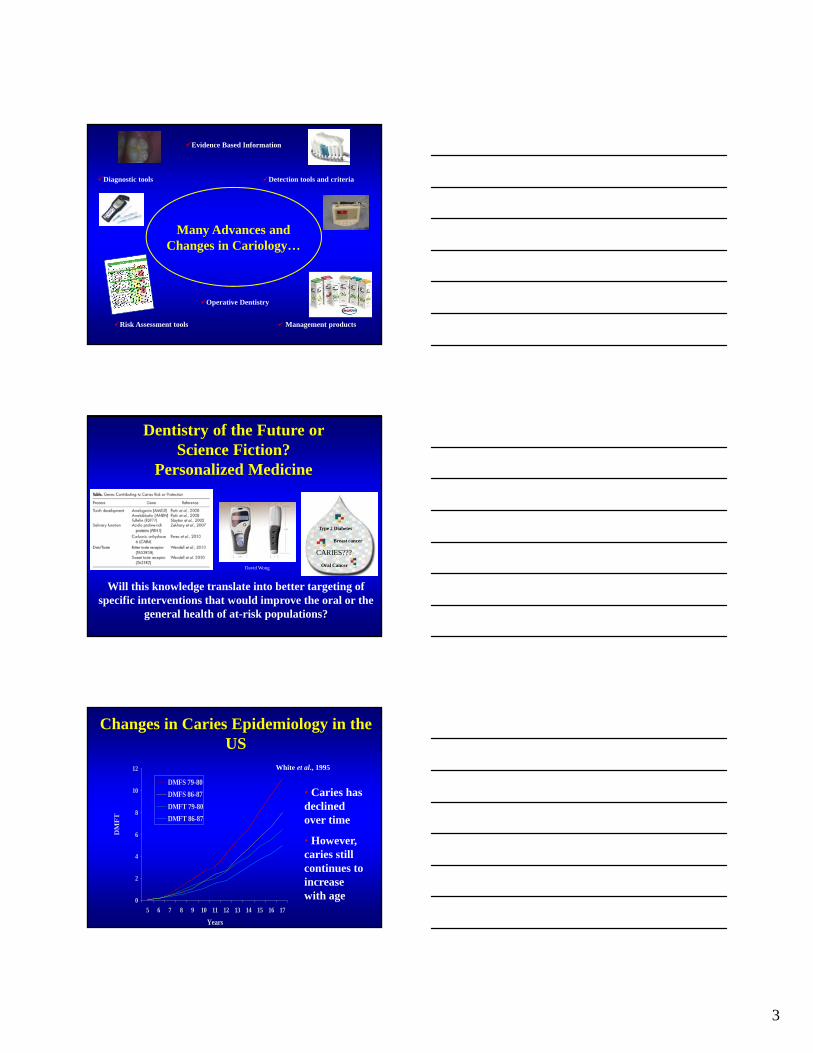
1) Overview of Detection and Diagnosis:
Traditional Caries Detection Methods
Hidden Caries New Methods of Caries
AssessmentVisualTechnology-based
Caries Lesion Activity Status
Outline2) Review of Sealant Recommendations and Diagnostic Thresholds for Placing SealantsSoundCarious Lesions
Incipient (non-cavitated)Cavitated
2
EducationResearch
Clinical Practice
ImprovedHealth
[Adapted from Sung et al. (2003) JAMA, 289, 1278-89]
0
5
10
15
20
25
30
35
40
45
Les
ion
Dep
th (m
icro
0 ppm F 1100 ppm F 1100 ppm F + Triclosan
Dentifrice
Single Species Biofilm
Evidence-Based Practice
“Half of what you are taught as medical students will in 10 years have been shown to be wrong. And the trouble is, none of your teachers knows which half”(Dr. Burwell, Dean of Harvard Medical School; 2002)
Risk-based prevention and disease
management have been recognized as the
cornerstones of modern caries management
(Featherstone, 2003; Fontana and Zero, 2006)
Best available Evidence
Patient-Centered
Dentist’s knowledge and experience
Risk-Based Management and Prevention
Modern Caries Management
Focus on Disease and Minimal intervention
How much evidence is needed to start making
changes in our practice of dentistry?
Changing Environment
2008 2009
3
Many Advances and Changes in Cariology…
Evidence Based Information
Detection tools and criteria
Management productsRisk Assessment tools
Diagnostic tools
Operative Dentistry
Type 2 Diabetes
Breast cancer
Oral Cancer
Dentistry of the Future orScience Fiction?
Personalized Medicine
David Wong
Will this knowledge translate into better targeting of specific interventions that would improve the oral or the
general health of at-risk populations?
CARIES???
Changes in Caries Epidemiology in the US
0
2
4
6
8
10
12
5 6 7 8 9 10 11 12 13 14 15 16 17
Years
DMFS 79-80
DMFS 86-87
DMFT 79-80
DMFT 86-87
White et al., 1995
• Caries has declined over time
• However, caries still continues to increase with age
DM
FT

4
It is true evidence for many of our daily caries intervention choices should be much stronger (most reviews conclude the evidence is weak)
So…what can be safely incorporated into practice?
and how?
If evidence challenges and questions current clinical practice, it will affect implementation
Article 1
Article 2
Internet
Expert Textbook
Maintaining an evidence-based knowledge on every aspect of
dental practice can be an overwhelming (volume, difficulty to:
locate, interpret, what if contradictory?)
Clinicians not trained to critically search, evaluate,
incorporate research findings (best evidence)
into their everyday practice model.
Dental schools, continuing professional education, and organized dentistry have very important roles :
• educate practitioners in the techniques of EBD
• provide repositories of best practices based on soundevidence
(Baelum and Kidd , 2008)
5
Variety of options change by setting, but the scientific evidence supporting management
strategies should be the same
Susceptible
Host
Carbohydrate-
rich diet
Cariogenic Microflora
Caries
X
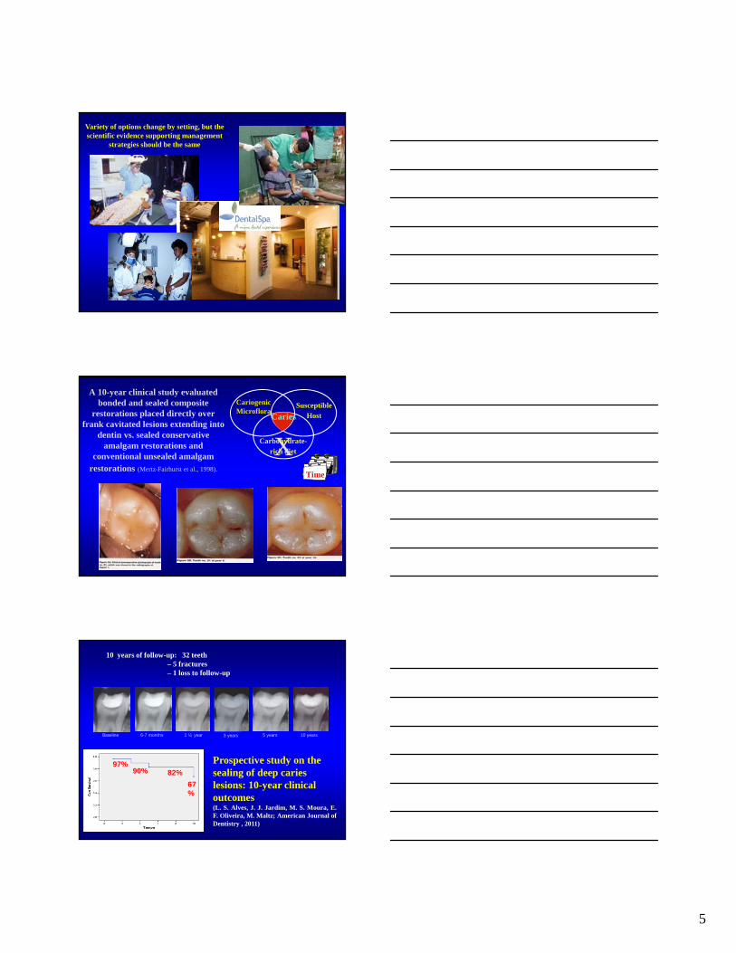
A 10-year clinical study evaluated bonded and sealed composite
restorations placed directly over frank cavitated lesions extending into
dentin vs. sealed conservative amalgam restorations and
conventional unsealed amalgam restorations (Mertz-Fairhurst et al., 1998).
Time
Baseline 6-7 months 1 ½ year 3 years 5 years 10 years
10 years of follow-up: 32 teeth– 5 fractures– 1 loss to follow-up
Prospective study on the sealing of deep caries lesions: 10-year clinical outcomes (L. S. Alves, J. J. Jardim, M. S. Moura, E.F. Oliveira, M. Maltz; American Journal ofDentistry , 2011)
67%
82%90%97%
6
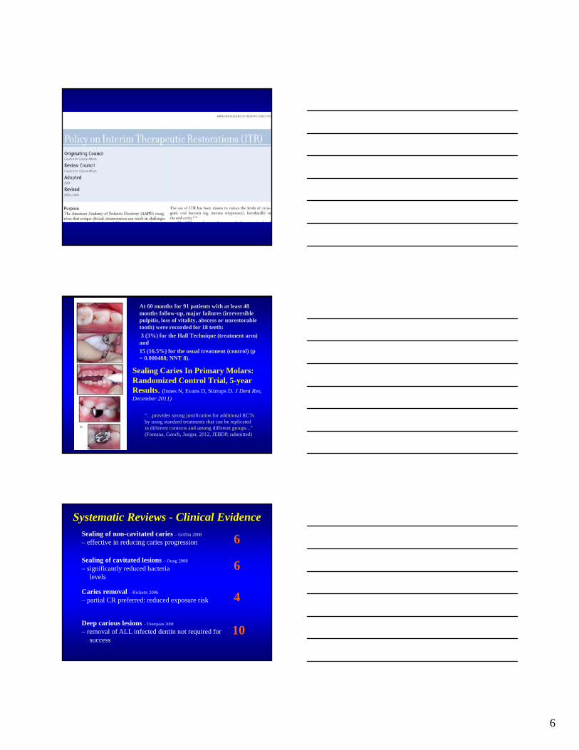
Sealing Caries In Primary Molars: Randomized Control Trial, 5-year Results. (Innes N, Evans D, Stirrups D. J Dent Res, December 2011)
At 60 months for 91 patients with at least 48 months follow-up, major failures (irreversible pulpitis, loss of vitality, abscess or unrestorable tooth) were recorded for 18 teeth:
3 (3%) for the Hall Technique (treatment arm) and
15 (16.5%) for the usual treatment (control) (p = 0.000488; NNT 8).
“…provides strong justification for additional RCTs by using standard treatments that can be replicated in different contexts and among different groups...” (Fontana, Gooch, Junger, 2012, JEBDP, submitted)
Deep carious lesions – Thompson 2008
– removal of ALL infected dentin not required for success
Caries removal – Ricketts 2006
– partial CR preferred: reduced exposure risk
Sealing of non-cavitated caries – Griffin 2008
– effective in reducing caries progression
Sealing of cavitated lesions – Oong 2008
– significantly reduced bacteria levels
Systematic Reviews - Clinical Evidence
6
6
4
10
7
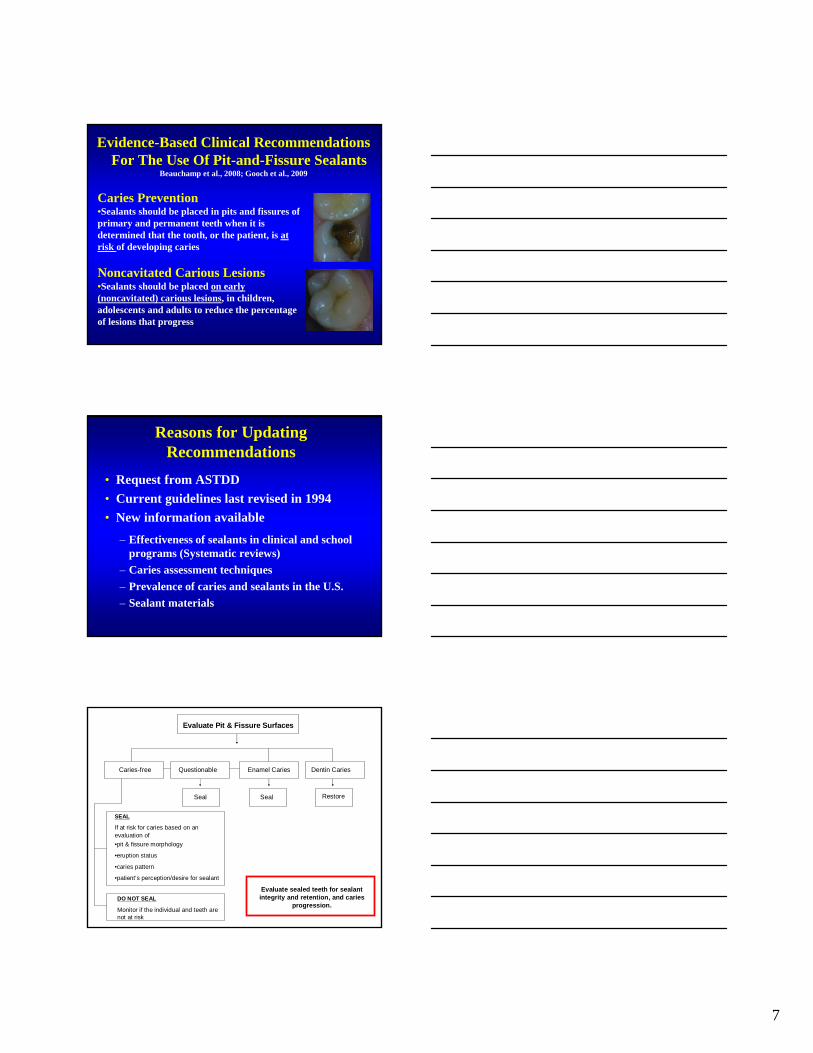
Evidence-Based Clinical Recommendations For The Use Of Pit-and-Fissure Sealants
Beauchamp et al., 2008; Gooch et al., 2009
Caries Prevention•Sealants should be placed in pits and fissures of primary and permanent teeth when it is determined that the tooth, or the patient, is at risk of developing caries
Noncavitated Carious Lesions•Sealants should be placed on early (noncavitated) carious lesions, in children, adolescents and adults to reduce the percentage of lesions that progress
Reasons for Updating Recommendations
• Request from ASTDD
• Current guidelines last revised in 1994
• New information available
– Effectiveness of sealants in clinical and school programs (Systematic reviews)
– Caries assessment techniques
– Prevalence of caries and sealants in the U.S.
– Sealant materials
Caries-free Questionable Enamel Caries Dentin Caries
Evaluate Pit & Fissure Surfaces
Seal Seal Restore
SEAL
If at risk for caries based on an evaluation of
•pit & fissure morphology
•eruption status
•caries pattern
•patient’s perception/desire for sealant
DO NOT SEAL
Monitor if the individual and teeth are not at risk
Evaluate sealed teeth for sealant integrity and retention, and caries
progression.
8
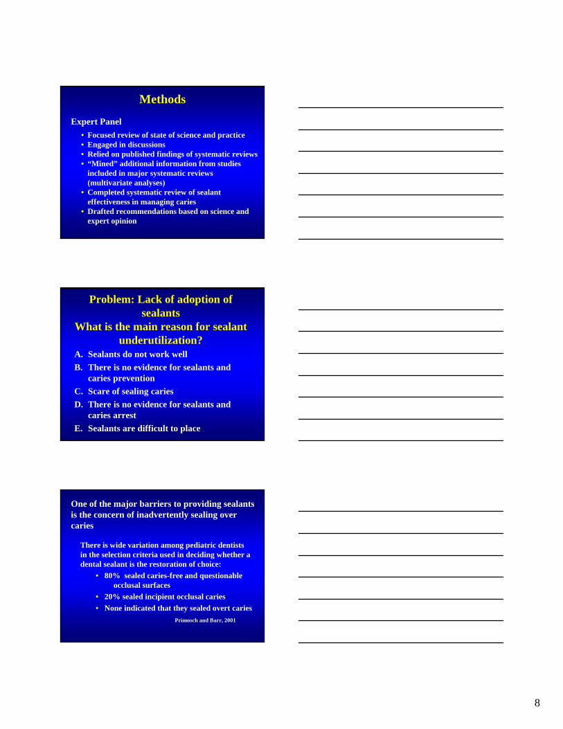
Methods
Expert Panel
• Focused review of state of science and practice • Engaged in discussions• Relied on published findings of systematic reviews• “Mined” additional information from studies
included in major systematic reviews (multivariate analyses)
• Completed systematic review of sealant effectiveness in managing caries
• Drafted recommendations based on science and expert opinion
Problem: Lack of adoption of sealants
What is the main reason for sealant underutilization?
A. Sealants do not work well
B. There is no evidence for sealants and caries prevention
C. Scare of sealing caries
D. There is no evidence for sealants and caries arrest
E. Sealants are difficult to place
One of the major barriers to providing sealants is the concern of inadvertently sealing over caries
There is wide variation among pediatric dentists in the selection criteria used in deciding whether a dental sealant is the restoration of choice:
• 80% sealed caries-free and questionable occlusal surfaces
• 20% sealed incipient occlusal caries
• None indicated that they sealed overt caries
Primosch and Barr, 2001
9
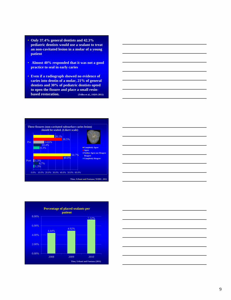
• Only 37.4% general dentists and 42.3% pediatric dentists would use a sealant to treat an non-cavitated lesion in a molar of a young patient
• Almost 40% responded that it was not a good practice to seal in early caries
• Even if a radiograph showed no evidence of caries into dentin of a molar, 21% of general dentists and 30% of pediatric dentists opted to open the fissure and place a small resin-based restoration. (Tellez et al., JADA 2011)
1.3%
8.3%
6.7%
10.4%
1.3%
14.6%
40.0%
38.5%
50.7%
28.1%
0.0% 10.0% 20.0% 30.0% 40.0% 50.0% 60.0%
Post
Pre
These fissures (non-cavitated subsurface caries lesion) should be sealed. (Likert scale)
Completely Agree
Agree
Neither Agree nor Disagree
Disagree
Completely Disagree
Titus, Urbani and Fontana; NOHC, 2011
4.44%4.92%
7.32%
0.00%
2.00%
4.00%
6.00%
8.00%
2008 2009 2010
Percentage of placed sealants per patient
Titus, Urbani and Fontana (2011)
10
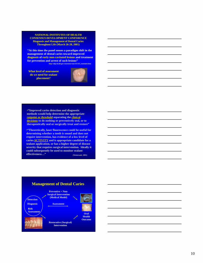
“At this time the panel senses a paradigm shift in the management of dental caries toward improved diagnosis of early non-cavitated lesions and treatment for prevention and arrest of such lesions”
NATIONAL INSTITUTES OF HEALTHCONSENSUS DEVELOPMENT CONFERENCE
Diagnosis and Management of Dental Caries Throughout Life (March 26-28, 2001)
http://odp.od.nih.gov/consensus/cons/115/115_statement.htm
What level of assessment do we need for sealant
placement?
•“Improved caries detection and diagnostic methods would help determine the appropriate cutpoint or threshold separating the clinical decisions to do nothing or preventively seal, or to therapeutically seal or surgically treat and restore”
•“Theoretically, laser fluorescence could be useful for determining whether a tooth is sound and does not require intervention, has evidence of a low level of caries ACTIVITY and is appropriate candidate for a sealant application, or has a higher degree of disease severity that requires surgical intervention. Ideally it could subsequently be used to monitor sealant effectiveness…” (Weintraub, 2001)
Management of Dental Caries
Diagnosis
Risk Assessment
Preventive + Non-Surgical Intervention
(Medical Model)
Restorative (Surgical) Intervention
Oral Health
Outcome
Assessment
Detection
11
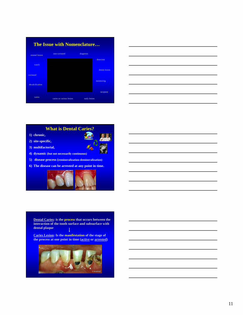
The Issue with Nomenclature…
detection
monitoring
watch
non-cavitated
cavitated
incipient
early lesioncaries or carious lesion
decalcification
caries
enamel lesion
dentin lesion
diagnosis
What is Dental Caries?1) chronic,
2) site-specific,
3) multifactorial,
4) dynamic (but not necessarily continuous)
5) disease process (remineralization-demineralization)
6) The disease can be arrested at any point in time.
Dental Caries: is the process that occurs between the interaction of the tooth surface and subsurface with dental plaque
Caries Lesion: Is the manifestation of the stage of the process at one point in time (active or arrested)
D Bratthall
12
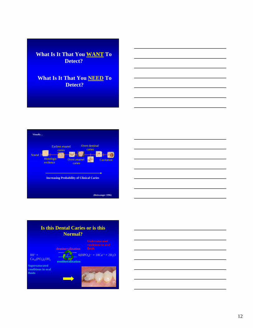
What Is It That You WANT To Detect?
What Is It That You NEED ToDetect?
(Beiswanger 1996)
SoundHistologicevidence
Earliest enamelcaries
Overt enamel caries
Overt dentinal caries
Cavitation
Increasing Probability of Clinical Caries
Visually…
8H+ + Ca10(PO4)6OH2
6(HPO4)- - + 10Ca++ + 2H2O
demineralization
remineralization
F-
Is this Dental Caries or is this Normal?
Undersaturated conditions in oral fluids
Supersaturated conditions in oral fluids
13
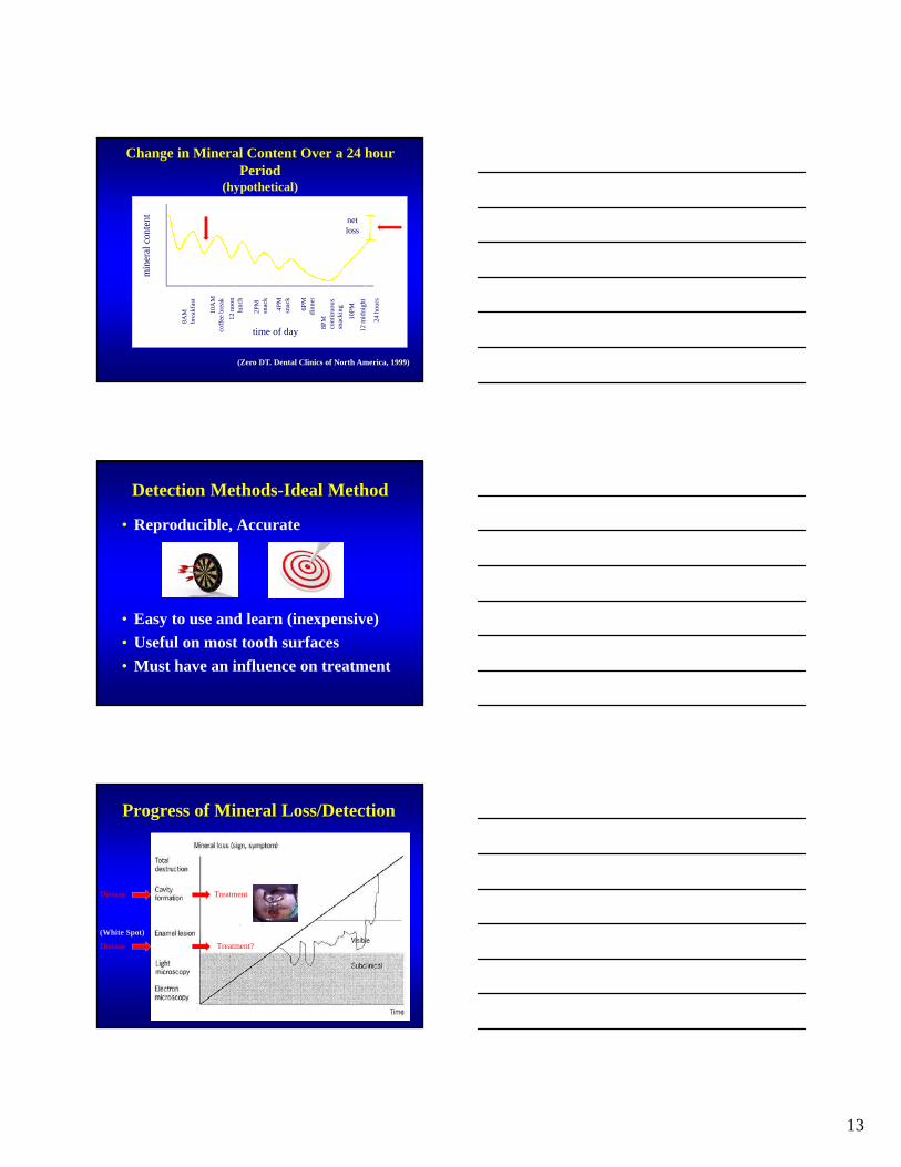
Change in Mineral Content Over a 24 hour Period
(hypothetical)8A
Mbr
eakf
ast
10A
M
coff
ee b
reak
12 n
oon
lunc
h
2PM
sn
ack
4PM
snac
k
6PM
dinn
er
8PM
cont
inuo
ussn
acki
ng
10P
M
12 m
idni
ght
24 h
ours
time of day
min
eral
con
tent net
loss
(Zero DT. Dental Clinics of North America, 1999)
Detection Methods-Ideal Method
• Reproducible, Accurate
• Easy to use and learn (inexpensive)
• Useful on most tooth surfaces
• Must have an influence on treatment
Progress of Mineral Loss/Detection
(White Spot)
Disease
Treatment?Disease
Treatment
14
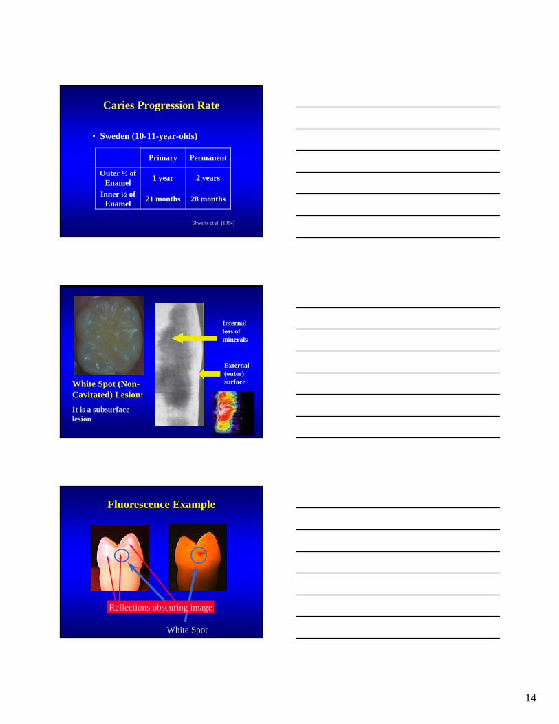
Caries Progression Rate
• Sweden (10-11-year-olds)
Shwartz et al. (1984)
Primary Permanent
Outer ½ of Enamel
1 year 2 years
Inner ½ of Enamel
21 months 28 months
White Spot (Non-Cavitated) Lesion:
It is a subsurface lesion
External (outer) surface
Internal loss of minerals
Fluorescence Example
White Spot
Reflections obscuring image
15
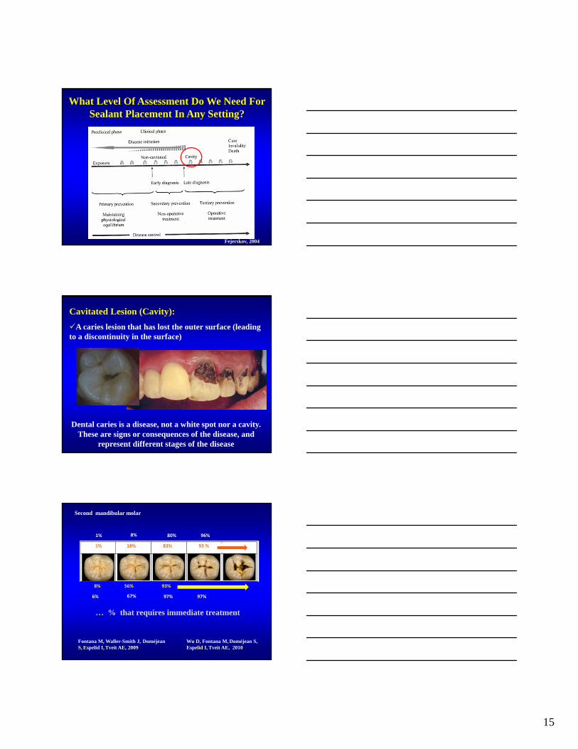
What Level Of Assessment Do We Need For Sealant Placement In Any Setting?
Fejerskov, 2004
Dental caries is a disease, not a white spot nor a cavity. These are signs or consequences of the disease, and
represent different stages of the disease
Cavitated Lesion (Cavity):
A caries lesion that has lost the outer surface (leading to a discontinuity in the surface)
1% 18% 83%
8% 56% 93%
93 %
Fontana M, Waller-Smith J, Doméjean S, Espelid I, Tveit AE, 2009
1% 8% 80% 96%
6% 67% 97% 97%
Wu D, Fontana M, Doméjean S,Espelid I, Tveit AE, 2010
Second mandibular molar
… % that requires immediate treatment
16
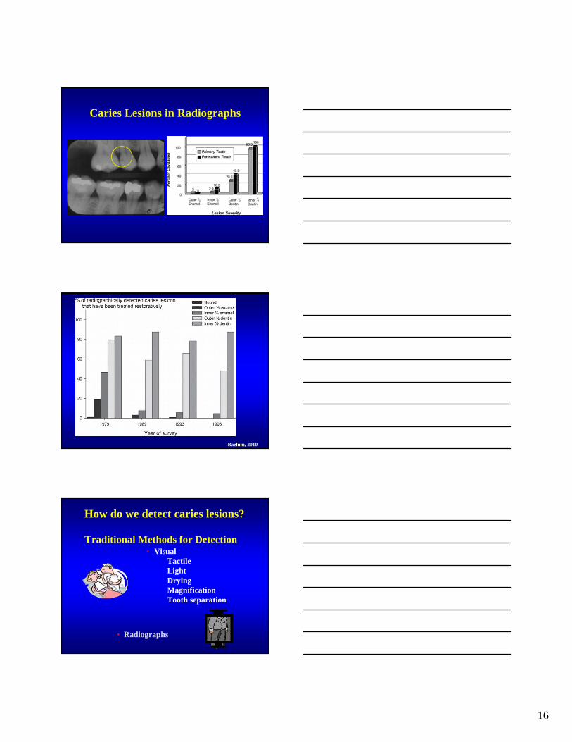
Caries Lesions in Radiographs
Baelum, 2010
Traditional Methods for Detection • Visual
Tactile LightDryingMagnificationTooth separation
• Radiographs
How do we detect caries lesions?
17
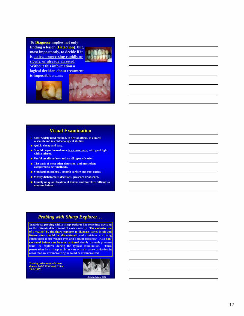
To Diagnose implies not only finding a lesion (Detection), but, most importantly, to decide if it is active, progressing rapidly or slowly, or already arrested.Without this information a logical decision about treatment is impossible (Kidd, 2001)
Visual Examination Most widely used method, in dental offices, in clinical
research and in epidemiological studies.
Quick, cheap and easy.
Should be performed on a dry, clean tooth, with good light, with a mirror.
Useful on all surfaces and on all types of caries.
The basis of most other detection, and most often compared to new methods.
Standard on occlusal, smooth surface and root caries.
Mostly dichotomous decisions: presence or absence.
Usually no quantification of lesions and therefore difficult to monitor lesions.
Probing with Sharp Explorer…
Ekstrand et al., 1987
Traditional probing with a sharp explorer has come into questionas the ultimate determinant of caries activity. The exclusive useof a “catch” by the sharp explorer to diagnose caries in pit andfissure sites should be discontinued and clinicians are beingcalled upon to use “sharp eyes and a blunt explorer.” Also non-cavitated lesions can become cavitated simply through pressurefrom the explorer during the typical examination. Thus,penetration by a sharp explorer can actually cause cavitation inareas that are remineralizing or could be remineralized.
Treating caries as an infectious disease. JADA 125 (June): 2-S to 15-S (1995)
18
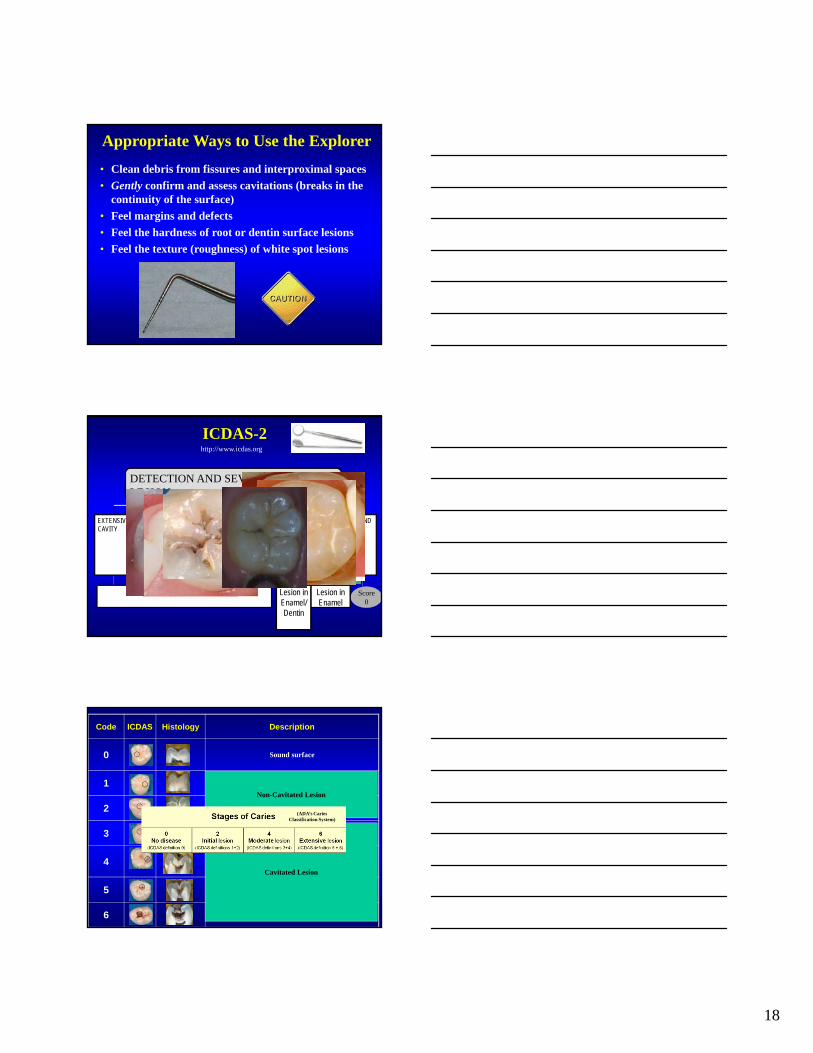
Appropriate Ways to Use the Explorer
• Clean debris from fissures and interproximal spaces
• Gently confirm and assess cavitations (breaks in the continuity of the surface)
• Feel margins and defects
• Feel the hardness of root or dentin surface lesions
• Feel the texture (roughness) of white spot lesions
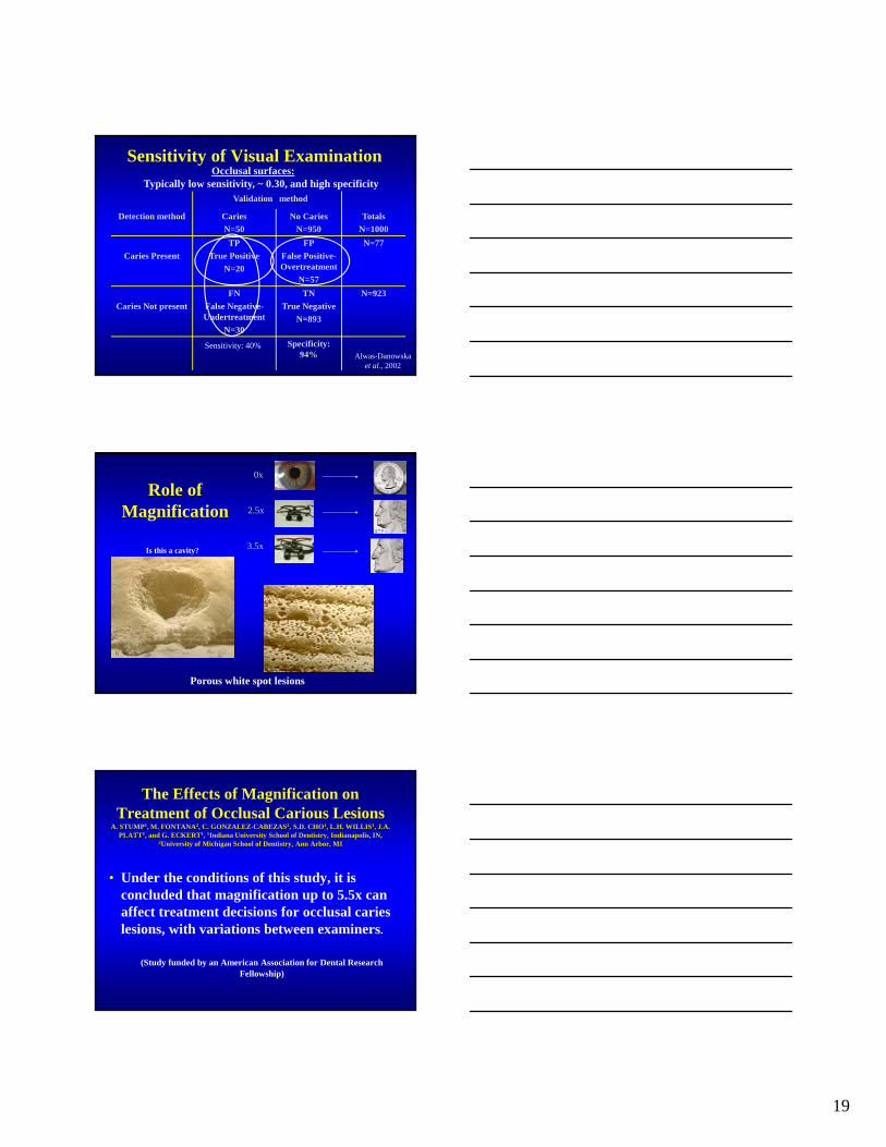
2 A. VISUAL APPEARANCE
ICDAS-2
Score5
DISTINCT CAVITY
Score6
EXTENSIVE CAVITY
SOUND
Score0
2. ACTIVITYDETECTION AND SEVERITY OF THE LESION
SURFACE INTEGRITYLOSS
Score3
OPACITYwithout air-drying: WHITE,BROWN
Scores2W,2B
OPACITYwith air-drying: WHITE, BROWN
Scores1W,1B
UNDERLYING GREY SHADOW
Score4
Lesion in Dentin Lesion in Enamel
Lesion in Enamel/Dentin
http://www.icdas.org
Code ICDAS Histology Description
0 Sound surface
1 First visual change in enamel
2 Distinct visual change in enamel
3 Localized enamel breakdown due to caries with no visible dentin
4Non-cavitated surface with underlying dark shadow from dentin
5 Distinct cavity with visible dentin
6 Extensive distinct cavity with visible dentin.
Non-Cavitated Lesion
Cavitated Lesion
(ADA’s Caries Classification System)
19
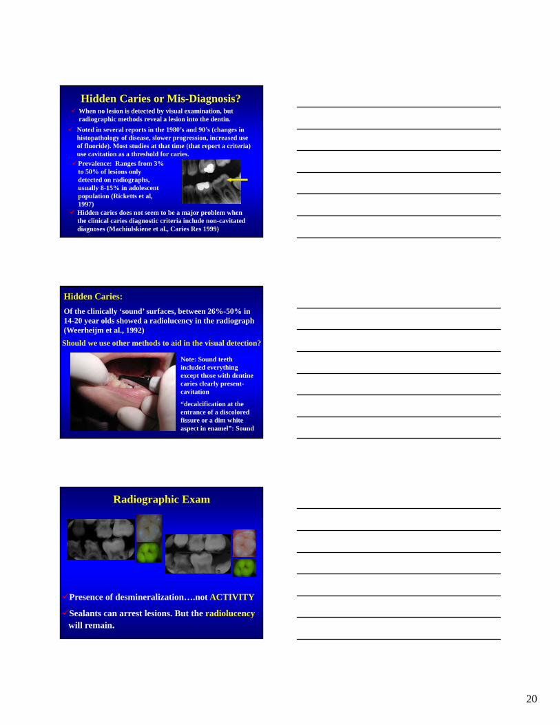
Validation method
Detection method Caries
N=50
No Caries
N=950
Totals
N=1000
Caries Present
TP
True Positive
N=20
FP
False Positive-Overtreatment
N=57
N=77
Caries Not present
FN
False Negative-Undertreatment
N=30
TN
True Negative
N=893
N=923
Specificity: 94%
Sensitivity of Visual Examination
Sensitivity: 40%Alwas-Danowska
et al., 2002
Occlusal surfaces:Typically low sensitivity, ~ 0.30, and high specificity
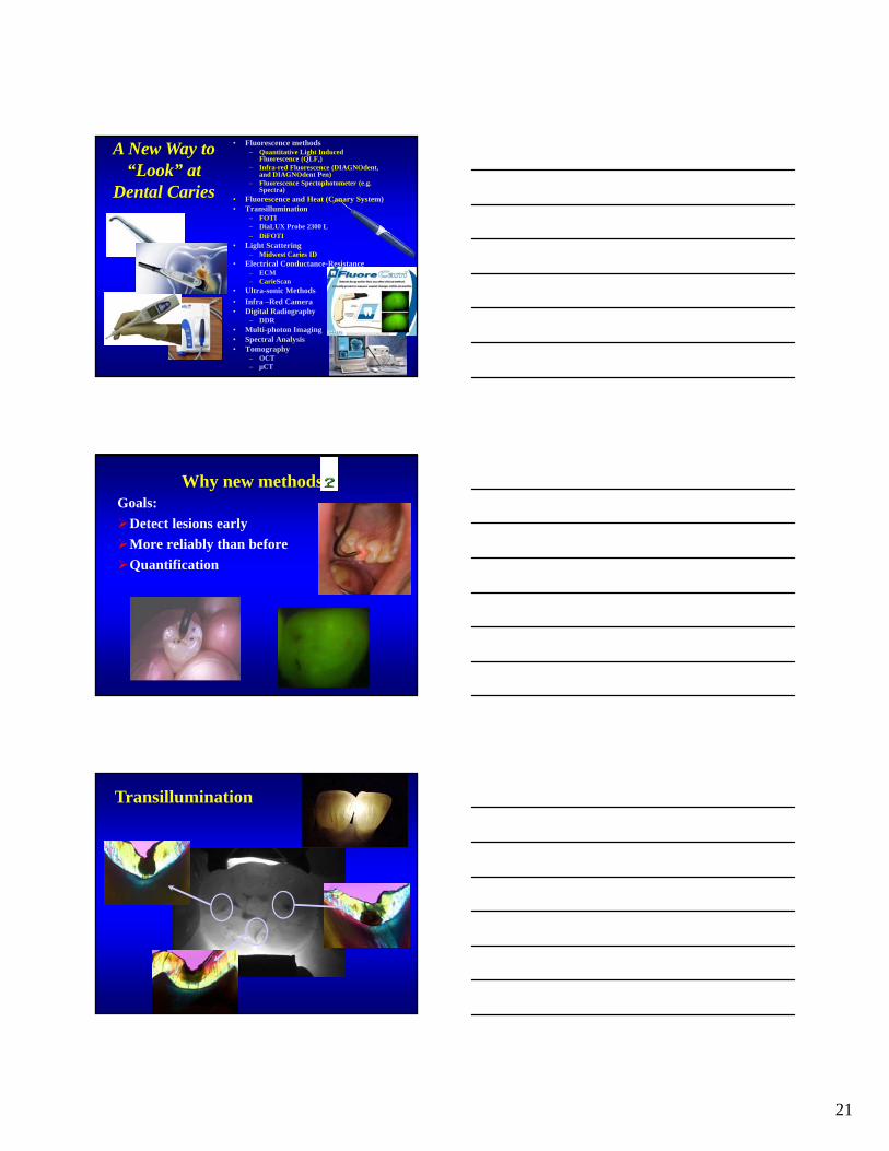
0x
2.5x
3.5x
Role of Magnification
Porous white spot lesions
Is this a cavity?
The Effects of Magnification on Treatment of Occlusal Carious Lesions
A. STUMP1, M. FONTANA2, C. GONZALEZ-CABEZAS2, S.D. CHO1, L.H. WILLIS1, J.A. PLATT1, and G. ECKERT1, 1Indiana University School of Dentistry, Indianapolis, IN,
2University of Michigan School of Dentistry, Ann Arbor, MI
• Under the conditions of this study, it is concluded that magnification up to 5.5x can affect treatment decisions for occlusal caries lesions, with variations between examiners.
(Study funded by an American Association for Dental Research Fellowship)
20
Hidden Caries or Mis-Diagnosis? When no lesion is detected by visual examination, but
radiographic methods reveal a lesion into the dentin.
Noted in several reports in the 1980’s and 90’s (changes in histopathology of disease, slower progression, increased use of fluoride). Most studies at that time (that report a criteria) use cavitation as a threshold for caries.
Prevalence: Ranges from 3% to 50% of lesions only detected on radiographs, usually 8-15% in adolescent population (Ricketts et al, 1997)
Hidden caries does not seem to be a major problem when the clinical caries diagnostic criteria include non-cavitated diagnoses (Machiulskiene et al., Caries Res 1999)
Hidden Caries:
Of the clinically ‘sound’ surfaces, between 26%-50% in 14-20 year olds showed a radiolucency in the radiograph (Weerheijm et al., 1992)
Should we use other methods to aid in the visual detection?
Note: Sound teeth included everything except those with dentine caries clearly present-cavitation
“decalcification at the entrance of a discolored fissure or a dim white aspect in enamel”: Sound
Presence of desmineralization….not ACTIVITY
Sealants can arrest lesions. But the radiolucencywill remain.
Radiographic Exam
21
A New Way to “Look” at
Dental Caries
• Fluorescence methods– Quantitative Light Induced
Fluorescence (QLF,)– Infra-red Fluorescence (DIAGNOdent,
and DIAGNOdent Pen)– Fluorescence Spectophotometer (e.g.
Spectra)• Fluorescence and Heat (Canary System)• Transillumination
– FOTI– DiaLUX Probe 2300 L – DiFOTI
• Light Scattering– Midwest Caries ID
• Electrical Conductance-Resistance– ECM– CarieScan
• Ultra-sonic Methods• Infra –Red Camera• Digital Radiography
– DDR• Multi-photon Imaging• Spectral Analysis• Tomography
– OCT– µCT
Why new methods?Goals:
Detect lesions early
More reliably than before
Quantification
Transillumination
22
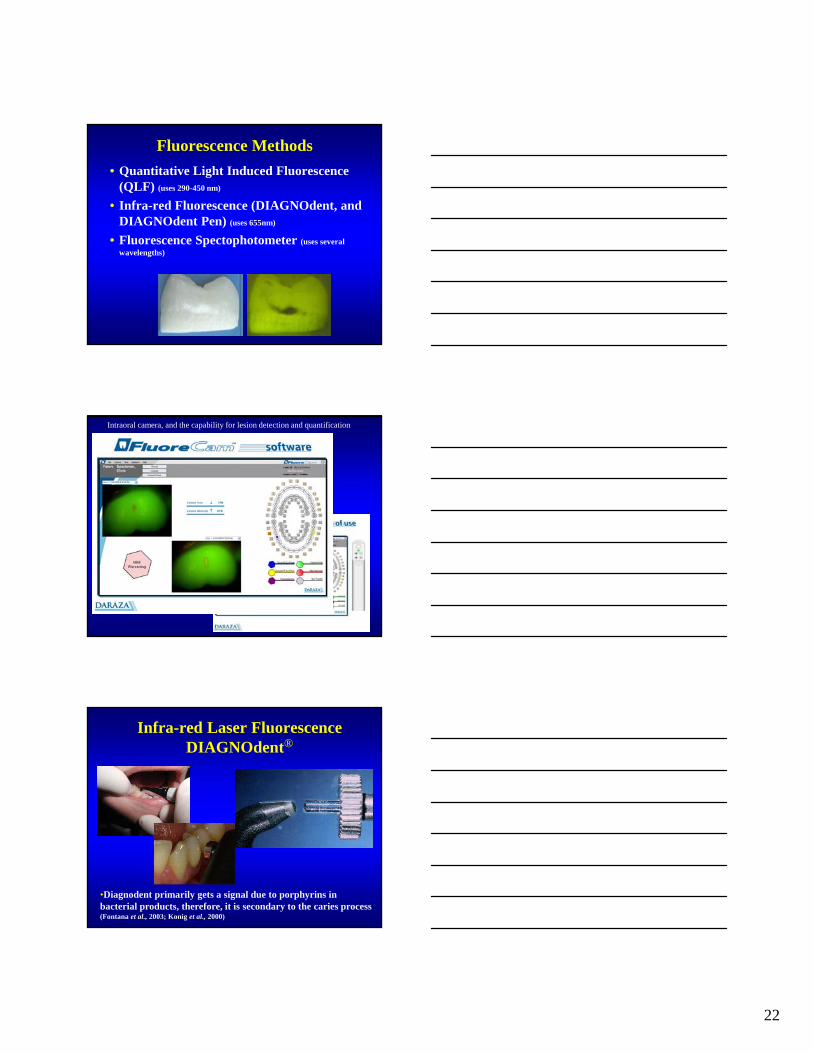
Fluorescence Methods
• Quantitative Light Induced Fluorescence (QLF) (uses 290-450 nm)
• Infra-red Fluorescence (DIAGNOdent, and DIAGNOdent Pen) (uses 655nm)
• Fluorescence Spectophotometer (uses several wavelengths)
Intraoral camera, and the capability for lesion detection and quantification
Infra-red Laser Fluorescence DIAGNOdent®
•Diagnodent primarily gets a signal due to porphyrins in bacterial products, therefore, it is secondary to the caries process (Fontana et al., 2003; Konig et al., 2000)
23
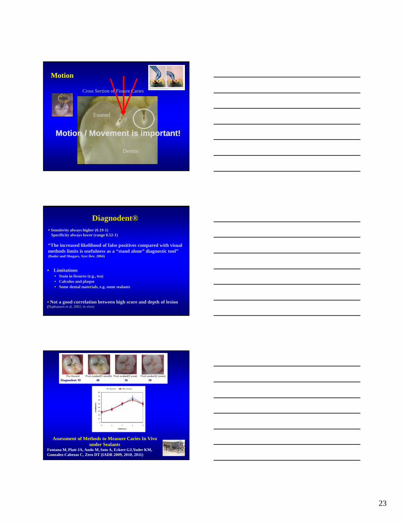
Motion / Movement is important!Motion / Movement is important!
Motion
Cross Section of Fissure Caries
Enamel
Dentin
Limitations• Stain in fissures (e.g., tea)• Calculus and plaque• Some dental materials, e.g. some sealants
Not a good correlation between high score and depth of lesion (Naphausen et al, 2002; in vivo)
Sensitivity always higher (0.19-1)Specificity always lower (range 0.52-1)
“The increased likelihood of false positives compared with visual methods limits is usefulness as a “stand alone” diagnostic tool” (Bader and Shugars, Syst Rev, 2004)
Diagnodent®
69
0
10
20
30
40
50
60
70
80
0 1 2 3 4
DIAGNOdent
ICDAS Score
Baseline After Sealant
Assessment of Methods to Measure Caries In Vivo under Sealants
Fontana M, Platt JA, Ando M, Soto A, Eckert GJ,Yoder KM, Gonzalez-Cabezas C, Zero DT (IADR 2009, 2010, 2011)
Diagnodent 39 40 36 30
24
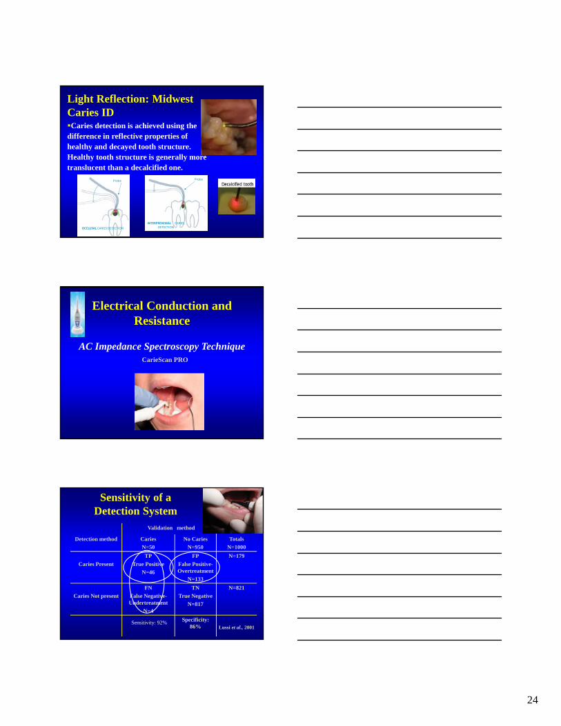
Caries detection is achieved using the difference in reflective properties of healthy and decayed tooth structure. Healthy tooth structure is generally more translucent than a decalcified one.
Light Reflection: Midwest Caries ID
OCCLUSAL CARIES DETECTION
Probe
INTERPROXIMAL CARIES DETECTION
Probe
Electrical Conduction and Resistance
AC Impedance Spectroscopy TechniqueCarieScan PRO
Validation method
Detection method Caries
N=50
No Caries
N=950
Totals
N=1000
Caries Present
TP
True Positive
N=46
FP
False Positive-Overtreatment
N=133
N=179
Caries Not present
FN
False Negative-Undertreatment
N=4
TN
True Negative
N=817
N=821
Specificity: 86% Lussi et al., 2001
Sensitivity of a Detection System
Sensitivity: 92%
25
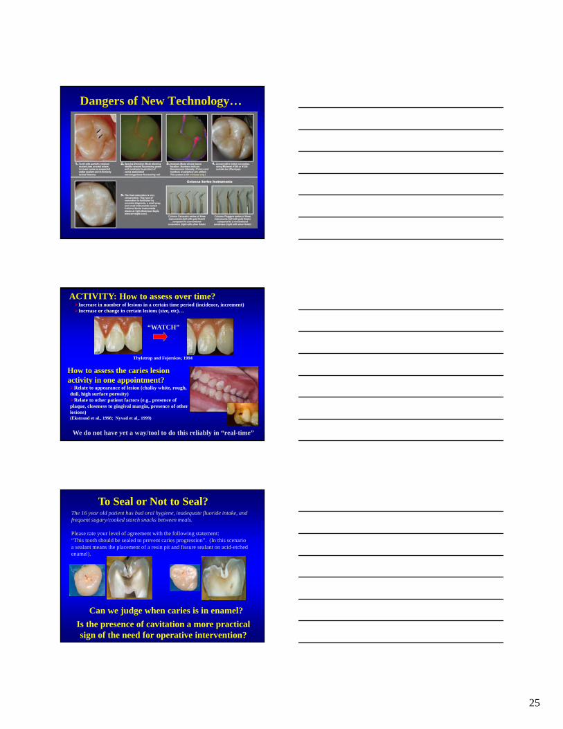
Dangers of New Technology…
ACTIVITY: How to assess over time?Increase in number of lesions in a certain time period (incidence, increment)Increase or change in certain lesions (size, etc)…
How to assess the caries lesion activity in one appointment?Relate to appearance of lesion (chalky white, rough, dull, high surface porosity)Relate to other patient factors (e.g., presence of plaque, closeness to gingival margin, presence of other lesions)(Ekstrand et al., 1998; Nyvad et al., 1999)
We do not have yet a way/tool to do this reliably in “real-time”
Thylstrup and Fejerskov, 1994
“WATCH”
Can we judge when caries is in enamel?
Is the presence of cavitation a more practical sign of the need for operative intervention?
To Seal or Not to Seal?The 16 year old patient has bad oral hygiene, inadequate fluoride intake, and frequent sugary/cooked starch snacks between meals.
Please rate your level of agreement with the following statement:“This tooth should be sealed to prevent caries progression”. (In this scenario a sealant means the placement of a resin pit and fissure sealant on acid-etched enamel).
26
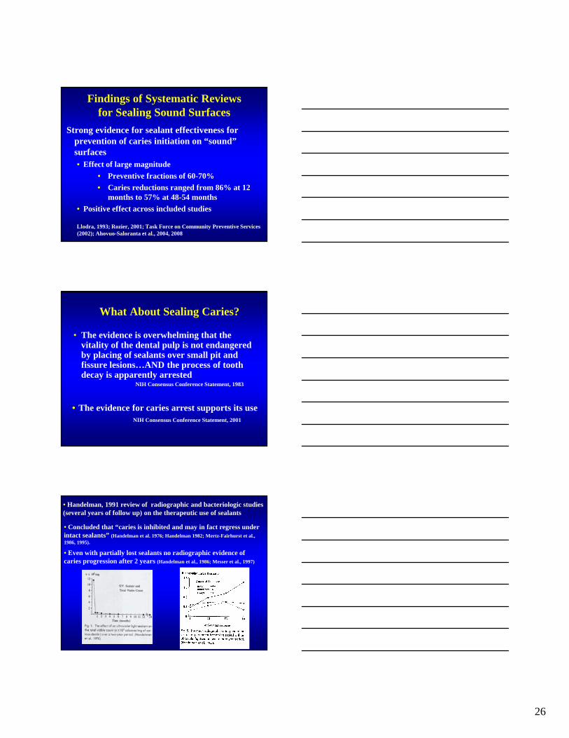
Findings of Systematic Reviews for Sealing Sound Surfaces
Strong evidence for sealant effectiveness for prevention of caries initiation on “sound” surfaces• Effect of large magnitude
• Preventive fractions of 60-70%
• Caries reductions ranged from 86% at 12 months to 57% at 48-54 months
• Positive effect across included studies
Llodra, 1993; Rozier, 2001; Task Force on Community Preventive Services (2002); Ahovuo-Saloranta et al., 2004, 2008
What About Sealing Caries?
• The evidence is overwhelming that the vitality of the dental pulp is not endangered by placing of sealants over small pit and fissure lesions…AND the process of tooth decay is apparently arrested
NIH Consensus Conference Statement, 1983
• The evidence for caries arrest supports its use NIH Consensus Conference Statement, 2001
• Handelman, 1991 review of radiographic and bacteriologic studies (several years of follow up) on the therapeutic use of sealants
• Concluded that “caries is inhibited and may in fact regress under intact sealants” (Handelman et al. 1976; Handelman 1982; Mertz-Fairhurst et al., 1986, 1995).
• Even with partially lost sealants no radiographic evidence of caries progression after 2 years (Handelman et al., 1986; Messer et al., 1997)
27
Heller et al. (1995) found in a 5 year longitudinal study in a fluoridated community that…
• initially sound surfaces did not benefit greatly from the application of sealants (caries rate: 13% if not sealed vs. 8% if sealed)
• there were clear benefits in sealing incipient caries (52% if not sealed vs. 11% if sealed)
[Incipient if dark staining; chalky appearance, or if explorer sticks, but no frank caries (cavitation). When in doubt used this classification]
The Effectiveness of Sealants in Managing Caries Lesions
Griffin et al., J Dent Res 2008
•No matter how studies were grouped (e.g., by material, by study duration) effect of sealants was strong and consistent
•Sealed non-cavitated lesions consistently had better outcomes than not sealed lesions
•% of sealed carious surfaces progressing was low
•Caries reduction was about 71%
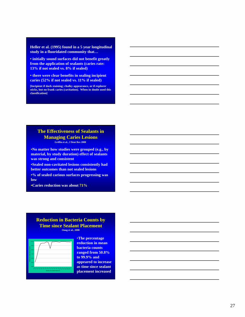
Reduction in Bacteria Counts by Time since Sealant Placement
Oong et al., 2008
40.0%
50.0%
60.0%
70.0%
80.0%
90.0%
100.0%
0.03
0.15
0.23
0.35
0.50
1.00
1.00
2.00
2.00
4.00
4.00
6.00
6.00
7.00
12.00
12.00
24.00
60.00
60.00
60.00
Months since Sealant Placement
% R
edu
ctio
n in
Mea
n B
acte
ria
Co
un
ts
•The percentage reduction in mean bacteria counts ranged from 50.8% to 99.9% and appeared to increase as time since sealant placement increased
28
Risk of CariesLost of Sealant vs. Never Sealed
Time
(years)
# Studies Relative Risk
1 3 1.0
2 3 1.2
3 3 1.0
4 2 1.2
Griffin et al., JADA, 2009
• Four-handed sealant placement is associated with higher retention rates
Griffin et al., JADA 2008
• Sealant retention rates for teeth cleaned with a toothbrush are at least as high as for teeth cleaned with a handpiece
Kolavic Gray et al., JADA 2009
• Visual assessment is appropriate• Teeth can be dried with cotton rolls, gauze or compressed air• Explorer may be used to clean the fissures and “gently” confirm
cavitation; do not use sharp explorer under force• Magnification (2x-4x) can be used, but is not required due to
insufficient evidence on its effect in assessing cavitation• Radiographs are unnecessary, especially in programs targeting
children in grades 2 – 3• Insufficient evidence to recommend other technologies to
determine presence or absence of cavitation
How To Assess Teeth For Sealant
Placement?Fontana et al., 2010
J Pub Health Dent, 1995** *
Non-CavitatedCavitated
29
http://ohiodentalclinics.com/curricula/sealant/mod3_0.html
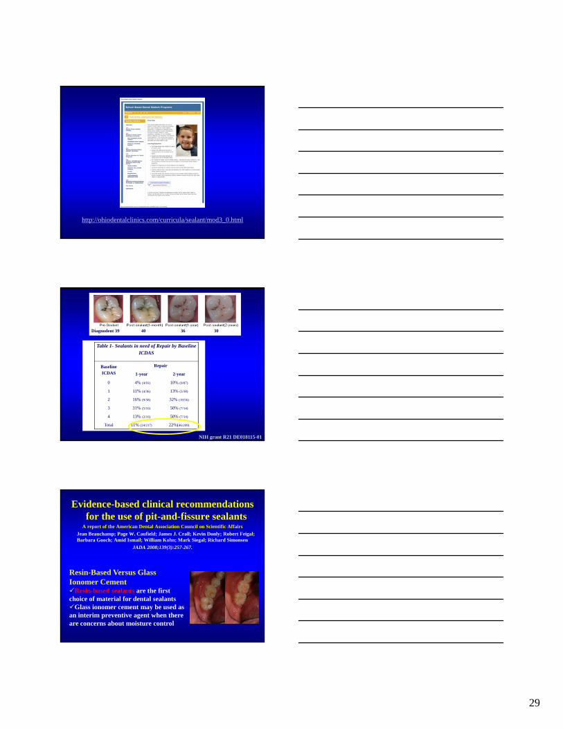
Table 1- Sealants in need of Repair by Baseline ICDAS
Baseline
ICDAS
Repair
1-year 2-year
0 4% (4/91) 10% (9/87)
1 11% (4/36) 13% (5/38)
2 16% (9/58) 32% (18/56)
3 31% (5/16) 50% (7/14)
4 13% (2/16) 50% (7/14)
Total 11% (24/217) 22%(46/209)
Diagnodent 39 40 36 30
NIH grant R21 DE018115-01
Resin-Based Versus Glass Ionomer CementResin-based sealants are the first choice of material for dental sealantsGlass ionomer cement may be used as an interim preventive agent when there are concerns about moisture control
Evidence-based clinical recommendations for the use of pit-and-fissure sealants
A report of the American Dental Association Council on Scientific Affairs
Jean Beauchamp; Page W. Caufield; James J. Crall; Kevin Donly; Robert Feigal; Barbara Gooch; Amid Ismail; William Kohn; Mark Siegal; Richard Simonsen
JADA 2008;139(3):257-267.
30
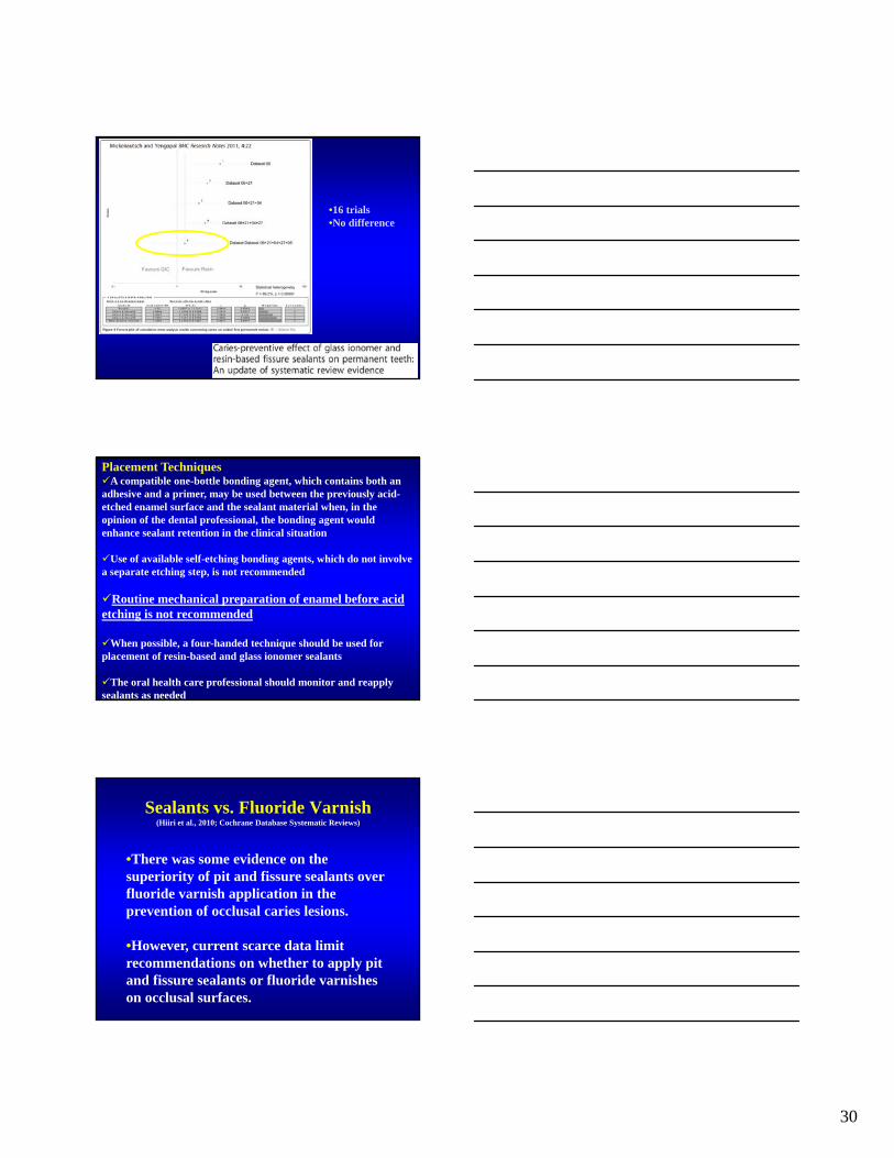
•16 trials•No difference
Placement TechniquesA compatible one-bottle bonding agent, which contains both an adhesive and a primer, may be used between the previously acid-etched enamel surface and the sealant material when, in the opinion of the dental professional, the bonding agent would enhance sealant retention in the clinical situation
Use of available self-etching bonding agents, which do not involve a separate etching step, is not recommended
Routine mechanical preparation of enamel before acid etching is not recommended
When possible, a four-handed technique should be used for placement of resin-based and glass ionomer sealants
The oral health care professional should monitor and reapply sealants as needed
Sealants vs. Fluoride Varnish(Hiiri et al., 2010; Cochrane Database Systematic Reviews)
•There was some evidence on the superiority of pit and fissure sealants over fluoride varnish application in the prevention of occlusal caries lesions.
•However, current scarce data limit recommendations on whether to apply pit and fissure sealants or fluoride varnishes on occlusal surfaces.
31
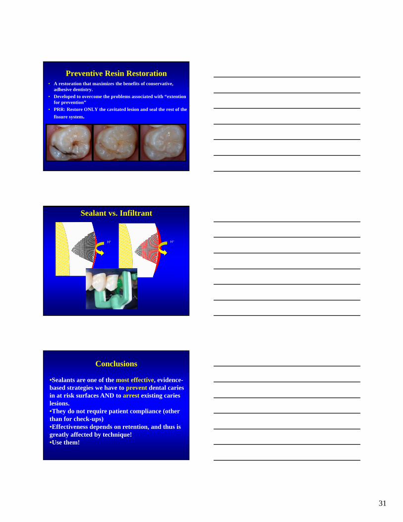
Preventive Resin Restoration• A restoration that maximizes the benefits of conservative,
adhesive dentistry.
• Developed to overcome the problems associated with “extention for prevention”
• PRR: Restore ONLY the cavitated lesion and seal the rest of the
fissure system.
http://www.dentaljuce.com/fruit/images/prr/BandA.jpg
sealingH+ H+
Sealant vs. Infiltrant
Conclusions
•Sealants are one of the most effective, evidence-based strategies we have to prevent dental caries in at risk surfaces AND to arrest existing caries lesions.•They do not require patient compliance (other than for check-ups)•Effectiveness depends on retention, and thus is greatly affected by technique!•Use them!
32
Passive dissemination of information is generally ineffective
•Educational materials (e.g., clinical practice guidelines, audiovisual materials, publications) •Didactic educational meetings (e.g., lectures)
Bero et al., BMJ. 1998
Active training programs have to be developed (resources planned and allocated) to educate teachers/practitioners: case discussions, ground rounds, etc.
Thank you…