biocontrol of bacillus subtilis against infection of - plant physiology
TRANSCRIPT
Biocontrol of Bacillus subtilis against Infection ofArabidopsis Roots by Pseudomonas syringae IsFacilitated by Biofilm Formation and SurfactinProduction1
Harsh Pal Bais, Ray Fall, and Jorge M. Vivanco*
Department of Horticulture and Landscape Architecture (H.P.B., J.M.V.) and Cell and Molecular BiologyProgram (J.M.V.), Colorado State University, Fort Collins, Colorado 80523–1173; and Department ofChemistry and Biochemistry and Cooperative Institute for Research in Environmental Sciences, University ofColorado, Boulder, Colorado 80309–0215 (R.F.)
Relatively little is known about the exact mechanisms used by Bacillus subtilis in its behavior as a biocontrol agent on plants.Here, we report the development of a sensitive plant infection model demonstrating that the bacterial pathogen Pseudomonassyringae pv tomato DC3000 is capable of infecting Arabidopsis roots both in vitro and in soil. Using this infection model, wedemonstrated the biocontrol ability of a wild-type B. subtilis strain 6051 against P. syringae. Arabidopsis root surfaces treatedwith B. subtilis were analyzed with confocal scanning laser microscopy to reveal a three-dimensional B. subtilis biofilm. It isknown that formation of biofilms by B. subtilis is a complex process that includes secretion of surfactin, a lipopeptideantimicrobial agent. To determine the role of surfactin in biocontrol by B. subtilis, we tested a mutant strain, M1, with adeletion in a surfactin synthase gene and, thus, deficient in surfactin production. B. subtilis M1 was ineffective as a biocontrolagent against P. syringae infectivity in Arabidopsis and also failed to form robust biofilms on either roots or inert surfaces.The antibacterial activity of surfactin against P. syringae was determined in both broth and agar cultures and also bylive-dead staining methods. Although the minimum inhibitory concentrations determined were relatively high (25 �gmL�1), the levels of the lipopeptide in roots colonized by B. subtilis are likely to be sufficient to kill P. syringae. Our resultscollectively indicate that upon root colonization, B. subtilis 6051 forms a stable, extensive biofilm and secretes surfactin,which act together to protect plants against attack by pathogenic bacteria.
Beneficial plant rhizobacteria (PR) are associatedwith the surfaces of plant roots and may increaseplant yield by mechanisms that impart improvedmineral nutrient uptake, disease suppression, or phy-tohormone production (Kloepper et al., 1991; Luten-berg et al., 1991; Costacurta and Vanderleyden, 1995;Defago and Keel, 1995). An important trait of PR istheir ability to effectively colonize the rhizosphere andmaintain a stable relationship with the surface of plantroots (Lutenberg and Dekkers, 1999). PR may alsointeract with a variety of soil microorganisms that arenormally present in the rhizosphere, in some casesacting as a biocontrol agent against pathogenic bacte-ria (Pinton et al., 2001). Interestingly, poor root colo-nization by PR may result in decreased biocontrolactivity (Schippers et al., 1987). One beneficial rhi-
zobacterium is Bacillus subtilis, which is ubiquitous insoil, can promote plant growth, protect against fungalpathogen attack (Utkhede and Smith, 1992; Asaka andShoda, 1996; Emmert and Handelsman, 1999), andplay a role in the degradation of organic polymers inthe soil (Emmert and Handelsman, 1999). Among thefirst successful biocontrol agents used against insectsand pathogens were members of the genus Bacillus(Powell and Jutsum, 1993). Commercial strains of B.subtilis have been marketed as biocontrol agents forfungal diseases of crops (Emmert and Handelsman,1999; Warrior et al., 2002). The commercial biofungi-cide, Serenade, which contains a B. subtilis strain, isreported to be effective against a variety of patho-genic bacteria, including Erwina, Pseudomonas, andXanthomonas strains (http://www.agraquest.com.The mechanism of this antibacterial effect is uncer-tain, although it is known that B. subtilis can producea variety of antibacterial agents, including a broadspectrum of lipopeptides, such as surfactin, that arepotent biosurfactants (Zuber et al., 1993; Peypoux etal., 1999).
It is now widely recognized that most bacteriafound in natural, clinical, and industrial settings per-sist in association with surfaces by forming biofilms(Davey and O’Toole, 2000). Biofilms are structuredcommunities of cells adherent to a surface and en-
1 This work was supported by the Colorado State UniversityAgricultural Experiment Station (grant to J.M.V.), by the NationalScience Foundation-CAREER (grant no. MCB 0093014 to J.M.V.),by the State of Colorado (Invasive Weeds Initiative to J.M.V.),bythe Lindbergh Foundation (to J.M.V.), and by the U.S. Departmentof Energy (grant no. DE–FG03–97ER20274 to R.F.).
* Corresponding author; e-mail [email protected];fax 970 – 491–7745.
Article, publication date, and citation information can be foundat http://www.plantphysiol.org/cgi/doi/10.1104/pp.103.028712.
Plant Physiology, January 2004, Vol. 134, pp. 307–319, www.plantphysiol.org © 2004 American Society of Plant Biologists 307 www.plantphysiol.orgon November 28, 2018 - Published by Downloaded from Copyright © 2004 American Society of Plant Biologists. All rights reserved.

cased in an extracellular polymeric matrix (Watnickand Kolter, 1999). Furthermore, these microbial com-munities are often composed of multiple species thatinteract with each other and their environment (Cos-terton et al., 1995). The site of one such ecologicallybeneficial bacterial community is the rhizosphere,where a rich microflora develops around the readilyavailable nutrients released by roots (Weller andThomashow, 1994). It is hypothesized that in thisenvironment, the microbial populations attached tothe roots and the surrounding soil particles may formbiofilm communities. Bacteria attach to the root sur-face using a variety of cell surface components, suchas outer membrane proteins, wall polysaccharides(capsules), lipopolysaccharide, cell surface aggluti-nin, and exopolysaccharide (Michiels et al., 1991;Amellal et al., 1998). Characterization of mutantsdefective in biofilm formation and development inseveral genera of both Gram-positive and -negativebacteria have begun to reveal some of the gene prod-ucts that are involved in biofilm formation; thesegene products include motility, cell surface struc-tures, and exopolysaccharide (Pratt and Kolter, 1999;Davey and O’Toole, 2000).
B. subtilis has been a model organism for the studyof Gram-positive bacterial physiology. Recently, ithas been reported that B. subtilis forms adheringbiofilms on inert surfaces under the control of avariety of transcription factors (Hamon and La-zazzera, 2001; Stanley et al., 2003). Of interest for thework presented here, both Branda et al. (2001) andKinsinger et al. (2003) have noted that biofilm forma-tion is much more robust in wild-type B. subtilisisolates than in highly subcultured laboratory strainsand that biofilm-like structures (pellicles on liquidmedia or on semisolid media) are dependent on thesecretion of surfactin, the lipopeptide mentionedabove. There is a growing recognition that biosurfac-tant production not only affects biofilm architecturebut can influence the attachment of bacteria to sur-faces (Davey et al., 2003). However, there is very littlepublished evidence that B. subtilis uses biofilm for-mation on plant roots to produce the biocontrol ef-fects noted above. Shoda (2000) summarizes the ac-cepted view that B. subtilis is a common microbe insoils but is not widespread in the rhizosphere exceptwhen introduced massively into soil. However, thereis emerging evidence that B. subtilis is a commoninhabitant of certain types of plant roots, probablybecause of biofilm formation (Fall et al., 2003).
In this study, we used Arabidopsis as a plant hostbecause it has been shown to be susceptible toPseudomonas syringae infections (Jakob et al., 2002). P.syringae, a Gram-negative bacteria, is found in therhizosphere of Arabidopsis, and Arabidopsis plantswith a P. syringae-populated rhizosphere showed se-vere disease symptoms (Jakob et al., 2002). AlthoughP. syringae infections in Arabidopsis are not of eco-
nomic importance, the interaction of P. syringae withArabidopsis is well described and considered to be amodel system for studying plant-microbe interac-tions (Tornero and Dangl, 2001). P. syringae is largelyan epiphytic foliar bacterium (Bashan and Bashan,2002). Nevertheless, it is capable of colonizing seedsand roots (Tornero and Dangl, 2001), although P.syringae’s root pathogenicity has not been described.In this study, we reasoned that P. syringae-Arabidopsis root interactions would provide an ex-cellent working model system to assess the biocon-trol ability of B. subtilis against infection by P.syringae. It was of particular interest to evaluate theinteractions of Gram-positive (B. subtilis) and-negative (P. syringae) bacteria on root surfaces be-cause these are likely to occur in the natural rhizo-sphere. Hence, for this study, our goals were thefollowing: (a) to determine if a wild-type B. subtiliswould colonize and form biofilms on Arabidopsisroots, (b) to assess whether such an associationwould provide biocontrol against a pathogenic P.syringae, and (c) to determine if surfactin formation isessential for B. subtilis biofilm formation and biocon-trol in vivo, by using the B. subtilis mutant strain M1with a deletion in a surfactin synthase gene and, thus,deficient in surfactin production. In addition, thiswork aimed to develop an experimental system tostudy P. syringae pathogenicity by using Arabidopsisroots as the host.
RESULTS
Root Pathogenicity of P. syringae pv tomato DC3000
The root pathogenicity of P. syringae pv tomatoDC3000 (P. syringae) was tested in vitro and in soil asdetermined by infection of Arabidopsis roots. Rootpathogenicity because of P. syringae infection wasassessed by quantifying the mortality rate of infectedArabidopsis plants. When P. syringae was applied tothe liquid media in which Arabidopsis plants weregrown, the bacterium caused characteristic disease-like symptoms such as black necrotic regions androtting on the roots submerged in the media (data notshown). Arabidopsis roots infected with P. syringaealso displayed other symptoms, including water-soaked translucent spots that later became necrotic,leading to plant mortality 7 d postinoculation (datanot shown; Fig. 1A). This result was expected be-cause previous studies have revealed similar diseasesymptoms and mortality in Arabidopsis leaves ex-posed to P. syringae (Jakob et al., 2002). In addition toin vitro studies, we tested the ability of P. syringae toinfect soil-grown Arabidopsis plants. P. syringaecaused plant mortality approximately 7 d postinocu-lation when infiltrated into the soil immediately sur-rounding the root system, and plants showed mor-tality similar to that described above (Fig. 1B). P.
Bais et al.
308 Plant Physiol. Vol. 134, 2004 www.plantphysiol.orgon November 28, 2018 - Published by Downloaded from Copyright © 2004 American Society of Plant Biologists. All rights reserved.

syringae also caused plant mortality when infiltratedinto Arabidopsis leaves (data not shown).
The P. syringae-Arabidopsis roots pathogenicitysystem was then used to test the effectiveness of B.subtilis as a biocontrol against P. syringae. The B.subtilis Marburg strain (ATCC 6051) was used as it isarguably the wild-type parent or is closely related tothe parent of the B. subtilis 168-derived strains widelyused for genetic and genomic studies (Hemphill andWhitely, 1975; Kunst et al., 1997). Arabidopsis treatedwith B. subtilis 6051 alone did not exhibit any plantmortality (Fig. 1C) under in vitro and soil conditions.Plant mortality induced by P. syringae was reduced inArabidopsis plants previously cocultivated with B.subtilis 6051 (see “Materials and Methods”); this wastrue both for plants cultured in vitro and those insterile soil (Fig. 1, C and D). Further quantified re-sults of this type are presented below.
B. subtilis 6051 Forms a Biofilm onRoot surfaces of Arabidopsis
As part of establishing the mechanism of biocontrolby B. subtilis 6051, we determined that this strainadheres to root surfaces of Arabidopsis. Four dayspost-cocultivation of Arabidopsis roots and B. subtilis6051 in Murashige and Skoog medium, roots wereviewed by phase-contrast and confocal scanning lasermicroscopy (CSLM). We observed that B. subtilis 6051cells had colonized virtually the entire root surface(Fig. 2A). Phase contrast and CSLM revealed that rootsof Arabidopsis were surrounded by phase-bright ma-terial suggestive of an extracellular matrix (Fig. 2A, IIand III). Arabidopsis grown in sterile soil with B.subtilis 6051 showed a similar root colonization (Fig.2A, IV), suggesting that B. subtilis 6051 forms a stable,nonpathogenic biofilm on these roots. For CSLM,roots were stained with a bacterial viable cell proce-dure (Bianciotto et al., 2001), where green fluorescencerevealed an extensive B. subtilis biofilm of live cellsunder in vitro and soil conditions (Fig. 2A, III and IV).
M1, a Surfactin-Deficient Mutant Strain of B. subtilis,Is Ineffective at Controlling P. syringae Infection
Although plant mortality induced by P. syringaewas reduced from about 85% to 10% in plants cocul-tivated with B. subtilis 6051, both in vitro and insterile soil (Fig. 2, B–D), a surfactin-deficient mutantknown as M1 provided virtually no protectionagainst the pathogen. As reviewed above, biofilmformation in B. subtilis is much more robust in wild-type B. subtilis isolates than in highly subculturedlaboratory strains and is dependent on the secretionof the extracellular, antimicrobial lipopeptide surfac-tin. Recently, one of us has constructed a surfactin-deficient mutant in the B. subtilis 6051 genetic back-ground and determined that this mutant (strain M1)
Figure 1. A, Pathogenicity of the P. syringae strain DC3000 againstArabidopsis in vitro. P. syringae was infiltrated into the liquid Murash-ige and Skoog medium of in vitro-grown plants, and plant mortalitywas recorded 7 d postinoculation. B, Pathogenicity of P. syringaestrain DC3000 against Arabidopsis in soil. Bacteria were added tosterile soil of plants and plant mortality was again recorded 7 dpostinoculation. C, In vitro- and soil-grown Arabidopsis plants cocul-tivated with B. subtilis 6051. Arabidopsis treated with B. subtilis 6051alone did not exhibit any plant mortality under in vitro and soilconditions. D, The biocontrol ability of B. subtilis 6051 was checkedby inoculating a known concentration of bacterial inoculum into theliquid medium of in vitro- and soil-grown Arabidopsis plants. Arabi-dopsis plants cocultivated with B. subtilis 6051 and subsequentlyinfected with P. syringae DC3000 under in vitro and soil conditions. P.syringae DC3000 was added on the 4 d post-cocultivation of B. subtiliswith Arabidopsis roots. Pretreatment with B. subtilis 6051 reducedplant mortality by approximately 70%.
Biofilm Formation and Surfactin Production
Plant Physiol. Vol. 134, 2004 309 www.plantphysiol.orgon November 28, 2018 - Published by Downloaded from Copyright © 2004 American Society of Plant Biologists. All rights reserved.

is deficient in both surface motility and biofilm for-mation (Kinsinger et al., 2003; see “Materials andMethods”). The availability of the M1 mutant al-lowed us to test whether surfactin secretion is essen-tial for the colonization and/or biocontrol of B. sub-tilis on Arabidopsis roots. In contrast to the resultsobtained for the wild-type B. subtilis 6051, Arabidop-sis plants cocultivated with B. subtilis M1 and subse-quently infected with P. syringae under in vitro con-ditions showed high plant mortality rates similar tothose observed with P. syringae alone (Fig. 2, B andD). A similar lack of protection by strain M1 was seenunder soil conditions (Fig. 2, C and D).
B. subtilis M1 Forms Less Biofilm onRoot Surfaces of Arabidopsis
To visualize the differences in colonization andbiofilm formation in planta between B. subtilis 6051and the B. subtilis M1 mutant, cocultivated roots wereviewed by phase-contrast and CSLM. Unlike thewild-type strain, the B. subtilis M1 mutant failed tocolonize the entire root surface (Fig. 3A). Phase con-trast microscopy revealed that roots of Arabidopsiscolonized with the B. subtilis M1 mutant were sur-rounded by much less phase-bright material, sugges-tive of a reduced biofilm formation, compared with
Figure 2. A, Phase contrast microscopy andCSLM of Arabidopsis roots cocultivated with B.subtilis 6051 under in vitro and soil conditionsfor 4 d showed mature biofilm formation by B.subtilis 6051. I, Phase contrast image of un-treated roots (the control). II, Roots treated withthe B. subtilis 6051 strain under in vitro condi-tions; note the marked area indicating a phase-bright material suggestive of biofilm surround-ing the roots. III, CSLM image of roots treatedwith B. subtilis 6051 strain under in vitro con-ditions. IV, CSLM image of roots visualized aftercocultivation with B. subtilis 6051 strain undersoil conditions. Double brackets in I to IV indi-cate the roots. Bars � 50 �m. A live-dead Ba-cLight bacterial viability kit was used to detectthe live bacteria. I to IV depict a section of theroot (from the root tip to the central elongationzone). B, Interaction of Arabidopsis-B. subtilis6051 and Arabidopsis-B. subtilis M1 cocultureswith P. syringae was checked by inoculating aknown concentration of P. syringae inoculumunder in vitro conditions. B, Lack of biocontrolability in B. subtilis M1. C, P. syringae wasadded on the 4th d post-cocultivation of the twoB. subtilis strains with Arabidopsis in soil. D,Plant mortality was quantified for each treat-ment by scoring the percentage of dead plantsover the percentage of live ones to measurebiocontrol under both in vitro and soil condi-tions. Bars � one SE. Two-way ANOVA for plantmortality: Ftreatment, 12.12; degrees of freedom,1.26; and P � 0.001.
Bais et al.
310 Plant Physiol. Vol. 134, 2004 www.plantphysiol.orgon November 28, 2018 - Published by Downloaded from Copyright © 2004 American Society of Plant Biologists. All rights reserved.
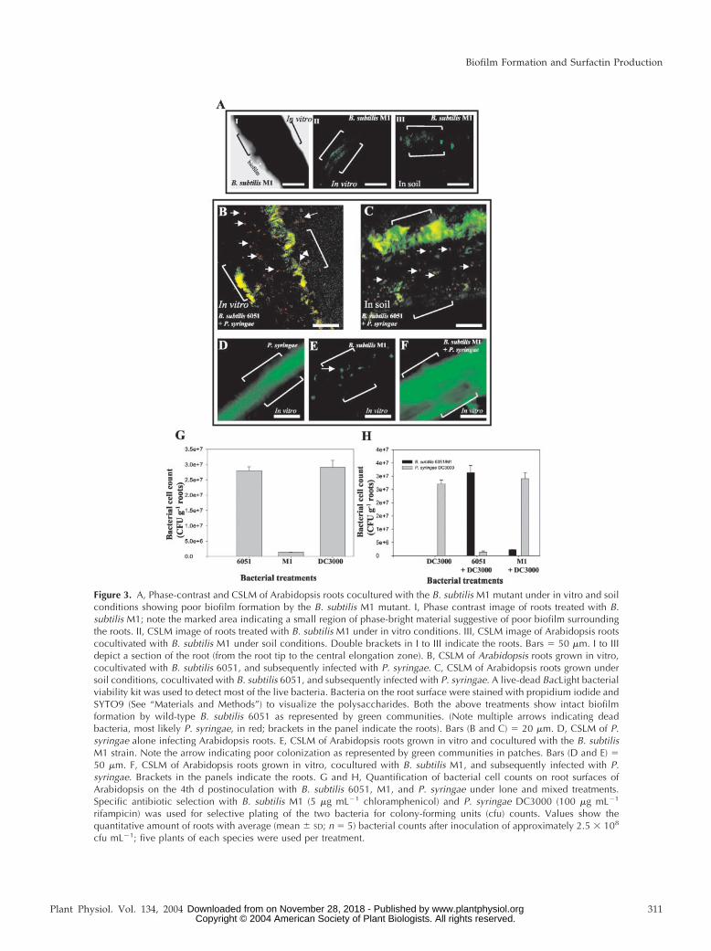
Figure 3. A, Phase-contrast and CSLM of Arabidopsis roots cocultured with the B. subtilis M1 mutant under in vitro and soilconditions showing poor biofilm formation by the B. subtilis M1 mutant. I, Phase contrast image of roots treated with B.subtilis M1; note the marked area indicating a small region of phase-bright material suggestive of poor biofilm surroundingthe roots. II, CSLM image of roots treated with B. subtilis M1 under in vitro conditions. III, CSLM image of Arabidopsis rootscocultivated with B. subtilis M1 under soil conditions. Double brackets in I to III indicate the roots. Bars � 50 �m. I to IIIdepict a section of the root (from the root tip to the central elongation zone). B, CSLM of Arabidopsis roots grown in vitro,cocultivated with B. subtilis 6051, and subsequently infected with P. syringae. C, CSLM of Arabidopsis roots grown undersoil conditions, cocultivated with B. subtilis 6051, and subsequently infected with P. syringae. A live-dead BacLight bacterialviability kit was used to detect most of the live bacteria. Bacteria on the root surface were stained with propidium iodide andSYTO9 (See “Materials and Methods”) to visualize the polysaccharides. Both the above treatments show intact biofilmformation by wild-type B. subtilis 6051 as represented by green communities. (Note multiple arrows indicating deadbacteria, most likely P. syringae, in red; brackets in the panel indicate the roots). Bars (B and C) � 20 �m. D, CSLM of P.syringae alone infecting Arabidopsis roots. E, CSLM of Arabidopsis roots grown in vitro and cocultured with the B. subtilisM1 strain. Note the arrow indicating poor colonization as represented by green communities in patches. Bars (D and E) �50 �m. F, CSLM of Arabidopsis roots grown in vitro, cocultured with B. subtilis M1, and subsequently infected with P.syringae. Brackets in the panels indicate the roots. G and H, Quantification of bacterial cell counts on root surfaces ofArabidopsis on the 4th d postinoculation with B. subtilis 6051, M1, and P. syringae under lone and mixed treatments.Specific antibiotic selection with B. subtilis M1 (5 �g mL�1 chloramphenicol) and P. syringae DC3000 (100 �g mL�1
rifampicin) was used for selective plating of the two bacteria for colony-forming units (cfu) counts. Values show thequantitative amount of roots with average (mean � SD; n � 5) bacterial counts after inoculation of approximately 2.5 � 108
cfu mL�1; five plants of each species were used per treatment.
Biofilm Formation and Surfactin Production
Plant Physiol. Vol. 134, 2004 311 www.plantphysiol.orgon November 28, 2018 - Published by Downloaded from Copyright © 2004 American Society of Plant Biologists. All rights reserved.

B. subtilis 6051 (compare Fig. 2A, II with Fig. 3A, I).Similarly, visualization of viable B. subtilis M1 cellsby CSLM using the viable cell procedure (see “Ma-terials and Methods”) revealed that only few andsmall regions in the roots were colonized by the M1mutant, and significantly reduced biofilm formationwas observed compared with B. subtilis 6051 (Fig. 3A,II and III). Soil-grown Arabidopsis roots cocultivatedwith the M1 mutant showed similarly reduced bio-film formation (Fig. 3A, III).
Interaction of B. subtilis Strains 6051 and M1 withP. syringae on Root Surfaces of Arabidopsis
To further visualize the differences in biocontrolefficiencies of B. subtilis 6051 and M1, P. syringae wasinoculated in the liquid growth media in which Ara-bidopsis plants were grown, 4 d post-treatment witheither B. subtilis strain. Viable cell microscopy of rootsprecultured with the 6051 strain revealed extensiveregions of green fluorescence, indicative of viablecells, with numerous clusters of red fluorescence pro-duced by dead bacteria (Fig. 3, B and C). In compari-son with roots similarly precultured with strain 6051and stained this way (Fig. 2A, III and IV) and based onthe protective effect of strain 6051 against P. syringaepathogencity, we inferred that the green-stained, via-ble cells are B. subtilis 6051 (Fig. 3H), and the red-stained dead cells are P. syringae. A control experimentwith roots treated with only P. syringae (Fig. 3D)shows that in the absence of B. subtilis 6051, thepseudomonad forms an extensive viable cell biofilm.
In contrast to the cocultivation with B. subtilis 6051,Arabidopsis roots precultured with the B. subtilis M1mutant and then infected with P. syringae showed nosigns of bacterial (P. syringae) mortality, instead re-vealing an intact pathogenic biofilm under in vitroconditions (Fig. 3F). Again, the M1 mutant showedonly small patches of colonization and biofilm for-mation (Fig. 3E), whereas the subsequent addition ofP. syringae resulted in an extensive viable biofilm(Fig. 3F). Similar results were obtained under soilconditions (data not shown).
To evaluate the correlation between plant mortalityand the size and quality of bacterial biofilms, we nextexamined bacterial colonization in plant roots culti-vated with lone treatments of B. subtilis 6051, M1, andP. syringae in contrast to roots cultivated with amixed treatment of B. subtilis 6051 � P. syringae andB. subtilis M1 � P. syringae under in vitro conditions.In accordance with our CSLM and plant mortalityresults, the B. subtilis M1 demonstrated poor rootcolonization 4 d post-treatment compared with thecolonization ability of B. subtilis 6051 or P. syringae(Fig. 3G). To confirm the likelihood that B. subtilis6051 out-competed P. syringae multiplication on Ara-bidopsis roots, we also examined the roots grownunder mixed treatments of B. subtilis 6051 with P.syringae. Four days after inoculation, the root-
localized bacterial number increased drastically forthe nonpathogenic B. subtilis 6051 compared with theP. syringae counts (Fig. 3H). In contrast, Arabidopsisroots precultured with the B. subtilis M1 mutant andthen infected with P. syringae showed reduced bac-terial counts for the M1 mutant (Fig. 3H), whereas thesubsequent addition of P. syringae resulted in anextensive multiplication of P. syringae on the Arabi-dopsis root surface (Fig. 3H). Specific antibiotic se-lection for B. subtilis M1 (5 �g mL�1 chlorampheni-col) and P. syringae DC3000 (100 �g mL�1 rifampicin)resulted in selective plating of the two bacteria forcfu counts. These results strongly support our hy-pothesis that B. subtilis 6051 acts as a potent biocon-trol against P. syringae infection in Arabidopsis.
Adherence of B. subtilis to Abiotic Surfaces
To further test whether the B. subtilis 6051 and M1strains differ in their ability to form adhering bio-films, we used the recently described methods ofHamon and Lazazzera (2001) to measure adherenceof the bacterium to the wells of a microtiter plate.Each strain was grown in the wells of a polyvinyl-chloride (PVC) microtiter plate in a complex biofilmgrowth medium, and the amount of adhering cellswas quantified by crystal violet (CV) staining (de-scribed in “Materials and Methods”). As shown inFigure 4A, measurable levels of adhered cells pro-gressively increased approximately 7-fold from 16 to30 h of incubation and then plateaued 60 to 70 h afterinoculation (Fig. 4A). This increase in CV stainingappeared to result from an increase in the number ofadhered cells because it corresponded to an increasein the number of cfu of adhered cells (data notshown). The B. subtilis M1 mutant produced approx-imately one-half the biofilm on microtiter plates com-pared with the B. subtilis 6051 (Fig. 4A).
To determine if the cells that were adhering to inertsurfaces were actually forming a three-dimensionalbiofilm, cells were analyzed by CSLM. Adhering B.subtilis 6051 cells (grown on glass cover slips) ap-peared to form a three-dimensional, multicellularstructure typical of a biofilm (Fig. 4, B and C). View-ing the cells in an x-y plane, it appears that theadhered cells form a mat of bacteria. From the x-zplane, it can be observed that the adhered cells forma structure with significant depth. As observed withCV staining, CSLM also revealed that the M1 mutantformed less biofilm compared with the B. subtilisstrain 6051 (Fig. 4, B and C).
Competition between B. subtilis Strains andP. syringae on Agar Surfaces
Because of the observed P. syringae mortality onArabidopsis root surfaces previously cocultivatedwith B. subtilis 6051, but not those cocultivated withM1, we investigated in situ P. syringae-B. subtilis com-
Bais et al.
312 Plant Physiol. Vol. 134, 2004 www.plantphysiol.orgon November 28, 2018 - Published by Downloaded from Copyright © 2004 American Society of Plant Biologists. All rights reserved.

petition on plates. Nutrient broth (NB) agar mediumwas co-inoculated with pairs of all three tested bac-teria (P. syringae-B. subtilis 6051 and P. syringae-B.subtilis M1). Sixteen hours after inoculation, B. subtilis6051 and P. syringae each formed a bacterial film overa large surface of their respective plates. Interest-ingly, when B. subtilis 6051 was cocultured with P.syringae, a distinct inhibition zone between the twobacteria was observed after 16 h of bacterial chal-lenge (Fig. 5A). The inhibition zone remained intacteven after 48 h (data not shown). In contrast, the B.subtilis M1 mutant failed to show any inhibition zoneagainst P. syringae and was overgrown by the latter(Fig. 5B). B. subtilis 6051 alone swarmed vigorouslyon NB agar plates, and the B. subtilis M1 mutantalone swarmed efficiently on NB agar plates as well,suggesting that the surfactin mutation did not impairits growth on this medium (Fig. 5, C and D). Thisphenomenon of selective inhibition of P. syringaegrowth by strain 6051 but not M1 was also seen onLuria-Bertani (LB) agar media (data not shown). Theessential difference between B. subtilis strains 6051and M1 was the formation of surfactin, which, basedon these results, appears to have an antimicrobialeffect on P. syringae (Wei and Chu, 1998). Thus, our
results show that the inhibition was clearly producedby B. subtilis 6051 against P. syringae.
Quantification of Surfactin from B. subtilisStrains 6051 and M1
Using HPLC conditions developed in the presentstudy (see “Materials and Methods”), surfactin pro-duction in liquid cultures of B. subtilis 6051 and M1strains was analyzed (Fig. 5E). As the typical chro-matogram (Fig. 5E, I) shows, the commercially pur-chased standard surfactin produces six differentpeaks depicting different isomers of surfactin; a sim-ilar result is shown by Wei and Chu (1998). We chosethese six major peaks through calibration to quantifysurfactin production. B. subtilis 6051 produced a sub-stance with a profile very similar to that of standardsurfactin (Fig. 5E, II). Interestingly, methanolic ex-tracts from the inhibition zone between B. subtilis6051 and P. syringae bacterial colonies from the in situchallenge experiments showed a chromatographicprofile similar to that observed for B. subtilis 6051 andstandard surfactin (Fig. 5E, III), suggesting that theinhibition zone was because of the abundant pres-ence of surfactin. In contrast, the B. subtilis M1 mu-
Figure 4. A, OD readings from microtiter plateassays of biofilm formation by B. subtilis 6051wild-type and M1 mutant strains. OD570 of sol-ubilized CV from microtiter assays over time forthe two tested B. subtilis strains; inset shows thesolubilized CV in polypropylene tubes depictingadherent biofilms: 1, B. subtilis 6051; and 2, B.subtilis M1 (values are mean � SD, n � 5). B,CSLM visualization of wild-type 6051 and M1mutant strains of B. subtilis grown on glass cov-erslips. C, An artist’s rendering of the three-dimensional structure of biofilm formed by wild-type (6051) and M1 mutant strains of B. subtilison x-z planes as visualized by CSLM. Bars �5 �m.
Biofilm Formation and Surfactin Production
Plant Physiol. Vol. 134, 2004 313 www.plantphysiol.orgon November 28, 2018 - Published by Downloaded from Copyright © 2004 American Society of Plant Biologists. All rights reserved.

tant did not appear to produce any of the surfactinisomers seen in the wild-type B. subtilis 6051 (Fig. 5E,IV). This analysis confirms that the �srfA-A mutationin strain M1 blocks formation of all six of the surfac-tin lipopeptides and that these polipeptides may ac-count for the antibacterial effect of B. subtilis on P.syringae.
Antibacterial Activity of Surfactin against P. syringae
The antibacterial activity of surfactin was testedagainst P. syringae planktonic cells by the broth mi-crodilution method in 96-well microtiter plates asdescribed in “Materials and Methods.” The mini-mum inhibitory concentration (MIC) of surfactin forP. syringae was determined to be 25 �g mL�1. TheMICs in Arabidopsis Murashige and Skoog basalmedia were comparable with MICs in cation-adjusted Mueller-Hinton broth (data not shown). Re-plating of the media from the 96-well microtiterplates (25–100 �g mL�1 surfactin) demonstrated thatthis lipopeptide is bactericidal (data not shown). Sur-factin also showed antibacterial activity against P.syringae on NB agar, evident by increasing zones ofinhibition (Fig. 6, A and B). Fluorescence microscopicvisualization by live-dead staining exhibited the bac-tericidal activity of surfactin against P. syringae in thetiter plate assay (Fig. 6, C–F). Fluorescence micros-copy of the P. syringae treated with MIC levels ofsurfactin (25 �g mL�1) revealed mortality as shownby red florescence compared with the untreated con-trol (Fig. 6, C–F). These results show that surfactinhas bactericidal activity against P. syringae.
Surfactin Formation by B. subtilis on Root Surfaces
To determine if surfactin was secreted from B. sub-tilis 6051 when grown on plant roots, we comparedthe levels of surfactin (a sum of the six isomers) in invitro cultures and during coculture of B. subtilis withArabidopsis roots in a defined Murashige and Skoogmedium. For comparison, the secretion of surfactinwas also measured in typical bacterial growth media,LB and NB, and in Arabidopsis Murashige and Skoogmedia, which also supported the growth of B. subtilis6051. Samples (either rinsed roots or culture mediasupernatants) were collected consecutively for 7 dand analyzed for surfactin by HPLC analysis (see“Materials and Methods”). As shown in Figure 6G,surfactin production by B. subtilis 6051 growing onroot surfaces of Arabidopsis was evident, with a finalconcentration in root extracts of 151.6 �g mL�1 per 50mg of roots fresh weight (Fig. 6G). Interestingly,surfactin production was elicited approximately 2.0-fold after administration of P. syringae into theArabidopsis-B. subtilis 6051 system (Fig. 6G; the ar-row depicts the sudden increase in surfactin produc-tion). Surfactin was not detected in root extracts ofArabidopsis plants cocultivated with B. subtilis M1and infected with P. syringae (data not shown).Growth of B. subtilis 6051 in common culture media,such as LB and NB, showed an expected linear in-crease of surfactin concentration during the timecourse until 160 h, followed by a gradual decrease(Fig. 6G). Substantial surfactin was also secreted byB. subtilis 6051 cultured in the Arabidopsis Murash-ige and Skoog medium.
Figure 5. A and B, In situ challenge of B. subtilis 6051 and P.syringae bacterial cultures. Bacterial cultures were inoculated with asterile toothpick on each one-half of the petri plate. Inhibition zoneswere visualized and photographed 48 h postinoculation. Plates in-dicate formation of surface film in NB agar medium (see “Materialsand Methods”). A, B. subtilis 6051 shows a clear and distinct inhi-bition zone repelling P. syringae, independent of the medium used(note the arrow indicating the bacterial inocula). B, B. subtilis M1was out-competed by P. syringae, a result independent of mediumused (note the arrows indicating the bacterial inocula). C and D,Lone B. subtilis 6051 and M1 growth plates on NB agar medium(note the arrows indicating bacterial inocula). E, HPLC spectrogramsfor surfactin production. Six peaks were used to quantify surfactincontent in B. subtilis. (Note the arrows indicating the six differentisoforms of surfactin). I, Standard surfactin. II, Surfactin content in B.subtilis 6051 broth cultures. III, Surfactin content isolated from theinterface of the inhibition zone observed during competition exper-iment between B. subtilis 6051 and P. syringae. IV, No surfactin wasfound from the B. subtilis M1 mutant.
Bais et al.
314 Plant Physiol. Vol. 134, 2004 www.plantphysiol.orgon November 28, 2018 - Published by Downloaded from Copyright © 2004 American Society of Plant Biologists. All rights reserved.

DISCUSSION
In this communication, we have described a newroot pathogenicity system (Arabidopsis roots-P. sy-ringae) and provide evidence for a unique biocontrolstrategy using the ubiquitous soil bacterium B. subti-lis—the formation of protective and antibacterial bio-films. First, we developed this experimental systemby using in vitro and soil cultures of Arabidopsis totest the root pathogenicity of P. syringae pv tomatoDC3000, a strain that has been identified as a potentleaf pathogen in Arabidopsis (Davis et al., 1991). Asshown here, the root pathogenicity of P. syringae, asbased on mortality rates, was similar both in vitroand in soil. Our experimental system provides ameans to disrupt the pathogenic interaction betweenP. syringae and Arabidopsis roots. We attempted todo this by using the wild-type B. subtilis strain 6051as a potential biocontrol agent. B. subtilis 6051 waschosen because this species is gaining recognition forbiocontrol in a variety of plants, albeit mainly for useas a seed protectant and antifungal agent (Chanway,2002; Warrior et al., 2002) and because of new evi-dence that some plant roots contain tightly bound B.subtilis strains that form abundant biofilms in vitro(Fall et al., 2003; Kinsinger et al., 2003).
When we tested the biocontrol efficiency of B. sub-tilis 6051 against root infection by P. syringae, reducedmortality of Arabidopsis was observed, both in cul-ture and in soil. The reason for this biocontrol effi-ciency was traced to the formation of anantimicrobial-producing biofilm, allowing for coloni-zation of the root surface of Arabidopsis, and to thesecretion of a lipopeptide antibiotic, surfactin. In ourstudies, we documented that the ability of B. subtilis6051 to control P. syringae infectivity of Arabidopsiswas directly proportional to its ability to colonizeand form biofilms on plant root surfaces.
Biofilm formation is a major bacterial adaptivestrategy to environmental conditions in aquatic andother settings (Emmert and Handelsman, 1999;Davey and O’Toole, 2000). However, the ability ofrhizosphere microorganisms to form protective bio-films is less well understood. Earlier studies sug-gested that biocontrol mechanisms could be relatedto biofilm formation on roots, which could protectagainst pathogenic infection (van Veen et al., 1997;O’Toole and Kolter, 1998). Pseudomonas fluorescens, aGram-negative soil bacterium and biocontrol agent,has been thought to form biofilms on plant roots, butthe relationship between biofilm formation and bio-control has not been confirmed (O’Toole and Kolter,1998; Bianciotto et al., 2001). In the case of strains ofB. subtilis and its close relatives that have been usedas biocontrol agents, numerous mechanisms for bio-control have been outlined (for review, see Chanway,2002), but no specific role for biofilm formation bythese bacteria on plant roots has been characterizedyet. However, in support of our results, it has beenfound recently that B. subtilis can form biofilms on
Figure 6. A and B, Antibacterial activity of 1 to 25 �g mL�1 surfactinon the growth of P. syringae. Standard surfactin was applied to thefilter discs. A bacterial inoculum (approximately 2 � 108 cells mL�1)of P. syringae was plated and spread on the petri dish, and the radialinhibition was observed on an hourly basis. A filter disc treated withonly solvent (2.5% [v/v] dimethyl sulfoxide) was used as a negativecontrol. A, Radial and efficient growth of P. syringae on NB agarplates in the absence of surfactin. B, Radial growth inhibition of P.syringae in the presence of different concentrations of surfactin. C toF, Fluorescence microscopic visualization of live-dead staining toshow bactericidal activity of surfactin against P. syringae in the titerplate assay. Bacterial suspensions treated with sub-MIC (0–5 �gmL�1), MIC (25 �g mL�1), and double MIC levels (50 �g mL�1) ofsurfactin were stained with propidium iodide and SYTO9 (see “Ma-terials and Methods”) to visualize the polysaccharides and nuclei,respectively. The green fluorescence in C and D depict live materialsurrounding/inside the bacterial colony. Red fluorescence showsdead cells (E and F). Scale bars � 20 �m. G, Surfactin profiles underdifferent media conditions (LB, NB, and Murashige and Skoog) andon the root surface of Arabidopsis grown alone, cocultured with B.subtilis strains, and infected with P. syringae. (Note the arrow indi-cating 96 h, the time of addition of P. syringae to the Arabidopsis-B.subtilis 6051 coculture). Arabidopsis roots were weighed (50 mgfresh weight) and extracted for surfactin analysis. (Values are mean �SD, n � 5).
Biofilm Formation and Surfactin Production
Plant Physiol. Vol. 134, 2004 315 www.plantphysiol.orgon November 28, 2018 - Published by Downloaded from Copyright © 2004 American Society of Plant Biologists. All rights reserved.

abiotic surfaces (Branda et al., 2001; Hamon and La-zazzera, 2001; Stanley et al., 2003).
The biocontrol ability of B. subtilis against the fun-gal pathogen Rhizoctonia solani has been shown to beachieved by virtue of the production of surfactin anditurin A, which are lipopeptides that contain a hy-droxy fatty acid connected by an ester peptide link-age to a cyclic heptapeptide (Peypoux et al., 1999).Interestingly, Asaka and Shoda (1996) showed thatthe persistence of surfactin in soil is better than thatof iturin A, suggesting a prolonged stable role forsurfactin in the rhizosphere. Here, we demonstratethat the biocontrol of P. syringae by B. subtilis 6051 isrelated to surfactin formation. We found that surfac-tin has an MIC of approximately 25 �g mL�1 againstP. syringae, which is relatively high for an antimicro-bial agent but may be reasonable for the exigenciesrhizosphere settings (Vivanco et al., 1999; Park et al.,2002). In the experiments with Arabidopsis roots thatwere precultured with B. subtilis 6051 (Fig. 6G), levelsof surfactin in rinsed roots were substantial (on theorder of 151.6 �g mL�1 per 50 mg root fresh weight).Thus, it is possible that at the root surface, the levelsof dissolved surfactin are substantially higher thanthe MIC against P. syringae determined in vitro, sug-gesting that the biocontrol exhibited against P. syrin-gae is linked to the formation of this antibiotic at theroot surface by a B. subtilis biofilm. The exact mech-anism by which surfactin acts as an antibacterialagent is not yet known, but is likely to be related toits ability to disrupt membranes (Peypoux et al.,1999) or to alter the physical and chemical propertiesof the biofilm growth of competing organisms (Neu,1996). For example, it was shown previously thatsurfactin inhibits biofilm formation of Salmonella en-terica at levels as low as 50 �g mL�1 and Escherichiacoli and Proteus mirabilis at higher levels in vitro(Mireles et al., 2001). Taking these observations to-gether, it is possible that the presence of B. subtilis6051 surfactin may prevent the planktonic cells ofother microbes from colonizing biological surfaces,including plant roots. This conclusion could explainthe biocontrol of P. syringae seen here in the Arabi-dopsis root system.
To further verify the role of surfactin in the biocon-trol of P. syringae, we utilized a surfactin-minus mu-tant of B. subtilis, strain M1, constructed using the B.subtilis 6051 background. The B. subtilis M1 mutantshowed normal growth in typical laboratory media,but when precultured with Arabidopsis, it was noteffective in controlling P. syringae pathogenicity andalso exhibited poor biofilm formation on roots, asshown by CSLM. It also formed less robust biofilmsthan the parent strain on inert surfaces. Although wecannot rule out pleiotropic effects resulting from thedeletion in the srfA-A gene of the M1 mutant, thesefindings strongly suggest that the production of sur-factin is essential for biofilm formation and coloniza-tion of Arabidopsis roots (and perhaps other plant
roots), and surfactin formation may be an essentialtrait for effective B. subtilis biocontrol strains.
As mentioned above, some plant roots containtightly bound B. subtilis strains, and we have shownthat many of these strains also produce surfactin (Fallet al., 2003; Kinsinger et al., 2003). It is possible thatbiofilm and surfactin formation may allow B. subtilisand its close relatives to efficiently colonize plantroots and also provide protection to their host.Whether this apparent “symbiotic” relationship iscommon in the roots of plants is unknown, but it is ofinterest to note that the roots of some plants containhigh populations of B. subtilis and other Bacillus spp.(Lilley et al., 1996; Pandey and Palni, 1997; Germidaet al., 1998).
The use of microorganisms to control plant dis-eases offers an attractive alternative to the use ofsynthetic chemicals (Emmert and Handelsman, 1999;Shoda, 2000; Warrior et al., 2002). The abundance ofa beneficial strain of microorganism in the vicinity ofplant roots may suppress plant pathogens withoutproducing lasting effects on the rest of the soil mi-crobial and plant communities (Howarth, 1991; Os-burn et al., 1995; van Veen et al., 1997). In accordance,the diversity of microbial communities provides arich source of potential biocontrol agents. With theroot pathogenicity system and methods describedhere, it should be possible to identify new Bacillusand other bacterial isolates that are effective in for-mation of protective and antibacterial biofilms.
MATERIALS AND METHODS
Plant Material and Growth Conditions ofArabidopsis in Vitro and in Soil
Seeds of wild-type Arabidopsis ecotype Columbia were obtained fromLehle Seeds (Round Rock, TX). Seeds were surface sterilized using 0.3%(v/v) sodium hypochlorite for 10 to 12 min and then washed four times insterile double distilled water. For root cultures, seeds were placed on staticMurashige and Skoog (1962) basal media in petri dishes for germination andincubated in a growth chamber. Twenty-five-day-old seedlings were indi-vidually transferred to 6-mL 12-well culture plates (Fisher Scientific, Lough-borough, Leicestershire, UK), each containing 2 mL of liquid Murashige andSkoog basal media. Plant cultures were maintained on an orbital platformshaker (Lab-Line Instruments, Melrose Park, IL) set at 90 rpm with aphotoperiod of 16 h of light and 8 h of dark at 25°C � 2°C.
For in-soil experiments, 25-d-old seedlings were transplanted from staticMurashige and Skoog media to 10-cm black plastic pots containing 50 g (dryweight) of PM-O5 Arabidopsis growing medium (Lehle Seeds). Plants wereincubated in a growth chamber at 30°C with 12 h of light and watered dailyfor 2 weeks before inoculation with bacteria.
Bacterial Strains and Culture Conditions
The following Bacillus subtilis strains were used in this study. Wild-typeB. subtilis 6051, the Marburg strain, was obtained from the American TypeCulture Collection (Manassas, VA). As described in detail elsewhere (Kins-inger et al., 2003), a surfactin-deficient mutant was constructed in the 6051strain by disruption of the srfA-A gene using the pJM103 integration vector(Perego, 1993). In brief, a 1,040-bp DNA fragment of the srfA-A gene wasamplified by PCR from B. subtilis 6051 chromosomal DNA, cloned betweenBamH1 and EcoR1 sites in pJM103, and the construct was used to transformB. subtilis 6051 using the method described by Anangnostopolous andSpizizen (1961). Although the transformation frequency with the 6051 strain
Bais et al.
316 Plant Physiol. Vol. 134, 2004 www.plantphysiol.orgon November 28, 2018 - Published by Downloaded from Copyright © 2004 American Society of Plant Biologists. All rights reserved.
was very low, mutants resistant to chloramphenicol were obtained, indica-tive of single insertions into and disruption of the srfA-A gene. One mutant,designated M1 (srfA-A::cam), grew normally in LB medium but secreted nosurfactin (as confirmed here), consistent with disruption of surfactin syn-thesis. The surface motility of the M1 mutant (Kinsinger et al., 2003) and itsability to form adhering biofilms on plastic surfaces (R. Kinsinger and R.Fall, unpublished data) were disrupted unless authentic surfactin wasadded to the growth media; the surfactin-minus phenotype of mutant M1was quite stable even after repeated subculture without added chloram-phenicol. Freshly plated cells from frozen stock cultures were used for allexperiments. Each strain was typically grown on LB agar plates and incu-bated at 37°C; chloramphenicol (5 �g mL�1) was added to maintain the M1mutant. Plated cells were suspended in 5 mL of LB broth for overnightgrowth at 37°C and shaken at 250 rpm. Pseudomonas syringae pv tomatoDC3000, a wild-type isolate that is pathogenic toward Arabidopsis leaves(Davis et al., 1991), was obtained from the laboratory of Dr. Christopher B.Lawrence (Department of Bioagricultural Sciences and Pest Management,Colorado State University, Fort Collins); it was maintained and grown on LBmedium with specified antibiotic selection (Rifampicin 100 �g mL�1) at28°C. In some experiments, bacterial cells were grown in NB (DIFCOLaboratories, Detroit) or Murashige and Skoog medium as described above.
In Vitro Root Pathogenicity Assay
P. syringae strains were grown to OD600 � 0.2 to 0.4 and added separatelyto the 2 mL of Murashige and Skoog media supporting each plant to reachan initial OD600 � 0.02 (approximately 2.5 � 107 cfu mL�2). Murashige andSkoog basal media (2 mL) without plant material was inoculated with thesame volume of each bacterial strain tested. By inoculation, we refer to theaddition of bacterial solution into the Murashige and Skoog medium wherethe roots were floating. A noninfected plant control was maintained underthe same conditions. All the treatments and controls were incubated at 30°Cin a controlled environment incubator shaker (New Brunswick Scientific,Edison, NJ) set at 30 rpm with a photoperiod of 16 h of light and 8 h of dark.Ten plants per treatment were used for analysis of mortality rates. Experi-ments were repeated twice in triplicate to standardize the observations.
Leaf Pathogenicity Assay
For leaf assays, P. syringae strains were grown in LB at 37°C to OD600 �0.2 to 0.3 and diluted 1:100 (w/v). Diluted suspensions were individuallyinjected with the blunt end of a hypodermic needle into intact leaves ofArabidopsis at a dose of approximately 1 � 103 cfu cm�2 as previouslydescribed (Jakob et al., 2002). Infiltrated plants were incubated in a growthchamber at 30°C and 80% relative humidity with 16 h of light and 8 h ofdark. Five leaves per plant were used for scoring of disease symptoms.
In-Soil Pathogenicity Assay
For soil infiltration, the 10-cm pots (with 50 g of soil) containing Arabi-dopsis were each flooded with 10 mL of P. syringae bacterial suspension togive an inoculum concentration of approximately 1 to 5 � 108 cfu g�1 of soil.Plants were incubated under identical conditions as those used for leafinfiltration assays. Ten plants per treatment were used for analysis ofmortality rates.
Analyzing the Biocontrol Efficiency of B. subtilisStrains 6051 and M1 against P. syringae
The wild-type B. subtilis 6051 and the M1 mutant were tested for theirbiocontrol ability on Arabidopsis roots both in vitro and under soil condi-tions. Bacterial strains were grown to OD600 � 0.3 to 0.4 and added sepa-rately to the 2 mL of Murashige and Skoog media of each in vitro plant toreach an initial OD600 � 0.02 (approximately 2.5 � 108 cfu mL�1). For soilinfiltration, the 10-cm pots (with 50 g of soil) containing Arabidopsis wereeach flooded with 10 mL of B. subtilis bacterial suspension to give aninoculum concentration of approximately 5 � 105 cfu g�1 of soil. A nonin-fected plant control was maintained under the same conditions. All thetreatments and controls were incubated at 30°C in a controlled environmentincubator shaker (New Brunswick Scientific) set at 30 rpm with a photope-
riod of 16 h of light and 8 h of dark. To analyze the biocontrol efficiency ofB. subtilis strains, P. syringae was inoculated under in vitro and soil condi-tions (as previously described) 4 d post-treatment with B. subtilis strainsusing the inoculum sizes mentioned above. Ten plants per treatment wereused for analysis of mortality rates. For bacterial counts on root surfaces, invitro-grown Arabidopsis root tissues (500 mg fresh weight) with mixedtreatments of B. subtilis 6051, M1, and P. syringae were washed with distilledwater and homogenized in 1 mL of saline (0.2% [w/v] sodium chloride)with a tissue grinder (size C, Kontes, Rochester, NY), and the suspensionwas filtered, diluted in saline, and plated on LB agar plates with specifiedantibiotic selection to determine bacterial cell counts. Specific antibioticselection with B. subtilis M1 (5 �g mL�1chloramphenicol) and P. syringaeDC3000 (100 �g mL�1rifampicin) was used for selective plating of the twobacteria for cfu counts. Each data point represents five replicates. All bac-terial growth assays were repeated, and only results that were observedconsistently are shown.
Microscopy
CSLM for biofilm formation was performed using the Live-dead BacLightBacterial Viability Kit (Molecular Probes, Eugene, OR) by incubating B.subtilis-P. syringae colonized Arabidopsis roots at room temperature in thedark for 15 min, according to the manufacturer’s manual. The samples weremounted with Citifluor antifading (Sigma, St. Louis) and observed forfluorescence with a confocal laser microscope (Fluroview LGPS-2, Olympus,Minneopolis). For observation of B. subtilis biofilms on glass coverslides byCSLM, B. subtilis biofilms were grown using the method described byWatnick and Kolter (1999). Biofilms of B. subtilis 6051 and M1 strains weregrown on glass coverslides (Fisher Scientific) in 6 mL of biofilm growthmedium in 50-mL polypropylene conical tubes. To determine whether B.subtilis biofilms were encased in a polysaccharide matrix, we stained thebiofilm with Calcofluor, a polysaccharide-binding dye. After rinsing, theslides were stained for 20 min with 10 mL of 75 �g mL�1 Calcofluor (Sigma;fluostain) in wash buffer. The stained biofilm was then analyzed by CSLMas described previously (Hamon and Lazazzera, 2001; Hogan and Kolter,2002). CSLM scans in both x-y and the x-z planes can be used to view athree-dimensional structure. To view adhered B. subtilis cells by CSLM, bothB. subtilis strains were stained with Calcofluor dye adhered to glass slides.Phase contrast images of B. subtilis-colonized root tissues were capturedwith a 10� objective on an Olympus BX60 microscope equipped withCoolSnap imaging software (San Diego) as described previously (Bianciottoet al., 2001). Phase contrast and CSLM were performed 4 d postinoculation.Samples were analyzed for fluorescence with a confocal laser microscope(Fluroview LGPS-2, Olympus). Samples were viewed using 488 nm as theexcitation wavelength.
Microtiter Plate Assay of B. subtilis Biofilm Formation
B. subtilis biofilm formation was monitored separately using a microtiterplate assay based on the methods of O’Toole et al. (1999). B. subtilis cellswere grown in 96-well PVC microtiter plates (Fischer Scientific) at 37°C inbiofilm growth medium. Biofilm growth medium based on Hamon andLazazzera (2001) was LB medium plus 0.15 m ammonium sulfate, 100 mmpotassium phosphate (pH 7), 34 mm sodium citrate, 1 mm MgSO4, and 0.1%(w/v) Glc. The inocula for the microtiter plates were obtained by growingthe cells in biofilm growth medium and shaking to midexponential growthand then diluting the cells to OD600 of 0.01 in fresh biofilm growth medium.Samples of 100 �L of the diluted cells were aliquoted to each well of 96-wellPVC microtiter plates. The microtiter plates were incubated at stationaryconditions. Cells that had adhered to the wells were stained with 0.1%(w/v) CV in wash buffer (0.15 m ammonium sulfate, 100 mm potassiumphosphate [pH 7], 34 mm sodium citrate, and 1 mm MgSO4) at roomtemperature for 20 min. Excess CV was then removed, and the wells wererinsed with water. The CV that had stained the cells was then solubilized in200 �L of 80% (v/v) ethanol and 20% (v/v) acetone. Biofilm formation wasquantified by measuring the OD570 for each well using an Opsys MR-Dynexplate reader (Chantilly, VA).
Biofilm Formation and Surfactin Production
Plant Physiol. Vol. 134, 2004 317 www.plantphysiol.orgon November 28, 2018 - Published by Downloaded from Copyright © 2004 American Society of Plant Biologists. All rights reserved.

In Situ Challenge between B. subtilis 6051 or M1 andP. syringae
LB and NB medium supplemented with 2.5 g L�1 tryptone, Glc (5 g L�1)with 0.4% (w/v) agar was incubated at 37°C. Swarm plates were typicallyallowed to dry at room temperature overnight before being used. Swarmplates were inoculated with bacteria using a sterile toothpick on both sidesof the petri plates to visualize competitive interactions. The plates were thenwrapped with plastic wrap to prevent dehydration and incubated at 37°Cfor 12 to 14 h.
Antibacterial Assays with Surfactin
MICs of surfactin against planktonic cells of P. syringae were determinedby the broth microdilution method using an inoculum of approximately 1 �105 cfu mL�1. Microtiter plates (96 well, Nalge Nunc International, Roch-ester, NY) were prepared with serial 2-fold dilutions of surfactin (Sigma) incation-adjusted Mueller-Hinton broth (DIFCO Laboratories). Surfactin wasadded from a 1 mg mL�1 stock solution in 2.5% (v/v) dimethyl sulfoxide.The MIC was visually defined as the lowest concentration of an antibioticthat completely inhibited cell growth after incubation for 22 h at 37°C. Allsusceptibility trials were conducted in triplicate. To check the bactericidalactivity of surfactin against P. syringae, sub-MIC (0–5 �g mL�1), MIC (25 �gmL�1), and double the MIC levels (50 �g mL�1) of surfactin-treated bacte-rial cells in microtiter plates were stained with Molecular Probes BacLightBacterial Viability Kit by incubating bacterial suspension at room tempera-ture in the dark for 20 min, according to the manufacturer’s manual. Thesamples were mounted with Citifluor antifading (Sigma) and observed forfluorescence with a fluorescence microscope (Fluroview LGPS-2, Olympus).
Quantificational Analysis of Surfactin
Surfactin concentration was analyzed by an HPLC procedure. B. subtiliscultures grown at different time points were withdrawn aseptically andcentrifuged at 8,000g for 20 min to pellet the cells. The supernatant wasextracted in methanol, concentrated and was further analyzed using anHPLC system consisting of P580 pumps (Dionex Co., Sunnyvale, CA) con-nected to an ASI-100 Automated Sample Injector (Dionex Co.), and a PDA-100 photodiode array variable UV/VIS detector (Dionex Co.). A C18 reverse-phase column (25.8 � 15 � 7 mm) was used for the separation of theextracts. Mobile phase solution A consisted of 3.8 mm trifluoroacetic acid inwater and acetonitrile (solution B; Fisher Scientific). Standard surfactin waspurchased from Sigma. An isocratic program with 20% (v/v) solution A and80% (v/v) solution B for 35 min was used for all separations with an initialinjection volume of 15 �L and a flow rate of 1 mL min�1. Chromeleonsoftware (Dionex Co.) was used to identify and quantify peaks. In a methodsimilar to the in situ challenge and root colonization experiments, extrac-tions for surfactin were performed by using the interface between the twobacterial colonies by carefully cutting the agar piece (500 mg) and thenextracting the agar piece and the intact B. subtilis colonized roots (mainlyroot tips and elongation zone region; approximately 5 cm long; 50 mg freshweight) in methanol; post-centrifugation, the supernatant was analyzed byreverse-phase HPLC as described above. The data presented here are fromfive independent experiments, and quantification of surfactin was per-formed by combining these experiments to calculate the average mean andto standardize conditions for a representative spectrogram.
Received June 17, 2003; returned for revision July 21, 2003; accepted No-vember 3, 2003.
LITERATURE CITED
Amellal N, Burtin G, Bartoli F, Heulin T (1998) Colonization of wheat rootsby an exopolysaccharide-producing pantoea agglomerans strain and itseffect on rhizosphere soil aggregation. Appl Environ Microbiol 64:3740–3747
Anagnostopolous C, Spizizen J (1961) Requirements for transformation inBacillus subtilis. J Bacteriol 81: 741–746
Asaka O, Shoda M (1996) Biocontrol of Rhizoctonia solani damping off ofTomato with Bacillus subtilis RB14. Appl Environ Microbiol 62: 4081–4085
Bashan Y, Bashan LE (2002) Protection of tomato seedlings against infectionby Pseudomonas syringae pv. tomato by using the plant growth-promotingbacterium Azospirillum brasilense. Appl Environ Microbiol 68: 2637–2643
Bianciotto V, Andreotti S, Balestrini R, Bonfante R, Perotto S (2001)Mucoid mutants of the biocontrol strain Pseudomonas fluorescens CHA0show increased ability in biofilm formation on mycorrhizal and non-mycorrhizal carrot roots. Mol Plant-Microbe Interact 14: 255–260
Branda SS, Gonzalez-Pastor JE, Ben-Yehuda S, Losick R, Kolter R (2001)Fruiting body formation by Bacillus subtilis. Proc Natl Acad Sci USA 98:11621–11626
Chanway CP (2002) Plant growth promotion by Bacillus and relatives. In RBerkeley, M Heyndrickx, N Logan, P De Vos, eds, B. subtilis for biocontrolin variety of plants. Blackwell Publishing, Malden, MA, pp 219–235
Costacurta A, Vanderleyden J (1995) Synthesis of phytohormones by plant-associated bacteria. Crit Rev Microbiol 21: 1–18
Costerton JW, Lewandowski Z, Caldwell DE, Korber DR, Lappin-ScottHM (1995) Microbial biofilms. Annu Rev Microbiol 49: 711–745
Davey ME, Caiazza NC, O’Toole GA (2003) Rhamnolipid surfactant pro-duction affects biofilm architecture in Pseudomonas aeruginosa PAO1. JBacteriol 185: 1027–1036
Davis KR, Schott E, Ausubel FM (1991) Virulence of selected phytopathicpseudomonads in Arabidopsis thaliana. Mol Plant-Microbe Interact 4:477–488
Davey ME, O’Toole GA (2000) Microbial biofilm: from ecology to moleculargenetics. Microbiol Mol Biol Rev 64: 847–867
Defago G, Keel C (1995) Pseudomonads as biocontrol agents of diseasescaused by soil borne pathogens. In HMT Hokkanen, JM Lynch, eds,Benefits and Risks of Introducing Biocontrol Agents. University Press,Cambridge, UK, pp 137–148
Emmert EAB, Handelsman J (1999) Biocontrol of plant disease: a Gram-positive perspective. FEMS Microbiol Lett 171: 1–9
Fall R, Kinsinger RF, Wheeler KA (2003) A simple method to isolate biofilmforming Bacillus subtilis and related species from plant roots. Appl Envi-ron Microbiol (in press)
Germida JJ, Siciliano SD, Frietas JRD, Seib AM (1998) Diversity of root-associated bacteria associated with field-grown canola (Brassica napus L.)and wheat (Triticum aestivum L.). FEMS Microbiol Ecol 26: 43–49
Hamon MA, Lazazzera BA (2001) The sporulation transcription factorSpo0A is required for biofilm development in Bacillus subtilis. Mol Mi-crobiol 42: 1119–1209
Hemphill HE, Whitely HR (1975) Bacteriophages of Bacillus subtilis. Bacte-riol Rev 39: 257–315
Hogan A, Kolter R (2002) Pseudomonas-Candida interactions: an ecologicalrole for virulence factors. Science 296: 2229–2232
Howarth FG (1991) Environmental impacts of classical biological control.Annu Rev Entomol 336: 485–509
Jakob K, Goss EM, Araki H, Van T, Krietman M, Bergelson J (2002)Pseudomonas viridiflava and Pseudomonas syringae-natural pathogens ofArabidopsis thaliana. Mol Plant-Microbe Interact 15: 1195–1203
Kinsinger RF, Shirk MC, Fall R (2003) Rapid surface motility and biofilmformation in Bacillus subtilis is dependent on extracellular surfactin andpotassium ion. J Bacteriol 185: 5627–5631
Kloepper JW, Zablotowick RM, Tipping EM, Lifshitz R (1991) Plantgrowth promotion mediated by bacterial rhizosphere colonizers. In DLKliester, PG Cregan, eds, The Rhizosphere and Plant Growth. KluwerAcademic Press, Dordrecht, The Netherlands, pp 315–326
Kunst F, Ogasawara N, Moszer I, Albertini AM, Alloni G, Azevedo V,Berkro MG, Bessieres P, Bolotin A, Borchert S (1997) The completegenome sequence of the Gram-positive bacterium Bacillus subtilis. Na-ture 390: 249–256
Lilley AK, Fry JC, Bailey MJ, Day MJ (1996) Comparison of aerobicheterotrophic taxa isolated from four root domains of mature sugar beet(Beta vulgaris). FEMS Microbiol 21: 231–242
Lutenberg BJJ, De Weger LA, Bennett JW (1991) Microbial stimulation ofplant growth and protection from disease. Curr Opin Micrbiol 2: 457–464
Lutenberg BJJ, Dekkers LC (1999) What makes Pseudomonas bacteria rhi-zosphere competent? Environ Microbiol 1: 9–13
Michiels KW, Croes CL, Vanderleyden L (1991) Two different modes ofattachment of Azospirillium brasilense Sp7 to wheat roots. J Gen Microbiol137: 2241–2246
Mireles JR-II, Toguchi A, Harshey RM (2001) Salmonella enterica serovarTyphimurium swarming mutants with altered biofilm-forming abilities:surfactin inhibits biofilm formation. J Bacteriol 183: 5848–5854
Bais et al.
318 Plant Physiol. Vol. 134, 2004 www.plantphysiol.orgon November 28, 2018 - Published by Downloaded from Copyright © 2004 American Society of Plant Biologists. All rights reserved.
Murashige T, Skoog F (1962) A revised medium for rapid growth andbioassay with tissue culture. Physiol Plant 15: 473–497
Neu TR (1996) Significance of bacterial surface-active compounds in inter-action of bacteria with interfaces. Micobiol Rev 60: 151–166
Osburn RM, Milner JL, Oplinger ES, Smith RS Handelsman J (1995) Effectof Bacillus cereus UW85 on the yield of soybean at two field sites inWisconsin. Plant Dis 79: 551–556
O’Toole GA, Kolter R (1998) Initiation of biofilm formation in Pseudomonasfluorescens WCS365 proceeds via multiple, convergent signaling path-ways: a genetic analysis. Mol Microbiol 30: 295–304
O’Toole GA, Pratt LA, Watnick PI, Newman DK, Weaver VB, Kolter R(1999) Genetic approaches to the study of biofilms. Methods Enzymol310: 91–109
Pandey A, Palni LMS (1997) Bacillus species: the dominant bacteria of therhizosphere of established tea bushes. Microbiol Res 152: 359–365
Park S-W, Lawrence CB, Linden JC, Vivanco JM (2002) Isolation andcharacterization of a novel ribosome-inactivating protein from root cul-tures of pokeweed and its mechanism of secretion from roots. PlantPhysiol 130: 164–178
Perego M (1993) Integrational vectors for genetic manipulation in Bacillussubtilis. In AL Sonenshein, JA Hoch, R Losick, eds, Bacillus subtilis andOther Gram-Positive Bacteria: Biochemistry, Physiology, and MolecularGenetics. American Society for Microbiology, Washington, DC, pp615–624
Peypoux F, Bonmatin JM, Wallach J (1999) Recent trends in the biochem-istry of surfactin. Appl Microbiol Biotechnol 51: 553–563
Pinton R, Varanini Z, Nannipieri P (2001) The rhizosphere as a site ofbiochemical interactions among soil components, plants and microorgan-isms. In R Pinton, Z Varanini, P Nannipieri, eds The Rhizosphere: Bio-chemistry and Organic Substances in the Soil-Plant Interface. MarcelDekker, New York, pp 1–17
Powell KA, Jutsum AR (1993) Technical and commercial aspects of biolog-ical control products. Pestic Sci 37: 315–321
Pratt LA, Kolter R (1999) Genetic analyses of bacterial biofilm formation.Curr Opin Microbiol 2: 598–603
Schippers B, Baker AW, Bakker PAHM (1987) Interactions of deleteriousand beneficial rhizosphere microorganisms and the effect of croppingpractices. Annu Rev Phytopathol 25: 339–358
Shoda M (2000) Bacterial control of plant diseases. J Biosci Bioeng 89:515–521
Stanley NR, Britton RA, Grossman AD, Lazazzera BA (2003) Identificationof catabolite repression as a physiological regulator of biofilm formationby Bacillus subtilis by use of DNA microarrays. J Bacteriol 185: 1951–1957
Tornero P, Dangl JL (2001) A high-throughput method for quantifyinggrowth of phytopathogenic bacteria in Arabidopsis thaliana. Plant J 28:475–481
Utkhede RS, Smith EM (1992) Promotion of apple tree growth and fruitproduction by the EBW-4 strain of Bacillus subtilis in apple replant diseasesoil. Can J Microbiol 38: 1270–1273
van Veen JA, van Overbeek LS, van Elsas JD (1997) Fate and activity ofmicroorganisms introduced into soil. Microbiol Mol Biol Rev 61: 121–135
Vivanco JM, Savary BJ, Flores HE (1999) Characterization of two novel typeI ribosome-inactivating proteins from the storage roots of the Andeancrop Mirabilis expansa. Plant Physiol 119: 1447–1456
Warrior P, Konduru K, Vasudevan P (2002) Formulation of biologicalcontrol agents for pest and disease management. In SS Gnanamanickam,ed, Biological Control of Crop Diseases. Marcel Dekker, New York, pp421–442
Watnick PI, Kolter R (1999) Steps in the development of a Vibrio cholerae ElTor biofilm. Mol Microbiol 34: 586–595
Wei YH, Chu IM (1998) Enhancement of surfactin production in iron-enriched media by Bacillus subtilis ATCC 21332. Enzy Microbe Technol22: 724–728
Weller DM, Thomashow LS (1994) Current challenges in introducing ben-eficial microorganisms into the rhizosphere. In F O’Gara, DN Dowling, BBoesten, eds, Molecular Ecology of Rhizosphere Microorganisms. VCH,New York, pp 1–18
Zuber P, Nakano MM, Marahiel MA (1993) Peptide antibiotics. In ALSonenshein, JA Hoch, R Losick, eds, Bacillus subtilis and Other Gram-Positive Bacteria: Biochemistry, Physiology, and Molecular Genetics.American Society for Microbiology, Washington, DC, pp 897–916
Biofilm Formation and Surfactin Production
Plant Physiol. Vol. 134, 2004 319 www.plantphysiol.orgon November 28, 2018 - Published by Downloaded from Copyright © 2004 American Society of Plant Biologists. All rights reserved.