biologic consideration of enamel and its clinical...
TRANSCRIPT

BIOLOGIC CONSIDERATION OF ENAMEL AND ITS CLINICAL
SIGNIFICANCE IN PRACTICE OF OPERATIVE DENTISTRY
Dr.Ahmed Al-Jobory

Introduction
Good knowledge of The 4 main
dental tissues and their
relationships to each other
and of the supporting
structures is necessary for
excellence in the performance
of operative procedures.
• Enamel
• Dentin
• Cementum
• Dental Pulp
Enamel structure
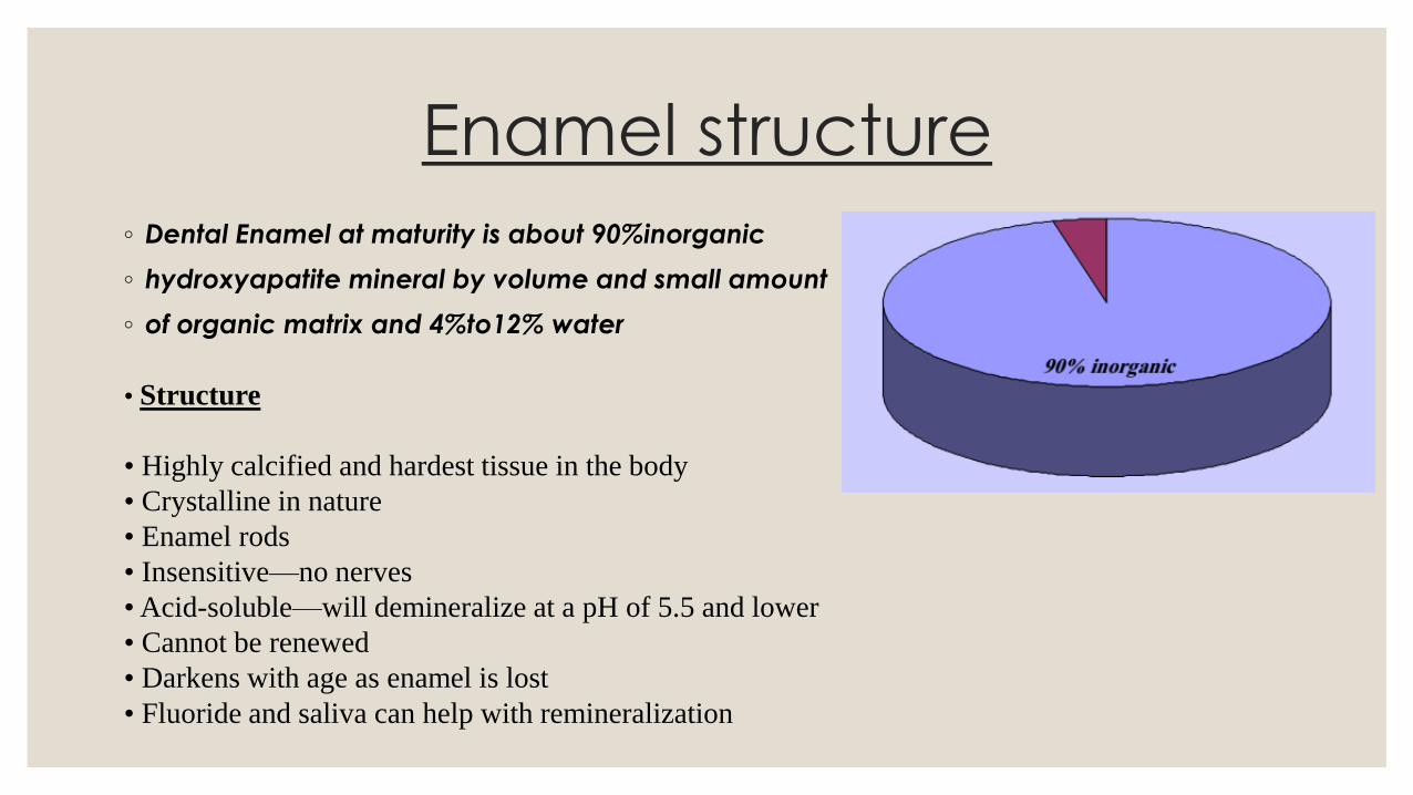
◦ Dental Enamel at maturity is about 90%inorganic
◦ hydroxyapatite mineral by volume and small amount
◦ of organic matrix and 4%to12% water
• Structure
• Highly calcified and hardest tissue in the body
• Crystalline in nature
• Enamel rods
• Insensitive—no nerves
• Acid-soluble—will demineralize at a pH of 5.5 and lower
• Cannot be renewed
• Darkens with age as enamel is lost
• Fluoride and saliva can help with remineralization

Morphologic and Histologic review
◦ Enamel formed by cells called ameloblast.
◦ Enamel provides a hard, durable shape for the functions of teeth & a protective cap
for the vital tissues of dentin & pulp.
◦ Both color & form contribute to the esthetic appearance of enamel.
◦ Enamel is incapable of repairing itself once destroyed because the ameloblast cell
degenerates after formation of enamel rod.

◦ The final act of ameloblast cell is secretion of a membrane covering the end
of enamel rod. This layer referred to as Nasmyth membrane or the primary
enamel cuticle, which covers the newly erupted tooth and is worn away by
mastication and cleaning. The membrane replaced by an organic deposit
called a pellicle, which is a precipitate of salivary proteins. Microorganisms
may invade the pellicle to form bacterial plaque, a potential precursor to
dental disease.

Permeability◦ at maturity, enamel is about 90% inorganic hydroxyapatite mineral
by volume. E. also contains a small amount of organic matrix & 4%
to 12% water, which is contained in the intercrystalline spaces & in
a network of micropores opening to the external surface. The
micropores form a dynamic connection between the oral cavity &
the systemic pulpal & dentinal tubule fluids.

◦ Various fluids, ions & low molecular weight substance can diffuse through the
semipermeable enamel. Therefore, the dynamics of acid demineralization,
caries, remineralization, fluoride uptake are not limited to the surface but are
active in three dimensions.

Solubility◦ Enamel is soluble when exposed to an acid medium, the solubility of
surface enamel decreased when fluorides are present during
enamel formation or topically applied to enamel surface. Fluoride
additions can affect the chemical and physical properties of the
apatite mineral and influence the hardness, chemical reactivity and
stability of enamel by lowering acid solubility, decreasing the rate of
demineralization and enhancing the rate of remineralization.

Clinical appearance and diagnosis
◦ The dentist must pay close attention to the surface characteristics of E. for
evidence of pathologic or traumatic conditions.
◦ 1. Color changes associated with demineralization
◦ 2. Cavitation
◦ 3. Wear
◦ 4. Faults and fissures
◦ 5. Cracks

1. Color changes associated with demineralization:
◦ E. is relatively translucent; its color is primarily a function of its thickness & the
color of underlying dentin.
◦ The thickness is more at the cusps tips & incisal edges & decreases below
deep fissures & become thin cervically at the junction with cementum.
◦ Color changes related to E. demineralization & caries are critical diagnostic
observation.
◦ Subsurface E. porosity from carious demineralization is manifested clinically by a milky white opacity called (white spot lesion); when located on smooth
surfaces. In later stages of caries, internal demineralization of E. at the DEJ,
subsurface cavitation imparts a blue or gray color to the overlying enamel.


◦2. Cavitation:
◦ The dentin affected until enamel breaks away to create a cavity, then a
restoration must be place.
◦ If untreated the cavitation expands to compromise the structural strength of
the crown and microorganisms infiltrate into deep dentin to affect the vitality
of the tooth.

◦3. Wear:
◦ Enamel is as hard as steel, however enamel will wear because of attrition or
frictional contact against opposing enamel or harder restorative materials
such as porcelain.
◦ Heavy occlusal wear demonstrated when rounded cuspal contacts are
ground to flat facets.
◦ Depending on factors such as bruxism, malocclusion, age and diet; cusps
may be completely lost & enamel abraded away so that the dentin is exposed.

◦4. Faults and fissures: ◦ A deep fissure formed by incomplete fusion of lobes of cuspal enamel in the
developing tooth.
◦ The resulting narrow clefts provide protected area for acidogenic bacteria.
◦ Pits & fissures defects are eight times more vulnerable to caries than are smooth
surfaces.
◦ Careful observation of enamel surrounding fissures for evidence of demineralization or
cavitation is necessary to determine the need for restorative intervention.

◦5. Cracks:◦ Pronounced cracks that extend from developmental grooves across marginal ridges
to axial walls or from the margins of large restorations may cause cuspal fracture.
◦ When this crack extends through dentin or when the patient has pain when chewing;
the tooth requires a restoration that provide complete cuspal coverage.

Crystal structure and enamel rods

Structurally enamel is composed of millions of
E. rods or prisms. The rods are densely packed
& have a wavy course & each extends from the
DEJ to the external surface of the tooth.
E. is the hardest substance of human body, E.
is very brittle, and so it requires a base of dentin to
withstand the masticatory stress. Enamel rods that
fail to possess a dentin base because of caries or
improper cavity design are easily fracture away
from neighboring rods.

◦ The structural components of enamel prisms are millions of small-elongated
apatite crystal, which are tightly packed, in a distinct pattern of orientation
that gives strength & structural identity to the enamel prisms.
◦ An organic matrix or prism sheath surrounds individual crystal.
◦ The spacing & orientation of the crystals & the amount of organic matrix make
the enamel rod boundary & the central core differentially soluble when
exposed for a short time to weak acids.

◦ The acid- treated E. surface has an irregular and pitted surface with
numerous microscopic undercuts, the etched enamel has a higher
surface energy, so resin monomer flows into & adheres to the etched
depressions to polymerize & form retentive resin tags. Because there
are (30,000) to (40,000) E. rods/ mm2 & acid etch penetration increases
the bondable surface area to (10) to (20) folds, micromechanical
bonding of resin restorative materials to E. is significant.
◦ Acid-etch modification of E. for restoration retention provides a
conservative, reliable, alternative to traditional surgical methods of
tooth preparation & restorations, (retentive grooves, pins, extension for
prevention).

◦ Starting at 1 mm from CEJ the rods run occlusally or incisally at 60 degree inclination and
progressively incline approaching the marginal ridges and cusp tips where the rods are parallel
to the long axis of the crown. In the cervical region the rods are oriented slightly in apical
direction.

◦ Loss of enamel rods that form the cavity wall of cavomargin of dental
restorations creates a gap defect, leakage of bacteria & their products
that may lead to secondary caries. Therefore, a basic principle of cavity
wall preparation is to bevel or parallel the direction of E. rods & avoid
undercutting them.

◦ Understanding enamel orientation is very important in restorative dentistry,
because enamel unsupported by underlying dentin is prone to fracture.

Clinical sites for caries initiation

◦ There are three distinctly different clinical sites on teeth where
cariogenic plaque may originate:
◦ 1- Pits and fissures
◦ 2- Smooth enamel surface
◦ 3- Root surface

◦ 1-Pits & fissures of enamel:
◦ this is the most susceptible site. The path of carious lesion is roughly
parallel to the long axis of enamel rods; the entry site may appear
much smaller than the actual lesion making clinical diagnosis
difficult. In cross section, the appearance of the lesion is an inverted
V with a wide area of involvement at DEJ.
◦ Pit & fissure caries most commonly found on occlusal surface of
posterior teeth, lingual surface of maxillary anterior teeth & buccal
and lingual pits of molars.


◦ 2-Smooth enamel surface:
◦ plaque usually develops only on smooth surface that is near the
gingival or under proximal contacts; the path of ingress of the lesion
is roughly parallel to the long axis of the enamel rods in the region.
Across section shows, a V shape with wide area of origin & the
apex of the V directed towards the DEJ. It includes proximal caries
& lesions on other smooth surfaces.
◦ After caries, extension to the DEJ there is both lateral spread along
the junction & extension pulpally.
◦ A cross section of caries in dentin is always V shaped with its base
at DEJ and the apex directed pulpally.

◦ 3-Root surface:
◦ the cementum covering the root surface is extremely thin and provides little
resistance to caries attack .Root lesions have less well-defined margins tend to
be U-shaped in cross section.
