biological pathways for sepsis-associated acute
TRANSCRIPT

Page 1/22
Bioinformatic Identi�cation of Hub Genes andBiological Pathways for Sepsis-Associated AcuteLung InjuryChao Zhang
Chongqing Medical University Pediatric College: Chongqing Medical University A�liated Children'sHospitalFeng Xu ( [email protected] )
Chongqing Medical University https://orcid.org/0000-0002-1122-0581Fang Fang
Chongqing Medical University
Research
Keywords: Sepsis, Acute lung injury, Bioinformatics, Differentially expressed genes, LPS, HMGB1
Posted Date: July 13th, 2021
DOI: https://doi.org/10.21203/rs.3.rs-623296/v1
License: This work is licensed under a Creative Commons Attribution 4.0 International License. Read Full License

Page 2/22
AbstractBackground: Sepsis-associated acute lung injury (ALI) is a potentially lethal complication associated witha poor prognosis and high mortality worldwide, especially in the outbreak of COVID-19. However, thefundamental mechanisms of this complication were still not fully elucidated. Thus, we conducted thisstudy to identify hub genes and biological pathways of sepsis-associated ALI, mainly focus on twopathways of LPS and HMGB1.
Methods: Gene expression pro�le GSE3037 were downloaded from Gene Expression Omnibus (GEO)database, including 8 patients with sepsis-induced acute lung injury, with 8 unstimulated bloodneutrophils, 8 LPS- induced neutrophils and 8 HMGB1-induced neutrophils. Differentially expressed genes(DEGs) identi�cations, Gene Ontology (GO) function analysis, Kyoto Encyclopedia of Genes andGenomes (KEGG) pathways analysis, Gene Set Enrichment Analysis (GSEA) and protein-proteininteraction (PPI) network constructions were performed to obtain hub genes and relevant biologicalpathways.
Results: We identi�ed 534 and 317 DEGs for LPS- and HMGB1-induced ALI, respectively. The biologicalpathways involved in LPS- and HMGB1-induced ALI were also identi�ed accordingly. By PPI networkanalysis, we found that ten hub genes for LPS-induced ALI (CXCL8, TNF, IL6, IL1B, ICAM1, CXCL1, CXCL2,IL1A, IL1RN and CXCL3) and another ten hub genes for HMGB1-induced ALI (CCL20, CXCL2, CXCL1,CCL4, CXCL3, CXCL9, CCL21, CXCR6, KNG1 and SST). Furthermore, by combining analysis, the resultsrevealed that genes of TNF, CCL20, IL1B, NFKBIA, CCL4, PTGS2, TNFAIP3, CXCL2, CXCL1 and CXCL3were potential biomarkers for sepsis-associated ALI.
Conclusions: Our study revealed that ten hub genes associated with sepsis-induced ALI were TNF, CCL20,IL1B, NFKBIA, CCL4, PTGS2, TNFAIP3, CXCL2, CXCL1 and CXCL3, which may serve as genetic biomarkersand be further veri�ed in prospective experimental trials.
BackgroundSepsis is a potentially lethal complication associated with a poor prognosis and high mortality [1]. It isestimated that nineteen millions of patients affected by sepsis worldwide annually, and the actualmortality of sepsis have unacceptably reached as high as 32.6%![2] Meanwhile, sepsis is oftencomplicated with acute lung injury (ALI), which is manifested as acute respiratory distress syndrome(ARDS), even as subsequent multiple organ dysfunction syndrome (MODS), especially in the situation ofCOVID-19 infection [3]. ARDS, as well as MODS, is considered as a milestone that predict poorerprognosis and higher mortality. Unfortunately, treatments of sepsis-induced acute lung injury are stilllimited [4]. Moreover, mechanisms of this disease have not been fully elucidated. Thus, it is criticallyimportant to investigate the mechanism of sepsis induced ALI to �nd out novel therapeutic targets.
The role of lipopolysaccharide (LPS) and high mobility group box-1 (HMGB1) in sepsis-associated ALIreceived more and more concerns in recent years. Several studies were conducted to explore relevant

Page 3/22
pathways for LPS and HMGB1, alone or together, that cause or aggravate sepsis-associated ALI [5–8].However, there are still a lack of bioinformatics evidence exploring the potential hub genes and biologicalpathways for LPS- and HMGB1- induced sepsis-associated ALI. To �ll in the gap of this �eld, weconducted the bioinformatics analysis by extracting GSE3037 datasets from GEO database, attemptingto identify the hub genes and pathways involving in sepsis-associated ALI, mainly focus on these twopathways of LPS and HMGB1.
Materials And Methods
Sepsis Induced ALI DatasetsExpression pro�le data of GSE3037 was extracted from Gene Expression Omnibus (GEO) database viamicroarray GPL96 platform. GSE3037 datasets include 8 patients with sepsis-induced ALI. The baselinedata of these patients were listed in Table 1. Blood neutrophils of these eight patients were cultured withLPS, cultured with HMGB1 or left unstimulated. Therefore, 8 unstimulated blood neutrophils samples(GSM66867-GSM66874), 8 HMGB1-induced samples (GSM66875-GSM66882) and 8 LPS-inducedsamples (GSM66883-GSM66890) were collected for further analysis.
Table 1Baseline data of extracted sepsis-induced acute lung injury patients
Sample no. sex age Site of infection Sepsis shock at ICU?
1 Male 66 Urosepsis No
2 Male 34 Pneumonia Yes
3 Male 43 Pneumonia No
4 Male 44 Cellulitis No
5 Male 41 Pneumonia No
6 Female 42 Pneumonia Yes
7 Male 43 Pneumonia Yes
8 Female 50 Pneumonia No
Identi�cations of DEGsGene expression matrix �les were extracted and further analyzed, referring to “hgu133a.db” annotations,which translated raw microarray data to gene expression data. Differential expression analysis wasperformed by “limma” [9] with criteria for adjust P-value < 0.1 and |log fold change (FC)|>1.2.
Enrichment analysis of DEGs1. GO function and KEGG pathway analysis of upregulated and downregulated DEGs were performed
by metascape (https://metascape.org/) [10] (minimum overlap:3, P-value cut-off:0.01 and minimum

Page 4/22
enrichment:1.5). The further chordal graph and heatmap analysis was conducted by GOplotsoftware [11].
GSEA analysisGene Set Enrichment Analysis (GSEA) was performed to study the enrichment of a gene set in a largenumber of genes, which often consists of multiple gene sets [12]. We chose “h.all.v7.2.symbols.gmt” forreference and set times of permutation as 1000 and type of permutation as “gene set”. The enrichmentwas considered as signi�cant when FDR < 0.05. Meanwhile, Normalized Enrichment Score (NES) wasapplied to assess the extent of enrichment.
PPI network construction and modular analysisSearch Tool for the Retrieval of Interacting Genes (STRING) online database (https://string-db.org/) [13]was performed to evaluate of protein–protein interaction (PPI). Cytoscape [14]and Molecular ComplexDetection (MCODE; cut-off: scores > 3 and nodes > 4) was used to �nalize module screening. Top ten hubgenes were identi�ed by further analysis via CytoHubba.
Results
Identi�cation of DEGs for LPS- and HMGB1-inducedpathwaysGene expression value in LPS and HMGB1-induced neutrophils samples was similar with the value inunstimulated neutrophils samples, respectively (Fig. 1). For LPS-induced ALI, volcano plots and heatmaprevealed that a total of 534 DEGs were identi�ed, with 296 up-regulated DEGs and 238 down-regulatedDEGs (Fig. 2). For HMGB1-induced ALI, the results revealed that totally 317 DEGs were identi�ed, with 158up-regulated DEGs and 159 down-regulated DEGs (Fig. 3).
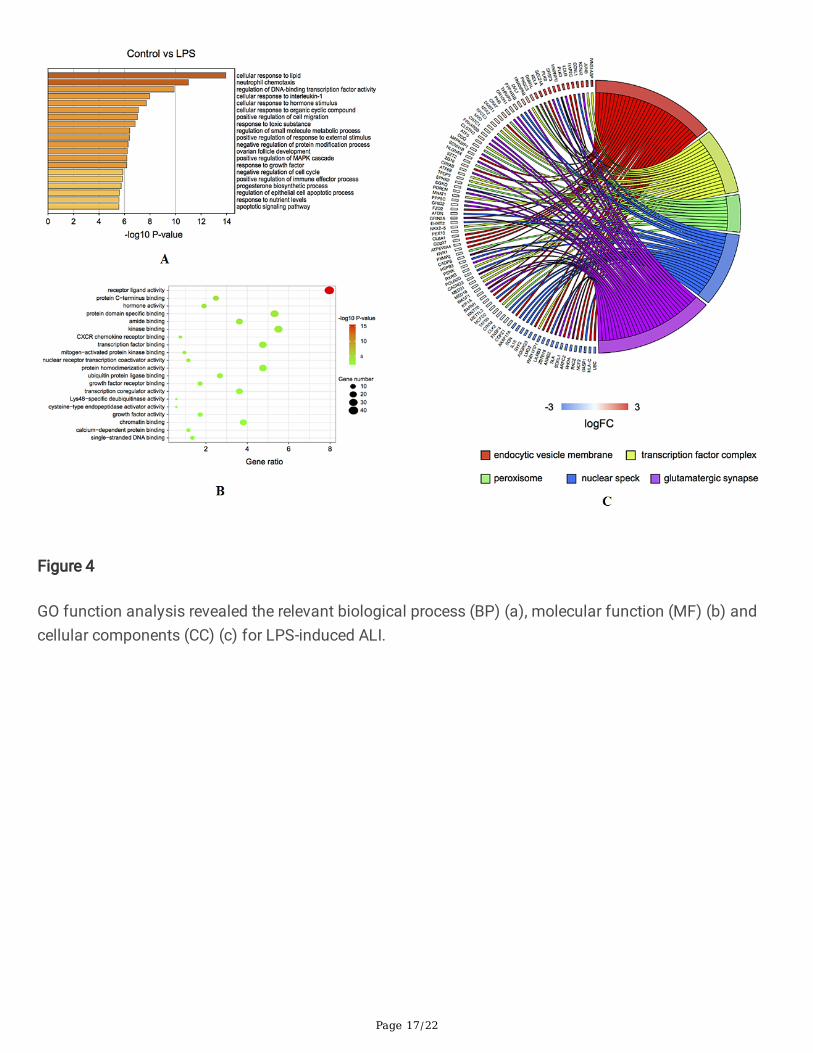
GO function analysisGO function analysis revealed that relevant biological process for LPS-induced ALI included cellularresponse to lipid, neutrophil chemotaxis, regulation of DNA-binding transcription factor activity (Fig. 4A).For molecular function, receptor ligand activity, protein C-terminus binding and hormone activity (Fig. 4B)were obtained. Corresponding cellular components included endocytic vesicle membrane, transcriptionfactor complex, peroxisome, etc. (Fig. 4C). Besides, for HMGB1-induced ALI, relevant biological processincluded response to chemokine, cellular response to lipopolysaccharide and fever generation (Fig. 5A).For molecular function, receptor ligand activity, protein kinase regulatory activity and cytokine binding(Fig. 5B) were identi�ed. Relevant cellular components included endoplasmic reticulum lumen,extracellular matrix, Schaffer collateral CA1 synapse and microtubule organizing center part, etc.(Fig. 5C).
KEGG pathways analysis

Page 5/22
For LPS-induced ALI, KEGG pathway analysis revealed that the signi�cant enrichment pathways includedIL-17 signaling pathway, Cytokine-cytokine receptor interaction, NF-kB signaling pathway, etc. (Fig. 6).Meanwhile, for HMGB1-induced ALI, KEGG pathway analysis revealed that the signi�cant enrichmentpathways involved cytokine-cytokine receptor interaction, TNF signaling pathway and cancer pathways,etc. (Fig. 7).
GSEA analysisFor LPS-induced ALI, GSEA analysis showed the signi�cant enrichment biological processes/statesinvolved TNFA-signaling via NF-kB, in�ammatory response, apoptosis, ultraviolet response up, hypoxiaand cholesterol homeostasis (Fig. 8). In addition, for HMGB1-induced ALI, GSEA analysis showed thesigni�cant enrichment biological processes/states involved TNFA-signaling via NF-kB, in�ammatoryresponse, apoptosis, ultraviolet response, hypoxia and KRAS signaling up (Fig. 9).
PPI network construction and hub genes identi�cationFor LPS-induced ALI, we identi�ed 10 modules in PPI network (Fig. 10A-10B). Using CytoHubba, weidenti�ed 10 potential hub genes related to LPS-induced ALI, including CXCL8, TNF, IL6, IL1B, ICAM1,CXCL1, CXCL2, IL1A, IL1RN and CXCL3 (Fig. 10C). Meanwhile, for HMGB1-induced ALI, we also identi�edten modules in PPI network (Fig. 11A-11B). We identi�ed ten hub genes related to HMGB1-induced ALI,including CCL20, CXCL2, CXCL1, CCL4, CXCL3, CXCL9, CCL21, CXCR6, KNG1 and SST (Fig. 11C).
Combining analysisWe subsequently performed a combining analysis to extract the overlapping data between LPS-inducedand HMGB1-induced ALI. Then a total of 88 DEGs were overlapping (Fig. 12A). By PPI networkconstruction, notably, we found a signi�cant module in PPI network (Fig. 12B-C). In this module, genes ofTNF, CCL20, IL1B, NFKBIA, CCL4, PTGS2, TNFAIP3, CXCL2, CXCL1, CXCL3 were identi�ed (Fig. 12D). Inaddition, we also performed GO function analysis, KEGG pathways analysis and GSEA analysis forcombining DEGs, the results were not shown.
DiscussionThe present study is the �rst study to conduct a comprehensive bioinformatics analysis to explore hubgenes and biological pathways related to LPS and HMGB1 triggered sepsis-associated ALI. The presentstudy identi�ed 534 and 317 DEGs for LPS- and HMGB1-induced ALI, respectively. The biologicalpathways involved in LPS- and HMGB1-induced ALI were also demonstrated accordingly. By conductingPPI network analysis, we found that ten hub genes for LPS-induced ALI (CXCL8, TNF, IL6, IL1B, ICAM1,CXCL1, CXCL2, IL1A, IL1RN and CXCL3) and another ten hub genes for HMGB1-induced ALI (CCL20,CXCL2, CXCL1, CCL4, CXCL3, CXCL9, CCL21, CXCR6, KNG1 and SST). Notably, by combining analysis,the results revealed a module of genes TNF, CCL20, IL1B, NFKBIA, CCL4, PTGS2, TNFAIP3, CXCL2, CXCL1and CXCL3 were potential biomarkers for sepsis-associated ALI.

Page 6/22
To further explore the mechanisms of LPS-triggered sepsis-associated ALI, we searched the literatures forthe functions of LPS relevant 10 genes (CXCL8, TNF, IL6, IL1B, ICAM1, CXCL1, CXCL2, IL1A, IL1RN andCXCL3). The functions of these ten genes were summarized in Table 2. The cellular components encodedby these 10 genes were all previously proved to be participated in the progression of in�ammations insepsis patients or animal models [15–24]. For chemokine ligand family, CXCL1, CXCL2, CXCL3 andCXCL8, the common functions are all chemoattractant for neutrophils [15, 20–21, 24]. And for interleukinfamily, IL1A, IL1B, ILRN and IL6, their common functions are rapid increasing in levels and evoking“cytokine storm” [17–18, 22–23]. For ICAM1, stimulated by LPS, resulted in increases in neutrophil-neutrophil adherence and aggregation [19]. And for TNF, the most common mediator of sepsis, elicit therelease of in�ammatory mediators [16]. LPS may stimulate these cellular components to aggravate theprogression of sepsis-induced lung injury.

Page 7/22
Table 2Hub genes and their functions in in�ammation of sepsis based on previous literatures (for LPS-induced
ALI)Hubgenes
Cellularcomponents
Reference Functions mentioned in the literatures
CXCL8 Chemokine(C-X-Cmotif)ligand 8
T MCunha etal [15]
Recruiting leukocytes from the blood into tissues duringin�ammation
TNF Tumornecrosisfactor
Lucy KReiss etal [16]
Eliciting the release of in�ammatory mediators, and mediatorof sepsis itself
IL6 Interleukin 6 Tanaka Tet al [17]
Excessive synthesis of IL-6 while �ghting environmentalstress leads to an acute severe systemic in�ammatoryresponse known as 'cytokine storm'
IL1B Interleukin-1-B
Varljen Tet al [18]
Proin�ammatory mediator whose level is rapidly increasedduring infection
ICAM1 Intercellular-adhesion-molecule-1
Ode Y etal [19]
An increased expression of ICAM-1 on neutrophils afterstimulation with LPS has been shown, which resulted insigni�cant increases in neutrophil-neutrophil adherence andaggregation
CXCL1 Chemokine(C-X-Cmotif)ligand 1
Paudel Set al [20]
CXCL1 induces neutrophil in�ux during bacterial infections, inaddition, it is a central player in host defense, granulopoiesis,and mobilization of neutrophils during sepsis.
CXCL2 Chemokine(C-X-Cmotif)ligand 2
De Filippoet al [21]
CXCL2 is secreted, especially by mast cells, monocytes, andmacrophages at the site of in�ammation, and is an importantchemotactic for neutrophils during infections
IL1A Interleukin-1-A
Zhu W etal [22]
IL-1 signaling via the type 1 IL-1 receptor leads to multiplein�ammatory effects including vasodilation, increasedvascular permeability
IL1RN Interleukin-1-RN
Meyer etal [23]
C allele of a synonymous coding variant (rs315952-C) inIL1RN is associated with reduced risk for ARDS and increasedplasma IL-1ra
CXCL3 Chemokine(C-X-Cmotif)ligand 3
Rebollo J[24]
CXCL3 has been shown to act as a chemoattractant forneutrophils to and for cerebellar progenitor cells
Besides, we also searched previous literatures and screened for the studies with the ten hub genes forHMGB1 triggered ALI. The cellular components encoded by these 10 genes were all previously proved tobe participated in the progression of in�ammations in sepsis patients or animal models. [20–21, 24, 25–31] For chemokine ligand family, CXCL1, CXCL2, CXCL3 and CXCL9, the common functions are allchemoattractant for neutrophils. [25, 28–29] For CC chemokine family, CCL4 plays key roles in

Page 8/22
chemotaxis as well as enhancement of the in�ammatory response, while CCL21 regulates leukocytetra�cking and homing to the lymph nodes. [26, 28] In addition, CCL20 is an early trigger of neutrophil cellrecruitment and activation of T cells in the small intestine. [27] For CXCR6, it could mediate iNKT-cellaccumulation, clustering and arrest within the liver in several in�ammatory conditions. [29] For KNG1, itcould accelerate the progress of cellular in�ammation. [30] Besides, SST has a protective effect onintestinal barrier dysfunction through the suppression of NF-κB. [31] HMGB1 may stimulate all thesecellular biomarkers to aggravate the progression of sepsis-induced lung injury. These functions of tenhub genes for HMGB1-induced ALI were listed in Table 3.

Page 9/22
Table 3Hub genes and their functions in in�ammation of sepsis based on previous literatures (for HMGB1-
induced ALI)Hubgenes
Cellularcomponents
Reference Functions mentioned in the literatures
CXCL9 Chemokine(C-X-C motif)ligand 9
Cavalcantiet al [15]
CXCL9 induces the CXC chemokines which act preferentiallyon T and B lymphocytes and natural killer cells (NK)
CCL4 Chemokine(C-C motif)ligand 4
Nowak etal [16]
CCL4 is a member of the CC chemokine family, and isproduced by numerous cell types including monocytes,neutrophils, T and B lymphocytes, NK cells, and dendriticcells. It plays a key role in chemotaxis as well asenhancement of the in�ammatory response
CCL20 Chemokine(C-C motif)ligand 20
KitagawaY et al [17]
CCL20 has been identi�ed as an early trigger of neutrophilcell recruitment and activation of T cells in the smallintestine.
CCL21 Chemokine(C-C motif)ligand 21
Castro Ret al [18]
The regulation of leukocyte tra�cking and homing to thelymph nodes (LNs)
CXCR6 Chemokine(C-X-C motif)receptor 6
Young etal [19]
CXCR6 has been shown to mediate iNKT-cell accumulation,clustering and arrest within the liver in several in�ammatoryconditions
CXCL1 Chemokine(C-X-C motif)ligand 1
Paudel Set al [20]
CXCL1 induces neutrophil in�ux during bacterial infections,in addition, it is a central player in host defense,granulopoiesis, and mobilization of neutrophils duringsepsis.
CXCL2 Chemokine(C-X-C motif)ligand 2
De Filippoet al [21]
CXCL2 is secreted, especially by mast cells, monocytes, andmacrophages at the site of in�ammation, and is animportant chemotactic for neutrophils during infections
KNG1 Kininogen-1 Hu Q et al[22]
As a pro-in�ammatory cytokine, KNG1 has beendemonstrated to accelerate the progress of in�ammation.Inhibition of KNG1 could relieve cellular in�ammation
SST Somatostatin Xu X [23] SST has a protective effect on intestinal barrier dysfunctionthrough the suppression of NF-κB
CXCL3 Chemokine(C-X-C motif)ligand 3
Rebollo J[24]
CXCL3 has been shown to act as a chemoattractant forneutrophils to and for cerebellar progenitor cells
Finally, we searched previous publications and screened for studies with the ten potential key genes forsepsis-associated ALI. As we descripted previously, TNF, CCL20, IL1B, CCL4, CXCL2, CXCL1, CXCL3 werelisted in Table 2 and Table 3. However, NFKB1A, PTGS2 and TNFAIP3 were newly recognized in thecombining analysis, as shown in Table 4.

Page 10/22
Table 4Hub genes and their functions in in�ammation of sepsis based on previous literatures (for sepsis-
associated ALI)Hubgenes
Reference Functions mentioned in the literatures
NFKB1A Yenmis etal [32]
NFKB1A have a genetic effect on the development of a variety ofin�ammatory diseases associated with altered immune response
PTGS2 Li N et al[16]
LPS increased the levels of ferroptotic markers involving prostaglandinendoperoxide synthase 2 (PTGS2)
CCL20 Jae-MinYuk et al[17]
Toll-like receptor (TLR)-induced in�ammation could be promoted by Tnfaip3transcription and �ne-tuning of metabolic reprogramming in macrophages
In spite that we identi�ed several novel genes that may involve in the progression of sepsis-associatedALI, there are still several limitations in this study. Firstly, lack of experimental veri�cation is the maindrawback of this study. Further explorable studies in vivo or in animals should be conducted toinvestigate the weighted value of these hub genes. Secondly, due to limited sample size of neutrophils ofsepsis patients, we failed to conduct a survival analysis based on these data. Nevertheless, our study stillprovided substantial implications for diagnosing and treating sepsis-induced ALI.
ConclusionsBioinformatics results showed that ten hub genes of LPS-induced sepsis-associated ALI were CXCL8,TNF, IL6, IL1B, ICAM1, CXCL1, CXCL2, IL1A, IL1RN and CXCL3, while another ten hub genes of HMGB1-induced sepsis-associated are CCL20, CXCL2, CXCL1, CCL4, CXCL3, CXCL9, CCL21, CXCR6, KNG1 andSST. By combining analysis, we found that the key genes for sepsis-induced ALI are TNF, CCL20, IL1B,NFKBIA, CCL4, PTGS2, TNFAIP3, CXCL2, CXCL1 and CXCL3, which may serve as genetic biomarkers fordiagnosing and treating sepsis-associated ALI. In addition, these �ndings should be further veri�ed insubsequent experimental studies.
AbbreviationsALI: acute lung injury; ARDS: acute respiratory distress syndrome; DEGs: differentially expressed genes;FC: fold change; GEO: gene expression omnibus; GO: gene ontology; GSEA: gene set enrichment analysis;HMGB1: high mobility group box-1; KEGG: kyoto encyclopedia of genes and genomes; LPS:lipopolysaccharide; MCODE: molecular complex detection; MODS: multiple organ dysfunction syndrome;NES: normalized enrichment score; PPI: protein-protein interaction; STRING: search tool for the retrieval ofinteracting genes.
DeclarationsEthical Approval and Consent to participate

Page 11/22
Not applicable.
Consent for publication
All authors are consented and approve the publication of this article.
Availability of supporting data
Not applicable.
Competing interests
The authors declare that they have no competing interests.
Funding
This work was supported by grants from Chongqing Yuzhong District Science and TechnologyCommission Project (20170110).
Author’s contributions
CZ performed data analysis work and wrote the manuscript. FX designed the study and assisted inwriting this manuscript. And FF edited this manuscript. All three authors fully read and approved the �nalversion of manuscript.
Acknowledgements
Not applicable.
Authors' information
Chao Zhang1,2,3, MD, PhD, Feng Xu1,2,3 MD, PhD, Fang Fang1,2,3 MD, PhD
1 Department of Pediatric Intensive Care Unit, Children’s Hospital of Chongqing Medical University,Chongqing, China
2 Ministry of Education Key Laboratory of Child Development and Disorders, National Clinical ResearchCenter for Child Health and Disorders (Chongqing), Chongqing, China
3 China International Science and Technology Cooperation base of Child Development and CriticalDisorders, Chongqing Key Laboratory of Pediatrics, Chongqing, China
References1. Faix JD. Biomarkers of sepsis. Crit Rev Clin Lab Sci. 2013;50(1):23–36.

Page 12/22
2. Anders Perner M, Cecconi M. Cronhjort et al., “Expert statement for the management of hypovolemiain sepsis. Intensive Care Med. 2018;44(6):791–8.
3. Khodir AE, Samra YA, Said E. “A novel role of nifuroxazide in attenuation of sepsis-associated acutelung and myocardial injuries; role of TLR4/NLPR3/IL-1beta signaling interruption,” Life Sci, vol. 256,pp.117907, 2020.
4. Galvão AM, Wanderley MS, Silva RA, et al. Intratracheal co-administration of antioxidants andceftriaxone reduces pulmonary injury and mortality rate in an experimental model of sepsis.Respirology. 2014;19(7):1080–7.
5. Wang YM, Ji R, Chen WW, et al. Paclitaxel alleviated sepsis-induced acute lung injury by activatingMUC1 and suppressing TLR-4/NF-κB pathway. Drug Des Devel Ther. 2019;13:3391–404.
�. Li YM, Zhang J, Su LJ, et al. Downregulation of TIMP2 attenuates sepsis-induced AKI through theNF-κb pathway. Biochim Biophys Acta Mol Basis Dis. 2019;1865(3):558–69.
7. Chen Q, Liu J, Wang W, et al., “Sini decoction ameliorates sepsis-induced acute lung injury viaregulating ACE2-Ang (1–7)-Mas axis and inhibiting the MAPK signaling pathway,” BiomedPharmacother, vol.115, pp.108971, 2019.
�. Zhang Z, Chen Z, Liu R, et al., “Bcl-2 Proteins Regulate Mitophagy in Lipopolysaccharide-InducedAcute Lung Injury via PINK1/Parkin Signaling Pathway,” Oxid Med Cell Longev, vol.2020,pp. 6579696, 2020.
9. Ritchie ME, Phipson B, Wu DI, et al., “limma powers differential expression analyses for RNA-sequencing and microarray studies,” Nucleic acids research, vol.43, no.7, pp. e47-e47,2015.
10. Zhou Y, Zhou B, Pache L, et al. Metascape provides a biologist-oriented resource for the analysis ofsystems-level datasets. Nature communications. 2019;10(1):1–10.
11. Wencke Walter, Fátima Sánchez-Cabo, M, Ricote, “GOplot: an R package for visually combiningexpression data with functional analysis,” Bioinformatics, vol.31, no.17, pp.2912–2914, 2015.
12. Subramanian A, Kuehn H, Gould J, et al. GSEA-P: a desktop application for Gene Set EnrichmentAnalysis. Bioinformatics. 2007;23(23):3251–3.
13. Mering C, Huynen M, Jaeggi D, et al. STRING: a database of predicted functional associationsbetween proteins. Nucleic acids research. 2003;31(1):258–61.
14. Shannon P, Markiel A, Ozier O, et al. Cytoscape: a software environment for integrated models ofbiomolecular interaction networks. Genome research. 2003;13(11):2498–504.
15. Cunha TM, Barsante MM, Guerrero AT, et al. Treatment with DF 2162, a non-competitive allostericinhibitor of CXCR1/2, diminishes neutrophil in�ux and in�ammatory hypernociception in mice. Br JPharmacol. 2008;154(2):460–70.
1�. Reiss LK, Raffetseder U, Gibbert L, et al., “Reevaluation of Lung Injury in TNF-Induced Shock: TheRole of the Acid Sphingomyelinase,” Mediators In�amm, vol. 2020, pp.3650508, 2020.
17. Tanaka T, Narazaki M, Kishimoto T. Immunotherapeutic implications of IL-6 blockade for cytokinestorm. Immunotherapy. 2016;8(8):959–70.

Page 13/22
1�. Varljen T, Sekulovic G, Rakic O, et al. Genetic variant rs16944 in IL1B gene is a risk factor for early-onset sepsis susceptibility and outcome in preterm infants. In�amm Res. 2020;69(2):155–7.
19. Ode Y, Aziz M, Wang P. CIRP increases ICAM-1 + phenotype of neutrophils exhibiting elevated iNOSand NETs in sepsis. J Leukoc Biol. 2018;103(4):693–707.
20. Paudel S, Baral P, Ghimire L, et al. CXCL1 regulates neutrophil homeostasis in pneumonia-derivedsepsis caused by Streptococcus pneumoniae serotype 3. Blood. 2019;133(12):1335–45.
21. De Filippo K, Dudeck A, Hasenberg M, et al. Mast cell and macrophage chemokines CXCL1/CXCL2control the early stage of neutrophil recruitment during tissue in�ammation. Blood.2013;121(24):4930–7.
22. Zhu W, London NR, Gibson CC, et al. Interleukin receptor activates a MYD88-ARNO-ARF6 cascade todisrupt vascular stability. Nature. 2012;492(7428):252–5.
23. Meyer NJ, Feng R, Li M, et al. IL1RN coding variant is associated with lower risk of acute respiratorydistress syndrome and increased plasma IL-1 receptor antagonist. Am J Respir Crit Care Med.2013;187(9):950–9.
24. Rebollo J, Geliebter J, Reyes N. ESM-1 siRNA Knockdown Decreased Migration and Expression ofCXCL3 in Prostate Cancer Cells. Int J Biomed Sci. 2017;13(1):35–42.
25. Cavalcanti NV, Torres LC, da Matta MC, et al. Chemokine Patterns in Children with Acute BacterialInfections. Scand J Immunol. 2016;84(6):338–43.
2�. Nowak JE, Wheeler DS, Harmon KK, Wong HR. Admission Chemokine (C-C motif) Ligand 4 LevelsPredict Survival in Pediatric Septic Shock. Pediatr Crit Care Med. 2010;11(2):213–6.
27. Kitagawa Y, Kikuchi S, Arita Y, et al. Inhibition of CCL20 increases mortality in models of mousesepsis with intestinal apoptosis. Surgery. 2013;154(1):78–88.
2�. Castro R, Bromage E, Abós B, et al. CCR7 is mainly expressed in teleost gills, where it de�nes an IgD +IgM- B lymphocyte subset. J Immunol. 2014;192(3):1257–66.
29. Young JS, Heffernan DS, Chung CS, et al. Effect of PD-1:PD-L1 in Invariant Natural Killer T CellEmigration and Chemotaxis Following Sepsis. Shock. 2016;45(5):534–9.
30. Hu Q, Wang Q, Han C, Yang Y. Sufentanil attenuates in�ammation and oxidative stress in sepsis-induced acute lung injury by downregulating KNG1 expression. Mol Med Rep. 2020;22(5):4298–306.
31. Xu X, Zhu Q, Li G, Ma J, Pan Z, Wu W. Protective Role of Somatostatin in Sepsis-Induced IntestinalBarrier Dysfunction through Inhibiting the Activation of NF- κ B Pathway. Gastroenterol Res Pract.2020;2020:2549486.
32. Yenmis G, Oner T, Cam C, Koc A, Kucuk OS, Yakicier MC, et al. 2015 Association of NFKB1 andNFKBIA polymorphisms in relation to susceptibility of Behçet’s disease. Scand J. Immunol. 81, 81–86.
33. Li N, Wang W, Zhou H, Wu Q, Duan M, Liu C, et al. Ferritinophagy-mediated ferroptosis is involved insepsis-induced cardiac injury. Free Radic Biol Med. 2020;160:303–18.

Page 14/22
34. Yuk JM, Kim TS, Kim SY, Lee HM, Han J, Dufour CR, et al. Orphan Nuclear Receptor ERRα ControlsMacrophage Metabolic Signaling and A20 Expression to Negatively Regulate TLR-InducedIn�ammation. Immunity. 2015;43(1):80–91.
Figures
Figure 1
Gene expression value in HMGB1-induced neutrophils samples (red color), LPS-induced neutrophils (bluecolor) and in unstimulated neutrophils (green color). a Before adjusting, b After adjusting.

Page 15/22
Figure 2
Volcano plots (a) and heatmap (b) of identi�ed DEGs of LPS-induced ALI. In volcano plots, the orangecolor plots represent up-regulated DEGs and blue color plots represent down-regulated DEGs.

Page 16/22
Figure 3
Volcano plots (a) and heatmap (b) of identi�ed DEGs of HMGB1-induced ALI. In volcano plots, the orangecolor plots represent up-regulated DEGs and blue color plots represent down-regulated DEGs.
Page 17/22
Figure 4
GO function analysis revealed the relevant biological process (BP) (a), molecular function (MF) (b) andcellular components (CC) (c) for LPS-induced ALI.

Page 18/22
Figure 5
GO function analysis revealed the relevant biological process (BP) (a), molecular function (MF) (b) andcellular components (CC) (c) for HMGB1-induced ALI.
Figure 6
Kyoto Encyclopedia of Genes and Genomes (KEGG) analysis for LPS-induced ALI.

Page 19/22
Figure 7
Kyoto Encyclopedia of Genes and Genomes (KEGG) analysis for HMGB1-induced ALI.
Figure 8
GSEA analysis revealed of enrichment biological processes/states of LPS-induced ALI including TNFA-signaling via NF-kB (a), in�ammatory response (b), apoptosis (c), ultraviolet response up (d), hypoxia(e)and cholesterol homeostasis (f).

Page 20/22
Figure 9
GSEA analysis revealed of enrichment biological processes/states of HMGB1-induced ALI includingTNFA-signaling via NF-kB (a), in�ammatory response (b), apoptosis (c), ultraviolet response up (d),hypoxia(e) and KRAS-signaling (f).
Figure 10
PPI network construction: a. PPI network of LPS induced ALI, b. PPI network of LPS-induced ALI afterclustering analysis, c. PPI network for top ten hub genes.

Page 21/22
Figure 11
PPI network construction: a. PPI network of HMGB1-induced ALI, b. PPI network of HMGB1-induced ALIafter clustering analysis, c. PPI network for top ten hub genes.
Figure 12

Page 22/22
a Overlapping data of DEGs for both HMGB1-induced ALI and LPS-induced ALI, b PPI network for sepsis-associated ALI; c Module highlighted in the PPI network; d. Hub genes for sepsis-associated ALI.