biology in focus - chapter 35
TRANSCRIPT
CAMPBELL BIOLOGY IN FOCUS
© 2014 Pearson Education, Inc.
Urry • Cain • Wasserman • Minorsky • Jackson • Reece
Lecture Presentations by Kathleen Fitzpatrick and Nicole Tunbridge
35The Immune System

© 2014 Pearson Education, Inc.
Overview: Recognition and Response
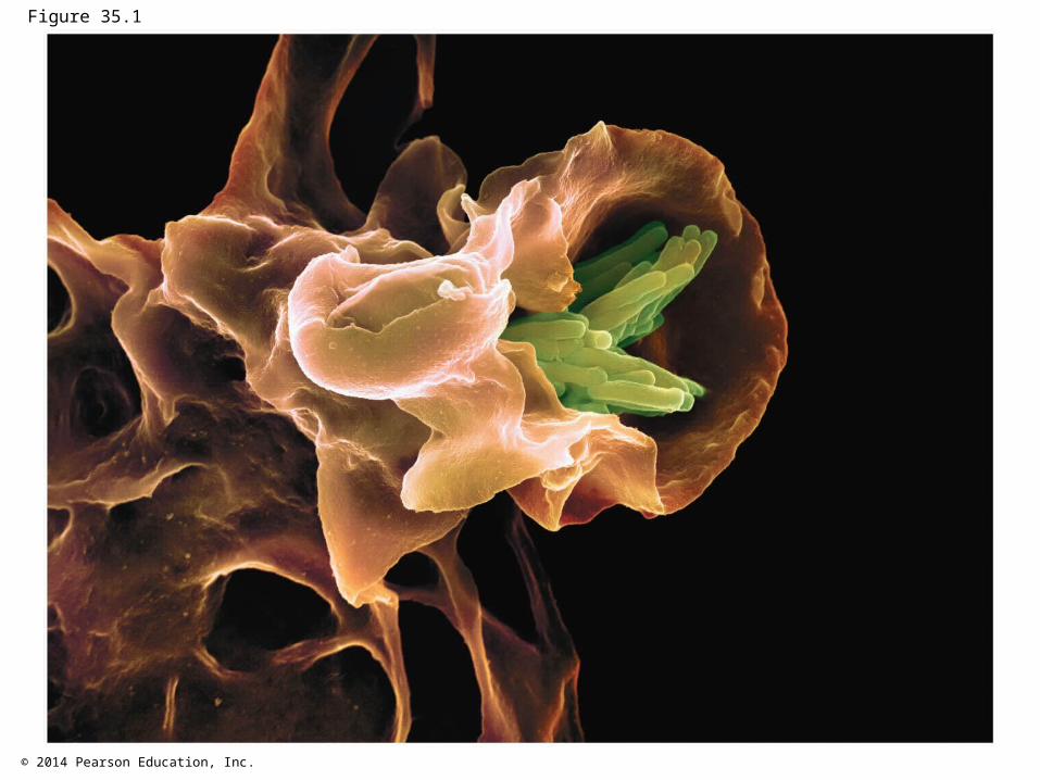
Pathogens, agents that cause disease, infect a wide range of animals, including humans
The immune system enables an animal to avoid or limit many infections
All animals have innate immunity, a defense that is active immediately upon infection
Vertebrates also have adaptive immunity
© 2014 Pearson Education, Inc.
Figure 35.1
© 2014 Pearson Education, Inc.
Innate immunity is present before any exposure to pathogens and is effective from the time of birth
It involves nonspecific responses to pathogens Innate immunity consists of external barriers plus
internal cellular and chemical defenses
© 2014 Pearson Education, Inc.
Adaptive immunity, or acquired immunity, develops after exposure to agents such as microbes, toxins, or other foreign substances
It involves a very specific response to pathogens

© 2014 Pearson Education, Inc.
Figure 35.2
INNATE IMMUNITY(all animals)• Recognition of traits shared
by broad ranges ofpathogens, using a smallset of receptors
• Rapid response
Barrier defenses:SkinMucous membranesSecretionsInternal defenses:Phagocytic cellsNatural killer cellsAntimicrobial proteinsInflammatory response
Humoral response:Antibodies defend againstinfection in body fluids.
Cytotoxic cells defendagainst infection in body cells.
Cell-mediated response:
ADAPTIVE IMMUNITY(vertebrates only)• Recognition of traits
specific to particularpathogens, using a vastarray of receptors
• Slower response
Pathogens(such as bacteria,fungi, and viruses)
© 2014 Pearson Education, Inc.
Concept 35.1: In innate immunity, recognition and response rely on traits common to groups of pathogens
Innate immunity is found in all animals and plants In vertebrates, innate immunity is a first response to
infections and serves as the foundation of adaptive immunity

© 2014 Pearson Education, Inc.
Innate Immunity of Invertebrates
Insects rely on their exoskeleton as a first line of defense against infection
In the digestive system, the enzyme lysozyme breaks down bacterial cell walls, protecting against pathogens ingested along with food
Hemocytes circulate within hemolymph and carry out phagocytosis, the ingestion and breakdown of foreign substances including bacteria

© 2014 Pearson Education, Inc.
Figure 35.3
Pathogen
Vacuole
PHAGOCYTICCELL
Lysosomecontainingenzymes
Pseudopodiasurround pathogens.
engulfed byendocytosis.
Pathogens
Vacuole forms.
Vacuole andlysosome fuse.
Pathogensdestroyed.
Debris frompathogens released.
1
2
3
4
5
6

© 2014 Pearson Education, Inc.
Hemocytes also release antimicrobial peptides that disrupt the plasma membranes of fungi and bacteria
The immune system recognizes bacteria and fungi by structures on their cell walls
An immune response is specific for each class of pathogen

© 2014 Pearson Education, Inc.
Innate Immunity of Vertebrates
The immune system of mammals is the best understood of the vertebrates
Innate defenses include barrier defenses, phagocytosis, and antimicrobial peptides
Additional defenses are unique to vertebrates: natural killer cells, interferons, and the inflammatory response
© 2014 Pearson Education, Inc.
Barrier Defenses
Barrier defenses include the skin and mucous membranes of the respiratory, urinary, and reproductive tracts
Mucus traps and allows for the removal of microbes Many body fluids including saliva, mucus, and tears
are hostile to many microbes The low pH of skin and the digestive system prevents
growth of many bacteria

© 2014 Pearson Education, Inc.
Cellular Innate Defenses
Pathogens entering the mammalian body are subject to phagocytosis
Phagocytic cells recognize groups of pathogens by Toll-like receptors (TLRs)
Each mammalian TLR binds to fragments of molecules characteristic to a set of pathogens

© 2014 Pearson Education, Inc.
Figure 35.4
VESICLE
CpG DNA
dsRNA
TLR9
TLR3 Innate immuneresponses
TLR5
TLR4Flagellin
LipopolysaccharideHelperprotein
PHAGOCYTICCELL
EXTRACELLULARFLUID

© 2014 Pearson Education, Inc.
There are two main types of phagocytic cells in mammals Neutrophils circulate in the blood and are attracted
by signals from infected tissues Macrophages are found throughout the body
© 2014 Pearson Education, Inc.
Two additional types of phagocytic cells Dendritic cells stimulate development of adaptive
immunity in cells that contact the environment (such as skin)
Eosinophils discharge destructive enzymes beneath mucosal surfaces

© 2014 Pearson Education, Inc.
Cellular innate defenses in vertebrates also involve natural killer cells
These circulate through the body and detect abnormal cells
They release chemicals leading to cell death, inhibiting the spread of virally infected or cancerous cells
Many cellular innate defenses involve the lymphatic system

© 2014 Pearson Education, Inc.
Antimicrobial Peptides and Proteins
In mammals, pathogen recognition triggers release of peptides and proteins that attack pathogens or impede their reproduction
Interferons provide innate defense, interfering with viruses and helping activate macrophages
The complement system consists of about 30 proteins that are activated by substances on microbe surfaces
Activation can lead to lysis of invading cells

© 2014 Pearson Education, Inc.
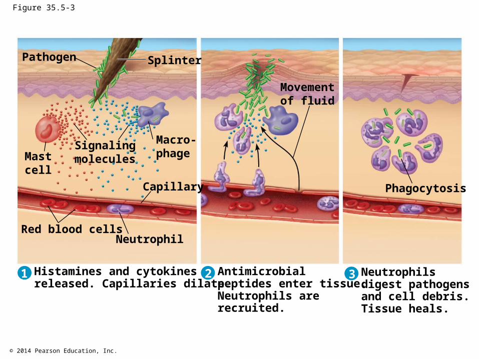
Inflammatory Response
The inflammatory response, such as pain and swelling, is brought about by molecules released upon injury of infection
Mast cells release histamine, which triggers blood vessels to dilate and become more permeable
Activated macrophages and neutrophils release cytokines, signaling molecules that modulate the immune response and promote blood flow to the site of injury or infection

© 2014 Pearson Education, Inc.
Enhanced blood flow to the site helps deliver antimicrobial peptides
This results in an accumulation of pus, a fluid rich in white blood cells, dead pathogens, and cell debris from damaged tissues
Video: Leukocyte Rolling
Video: Neutrophil Chemotaxis

© 2014 Pearson Education, Inc.
Figure 35.5-1
Pathogen Splinter
Macro-phage
Capillary
NeutrophilRed blood cells
Mastcell
Signalingmolecules
1 Histamines and cytokinesreleased. Capillaries dilate.

© 2014 Pearson Education, Inc.
Figure 35.5-2
Pathogen Splinter
Macro-phage
Capillary
NeutrophilRed blood cells
Mastcell
Signalingmolecules
1 Histamines and cytokinesreleased. Capillaries dilate.
Movementof fluid
Antimicrobialpeptides enter tissue.Neutrophils arerecruited.
2
© 2014 Pearson Education, Inc.
Figure 35.5-3
Pathogen Splinter
Macro-phage
Capillary
NeutrophilRed blood cells
Mastcell
Signalingmolecules
1 Histamines and cytokinesreleased. Capillaries dilate.
Movementof fluid
2 Antimicrobialpeptides enter tissue.Neutrophils arerecruited.
Phagocytosis
3 Neutrophilsdigest pathogensand cell debris.Tissue heals.

© 2014 Pearson Education, Inc.
Inflammation can be either local or systemic (throughout the body)
Fever is a systemic inflammatory response triggered by substances released by macrophages
Septic shock is a life-threatening condition caused by an overwhelming inflammatory response
Chronic inflammation can also threaten human health

© 2014 Pearson Education, Inc.
Evasion of Innate Immunity by Pathogens
Adaptations have evolved in some pathogens that enable them to avoid destruction by phagocytic cells
Tuberculosis (TB) resists breakdown within lysosomes after being engulfed by a host cell
Mechanisms like this make certain fungi and bacteria substantial pathogenic threats

© 2014 Pearson Education, Inc.
Concept 35.2: In adaptive immunity, receptors provide pathogen-specific recognition
The adaptive response relies on two types of lymphocytes, or white blood cells
Lymphocytes that mature in the thymus above the heart are called T cells, and those that mature in bone marrow are called B cells

© 2014 Pearson Education, Inc.
Figure 35.UN01
Antigenreceptors
Mature B cell Mature T cell
© 2014 Pearson Education, Inc.
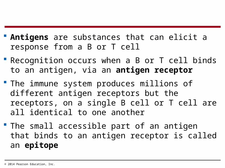
Antigens are substances that can elicit a response from a B or T cell
Recognition occurs when a B or T cell binds to an antigen, via an antigen receptor
The immune system produces millions of different antigen receptors but the receptors, on a single B cell or T cell are all identical to one another
The small accessible part of an antigen that binds to an antigen receptor is called an epitope

© 2014 Pearson Education, Inc.
Antigen Recognition by B Cells and Antibodies
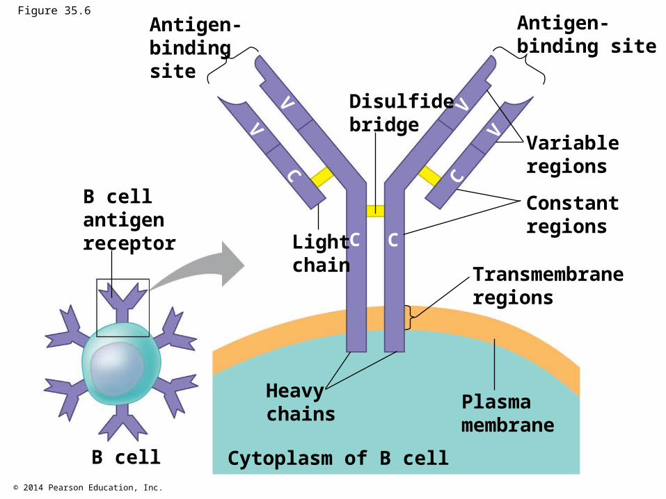
Each B cell antigen receptor is a Y-shaped molecule with two identical heavy chains and two identical light chains
The constant (C) regions of the chains vary little among B cells, whereas the variable (V) regions differ greatly
Together, the V regions of the heavy and light chains form an antigen-binding site
© 2014 Pearson Education, Inc.
Figure 35.6Antigen-bindingsite
B cellantigenreceptor
Disulfidebridge
Lightchain
Antigen-binding site
Variableregions
Constantregions
Transmembraneregions
Plasmamembrane
Heavychains
Cytoplasm of B cellB cell
VV
C
C C
C
VV
© 2014 Pearson Education, Inc.
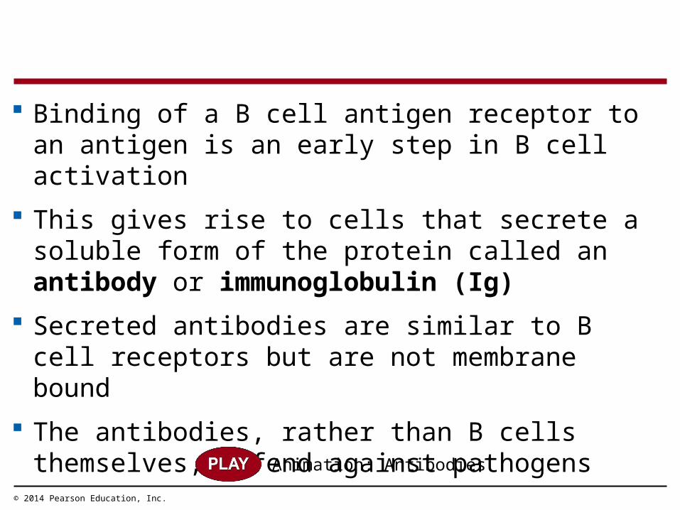
Binding of a B cell antigen receptor to an antigen is an early step in B cell activation
This gives rise to cells that secrete a soluble form of the protein called an antibody or immunoglobulin (Ig)
Secreted antibodies are similar to B cell receptors but are not membrane bound
The antibodies, rather than B cells themselves, defend against pathogens
Animation: Antibodies

© 2014 Pearson Education, Inc.
Figure 35.7Antigen-receptor Antibody
B cell
Antigen Epitope
Pathogen(a) B cell antigen receptors and antibodies
Antibody CAntibody A
Antibody B
Antigen
(b) Antigen receptor specificity

© 2014 Pearson Education, Inc.
Each T cell receptor consists of two different polypeptide chains (called and )
The tips of the chain form a variable (V) region; the rest is a constant (C) region
The V regions of the and chains together form an antigen-binding site
Antigen Recognition by T Cells

© 2014 Pearson Education, Inc.
Figure 35.8Antigen-bindingsite
T cellantigenreceptor
Disulfidebridge
T cell Cytoplasm of T cell
chain chain
Variableregions
Constantregions
Transmembraneregion
Plasmamembrane
V V
CC

© 2014 Pearson Education, Inc.
T cells bind only to antigen fragments displayed or presented on a host cell
MHC (major histocompatibility complex) molecules are host proteins that display the antigen fragments on the cell surface
© 2014 Pearson Education, Inc.
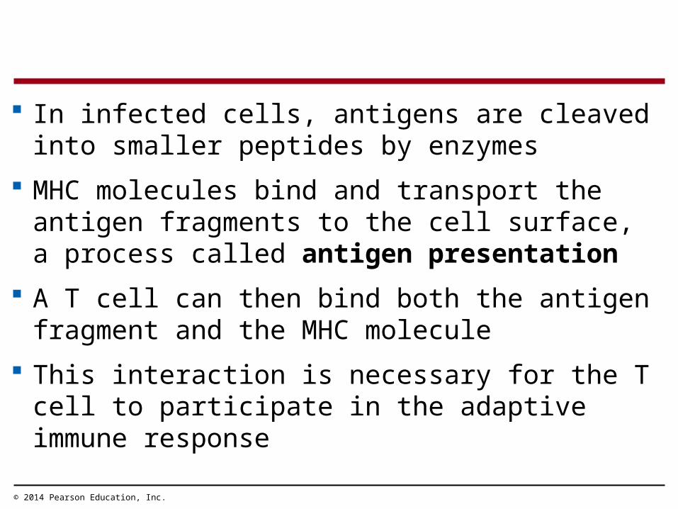
In infected cells, antigens are cleaved into smaller peptides by enzymes
MHC molecules bind and transport the antigen fragments to the cell surface, a process called antigen presentation
A T cell can then bind both the antigen fragment and the MHC molecule
This interaction is necessary for the T cell to participate in the adaptive immune response

© 2014 Pearson Education, Inc.
Figure 35.9
Host cell
Displayedantigenfragment
MHCmolecule
Antigen fragment
Pathogen
T cell
T cell antigenreceptor

© 2014 Pearson Education, Inc.
B Cell and T Cell Development
The adaptive immune system has four major characteristics Diversity of lymphocytes and receptors Self-tolerance; lack of reactivity against an animal’s
own molecules Proliferation of B and T cells after activation Immunological memory

© 2014 Pearson Education, Inc.
Generation of B and T Cell Diversity
By combining variable elements, the immune system assembles a diverse variety of antigen receptors
The capacity to generate diversity is built into the structure of Ig genes
Many different chains can be produced from the same gene by rearrangement of the DNA
Rearranged DNA is transcribed and translated and the antigen receptor formed
© 2014 Pearson Education, Inc.
For example, a receptor light-chain gene contains a variable (V) segment, a joining (J) segment, and a constant (C) segment
The gene contains one C segment, 40 different V segments, and 5 different J segments
These can be combined in 200 different ways The number of heavy-chain combinations is even
greater

© 2014 Pearson Education, Inc.
Figure 35.10
pre-mRNA
mRNA
DNA ofdifferentiatedB cell
DNA ofundifferentiatedB cell
RNA processing
Transcription of permanentlyrearranged, functional gene
Recombination deletes DNA betweenrandomly selected V segment and J segment
Functional gene
1
2
3
4 Translation
Light-chain polypeptide
Variableregion
Constantregion B cell
Antigen receptorV C
CCap V39 J5 Poly-A tail
VV V
V
C
C C
C
CV39 J5 Intron
C
V39 J5 IntronV38V37
V39V38 V40V37 J5J4J3J2J1 Intron
C

© 2014 Pearson Education, Inc.
Figure 35.10a
DNA ofundifferentiatedB cell
V39V38
V40
V37
J5J4J3J2J1 Intron
DNA ofdifferentiatedB cell
pre-mRNA
Transcription of permanentlyrearranged, functional gene
Recombination deletes DNA betweenrandomly selected V segment and J segment
Functional gene
CV39V38V37
J5 Intron C
Intron CV39 J5
2
1

© 2014 Pearson Education, Inc.
Figure 35.10b
pre-mRNA Intron CV39 J5
mRNA
RNA processing
CCap V39 J5 Poly-A tail
Translation
Light-chainpolypeptide
Variableregion
Constantregion B cell
Antigen receptorV C
VV V
V
C C
C C
4
3
© 2014 Pearson Education, Inc.
Origin of Self-Tolerance
Antigen receptors are generated by random rearrangement of DNA
As lymphocytes mature in bone marrow or the thymus, they are tested for self-reactivity
Some B and T cells with receptors specific for the body’s own molecules are destroyed by apoptosis, or programmed cell death
The remainder are rendered nonfunctional
© 2014 Pearson Education, Inc.
Proliferation of B Cells and T Cells
In the body only a tiny fraction of antigen receptors are specific for a given epitope
In the lymph nodes, an antigen is exposed to a steady stream of lymphocytes until a match is made
This binding of a mature lymphocyte to an antigen initiates events that activate the lymphocyte
© 2014 Pearson Education, Inc.
Once activated, a B or T cell undergoes multiple cell divisions to produce a clone of identical cells (called clonal selection)
Two types of clones are produced Short-lived activated effector cells that act
immediately against the antigen Long-lived memory cells that can give rise to effector
cells if the same antigen is encountered again
Animation: Role of B Cells

© 2014 Pearson Education, Inc.
Figure 35.11
B cells thatdiffer inantigenspecificity
AntigenAntigenreceptor
Antibody
Plasma cellsMemory cells

© 2014 Pearson Education, Inc.
Immunological memory is responsible for long-term protections against diseases, due to a prior infection
The first exposure to a specific antigen represents the primary immune response
During this time, selected B and T cells give rise to their effector forms
In the secondary immune response, memory cells facilitate a faster, stronger, and longer response
Immunological memory can span many decades
Immunological Memory

© 2014 Pearson Education, Inc.
Figure 35.12
Primary immuneresponse toantigen A
Secondaryimmune responseto antigen A
Antibodiesto A Antibodies
to B
101
103
104
100
0 7 14 21 28 35 42 49 56
Primary immuneresponse to antigen B
Ant
ibod
y co
ncen
trat
ion
(arb
itrar
y un
its)
Exposureto antigen A
Exposure to antigens A and BTime (days)
102

© 2014 Pearson Education, Inc.
Concept 35.3: Adaptive immunity defends against infection of body fluids and body cells
B and T lymphocytes produce a humoral immune response and a cell-mediated immune response
In the humoral immune response, antibodies help neutralize or eliminate toxins and pathogens in the blood and lymph
In the cell-mediated immune response specialized T cells destroy infected host cells

© 2014 Pearson Education, Inc.
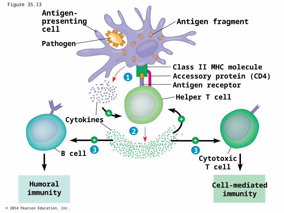
Helper T Cells: A Response to Nearly All Antigens
A type of T cell called a helper T cell triggers both the humoral and cell-mediated immune responses
An antigen must be displayed on the surface of an antigen-presenting cell and bind specifically to the antigen receptor of a T cell
Antigen-presenting cells have class I and class II MHC molecules on their surfaces

© 2014 Pearson Education, Inc.
Antigen-presenting cells are recognized based on their class II MHC molecules
Antigen receptors on the surface of helper T cells bind to the antigen and the class II MHC molecule
Signals are then exchanged between the two cells The helper T cell is activated, proliferates, and forms
a clone of helper T cells, which then activate the appropriate B cells
Animation: Helper T Cells
Video: Cell Receptors
© 2014 Pearson Education, Inc.
Figure 35.13
Antigen-presentingcell
Antigen fragment
Class II MHC moleculeAccessory protein (CD4)Antigen receptorHelper T cell
Pathogen
Cytokines
B cell
1
2
3 3Cytotoxic
T cell
Cell-mediatedimmunity
Humoralimmunity

© 2014 Pearson Education, Inc.
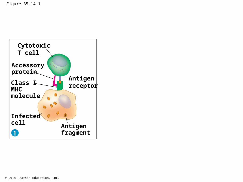
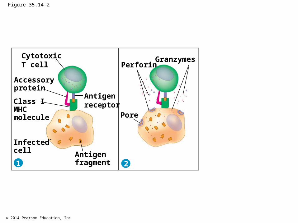
Cytotoxic T Cells: A Response to Infected Cells
Cytotoxic T cells are the effector cells in the cell-mediated immune response
Cytotoxic T cells recognize fragments of foreign proteins produced by infected cells and possess an accessory protein that binds to class I MHC molecules
The activated cytotoxic T cell secretes proteins that disrupt the membranes of target cells and trigger apoptosis
Animation: Cytotoxic T Cells
© 2014 Pearson Education, Inc.
Figure 35.14-1
Antigen fragment
Class I MHC molecule
Accessory protein
Antigen receptor
CytotoxicT cell
Infectedcell1
© 2014 Pearson Education, Inc.
Figure 35.14-2
Antigen fragment
Class I MHC molecule
Accessory protein
Antigen receptor
CytotoxicT cell
Infectedcell1
Pore
2
PerforinGranzymes

© 2014 Pearson Education, Inc.
Figure 35.14-3
Antigen fragment
Class I MHC molecule
Accessory protein
Antigen receptor
CytotoxicT cell
Infectedcell1
Pore
2
PerforinGranzymes Released
cytotoxicT cell
Dyinginfected cell
3

© 2014 Pearson Education, Inc.
B Cells and Antibodies: A Response to Extracellular Pathogens
The humoral response is characterized by secretion of antibodies by clonally selected B cells
Activation of B cells involves helper T cells and proteins on the surface of pathogens
In response to cytokines from helper T cells and an antigen, a B cell proliferates and differentiates into memory B cells and antibody-secreting effector cells called plasma cells

© 2014 Pearson Education, Inc.
Figure 35.15-1
Helper T cell
Antigenfragments
Pathogen
CD4
Antigen-presenting cell
Antigenreceptor
1
MHC

© 2014 Pearson Education, Inc.
Figure 35.15-2
Helper T cell
Antigenfragments
Pathogen
CD4
Antigen-presenting cell
Antigenreceptor
MHC
B cell
Cytokines
Activatedhelper T cell21

© 2014 Pearson Education, Inc.
Figure 35.15-3
Helper T cell
Antigenfragments
Pathogen
CD4
Antigen-presenting cell
Antigenreceptor
MHC
B cell
Cytokines
Activatedhelper T cell21 3
Plasma cellsSecreted
antibodies
Memory B cells

© 2014 Pearson Education, Inc.
Antibodies do not kill pathogens; instead, they mark pathogens for destruction
In neutralization, antibodies bind to viral surface proteins, preventing infection of a host cell
Antibodies may also bind to toxins in body fluids and prevent them from entering body cells
© 2014 Pearson Education, Inc.
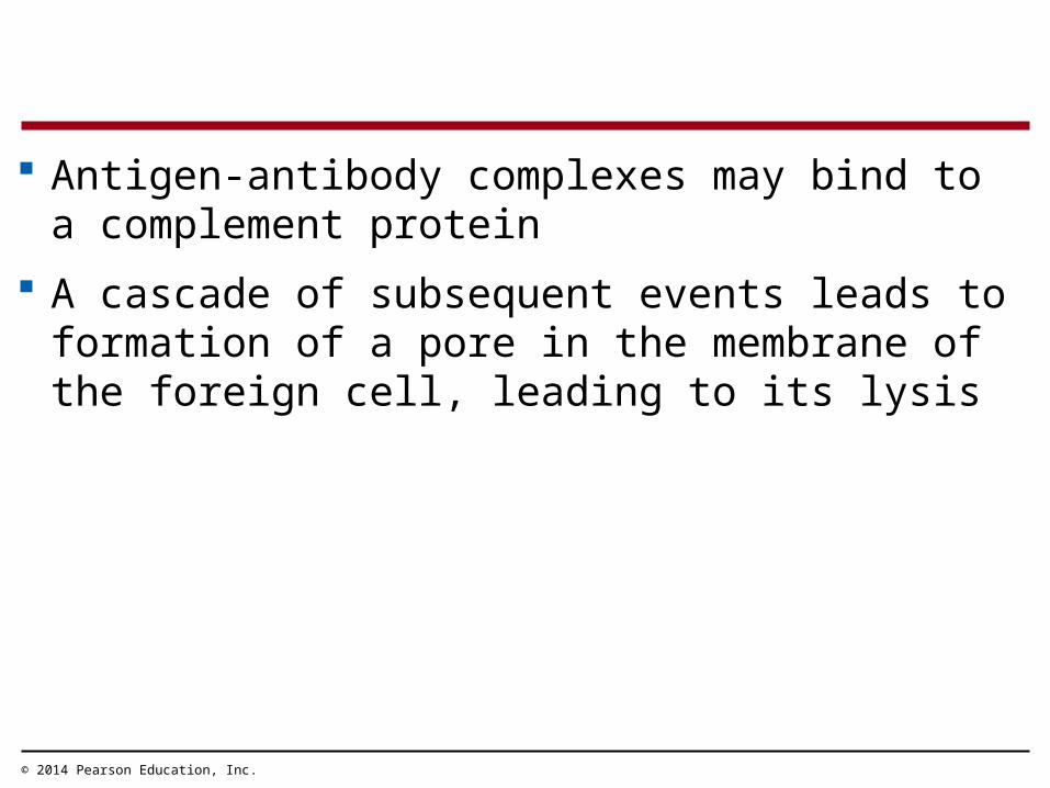
Antigen-antibody complexes may bind to a complement protein
A cascade of subsequent events leads to formation of a pore in the membrane of the foreign cell, leading to its lysis

© 2014 Pearson Education, Inc.
B cells can express five different forms (or classes) of immunoglobulin (Ig) with similar antigen-binding specificity but different heavy-chain C regions
One type, the B cell antigen receptor, is membrane bound
The others are soluble and include those found in blood, tears, saliva, and breast milk

© 2014 Pearson Education, Inc.
Both the humoral and cell-mediated responses can include primary and secondary immune responses
Memory cells enable the secondary response
Summary of the Humoral and Cell-Mediated Immune Responses
Animation: Immunology

© 2014 Pearson Education, Inc.
Figure 35.16
B cell Helper T cell
Antigen-presenting cell
Engulfed by
Antigen (1st exposure)
Humoral (antibody-mediated)immune response Cell-mediated immune response
KeyStimulatesGives rise to
CytotoxicT cell
Memoryhelper T cell
Memorycytotoxic T cells
MemoryB cells
Antigen (2nd exposure)
Activecytotoxic T cells
Defend against intracellularpathogens and cancer
Defend againstextracellular pathogens
Plasmacells
Secretedantibodies
© 2014 Pearson Education, Inc.
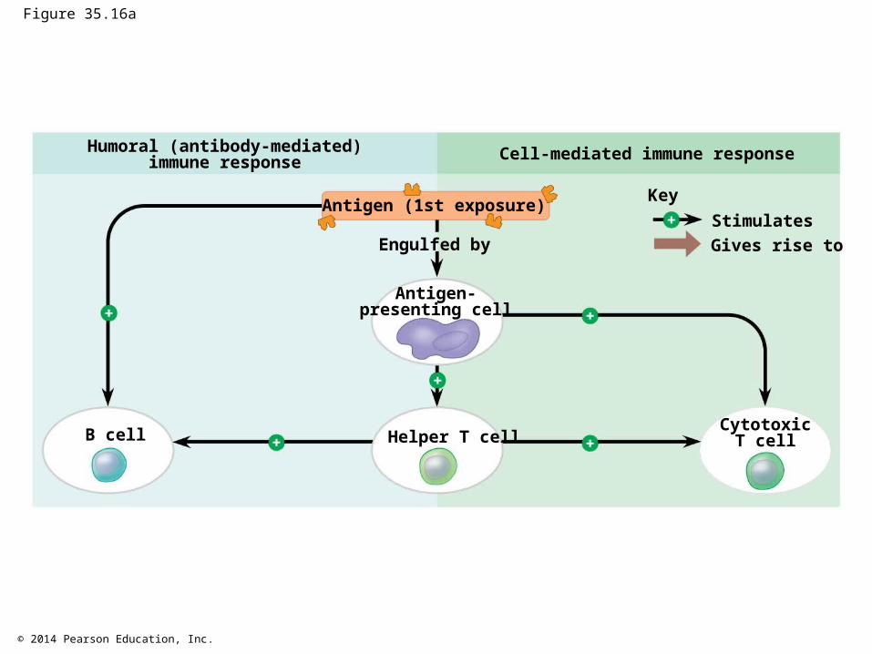
Figure 35.16a
B cell Helper T cell
Antigen-presenting cell
Engulfed by
Antigen (1st exposure)
Humoral (antibody-mediated)immune response Cell-mediated immune response
KeyStimulatesGives rise to
CytotoxicT cell

© 2014 Pearson Education, Inc.
Figure 35.16b
B cell Helper T cell
Humoral (antibody-mediated)immune response Cell-mediated immune response
CytotoxicT cell
KeyStimulatesGives rise to
Memoryhelper T cell
Memorycytotoxic T cells
MemoryB cells
Antigen (2nd exposure)
Activecytotoxic T cells
Defend against intracellularpathogens and cancer
Defend againstextracellular pathogens
Plasmacells
Secretedantibodies

© 2014 Pearson Education, Inc.
Active and Passive Immunization
Active immunity occurs naturally when a pathogen infects the body
Passive immunity provides immediate, short-term protection
It is conferred naturally when antibodies cross the placenta from mother to fetus or pass from mother to infant in breast milk
Both active and passive immunity can be induced artificially
© 2014 Pearson Education, Inc.
Active immunity is induced when antigens are introduced into the body in vaccines
In this process of immunization, inactivated bacterial toxins or weakened or killed pathogens are introduced
Passive immunity can be conferred artificially by injecting antibodies into a nonimmune person

© 2014 Pearson Education, Inc.
Antibodies as Tools
Polyclonal antibodies, produced following exposure to an antigen, are products of many different clones of plasma cells, each specific for a different epitope
Monoclonal antibodies are prepared from a single clone of B cells grown in culture
Monoclonal antibodies have provided the basis for many recent advances in medical diagnosis and treatment

© 2014 Pearson Education, Inc.
Immune Rejection
Cells transferred from one person to another can be attacked by the recipient’s immune defenses
This complicates blood transfusions and the transplant of tissues or organs
To minimize rejection, physicians use donor tissue that closely matches the MHC molecules of the recipient
Recipients also take medicines that suppress their immune responses

© 2014 Pearson Education, Inc.
Disruptions in Immune System Function
Although adaptive immunity offers significant protection against many pathogens, it is not fail-safe

© 2014 Pearson Education, Inc.
Allergies
Allergies are exaggerated (hypersensitive) responses to antigens called allergens
In localized allergies such as hay fever, plasma cells secrete antibodies specific for antigens on the surface of pollen grains
This triggers immune cells in connective tissue to release histamine and other inflammatory chemicals
Antihistamines block receptors for histamine and diminish allergy symptoms
© 2014 Pearson Education, Inc.
Figure 35.17
IgE
Allergen
GranuleMast cell
1
2
Histamine
3

© 2014 Pearson Education, Inc.
An acute allergic response can lead to anaphylactic shock, a life-threatening reaction
Substances that can trigger anaphylactic shock in allergic individuals include bee venom, penicillin, peanuts, and shellfish
People with these hypersensitivities often carry epinephrine to counteract the allergic response
© 2014 Pearson Education, Inc.
Autoimmune Diseases
In individuals with autoimmune diseases, the immune system targets certain molecules of the body
Autoimmune diseases include systemic lupus erythematosus, rheumatoid arthritis, insulin-dependent diabetes mellitus, and multiple sclerosis

© 2014 Pearson Education, Inc.
Figure 35.18

© 2014 Pearson Education, Inc.
Immune System Avoidance
Mechanisms to thwart immune responses have evolved in pathogens
A pathogen may alter how it appears to the immune system by changing the epitopes it expresses
Such changes are called antigenic variation This mechanism is seen in the parasite that causes
sleeping sickness and in the influenza virus

© 2014 Pearson Education, Inc.
Some viruses avoid an immune response by infecting cells and then entering an inactive state called latency
The virus (such as herpes simplex) remains latent until a stimulus reactivates it
Stimuli include stress, fever, or menstruation
© 2014 Pearson Education, Inc.
Acquired immunodeficiency syndrome (AIDS) is caused by HIV (human immunodeficiency virus), which both attacks and escapes the immune system
It infects helper T cells with high efficiency It escapes the immune system through its high
mutation rate, which reduces the ability of the immune system to eliminate the infection
It also can undergo latency

© 2014 Pearson Education, Inc.
People with AIDS are highly susceptible to infections and cancers that a healthy immune system would normally defeat
Unprotected sex and transmission via HIV-contaminated needles account for the majority of HIV infections
HIV cannot be cured, but drugs have been developed to slow HIV replication and progression to AIDS
© 2014 Pearson Education, Inc.
Cancer and Immunity
The frequency of certain cancers increases when adaptive immunity is impaired
20% of all human cancers involve viruses The immune system can act as a defense against
viruses that cause cancer and against cancer cells that harbor viruses
In 2006, a vaccine was released that acts against human papillomavirus (HPV), a virus associated with cervical cancer

© 2014 Pearson Education, Inc.
Figure 35.UN02a
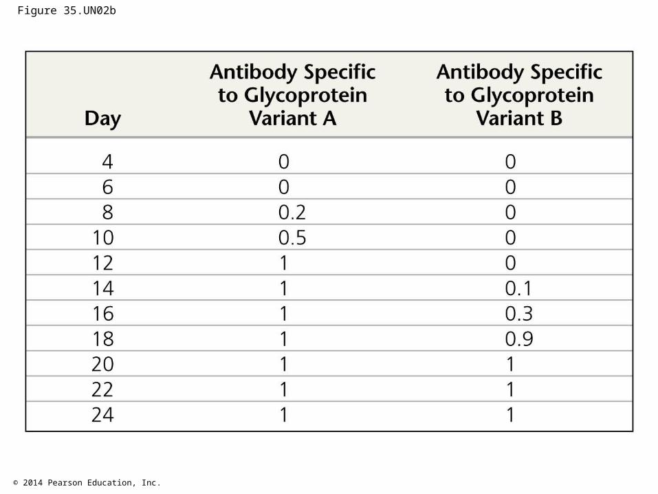
© 2014 Pearson Education, Inc.
Figure 35.UN02b
© 2014 Pearson Education, Inc.
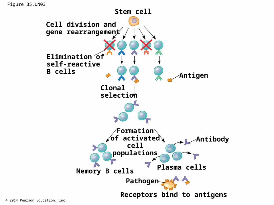
Figure 35.UN03
Stem cell
Antigen
Antibody
Cell division andgene rearrangement
Elimination ofself-reactiveB cells
Clonalselection
Memory B cells
Formationof activated
cellpopulations
Plasma cells
Pathogen
Receptors bind to antigens