bioprinting cell-laden matrigel for radioprotection study...
TRANSCRIPT

Bioprinting cell-laden matrigel for radioprotection study of liver by pro-drug conversion in a
dual-tissue microfluidic chip
This article has been downloaded from IOPscience. Please scroll down to see the full text article.
2011 Biofabrication 3 034112
(http://iopscience.iop.org/1758-5090/3/3/034112)
Download details:
IP Address: 129.25.13.163
The article was downloaded on 16/09/2011 at 13:59
Please note that terms and conditions apply.
View the table of contents for this issue, or go to the journal homepage for more
Home Search Collections Journals About Contact us My IOPscience
IOP PUBLISHING BIOFABRICATION
Biofabrication 3 (2011) 034112 (9pp) doi:10.1088/1758-5082/3/3/034112
Bioprinting cell-laden matrigel forradioprotection study of liver by pro-drugconversion in a dual-tissue microfluidicchipJ E Snyder1, Q Hamid1, C Wang1, R Chang1, K Emami2, H Wu2 andW Sun1,3
1 Department of Mechanical Engineering, Drexel University, Philadelphia, PA 19104, USA2 Radiation Biophysics Lab, NASA Johnson Space Center, Houston, TX 77586, USA3 Department of Mechanical Engineering, Tsinghua University, Beijing, 100084,People’s Republic of China
E-mail: [email protected] and [email protected]
Received 15 January 2011Accepted for publication 16 August 2011Published 1 September 2011Online at stacks.iop.org/BF/3/034112
AbstractThe objective of this paper is to introduce a novel cell printing and microfluidic system toserve as a portable ground model for the study of drug conversion and radiation protection ofliving liver tissue analogs. The system is applied to study behavior in ground models of spacestress, particularly radiation. A microfluidic environment is engineered by two cell types toprepare an improved higher fidelity in vitro micro-liver tissue analog. Cell-laden Matrigelprinting and microfluidic chips were used to test radiation shielding to liver cells by thepro-drug amifostine. In this work, the sealed microfluidic chip regulates three variables ofinterest: radiation exposure, anti-radiation drug treatment and single- or dual-tissue cultureenvironments. This application is intended to obtain a scientific understanding of the responseof the multi-cellular biological system for long-term manned space exploration, diseasemodels and biosensors.
(Some figures in this article are in colour only in the electronic version)
1. Introduction
Bioactive microfluidic systems offer a promising platform fora small-scale study of pharmacokinetics/pharmacodynamics,drug absorption, elimination and toxicity on humanbiological material [1–3]. In addition, the liquid cellconfiguration enables real-time studies of bioaffinity reactionsat predetermined flow rates and temperatures [4]. The fidelityof the in vitro test platform and reliability of the biologicalanalog are directly related to the manufacturing and cellassembly capability [5]. Constituent components and theirspatial arrangement are determined from the analysis of targetbiological systems, which is realized as physical prototypes.Manufacturing of those physical prototypes is limited by
process resolution and material handling, meaning preservingcell viability and function.
In vitro drug discovery platforms and sophisticatedcell assembly technology are independently recognizedas bioadditive techniques with the potential for in vitrobiomimicry and monitoring [6, 7]. One current trend in drugdiscovery and tissue engineering research is to use a combinedthree-dimensional cell assembly and microfluidic environmentto probe and analyze the biological condition, response, anddrug effectiveness [8, 9]. This becomes possible as appropriatearchitectural length scales and biological components areidentified and relevant manufacturing capability is developed.In this work, the authors introduce a novel cell printingsystem and microfluidic chip to study dual-tissue drug
1758-5082/11/034112+09$33.00 1 © 2011 IOP Publishing Ltd Printed in the UK
Biofabrication 3 (2011) 034112 J E Snyder et al
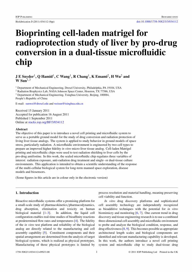
Figure 1. Schematic representation of the direct cell writing process for biofabrication of micro-tissues onto a microfluidic device.Reprinted with permission [9].
conversion and effectiveness of the radioprotective pro-drugamifostine against radiation of the magnitude found outsidethe Earth’s orbit [10]. This work is done in part to prepare amanufacturing and cell culture method to test pharmaceuticalsin the space environment to make informed recommendationsabout medical preparedness for long-term manned spaceexploration [11].
The presented microfluidic system improves in vitromodeling beyond single cell activity by including multiple celltypes [12, 13]. This works using two cell types: (1) epithelialcells as the cell type lining the body’s lumen, which the drugwould pass through before moving from the blood streamto the target cell type and (2) hepatocytes as the target celltype we are studying for evidence of radioprotection. In themicrofluidic environment the drug passes serially through theepithelial construct and then the hepatyocytes, as this wouldbe the path of the drug diffusing from the blood stream to thetissue. Each cell type is physically segregated into separatechambers of a microfluidic system and dynamically perfusedby a syringe pump. The transient interaction between celltypes is controlled by the flow rate of the pump. Soluble cuesand experimental variables are introduced to cell by perfusionthrough the microfluidic chambers. Engineered devices, suchas the direct cell writing process and microfluidic device, canbe used to assemble biological materials to approximate invivo pathogenesis, as presented in figure 1.
Macro and micro patternings of cells are part ofmechanical cues to promote biomimetic function [14].Cells, as dynamic spatial entities, rely on paracrinesignaling and other environmental cues to define their ownbehavior. The cell culture environment is engineeredto approximate in vivo conditions in vitro to elicit thebiomimetic function. A motion-controlled bioprinting systemand extracellular matrix provide structure and patterningcontrol to leverage biologists characterization of knownfunctional tissues for metabolism studies. A promising
extracellular matrix is a gelatinous protein mixture Matrigel,which improved biomimetic cell function through bioactivefactors and essential macromolecules [15–17]. However,existing printing techniques are unable to dispense cell-laden Matrigel because the devices operate at or aboveroom temperature. Matrigel thermally cross-links at above4 ◦C, and this occludes dispensing capillary and preventsfurther extrusion. A novel temperature-controlled printingsystem is presented in this work to pattern Matrigel andleverage both biological cues from the Matrigel matrix andphysical cues from physiologically derived cell patterningto improve the biomimetic function of in vitro tissueanalog [18, 19]. Printing technology allows researchersto leverage geometric positioning and proximity of specificbiologics to bring functional abilities to cell aggregates[20–22]. In this work, cells are printed in a squarewave form for nutrient and drug diffusion to the core ofthe extruded filament, to apply structural cues to cells,and for quality control over equal dispensing for eachsample.
In our study here, we present the integration of amicrofluidic cell culture environment with the novel cellprinting technique to create an in vitro analog using twodifferent cell types to study pro-drug conversion in a dual-tissue microfluidic chip and resultant radioprotection to liver.Specifically, we are testing radiation protection afforded bypro-drug amifostine against space-like radiation exposure.The in vitro environment provides engineering control over theconstituent components and spatial arrangement to focus thestudy of causality by isolating particular biologics of interest.The authors claim improved model fidelity, over single-cell-type models, by a combined microfluidic environment andcell assembly technique using a matrix Matrigel, based on theobservation of radiation shielding.
2
Biofabrication 3 (2011) 034112 J E Snyder et al
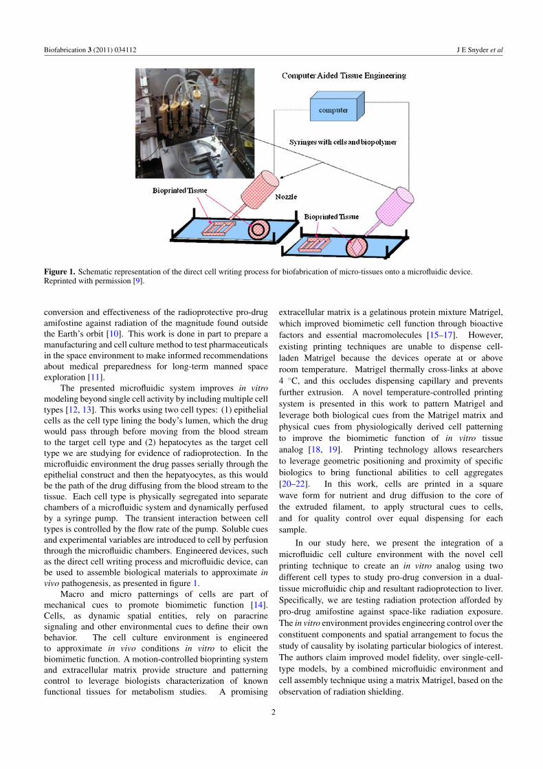
Figure 2. Schematic representation of the subsystems of temperature-controlled printing.
2. Materials and methods
2.1. Cell culture and encapsulation
Two immortalized cell lines are co-cultured in the dual-tissueenvironment. Human hepatic carcinoma cells of the cell lineHepG2 (ATCC) and human mammary epithelial of the cellline M10 (ATCC) are cultured in the alpha modification ofEagle’s medium supplemented with 10% (v/v) fetal bovineserum and 1% (v/v) penicillin streptomycin. Half volume ofthe culture medium is changed every other day. Culturesare maintained at 37 ◦C and 5% carbon dioxide. Thecell culture medium and supplements are purchased fromInvitrogen unless otherwise noted.
Separate cell-laden solutions of epithelial and hepatocytesare created in an identical manner. Cells are rinsed withphosphate buffer solution (PBS), trypsinized and countedusing exclusion dye Trypan Blue and a hemocytometer. Oncecounted, cells are mixed with the gelatinous protein mixturebasement phenol red-free Matrigel (BD Bioscience) over iceto prevent gelation. Cells are homogenously distributedthroughout the Matrigel using a gentle tapping technique withminimal pipetting to a concentration of 1.0 × 10⁶ cells mL−1.The final printing solution is 50% cells and 50% Matrigel (v/v)solution. The cell-laden solution is stored on ice for 5–10 minprior to printing to prevent gelation.
2.2. Temperature motion controlled printing
Cell printing is a powerful manufacturing tool to assemblebiologics to bring structure and functional architecture to cellaggregates. Here we present a temperature-controlled cellprinting system inspired by rapid prototyping technology.A CAD/CAM platform is integrated with solid freeformautomation to assemble biologics in a three-dimensional spaceusing layer-by-layer manufacturing techniques. This systemexecutes micron-scale deposition and printing to repeatedlygenerate cell-laden constructs for scaffold-guided tissueengineering applications to include regenerative medicine,in vitro drug trials and disease analogs. A depiction ofthe subsystem components is presented in figure 2.
The system integrates a temperature-controlled enclosure,dispensing system and plotter to allow for the printingof thermally cross-linking materials, such as Matrigel or
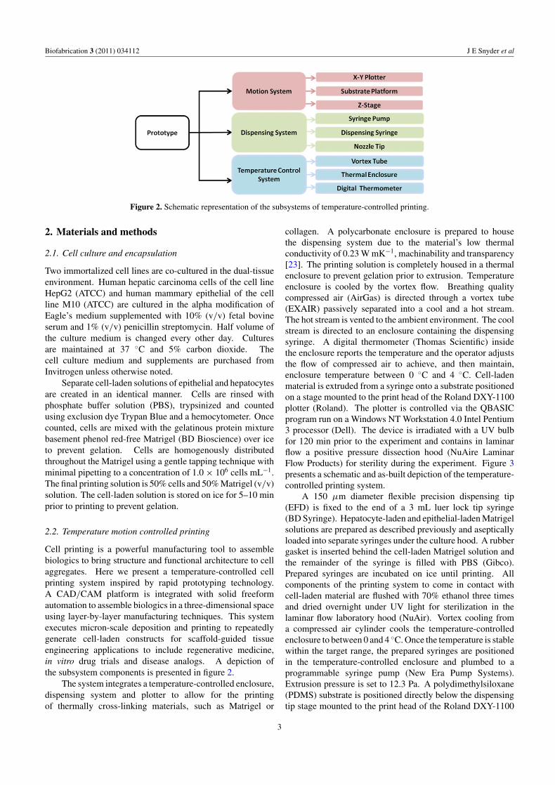
collagen. A polycarbonate enclosure is prepared to housethe dispensing system due to the material’s low thermalconductivity of 0.23 W mK−1, machinability and transparency[23]. The printing solution is completely housed in a thermalenclosure to prevent gelation prior to extrusion. Temperatureenclosure is cooled by the vortex flow. Breathing qualitycompressed air (AirGas) is directed through a vortex tube(EXAIR) passively separated into a cool and a hot stream.The hot stream is vented to the ambient environment. The coolstream is directed to an enclosure containing the dispensingsyringe. A digital thermometer (Thomas Scientific) insidethe enclosure reports the temperature and the operator adjuststhe flow of compressed air to achieve, and then maintain,enclosure temperature between 0 ◦C and 4 ◦C. Cell-ladenmaterial is extruded from a syringe onto a substrate positionedon a stage mounted to the print head of the Roland DXY-1100plotter (Roland). The plotter is controlled via the QBASICprogram run on a Windows NT Workstation 4.0 Intel Pentium3 processor (Dell). The device is irradiated with a UV bulbfor 120 min prior to the experiment and contains in laminarflow a positive pressure dissection hood (NuAire LaminarFlow Products) for sterility during the experiment. Figure 3presents a schematic and as-built depiction of the temperature-controlled printing system.
A 150 μm diameter flexible precision dispensing tip(EFD) is fixed to the end of a 3 mL luer lock tip syringe(BD Syringe). Hepatocyte-laden and epithelial-laden Matrigelsolutions are prepared as described previously and asepticallyloaded into separate syringes under the culture hood. A rubbergasket is inserted behind the cell-laden Matrigel solution andthe remainder of the syringe is filled with PBS (Gibco).Prepared syringes are incubated on ice until printing. Allcomponents of the printing system to come in contact withcell-laden material are flushed with 70% ethanol three timesand dried overnight under UV light for sterilization in thelaminar flow laboratory hood (NuAir). Vortex cooling froma compressed air cylinder cools the temperature-controlledenclosure to between 0 and 4 ◦C. Once the temperature is stablewithin the target range, the prepared syringes are positionedin the temperature-controlled enclosure and plumbed to aprogrammable syringe pump (New Era Pump Systems).Extrusion pressure is set to 12.3 Pa. A polydimethylsiloxane(PDMS) substrate is positioned directly below the dispensingtip stage mounted to the print head of the Roland DXY-1100
3
Biofabrication 3 (2011) 034112 J E Snyder et al
Figure 3. Schematic and as-built photograph of a temperature-controlled printing system.
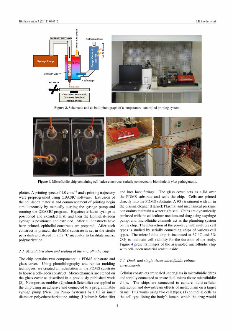
Figure 4. Microfluidic chip containing cell-laden constructs serially connected to biomimic in vivo pathogenesis.
plotter. A printing speed of 1.0 cm s−1 and a printing trajectorywere preprogramed using QBASIC software. Extrusion ofthe cell-laden material and commencement of printing beginsimultaneously by manually starting the syringe pump andrunning the QBASIC program. Hepatocyte-laden syringe ispositioned and extruded first, and then the Epithelial-ladensyringe is positioned and extruded. After all constructs havebeen printed, epithelial constructs are prepared. After eachconstruct is printed, the PDMS substrate is set in the sterilepetri dish and stored in a 37 ◦C incubator to facilitate matrixpolymerization.
2.3. Microfabrication and sealing of the microfluidic chip
The chip contains two components: a PDMS substrate andglass cover. Using photolithography and replica moldingtechniques, we created an indentation in the PDMS substrateto house a cell-laden construct. Micro-channels are etched onthe glass cover as described in a previously published work[8]. Nanoport assemblies (Upchurch Scientific) are applied tothe chip using an adhesive and connected to a programmablesyringe pump (New Era Pump Systems) by 0.02 in innerdiameter polyetheretherketone tubing (Upchurch Scientific)
and luer lock fittings. The glass cover acts as a lid overthe PDMS substrate and seals the chip. Cells are printeddirectly into the PDMS substrate. A 90 s treatment with air inthe plasma cleaner (Harrick Plasma) and mechanical pressureconstraints maintain a water tight seal. Chips are dynamicallyperfused with the cell culture medium and drug using a syringepump, and microfluidic channels act as the plumbing systemon the chip. The interaction of the pro-drug with multiple celltypes is studied by serially connecting chips of various celltypes. The microfluidic chip is incubated at 37 ◦C and 5%CO2 to maintain cell viability for the duration of the study.Figure 4 presents images of the assembled microfluidic chipwith cell-laden material sealed inside.
2.4. Dual- and single-tissue microfluidic cultureenvironments
Cellular constructs are sealed under glass in microfluidic chipsand serially connected to create dual-micro-tissue microfluidicchips. The chips are connected to capture multi-cellularinteraction and downstream effects of metabolism on a targettissue. This works using two cell types, (1) epithelial cells asthe cell type lining the body’s lumen, which the drug would
4
Biofabrication 3 (2011) 034112 J E Snyder et al
(A) (B) (C)
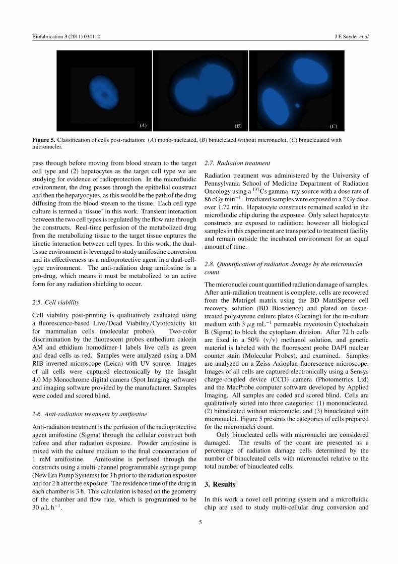
Figure 5. Classification of cells post-radiation: (A) mono-nucleated, (B) binucleated without micronuclei, (C) binucleuated withmicronuclei.
pass through before moving from blood stream to the targetcell type and (2) hepatocytes as the target cell type we arestudying for evidence of radioprotection. In the microfluidicenvironment, the drug passes through the epithelial constructand then the hepatyocytes, as this would be the path of the drugdiffusing from the blood stream to the tissue. Each cell typeculture is termed a ‘tissue’ in this work. Transient interactionbetween the two cell types is regulated by the flow rate throughthe constructs. Real-time perfusion of the metabolized drugfrom the metabolizing tissue to the target tissue captures thekinetic interaction between cell types. In this work, the dual-tissue environment is leveraged to study amifostine conversionand its effectiveness as a radioprotective agent in a dual-cell-type environment. The anti-radiation drug amifostine is apro-drug, which means it must be metabolized to an activeform for any radiation shielding to occur.
2.5. Cell viability
Cell viability post-printing is qualitatively evaluated usinga fluorescence-based Live/Dead Viability/Cytotoxicity kitfor mammalian cells (molecular probes). Two-colordiscrimination by the fluorescent probes enthedium calceinAM and ethidium homodimer-1 labels live cells as greenand dead cells as red. Samples were analyzed using a DMRIB inverted microscope (Leica) with UV source. Imagesof all cells were captured electronically by the Insight4.0 Mp Monochrome digital camera (Spot Imaging software)and imaging software provided by the manufacturer. Sampleswere coded and scored blind.
2.6. Anti-radiation treatment by amifostine
Anti-radiation treatment is the perfusion of the radioprotectiveagent amifostine (Sigma) through the cellular construct bothbefore and after radiation exposure. Powder amifostine ismixed with the culture medium to the final concentration of1 mM amifostine. Amifostine is perfused through theconstructs using a multi-channel programmable syringe pump(New Era Pump Systems) for 3 h prior to the radiation exposureand for 2 h after the exposure. The residence time of the drug ineach chamber is 3 h. This calculation is based on the geometryof the chamber and flow rate, which is programmed to be30 μL h−1.
2.7. Radiation treatment
Radiation treatment was administered by the University ofPennsylvania School of Medicine Department of RadiationOncology using a 137Cs gamma -ray source with a dose rate of86 cGy min−1. Irradiated samples were exposed to a 2 Gy doseover 1.72 min. Hepatocyte constructs remained sealed in themicrofluidic chip during the exposure. Only select hepatocyteconstructs are exposed to radiation; however all biologicalsamples in this experiment are transported to treatment facilityand remain outside the incubated environment for an equalamount of time.
2.8. Quantification of radiation damage by the micronucleicount
The micronuclei count quantified radiation damage of samples.After anti-radiation treatment is complete, cells are recoveredfrom the Matrigel matrix using the BD MatriSperse cellrecovery solution (BD Bioscience) and plated on tissue-treated polystyrene culture plates (Corning) for the in-culturemedium with 3 μg mL−1 permeable mycotoxin CytochalasinB (Sigma) to block the cytoplasm division. After 72 h cellsare fixed in a 50% (v/v) methanol solution, and geneticmaterial is labeled with the fluorescent probe DAPI nuclearcounter stain (Molecular Probes), and examined. Samplesare analyzed on a Zeiss Axioplan fluorescence microscope.Images of all cells are captured electronically using a Sensyscharge-coupled device (CCD) camera (Photometrics Ltd)and the MacProbe computer software developed by AppliedImaging. All samples are coded and scored blind. Cells arequalitatively sorted into three categories: (1) mononucleated,(2) binucleated without micronuclei and (3) binucleated withmicronuclei. Figure 5 presents the categories of cells preparedfor the micronuclei count.
Only binucleated cells with micronuclei are considereddamaged. The results of the count are presented as apercentage of radiation damage cells determined by thenumber of binucleated cells with micronuclei relative to thetotal number of binucleated cells.
3. Results
In this work a novel cell printing system and a microfluidicchip are used to study multi-cellular drug conversion and
5
Biofabrication 3 (2011) 034112 J E Snyder et al
(A) (B)
(C) (D)
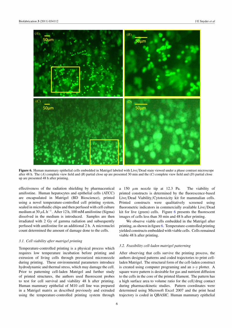
Figure 6. Human mammary epithelial cells embedded in Matrigel labeled with Live/Dead stain viewed under a phase contrast microscopeafter 48 h. The (A) complete view field and (B) partial close up are presented 30 min and the (C) complete view field and (D) partial closeup are presented 48 h after printing.
effectiveness of the radiation shielding by pharmaceuticalamifostine. Human hepatocytes and epithelial cells (ATCC)are encapsulated in Matrigel (BD Bioscience), printedusing a novel temperature-controlled cell printing system,sealed in microfluidic chips and then perfused with cell culturemedium at 30 μL h−1. After 12 h, 100 mM amifostine (Sigma)dissolved in the medium is introduced. Samples are thenirradiated with 2 Gy of gamma radiation and subsequentlyperfused with amifostine for an additional 2 h. A micronucleicount determined the amount of damage done to the cells.
3.1. Cell viability after matrigel printing
Temperature-controlled printing is a physical process whichrequires low temperature incubation before printing andextrusion of living cells through pressurized micronozzleduring printing. These environmental parameters introducehydrodynamic and thermal stress, which may damage the cell.Prior to patterning cell-laden Matrigel and further studyof printed structures, the authors used fluorescent probesto test for cell survival and viability 48 h after printing.Human mammary epithelial of M10 cell line was preparedin a Matrigel matrix as described previously and extrudedusing the temperature-controlled printing system through
a 150 μm nozzle tip at 12.3 Pa. The viability ofprinted constructs is determined by the fluorescence-basedLive/Dead Viability/Cytotoxicity kit for mammalian cells.Printed constructs were qualitatively screened usingfluorometric indicators in commercially available Live/Deadkit for live (green) cells. Figure 6 presents the fluorescentimages of cells less than 30 min and 48 h after printing.
We observe viable cells embedded in the Matrigel afterprinting, as shown in figure 6. Temperature-controlled printingyielded constructs embedded with viable cells. Cells remainedviable 48 h after printing.
3.2. Feasibility cell-laden matrigel patterning
After observing that cells survive the printing process, theauthors designed patterns and coded trajectories to print cell-laden Matrigel. The structural form of the cell-laden constructis created using computer programing and an x–y plotter. Asquare wave pattern is desirable for gas and nutrient diffusionto the cells in the core of the printed filament. The pattern hasa high surface area to volume ratio for the cell/drug contactduring pharmacokinetic studies. Pattern coordinates weredetermined using Microsoft Excel 2007 and the print headtrajectory is coded in QBASIC. Human mammary epithelial
6
Biofabrication 3 (2011) 034112 J E Snyder et al
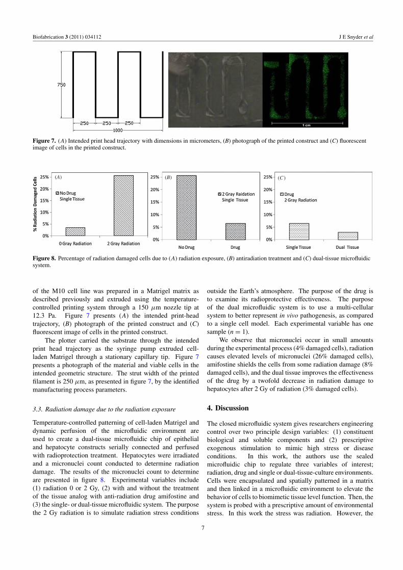
Figure 7. (A) Intended print head trajectory with dimensions in micrometers, (B) photograph of the printed construct and (C) fluorescentimage of cells in the printed construct.
(A) (B) (C)
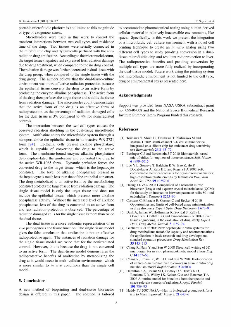
Figure 8. Percentage of radiation damaged cells due to (A) radiation exposure, (B) antiradiation treatment and (C) dual-tissue microfluidicsystem.
of the M10 cell line was prepared in a Matrigel matrix asdescribed previously and extruded using the temperature-controlled printing system through a 150 μm nozzle tip at12.3 Pa. Figure 7 presents (A) the intended print-headtrajectory, (B) photograph of the printed construct and (C)fluorescent image of cells in the printed construct.
The plotter carried the substrate through the intendedprint head trajectory as the syringe pump extruded cell-laden Matrigel through a stationary capillary tip. Figure 7presents a photograph of the material and viable cells in theintended geometric structure. The strut width of the printedfilament is 250 μm, as presented in figure 7, by the identifiedmanufacturing process parameters.
3.3. Radiation damage due to the radiation exposure
Temperature-controlled patterning of cell-laden Matrigel anddynamic perfusion of the microfluidic environment areused to create a dual-tissue microfluidic chip of epithelialand hepatocyte constructs serially connected and perfusedwith radioprotection treatment. Hepatocytes were irradiatedand a micronuclei count conducted to determine radiationdamage. The results of the micronuclei count to determineare presented in figure 8. Experimental variables include(1) radiation 0 or 2 Gy, (2) with and without the treatmentof the tissue analog with anti-radiation drug amifostine and(3) the single- or dual-tissue microfluidic system. The purposethe 2 Gy radiation is to simulate radiation stress conditions
outside the Earth’s atmosphere. The purpose of the drug isto examine its radioprotective effectiveness. The purposeof the dual microfluidic system is to use a multi-cellularsystem to better represent in vivo pathogenesis, as comparedto a single cell model. Each experimental variable has onesample (n = 1).
We observe that micronuclei occur in small amountsduring the experimental process (4% damaged cells), radiationcauses elevated levels of micronuclei (26% damaged cells),amifostine shields the cells from some radiation damage (8%damaged cells), and the dual tissue improves the effectivenessof the drug by a twofold decrease in radiation damage tohepatocytes after 2 Gy of radiation (3% damaged cells).
4. Discussion
The closed microfluidic system gives researchers engineeringcontrol over two principle design variables: (1) constituentbiological and soluble components and (2) prescriptiveexogenous stimulation to mimic high stress or diseaseconditions. In this work, the authors use the sealedmicrofluidic chip to regulate three variables of interest;radiation, drug and single or dual-tissue-culture environments.Cells were encapsulated and spatially patterned in a matrixand then linked in a microfluidic environment to elevate thebehavior of cells to biomimetic tissue level function. Then, thesystem is probed with a prescriptive amount of environmentalstress. In this work the stress was radiation. However, the
7
Biofabrication 3 (2011) 034112 J E Snyder et al
portable microfluidic platform is not limited to this magnitudeor type of exogenous stress.
Microfluidics were used in this work to control thetransient interactions between two cell types and residencetime of the drug. Two tissues were serially connected inthe microfluidic chip and dynamically perfused with the anti-radiation drug amifostine. According to the micronuclei count,the target tissue (hepatocytes) expressed less radiation damagedue to drug treatment, when compared to the no drug control.The radiation damage was further decreased in dual tissue withthe drug group, when compared to the single tissue with thedrug group. The authors believe that the dual-tissue-cultureenvironment was more effective radiation protection becausethe epithelial tissue converts the drug to an active form byproducing the enzyme alkaline phosphatase. The active formof the drug then perfuses the target tissue and shields the tissuefrom radiation damage. The micronuclei count demonstratesthat the active form of the drug is an effective form ofradioprotection, as the percentage of radiation-damaged cellsfor the dual tissue is 3% compared to 4% for nonirradiatedcontrols.
The interaction between the two cell types caused theobserved radiation shielding in the dual-tissue microfluidicsystem. Amifostine enters the microfluidic system through ananoport above the epithelial tissue in its inactive WR-2721form [24]. Epithelial cells present alkaline phosphatase,which is capable of converting the drug to the activeform. The membrane-bound enzyme alkaline phosphatasede-phosphorylated the amifostine and converted the drug tothe active WR-1065 form. Dynamic perfusion forces theconverted drug to the target tissue, which is the hepatocyteconstruct. The level of alkaline phosphatase present inthe hepatocyte is much less than that of the epithelial construct.The drug metabolized to an active form by the metabolizingconstruct protects the target tissue from radiation damage. Thesingle tissue model is only the target tissue and does notinclude the epithelial tissue, which presents more alkalinephosphatase activity. Without the increased level of alkalinephosphatase, less of the drug is converted to an active formand less radiation-protection is observed. The percentage ofradiation damaged cells for the single tissue is more than twicethe dual tissue.
The dual tissue is a more authentic representation of invivo pathogenesis and tissue function. The single tissue modelgives the false conclusion that amifostine is not an effectiveradioprotective agent. The instances of radiation damage forthe single tissue model are twice that for the nonirradiatedcontrol. However, this is because the drug is not convertedto an active form. The dual-tissue model demonstrates theradioprotective benefits of amifostine by metabolizing thedrug as it would occur in multi-cellular environments, whichis more similar to in vivo conditions than the single cellmodel.
5. Conclusions
A new method of bioprinting and dual-tissue bioreactordesign is offered in this paper. The solution is tailored
to accommodate pharmaceutical testing using human-derivedcellular material in relatively inaccessible environments, likespace. Specifically, in this work we present the integrationof a microfluidic cell culture environment with a novel cellprinting technique to create an in vitro analog using twodifferent cell types to study pro-drug conversion in a dual-tissue microfluidic chip and resultant radioprotection to liver.The radioprotective benefits and pro-drug conversion bymultiple cell types are more fully realized by incorporatingthe dual-tissue model. Future work using the printing systemand microfluidic environment is not limited to the cell type,drug or environmental stress presented here.
Acknowledgments
Support was provided from NASA USRA subcontract grantno. 09940-008 and the National Space Biomedical ResearchInstitute Summer Intern Program funded this research.
References
[1] Torisawa Y, Shiku H, Yasukawa T, Nishizawa M andMatsue T 2005 Multi-channel 3-D cell culture deviceintegrated on a silicon chip for anticancer drug sensitivitytest Biomaterials 26 2165–72
[2] Bettinger C J and Borenstein J T 2010 Biomaterials-basedmicrofluidics for engineered tissue constructs Soft. Matter.6 4999–5015
[3] Loo Y L, Someya T, Baldwin K W, Bao Z, Ho P,Dodabalapur A, Katz H E and Rogers J A 2002 Soft,conformable electrical contacts for organic semiconductors:high-resolution plastic circuits by lamination Proc. NatlAcad. Sci. USA 99 10252–6
[4] Huang J D et al 2008 Comparison of a resonant mirrorbiosensor (IAsys) and a quartz crystal microbalance (QCM)for the study on interaction between paeoniae radix 801 andendothelin-1 Sensors 8 8275–90
[5] Carstens C, Elbracht R, Gartner C and Becker H 2010Opportunities and limits of cell-based assay miniaturizationin drug discovery Expert Opin. Drug Discovery 5 673–9
[6] Dash A, Inman W, Hoffmaster K, Sevidal S, Kelly J,Obach R S, Griffith L G and Tannenbaum S R 2009 Livertissue engineering in the evaluation of drug safety ExpertOpin. Drug Metab. Toxicol. 5 1159–74
[7] Gebhardt R et al 2003 New hepatocyte in vitro systems fordrug metabolism: metabolic capacity and recommendationsfor application in basic research and drug development,standard operation procedures Drug Metabolism Rev.35 145–213
[8] Chang R, Nam Y and Sun W 2008 Direct cell writing of 3Dmicroorgan for in vitro pharmacokinetic model Tissue Eng.C 14 157–66
[9] Chang R, Emami K, Wu H L and Sun W 2010 Biofabricationof a three-dimensional liver micro-organ as an in vitro drugmetabolism model Biofabrication 2 045004
[10] Hamilton S A, Pecaut M J, Gridley D S, Travis N D,Bandstra E R, Willey J S, Nelson G A and Bateman T A2006 A murine model for bone loss from therapeutic andspace-relevant sources of radiation J. Appl. Physiol.101 789–93
[11] Haddy F J 2007 NASA—Has its biological groundwork for atrip to Mars improved? Faseb J. 21 643–6
8
Biofabrication 3 (2011) 034112 J E Snyder et al
[12] Tourovskaia A, Figueroa-Masot X and Folch A 2005Differentiation-on-a-chip: a microfluidic platform forlong-term cell culture studies Lab Chip 5 14–9
[13] Lee P J, Hung P J and Lee L P 2007 An artificial liver sinusoidwith a microfluidic endothelial-like barrier for primaryhepatocyte culture Biotechnol. Bioeng. 97 1340–6
[14] Underhill G H, Chen A A, Albrecht D R and Bhatia S N 2007Assessment of hepatocellular function within PEGhydrogels Biomaterials 28 256–70
[15] Kleinman H K and Martin G R 2005 Matrigel: basementmembrane matrix with biological activity Semin. CancerBiol. 15 378–86
[16] Dutta R C and Dutta A K 2010 Comprehension of ECM-celldynamics: a prerequisite for tissue regeneration Biotechnol.Adv. 28 764–9
[17] Moghe P V, Berthiaume F, Ezzell R M, Toner M,Tompkins R G and Yarmush M L 1996 Culture matrixconfiguration and composition in the maintenance ofhepatocyte polarity and function Biomaterials17 373–85
[18] Nahmias Y, Schwartz R E, Hu W S, Verfaillie C M andOdde D J 2006 Endothelium-mediated hepatocyterecruitment in the establishment of liver-like tissue in vitroTissue Eng. 12 1627–38
[19] Allen J W, Khetani S R and Bhatia S N 2005 In vitro zonationand toxicity in a hepatocyte bioreactor Toxicol. Sci.84 110–9
[20] Murray P E, Garcia-Godoy F and Hargreaves K M 2007Regenerative endodontics: a review of current status and acall for action J. Endod. 33 377–90
[21] Ciocca L, De Crescenzio F, Fantini M and Scotti R 2009CAD/CAM and rapid prototyped scaffold construction forbone regenerative medicine and surgical transfer of virtualplanning: a pilot study Comput. Med. Imaging Graph.33 58–62
[22] Barron J A, Wu P, Ladouceur H D and Ringeisen B R 2004Biological laser printing: a novel technique for creatingheterogeneous 3-dimensional cell patterns Biomed.Microdevices 6 139–47
[23] Zhang X, Hendro W, Fujii M, Tomimura T and Imaishi N2002 Measurements of the thermal conductivity andthermal diffusivity of polymer melts with the short-hot-wiremethod Int. J. Thermophys. 23 1077–90
[24] Aydemir N, Sevim N, Celikler S, Vatan O andBilaloglu R 2009 Antimutagenicity of amifostine againstthe anticancer drug fotemustine in the Drosophila somaticmutation and recombination (SMART) test Mutat. Res.:Genet. Toxicol. Environ. Mutag. 679 1–5
9