biotransformations of rifamycins: process …ychisti/rifaba.pdf · rifamycins biotransformations...
TRANSCRIPT
Biotech. Adv. Vol. 10, pp. 577-595,1992 0734-9750/92 $15.00 Printed in Great Britain. All Rights Reserved. © 1992 Pergamon Press Ltd
BIOTRANSFORMATIONS OF RIFAMYCINS: PROCESS POSSIBILITIES
U. C. BANERJEE,* B. SAXENA* and Y. CHISTI?
*Biochemical Engineering Research and Process Development Centre, Institute of Microbial Technology, P.O. Box 1304, Sector 39A, C~ndigarh 160, 014, India
"/'Department of Chemical Engineering, University of Waterloo, Waterloo, Ontario, Canada N2L 3G1
Abstract
Rifampicin, an important antibiotic, is manufactured by chemical conversion of rifamycin S which is obtained by the chemical modification of rifamycin B. Rifamycin B is a product of Nocardia mediterranei fermentations. The chemical conversion of rifamycin B to rifamycin S has many disadvantages: Strong acidic conditions are required, heavy foam formation accompanies transformation and the yields are low. This review highlights the developments in alternative, biochemical transformations using enzymes and cells; the main focus is on transformations carried out by rifamycin oxidase.
Key Words
Rifamycin antibiotics, biotransformation, rifamycin oxidase, Monocillium sp., Humicola sp., Curvularia lunata.
Introduction
Chemical or biochemical transformation of naturally produced antibiotics can
potentially be used to alter some of the characteristics of the natural substances.
Changes in antimicrobial activity, spectrum of action, oral absorption, toxicity and
allergenic responses may be achieved. A commercially used example of such a
transformation is the conversion of penicillins (predominantly benzyl penicillin) to
577
578 U.C. BANERJEE etal.
6-amino penicillanic acid, 6-APA, which is a precursor for other semi-synthetic
penicillins. The enzyme penicillin acylase produced by Escherichia coli, other
bacteria and fungi [1], is the basis of this conversion which involves selective
hydrolysis of the amide bond in the penicillin side-chain.
Rifampicin, a powerful antibiotic against certain important diseases, is
currently produced by a scheme utilising fermentation followed by chemical
conversion: The relatively inactive precursor, rifamycin B, is produced by Nocardia
medilerranei in submerged culture; it is converted to the active rifamycin S in a
chemical process. The low pH requirements of the chemical conversion necessitate
expensive acid-resistant equipment, the yields are low and excessive foam
formation causes operational problems. As a result, rifampicin is one of the more
expensive antibiotics. In view of the shortcomings of the chemical conversion step,
alternative biotransformations of rifamycin B to S have been investigated [2, 3].
Here we review these biotransformation methods based in several forms of
biocatalysts: immobilized enzymes, growing and resting whole cells, and pretreated
cells.
Rifamycins
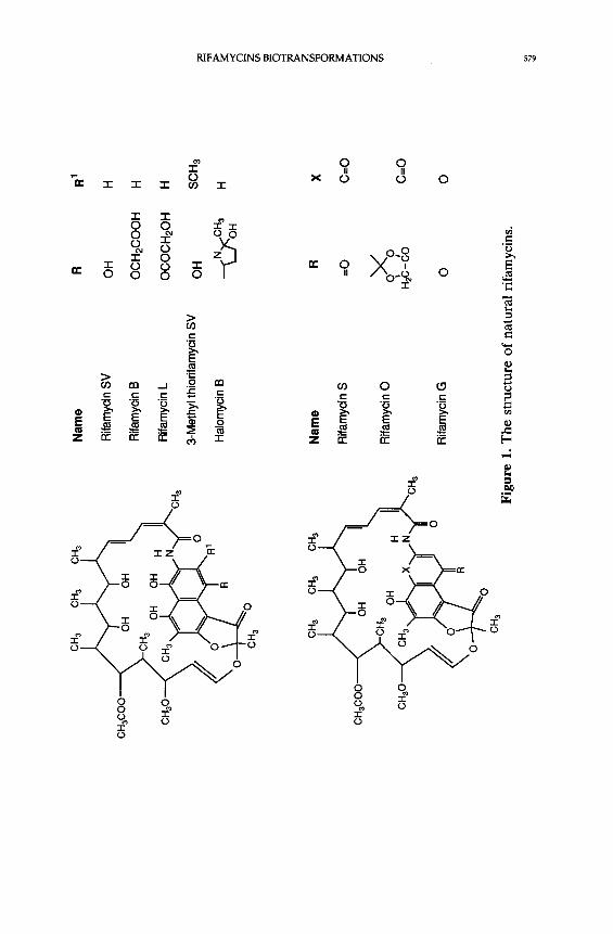
From the culture filtrate of the original strain of N. mediterranei, seven metabolites
(rifamycins A-E, G and Y) were isolated [4, 5-7]. The structures of these natural
rifamycins are shown in Figure 1 [8]. Of these, rifamycin B was chosen for
development because of its stability, ease of purification and solubility at
physiological pH's. Addition of barbiturates to the fermentation media was used
to dramatically alter the product mix, favouring the formation of rifamycin B, and
hence establishing a practical method for its production [9, 10]. Rifamycin B was
demonstrated to be microbiologically inactive [11]; its apparent activity depended
on its transformation into active products in test cultures or in body fluids. The
transformation products (rifamycin O, SV and S) were isolated and found to be
microbiologically very active. Due to its good in-vivo activity, tolerability and
solubility properties, rifamycin SV was chosen for further studies. Rifamycin SV
has been marketed in several countries for the treatment of infections from gram-
.CH
3 O
H 3
CH
3
CH
aC
OO
~
Nam
e R
R
1
Rifa
myc
in SV
O
H
H
Rifa
myc
in B
OCH
2CO
OH
H
Rifa
myc
in L
OCO
CH2O
H H
3-M
ethy
l thio
rifam
ycin
SV
OH
SC
H3
Halom
ycin B
~N
,)<
"CH3
H
~
/ O
H
CH3C
OO
CHa
Na
me
R
Rifa
myc
in S
=O
Rifa
myc
in O
O
~'O
t
/ H
zC
-CO
Rifa
myc
in G
O
Fig
ure
1. T
he
stru
ctur
e of
nat
ural
rif
amyc
ins.
X C=O
C=O
O
_=
z o z o r./3
3s{J U.C. BANERJEE et al.
positive bacteria, but it is poor in anti-tuberculotic activity. Hundreds of
compounds were synthesized from rifamycin B, O and S with the aim of obtaining
a product superior to rifamycin SV in at least two respects: activity by oral
administration and greater efficacy in curing tuberculosis. These goals were
attained with rifampicin which is now widely used for the t reatment of leprosy,
tuberculosis and several other infectious diseases [12], Rifampicin is at present
also used in several other infections, notably in severe infections by Staphylococci
resistant to I[}-lactam antibiotics and infections caused by intracellular bacteria.
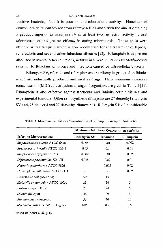
Rifamycin SV, rifamide and rifampicin are the rifamycin group of antibiotics
which are industrially produced and used as drugs. Their minimum inhibitory
concentrat ion (MIC) values against a range of organisms are given in Table 1 [11].
Rifampicin is also effective against t rachoma and inhibits certain viruses and
experimental tumours. Other semi-synthetic rifamycins are 27-demethyl-rifamycin
SV and, 25-deacetyl and 27-demethyl rifamycin B. Rifamyein S is of considerable
Table 1. Minimum Inhibitory Concentrations of Rifamycin Group of Antibiotics
Infecting Microorganism
Minimum Inhibitory Concentration (pg/mL)
Rifamycin SV Ri famide Rifampicin
Staphylococcus aureus ATCC 6538 0.005
Streptococcus faecalis ATCC 10541 0.05
Streptococcus pyogenes C 203 0.002
Diplococcus pneumoniae XXLIIL 0.025
Neisseria gonorrhoeae ATCC 9826
Haemophilus bzfluenzae ATCC 9334
Escherichia coli (McLeod) 50
Klebsiella pneumoniae ATCC 10031 25
Proteus vulgaris X 19 25
Salmonella typhi 100
Pseudornonas aentginosa 50
Mycobacterium tuberculosis H37 Rv 0.05
0.01 0.002
0.1 0.01
0.01 0.02
0.02 0.01
0.005 0.02
0.02
10 1
20 5
20 5
20 5
50 10
0.2 0.5
Based on Sensi el al. [11].
RIFAMYCINS BIOTRANSFORMATIONS 581
economic importance because it is a precursor for the majority of semi-synthetic
rifamycin antibiotics. Although, it is possible to produce the biologically active
rifamycin S by fermentation from a blocked mutant, its yield is low compared to
that of rifamycin B. Addition of sodium diethyl barbiturate to the fermentation
medium results essentially in the formation of a single product, rifamycin B.
Biotransformation of Rifamycins
Biotransformation of rifamycin was first reported by Lancini et al. [13] who found
that washed mycelia of Nocardia mediterranei could convert rifamycin B to
rifamycin Y. It was further shown that rifamycin Y was not produced by "ex novo"
synthesis, but originated from rifamycin B; this transformation did not require the
addition of diethyl barbituric acid. This work provided strong evidence that
rifamycin B was the natural precursor of rifamycin Y. In 1969 biotransformation
of rifamycin S to rifamycin B and rifamycin L by washed cells of N. mediterranei
was reported [14]. Acetylation (rifamycin S to rifamycin B) and esterification
(rifamycin S to rifamycin L) reactions were involved. Based on these observations
Lancini et al. [14] proposed that in N. mediterranei fermentations, rifamycin S or
its reduced form (rifamycin SV) is the first microbiologically active product of the
biosynthetic pathway, and is the precursor of rifamycins B and L. In the same
year Lancini and Hengeller noted that N. mediterranei also deacetylates rifamycin
B [15]. A little later, White et al. discovered that the washed mycelium from a
rifamycin B producing strain of N. rnediterranei transformed rifamycin W into
rifamycin B [16]. This biotransformation proved that the mycelia of N.
mediterranei are capable of carrying out the necessary modifications,/.e., removal
of methyl group and introduction of oxygen molecule, to transform the common
progenitor into rifamycin S. Lancini et al. demonstrated that washed cells of N.
mediterranei transformed the quinone ring of rifamycin S into pyrone ring of
rifamycin G [7]. Later, Ghisalba et al. showed that a strain of N. mediterranei
N813 (rifamycin B producer) partially transformed protorifamycin I to
protorifamycin W [17]. The formation of a small amount of rifamycin B from
protorifamycin I could also be detected [17].
CH
3 O
H 3
OH
3 C
H 3
OH
3 O
H3
CH
3C
OO
~
Rifam
ycinO
xidas
e CH
3C
OO
~
~ .,~
/ OC
H2CO
OH
~"~,
,'~o
\o~
~~
° C
H3
OH
3
Rifam
ycin B
[
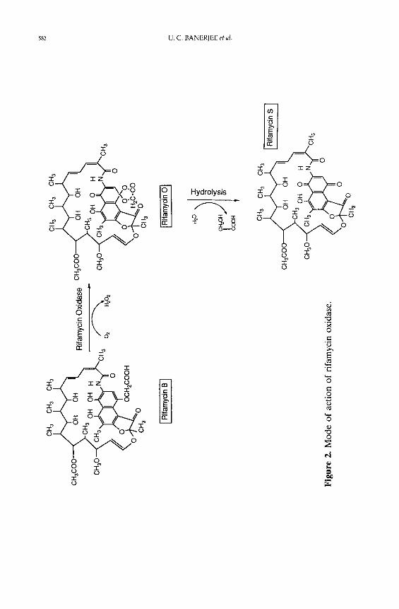
Fig
ure
2. M
ode
of
acti
on o
f ri
fam
ycin
oxi
dase
.
I Rifam
ycin O
I
COOH
~
CH
3 C
H 3
CH
3
[ Rifa-
.-_myci
n S l
~..coo
)~o:.
:. ,
~,
~J" /
,~O
CH
3
(q Z rn [11
RIFAMYCINS BIOTRANSFORMATIONS 583
More recently, Schupp et al. of Ciba-Geigy determined that the washed
mycelia of a recombinant strain of N. mediterranei R-21 transformed 50% of the
added rifamycin S to rifamycin B in 24 hours and 90% in 48 hours [18]. No other
transformation of rifamycins was detectable under the conditions used. Ghislba et
al. were able to coax permeabilized cells of N. mediterranei and E. coli into
transforming rifamycin S to rifamycin SV [19]. This transformation was NADH
dependent, but was not specific for the rifamycin biosynthetic pathway [19]. The
reduction was probably due to the reducing conditions of the cells.
The above cited biotransformation studies were carried out to elucidate the
biosynthetic pathways of rifamycins.
Bioprocesses for rifamycin S production
Possible advantages of biotransformation over the conventional chemical means
prompted researchers to look for new microbial strains with rifamycin B
transforming activity. In 1983 the first real biotransformation of inactive rifamycin
B to the active S-form was reported by Han et al. [2]. They isolated two fungi
imperfecti, Humicola sp. (ATCC 20620) and Monocill ium sp. (ATCC 20621),
which could catalyze the conversion of rifamycin B to rifamycin O and on to
rifamycin S, which is the reverse reaction of rifamycin B biosynthesis. The enzyme
from Monocil l ium sp. catalysed the oxidative reaction of rifamycin B to rifamycin
O [2]. The identification of the reaction products suggested that the reaction
proceeded by oxidative cyclization of rifamycin B to give rifamycin O, which on
spontaneous hydrolysis, gave rifamycin S in neutral aqueous solution. The
characterization of the enzyme was different as compared with that of other
polyphenol oxidases such as laccase. They classified this enzyme into a sub group
EC.1.10.3.6 with a trivial name rifamycin oxidase [2].
The mode of action of rifamycin oxidase, as presented in Figure 2, was
suggested [2]. In the two-step transformation reaction, one mole of rifamycin B
reacts with one mole of oxygen to produce one mole each of rifamycin O and
hydrogen peroxide. Further hydrolysis of one mole of rifamycin O produces one
mole each of rifamycin S and glycolic acid. From the stoichiometry it is also
584.
OH OH
OCH2COOH
[ Rifamycin B l
U. C. BANERJEE et al.
02
OH O- H ' / ~ ' ~ C H a ~ ~ !
H~C--C --O-H H ~ ; "
OH O C H 3 " ~ ~ ~
~ l O/NO I I
HbC- -C~ 0 H
I Rifamy cin O I
+ H202
H20
OH O CH3 @ ~ ~
-
H~ C--C~
OH O
C H 3 ~ ~ + H +
H~ C__CIO__H
H 0
OH O
+ H.~. C--C --OH /
H O
I Rifamycin S J I Glycolic acid I
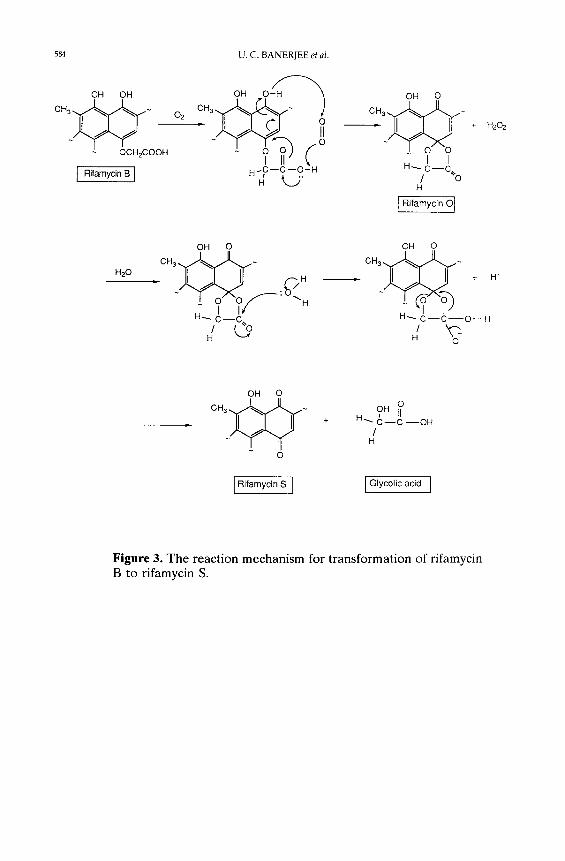
Figure 3. The reaction mechanism for transformation of rifamycin B to rifamycin S.

RIFAMYCINS BIOTRANSFORMATIONS 585
known that 100 g of rifamycin B yields 92 g of rifamycin S. From the action of
rifamycin oxidase on rifamycin B, it seems that substituted hydroquinone moiety
of rifamycin B is converted to quinone form of rifamycin B, Le., to rifamycin S.
The proposed organic reaction mechanism of this transformation is shown in
Figure 3.
The same group of workers also published the isolation procedure of
Monocillium sp. and its morphological and culture characteristics [20]. They
further demonstrated that rifamycin S can be prepared in high yield by microbial
transformation of rifamycin B [20].
In 1989 Vohra et al. reported a highly active extracellular rifamycin oxidase
from Curvularia lunata var aeria [3]. This enzyme could effectively transform
rifamycin B to S. The time course of this conversion, monitored by HPLC [21],
showed rapid consumption of rifamycin B, with 90% of it disappearing within the
first twenty minutes of incubation. The production of rifamycin O was
correspondingly fast and it underwent spontaneous hydrolysis to rifamycin S.
Within an hour all the rifamycin B was converted to rifamycin O and most of the
latter was hydrolysed to rifamycin S.
Production of rifamycin oxidase
In order to increase the intracellular rifamycin oxidase production by Humicola
sp., biochemical engineering studies were conducted by Kim et al. [22]. The
maximum specific enzyme activity in fermentations without pH control was
reported to be 9.5 IU/g dry cell, and 15 IU/g dry cell for cells cultured at
controlled pH of 8.0 [22]. Rifamycin oxidase production was accompanied by
biomass growth [22]. Antifoam (Neorin 202) concentrations lower than 1% (v/v)
did not have any significant effect on cell growth or on enzyme productivity [22].
pH control enhanced cell growth as well as enzyme production. The maximum
specific cell growth rate was 0.24 h -1 at a constant pH 7.0, but it was only 0.19 h -1
in absence of pH control. Highest specific enzyme activity and productivity were
observed at pH 8.0. No significant repression of enzyme production by high
glucose concentrations was apparent up to 60 g/L glucose [22]. At this glucose
586 U.C. BANERJEE et al.
concentration, 15 IU of enzyme activity per gram dry cell weight were obtained
(with pH control) and total enzyme production was 300 IU/L.
The effects of different carbon and nitrogen sources and of environmental
factors (pH, temperature) on the production of rifamycin oxidase by C. lunata
have been studied [23, 24]. Carboxymethylcellulose, yeast extract and peptone (1%
w/v, each) and ammonium sulphate (0.04 % nitrogen content) were determined
to be the best combination for maximum rifamycin oxidase production [24]. Other
synthetic media were also tested, but no growth or enzyme production occurred
in those media [23]. When carboxymethylcellulose was used as a carbon source,
the organism (C. lunata) formed mycelial type of growth rather than pellet type
[23]. With other substrates, pellet type of growth prevailed [23]. Mycelial growth
always yielded higher enzyme activity [24]. Enzyme activity was reduced under
conditions which led to intraceilular black pigment production. C. lunata could
produce high amounts of extracellular rifamycin oxidase within 40-50 hours of
fermentation. The enzyme yield was found to be 3400 IU/g (dry weight) cell mass.
The C. lunata rifamycin oxidase being an extracellular enzyme requires simpler
downstream processing relative to the enzyme from Humicola sp. Filtered broths
of C. lunata can be used for the transformation reaction. The intracellular enzyme
from the Humicola sp. may require cell disruption for extraction of enzyme
activity. Alternatively, delipidation of Humi¢ola sp. may be used to reduce the
diffusional resistances during the transformation reactions while fixing the enzyme
within the cells. Compared to Humicola sp. enzyme, the enzyme from C. lunata
has an important advantage for industrial applications. So far there is no
literature on the production of rifamycin oxidase by Monocillium sp.
Characteristics of rifamycin oxidase
The characteristics of rifamycin oxidase from Humicola sp. are different from that
of Monocillium sp. The enzymes differ noticeably in optimum pH, temperature,
degree of inhibition by substrates and in activities towardp-hydroquinone. Further,
the rifamycin oxidase from Monocillium sp. has quite different properties as
compared with other similar enzymes such as catechol oxidase, laccase, ascorbate
RIFAMYCINS BIOTRANSFORMATIONS 587
oxidase, o-aminophenol oxidase and 3-hydroxyanthranilate oxidase, which utilize
diphenols and related compounds as electron donors and oxygen as an electron
acceptor [2]. The substrate specificity of rifamycin oxidase from Monocillium sp.
was also different from that of the laccase from different sources. This enzyme
oxidised hydroquinone and rifamycin B very rapidly, while laccase showed
relatively low activity on those compounds [2] . Rifamycin oxidase from
Monocillium sp. also oxidised rifamycin SV, 3-formyl rifamycin SV, pyrogallol and
catechol. Resorcinol and the mannich derivative of rifamycin SV (3-
diethylaminomethyl rifamycin SV) were not attacked at all. The enzyme activity
was neither inhibited by Ag + or Hg +2 nor activated by Cu +2. Other metal ions
such as Ca +2, Mg +2, Fe +2, Fe +3, Co +2, Mn +2, Zn +2 and Mo +2 did not affect
the enzyme activity [2]. EDTA caused no marked inhibition of the enzyme. The
enzyme was neither a cupro-protein nor a flavo-protein; it did not contain any
heine or non-heine ion nor any metals as co-factors [2]. Rifamycin oxidase from
Monocillium sp. has pH and temperature optima of pH 7.8 and 40 °C,
respectively.
The catalytic properties of partially purified rifamycin oxidase produced
intracellularly by Humicola sp. were reported by Seong et al. [25]. The enzyme
was most specific for rifamycin B among the various rifamycin derivatives tested
[26]. The substrate specificity of the enzyme was investigated using various
substrate analogues, including rifamycin B derivatives and other polyol compounds.
P-hydroxyphenoxyacetic acid and p-hydroquinone which are corresponding
structural analogues in the quinonoid moiety, caused a significant decrease in the
enzyme activity compared to rifamycin B and rifamycin SV, respectively [25]. The
enzyme showed highest catalytic activity on rifamycin B among the various
substrates used. The Kin-value of this enzyme from Humicola sp. was found to be
0.05 mM [25]. Substrate inhibition was observed at substrate concentrations above
2 mM. A pH of 7.8 and a temperature of 45 ° C were optimal for enzyme activity,
catalytic activity was greatly reduced above this temperature. The rifamycin
oxidase activity was strongly inhibited by Fe +2 and Hg +2, but not by other metal
588 U.C. BANERJEE et al.
ions [25]. Presence of chelating agents such as EDTA did not affect the enzyme
activity [25].
The optimum pH and temperature of rifamycin oxidase from Curvularia
lunata are pH 6.5 and 50 °C, respectively [3]. The kinetic constants for the C.
lunata enzyme have also been reported [26]. Under optimal conditions, Michaelis-
Menten type kinetics were observed [26]; the K m and Vma x values, with rifamycin
B as substrate, were found to be 0.67 mM and 11 IU/mL, respectively [26]. No
inhibition of enzyme activity was noted up to 10 mM rifamycin B [26]. The
activation and deactivation energies of the partially purified enzyme, calculated
from Arrhenius plots, were 5.80 and 35.10 kcal/mole respectively [26]. The C.
lunata rifamycin oxidase activity was inhibited by Fe +2, Ag + and Hg +2, but was
not affected by the chelating agent, E D T A [26].
Biotransformation with soluble enzymes
Reports on biotransformation of rifamycin B to rifamycin S with soluble rifamycin
oxidases are very limited. Intracellular rifamycin oxidases from Monocillium sp.
and Humicola sp. were isolated from the cells and purified, but no systematic work
was clone to transform rifamycin B to S with the soluble enzyme. Rifamycin B was
used as a substrate simply to ascertain enzyme activity; rifamycin S did form in the
reaction mixture.
Extracellular, soluble rifamycin oxidase from C. lunata was used by Banerjee
et al. for the transformation of rifamycin B to S [27]. The transformation
conditions were optimized using partially purified rifamycin oxidase [27].
Transformation reactions were carried out at 28, 37 and 50 °C, with or without
agitation. Incubation temperature, as well as the mixing rate, significantly affected
the transformation rate [27]. Although at higher temperatures (37 and 50 ° C), the
transformation time (2 hours) was less than at 28 °C (3 hours), for practical
purposes, the lower temperature is preferred: Rifamycin oxidase has better
thermostability at 28 °C and oxygen (a required reactant) is more soluble in
aqueous solutions at lower temperatures. The transformation time in the absence
of mixing was almost 50 hours at 28 ° C. Optimal agitation speed was found to be
RIFAMYCINS BIOTRANSFORMATIONS 589
200 rpm. Different percentages (2-8 % v/v) of enzyme solution were used, and
5% (v/v) enzyme concentration was found to be optimal. Suitable aeration rate for
the transformation, optimized in a 0.5 L MBR bioreactor, was 1-1.25 vvm.
Antifoam agent (polypropylene glycol) was added to suppress the foam. No
adverse effect of the antifoam on enzyme activity was noted.
Biotransformation with immobilized enzymes
Rifamycin oxidase from C. lunata was immobilized on nylon fibres using
glutaraldehyde as the crosslinking agent [28]. An activity of 18 IU/g fibres, with
a binding efficiency of 37 %, was achieved. The immobilized enzyme showed an
operational stability of 7-days and was protected against thermal inactivation. It
exhibited a gm(app) of 2.0 mM. A polyacrylamide-based immobilization procedure
has also been described for this enzyme [26]. Various percentages of acrylamide
(6-10 % w/v) were used for the immobilization; 10 % (w/v) gel strength was found
to be optimal. Compared to free enzyme, the optimum pH (6.0-6.5) for the
immobilized preparation shifted slightly to the acidic region, but the temperature
optima were unaffected at 50 °C. In biotransformation studies with the
immobilized enzyme preparations, it took nearly 9 hours to complete the
transformation whereas with soluble enzyme, with the same percentage of
rifamycin B ( 1 % w/v), the reaction took 3 hours. The immobilized enzyme
preparations were tested for reusability. Upon reuse, the beads acquired colour
and after 3-4 uses, the transformation was additionally diffusionally limited. The
time required for complete transformation increased with the number of use
cycles. Transformation time for the fourth cycle was 10.5 hours, and in the sixth
and seventh cycles the transformation time was 20 and 25 hours, respectively.
Some adsorption of rifamycin on the beads was noted and it led to lower than
expected recovery of rifamycin S after the transformation. So far, there are no
reports of immobilization of soluble rifamycin oxidase from Monocillium or
Humicola sp.
59o U, C. BANERJEE et al.
Biotransformation with whole cells in suspension
As an enzyme source for industrial applications, whole cell preparations of
H u m i c o l a sp. were tested by Seong et al. [25]. A time lag in the conversion time
profile was observed in the initial stages of reaction with whole cells, but no such
trend was noticed with acetone dried (delipidated) cells. Diffusional resistances
to the substrates and products through the cell membrane, may have been
responsible for the initial lag in conversion. Acetone treatment removes the major
portion of lipids of the cell membrane making the cell more porous and removing
any diffusional limitations. Also, the lag could be reduced by increasing the
temperature. Enhanced mobility of membrane of whole cells at higher
temperatures was a possible explanation for the reduced lag.
Substrate inhibition was observed with whole cells as well as with acetone
treated cells when substrate concentration in the transformation reaction
increased, but up to 4 mM rifamycin B concentration there was no significant
reduction in the initial rate of reaction [25]. Rifamycin O and rifamycin S are
relatively hydrophobic and have an increased affinity to the cell membrane. This
could cause their accumulation in the membrane resulting in further interference
with the diffusion of substrate.
Han et al. clearly indicated the use of delipidated whole cells of Hurnicola
sp. for attaining maximal conversion yield of rifamycin B to rifamycin S [29, 30].
Banerjee reported the transformation of rifamycin B to rifamycin S using growing
and resting cells of C. lunata [26]. The organism could grow in the presence of
rifamycin B and simultaneously convert rifamycin B to rifamycin S [26]. With
growing cells, rifamycin B was added at the time of inoculation in the medium.
With 1% (w/v) of rifamycin B in the medium, growth of the organism was
reduced, but the transformation was completed within 20 hours of inoculation [26].
In other experiments, cells were grown up to the stationary phase and rifamycin
B was added at different phases of growth. Twelve-hours grown cells in the
culture broth took 16-hours to complete the transformation, whereas with 24-
hours grown cells it took only 4-hours. It took nearly 6-hours for 48-hours grown
cells to complete the transformation. Seventy-two hours grown cells of C. lunata
RIFAMYCINS BIOTRANSFORMATIONS 591
transformed only 10% of the added rifamycin B, and 96 and 120-hours grown cells
did not transform any rifamycin B. Resting cells in both exponential and
stationary phases could transform rifamycin B to rifamycin S and both could be
reused for the transformation reaction. It took 3.6-hours to convert rifamycin B
(1%, w/v) to rifamycin S by 24-hours grown, resting cells. The cells could be
conveniently recycled up to ten-times. At the 10th cycle, 18-hours were taken to
complete the transformation. Transformation of rifamycin B with growing and
resting cells did not give stoichiometric yields; less than expected rifamycin S was
obtained.
Biotransformation with immobilized whole cells
De-fatted whole cells of H u m i c o l a sp. with rifamycin oxidase activity have been
immobilized by copolymerization with acrylamide for use in biotransformations of
rifamycin [31]. The enzyme activity remaining after the immobilization step was
~50 % of the initial activity. The recovery of activity declined with increasing cell
volume added into the original mixture [31]. This may have been due to increase
in mass transfer resistances, or, to overpacking of the polyacrylamide gel matrix
with cells. The optimal reaction pH for the immobilized, acetone de-fatted, cells
was pH 7.8, while the optimal temperature for both free and immobilized de-
fatted whole cells was found to be 50 ° C. Experiments were conducted at a lower
temperature of 40 ° C to reduce enzyme inactivation. No appreciable activity loss
occurred for the immobilized acetone de-fatted cells during one-month storage at
4 °C and pH 7.8 [30]. The half life of the treated cells at 40 °C and pH 8 was ca.
8-days.
The Michaelis-Menten cons tan t s , Km(app) , and the substrate inhibition
constant, Kin(app), of the immobilized, acetone de-fatted, cells were found to be 0.6
mM and 19.6 mM, respectively [31]. The Kin-value of the acetone de-fatted cells
592 U.C. BANERJEE et al.
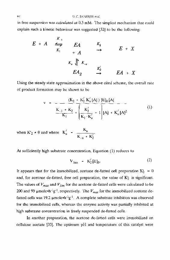
in free suspension was calculated at 0.3 mM. The simplest mechanism that could
explain such a kinetic behaviour was suggested [32] to be the following:
E + A K_ 1
K1
EA x2
+ A
K; E A 2 -- .
E + X
EA + X
Using the steady state approximation in the above cited scheme, the overall rate
of product formation may be shown to be
V (K 2 + K~ K~IA])[E]0[A ]
K-1 + K2 + I K~/ ] / K1 [ K~'K/a + 1j [A] ÷ K a [A] /
(1)
K a when K' 2 e 0 and where K/a -
K_ a + K~/"
At sufficiently high substrate concentration, Equation (1) reduces to
Vlim _ K~ / [El0" (2)
It appears that for the immobilized, acetone de-fatted cell preparation K~ = 0
and, for acetone de-fatted, free cell preparation, the value of K~ is significant.
The values of Vma x and Vli m for the acetone de-fatted cells were calculated to be
200 and 99 ~moleoh-lg -1, respectively. The Vma x for the immobilized acetone de-
fatted cells was 19.2 ~.mole.h-lg 1. A complete substrate inhibition was observed
for the immobilized cells, whereas the enzyme activity was partially inhibited at
high substrate concentration in freely suspended de-fatted cells.
In another preparation, the acetone de-fatted cells were immobilized on
cellulose acetate [33]. The optimum pH and temperature of this catalyst were
RIFAMYCINS BIOTRANSFORMATIONS 593
found to be pH 7.2 and 50-55 °C, respectively. Compared to the free enzyme, the
immobilized preparations were less sensitive to temperature and pH changes.
Twenty percent of the enzyme activity was recovered when the treated cells were
immobilized on 3 mm beads [33]. Recovery of the immobilized enzyme activity
increased as the bead size became smaller. Mass transfer limitations were
concluded to be the main reason for lower activity of the immobilized enzyme
[33]. The optimal loading of the acetone treated cell powder for cellulose acetate
beads was one gram powder in 50 mL acetone/dimethylsulfoxide (3:2 v/v) mixture
[33]. Higher enzyme loadings were not practical because of the precipitation of
acetone treated cell powder during the immobilization process which caused
problems with homogeneity of the catalyst. The physical strength of the cellulose
acetate beads containing acetone de-fatted t t umico la sp. cells was sufficient for
use in packed bed reactors.
Besides the characterization of the enzyme rifamycin oxidase in free cells,
whole cells and treated cells, Chung et al. did biotransformation studies in
fluidized bed and rotating packed disc reactors [33]. A fluidized bed system with
immobilized whole cells o f H u m i c o l a sp. was used also by Lee el al. [34]. A linear
relationship between the loading of the immobilized whole cells and conversion
in both batch and continuous operations were found [34]; however, the conversion
efficiency was higher in the batch mode for any given residence time [34].
Moreover, the aeration effect on the reaction rates in continuous operation was
different from that in batch operation. Among the two reactor geometries used,
the one which produced better mixing also gave 10 % better yield.
For the biotransformation with immobilized whole cells in a rotating packed
disc reactor (RPDR), Chung et al. found that the initial reaction rate and the total
productivity were dependent upon the degree of submergence of the discs [35].
The optimal submergence was 0.5; the rotational speed of the disc did not affect
the conversion very much. Higher conversions were attained with longer residence
times; however, the productivity declined with increasing residence times. Higher
volumetric productivities may be achieved if more enzyme is loaded in the
immobilized enzyme beads, or, more discs are installed in the RPDR. A RPDR
594 U.C. BANERJEE et al.
may be more amenable to plug flow operat ion than a fluidized bed reactor, which
may increase the relative conversion efficiency in the RPDR. No reports of
t ransformation of rifamycin B using immobilized whole cells or treated cells of
Monocil l ium sp. or C. lunata have appeared so far.
Conclus ions
The transformation of rifamycin B to rifamycin S can be successfully performed
by rifamycin oxidase enzyme. Intracellular enzyme from Humicola sp. and
Monocil l ium sp. may be used after extraction from the cells. Alternatively,
delipidation of Humicola sp. may be employed to decrease the diffusional
limitations in a process with the enzyme fixed within the cells. The transformation
may also be achieved less expensively with the extracellular enzyme of C. lunata.
The latter system has important processing advantages and either partially purified
enzyme, or crude extracts of C. lunata can be used. Immobilized rifamycin oxidase
preparat ions can also be employed with the advantages of greater stability,
reusability and productivity. It is now clear that several biotransformation options
may be used to prepare rifamycin S from rifamycin B. In view of the importance
of rifamycin S as a key intermediate for the preparat ion of many semi-synthetic
rifamycin derivatives of therapeutic value, further process improvements and scale-
up considerations need to be investigated.
Acknowledgements
The financial support from the Council for Scientific and Industrial Research and the Department of Biotechnology, India, is gratefully acknowledged.
References
1. Vandamme, E.J. and Voets, J.P. (1974), Adv. Appl. Microbiol., 17, 311. 2. Han, M.H., Seong, B.L., Son, H.J. and Mheen, T.I. (1983), FEBS Lett., 151, 36. 3. Vohra, R.M., Banerjee, U.C., Das, S. and Dube, S. (1989), Biotechnol. Lett., 11, 851. 4. Oppolzer, W., Prelog, V. and Sensi, P. (1964), Experientia, 20, 336. 5. Lancini, G.C. (1986), "Ansamycins," In Biotechnology, Volume 4, (Rehm, H.J. and Reed,
G., Editors), Verlag Chemie, Weinheim. 6. Leitich, J., Prelog, V. and Sensi, P. (1967), Experientia, 23, 505. 7. Lancini, G.C. and Sartori, G. (1976), J. Antibiot., 29, 466. 8. Lancini, G.C. and Sensi, P. (1967), In Proceedings of the 5th h~ternational Congress on
RIFAMYCINS BIOTRANSFORMATIONS 595
Chemotherapy, Volume 1, (Spitz, K.H. and Haschek, H., Editors), Verlag der wiener Medizinischen Akademie, p. 41.
9. Margalith, P. and Pagani, H. (1961), AppL Microbiol., 9, 320. 10. Margalith, P. and Pagani, H. (1961), Appl. Microbiol., 9, 325. 11. Sensi, P., Ballota, R., Greco, A.M. and Gallo, G.G. (1961), Farmaco (Ed. Sci.), 16, 165. 12. Sensi, P. and Thiemann, J.E. (1967), Progress Ind. Microbiol., 6, 21. 13. Lancini, G.C., Thiemann, J.E., Sartori, G. and Sensi, P. (1967), Expevientia, 23, 899. 14. Lancini, G.C., Gallo, G.G., Sartori, G. and Sensi, P. (1969), J. Antibiot., 22, 369. 15. Lancini, G.C., Hengheller, C. (1969), J. Antibiot., 22, 637. 16. White, R.J., Martenelli, E. and Lancini G.C. (1974), Proceedings Nat. Acad. Sci., USA, 71(8),
3260. 17. Ghisalba, O., Traxler, P. and Nuesch, J. (1978), J. Antibiot., 31, 1124. 18. Schupp, T., Traxler, P. and Auden, J.A.L. (1981), J. Antibiot., 34, 965. 19. Ghisalba, O., Roos, R., Schuup, T. and Nuesch, J. (1981), J. Antibiot., 35, 74. 20. Seong, B.L., Son, H.J., Mheen, T.I. and Han, M.H. (1983), J. Antibiot., 36, 1402. 21. Vohra, R.M. and Dube, S. (1989), J. Chromatogr., 477, 463. 22. Kim, E.K., Choi, C.Y., Pask, J.M., Han, M.H. and Park, Y.H. (1984), J. Ferment. Technol.,
62, 117. 23. Banerjee, U.C., Vohra, R.M., Jain, S.C. and Das, S (1992), J. Ferment. Technol. Bioeng., In
press. 24. Banerjee, U.C. and Srivastava, J.P. (1992), J. Biotechnol., In press. 25. Seong, B.L., Som, H.J., Mheen, T.I., Park, Y.H. and Han, M.H. (1985), J. Ferment.
TechnoL, 63, 515. 26. Banerjee, U.C. (1990), PhD Thesis, Panjab University, Chandigarh, India. 27. Banerjee, U.C. (1992), J. Biotechnol., In press. 28. Vohra, R.M. and Vyas, V.V. (1992), Biotechnol. Techniques, 6, 6l. 29. Han, M.H., Mheen, T.I., Seong, B.L. and Som, H.J. (1984), U.S. Patent 4,43L735. 30. Han, M.H., Mheen, T.I., Seong, B.L. and Som, H.J. (1983), Japan Kokai 58,138,390. 31. Lee, G.M., Choi, C.Y., Park, J.M. and Han, M.H. (1984), Biotechnol. Lett., 6, 143. 32. Laidler, K.J. and Bunting, P.S. (1973), The Chemical Kinetics of Enzyme Action, 2nd edition,
Clarendon Press (Oxford). 33. Chung, B.H., Chang, H.N. and Han, M.H. (1985), KoreanJ. Appl. MicrobioL Bioeng., 13(2),
115. 34. Lee, G.M., Choi, C.Y., Park, J.M. and Han, M.H. (1985), J. Chem. Technol. Biotechnol.,
35B, 3. 35. Chung, B.H., Chang, H.N. and Han, M.H. (1986), J. Ferment. Technol., 64, 343.