bone and soft tissue sarcomas 2008 - columbia … zradiation induced sarcomas ... zthe interval...
TRANSCRIPT

1
BONE AND SOFT BONE AND SOFT TISSUE TUMORSTISSUE TUMORS
Fabrizio Remotti MD
BONE AND SOFT TISSUE BONE AND SOFT TISSUE TUMORSTUMORS
Traditionally bone and soft tissue tumors have been treated separately.This separation will be maintained in the following presentation.Soft tissue sarcomas will be treated first and the sarcomas of bone will follow.

2
DEFINITIONDEFINITIONSoft tissue pathology deals with tumors of the connective tissues.The concept of soft tissue is understood broadly to include non-osseous tumors of extremities, trunk wall, retroperitoneum and mediastinum, and head & neck.Excluded (with a few exceptions) are organ specific tumors.
EPIDEMIOLOGYEPIDEMIOLOGYSarcomas are rare tumors compared to other malignancies: 8,700 new sarcomas in 2001, with 4,400 deaths.The incidence of sarcomas is around 3-4/100,000.Slight male predominance (with some subtypes more common in women).Majority of soft tissue tumors affect older adults, but important sub-groups occur predominantly or exclusively in children.Incidence of benign soft tissue tumors not known, but probably outnumber malignant tumors 100:1.

3
SOFT TISSUE TUMORSSOFT TISSUE TUMORS
Nowhere in the picture…..
Histological Histological classification classification of soft tissue of soft tissue
tumorstumors

4
Histological Histological classification classification of soft tissue of soft tissue
tumorstumors
ETIOLOGYETIOLOGY
The etiology of soft tissue sarcomas is poorly understood, and what is known apply only to a small fraction of the group.The known etiologic agents are ionizing radiation, oncogenic viruses, and chemicals.These agents are able to cause genetic alterations that can lead to tumorigenesis.

5
ETIOLOGYETIOLOGYRadiation induced sarcomas develop in 1% of patients who have undergone therapeutic irradiation.The interval between irradiation and diagnosis of sarcoma varies between 5 and 10 years.The majority of radiation-induced sarcomas are high grade and poorly differentiated (MFH, FS, OS, and AS).
ETIOLOGYETIOLOGY
Oncogenic viruses introduce new genomic material in the cell, which encode for oncogenic proteins that disrupt the regulation of cellular proliferation.Two DNA viruses have been linked to soft tissue sarcomas:
– Human herpes virus 8 (HHV8) linked to Kaposi’s sarcoma
– Epstein-Barr virus (EBV) linked to subtypes of leiomyosarcoma
In both instances the connection between viral infection and sarcoma is more common in immunosuppressed hosts.

6
ETIOLOGYETIOLOGY
Herbicides (“agent orange”) and peripheral soft tissue sarcomasRetained metal objects (shrapnel, surgical devices) and AS and MFHVinyl chloride, inorganic arsenic, Thorotrast, anabolic steroids linked to AS and MFH.
ETIOLOGYETIOLOGYHost factors may also play a role in the development of soft tissue sarcomas.– Immunosuppression,
besides Kaposi’s sarcoma, may be associated with sarcomas.
– Lymphedema, congenital or acquired (post-mastectomy) is a rare cause of extremity-based AS.
AS in lymphedema

7
SOFT TISSUE TUMORSSOFT TISSUE TUMORS
CONGENITAL SYNDROMES ASSOCIATED WITH BONE AND SOFT TISSUE TUMORSCONGENITAL SYNDROMES ASSOCIATED WITH BONE AND SOFT TISSUE TUMORS
Desmoid tumorsAPC5q21ADFamilial infiltrative fibromatosis
Osteochondromas, chondrosarcomasEXT18q24SporadicLanger- Giedion syndrome
Osteosarcomas, RMS, other sarcomasTP53CHEK2
17p1322q11
ADLi-Fraumeni syndrome
Lipomas--ADFamilial multiple lipomas
-
TNFRSF11A
APC
-
PTEN
-
-
PRKAR1AK
BLM
Complex
PTEN
GNAS1
Gene
-
18q21
5q21
9p21-22
10q23
-
7q33
17q23-242p16
15q26
11p15
10q23
20q13
Locus
Sporadic
AD
AD
AD
AD
Sporadic
AD
AD
AR
Sp/AD
AD
AD
Inheritance
Symmetrical lipomatosis
Familial expansile osteolysis
Familial adenomatous polyposis
Diaphyseal medullary stenosis
Cowden disease (Multiple hamartoma syndrome)
Costello syndrome
Familial chordoma
Carney complex(Familial myxoma syndrome)
Bloom syndrome
Beckwith- Wiedemann syndrome
Bannayan -Riley- Ruvalcaba syndrome
Albright hereditary osteodystrophy
Disorder
Lipomas, lipomatosis of head and neck
Osteosarcomas
Craniofacial osteomas, desmoid tumors
MFH
Lipomas, Hemangiomas
Rhabdomyosarcomas
Chordomas
Myxomas and pigmented schwannomas
Osteosarcoma
Embryonal RMS, myxomas, fibromas, hamartomas
Lipomas, hemangiomas
Soft tissue calcifications and osteomas
Tumor

8
CONGENITAL SYNDROMES ASSOCIATED WITH BONE AND SOFT TISSUE TUMORSCONGENITAL SYNDROMES ASSOCIATED WITH BONE AND SOFT TISSUE TUMORS
Bone and soft tissue sarcomasWRN8p11-12ARWerner syndrome
Glomus tumors-1p21-22ADVenous malf. With glomus cells
RhabdomyosarcomasCREBBP16p13ADRubinstein- Taybi syndrome
OsteosarcomasRECQL48q24ARRothmund- Thompson syndrome
Malignant rhabdoid tumorsSMARCB122q11ADRhabdoid predisposition syndrome
Osteosarcomas, soft tissue sarcomasRB113q14ADRetinoblastoma
Lipomas--SporadicProteus syndrome
Osteosarcomas18q215q315q35
ADPaget disease of bone, familial
Enchondromas, chondrosarcomasPTHR13p21-22SporadicOllier disease
SchwannomasNF222q12ADNeurofibromatosis type 2
Neurofibromas, MPNSTNF117q11ADNeurofibromatosis type 1
Myofibromas--ARMyofibromatosis
Osteochondromas, chondrosarcomasEXT1EXT2
8q2411p11-12
ADMultiple osteochondromas,non- syndromic
Fibrous dysplasia, osteosarcomasGNAS120q13SporadicMcCune –Albright syndrome
Fibrous dysplasia, OS, IM myxomasGNAS120q13SporadicMazabraud syndrome
Enchondromas, CS, hemangiomas, AS--SporadicMaffucci syndrome
TumorGeneLocus InheritanceDisorder
CLASSIFICATIONCLASSIFICATIONAll tumors are derived from stem cells that are programmed to differentiate into various mature cell types.Some of the stem cells probably belong to local, organ-specific pools, as underscored by the fact that many tumors resemble tissues present in the regionOther involved stem cells may be bone marrow derived.
Vascular leiomyosarcoma
9
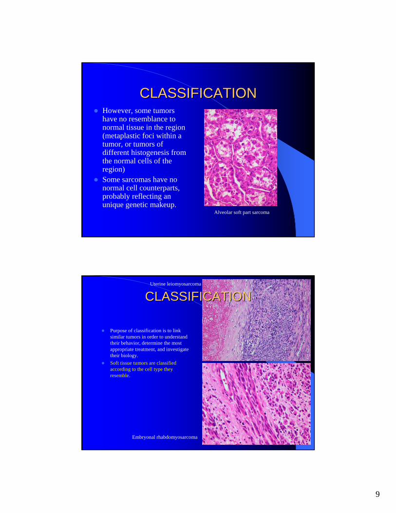
CLASSIFICATIONCLASSIFICATIONHowever, some tumors have no resemblance to normal tissue in the region (metaplastic foci within a tumor, or tumors of different histogenesis from the normal cells of the region)Some sarcomas have no normal cell counterparts, probably reflecting an unique genetic makeup.
Alveolar soft part sarcoma
CLASSIFICATIONCLASSIFICATION
Purpose of classification is to link similar tumors in order to understand their behavior, determine the most appropriate treatment, and investigate their biology.Soft tissue tumors are classified according to the cell type they resemble.
Uterine leiomyosarcoma
Embryonal rhabdomyosarcoma

10
CLASSIFICATIONCLASSIFICATION
Refinements are coming from cytogenetics, molecular, and gene expression studies.The majority arise from -or show differentiation toward- mesenchymal cells, but some show other differentiation (neuroectodermal, histiocytic).A small subset is of unknown histogenesis.
CLASSIFICATIONCLASSIFICATION
Tumors are also classified according their biologic potential.A three-tiered system is used:– 1. Benign– 2. Borderline (intermediate malignant)– 3. Malignant.

11
SOFT TISSUE TUMORSSOFT TISSUE TUMORSMAJOR TYPES OF SOFT TISSUE TUMORS Cell type Benign tumor Malignant tumor(Myo)fibroblast Fibroma, myxoma Fibrosarcoma, MFHAdipocyte Lipoma LiposarcomaSmooth muscle cell Leiomyoma LeiomyosarcomaSkeletal muscle cell Rhabdomyoma RhabdomyosarcomaEndothelial cell Hemangioma AngiosarcomaSchwann cell Schwannoma, neurofibroma MPNSTCartilage cell Chondroma ChondrosarcomaInterstitial cell GIST GISTHistiocyte JXG, GCTTS, RDD True histiocytic sarcomaUnknown No benign counterparts ES, SS, ES, ASPS
SOFT TISSUE TUMORSSOFT TISSUE TUMORSFibrous/myofibroblastic tumors
Fibroma-benign
Desmoid-borderline
Fibrosarcoma-malignant

12
SOFT TISSUE TUMORSSOFT TISSUE TUMORSLipomatous tumors Lipoma-benign
Liposarcoma-malignant
SOFT TISSUE TUMORSSOFT TISSUE TUMORSSmooth muscle tumors
Leiomyoma
Leiomyosarcoma, low grade
Leiomyosarcoma, high grade

13
IMMUNOHISTOCHEMISTRYIMMUNOHISTOCHEMISTRY
Immunohistochemistry is the most practical way to evaluate the presence of certain protein and carbohydrate epitopes on tissue sections. Evaluation of cell- or tumor-type specific or cell-cycle related markers may have diagnostic significance.Very few markers are specific for one tumor type.No cell-cycle marker is able to separate benign and malignant tumors.
IMMUNOHISTOCHEMISTRYIMMUNOHISTOCHEMISTRYMyofibroblastic tumors: SMA, HHF35Smooth muscle tumors: desmin, SMA, HHF35Skeletal muscle tumors: desmin, myogenin, Myo-D1, myoglobinNerve sheath tumors: S-100 protein, CD34, EMAFatty tumors: S-100 proteinSynovial sarcoma: CK, EMA, S-100Epithelioid sarcoma: CK, CD34Carcinomas: CK, EMAMelanoma: S-100, HMB45, tyrosinase, Melan A
Cam 5.2- synovial sarcoma

14
GRADINGGRADING
Grading is an arbitrary estimate of the degree of malignancy of a neoplasm (basically an attempt to determine the biological potential of a tumor).The purpose of grading is to provide guidance for prognostic prediction and treatment (mainly to determine the need for adjuvant therapy). Other independent variables evaluated with grading are tumor size and depth, margins of resection, and clinical situation.
GRADINGGRADING
Grading is an element of any current staging system.Correct grading requires correct histologic typing of the sarcoma, as demonstrated by the inclusion of the histologic type as a grading variable.

15
GRADINGGRADING
Grading applies best to excision specimen because biopsies may be non-representative of the correct grade.Preoperative treatments, such as radiation, chemotherapy, or embolization, can make grading inapplicable.Weak points of grading: – Subjective elements (number of mitoses, percent of
necrosis, tumor differentiation)– Frequent vs. rare tumors
GRADINGGRADINGGRADING SYSTEM SOFT TISSUE SARCOMAS (FFCC)
Score (1-3)TUMOR DIFFERENTIATIONwell diff 1defined histogenetic types 2poorly diff & undef histogenesis 3
MITOTIC COUNT0-9/10HPF 110-19/HPF 2>20 HPF 3
TUMOR NECROSISnone 0<50% 1>50% 2
HISTOLOGIC GRADE Sum of scores1 2 or 32 4 or 53 6, 7 or 8

16
GRADINGGRADINGDIFFERENTIATION SCORE 1
Well differentiated sarcoma (fibro-, lipo-, leiomyo-, chondro-)Well differentiated MPNST (neurofibroma with malignant transformation)
DIFFERENTIATION SCORE 2Conventional fibrosarcoma, leiomyosarcoma, angiosarcomaConventional MPNSTMyxoid sarcomas (MFH, liposarcoma, chondrosarcoma)Storiform-pleomorphic MFH
DIFFERENTIATION SCORE 3Sarcomas of undefined histog. (ASPS, SS,ES,CCS, undiff. Sarc.,malig. rhabdoid tumor)Ewing family of tumorsPleomorphic sarcomas (lipo-, leio-)Round cell and pleomorphic liposarcomaRhabdomyosarcoma (except botryoid and spindle cell)Poorly differentiated angiosarcomaTriton tumor, epithelioid MPNSTExtraskeletal mesenchymal CS, and osteosarcomaGiant-cell and inflammatory MFH
GRADINGGRADINGDifferentiation score 1
Fibrosarcoma

17
GRADINGGRADINGDifferentiation score 2
MFH
GRADINGGRADINGDifferentiation score 3
Ewing sarcoma

18
STAGINGSTAGING
The stage is an estimate of the extent or dissemination of a tumor (and in the current systems includes tumor grade).Staging is important for planning of treatment and prognostication.Clinical data and imaging studies are part of staging process(Visceral sarcomas excluded)
STAGING (GSTAGING (G--TNM)TNM)
PRESENTPOSITIVE OR NEGATIVEANYANY
ABSENTPOSITIVEANYANYIV
ABSENTNEGATIVET2bHIGHIII
ABSENTNEGATIVET2aHIGHIIB
ABSENTNEGATIVET1a or T1bHIGHIIA
ABSENTNEGATIVET2a or T2bLOWIB
ABSENTNEGATIVET1a or T1bLOWIA
ABSENT/PRESENTNEG/POST1 (<5 CM) OR T2 (>5 CM)LOW OR HIGHI - IV
METASTASISLYMPH NODESPRIMARY TUMORGRADESTAGE
“a” superficial tumors of trunk and extremities (above fascia)“b” deep tumors of trunk and extremities or intra-abdominal, intra-thoracic or retro-peritoneal

19
STAGING OF SARCOMASSTAGING OF SARCOMAS
10-20IV
52III
72II
86I
%Stage
5-yr survival
NEJM 2005; 353: 701-711
SOFT TISSUE SARCOMASSOFT TISSUE SARCOMAS--COMPREHENSIVE ANALYSISCOMPREHENSIVE ANALYSIS
Gross examinationEvaluation of inked marginsGross description and tumor measurementsPhotographSampling of tumor and marginsFrozen sections for diagnostic or triaging purposes
Frozen tissue procurementFormalin fixationCytogenetics(E.M.)

20
SOFT TISSUE SARCOMASSOFT TISSUE SARCOMASMARGINSMARGINS
A margin obtained by the surgical re-excision of the wound previously found to be microscopically intra-lesional in the same operative procedure.
CONTAMINATED
The surgical margins are all wide and include the entire anatomical compartment(s) involved by the tumor.
RADICAL
The surgical plane of dissection passes outside the reactive zone and through normal tissue.
WIDE
The surgical plane of dissection passes through the pseudocapsule, without microscopic evidence of tumor.
MARGINAL
The surgical plane of dissection passes through tumor tissue.
INTRALESIONALINTERPRETATIONDESCRIPTION
PARAMETERS TO BE INCLUDED IN PARAMETERS TO BE INCLUDED IN REPORT OF A SARCOMAREPORT OF A SARCOMA
FINAL REPORT– 1. Tumor site, type of
excision– 2. Depth of the tumor– 3. Tumor type and
variant– 4. Grade (if possible)– 5. Tumor size– 6. Status of margins &
L.N.– 7. Percent of necrosis– 8. Vascular invasion, if
present
ADDENDUM REPORT(S)– 1. Immunohistochemistry– 2. Electron microscopy– 3. Cytogenetics

21
IMAGING STUDIESIMAGING STUDIESThe ultimate goal is:– 1. Detecting lesions– 2. Giving a specific
diagnosis or a reasonable differential diagnosis
– 3. Staging the lesion
Liposarcoma
MFH
IMAGING STUDIESIMAGING STUDIESCT and particularly MRI allow detection and and staging by delineating anatomical extent in virtually all cases.A relatively specific diagnosis can be given in approximately 25-50% of cases, according to the type.
65 W, FS thigh (MRI)

22
GENETICS OF SOFT TISSUE GENETICS OF SOFT TISSUE TUMORSTUMORS
Numerous cancer-specific genetic alterations have been described.Some of them (such as translocations, numerical changes, large deletions and gene amplifications) are seen at the cytogenetic level.Subtle changes (such as single base pair substitutions, small deletions) require molecular genetic detection.
GENETICS OF SOFT TISSUE GENETICS OF SOFT TISSUE TUMORSTUMORS
Many chromosomal translocations and other genetic rearrangements lead to formation of oncogenic gene fusions or overexpression of normal genes.Many of these changes may be used for diagnosis or confirmation of diagnosis.
FISH-F: Ewing
FISH-BA: Ewing
t(11;22)(q24;q12)

23
GENE FUSIONS IN SARCOMASGENE FUSIONS IN SARCOMAS
Nonrandom translocations were described first in hematopoietic malignancies.Identified in many types of sarcomas.Also identified in benign soft tissue tumors.Each translocation results in a specific gene fusion.Each gene fusion is present in most cases of a specific sarcoma category, and is not present in any other sarcoma type.These genetic events demonstrate consistency and specificity.

24
GENE FUSIONS IN SARCOMASGENE FUSIONS IN SARCOMAS
These translocations:1. represent fundamental genetic steps in
the development of these cancers2. are useful markers for the diagnosis3. may constitute new therapeutic targets
GENE FUSIONS IN SARCOMASGENE FUSIONS IN SARCOMAS
Investigation of these translocation may:1. clarify the molecular etiology of these
cancers2. help in identifying new markers for
diagnosis and monitoring3. lead to new therapeutic strategies
against tumor-specific markers.

25
GENE FUSIONS IN SARCOMASGENE FUSIONS IN SARCOMAS1. These translocations disrupt genes
located at the chromosomal breakpoints and juxtapose portions of these genes to create two reciprocal chimeric genes.
2. The breaks are confined to one or a few introns within the coding region of each gene.
3. The chimeric genes are transcribed to generate chimeric transcripts.
4. The chimeric transcripts are translated into chimeric proteins.
FISH with dual color break-apart probe cocktail flanking the EWS breakpoint region at 22q12
GENE FUSIONS IN SARCOMASGENE FUSIONS IN SARCOMAS
The novel protein products have significantly altered functional properties.In many cases, one or both involved genes are transcription factors, and the chimeric product is a novel transcription factor.
26
SOFT TISSUE TUMORSSOFT TISSUE TUMORSSUMMARYSUMMARY
Tumors of connective tissue.Rare (sarcomas: 3-4 cases per 100,000).Etiology unclear, with a few exceptions.Classified according to tissue they resemble.Biologically: benign, borderline or malignant.Grading and staging crucial elements to be added to diagnosis.Some of the lesions have specific translocations.
BONE TUMORSBONE TUMORS

27
BONE TUMORSBONE TUMORS• The majority of tumors involving bone
are secondary (or metastatic):- secondary (metastases) (95%)- primary (5%)
MELANOMA TO PROXIMAL HUMERUS
BREAST CANCER TO HIP
METASTATIC BONE TUMORSMETASTATIC BONE TUMORS
Carcinomas are the most common metastatic tumors to bone.Other neoplasms may also metastasize to bone (sarcomas, melanomas).
Mammary carcinoma
Uterine leiomyosarcoma

28
Secondary Tumors of BoneSecondary Tumors of Bone
• Lung• Breast• Prostate• G.I• Kidney• Thyroid
•The carcinomas most frequently involved with bone metastasis originate from:
BONE TUMORSBONE TUMORS
Primary bone tumors are rare.Sarcomas account for 0.2% of all neoplasms (SEER Cancer Statistics Review, 1973-1996).Soft tissue sarcomas are 10 times more common than primary bone sarcomas.

29
BONE TUMORSBONE TUMORS
In North America and Europe, the incidence rate for bone in males is approximately 0.8 new cases per 100,000 people a year.Osteosarcoma is the most common primary malignant tumor of bone (35%), followed by chondrosarcoma (25%) and Ewing sarcoma (16%).Chordomas and MFH represent 8 and 5% of the the tumors in the group respectively.
BONE TUMORSBONE TUMORS
The majority of bone sarcomas arise de novo.Some, however, develop in association with recognizable precursors.
Osteoblastoma and Chondroblastoma
Giant cell tumor
Osteogenesis imperfecta
Metallic and polyethylene implants
Chronic osteomyelitis
Bone infarct
Fibrous dysplasiaLOW RISK
Radiation osteitis
Polyostotic Paget disease
Multiple enchondromasMODERATE RISK
Rothmund-Thompson syndrome
Familial Retinoblastoma syndrome
Ollier and Maffucci syndromeHIGH RISK

30
WHO WHO CLASSIFICATION CLASSIFICATION
OF BONE TUMORSOF BONE TUMORS
Fibrosarcoma
Desmoplastic fibromaFibrohistiocytic tumors
Fibrosarcoma
Desmoplastic fibromaFibrogenic tumors
High grade surface
Periosteal
Parosteal
Secondary
Low grade central
Small cell
Telangiectatic
ConventionalOsteosarcoma
Osteoblastoma
Osteoid osteomaOsteogenic tumors
Clear cell
Mesenchymal
Dedifferentiated
Peripheral
CentralChondrosarcoma
Chondromyxoid fibroma
Chondroblastoma
Multiple chondromatosis
Periosteal chondroma
EnchondromaChondroma
OsteochondromaCartilage tumors
WHO WHO CLASSIFICATION CLASSIFICATION
OF BONE OF BONE TUMORSTUMORS
Synovial chondromatosisJoint lesions
Chest wall hamartoma
Erdheim -Chester disease
Langerhans cell histiocytosis
Osteofibrous dysplasia
Fibrous dysplasia
Simple cyst
Aneurysmal bone cystMiscellaneous lesions
Metastatic malignancy
AdamantinomaMiscellaneous tumors
SchwannomaNeural tumors
Liposarcoma
LipomaLipogenic tumors
Leiomyosarcoma
LeiomyomaSmooth muscle tumors
Angiosarcoma
HemangiomaVascular tumors
ChordomaNotochordal tumors
Malignant giant cell tumor
Giant cell tumorGiant cell tumor
Malignant lymphoma
Plasma cell myelomaHematopoietic tumors
Ewing sarcomaEwing/PNET

31
BONE TUMORSBONE TUMORS
01020304050607080
0 to
4
10 to
14
20 to
24
30 to
34
40 to
44
50 to
54
60 to
64
70 to
74
80 to
85
OSCSESCHMFH
•Bone sarcomas as a group have a bimodal distribution.•The first peak is in the second decade.•The second peak occurs in patients older than sixty.
BONE TUMORSBONE TUMORS
The clinical presentation of bone tumors is at the beginning non-specific, with pain and swelling presenting first.Later, limitation of movement and pathological fracture and general symptoms may occur.A long time may elapse until the tumor is diagnosed.

32
BONE TUMORS BONE BONE TUMORS BONE TUMORSTUMORS
The diagnosis is based on imaging and histological criteria.
CONVENTIONAL X-RAY
BENIGN
TREATMENT
QUESTIONABLERESULTS
SUSPICIOUS FORMALIGNANCY
STAGING
MRI
CT
BIOPSY
TREATMENT
CT
BIOPSYMRI
X-RAY
BONE TUMORSBONE TUMORSConventional radiographs are still important in the diagnosis of bone tumors.Many tumors are site-specific.Many tumors have a characteristic radiographic appearance.
1. Ewing sarcoma, lymphoma, myeloma2. Osteofibrous dysplasia, adamantinoma3. Osteoid osteoma4. Fibrous dysplasia5. Chondromyxoid fibroma6. Non-ossifying fibroma7. Bone cyst, osteoblastoma8. Osteochondroma9. Osteosarcoma10. Enchondroma, chondrosarcoma11. Giant-cell tumor12. Chondroblastoma

33
BONE TUMORSBONE TUMORS
Some fancy words from the world of shadows
BONE TUMORSBONE TUMORS
The imaging characteristics of some lesions are diagnostic.Even if not clear to the radiologist, the images may help somebody else down the diagnostic chain (e.g. the pathologist)
Geographic with sharp margins and flocculent calcifications(Enchondroma)
Sclerotic margin and lytic(Chondroblastoma)

34
BONE TUMORSBONE TUMORS
Geographic with ill defined margins: usually malignant (in this case a primary chondrosarcoma)
Previous biopsy site
BONE TUMORSBONE TUMORSMoth-eaten and permeated are bad news (unless it’s infection)
Previous biopsy site

35
BONE TUMORSBONE TUMORS
Periosteal reactions (such as spiculated, Codman’s angle, onion skin) are witnesses of cortical destruction and soft tissue extension ( usually bad news, unless infective)
BONE TUMORSBONE TUMORS
The tumor need to be graded (grading is an important element of the staging and determines if the tumor is stage I or II).The TNM system follows a 2 tier grading system: low- and high-grade.

36
BONE TUMORSBONE TUMORS
The staging of bone sarcomas follows the TNM system.
M1b: other sites
M1a: lung
Distant metastasis:M1
No distant metastasisM0
Distant metastasis cannot be assessedMXDistant metastases (M)
Regional lymph node metastasisN1
No regional lymph node metastasisNO
Regional lymph nodes cannot be assessedNXRegional lymph nodes (N)
Discontinuous tumors in the primary bone siteT3
Tumor equal or more than 8 cm in greatest dimensionT2
Tumor less or equal to 8 cm in greatest dimensionT1
No evidence of primary tumorT0
Primary tumor cannot be assessedTXPrimary tumor (T)
AJCC Cancer Staging Manual, 6th Edition, Springer, New York
BONE TUMORSBONE TUMORS
Any gradeM1bAny NAny T
Any gradeAny MN1Any TStage IVB
Any gradeM1aN0, NXAny TStage IVA
Any gradeM0N0, NXT3Stage III
High gradeM0N0, NXT2Stage IIB
High gradeM0N0, NXT1Stage IIA
Low gradeM0N0, NXT2Stage IB
Low gradeM0N0, NXT1Stage IA
AJCC Cancer Staging Manual, 6th Edition, Springer, New York
37
BONE TUMORSBONE TUMORS
Stage I: low grade intra-compartmental (risk of metastasis <25%)Stage II: high-grade extra-compartmental (risk of metastasis >25%)Stage III: any grade, discontinuous tumor in the primary bone siteStage IV: any grade, metastatic
BONEBONE--FORMING TUMORSFORMING TUMORS
High grade surface
Periosteal
Parosteal
Secondary
Low grade central
Small cell
Telangiectatic
ConventionalOsteosarcoma
Osteoblastoma
Osteoid osteomaOsteogenic tumors

38
OSTEOID OSTEOMAOSTEOID OSTEOMABenign bone forming tumor.Small size, limited growth potential and disproportionate pain.Most common in long bones, but every bone may be affected.It may be painful on physical examinationIt may be associated with redness of skin and swelling. Lesions close to a joint may be associated with joint effusion.
OSTEOID OSTEOMAOSTEOID OSTEOMAOn plain x-rays the lesion is characterized by dense cortical sclerosis surrounding a radiolucent nidus.CT scan best type of imaging study.
Nidus

39
OSTEOID OSTEOMAOSTEOID OSTEOMASmall, cortically based lesion, red and gritty, surrounded by sclerotic bone.The lesion is composed of a meshwork of osteoid trabeculae lined by plump osteoblasts.
OSTEOID OSTEOMAOSTEOID OSTEOMA
Near diploid karyotype.Two cases with involvement of 22q13 and loss of distal part of 17q.Excellent prognosis following local excision (nidus has to be removed completely).

40
OSTEOSARCOMAOSTEOSARCOMA
• Malignant primary neoplasm of bone that produces osteoid (osteoid directly produced by the tumor cells).
• Intra-medullary origin (conventional type).• Rare subtypes.• Most common, non-hematopoietic tumor of
bone (incidence 4-5 per million).
OSTEOSARCOMAOSTEOSARCOMALargely a disease of the young (60% <25 years)30 % >40 years.In older people rule out predisposing conditions (e.g. Paget’s disease of bone, radiation)Long bones of appendicular skeleton are favored91% metaphysis, 9% diaphysis

41
OSTEOSARCOMAOSTEOSARCOMACentral
- Low Grade- High Grade
Surface- Low Grade- High Grade
OSTEOSARCOMAOSTEOSARCOMA• Conventional:
- Osteoblastic (50%)- Chondroblastic (<25%) - Fibroblastic (<1-2 %)
• Telangiectatic (<4%)• Small cell (1.5%)• Low grade central (<1%)• Parosteal (4%)• Periosteal (<2%)• High-grade surface (<<1%)• Secondary (20% of OS in patients older
than 40)

42
OSTEOSARCOMAOSTEOSARCOMA
Anaplastic, pleomorphic tumor.Production of osteoid.Cartilage and fibrous tissue may also be produced.
OSTEOSARCOMAOSTEOSARCOMAIHC not useful.Complex clonal chromosomal aberrations (including numerical and structural alterations).Recurrent involvement of 1p11, 13, 1q11-12, 1q21-22, 11p14-15, 14p11-13, 15p11-13, 17p, 19q13.Imbalances of +1, -6q, -9, -10, -13 (retinoblastoma gene on chromosome 13) and -17.Gains in 3q26, 4q12-13, 5p13-14, 7q31-32, 8q21-23, 12q14-15 (MDM2 and PRIM1), and 17p11-12 (Li-Fraumeni syndrome).Over-expression of MET and FOS in >50% of OS, and MYC in <15%.

43
OSTEOSARCOMAOSTEOSARCOMAUntreated is fatal (aggressive local growth and rapid hematogenous systemic metastasis).When treated with surgery alone, survival is limited.Age, gender, location, size, stage and laboratory tests traditional prognostic factors.The most reliable indicator of survival is the response to preoperative chemotherapy (good prognosis >90% tumor necrosis).In good responders survival in 80-90% of cases is nor unusual.Bad responders, without change in chemotherapy, die in 80-90% of cases (but with change of regimen long-term survival can be greatly improved).
CARTILAGECARTILAGE--FORMING TUMORSFORMING TUMORS
Clear cell
Mesenchymal
Dedifferentiated
Peripheral
CentralChondrosarcoma
Chondromyxoid fibroma
Chondroblastoma
Multiple chondromatosis
Periosteal chondroma
EnchondromaChondroma
OsteochondromaCartilage tumors

44
ENCHONDROMAENCHONDROMABenign hyaline cartilage neoplasm of medullary bone.Usually single.Common (10-25% of all benign bone tumors), but incidence is probably significantly higher.Wide age distribution (5-80 years) with most patients between 2nd and 4th decade.Most common in hands, followed by long tubular bones. Rare in flat bones.Exceedingly rare in craniofacial bones.
ENCHONDROMAENCHONDROMA
Swelling of small bones of hands and feet, thinning of cortex and pathological fractures.Asymptomatic in long bones.Well marginated lesions on imaging; lytic or mineralized.Usually in metaphysis, less common in diaphysis, rare in epiphysis. Hypocellular, avascular with abundant hyaline cartilage.

45
CHONDROSARCOMA (PRIMARY)CHONDROSARCOMA (PRIMARY)Malignant neoplasm with pure hyaline cartilage differentiation.Primary or conventional chondrosarcoma (90% of CS) arises centrally in a previously normal bone.Third most common primary malignancy of bone after myeloma and osteosarcoma.Tumor of adulthood (majority of patients >50 years).Genetics:
– Near diploid or pseudo-diploid karyotypes.
– Simple numerical changes (-X, -Y, +5).
– Rearrangements of 1p13-p22.
CHONDROSARCOMA (PRIMARY)CHONDROSARCOMA (PRIMARY)
Most commonly involved sites are: the pelvis, proximal femur and humerus, distal femur and ribs.Rare in the fingers (1%).Extremely rare in spine and craniofacial bones.Local swelling and pain.Metaphysis.Expansion of bone, thickening of the cortex with possible cortical erosion or destruction.

46
CHONDROSARCOMA (PRIMARY)CHONDROSARCOMA (PRIMARY)Cut surfaces translucent, blue-gray to white (cartilage), lobular, solid to myxoid areas, cortical erosion and soft tissue extension possible.Lobules of cartilage separated by fibrous septa. Host lamellar entrapment.Three grades:
– 1. Moderately cellular, oftentimes indistinguishable from enchondroma.
– 2. More cellular and atypical with more myxoid matrix.
– 3. More cellular,atypical and myxoid than grade 2 lesions. Mitoses are easily detected.
CHONDROSARCOMA (PRIMARY)CHONDROSARCOMA (PRIMARY)
The histological grade is the most important predictor of local recurrence and metastasis.89% of patients with grade 1 lesions are alive at 5 years.Only 53% of patients with grade 2 and 3 lesions are alive at 5 years.10% of recurrent tumors show increase in grade or dedifferentiate.Treatment is surgical (chemo and radiation resistant tumors).

47
Summary of bone sarcomas Summary of bone sarcomas Tumors of connective tissue.Very rare (bone sarcomas: 1/10 of soft tissue sarcomas).Etiology unclear, with a few exceptions.Classified according to tissue they resemble.Biologically: benign, borderline or malignant.Grading and staging crucial elements to be added to diagnosis.No recurrent diagnostic translocation (unless a soft tissue equivalent exists, e.g. Ewing sarcoma).