brain anatomy and function - sinoe medical association...
TRANSCRIPT

1
The Brain
DanilHammoudi.MD
http://publish.uwo.ca/~jkiernan/neuslide.htm
Cell bodies in CNS: nuclei
Cell bodies in PNS: ganglia
Nerves: bundles of axons!
Divisions of the Human Brain: 1 - Myelencephalon, which includes the medulla 2 - Metencephalon, which includes the ponsand cerebellum 3 - Mesencephalon, which includes the midbrain (tectum and tegmentum)4 - Diencephalon, which includes the thalamus and hypothalamus 5 - Telencephalon, which includes the cerebrum (cerebral cortex, basal ganglia, & medullary body)
New Terms:Brain Division
Telencephalon Diencephalon Mesencephalon Metencephalon Myelencephalon
Medulla
Telencephalon
–Cerebral Cortex
–Limbic system
–Basal Ganglia
Pons:
Cerebellum:

2
Cerebral Cortex
• Allows for sensation, voluntary movement, self-awareness, communication, recognition, and more.
• Gray matter! • 40% of brain mass, but
only 2-3 mm thick.• Each cerebral
hemisphere is concerned with the sensory and motor functions of the opposite side (a.k.a. contralateral side) of the body.
The standard areas of cortex (isocortex) is characterized as having six distinct layers. From outside inward:
1.Molecular layer2.External granular layer3.External pyramidal layer4.Internal granular layer5.Internal pyramidal layer6.Multiform layer.
I-Molecular layer or plexiform layer: Contains few cells and a rich nerve fiber plexus made up of axons and dendrites of cells in other laminae as well as cells in this lamina.
II-External granular layer: Closely packed small neurons.
III-External pyramidal layer:Composed mainly of pyramidal neurons and many granule cells and cells of Martinotti.*
IV-Internal granular layer: Composed chiefly of stellate cells that are closely packed.
V-Internal pyramidal or ganglioniclayer: Consists of medium-sized and large pyramidal cells intermingled with granule cells.
VI-Multiform layer or layer of fusiformcells: Contains a variety of cell types.
White matter: Contains incoming and outgoing nerve fibers.
Structures of the Brain:
Protection• What is the major protection for the
brain?• There are also 3 connective tissue
membranes called the meninges:• Cover and protect the CNS• Protect blood vessels• Contain cerebrospinal fluid
• The 3 meninges from superficial to deep:
• Dura mater• Arachnoid mater• Pia mater
Dura Mater•Leathery, strong meninx composed of two fibrous connective tissue layers •The two layers separate in certain areas and form dural sinuses•Three dural septa extend inward and limit excessive movement of the brain•Falx cerebri – fold that dips into the longitudinal fissure•Falx cerebelli – runs along the vermis of the cerebellum•Tentorium cerebelli – horizontal dural fold extends into the transverse fissure
Arachnoid Mater•The middle meninx, which forms a loose brain covering•It is separated from the dura mater by the subdural space•Beneath the arachnoid is a wide subarachnoid space filled with CSF and large blood vessels•Arachnoid villi protrude superiorly and permit CSF to be absorbed into venous blood
Pia Mater•Deep meninx composed of delicate connective tissue that clings tightly to the brain
MENINGES

3
Meninges,
(a)
Superiorsagittal sinus
Skin of scalpPeriosteum
Falx cerebri(in longitudinalfissure only)
Bloodvessel
Arachnoid villusPia materArachnoid mater
DuramaterMeningeal
Periosteal
SubduralspaceSubarachnoidspace
Bone of skull
Meninges,
(b)
Occipital lobe
Superiorsagittal sinus
Dura mater
Tranversesinus
Temporalbone
Scalp
Skull
Tentoriumcerebelli
Cerebellum
Arachnoid materover medullaoblongata
Blood-Brain BarrierProtective mechanism that helps maintain a stable environment for the brainBloodborne substances are separated from neurons by:Continuous endothelium of capillary wallsRelatively thick basal laminaBulbous feet of astrocytes
Blood-Brain Barrier: FunctionsSelective barrier that allows nutrients to pass freelyIs ineffective against substances that can diffuse through plasma membranesAbsent in some areas (vomiting center and the hypothalamus), allowing these areas to monitor the chemical composition of the bloodStress increases the ability of chemicals to pass through the blood-brain barrier
Cerebral Cortex
• 3 types of functional areas:1. Motor Control voluntary
motor functions2. Sensory Allow for conscious
recognition of stimuli3. Association Integration
4
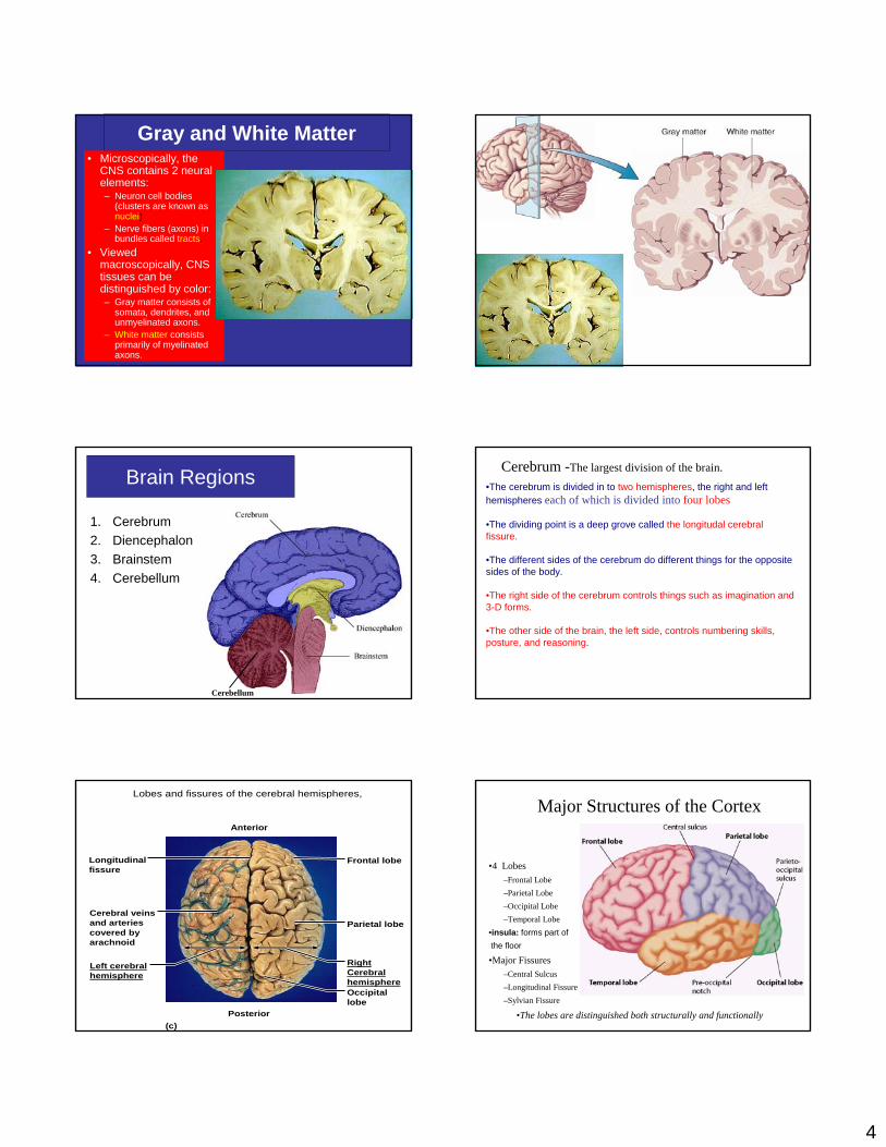
Gray and White Matter• Microscopically, the
CNS contains 2 neural elements:– Neuron cell bodies
(clusters are known asnuclei)
– Nerve fibers (axons) in bundles called tracts.
• Viewed macroscopically, CNS tissues can be distinguished by color:– Gray matter consists of
somata, dendrites, and unmyelinated axons.
– White matter consists primarily of myelinated axons.
Brain Regions
1. Cerebrum2. Diencephalon3. Brainstem4. Cerebellum
Cerebellum
•The cerebrum is divided in to two hemispheres, the right and left hemispheres each of which is divided into four lobes
•The dividing point is a deep grove called the longitudal cerebral fissure.
•The different sides of the cerebrum do different things for the opposite sides of the body.
•The right side of the cerebrum controls things such as imagination and 3-D forms.
•The other side of the brain, the left side, controls numbering skills, posture, and reasoning.
Cerebrum -The largest division of the brain.
Lobes and fissures of the cerebral hemispheres,
(c)
Parietal lobe
Frontal lobe
RightCerebralhemisphereOccipitallobe
Left cerebralhemisphere
Cerebral veinsand arteriescovered byarachnoid
Longitudinalfissure
Posterior
Anterior
•4 Lobes–Frontal Lobe–Parietal Lobe–Occipital Lobe–Temporal Lobe
•insula: forms part ofthe floor
•Major Fissures–Central Sulcus–Longitudinal Fissure–Sylvian Fissure
Major Structures of the Cortex
•The lobes are distinguished both structurally and functionally

5
Cortical Motor Areas1. Primary Motor
Cortex2. Premotor Cortex3. Broca’s Area4. Frontal Eye Field
Primary motor cortex
Broca’s Area
Premotor cortex
Frontal Eye Field
Primary (Somatic) Motor Cortex• Located in the precentral
gyrus of each cerebral hemisphere.
• Contains large neurons(pyramidal cells) whichproject to SC neurons which eventually synapse on skeletal muscles – Allowing for voluntary
motor control.– These pathways are known
as the corticospinal tracts or pyramidal tracts.
Primary (Somatic) Motor Cortex
• Somatotopy– The entire body is represented
spatially in the primary motor cortex, i.e., in one region we have neurons controlling hand movements and in another region leg movements, etc.
• Neurons controlling movement of different body regions do not intermingle.
• What does it mean to say that motor innervation is contralateral?
• Let’s look at the motor homunculus.
Lobes and fissures of the cerebral hemispheres,.
(a)
(b)
Postcentral gyrus
Central sulcus
Precentral gyrus
Frontal lobe Parietal lobeParieto-occipital sulcus(on medial surfaceof hemisphere)Lateral sulcus
Transversecerebral fissure
Occipital lobeTemporal lobe
Cerebellum
Medulla oblongataSpinal cord
Cortex(gray matter)
Fissure(a deep sulcus)
Gyrus
Sulcus
White matter
Central sulcusFrontal lobe
Temporal lobe(pulled down)
Gyri of insula
Pons

6
1.Gyri breves insulae2.Gyri longi insulae3.Limen insulae4.Sulcus centralis insulae5.Sulcus circularis insulae
1.Sulcus centralis2.Gyrus praecentralis3.Gyrus postcentralis4.Sulcus praecentralis5.Sulcus postcentralis
6.Sulcus frontalis superior 7.Sulcus frontalis inferior 8.Gyrus frontalis superior 9.Gyrus frontalis medius
10.Gyrus frontalis inferior 10a.Pars opercularis10b.Pars triangularis
10c.Pars orbitalisSulcus lateralis
11.Ramus ascendens12.Ramus anterior
13.Ramus posterior 14.Sulcus temporalis superior 15.Sulcus temporalis inferior 16.Gyrus temporalis supeior17.Gyrus temporalis medius18.Gyrus temporalis inferior 19.Gyrus supramarginalis
20.Gyrus angularis21.Sulcus parietooccipitalis20+21.Lobulus parietalis inf.
22.Lobus occipitalis23.Margo superior 24.Margo inferior
25.Sulcus intraparietalis26.Cerebellum
27.Lobulus parietalis sup.
Cerebral hemisphere (hemispherium cerebrale)
•Is defined as one of the two regions of the brain that are delineated by the body's median plane.
•The brain can thus be described as being divided into left and right cerebral hemispheres. Each of these hemispheres has an outer layer of grey matter called the cerebral cortex that is supported by an inner layer of white matter.
• The hemispheres are linked by the corpus The hemispheres are linked by the corpus callosumcallosum, a very large bundle of nerve , a very large bundle of nerve fibers, and also by other smaller fibers, and also by other smaller commissurescommissures, including the anterior , including the anterior commissurecommissure, , posterior posterior commissurecommissure, and , and hippocampalhippocampal commissurecommissure.
•These commissures transfer information between the two hemispheres to coordinate localized functions.
• The architecture, types of cells, types of neurotransmitters and receptor subtypes are all distributed among the two hemispheres in a markedly asymmetric fashion.
• However, it must be noted that, while some of these hemispheric distribution differences are consistent across human beings, or even across some species, many observable distribution differences vary from individual to individual within a given species.
Cerebral Features:
• Sulci – Small grooves dividing the gyri
– Central Sulcus – Divides the Frontal Lobe from the Parietal Lobe
• Fissures – Deep grooves, generally dividing large regions/lobes of the brain
– Longitudinal Fissure – Divides the two Cerebral Hemispheres
– Transverse Fissure – Separates the Cerebrum from the Cerebellum
– Sylvian/Lateral Fissure – Divides the Temporal Lobe from the Frontal and Parietal Lobes
• Gyri – Elevated ridges “winding” around the brain.
Gyri (ridge, circumvolution)
Fissure
(deep groove)
Sulci(groove)
http://williamcalvin.com/BrainForAllSeasons/img/bonoboLH-humanLH-viaTWD.gif
The medial longitudinal fissure (or longitudinal cerebral fissure, or longitudinal fissure, or interhemispheric fissure) is the deep groove which separates the two hemispheres of the vertebrate brain.
The falx cerebri, a dural brain covering, lies within the medial longitudinal fissure.
1. right cerebral cortex2. longitudinal fissure3. cerebellum4. frontal lobe5. central sulcus6. parietal lobe
7
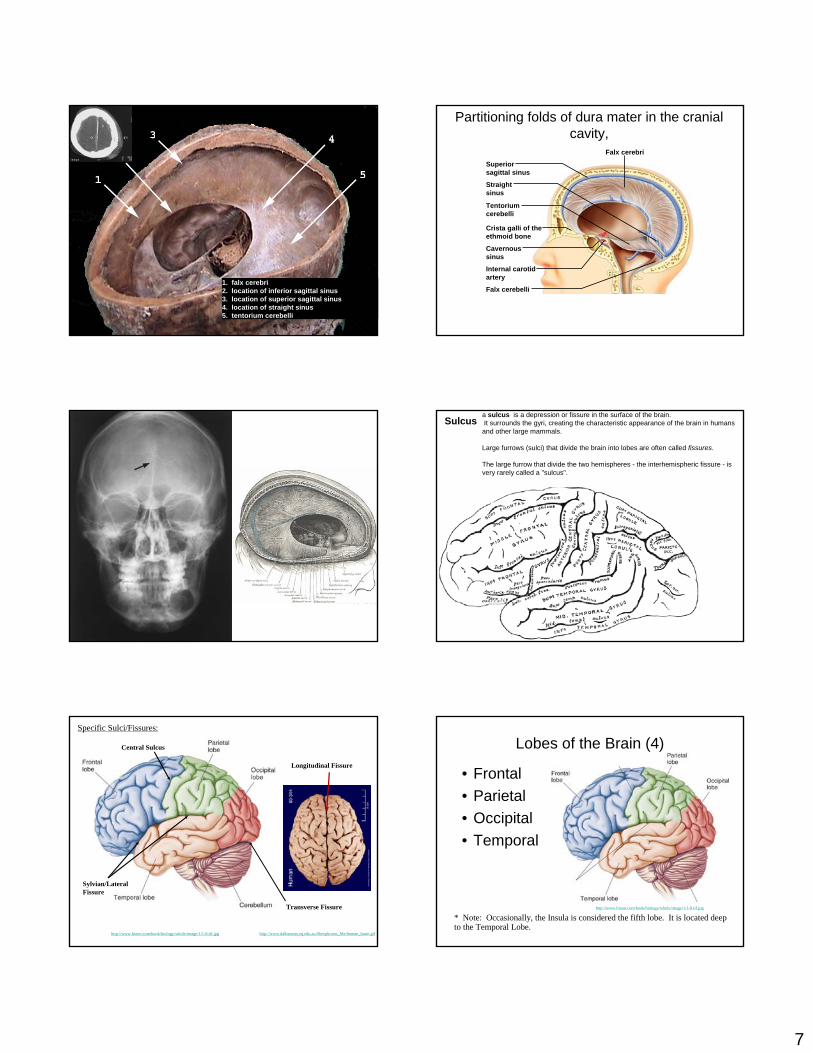
1. falx cerebri2. location of inferior sagittal sinus 3. location of superior sagittal sinus 4. location of straight sinus 5. tentorium cerebelli
Falx cerebri
Tentoriumcerebelli
Superiorsagittal sinus
Straightsinus
Crista galli of theethmoid bone
Cavernoussinus
Internal carotidartery
Falx cerebelli
Partitioning folds of dura mater in the cranial cavity,
Falx cerebriSulcus
a sulcus is a depression or fissure in the surface of the brain.It surrounds the gyri, creating the characteristic appearance of the brain in humans
and other large mammals.
Large furrows (sulci) that divide the brain into lobes are often called fissures.
The large furrow that divide the two hemispheres - the interhemispheric fissure - is very rarely called a "sulcus".
Longitudinal Fissure
Transverse Fissure
Sylvian/Lateral Fissure
Central Sulcus
http://www.bioon.com/book/biology/whole/image/1/1-8.tif.jpg http://www.dalbsoutss.eq.edu.au/Sheepbrains_Me/human_brain.gif
Specific Sulci/Fissures:
Lobes of the Brain (4)
• Frontal• Parietal• Occipital• Temporal
* Note: Occasionally, the Insula is considered the fifth lobe. It is located deep to the Temporal Lobe.
http://www.bioon.com/book/biology/whole/image/1/1-8.tif.jpg

8
Central sulcus= between frontal and parietal lobes.
Frontal lobe:precentral gyrus: motor neurons.
Parietal lobe: Poscentral gyrus: somatesthetic sensation (cutaneous touch, pain, heat, muscles and joints).
MAP of motor and of sensory control (homunculus)
Lobes and fissures of the cerebral hemispheres,
(d)
(c)
Left cerebralhemisphere
Transverse cerebralfissureCerebellumBrain stem
Parietal lobe
Frontal lobe
Right cerebralhemisphere
Occipitallobe
Left cerebralhemisphere
Cerebral veinsand arteriescovered byarachnoid
Longitudinalfissure
Posterior
Anterior
LOBESCortical Function •Frontal Lobe
–Higher thought processing; decision making; abstract thinking–Primary “precentral” motor area
•Parietal Lobe–Primary “postcentral” somatosensory area: sensation of muscles, organs, and skin
•Occipital Lobe–Visual processing
•Temporal Lobe–Auditory & equilibrium processing–Left temporal lobe involved in speech and comprehension of language
TheCerebralCortex
FrontalFrontalLobeLobe
HigherHigherIntellectualIntellectualFunctionsFunctions
PrimaryPrimaryMotorMotorAreaArea
PremotorPremotorAreaArea
SpeechSpeechMotorMotorAreaArea
leglegtrunktrunk
armarmhandhandfaceface
tonguetongue
ParietalParietalLobeLobePrimaryPrimary
SensorySensoryAreaArea
SensorySensoryAssociationAssociation
AreaArea
OccipitalOccipitalLobeLobe
PrimaryPrimaryVisualVisualAreaArea
VisualVisualAssociationAssociation
AreaArea
TemporalTemporalLobeLobe
MemoryMemory
PrimaryPrimaryAuditoryAuditory
AreaArea
LanguageLanguageComprehensionComprehension
& Formation& Formation
Thumb
Motor and sensory areas of the cerebral cortex,
GenitalsToes
SwallowingTongue
Jaw
Motor cortex(precentral gyrus)
Motor Sensory
Leg
Lips
Face
NeckBrowEye
FingersHand
Wrist
Elbow
Arm
ShoulderTrunk
Hip
Kn ee H
ip
Tru n
kN
eck
Hea
d
Arm
Elbo
wFo
rear
mHa
ndFi
nger
sTh
umb
Eye
Nose
Face
Lips
Teeth
Gums
JawTongue
Pharynx
Intra-abdominal

9
Functional and structural areas of the cerebral cortex, .
(a)
Primary motor area
Premotor cortex
Frontaleye field
Working memoryfor spatial tasks
Executive area fortask management
Working memory for object-recall tasks
Broca's area(outlined by dashes)
Solving complex,multitask problems
Prefrontal cortex
Central sulcusPrimary somatosensorycortexSomatosensoryassociation area
Somatic sensation
Gustatory cortex(in insula)
Taste
Wernicke's area(outlined by dashes)
Primary visualcortex
Visualassociation area
Vision
Auditoryassociation areaPrimary auditory cortex
Hearing
1147
45 44
8
6 43 1 2
5
43
7
19 1817
224241
22
Premotor Cortex• Located just anterior
to the primary motor cortex.
• Involved in learned or patterned skills.
• Involved in planning movements.
• How would damage to the primary motor cortex differ from damage to the premotor cortex?
Broca’s Area• Typically found in only
one hemisphere (often the left), anterior to the inferior portion of the premotor cortex.
• Directs muscles of tongue, lips, and throat that are used in speech production.
• Involved in planning speech production and possibly planning other activities.
Frontal Eye Field
• Controls voluntary eye movements.
• Found in and anterior to the premotor cortex, superior to Broca’s area.
• What muscles would be affected if this area was damaged?
Sensory Areas• Found in the parietal, occipital, and
temporal lobes.
1. Primary somatosensory cortex2. Somatosensory association cortex3. Visual areas4. Auditory areas5. Olfactory cortex6. Gustatory cortex7. Vestibular cortex
Lobes and fissures of the cerebral hemispheres,
(a)
Postcentral gyrus
Central sulcus
Precentral gyrus
Frontal lobe Parietal lobeParieto-occipital sulcus(on medial surfaceof hemisphere)Lateral sulcus
Transversecerebral fissure
Occipital lobeTemporal lobe
Cerebellum
Medulla oblongataSpinal cord
Cortex(gray matter)
Fissure(a deep sulcus)
Gyrus
Sulcus
White matter
Pons

10
(b)
Frontal eye field
Prefrontalcortex
Processes emotionsrelated to personaland social interactions
Olfactory bulb
Orbitofrontalcortex
Olfactory tract
Fornix
Temporal lobe
Corpuscallosum
Premotorcortex Primary
motor areaCingulategyrus
Central sulcus
Primary somatosensorycortex
Parietal lobe
Parieto-occipitalsulcus
Somatosensoryassociation area
Occipitallobe
Visual associationarea
Calcarinesulcus
Parahippocampalgyrus
Uncus
Primary olfactorycortex
Primaryvisual cortex
Functional and structural areas of the cerebral cortex,.
8
1-36
8
6 4
45
7
19
18
181734
28
Lobes of the Brain - Frontal
• The Frontal Lobe of the brain is located deep to the Frontal Bone of the skull.
(Investigation: Phineas Gage)
• It plays an integral role in the following functions/actions:
- Memory Formation
- Emotions
- Decision Making/Reasoning
- Personality
Investigation (Phineas Gage)
Modified from: http://www.bioon.com/book/biology/whole/image/1/1-8.tif.jpg
Frontal Lobe - Cortical Regions
• Orbitofrontal Cortex – Site of Frontal Lobotomies
• Primary Motor Cortex (Precentral Gyrus) – Cortical site involved with controlling movements of the body.
• Broca’s Area – Controls facial neurons, speech, and language comprehension. Located on Left Frontal Lobe.– Broca’s Aphasia – Results in the ability to comprehend speech, but the decreased motor ability (or inability) to speak and form words.
• Olfactory Bulb - Cranial Nerve I, Responsible for sensation of Smell
* Desired Effects:- Diminished Rage- Decreased Aggression- Poor Emotional Responses
* Possible Side Effects:- Epilepsy- Poor Emotional Responses- Perseveration (Uncontrolled, repetitive actions, gestures, or words)
Primary Motor Cortex/ PrecentralGyrus
Broca’s Area
OrbitofrontalCortex
Olfactory Bulb
Modified from: http://www.bioon.com/book/biology/whole/image/1/1-8.tif.jpg
Regions
Investigation (Phineas Gage)
Parietal Lobe - Cortical Regions
• Primary Somatosensory Cortex (PostcentralGyrus) – Site involved with processing of tactile and proprioceptive information.
• Somatosensory Association Cortex - Assists with the integration and interpretation of sensations relative to body position and orientation in space. May assist with visuo-motor coordination.
• Primary Gustatory Cortex – Primary site involved with the interpretation of the sensation of Taste.

11
Lobes of the Brain - Parietal Lobe• The Parietal Lobe of the brain is located deep to
the Parietal Bone of the skull.
• It plays a major role in the following functions/actions:
- Senses and integrates sensation(s)
- Spatial awareness and perception(Proprioception - Awareness of body/ body parts in space and in relation to each other)
Modified from: http://www.bioon.com/book/biology/whole/image/1/1-8.tif.jpg
Primary SomatosensoryCortex/ Postcentral Gyrus
Primary Gustatory Cortex
SomatosensoryAssociation Cortex
Regions
Modified from: http://www.bioon.com/book/biology/whole/image/1/1-8.tif.jpg
Lobes of the Brain – Occipital Lobe
• The Occipital Lobe of the Brain is located deep to the Occipital Bone of the Skull.
• Its primary function is the processing, integration, interpretation, etc. of VISION and visual stimuli.
Modified from: http://www.bioon.com/book/biology/whole/image/1/1-8.tif.jpg
Occipital Lobe – Cortical Regions
• Primary Visual Cortex – This is the primary area of the brain responsible for sight -recognition of size, color, light, motion, dimensions, etc.
• Visual Association Area – Interprets information acquired through the primary visual cortex.
Primary Visual Cortex
Visual Association Area
RegionsModified from: http://www.bioon.com/book/biology/whole/image/1/1-8.tif.jpg
Lobes of the Brain – Temporal Lobe
• The Temporal Lobes are located on the sides of the brain, deep to the Temporal Bones of the skull.
• They play an integral role in the following functions:
- Hearing
- Organization/Comprehensionof language
- Information Retrieval (Memory and Memory Formation)
Modified from: http://www.bioon.com/book/biology/whole/image/1/1-8.tif.jpg
12
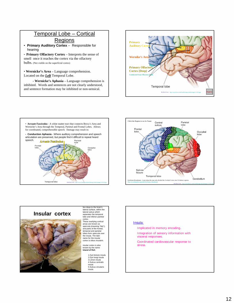
Temporal Lobe – Cortical Regions
• Primary Auditory Cortex – Responsible for hearing
• Primary Olfactory Cortex – Interprets the sense of smell once it reaches the cortex via the olfactory bulbs. (Not visible on the superficial cortex)
• Wernicke’s Area – Language comprehension. Located on the Left Temporal Lobe.
- Wernicke’s Aphasia – Language comprehension is inhibited. Words and sentences are not clearly understood, and sentence formation may be inhibited or non-sensical.
Primary Auditory Cortex
Wernike’s Area
Primary Olfactory Cortex (Deep)Conducted from Olfactory Bulb
RegionsModified from: http://www.bioon.com/book/biology/whole/image/1/1-8.tif.jpg
• Arcuate Fasciculus - A white matter tract that connects Broca’s Area and Wernicke’s Area through the Temporal, Parietal and Frontal Lobes. Allows for coordinated, comprehensible speech. Damage may result in:
- Conduction Aphasia - Where auditory comprehension and speech articulation are preserved, but people find it difficult to repeat heard speech.
Modified from: http://www.bioon.com/book/biology/whole/image/1/1-8.tif.jpg
Click the Region to see its Name
Korbinian Broadmann - Learn about the man who divided the Cerebral Cortex into 52 distinct regions: http://en.wikipedia.org/wiki/Korbinian_Brodmann
Modified from: http://www.bioon.com/book/biology/whole/image/1/1-8.tif.jpg
Insular cortex
1.Gyri breves insula2.Gyri longi insula3.Limen insula4.Sulcus centralisinsula5.Sulcus circularisinsula
lies deep to the brain's lateral surface, within the lateral sulcus which separates the temporal lobe and inferior parietal cortex. These overlying cortical areas are known as opercula (meaning "lids"), and parts of the frontal, temporal and parietal lobes form opercula over the insula. The latinname for the insular cortex is lobus insularis.
insular cortex is also known by the name Island of Reil,
Insula:Implicated in memory encoding.
Integration of sensory information with visceral responses.
Coordinated cardiovascular response to stress.

13
The insular cortex is a complex structure which contains areas that subserve•visceral sensory, •motor, •vestibular,• and somatosensory functions.
•The role of the insular cortex in auditory processing was poorly understood until recently. •However, recent case studies indicate that bilateral damage to the insulae may result in total auditory agnosia. •Functional imaging studies demonstrate that the insulae participate in several key auditory processes, such as allocating auditory attention and tuning in to novel auditory stimuli, temporal processing, phonological processing and visual-auditory integration. •These studies do not clarify the issue of further specialisation within the insular cortex, e.g. whether the posterior insulae are primarily sensory areas, while the anterior insulae serve mainly as integration/association auditory areas, two hypotheses that would be compatible with the cytoarchitectonic structure and connectivity of the insulae.
Primary Somatosensory Cortex• What does “somato”
mean?• Found in the
postcentral gyrus.• Neurons in this
cortical area receive info from sensory neurons in the skin and from proprioceptors which monitor joint position.
• Contralateral input.• How was the motor
somatotopic map arranged? – Do you think the
somatotopic map will be identical?
Somatosensory Association Cortex
• Found posterior to the primary somatosensory cortex and is neurally tied to it.
• Synthesizes multiple sensory inputs to create a complete comprehension of the object being felt.– How would damage to
this area differ from damage to the primary somatosensory cortex?
Primary Visual Cortex
• Found in the posterior and medial occipital lobe.
• Largest of the sensory cortices. – What does this
suggest?• Contralateral input.
Visual Association Area• Surrounds the primary
visual cortex.• Basically vision is the
sensation of bars of light on our retinal cells. The primary visual cortex tells which cells are being stimulated and how. The association area lets us “see” what we’re looking at.

14
Auditory Cortex• Found in the superior
margin of the temporal lobe, next to the lateral sulcus.
• Sound waves excite cochlear receptors in the inner ear which send info to the auditory cortex.
• There is also an auditory association area which lets us interpret and remember sounds.
Olfactory Cortex• Found in the frontal lobe
just above the orbits.• Receptors in the
olfactory epitheliumextend through the cribriform plate and are excited by the binding of oderants. They then send their info to the olfactory cortex.
• Very much involved in memory and emotion.
Gustatory and Vestibular Cortices
• Gustatory cortex is involved in taste and is in the parietal lobe just deep to the temporal lobe.
• Vestibular cortex is involved in balance and equilibrium and is in the posterior insula
Association Areas
• Allows for analysis of sensory input.
• Multiple inputs and outputs. Why?
1. Prefrontal cortex2. Language areas3. General interpretation area4. Visceral association area

15
Prefrontal Cortex
• Anterior frontal lobes• Involved in analysis,
cognition, thinking, personality, conscience, & much more.
• What would a frontal lobotomy result in?
• Look at its evolution
Language Areas• Large area for language understanding and production surrounding the lateral sulcus in the left (language-dominant) hemisphere
• Includes:– Wernicke’s area
understanding oral/written words
– Broca’s areaspeech production
– Lateral prefrontal cortex language comprehension and complex word analysis
– Lateral and ventral temporal cortexintegrates visual and auditory stimulate
General and Visceral Association Areas
• General area integrates multiple stimuli into a single cogent “understanding of the situation.”– Found on only one
hemisphere – typically left.
– Contained by 3 lobes: temporal, occipital, and parietal.
• Visceral association area is involved in perception of visceral sensations (such as disgust).– Located in insular cortex
Lateralization
• The fact that certain activities are the almost exclusive domain of one of the 2 hemispheres.
• In most people, the left hemisphere has a more control over language, math, and logic.
• While the right hemisphere is geared towards musical, artistic and other creative endeavors.
• Most individuals with left cerebral dominance are right-handed.
Cerebral White Matter
• Is white matter involved in communication?
• 3 types of fibers:1. Commissural –
connect corresponding areas of the 2 hemispheres. Largest is the corpus callosum.
2. Association fibers –connect different parts of the same hemisphere
3. Projection fibers –fibers entering and leaving the cerebral hemispheres from/to lower structures

16
Basal Nuclei
• Components of the extrapyramidal system which provides subconscious control of skeletal muscle tone and coordinates learned movement patterns and other somatic motor activities.
• Doesn’t initiate movements but once movement is underway, they assist in the pattern and rhythm (especially for trunk and proximal limb muscles
• Set of nuclei deep within the white matter.
• Includes the:– Caudate Nucleus– Lentiform Nucleus
• Globus pallidus• Putamen
Basal Nuclei• Info arrives at the caudate nucleus and the putamen from
sensory, motor, and association areas of the cortex.• Processing and integration occurs w/i the nuclei and then
info is sent from the globus pallidus to the motor cortex via the thalamus.
• The basal nuclei alter motor commands issued by the cerebral cortex via this feedback loop.
Parkinson’s Disease• Each side of the midbrain contains a nucleus called
the substantia nigra.• Neurons in the substantia nigra inhibit the activity of
basal nuclei by releasing dopamine.
Damage to SN neurons
Decrease in dopamine secretion
Increased activity of basal nuclei
Gradual increase in muscle tone
Appearance of symptoms of Parkinson’s disease: tremor, slow movement, inability to move, rigid
gait, reduced facial expression
Diencephalon• Forms the central
core of the forebrain
• 3 paired structures:1. Thalamus2. Hypothalamus3. Epithalamus
All 3 are gray matter
17
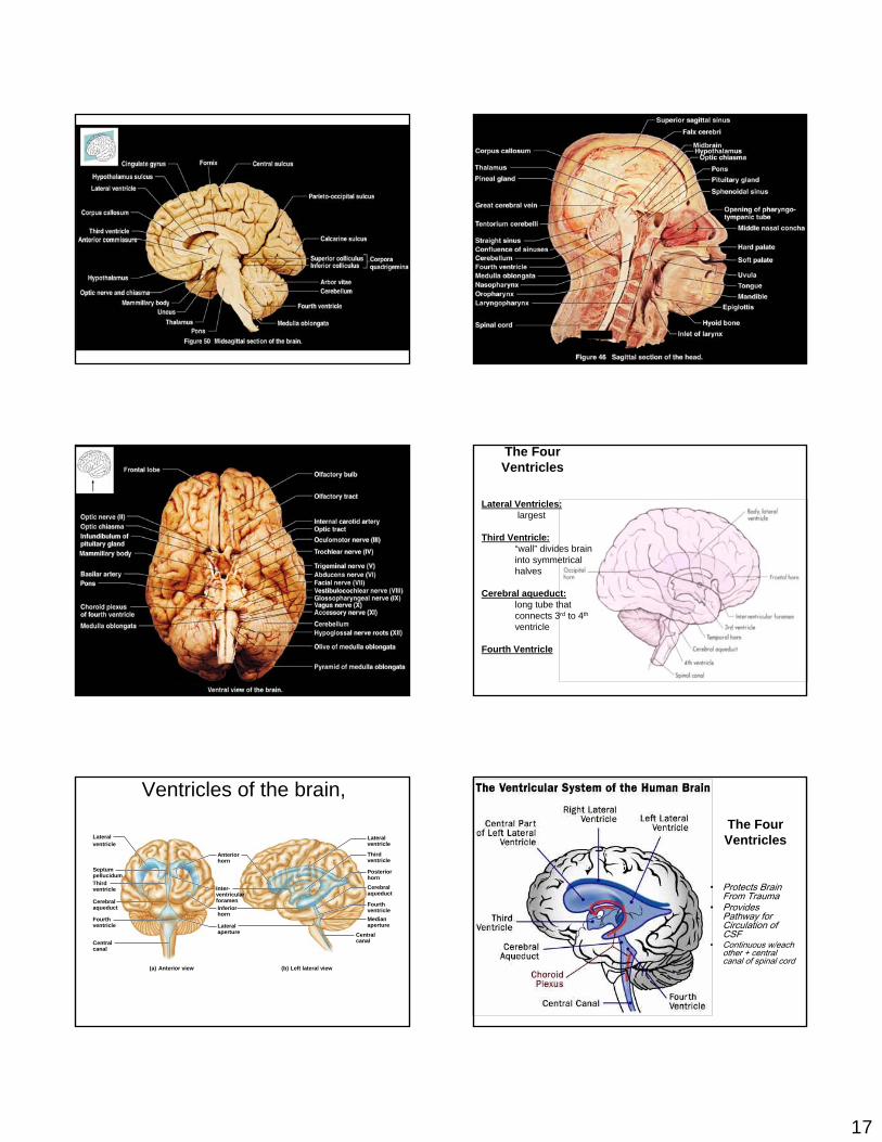
Lateral Ventricles:largest
Third Ventricle:“wall” divides brain into symmetrical halves
Cerebral aqueduct: long tube thatconnects 3rd to 4th
ventricle
Fourth Ventricle
The Four Ventricles
Ventricles of the brain,
(b)(a)
Lateralventricle
Posteriorhorn
Thirdventricle
Cerebralaqueduct
FourthventricleMedianaperture
Anteriorhorn
Inter-ventricularforamenInferiorhorn
Lateralaperture Central
canal
Lateralventricle
SeptumpellucidumThirdventricle
Cerebralaqueduct
Left lateral viewAnterior view
Fourthventricle
Centralcanal
The Four Ventricles
• Protects Brain From Trauma
• Provides Pathway for Circulation of CSF
• Continuous w/each other + central canal of spinal cord

18
Coronal Section Level of the LGB's
Types of fiber tracts in white matter,
(a)
(b)
Associationfibers
Thalamus and internal capsule
Corpus callosum(commissuralfibers)
Projection(internal capsule)fibers
Projectionfibers
Functional Brain System
• Networks of neurons working together and spanning wide areas of the brain
• The two systems are:– Limbic system– Reticular formation
Limbic System• Structures located on the medial aspects of
cerebral hemispheres and diencephalon• Includes the rhinencephalon, amygdala,
hypothalamus, and anterior nucleus of the thalamus

19
Limbic System
• Parts especially important in emotions:– Amygdala – deals with anger, danger, and
fear responses– Cingulate gyrus – plays a role in expressing
emotions via gestures, and resolves mental conflict
• Puts emotional responses to odors – e.g., skunks smell bad
Limbic System
Figure 12.18
Limbic System: Emotion and Cognition
• The limbic system interacts with the prefrontal lobes, therefore:
– One can react emotionally to conscious understandings
– One is consciously aware of emotion in one’s life
• Hippocampal structures – convert new information into long-term memories
Reticular Formation
• Composed of three broad columns along the length of the brain stem
– Raphe nuclei– Medial (large cell) group– Lateral (small cell) group
• Has far-flung axonal connections with hypothalamus, thalamus, cerebellum, and spinal cord
Reticular Formation
Figure 12.19
DIENCEPHALON
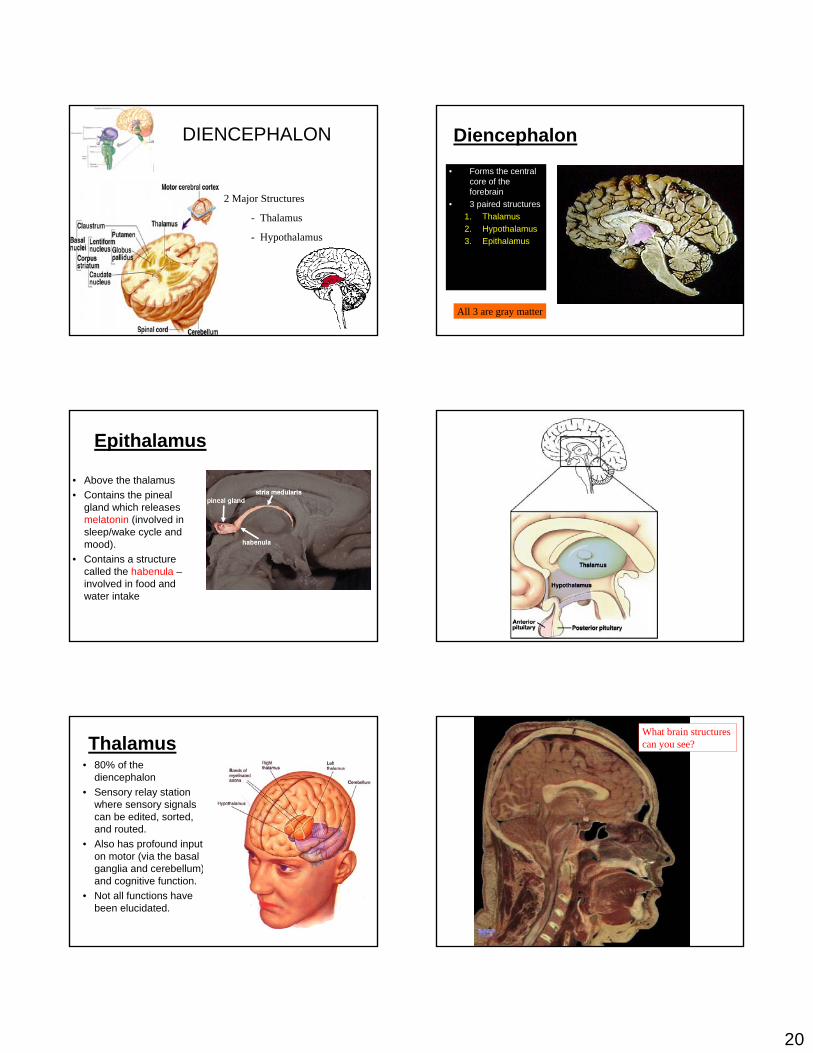
20
DIENCEPHALON
2 Major Structures
- Thalamus
- Hypothalamus
Diencephalon• Forms the central
core of the forebrain
• 3 paired structures:1. Thalamus2. Hypothalamus3. Epithalamus
All 3 are gray matter
Epithalamus
• Above the thalamus• Contains the pineal
gland which releases melatonin (involved in sleep/wake cycle and mood).
• Contains a structure called the habenula –involved in food and water intake
Thalamus• 80% of the
diencephalon • Sensory relay station
where sensory signals can be edited, sorted, and routed.
• Also has profound input on motor (via the basal ganglia and cerebellum) and cognitive function.
• Not all functions have been elucidated.
What brain structures can you see?

21
Midsagittal section of the brain illustrating the diencephalon and brain stem
Parietal lobe ofcerebral hemisphereCorpus callosum
Choroid plexusOccipital lobe ofcerebral hemisphereThalamus(encloses third ventricle)
Pineal body/gland(part of epithalamus)
Posterior commissure
CorporaquadrigeminaCerebralaqueduct
Arbor vitaeFourth ventricleChoroid plexusCerebellum
Septum pellucidum
Interthalamicadhesion(intermediatemass of thalamus)Frontal lobeof cerebralhemisphereInterventricularforamenAnteriorcommissure
HypothalamusOptic chiasmaPituitary gland
Temporal lobe ofcerebral hemisphere Mammillary body
PonsMedulla oblongataSpinal cord
Midbrain
Fornix
(a) (b)
Dorsal nuclei
Ventral nuclei
Medial
Anteriornucleargroup
Reticularnucleus
Ventralanterior
Ventrallateral
Ventralposteriorlateral
Lateralgeniculatebody
Medialgeniculatebody
Pulvinar
Lateraldorsal
Lateralposterior
Preopticnucleus
SupraopticnucleusSuprachiasmaticnucleus
Anteriorhypothalamicnucleus
Dorsomedialnucleus
Paraventricularnucleus
FornixAnteriorcommissure
PosteriorhypothalamicnucleusLateralhypothalamicareaVentromedialnucleus
OpticchiasmaInfundibulum(stalk of thepituitary gland)
Pituitarygland
Arcuatenucleus
Mammillarybody
Thalamus- Two lobes that relay sensory projection fiber info to the cerebral cortex
Hypothalamus
- Lies at the base of the brain
- Controls and regulates the endocrine system (hormones), autonomic system, species survival (the four Fs) and sleeping.
- Contains many nuclei and fiber tracts
• All sensory modalities relay through the thalamus
Thalamus Thalamus – “gateway” to the cerbral cortexAfferent impulses from all senses converge and
synapse in the thalamusImpulses of similar function are sorted out, “edited”, and relayed as a group to the appropriate area of the sensory cortex or association areasAll inputs ascending to the cerebral cortex pass through the thalamusPlays a key role in mediating sensation, motor activities, cortical arousal, learning, and memory
Hypothalamus• Functions:
– Autonomic regulatory center• Influences HR, BP, resp. rate,
GI motility, pupillary diameter.• Can you hold your
breath until you die?– Emotional response
• Involved in fear, loathing, pleasure• Drive center: sex, hunger
– Regulation of body temperature– Regulation of food intake
• Contains a satiety center– Regulation of water balance and thirst– Regulation of sleep/wake cycles– Hormonal control
• Releases hormones that influence hormonal secretion from the anterior pituitary gland.
• Releases oxytocin and vasopressin
HypothalamusBelow the thalamus, it caps the brainstem and forms the inferolateral walls of the third ventricleMammillary bodies - small, paired nuclei bulging anteriorly from the hypothalamus - relay stations for olfactory pathwaysInfundibulum – stalk of the hypothalamus connecting to the pituitary gland
Main visceral control center of the body, important to overall body homeostasis

22
Hypothalamus
• A group of nuclei critical for regulating homeostasis, the four Fs, and hormones
Hypothalamic NucleiHypothalamic Nuclei
Hypothalamic FunctionRegulates blood pressure, rate and force of heartbeat, digestive tract motility, respiratory rate and depth, pupil size, and many other visceral activitiesCenter for emotional response - involved in perception of pleasure, fear, rageRegulates body temperature – the body’s “thermostat”Regulates food intake - feelings of hunger and satiety Regulates sleep-wake cycle
Regulates ANS by controlling activity of centers in brains stem and spinal cord
Endocrine Functions of the HypothalamusEndocrine Functions of the HypothalamusReleasing hormones control the secretion of hormones by the anterior pituitary
Stimulates ADH release from the posterior pituitary
Anti-diuretic hormone- causes kidneys to retain water
Types of fiber tracts in white matter,
(c)
Coronaradiata
Projectionfibers
LongitudinalfissureGray matterWhite matterLateralventricle
Fornix
Thirdventricle
Thalamus
Pons
MedullaoblongataDecussation
of pyramids
Corpus callosum(commissuralfibers)
Basal nuclei(ganglia)
Internalcapsule
Superior
Caudate
Globus pallidus
Putamen
Basal nuclei
(b)
Corpus callosumAnterior hornof lateral ventricleCaudate nucleusThird ventricle
Putamen LentiformnucleusGlobus pallidus
Thalamus
Cerebral cortex
Cerebral white matter
Anterior
Posterior
Inferior hornof lateral ventricle
Basal nuclei,
(a)
Fibers ofcorona radiata
Corpusstriatum
Internal capsule(projection fibersrun deep to lentiform nucleus)
Caudatenucleus
Lentiformnucleus
Thalamus
Tail of caudatenucleus
23
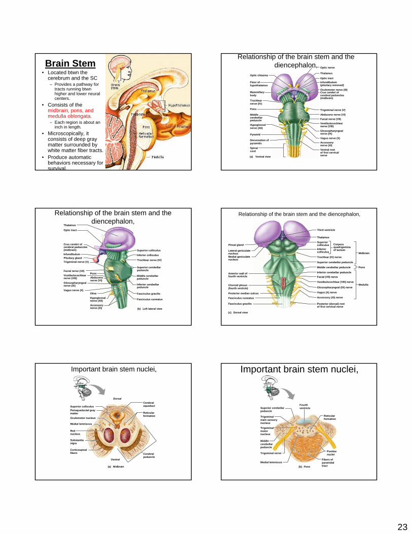
Brain Stem• Located btwn the
cerebrum and the SC– Provides a pathway for
tracts running btwn higher and lower neural centers.
• Consists of the midbrain, pons, and medulla oblongata.– Each region is about an
inch in length.• Microscopically, it
consists of deep gray matter surrounded by white matter fiber tracts.
• Produce automatic behaviors necessary for survival.
Relationship of the brain stem and the diencephalon,
(a)
Optic chiasma
Floor of hypothalamus
Mammillarybody
Trochlearnerve (IV)
Pons
Middle cerebellarpeduncleHypoglossal nerve (XII)
Pyramid
Decussation ofpyramids
Spinalcord
Optic nerve
Thalamus
Optic tractInfundibulum(pituitary removed)
Oculomotor nerve (III)Crus cerebri of cerebral peduncles (midbrain)
Trigeminal nerve (V)
Abducens nerve (VI)Facial nerve (VII)Vestibulocochlearnerve (VIII)
Glossopharyngealnerve (IX)Vagus nerve (X)Accessory nerve (XI)Ventral rootof first cervicalnerveVentral view
Relationship of the brain stem and the diencephalon,
(b)
Thalamus
Optic tract
Crus cerebri of cerebral peduncles (midbrain)
Trigeminal nerve (V)
Abducensnerve (VI)
Facial nerve (VII)Vestibulocochlearnerve (VIII)
Glossopharyngealnerve (IX)
Vagus nerve (X)
InfundibulumPituitary gland
Pons
OliveHypoglossalnerve (XII)Accessory nerve (XI)
Superior colliculus
Inferior colliculus
Trochlear nerve (IV)
Superior cerebellarpeduncle
Middle cerebellarpeduncle
Inferior cerebellarpeduncle
Fasciculus gracilis
Fasciculus cuneatus
Left lateral view
Relationship of the brain stem and the diencephalon,
(c)
Posterior median sulcus
Choroid plexus(fourth venticle)
Pineal gland
Lateral geniculatenucleusMedial geniculatenucleus
Anterior wall offourth ventricle
Fasciculus cuneatus
Fasciculus gracilis
Third ventricle
Thalamus
Superiorcolliculus
MidbrainInferiorcolliculus
Trochlear (IV) nerve
Superior cerebellar peduncle
Middle cerebellar peduncle Pons
Inferior cerebellar peduncleFacial (VII) nerve
Vestibulocochlear (VIII) nerve
Glossopharyngeal (IX) nerve
Vagus (X) nerve
Accessory (XI) nerve
Posterior (dorsal) rootof first cervical nerve
Medulla
Corporaquadrigeminaof tectum
Dorsal view
(a)
DorsalCerebralaqueduct
Reticularformation
Cerebralpeduncle
Ventral
Corticospinalfibers
Substantianigra
Red nucleus
Medial lemniscus
Oculomotor nucleus
Periaqueductal graymatter
Superior colliculus
Midbrain
Important brain stem nuclei, Important brain stem nuclei,
(b)
Reticularformation
Trigeminal nervePontinenuclei
Fibers of pyramidal tract
Middle cerebellarpeduncle
Trigeminalmain sensorynucleus
Trigeminal motor nucleus
Superior cerebellarpeduncle
Medial lemniscus
Fourthventricle
Pons

24
Pons:Connects other parts.
several nuclei associated with cranial nerves
respiratory centers.
Cerebellum:“little brain”
Receives input from proprioceptors (joints, muscles, tendons).
Refinement/coordination of movement.
Reticular formation -1 - portions located in the spinal cord, medulla, pons, midbrain, & hypothalamus
2 - needed for arousal from sleep & to maintain consciousness
Cerebellum -1 - functions in coordination, maintenance of posture, & balance
Cerebrum -1 - largest portion of the human brain
2 - consists of 2 hemispheres divided by a fissure
3 - includes cerebral cortex, medullary body, & basal ganglia: Cortex:
outer 2 - 4 mm of the cerebrum consists of gray matter (cell bodies & synapses; no myelin)
'folded', with upfolded areas called gyri & depressions or grooves called sulciconsists of four primary lobes
Medullary body: the 'white matter' of the cerebrum; consists of
myelinated axons types of axons include:
commissural fibers - conduct impulses between cerebral hemispheres (and form the corpus
callosum)
projection fibers - conduct impulses in & out of the cerebral hemispheresassociation fibers - conduct impulses within hemispheres
Basal ganglia: masses of gray matter in each cerebral hemisphere
important in control of voluntary muscle movements
Limbic System -1 - consists of a group of nuclei + fiber tracts
2 - located in part in cerebral cortex, thalamus, & hypothalamus 3 - Functions:
aggression fear
feeding sex (regulation of sexual drive & sexual behavior)
CEREBELLUM

25
Cerebellum,
(a)(b)
(c) (d)
AnteriorlobePrimaryfissure
Posteriorlobe
Vermis
Horizontalfissure
Vermis
White matterof cerebellum
Deep cerebellarnuclei Granule cells
in granular layer
Purkinjecells
Site of basket cellsand stellate cells inouter cortex (molecular layer)
Caudal(inferior)
Brain stem(midbrain)Cerebellarcortex
Vermis(cut)
Cerebellum• Lies inferior to the cerebrum and
occupies the posterior cranial fossa.
• 2nd largest region of the brain.• 10% of the brain by volume, but it
contains 50% of its neurons• Has 2 primary functions:
1. Adjusting the postural muscles of the body• Coordinates rapid, automatic adjustments, that maintain balance and
equilibrium2. Programming and fine-tuning movements controlled at the subconscious
and conscious levels• Refines learned movement patterns by regulating activity of both the
pyramidal and extrapyarmidal motor pathways of the cerebral cortex• Compares motor commands with sensory info from muscles and joints
and performs any adjustments to make the movement smooth
Do you see the cerebellum? What else can you see?
Cerebellum• Has a complex, convoluted
cortical surface with multiple folds (folia) which are less prominent than the gyri of the cerebrum.
• Has anterior and posterior lobes separated by the primary fissure.
• Along the midline, a narrow band of cortex called the vermis separates the cerebellar hemispheres.
• The floccunodular lobe lies anterior to the vermis and btwn the cerebellar hemispheres.
Cerebellum
• Cerebellar cortex contains huge, highly branched Purkinje cellswhose extensive dendrites can receive up to 200,000 synapses.
• Internally, the white matter forms a branching array that in a sectional view resembles a tree – for this reason, it’s called the arbor vitae

26
Cerebellum• Tracts that link the cerebellum w/ the brain
stem, cerebrum, and spinal cord leave the cerebellar hemispheres as the superior, middle, and inferior cerebellar peduncles.– SCP carries instructions from cerebellar
nuclei to the cerebral cortex via midbrain and thalamus
– MCP connects pontine nuclei to the cerebellum. This info ultimately came from the cerebral cortex and informs the cerebellum of voluntary motor activities
– ICP connects the cerebellum and the medulla oblongata and carries sensory information from muscles and from the vestibular apparatus of the inner ear.
Cerebellum
• The cerebellum can be permanently damaged by trauma or stroke or temporarily affected by drugs such as alcohol.
• These alterations can produce ataxia – a disturbance in balance.
LIMBIC SYSTEM
Midbrain
• Located btwn the diencephalon and the pons.– 2 bulging cerebral peduncles on
the ventral side. These contain:• Descending fibers that go to
the cerebellum via the pons• Descending pyramidal tracts
– Running thru the midbrain is the hollow cerebral aqueduct which connects the 3rd and 4th
ventricles of the brain.– The roof of the aqueduct ( the
tectum) contains the corpora quadrigemina
• 2 superior colliculi that control reflex movements of the eyes, head and neck in response to visual stimuli
• 2 inferior colliculi that control reflex movements of the head, neck, and trunk in response to auditory stimuli
•Cranial nerves 3&4 (oculomotor and trochlear) exit from the midbrain
•Midbrain also contains the headquarters of the reticular activating system
Midbrain• On each side, the midbrain
contains a red nucleus and a substantia nigra– Red nucleus contains
numerous blood vessels and receives info from the cerebrum and cerebellum and issues subconscious motor commands concerned w/ muscle tone & posture
– Lateral to the red nucleus is the melanin-containing substantia nigra which secretes dopamine to inhibit the excitatory neurons of the basal nuclei.
• Damage to the substantia nigra would cause what?
27
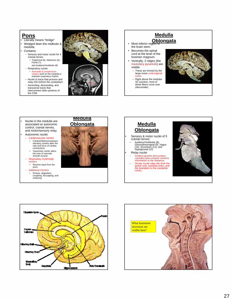
Pons• Literally means “bridge”• Wedged btwn the midbrain &
medulla. • Contains:
– Sensory and motor nuclei for 4 cranial nerves
• Trigeminal (5), Abducens (6), Facial (7), and Auditory/Vestibular (8)
– Respiratory nuclei:• Apneustic & pneumotaxic
centers work w/ the medulla to maintain respiratory rhythm
– Nuclei & tracts that process and relay info to/from the cerebellum
– Ascending, descending, and transverse tracts that interconnect other portions of the CNS
Medulla Oblongata
• Most inferior region of the brain stem.
• Becomes the spinal cord at the level of the foramen magnum.
• Ventrally, 2 ridges (the medullary pyramids) are visible. – These are formed by the
large motor corticospinal tracts.
– Right above the medulla-SC junction, most of these fibers cross-over (decussate).
Medulla Oblongata
• Nuclei in the medulla are associated w/ autonomic control, cranial nerves, and motor/sensory relay.
• Autonomic nuclei:– Cardiovascular centers
• Cardioinhibitory/cardioacceleratory centers alter the rate and force of cardiac contractions
• Vasomotor center alters the tone of vascular smooth muscle
– Respiratory rhythmicity centers• Receive input from the
pons– Additional Centers
• Emesis, deglutition, coughing, hiccupping, and sneezing
Medulla Oblongata
• Sensory & motor nuclei of 5 cranial nerves:
– Auditory/Vestibular (8), Glossopharyngeal (9), Vagus (10), Accessory (11), and Hypoglossal (12)
• Relay nuclei– Nucleus gracilis and nucleus
cuneatus pass somatic sensory information to the thalamus
– Olivary nuclei relay info from the spinal cord, cerebral cortex, and the brainstem to the cerebellar cortex.
What brainstem structures are visible here?

28
Limbic System
• Includes nuclei and tracts along the border btwn the cerebrum and the diencephalon.
• Functional grouping rather than anatomical
• Functions include:1. Establishing emotional states2. Linking conscious cerebral cortical
functions w/ unconscious functions of the brainstem
3. Facilitating memory storage and retrieval
• Limbic lobe of the cerebrum consists of 3 gyri that curve along the corpus callosum and medial surface of the temporal lobe.
• Limbic system the center of emotion – anger, fear, sexual arousal, pleasure, and sadness.
Reticular Formation
• Extensive network of neurons that runs thru the medulla and projects to thalamic nuclei that influence large areas of the cerebral cortex.– Midbrain portion of RAS most
likely is its center• Functions as a net or filter
for sensory input.– Filter out repetitive stimuli.
Such as?– Allows passage of infrequent
or important stimuli to reach the cerebral cortex.
– Unless inhibited by other brain regions, it activates the cerebral cortex – keeping it alert and awake.
How might the “sleep centers” of your brain work? Why does alcohol make you tired?
The limbic system,
Cingulategyrus Corpus
callosumFornix
Anteriorthalamic nuclei(flanking3rd ventricle)Amygdala
Parahippocampalgyrus
Septumpellucidum
Septal nuclei
Hypothalamus
Anterior pellucidum
Olfactory bulb
Mammillary body
Hippocampus
Dentate gyrus(deep to theparahippocampalgyrus)
The reticular formation,
Visualimpulses
Reticular formation
Ascending generalsensory tracts(touch, pain, temperature)
Descendingmotor projectionsto spinal cord
Auditoryimpulses
Radiationsto cerebralcortex
Memory processing, Outside stimuli
General and special sensory receptors
Short-termmemory
Data transferinfluenced by:ExcitementRehearsalAssociation ofold and new data
Long-termmemory
Data permanently lost
Afferent inputs
Retrieval
Forget
ForgetData selectedfor transferAutomatic
memory
Data unretrievable
Temporary storage(buffer) in cerebral cortex
Proposed memory circuits,
(a) (b)
BasalforebrainPrefrontalcortex
Smell Taste
Thalamus
Touch
Hearing
Vision
Hippocampus
Thalamus
ThalamusPremotorcortex
Basalforebrain
Substantianigra
Prefrontalcortex
Associationcortex
Dopamine
Associationcortex
Basalnuclei
Sensory andmotor inputs
Medial temporal lobe(hippocampus, etc.)
Premotorcortex
Thalamus
Substantianigra
Basalnuclei
Sensoryinput
AChACh

29
CSFCerebrospinal Fluid (CSF)
Watery solution similar in composition to blood plasma
Contains less protein and different ion concentrations than
plasma
Forms a liquid cushion that gives buoyancy to the CNS organs
Prevents the brain from crushing under its own weight
Protects the CNS from blows and other trauma
Nourishes the brain and carries chemical signals throughout it
HYDROCEPHALUS WHEN CSF DO NOT CIRCULATE
INCREASING ITS PRESSURE
Formation, location, and circulation of CSF,
Ependymalcells
Superiorsagittal sinus
ArachnoidvillusSubarachnoid spaceArachnoid materMeningeal dura materPeriosteal dura materGreat cerebral vein
Tentorium cerebelliStraight sinusConfluence of sinuses
CerebellumChoroid plexusCerebral vesselsthat supplychoroid plexusCentral canalof spinal cordSpinal dura mater
Inferior end ofspinal cord
Filum terminale(inferior endof pia mater)
Superiorcerebral veinChoroid plexusCerebrum coveredwith pia materSeptumpellucidumCorpuscallosumInterventricularforamenThird ventriclePituitary gland
Cerebral aqueductLateral apertureFourth ventricleMedian aperture
Capillary
Connectivetissue ofpia mater
Wastes andunnecessarysolutes absorbed
Filtrate containing glucose,oxygen, vitamins, andions (Na+, Cl–, Mg2+, etc.)
Sectionof choroidplexus
Cavity ofventricle
Formation, location, and circulation of CSF, Superiorsagittal sinus
ArachnoidvillusSubarachnoid spaceArachnoid materMeningeal dura materPeriosteal dura materGreat cerebral vein
Tentorium cerebelliStraight sinusConfluence of sinuses
CerebellumChoroid plexusCerebral vesselsthat supplychoroid plexus
Central canalof spinal cordSpinal dura mater
Inferior end ofspinal cord
Filum terminale(inferior endof pia mater)
Superiorcerebral veinChoroid plexusCerebrum coveredwith pia materSeptumpellucidumCorpuscallosumInterventricularforamenThird ventriclePituitary gland
Cerebral aqueductLateral apertureFourth ventricleMedian aperture
(b)
•It produces the cerebrospinal fluid (CSF) which is found within the ventricles of the brain and in the subarachnoid space around the brain and spinal cord.
•It is comprised of a rich capillary bed, pia mater, and choroid epithelial cells.•It is located in certain parts of the ventricular system of the brain.
Choroid PlexusesClusters of capillaries that
form tissue fluid filters, which hang from the roof of each
ventricleHave ion pumps that allow
them to alter ion concentrations of the CSF
Help cleanse CSF by removing wastes
Ventral aspect of the human brain, showing the three regions of the brain stem,
Frontal lobe
Olfactory bulb(synapse pointof cranial nerve I)
Optic chiasma
Optic nerve (II)
Optic tract
Mammillary body
Pons
Temporal lobe
Medulla
Cerebellum
Spinal cord
Midbrain
30
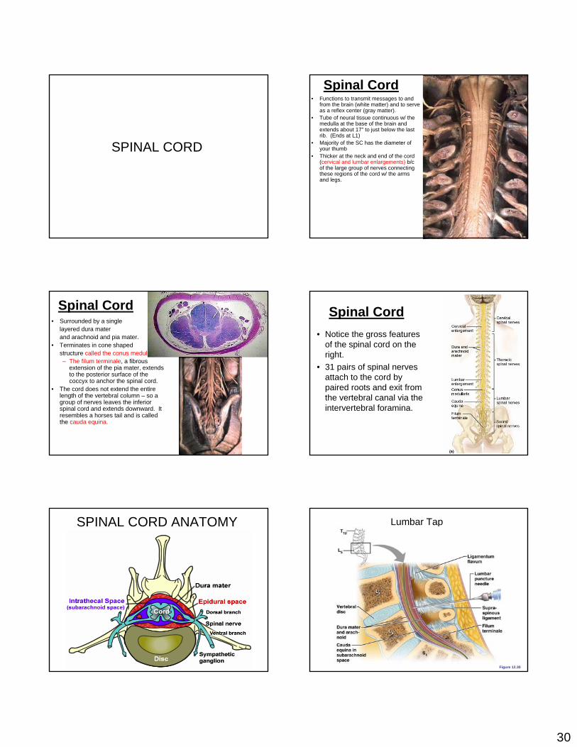
SPINAL CORD
Spinal Cord• Functions to transmit messages to and
from the brain (white matter) and to serve as a reflex center (gray matter).
• Tube of neural tissue continuous w/ the medulla at the base of the brain and extends about 17” to just below the last rib. (Ends at L1)
• Majority of the SC has the diameter of your thumb
• Thicker at the neck and end of the cord (cervical and lumbar enlargements) b/c of the large group of nerves connecting these regions of the cord w/ the arms and legs.
Spinal Cord• Surrounded by a single
layered dura mater and arachnoid and pia mater.
• Terminates in cone shaped structure called the conus medullaris.– The filum terminale, a fibrous
extension of the pia mater, extends to the posterior surface of the coccyx to anchor the spinal cord.
• The cord does not extend the entire length of the vertebral column – so a group of nerves leaves the inferior spinal cord and extends downward. It resembles a horses tail and is called the cauda equina.
Spinal Cord• Notice the gross features
of the spinal cord on the right.
• 31 pairs of spinal nerves attach to the cord by paired roots and exit from the vertebral canal via the intervertebral foramina.
SPINAL CORD ANATOMY Lumbar Tap
Figure 12.30
31
Spinal Cord
Figure 12.29a
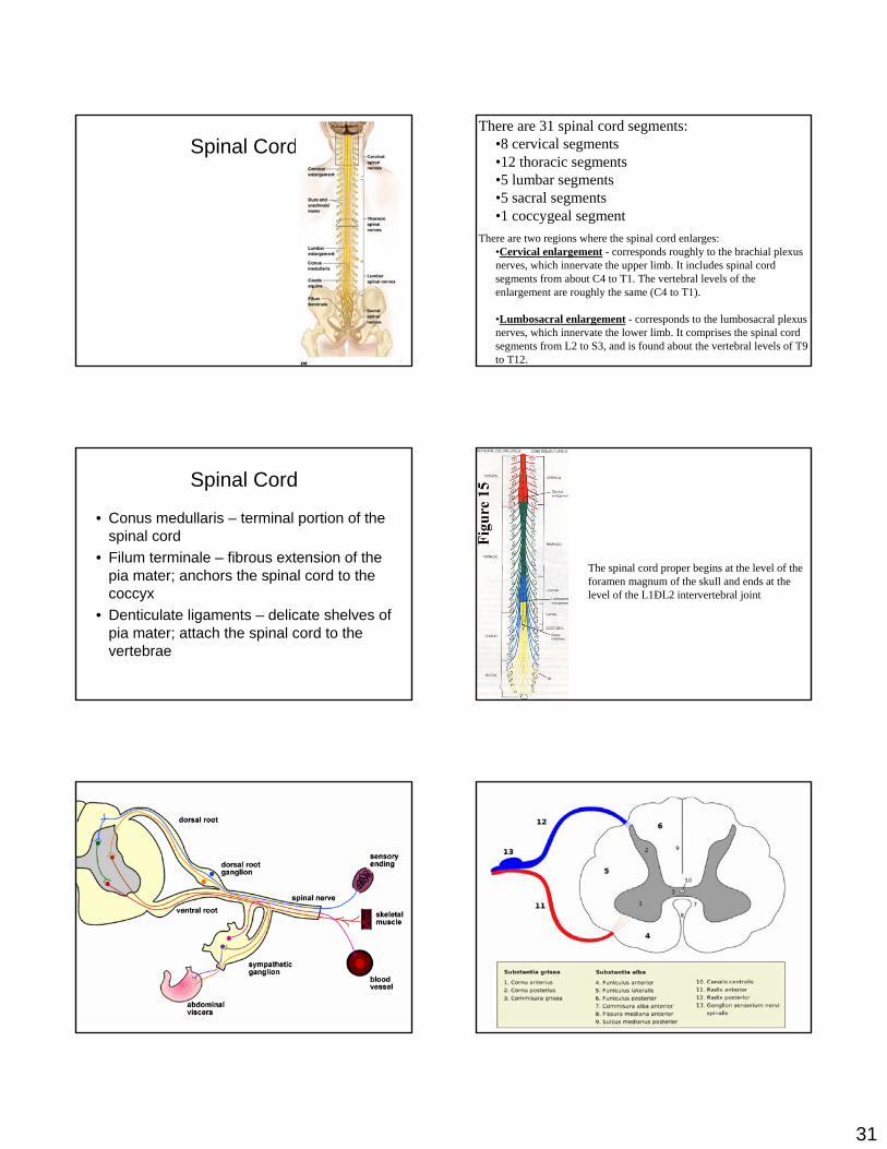
There are 31 spinal cord segments:•8 cervical segments•12 thoracic segments•5 lumbar segments•5 sacral segments•1 coccygeal segment
There are two regions where the spinal cord enlarges:•Cervical enlargement - corresponds roughly to the brachial plexus nerves, which innervate the upper limb. It includes spinal cord segments from about C4 to T1. The vertebral levels of the enlargement are roughly the same (C4 to T1).
•Lumbosacral enlargement - corresponds to the lumbosacral plexus nerves, which innervate the lower limb. It comprises the spinal cord segments from L2 to S3, and is found about the vertebral levels of T9 to T12.
Spinal Cord
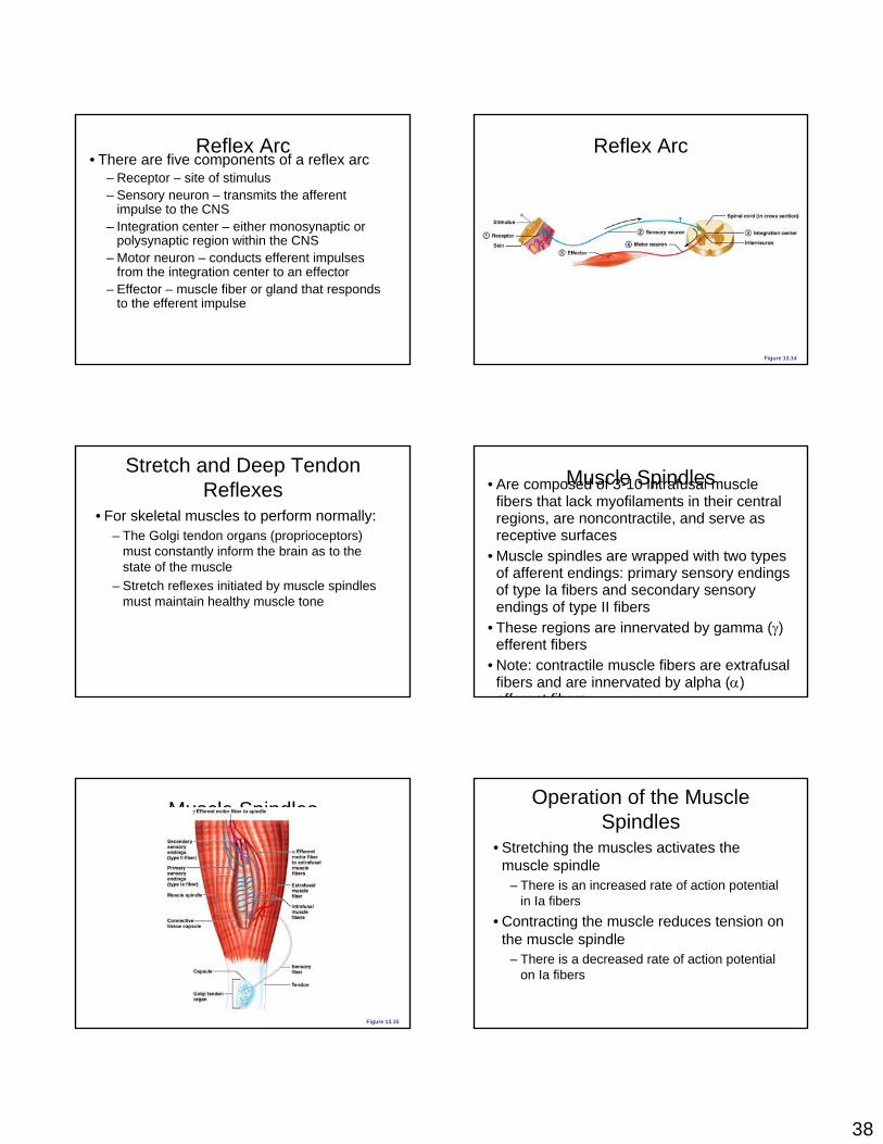
• Conus medullaris – terminal portion of the spinal cord
• Filum terminale – fibrous extension of the pia mater; anchors the spinal cord to the coccyx
• Denticulate ligaments – delicate shelves of pia mater; attach the spinal cord to the vertebrae
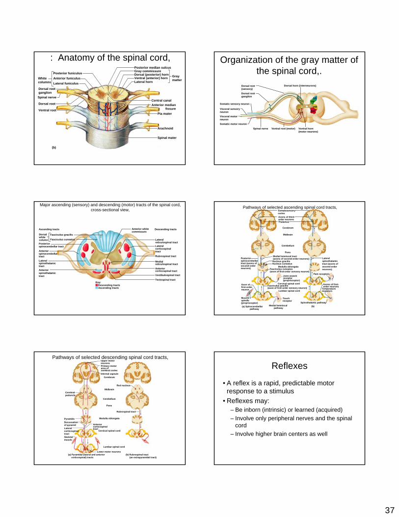
The spinal cord proper begins at the level of the foramen magnum of the skull and ends at the level of the L1ÐL2 intervertebral joint

32

33
a pia mater b subarachnoid space c dura mater d myelinatedaxon e unipolar neuron of the dorsal root ganglion surrounded by satellite cells (neuroglia).
a Pia mater b Subarachnoid space filled with cerebral spinal fluid, wastes and various cells. c Fibrocyte mixed in the blue collagen fibers of the dura mater. d Nucleus & nucleolus of unipolar neuron e Nucleus of one of many tiny satellite cells surrounding the large unipolar neuron. f Myelinated axon g Node of Ranvier h Nucleus of white Schwann cell
a Synaptic bulbs over the motor end plate -neuromuscular junction b Neuron axon terminal - black fibers
The central canal is the cerebrospinal fluid-filled space that runs longitudinally through the length of the entire spinal cord. The central canal is contiguous with the ventricular system of the brain.
The PNS is separated into 2 divisions:
1. the afferent division, which carries sensory information from sensory receptors of the PNS to the CNS. Receptors include neurons or specialized cellsthat detect changes or respond to stimuli, and complex sensory organs such as the eyes and ears.
2. the efferent division, which carries motor commands from the CNS to muscles and glands of the PNS. The cells or organs that respond to efferent signals by doing something are called effectors

34
The efferent division is divided into 2 parts:1. the somatic nervous system (SNS), which controls skeletal muscle contractionsa. voluntary muscle contractionsb. involuntary muscle contractions (reflexes)
2. the autonomic nervous system (ANS), which controls subconscious actions such as contractions of smooth muscle and cardiac muscle, and glandular secretions.
The ANS is separated into 2 divisions:1. the sympathetic division, which has a stimulating effect2. the parasympathetic division, which has a relaxing effect
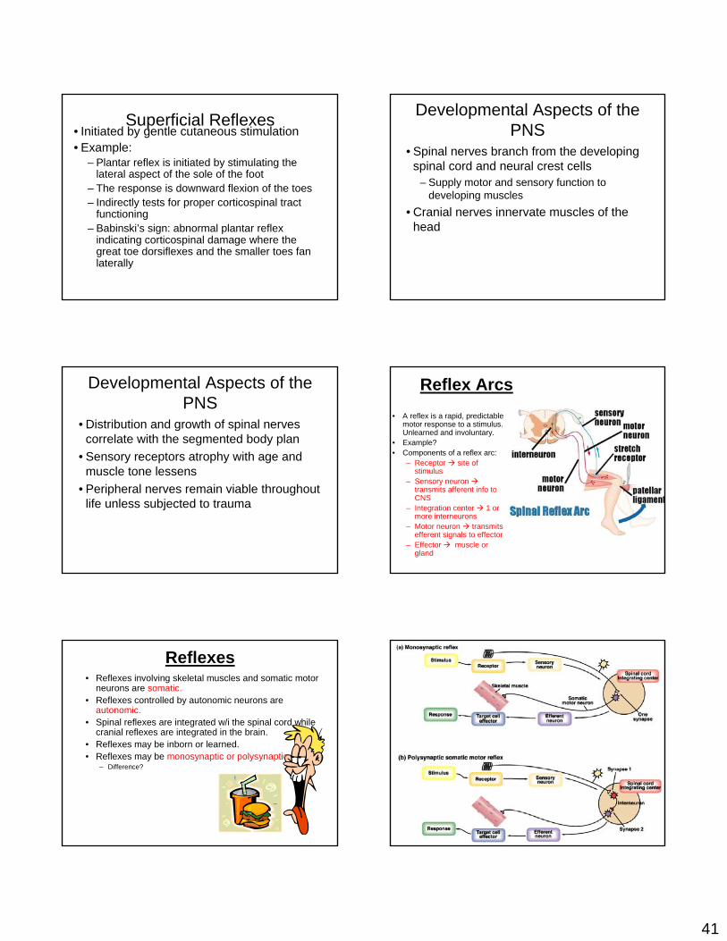
Cross Sectional Anatomy of the Spinal Cord
• Flattened from front to back.• Anterior median fissure and posterior median sulcus
partially divide it into left and right halves.• Gray matter is in the core of the cord and surrounded
by white matter.
• Resembles a butterfly.• 2 lateral gray masses connected by the gray
commissure.• Posterior projections are the posterior or dorsal horns.• Anterior projections are the anterior or ventral horns.• In the thoracic and lumbar cord, there also exist lateral horns.
Gray Matter• Posterior horns contain
interneurons.• Anterior horns contain some • interneurons as well as the cell
bodies of motor neurons.– These cell bodies project their
axons via the ventral roots of the spinal cord to the skeletal muscles.
– The amount of ventral gray matter at a given level of the spinal cord is proportional to the amount of skeletal muscle innervated.
Gray Matter• Lateral horn neurons are
sympathetic motor neurons serving visceral organs.– Their axons also exit via the
ventral root.• Afferent sensory fibers
carrying info from peripheral receptors form the dorsal roots of the spinal cord. The somata of these sensory fibers are found in an enlargement known as a dorsal root ganglion.
• The dorsal and ventral roots fuse to form spinal nerves.

35
White Matter• Myelinated nerve fibers.• Allows for communication btwn the brain and spinal cord or btwn different
regions of the spinal cord.• White matter on each side of the cord is divided into columns or funiculi.
– Typically, they are ascending or descending.• What does that mean?
Spinal Nerves• 31 nerves connecting the spinal
cord and various body regions.• 8 paired cervical nerves• 12 paired thoracic
nerves• 5 paired lumbar nerves• 5 paired sacral nerves• 1 pair of coccygeal
nerves
Spinal Nerves
• Each connects to the spinal cord by 2 roots – dorsal and ventral.
• Each root forms from a series of rootlets that attach along the whole length of the spinal cord segment.
• Ventral roots are motor while dorsal roots are sensory.
Spinal Nerves• The 2 roots join to
form a spinal nerve prior to exiting the vertebral column.
• Roots are short and horizontal in the cervical and thoracic regions while they are longer and more horizontal in the sacral and lumbar regions.
• Almost immediately after emerging from its intervertebral foramen, a spinal nerve will divide into a dorsal ramus, a ventral ramus, and a meningeal branch that reenters and innervates the meninges and associated blood vessels.
• Each ramus is mixed.• Joined to the base of the ventral rami of spinal nerves in the
thoracic region are the rami communicantes. These are sympathetic fibers that we’ll deal with shortly.
• Dorsal rami supply the posterior body trunk whereas the thicker ventral rami supply the rest of the body trunk and the limbs.

36
Plexuses• Except for T2 to T12,
all ventral rami branch extensively and join one another lateral to the vertebral column forming complicated nerve plexuses.
• W/i a plexus, fibers from different rami crisscross each other and become redistributed.
Sacral Plexus
Gross structure of the spinal cord, posterior view,
(b)
Terminus ofmedullaoblongataof brain
Dura mater
Sectionedpedicles ofcervicalvertebrae
Spinal roots
Posteriormedian sulcusof spinal cord
Gross structure of the spinal cord, posterior view,
(c)
Spinal cord
Denticulateligament
Arachnoidmater
Vertebralarch
Denticulateligament
Dorsal root
Dura mater
Posteriormediansulcus
Gross structure of the spinal cord, posterior view,
(d)
First lumbarvertebral arch(cut across)
Spinousprocess of second lumbarvertebra
Spinal cord Caudaequina
Conusmedullaris
Filumterminale
Diagrammatic view of a lumbar tap,
L5
L4
L5
Ligamentumflavum
Supra-spinousligament
Lumbarpunctureneedle
Filumterminale
Vertebraldisc
Caudaequina insubarachnoidspace
Dura materand arach-noid
T12
L5
S1
L4
Anatomy of the spinal cord,
(a)
Epidural space(contains fat)
Pia materSpinal meningesArachnoid
Dura mater
Bone ofvertebra
Subdural space
Subarachnoidspace
Dorsal rootganglion
Bodyof vertebra
37
: Anatomy of the spinal cord,
(b)
Posterior funiculus
Posterior median sulcus
Central canalAnterior median
fissurePia mater
Arachnoid
Spinal mater
Gray commissureDorsal (posterior) horn Gray
matterLateral hornVentral (anterior) hornAnterior funiculus
Lateral funiculusWhitecolumns
Dorsal rootganglion
Dorsal root
Ventral root
Spinal nerve
Organization of the gray matter of the spinal cord,.
Somatic sensory neuron
Dorsal root(sensory)
Dorsal rootganglion
Visceral sensoryneuron
Somatic motor neuron
Spinal nerve Ventral root (motor) Ventral horn(motor neurons)
Dorsal horn (interneurons)
Visceral motorneuron
SSVS
VMSM
Major ascending (sensory) and descending (motor) tracts of the spinal cord, cross-sectional view,
Ascending tracts Descending tracts
Fasciculus gracilisDorsalwhitecolumn Fasciculus cuneatusPosteriorspinocerebellar tract
LateralspinothalamictractAnteriorspinothalamictract
Anterior white commissure
Lateralcorticospinaltract
Lateralreticulospinal tract
Anteriorcorticospinal tract
Ascending tractsDescending tracts
Key:
Medialreticulospinal tract
Rubrospinal tract
Vestibulospinal tract
Tectospinal tract
Anteriorspinocerebellartract
Pathways of selected ascending spinal cord tracts,
Axons of first-order neurons
Lateralspinothalamictract (axons ofsecond-orderneurons)
Pain receptors
SomatosensorycortexAxons of third-order neuronsThalamus
Cerebrum
Midbrain
Spinocerebellarpathway
Spinothalamic pathway
Cerebellum
Pons
Medulla oblongataFasciculus cuneatus(axon of first-order sensory neuron)
Fasciculus gracilis(axon of first-order sensory neuron)
Axon offirst-orderneuron
Musclespindle(proprioceptor)
Joint stretchreceptor(proprioceptor)
Cervical spinal cord
Touchreceptor
Medial lemniscal tract(axons of second-order neurons)Posterior
spinocerebellartract (axons ofsecond-orderneurons)
Nucleus gracilisNucleus cuneatus
Lumbar spinal cord
Medial lemniscalpathway
Temperaturereceptors
(a) (b)
Pathways of selected descending spinal cord tracts,
(a) Pyramidal (lateral and anterior corticospinal) tracts
(b) Rubrospinal tract (an extrapyramidal tract)
Primary motorarea ofcerebral cortexInternal capsule
Cerebralpeduncle
Midbrain
Cerebellum
Cerebrum
Medulla oblongata
Cervical spinal cord
Skeletal muscle
PyramidsDecussationof pyramidLateralcorticospinaltract
Anteriorcorticospinaltract
Lumbar spinal cord
Lower motor neurons
Red nucleus
Pons
Rubrospinal tract
Upper motorneurons Reflexes
• A reflex is a rapid, predictable motor response to a stimulus
• Reflexes may: – Be inborn (intrinsic) or learned (acquired)– Involve only peripheral nerves and the spinal
cord – Involve higher brain centers as well
38
Reflex Arc• There are five components of a reflex arc
– Receptor – site of stimulus– Sensory neuron – transmits the afferent
impulse to the CNS– Integration center – either monosynaptic or
polysynaptic region within the CNS– Motor neuron – conducts efferent impulses
from the integration center to an effector– Effector – muscle fiber or gland that responds
to the efferent impulse
Reflex Arc
Figure 13.14
Stretch and Deep Tendon Reflexes
• For skeletal muscles to perform normally: – The Golgi tendon organs (proprioceptors)
must constantly inform the brain as to the state of the muscle
– Stretch reflexes initiated by muscle spindles must maintain healthy muscle tone
Muscle Spindles• Are composed of 3-10 intrafusal muscle fibers that lack myofilaments in their central regions, are noncontractile, and serve as receptive surfaces
• Muscle spindles are wrapped with two types of afferent endings: primary sensory endings of type Ia fibers and secondary sensory endings of type II fibers
• These regions are innervated by gamma (γ) efferent fibers
• Note: contractile muscle fibers are extrafusalfibers and are innervated by alpha (α) efferent fibers
Muscle Spindles
Figure 13.15
Operation of the Muscle Spindles
• Stretching the muscles activates the muscle spindle
– There is an increased rate of action potential in Ia fibers
• Contracting the muscle reduces tension on the muscle spindle
– There is a decreased rate of action potential on Ia fibers

39
Operation of the Muscle Spindle
Figure 13.17
Stretch Reflex• Stretching the muscle activates the muscle
spindle• Excited γ motor neurons of the spindle
cause the stretched muscle to contract• Afferent impulses from the spindle result in
inhibition of the antagonist• Example: patellar reflex
– Tapping the patellar tendon stretches the quadriceps and starts the reflex action
– The quadriceps contract and the antagonistic hamstrings relax
Stretch Reflex
Figure 13.16
Golgi Tendon Reflex
• The opposite of the stretch reflex• Contracting the muscle activates the Golgi
tendon organs • Afferent Golgi tendon neurons are
stimulated, neurons inhibit the contracting muscle, and the antagonistic muscle is activated
• As a result, the contracting muscle relaxes and the antagonist contracts
Golgi Tendon Reflex
Figure 13.18
Flexor and Crossed Extensor Reflexes
• The flexor reflex is initiated by a painful stimulus (actual or perceived) that causes automatic withdrawal of the threatened body part
• The crossed extensor reflex has two parts– The stimulated side is withdrawn– The contralateral side is extended

40
Afferentfiber
Efferentfibers
Extensorinhibited
Flexorstimulated
Right arm(site of stimulus)
Left arm (site ofreciprocal activation)
Arm movements
Interneurons
Key:+ Excitatory synapse– Inhibitory synapse
Efferentfibers
FlexorinhibitedExtensorstimulated
+
–+
–
+
+
Flexes
Extends
Figure 13.19
Crossed Extensor Reflex
Afferentfiber
Right arm(site of stimulus)
Key:+ Excitatory synapse– Inhibitory synapse
+
–+
–
+
+
Figure 13.19
Crossed Extensor Reflex
Afferentfiber
Right arm(site of stimulus)
Interneurons
Key:+ Excitatory synapse– Inhibitory synapse
+
–+
–
+
+
Figure 13.19
Crossed Extensor Reflex
Afferentfiber
Efferentfibers
Right arm(site of stimulus)
Left arm (site ofreciprocal activation)
Interneurons
Key:+ Excitatory synapse– Inhibitory synapse
Efferentfibers
+
–+
–
+
+
Figure 13.19
Crossed Extensor Reflex
Afferentfiber
Efferentfibers
Extensorinhibited
Flexorstimulated
Right arm(site of stimulus)
Left arm (site ofreciprocal activation)
Interneurons
Key:+ Excitatory synapse– Inhibitory synapse
Efferentfibers
FlexorinhibitedExtensorstimulated
+
–+
–
+
+
Figure 13.19
Crossed Extensor Reflex
Afferentfiber
Efferentfibers
Extensorinhibited
Flexorstimulated
Right arm(site of stimulus)
Left arm (site ofreciprocal activation)
Arm movements
Interneurons
Key:+ Excitatory synapse– Inhibitory synapse
Efferentfibers
FlexorinhibitedExtensorstimulated
+
–+
–
+
+
Flexes
Extends
Figure 13.19
Crossed Extensor Reflex
41
Superficial Reflexes• Initiated by gentle cutaneous stimulation• Example:
– Plantar reflex is initiated by stimulating the lateral aspect of the sole of the foot
– The response is downward flexion of the toes– Indirectly tests for proper corticospinal tract
functioning– Babinski’s sign: abnormal plantar reflex
indicating corticospinal damage where the great toe dorsiflexes and the smaller toes fan laterally
Developmental Aspects of the PNS
• Spinal nerves branch from the developing spinal cord and neural crest cells
– Supply motor and sensory function to developing muscles
• Cranial nerves innervate muscles of the head
Developmental Aspects of the PNS
• Distribution and growth of spinal nerves correlate with the segmented body plan
• Sensory receptors atrophy with age and muscle tone lessens
• Peripheral nerves remain viable throughout life unless subjected to trauma
Reflex Arcs• A reflex is a rapid, predictable
motor response to a stimulus. Unlearned and involuntary.
• Example?• Components of a reflex arc:
– Receptor site of stimulus
– Sensory neuron transmits afferent info to CNS
– Integration center 1 or more interneurons
– Motor neuron transmits efferent signals to effector
– Effector muscle or gland
Reflexes• Reflexes involving skeletal muscles and somatic motor
neurons are somatic.• Reflexes controlled by autonomic neurons are
autonomic.• Spinal reflexes are integrated w/i the spinal cord while
cranial reflexes are integrated in the brain.• Reflexes may be inborn or learned.• Reflexes may be monosynaptic or polysynaptic.
– Difference?

42
Somatic Reflexes• Let’s look at the
muscle spindle reflex and the Golgi tendon reflex and figure out:– What they are?– Why are they somatic?– Are they mono- or
polysynaptic?– Are they ipsilateral or
contralateral reflexes?
Muscle Spindle Reflex
Golgi Tendon Reflex Autonomic Reflexes• May be spinal (e.g.,
urination and defecation) or modified by higher brain structures.
• The thalamus, hypothalamus and brain stem are in charge of multiple reflexes – HR, BP, breathing, eating, osmotic balance, temperature, vomiting, gagging, sneezing.
• All are polysynaptic.
Gross structure of the spinal cord, posterior view,
(a)
Cervicalspinal nervesCervical
enlargement
Lumbarspinal nerves
Sacralspinal nerves
Thoracicspinal nerves
Dura andarachnoidmater
LumbarenlargementConusmedullarisCaudaequinaFilumterminale

43
Spinal cordThe spinal cord extends from the skull (foramen magnum) to the first lumbar vertebra.
Like the brain, the spinal cord consists of gray matter and white matter.
The gray matter (cell bodies & synapses) of the cord is located centrally & is surrounded by white matter (myelinated axons).
The white matter of the spinal cord consists of ascending and descending fiber tracts, with the ascending tracts transmitting sensory information (from receptors in the skin, skeletal muscles, tendons, joints, & various visceral receptors) and the descending tracts transmitting motor information (to skeletal muscles, smooth muscle, cardiac muscle, & glands).
The spinal cord is also responsible for spinal reflexes.
Reflex- rapid (and unconscious) response to changes in the internal or external environment needed to maintain homeostasis
Reflex arc - the neural pathway over which impulses travel during a reflex. The components of a reflex arc include:
1 - receptor - responds to the stimulus 2 - afferent pathway (sensory neuron) - transmits impulse into the
spinal cord 3 - Central Nervous System - the spinal cord processes information 4 - efferent pathway (motor neuron) - transmits impulse out of spinal
cord 5- effector - a muscle or gland that receives the impulse from the motor
neuron & carries out the desired response