cancer of the colon and rectum -...
TRANSCRIPT

ACTAUNIVERSITATISUPSALIENSISUPPSALA2006
Digital Comprehensive Summaries of Uppsala Dissertationsfrom the Faculty of Medicine 145
Cancer of the Colon and Rectum
Population Based Survival Analysis and Study onAdverse Effects of Radiation Therapy for RectalCancer
HELGI BIRGISSON
ISSN 1651-6206ISBN 91-554-6543-9urn:nbn:se:uu:diva-6824


Never too late to learn


List of studies
This doctoral thesis is based on the following studies, which are referred to in the text by the Roman numerals given below (I-IV):
I Birgisson H, Talbäck M, Gunnarsson U, Påhlman L, Glimelius B. Improved survival in cancer of the colon and rectum in Swe-den. European Journal of Surgical Oncology 2005; 31(8): 845-853.
II Birgisson H, Påhlman L, Gunnarsson U, Glimelius B. Occur-rence of second cancers in patients treated with radiotherapy for rectal cancer. Journal of Clinical Oncology 2005; 23(25): 6126-6131.
III Birgisson H, Påhlman L, Gunnarsson U, Glimelius B. Adverse effects of preoperative radiation therapy for rectal cancer: Long-term follow-up of the Swedish Rectal Cancer Trial. Journal of Clinical Oncology 2005; 23(34): 8697-8705.
IV Birgisson H, Påhlman L, Gunnarsson U, Glimelius B. Late gas-trointestinal disorders after surgery for rectal cancer and the rela-tionship to preoperative radiation therapy. Manuscript.
Reprints were made with the permission of the publishers.


Contents
Introduction...................................................................................................11Historical notes.........................................................................................11Definition .................................................................................................13Incidence ..................................................................................................14Aetiology..................................................................................................14Diagnosis..................................................................................................16Pathology and staging ..............................................................................16Treatment .................................................................................................18
Surgery.................................................................................................18Radiation therapy.................................................................................19Chemotherapy......................................................................................23
Prognosis ..................................................................................................24Recurrence ...........................................................................................24Metastasis ............................................................................................24Survival................................................................................................24
Aims of the investigation ..............................................................................26The specific aims were:............................................................................26
Subjects, material and methods.....................................................................27Study I ......................................................................................................27Study II.....................................................................................................29Study III ...................................................................................................29Study IV ...................................................................................................30The Swedish Cancer Register...................................................................30The Swedish Hospital Discharge Register ...............................................30Statistical analysis ....................................................................................31Ethics........................................................................................................31
Results...........................................................................................................32Study I ......................................................................................................32Study II.....................................................................................................36Study III ...................................................................................................41Study IV ...................................................................................................45
General discussion ........................................................................................48
Conclusions...................................................................................................59

Future perspectives .......................................................................................60
Summary in Swedish ....................................................................................62Sammanfattning av avhandlingens delarbeten .........................................62
Delarbete 1...........................................................................................62Delarbete 2...........................................................................................62Delarbete 3...........................................................................................63Delarbete 4...........................................................................................64
Summary in Icelandic ...................................................................................65Útdráttur ...................................................................................................65
Acknowledgements.......................................................................................67
References.....................................................................................................69

Abbreviations
APC adenomatous polyposis coli
CI confidence interval
CT computed tomography
FAP familial adenomatous polyposis coli syndrome
HNPCC hereditary non-polyposis colorectal cancer
ICD international classification of diseases
MRI magnetic resonance imaging
MV megavolt
RR relative risk
SRCT Swedish Rectal Cancer Trial
TME total mesorectal excision
TNM tumour, lymph node, metastasis (staging)

11
Introduction
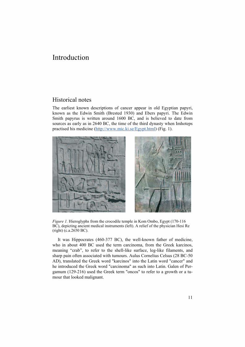
Historical notes The earliest known descriptions of cancer appear in old Egyptian papyri, known as the Edwin Smith (Brested 1930) and Ebers papyri. The Edwin Smith papyrus is written around 1600 BC, and is believed to date from sources as early as in 2640 BC, the time of the third dynasty when Imhoteps practised his medicine (http://www.mic.ki.se/Egypt.html) (Fig. 1).
Figure 1. Hieroglyphs from the crocodile temple in Kom Ombo, Egypt (170-116 BC), depicting ancient medical instruments (left). A relief of the physician Hesi Re (right) (c.a.2650 BC).
It was Hippocrates (460-377 BC), the well-known father of medicine, who in about 400 BC used the term carcinoma, from the Greek karcinos, meaning “crab”, to refer to the shell-like surface, leg-like filaments, and sharp pain often associated with tumours. Aulus Cornelius Celsus (28 BC-50 AD), translated the Greek word "karcinos" into the Latin word "cancer" and he introduced the Greek word "carcinoma" as such into Latin. Galen of Per-gamum (129-216) used the Greek term "oncos" to refer to a growth or a tu-mour that looked malignant.

12
Surgery for rectal cancer performed through the perineum has been de-scribed since the eighteenth century (Rankin et al. 1932), but at that time the success rate was low and the recurrence rate high. In 1908 Ernest Miles de-scribed abdominoperineal resection for cancer of the rectum (Hill 1979) and a successful technique for low anterior resection was developed at the Mayo Clinic, USA, in about 1939 (Dixon 1939). In the early days, the principal goals in the surgical treatment of abdominal diseases, such as cancer of the colon and rectum, were to control the patient’s pain and prevent infections and shock. Although elective abdominal surgery began in 1809 with the resection of an ovarian tumour by Ephraim MacDowell, it was not until an-aesthesia was introduced in 1846 by William Morton and antisepsis in 1867 by Joseph Lister that complicated surgery of the abdomen was made possible (Lister 1867). The discovery of blood groups by Karl Landsteiner in 1901 and of penicillin in 1928 by Alexander Fleming made surgical treatment much safer. In the following decades continued improvements were achieved to reach the modern standard that we are familiar with today.
The discovery of X-rays in 1895 by Wilhelm Conrad Röntgen (1845-1923) and that of radium by Marie and Pierre Curie in 1898 led to the use of irradiation for medical practice. Early on, radiation therapy was used with favourable results in treatment of other pelvic malignancies than rectal can-cer, such as cancer of the prostate, cervix and uterus. The use of radiation therapy in rectal cancer, was first described in the literature in 1959 (Stearns et al. 1959).
Paul Ehrlich (1854-1915), when describing treatment with antibiotics, coined the term chemotherapy. Alkylating agents represent the first class of chemotherapeutic drugs to be used in the clinical setting. Notably, they were a product of the secret gas warfare programme of the United States in both world wars. The exposure of military seamen to sulphur mustard gas in World War II led to the observation that alkylating agents caused marrow and lymphoid hypoplasia (Adair and Bogg 1931). This observation in turn led to the use of nitrogen mustard (mechlorethamine) against haematological neoplasms at the Yale Cancer Center, USA, in 1943 (Rhoads 1946). Around 1950, the effect of chemotherapy in colon cancer was studied, using thio-tepa, floxuridine and 5-fluorouracil. Since the results of a randomised trial were presented in 1958, 5-fluorouracil has been established as an effective drug against colon and rectal cancer (Curreri et al. 1958).

13
DefinitionAs cancer of the colon and rectum originates most often from the glands of the mucosa, it is classified as adenocarcinoma.
The synonym colorectal cancer is often applied for colon and rectal can-cer, but it is important to determine whether the cancer originates in the co-lon or in the rectum (Fig. 2), as there are considerable differences in the treatment. Patients with rectal cancer are operated on with a specific surgical technique and are often given pre- or postoperative radiation therapy; also there are probable differences in the effect of chemotherapy. Cancer located below the level of 15 cm from the anal verge as measured with a rigid recto-scope, or below the pelvic promontory as visualised by X-ray, computed tomography (CT), magnetic resonance imaging (MRI) or during surgery, is defined as rectal cancer in most classification systems, and 15 cm is the level used in most current trials (Fig. 2) (Kapiteijn et al. 2001).
Figure 2. The anatomy of the gastrointestinal tract (left) and the pelvis and rectum (right).

14
IncidenceTogether colon and rectal cancer is the third most common cancer in Swe-den, with 5475 new cases diagnosed in the year 2003; colon cancer ac-counted for two-thirds of these cases, with 3559 diagnoses in the year 2003. In Sweden, as well as in the other Nordic countries, there has been a minor increase in the incidence of colon and rectal cancer during the past decades (Malila and Hakulinen 2003). For colon cancer, which is the fourth most common cancer in males after prostate, lung and skin cancers, the world standardised incidence rate is 18.9 per 100 000 inhabitants a year. In females colon cancer is the second most common form of cancer in Sweden after breast cancer, with a world standardised incidence rate of 17.2. Cancer of the rectum and anus ranks sixth in both males and females, with world standard-ised incidence rates of 12.4 and 8.0 respectively (SOS 2003).
Cancer of the colon and rectum is rare in younger age groups (< 50 years), the median age at diagnosis of colon cancer in males being 71.5 years and in females 72.4 years. For rectal cancer the median ages are 70.2 and 71.3 years in males and females respectively (data from the Swedish Cancer Registry 1995-99).
Aetiology The transformation from a normal cell to a cancer cell is thought to depend on several steps of genetic changes (Fearon and Vogelstein 1990). The growth starts as an adenoma, which is a benign elevation of the mucosal surface, and then gradually changes to cancer. The time interval from the first genetic event to the end of this transformation is not known, but is probably individual, and can take years or even decades. The underlying cause of these genetic changes has not been clearly established, but envi-ronmental factors such as faecal mutagens, meat intake, bile acids, altered vitamin and mineral intake and faecal pH are thought to play a role (DeVita et al. 2000). Lifestyle, with smoking as the most important factor, is defi-nitely associated with lung, oesophagus and urinary bladder cancers. The association with colon and rectal cancer is less well documented, and the data are inconsistent, but from more recent studies associations of varying strengths have been reported (Mizoue et al. 2006). Alcohol is another factor that is inconsistently thought to be associated with colorectal cancer (Longnecker et al. 1990), with a high (>45 g) daily intake increasing the risk moderately (Cho et al. 2004).
Several groups of patients have been identified as being at higher risk for developing colorectal cancer and account for 1-2% of all cases of this can-cer. Patients with inflammatory bowel disease have a higher incidence of colon and rectal cancer compared to the general population (Sinclair et al.
15
1983). A meta-analysis of the colorectal cancer risk in patients with ulcera-tive colitis has shown that 2% of the patients will develop cancer after 10 years, 8% by 20 years and 18% by 30 years (Eaden et al. 2001). Besides the duration of symptoms, the risk is also increased with increased severity of the inflammation and with early age at diagnosis (Munkholm 2003). Patients with Crohn´s disease are also thought to have an increased risk of colorectal cancer, although the results are not as consistent as for ulcerative colitis (Munkholm 2003).
Entities of hereditary colorectal cancer have been defined, although more than 85% of colorectal cancers are considered to be sporadic (Kinzler and Vogelstein 1996). Among patients with first-degree relatives diagnosed with colorectal cancer there is a two- to threefold increase in the risk of develop-ing colon and rectal cancer. Hereditary syndromes with a markedly increased risk of colon and rectal cancer, such as hereditary non-polyposis colorectal cancer (HNPCC) and familial adenomatous polyposis coli syndrome (FAP) are well known, but uncommon. These two syndromes have been reported to account for 2-3% (Ponz de Leon 1994; Aaltonen et al. 1998; Pinol et al. 2004) and <1% (Jarvinen 1992; Bulow 2003) of all cases of colorectal can-cer, respectively. The diagnosis of HNPCC is now made with the help of the Amsterdam criteria (Table 1), but genetic techniques will probably be more widely used in the future (Aaltonen et al. 1998; Kievit et al. 2004).
Table 1. The revised Amsterdam criteria for hereditary non-polyposis colorectal cancer (HNPCC).
The Amsterdam criteria apply the 3-2-1 rule to classify HNPCC families:
There have been three cases of either colon or rectal cancer or other HNPCC-associated cancers — endometrial, small intestine, urinary tract, or kidney (renal pelvis) — in the family
Spread over at least two generations
With one cancer patient having being diagnosed before age 50
The known genetic changes in HNPCC and FAP are autosomal dominant, with the adenomatous polyposis coli (APC) gene on chromosome 5 affected in FAP (Renkonen et al. 2005) and changes in the mismatch repair genes in HNPCC (de Leon et al. 1999).

16
DiagnosisThe definitive diagnosis of the cancer is most often established endoscopi-cally in rectal cancer and with endoscopy or X-ray in cases of colon cancer. Preferably biopsies are taken for histopathological examination to confirm the diagnosis. Most often it is the patient’s clinical symptoms, i.e. rectal bleeding, changes in bowel habits and/or abdominal pain, or detection of anaemia, that initiate these investigations. Occasionally ultrasound or CT performed for other suspected diseases will disclose an asymptomatic pri-mary tumour or its metastatic lesions.
Because of the high incidence and the gradual progression of cancer from an early polypous or adenomatous lesion, screening for colon and rectal can-cer has been advocated. Several randomised trials of screening for colon and rectal cancer have been carried out and have shown lower mortality from colorectal cancer in the screened populations (Gérard et al. 1988; Hardcastle et al. 1996; Kronborg et al. 1996). In Sweden, a screening programme cov-ering the whole population has not been introduced (Hakama et al. 2005).
Pathology and staging Pathological examination is crucial in the diagnostics and staging of the dis-ease (Table 2). The definite tumour (T) and lymph node (N) staging is per-formed postoperatively at the pathological examination. Metastasis (M) stag-ing is mainly done by radiological examination, with the lungs and liver as the most common metastatic sites. Peritoneal metastases are most commonly identified during surgery.
Staging provides information about the extent of the infiltrative growth of the primary cancer and whether tumour cells have disseminated to lymph nodes and other organs. On the basis of this information it is possible to make decisions about treatment alternatives and to assess the patient’s prog-nosis.
Clinical T, N and M staging is achieved preoperatively with radiological imaging such as plain X-rays, ultrasound, CT and MRI (Thoeni 1997). Pre-operative staging is especially important in cases of rectal cancer, where preoperative radiation therapy, chemotherapy or both is dependent on the stage of the disease.
In the Uppsala-Örebro region in the year 2004 the distribution of tumour stage in colon cancer was 11.1% in stage I, 32.8% in stage II, 30.2% in stage III, and 22.6% in stage IV; in 3% the stage was unknown (ROC 2004a). For rectal cancer the corresponding figures were 21.1%, 31.0%, 32.0%, 13.6% and 2.1% respectively (ROC 2004b).

17
Table 2. The staging system most commonly used is the TNM (Tumour, lymph Node, Metastasis) staging (AJCC 2002; Sobin 2002).
T primary Tumour N regional lymph Nodes
TX Primary tumour cannot be assessed. NX Regional lymph nodes cannot be as-
sessed T0 No evidence of primary tumour N0 No regional lymph node metastasis Tis Carcinoma in situ: intraepithelial or
invasion of lamina propria N1 Metastasis in 1 to 3 regional lymph
nodesT1 Tumour invades submucosa T2 Tumour invades muscularis propria
N2 Metastasis in 4 or more regional lymph nodes
T3 Tumour invades through muscularis propria into subserosa or into non-peritonealised pericolic or perirectal tis-sues
M distant MetastasisMX Distant metastasis cannot be assessed M0 No distant metastases M1 Distant metastases
T4 Tumour directly invades other organs or structures and/or perforates visceral peritoneum.
T N M Stage 0 Tis N0 M0Stage I T1, T2 N0 M0Stage IIA T3 N0 M0Stage IIB T4 N0 M0Stage IIIA T1, T2 N1 M0Stage IIIB T3, T4 N1 M0 Stage IIIC any T N2 M0Stage IV any T any N M1
A sufficient number of tumour slices have to be taken for microscopic ex-amination to assess the extent of the infiltrative growth, i.e. how deep in the wall of the colon/rectum the tumour is growing. The size of the circumferen-tial margin, which is evaluated at the pathological examination, is important for deep infiltrating tumours and this margin is also a direct measure of the quality of the surgery. Examination of 12 lymph nodes is recommended but that number of lymph nodes can be difficult to achieve, especially with rectal cancers that have been treated with radiation therapy and/or chemotherapy preoperatively. At the pathological examination the differentiation of the tumour is also assessed and infiltration of tumour cells in vascular spaces and around nerves is reported if found. These factors are thought to be asso-ciated with the patient’s prognosis.

18
Treatment Treatment of colorectal cancer can be regarded as multidisciplinary, and aims at cure in all patients, and if this is not possible, at providing the best available palliative treatment.
Surgery The concept applied in surgical treatment of colon cancer has not changed greatly during the past decades, with the surgical dissection following the embryological planes and taking into account the tumour margins and the vascular and lymphoid anatomy (Fazio and Tjandra 1991; Mahteme and Påhlman 2005).
A free margin of 10 cm proximal and distal to the tumour should be suffi-cient for radical surgery. For the lateral margin it is important to resect the tumour en bloc if it has infiltrated other organs such as the omentum, small bowel or the abdominal wall, to prevent the spill of tumour cells.
The lymphoid drainage of the tumour is located alongside the vascular supply of the colon in the mesentery. To be able to resect as much of the untorn mesentery and as many lymph nodes as possible, it is important to dissect and divide the vascular trunk, supplying the affected part of the co-lon, as proximally as possible (Slanetz and Grimson 1997).
For rectal cancer, a surgical technique following the embryonic planes, namely total mesorectal excision (TME), has been developed in the past two decades (Heald and Ryall 1986; Enker et al. 1995). Before the introduction of TME, the rectum was dissected blunt, without respect to the borders of the mesorectal fascia. With the TME technique, the dissection is performed out-side the mesorectal fascia (Fig. 2) under visual control, preventing spill of tumour cells from the primary tumour and the lymph nodes of the mesorec-tum (Heald et al. 1982). TME, together with radiation therapy, has signifi-cantly reduced the incidence of local recurrences (SRCT 1997; CCCG 2001; Kapiteijn et al. 2001).
If the tumour is advanced, with overgrowth to adjacent organs such as the prostate or vagina, the same principles are followed as in colon cancer, with en bloc resection of these organs together with the rectum.
Although the long-term (oncological) results of the surgical treatment are important, the short-term outcome also has a significant impact on the total results. The postoperative morbidity and mortality depend on the surgeon, the technique and the peri- and postoperative care. Thus, the performance of an adequately trained colorectal surgeon together with the development of colorectal teams with higher volumes for each surgeon are determining for the surgical outcome (Dahlberg et al. 1998b; Martling et al. 2000; Smedh et al. 2001).

19
Large differences have been observed between patients operated on as emergencies and those treated as elective cases; about 25% of colon cancers are operated on as emergency cases (Jestin et al. 2005), but only 2-3% of the rectal cancers (Smedh et al. 2001). The morbidity and mortality are higher after emergency surgery, and both short- and long-term survival is impaired.
In cases of disseminated disease, the same surgical principles can be ap-plied for colon and rectal cancer. Most liver metastases are unresectable, but in cases of localised liver metastases there is a good possibility of cure, with 5-year survival of 30-40% in patients treated with a radical resection (Figueras et al. 2001; Lindner et al. 2003). Combination chemotherapy can induce down-sizing of liver metastasis, making surgery with curative inten-tion possible in a few cases (Bismuth and Adam 1998; Nordlinger et al. 2003). Patients with local recurrence and a solitary lung metastasis should also be evaluated with a view to possible resection (Vogelsang et al. 2004; Yoshidome et al. 2004). An improvement in the treatment of peritoneal car-cinomatosis, using peritonectomy and intraperitoneal chemotherapy, in pa-tients with no other sign of distant metastasis, has resulted in a 5-year sur-vival of 30% compared to less than 5% after treatment with chemotherapy only (Mahteme et al. 2004).
Radiation therapy Radiation therapy in colon and rectal cancer, in addition to surgery, is almost exclusively used in the treatment of rectal cancers. This therapy is used only occasionally, as palliative treatment, in advanced colon cancer.
The radiation therapy in patients with a resectable rectal cancer can be given preoperatively (neoadjuvant) or postoperatively (adjuvant). In Sweden it is now routine to give this therapy preoperatively, because of the poten-tially shorter duration of the treatment and the better effects in terms of re-duction of the local recurrence rate compared to postoperative irradiation (Frykholm et al. 1993; CCCG 2001; Marijnen et al. 2004; Sauer et al. 2004). Short-course preoperative 5x5 Gy treatment with immediate surgery is con-venient for use in patients with a resectable cancer when there are no signs of overgrowth to other organs requiring down-sizing or down-staging. If this is required, a prolonged course, usually in combination with chemotherapy, is preferable (Glimelius et al. 2003; Bujko et al. 2004; Bosset et al. 2005).
Preoperative radiation therapy was shown to increase the overall survival in the Swedish Rectal Cancer Trial (SRCT 1997) and in a meta-analysis of all randomised trials on preoperative radiation therapy using a radiation dose corresponding to a biologically effective dose of at least 30 Gy (CCCG 2001). Based on these randomised trials and the German trial comparing pre- and postoperative radiochemotherapy (Sauer et al. 2004), preoperative radia-

20
tion therapy is now considered as a gold standard in cases where radiation therapy is considered.
Radiation technique The radiation beams are presently delivered with linear accelerators using 8-16 megavolt (MV) photons. At present a three- (Fig. 3) or four-beam (portal) technique is used with the upper beam limit at the promontory and the lower border below or slightly above the anus, depending upon the tumour height (Fig. 4).
Figure 3. Schematic drawing of the radiation beams showing how they are directed to the target (the tumour and the lymph nodes).

21
Figure 4. Sagittal view of the female pelvis showing the directions of the posterior beam and how small bowel can be located in the pelvis.
The aim is to irradiate the mesorectal lymph nodes and the presacral lymph nodes, the lymph nodes along the internal iliac artery and the nodes of the obturator foramen (Fig. 5). Shielding of tissues not at risk of containing tumour cells is performed, presently mostly with multileaf collimators.
Figure 5. Schematic presentation of the internal iliac lymph nodes (right and left) and the presacral and the mesorectal lymph nodes.
The dose of the preoperative irradiation is dependent upon the preopera-tive staging. T1-2 tumours located within 6-15 cm from the anus are not considered to require radiation therapy, as local recurrences rarely occur in

22
those cases (Kapiteijn et al. 2001). In primarily resectable rectal cancer that is considered to be mobile on the basis of clinical investigation (T1-T3) and is located within 0-6 cm from the anus, as well as in T3 tumours located within 6-15 cm from the anus, the irradiation is frequently given as 5 Gy in five fractions. In patients with locally advanced rectal cancer, fixed on clini-cal examination (T4), long-term preoperative radiation therapy is given as 1.8-2 Gy in 25-28 fractions. The aim of this long-term radiation therapy is to achieve tumour regression and to increase the possibility of radical excision. Radiochemotherapy for advanced rectal cancer could be even more benefi-cial, but definitive evidence of superiority has not been available until re-cently (Glimelius et al. 2003; Bosset et al. 2005; Braendengen et al. 2005).
ComplicationsEarly complications that become apparent during radiation therapy or up to 3 months thereafter are well known, and include skin, gastrointestinal (Galland and Spencer 1985), genitourinary and neurological complaints (Marijnen et al. 2002). The early complications should preferably be evaluated according to their severity, where a scoring system according to the Radiation Therapy Oncology Group can be used (Trotti et al. 2003). In this system there are five grades from grade 0, representing no complaints, up to grade 5, with toxicity leading to death. With modern preoperative radiotherapy using 5x5 Gy and a four-beam technique, most of these early complications are of low grade, are transient, and require no intervention (Marijnen et al. 2002). However, a few patients can suffer from acute neurological toxicity, presenting as pain in the gluteal region or legs (SRCT 1993; Frykholm et al. 1996b; Marijnen et al. 2002). The radiation technique is of great importance when predicting the occurrence of grade 5 toxicity. Previously when anterior-posterior beams were used and the upper beam limit was higher up in the abdomen, increased postoperative mortality was noted in patients treated with anterior-posterior beams only and not with the three- or four-beam technique (SRCT 1993; Holm et al. 1996b). Anterior-posterior beams should therefore not be used today.
Studies with a long-term follow-up focused on adverse effects of radio-therapy for rectal cancer are sparse. A follow-up of the Stockholm I and II trials, on the short course preoperative radiation therapy for rectal cancer, showed an increase in femoral and pelvic fractures in patients randomised to radiotherapy in the Stockholm I trial. In the Stockholm II trial an increase in deep vein thrombosis and fistulas was seen (Holm et al. 1996b). When the Stockholm studies were analysed together, the irradiated patients showed an increased risk of bowel obstructions, venous thromboembolism, and femoral neck and pelvic fractures. Impaired bowel function, i.e. incontinence and frequent stools, has been reported in a long-term follow-up of the Stockholm Trials (Pollack et al. 2006), in a 5-year follow-up of the Swedish Rectal

23
Cancer Trial (Dahlberg et al. 1998a) and after 2 years of follow-up in the Dutch TME trial (Marijnen et al. 2005). Impaired sexual function has also been recorded in irradiated patients participating in the Dutch TME trial (Marijnen et al. 2005), but no difference was found in the quality of life.
To our knowledge there has been only one study which has compared the findings at late follow-up between pre- and postoperative radiotherapy. This study showed an increased risk of bowel obstruction in the postoperatively irradiated group (Frykholm et al. 1993). Lundby et al., analysed the outcome of postoperative radiation therapy with 50 Gy compared to no radiation ther-apy and found that irradiated patients had increased bowel frequency, ur-gency and incontinence of loose stools (Lundby et al. 1997; Lundby et al. 2005). Patients treated with postoperative chemoradiotherapy have a high incidence of bowel problems such as proctitis, enteritis, incontinence, in-creased bowel frequency and defecation at night (Kollmorgen et al. 1994; Miller et al. 1999; Sauer et al. 2004).
Chemotherapy Among curatively treated colon cancer patients, chemotherapy has still been proven beneficial for stage III patients only. The value of chemotherapy for stage II colon cancer remains controversial (Ragnhammar et al. 2001b), but many agree that it has a role also for the high risk stage II cancers, that is T4 tumours, for tumours with perforation and for tumours with vessel invasion. Most studies are based on leucovorin modulated 5-fluorouracil. The present standard for 5 fluorouracil-leucovorin treatment is 6 months (Ragnhammar et al. 2001b), although recently even better effects have been observed from the use of 5-fluorouracil-leucovorin with oxaliplatin (Andre et al. 2004). In rectal cancer there is no evidence for the benefits of adjuvant chemotherapy, but it may be beneficial in stage III cancers (Ragnhammar et al. 2001b).
In patients with advanced colon and rectal cancer (stage IV), prolonged survival and a decrease in tumour-related symptoms have been shown after chemotherapy, now often based on 5-fluorouracil-leucovorin and oxaliplatin or irinotecan (Simpson et al. 2003; Sorbye et al. 2004) .
New compounds which inhibit the angiogenic capability of the tumour by inhibiting vascular endothelial growth factor or the epidermal growth factor receptor have recently also been found to have beneficial effects (Hurwitz et al. 2004; Nygren et al. 2005).

24
Prognosis
RecurrenceA recurrence of colon and rectal cancer can be classified as local (loco-regional) or distal (metastasis). Local recurrence of rectal cancer was previ-ously a substantial problem, but with improved surgical techniques and ra-diation therapy the local recurrence rate has decreased from 20-30% to 5-10% (Arbman et al. 1996; Dahlberg et al. 1999; Martling et al. 2001).
The incidence of local recurrence is dependent on the stage of the disease. Five-year follow-up data from the Swedish Rectal Cancer Trial of patients treated with preoperative irradiation show a 4% recurrence rate in stage I disease, 10% in stage II and 20% in stage III (SRCT 1997). In that trial the surgical technique was not optimal, with an overall local recurrence of 29% in the surgery-only group versus 9% in the preoperatively treated group after 13 years of follow-up (Folkesson et al. 2005).
There is now substantial evidence that patients with rectal cancer operated on with TME have a lower local recurrence rate compared to those that were not operated on with the TME technique. Four-year results of the Dutch TME trial revealed that patients randomised to TME surgery only had a local recurrence rate of 11%, whereas in irradiated patients the corresponding rate was 6% (Kapiteijn et al. 2001; Marijnen et al. 2004).
MetastasisThe most important reason for failure and death in patients with colon and rectal cancer is the development of metastasis. One-third of the patients op-erated on for colon and rectal cancer will show recurrence with metastasis, where the most common sites are the liver and lungs. Only few of the pa-tients are potentially curable, as the metastatic disease is usually widespread and not amenable to surgical treatment. Chemotherapy is only partially ef-fective, giving a chance of only limited prolongation of survival (Simpson et al. 2003; Sorbye et al. 2004).
Survival Survival rates are dependent on the stage of the disease. Patients with stage I disease can be expected to have a 5-year relative survival of 80-95%; stage II 60-80%; stage III 30-55%; and stage IV <3% (Ries 2002).

25
There are several factors that influence survival irrespective of the stage of the disease; for example patients operated on acutely have a worse prog-nosis than those operated on electively (Jestin et al. 2005), and males have a worse prognosis than females (Talback et al. 2003). Recent studies have shown improved survival rates in colon and rectal cancer in Sweden (Talback et al. 2003), and this is at least partly due to better surgical (Martling et al. 2000) and oncological treatments (Ragnhammar et al. 2001a).
Other factors may also have contributed to the improvement, such as ear-lier detection of the tumours, better perioperative treatment, improved anaes-thesia and postoperative care, and centralisation of the treatment of rectal cancer to colorectal units (Smedh et al. 2001).

26
Aims of the investigation
The overall aim of this doctoral research was to analyse the survival of pa-tients with colon and rectal cancer in Sweden, and the risks associated with radiation therapy, in addition to surgery, for rectal cancer.
The specific aims were:
to investigate time-trends in survival of patients with colon and rectal cancer in Sweden (Study I)
to determine whether the combination of preoperative radiation therapy and TME, which was adopted by the county of Uppsala in the year 1986 and by the rest of Sweden between 1990 and 1995, has improved the survival of patients with rectal cancer (Study I)
to analyse the occurrence of second cancers in patients with rectal cancer (Study II)
to address the question whether radiation therapy, in addition to surgery, for rectal cancer increases the risk of second cancers in organs within or adja-cent to the irradiated volume (Study II)
to investigate the incidence of late hospital admissions in patients with rectal cancer treated with preoperative irradiation in comparison to those who were not (Study III)
to determine whether neoadjuvant radiation therapy, used in patients with rectal cancer, increases the risk of diseases in organs situated within or adja-cent to the irradiated volume (Study III)
to analyse in detail the relations of late gastrointestinal disorders, necessitat-ing hospitalisation, to preoperative radiation therapy for rectal cancer (Study IV)
27
Subjects, material and methods
Study I Survival analyses were performed on the basis of data obtained from the Swedish Cancer Register for all patients diagnosed with an carcinoma of the colon and rectum between 1960 and 1999.
Patients diagnosed with an invasive adenocarcinoma, an undifferentiated carcinoma, or unspecified malignant tumours were included in the study.
Patients diagnosed at autopsy, patients with zero survival and patients without follow-up information were excluded from the analyses. Patients who emigrated from the country were censored at the time of emigration. Only the first colon or rectal cancer in each case was included. All synchro-nous cancers were excluded and patients with a metachronous cancer of the colon or rectum were censored at the date of the second diagnosis (Table 3).
The end point of the study was defined as December 31, 2001, and all pa-tients alive were censored at that date.

28
Table 3. Patients with colon and rectal cancer in Sweden included and excluded in the analyses for each five-year period of diagnosis.
Colon cancer Rectal cancer
Period All cases Excluded Included All cases Excluded Included Au-
topsy, zero sur-vival
Notadeno-carci-noma
With-out
follow-up
Au-topsy, zero sur-vival
Notadeno-carci-noma
With-out
follow-up
1960-1964 8 200 1 045 184 103 6 868 4 972 357 189 49 4 377
1965-1969 9 975 1 283 233 93 8 366 5 682 409 222 46 5 005
1970-1974 11 370 1 415 243 28 9 684 6 318 440 271 11 5 596
1975-1979 12 429 1 265 308 5 10 851 7 081 287 380 0 6 414
1980-1984 13 682 1 212 358 10 12 102 8 017 320 461 2 7 234
1985-1989 14 527 1 177 351 8 12 991 8 413 285 510 3 7 615
1990-1994 15 182 785 373 5 14 019 8 991 227 582 3 8 179
1995-1999 15 488 529 339 38 14 582 9 010 162 632 15 8 201
Total 100 853 8711 2 389 290 89 463 58 484 2 487 3 247 129 52 621

29
Study II In the analysis of second cancers, the study population consisted of patients with rectal cancer participating in two randomised trials. In the first one, the Uppsala Trial, running from 1980 to 1985, there were 471 patients random-ised to preoperative 5x5.1 Gy radiation therapy or postoperative 30x2 Gy radiation therapy. The patients who received postoperative radiation therapy had stage II or III tumours (no irradiation for stage I tumours). In the second one, the Swedish Rectal Cancer Trial, running from 1987 to 1990, there were 1147 eligible patients randomised to preoperative 5x5 Gy radiation therapy or surgery only.
Data from the two trials were matched against the Swedish Cancer Regis-ter to identify second cancers, defined as any new cancer other than rectal cancer, detected more than 6 months after the day of surgery for the rectal cancer.
Study III In the analysis of late adverse effects of radiation therapy for rectal cancer, patients included in the Swedish Rectal Cancer Trial were matched against the Swedish Hospital Discharge Register to obtain information about diag-noses registered during hospital admissions after the primary treatment of the rectal cancer. Only curatively treated patients were included in the study, in order to avoid confounding from morbidity related to residual cancer. In patients diagnosed with local recurrence or metastasis, admissions up to three months before the diagnosis of the recurrence or metastasis were in-cluded to avoid confounding from the recurrent cancer.
Early and late hospital admissions were defined as admissions occurring before and after six months, respectively, from the resection of the primary rectal cancer.
Only the first admission registered for a specific diagnosis or diagnostic category during either the first 6 month period or thereafter, was included for each patient. If a patient received two or more diagnoses at the same admis-sion, all diagnoses were included unless they referred to the same specific diagnosis or diagnostic category. All secondary malignancies were excluded from the analysis, since these were studied separately (Study II).

30
Study IV In the study of gastrointestinal disorders possibly due to radiation therapy for rectal cancer, the same study population and exclusion criteria were used as in study III. The results from the matching with the hospital discharge regis-try were used to identify all patients with selected abdominal diagnoses.
The patients’ hospital records were reviewed to make a more detailed analysis of late gastrointestinal disorders possibly related to preoperative radiation therapy for rectal cancer and their relations to age, sex, immediate postoperative complications and radiation techniques.
The Swedish Cancer Register In Sweden and in other Nordic countries, great efforts have been devoted to building up health-related population based registers. The first such register in Sweden was the Cause of Death Register going back to 1749. The Swed-ish National Cancer Register has records of all diagnosed cancer patients in Sweden since 1958. According to the regulations, physicians are under obli-gation to report all cases of cancer to the Registry. In addition, pathologists and cytologists separately report to the Registry every cancer diagnosis made on surgically removed tissues, biopsy and cytology specimens, and at autop-sies. According to a quality control study there is a lower than 2% deficit in the registration of new cases in the database. Dates and causes of deaths are transferred annually to the Register from the Cause of Death Register by computerised linkage. Tumour stage and clinical data such as surgical cura-bility, radiation therapy or chemotherapy are not included in the Swedish National Cancer Register.
The Swedish Hospital Discharge RegisterSince 1987 the Hospital Discharge Register has covered all public, in-patient care in Sweden. Corresponding data for earlier years, 1964-1986, for somatic care and for the years 1973-1986 for psychiatric care are found at the Centre for Epidemiology, Stockholm.
The information recorded in the Register consists of data on the patient, such as personal identification number and sex, dates of admission and dis-charge, and medical data such as the main and secondary diagnoses and any surgical procedures.

31
Statistical analysis Survival was evaluated as observed and relative survival rates (Study I). Relative survival rate is the ratio of observed survival in the study population to the expected survival in a group of people in the general population simi-lar to the patient group in terms of sex and age attained in the year of diag-nosis of the cases. The cumulative relative survival rate was calculated ac-cording to the Hakulinen cohort method.
Actuarial life-table procedures were used to calculate person-years at risk, number of second cancers, and the cumulative proportion of second cancers in each group, whereas the log rank method was used for tests of signifi-cance (Study II). Relative risks (RR) were calculated with 95% confidence intervals (CI). The stratified risk from both trials was calculated according to the Mantel-Haenszel estimation.
In analyses of late adverse effects (Studies III and IV), the difference in duration from surgery to a new admission between preoperatively irradiated and surgery alone patients was tested for significance by the log-rank method. Relative risk analysis, with 95% confidence intervals, was by inten-tion-to-treat on time to admission, using Cox proportional-hazards models, where the calculated hazard ratio was used as the relative risk estimate. The length of time to admission was measured from the date of the primary treatment to the first admission or to the censoring time, which was three months before a local/distal recurrence, date of death or end of the study period (December 31, 2001).
EthicsThe Ethics Committee of Uppsala University approved the studies.

32
Results
Study I From 1960 to 1999, the observed (Fig. 6) and the relative survival rate (Fig. 7) in patients both with colon and with rectal cancer improved markedly. The 1- and 2-year relative survival rates were better for rectal than for colon cancer during all 5-year time periods. The 5- and 10-year relative survival rate, however, was always better for colon cancer except during the last two time periods, when the relative survival rate for rectal cancer came closer to those for colon cancer. In fact, during the most recent time period studied, 1995-99, the 5-year relative survival rate for rectal cancer was marginally higher than that for colon cancer (57.6% vs 57.2%) (Fig. 7).
Figure 6. One, 2-, 5- and 10-year observed survival rates for colon and rectal cancer in Sweden, in per cent, 5-year periods from 1960 to 1999.
0
10
20
30
40
50
60
70
80
90
100
1960-1964 1965-1969 1970-1974 1975-1979 1980-1984 1985-1989 1990-1994 1995-1999
Year of diagnosis
Obs
erve
d su
rviv
al ra
te
1yr Colon1yr Rectal2yr Colon2yr Rectal5yr Colon5yr Rectal10yr Colon10yr Rectal

33
Figure 7. One, 2-, 5- and 10-year relative survival rates for colon and rectal cancer in Sweden, in per cent, in 5-year periods from 1960 to 1999.
The cumulative relative survival rate for cancer of the colon and rectum only marginally improved during the first four 5-year periods (Figs. 8 and 9).From the time period 1980-84 a marked improvement in relative survival rate was seen for each 5-year period. In colon cancer this is almost entirely explained by an improvement during the first year (Fig. 8). After 5-6 years the relative survival rate in patients with colon cancer ceased to decrease. In patients with rectal cancer the same general pattern was seen, although the improvements continued slightly beyond the first year (Fig. 9), and the rela-tive survival rate did not cease to decrease until after 8-9 years.
0
10
20
30
40
50
60
70
80
90
100
1960-1964 1965-1969 1970-1974 1975-1979 1980-1984 1985-1989 1990-1994 1995-1999
Year of diagnosis
Rel
ativ
e su
rviv
al ra
te
1yr Colon2yr Colon5yr Colon10yr Colon1yr Rectal2yr Rectal5yr Rectal10yr Rectal

34
Figure 8. Cumulative relative survival rate for colon cancer in Sweden for the year 1 to 15 after diagnosis; comparison between 5-year time periods from 1960-1999.
Figure 9. Cumulative relative survival rate for rectal cancer in Sweden for the year 1 to 15 after diagnosis; comparison between 5-year time periods from 1960-1999.
0
10
20
30
40
50
60
70
80
90
100
0 1 2 3 4 5 6 7 8 9 10 11 12 13 14 15
Years since diagnosis
Rel
ativ
e su
rviv
al ra
te
1995-19991990-19941985-19891980-19841975-19791970-19741965-19691960-1964
0
10
20
30
40
50
60
70
80
90
100
0 1 2 3 4 5 6 7 8 9 10 11 12 13 14 15
Years since diagnosis
Rel
ativ
e su
rviv
al ra
te
1995-19991990-19941985-19891980-19841975-19791970-19741965-19691960-1964

35
Females had a lower relative risk of excess death than males, both with colon and with rectal cancer. Among patients with rectal cancer, the oldest age group (>75 years) showed a increased relative risk of excess death than younger ones, but in those with colon cancer no clear age difference was seen.
Patients diagnosed with rectal cancer in the county of Uppsala between 1985 and 1989 showed a lower relative risk of excess death than patients diagnosed with rectal cancer in the rest of Sweden (Table 4), but no signifi-cant differences were noted in the latest time periods 1990-94 and 1995-99.
Table 4. Relative risk (RR) of excess death from rectal cancer in the county of Upp-sala compared to the rest of Sweden.
Period RR of excess death * 95% CI P value
1960-1984 0.91 0.78 1.07 0.26
1985-1989 0.62 0.42 0.89 0.01
1990-1994 0.94 0.69 1.30 0.72
1995-1999 0.78 0.52 1.17 0.23
*RR of excess death in rest of Sweden = 1.00 for all periods

36
Study II In this study, 115 patients with 122 second cancer diagnoses other than rectal cancer were included in the analysis. The most common second cancers were cancers of the prostate, colon and urinary bladder (Table 5). Only one case of soft tissue sarcoma was identified, this patient received radiation therapy but the tumor was placed in the thorax region, outside of the irradi-ated target.
Table 5. Second cancers diagnosed in patients with rectal cancer from the Uppsala Trial and the Swedish Rectal Cancer Trial combined. Numbers refer to the numbers of second cancers.
Head and neck region 5 Skin and connective tissue 11
Brain 2 Within/adjacent to RT volume 1
Oral cavity 1 Outside RT volume 10
Thyroid 1 Breast 9
Eye 1 Gynaecological 8
Cervix 2
Upper GI 14 Uterus 6
Esophagus 3 Kidney 6
Stomach 6 Prostate 21
Duodenum 1 Urinary bladder & ureter 12
Liver 3 Haematological 9
Pancreas 1 AML 1
Small Bowel 1 CLL 1
CML 1
Colon 17 ET 1
NHL 3
Lung 9 Multiple myeloma 2 Abbreviations: RT, Radiotherapy; GI, Gastrointestinal; AML, Acute myeloid leukaemia; CLL, Chronic lymphocytic leukaemia; CML, Chronic myeloid leukaemia; ET, Essential thrombocytosis; NHL, Non-Hodgkin’s lymphoma

37
Thirty-seven (7.9%) of the 469 patients in the Uppsala Trial developed a second cancer, and the corresponding figure among the 1130 patients in the Swedish Rectal Cancer Trial was 78 (6.9%) (RR 0.88; 95% CI 0.60 – 1.31) (Table 6).
The overall stratified relative risk of developing a second cancer in irradi-ated patients was 1.85 (95% CI 1.23-2.78), but the increase was significant only in the Swedish Rectal Cancer Trial (RR 1.84; 95% CI 1.15-2.97) and not in the Uppsala Trial (RR 1.87; 95% CI 0.86-4.10).
Stratified analyses of the results from both trials showed, in the irradiated patients, a significantly increased risk for occurrence of second cancers within or adjacent to irradiated volumes (RR 2.04; 95% CI 1.10-3.79), but only a trend for an increased risk outside the irradiated volumes (RR 1.78; 95% CI 0.97-3.27).

38
Table 6. Relative risks of second cancers in patients treated for rectal cancer in the Uppsala Trial and the Swedish Rectal Cancer Trial. A comparison between radiation therapy (RT) and surgery only (no RT) groups.
Uppsala Trial SRCT
Subgroup of
Second Cancer
RT(pre:post)
n
no RT
nRR (95% CI)
RT
n
no RT
nRR (95% CI)
Lung 3 (2:1) 0* 5 1 4.36 (0.5-37.3)
Breast † 2 (2:0) 1 1.0 (0.1-11.4) 4 2 1.76 (0.3-9.6)
Gynaecological † 0* 1 6 1 5.27 (0.6-43.8)
Haematological 5 (5:0) 1 2.6 (0.3-22.1) 2 1 1.74 (0.2 -19.2)
Skin within RT volume 0* 0* 1 0*
Skin outside RT volume 1 (0:1) 0* 4 5 0.70 (0.2-2.6)
Head and neck 3 (2:1) 0* 1 1 0.89 (0.1-14.0)
Kidney 1 (0:1) 0* 3 2 1.31 (0.2-7.8)
Prostate ‡ 5 (5:0) 1 2.5 (0.3-21.0) 10 5 1.75 (0.6-5.1)
Urinary bladder and ureter 2 (2:0) 0* 6 4 1.31 (0.4-4.6)
Upper GI 4 (3:1) 0* 6 4 1.31 (0.4-4.6)
Small bowel 1 (1:0) 0* 0* 0*
Colon 2 (1:1) 4 0.3 (0.1-1.4) 10 1 8.72 (1.1-68.1)
Overall 29 (23:6) 8 1.87 (0.86-4.10) 53 25 1.84 (1.12-2.97)
Within/adjacent to the RT volume § 13 (13:0) 3 2.24 (0.64-7.86) 25 11 1.98 (0.97-4.02)
Outside RT vol-ume # 14 (9:5) 1 7.24 (0.95-55.1) 23 15 1.33 (0.70-2.56)
Abbreviations: SRCT, Swedish Rectal Cancer Trial; RT, Radiation therapy; N, Number of second cancers; Pre, Preoperatively; Post, Postoperatively; RR, Relative risk; CI, Confidence interval; GI, Gastrointestinal. * RR was not calculated when no cases were present. † Only females. ‡ Only males. § Gynaecological, haematological, prostate, urinary bladder, ureteric cancer, and skin within RT volume.# Lung, breast, head and neck, kidney, upper GI, and skin outside RT volume.

39
There were no differences in the relative risk of a second cancer between TNM stages, but when each TNM stage was analysed separately an in-creased risk of developing a second cancer in irradiated patients was ob-served among patients with stage I tumours, a tendency in stage II, but no difference in stage III (Table 7).
The median interval between inclusion in the trials and diagnosis of the second cancer was 6.5 (1-18) years. When the results were stratified by la-tency period, a significantly increased relative risk of second cancer in the irradiated groups was seen only in the time period 5-10 years from the pri-mary treatment (Table 7). The types of secondary cancers diagnosed did not differ between latency periods.
Table 7. Relative risk of second cancers in RT vs. no RT patients with rectal cancer from the Uppsala Trial and the Swedish Rectal Cancer Trial combined. Comparisons are made according to tumor stage and latency periods.
Number of sec-ond cancers
RT: no RT
Person years at risk
RT: no RT RR 95% CI
Stage I 35 : 15 14 848 : 14 940 2.35 1.28-4.30
Stage II 31 : 9 14 126 : 7 836 1.91 0.91-4.01
Stage III 16 : 9 7 906 : 4 638 1.04 0.46-2.36
Years from primary treatment
< 5 years 31 : 13 8 249 : 6 101 1.76 0.92-3.37
5-10 years 33 : 12 5 107 : 3 810 2.05 1.06-3.97
> 10 years 18 : 8 3 104 : 2 243 1.63 0.71-3.74
Abbreviations: RT, Radiation therapy; RR, Relative risk; CI, Confidence interval

40
Figure 10 illustrates the cumulative proportion of patients treated for rec-tal cancer developing second cancers in both trials and all radiation groups.
After 14 years of follow-up of the Swedish Rectal Cancer Trial, local re-currence had developed in 60 (10.8%) of 555 radiation-treated patients and in 152 (26.4%) of 575 patients in the surgery only group (RR 0.34; 95% CI 0.25-0.46). A second cancer occurred in 53 (9.5%) irradiated patients and in 25 (4.3%) patients who were not irradiated. Hence 20.3% of the irradiated patients got either a local recurrence or a second cancer, compared to 30.7% of the non-irradiated patients (RR 0.55; 95% CI 0.44-0.70).
Figure 10. Cumulative proportion of patients treated for rectal cancer developing second cancers. Comparison between the radiation therapy groups of the Uppsala Trial and the Swedish Rectal Cancer Trial.
Abbreviations: SRCT, Swedish Rectal Cancer Trial; RT, Radiation therapy; Preop, Preopera-tive; Postop, Postoperative.
0,0
0,5
1,0
1,5
2,0
2,5
3,0
3,5
1 2 3 4 5 6 7 8 9 10 11 12 13 14 15 16 17 18 19 20Years from diagnosis
SRCT PreopRT
SRCT NoRT
Uppsala Trial PreopRT
Uppsala Trial PostopRT
Uppsala Trial NoRT

41
Study III Of the 908 curatively treated patients included in the Swedish Rectal Cancer Trial, 661 patients (73%) were admitted once or more to a hospital after the treatment of the primary rectal cancer. After exclusion of all recurrent or secondary cancer diagnoses, no difference was seen in the relative risk for all admissions (RR 1.07; 95% CI 0.92-1.26). However, an almost two-fold in-crease in relative risk for early admissions was found for irradiated patients (RR 1.74; 95% CI 1.30-2.33), but there were no differences in relative risk for late admissions (RR 0.88; 95% CI 0.73-1.06) (Fig. 11).
0 2 4 6 8 10 12 14
0.0
0.2
0.4
0.6
0.8
1.0
Time (Years)
No radiation therapy
Radiation therapy P=1.08
Figure 11. Proportion of patients admitted for any reason. Comparison between curatively treated patients with rectal cancer participating in the Swedish Rectal Cancer Trial treated with preoperative radiation therapy (n=454) and those treated with surgery alone (n=454).

42
An increase in relative risk among irradiated patients during the first 6 months was observed for infections and gastrointestinal diagnoses and a trend for an increased relative risk was seen for endocrine and cardiovascular diagnoses (Table 8). Further analyses of the specific diagnoses did not reveal any differences.
Table 8. Diagnoses according to ICD chapters and specific diagnosis registered during early hospital admissions (<6 months) after primary treatment in patients with rectal cancer.* Relative risks and 95% confidence intervals, in patients treated with preoperative radiation therapy compared to those who underwent surgery alone.
ICD chapter
Specific diagnosis
ICD-9
Diagnostic codes
no RT
n
RT
n
RR 95% CI
Infections 008 - 138 ** 2 16 7.67 1.76 - 33.39
Endocrine 240 – 279 2 9 4.55 0.98 - 21.12
Cardiovascular 390 - 459, 786F 14 26 1.89 0.99 - 3.63
Gastrointestinal 530 –574, 787, 789** 21 53 2.57 1.55 - 4.26
Bowel obstruction 560B-X 5 11 2.20 0.76 - 6.36
Constipation 564A 5 10 1.94 0.66 - 5.68
Abdominal pain 789A 2 8 3.96 0.84 - 18.71
Urological 580 – 608** 16 22 1.37 0.72 - 2.62
Abbreviations: ICD, International classification of diseases; RR, Relative risk; CI, Confidence interval; RT, Preoperative radiation therapy; no RT, Surgery alone. * Diagnostic categories and specific diagnosis with fewer than 10 admissions are not pre-sented in the table. ** These infectious diagnoses were included in the infections category and excluded from the organ specific category: 466A, 481X-487W, 558X, 566X, 590A-W, 595A-X, 599A, 604A-X, 680C-682H, 684X-690X, 780G, 958D
Regarding late admissions, no diagnoses, categorised according to inter-national classifications of diseases (ICD), showed a significant increase in

43
relative risk in the irradiated group, but trends were seen for infectious and gastrointestinal diagnoses, and for fractures (Table 9).
Table 9. Diagnoses during late hospital admissions (>6 months) after primary treat-ment in patients with rectal cancer.* Relative risks and 95% confidence intervals are presented for patients treated with preoperative radiation therapy (RT, n=454) in comparison with those who underwent surgery alone (No RT, n=454).
ICD chapter ICD-9
Diagnostic codes
no RT
n
RT
n
RR 95% CI
Infections 008 - 138 ** 58 89 1.34 0.96 - 1.87
Endocrine 240 - 279 45 65 1.26 0.86 - 1.85
Psychiatry 290 - 319 22 18 0.69 0.37 – 1.28
Neurological 237, 330 - 359 19 26 1.17 0.64 – 2.11
Cardiovascular 390 - 459, 786F 147 154 0.88 0.71 - 1.11
Pulmonary 492 - 508 12 18 1.34 0.64 – 2.78
Gastrointestinal 530 – 574, 787, 789**
120 157 1.23 0.97 - 1.56
Urological 580 – 608** 49 42 0.74 0.49 - 1.12
Gynaecological 614 - 629 10 6 0.51 0.19 - 1.42
Skin 691 - 709 8 16 1.70 0.73 - 3.98
Orthopaedic &
rheumatological
710 - 732 36 53 1.33 0.87 - 2.04
Fractures 733B, 800 - 829 36 58 1.45 0.95 - 2.19
Abbreviations: ICD, International classification of diseases; RR, Relative risk; CI, Confidence interval; RT, Preoperative radiation therapy; no RT, Surgery alone. * Diagnostic categories with fewer than 10 admissions are not presented in the table. ** These infectious diagnoses were included in the infections category and excluded from the organ specific category: 466A, 481X-487W, 558X, 566X, 590A-W, 595A-X, 599A, 604A-X, 680C-682H, 684X-690X, 780G, 958D
In the group of gastrointestinal diagnoses, increased relative risks in irra-diated patients were observed for bowel obstruction, nausea and unspecific

44
abdominal pain, whereas the risk for inguinal hernia was lower among irra-diated patients (Table 10).
Table 10. Specific diagnosis registered during late hospital admissions (>6 months) due to gastrointestinal diagnoses after primary treatment in patients with rectal can-cer.* Relative risks and 95% confidence intervals for patients treated with preopera-tive radiation therapy in comparison with those who underwent surgery alone are shown.
Specific diagnosis ICD-9
Diagnostic codes
no RT
n
RT
n
RR 95% CI
Gastric ulcer 531A-X 8 7 0.78 0.28 - 2.15
Duodenal ulcer 532A-X 7 5 0.60 0.19 - 1.90
Fistulas 537E, 565B, 569C, 596B, 619B 5 9 1.56 0.52 - 4.67
Inguinal hernia 550X-552A 10 3 0.26 0.07 - 0.96
Incision hernia 552C-553X 14 24 1.52 0.79 - 2.94
Paralytic bowel 560B 8 16 1.85 0.79 - 4.32
Bowel obstruction 560D-X 20 42 1.88 1.10 - 3.20
Constipation 564A 17 33 1.72 0.96 - 3.09
Gastrointestinal haemorrhage 569D, 578A-X 10 13 1.13 0.49 - 2.58
Stoma problem 569G 7 14 1.73 0.70 - 4.28
Gallstones 574A-F 9 19 1.86 0.84 – 4.11
Nausea 787A 3 14 4.04 1.16 - 14.06
Abdominal pain 789A 21 44 1.92 1.14 - 3.23
Abbreviations: ICD, International classification of diseases; RR, Relative risk; CI, Confidence interval; RT, Preoperative radiation therapy; no RT, Surgery alone. * Specific disease diagnosis with fewer than 10 admissions are not presented in the table.

45
Study IV Overall, irradiated patients had increased risks for small bowel obstruction and abdominal pain but not for constipation, nausea, abscesses, fistulas, gas-trointestinal bleeding, stenosis of the anastomosis or stoma complaints. The increased risk for small bowel obstruction was not seen for early admissions but only for late ones.
Since there were no differences in the risk for early admission between ir-radiated and surgery only patients, and as the main purpose of this study was to analyse late adverse effects to radiation therapy, early admissions were not further considered.
In an analysis of patients treated surgically for bowel obstruction, a dif-ference between the irradiated and non-irradiated groups became obvious already after one to two years (RR 6.31; 95% CI 1.89-21.26) (Fig. 12).
Figure 12. Late bowel obstruction in patients treated surgically for rectal cancer. A comparison of irradiated (n=454) and non-irradiated (n=454) patients participating in the Swedish Rectal Cancer Trial. Abbreviations: RR, Relative risk; CI, Confi-dence interval.

46
When a small bowel obstruction was treated conservatively, a non-significant difference was noted after 7-8 years (RR 1.67; 95% CI 0.92-3.03) (Fig. 13). The incidence of surgically treated small bowel obstructions was estimated to be 6.8% (31/454) among irradiated patients, compared to 1.8% (8/454) among non-irradiated patients.
Figure 13. Late bowel obstruction in patients treated conservatively for rectal can-cer. A comparison of irradiated (n=454) and non-irradiated (n=454) patients partici-pating in the Swedish Rectal Cancer Trial. Abbreviations: RR, Relative risk; CI, Confidence interval.
Irradiated patients had increased risks for late small bowel obstruction ir-respective of which technique was used, with the numerically highest risk for those irradiated with anterior-posterior beams (RR 2.92; 95% CI 1.57-5.43): three-beam technique (RR 1.51; 95% CI 1.11-2.06) and four-beam technique (RR 1.39; 95% CI 1.03-1.88). There were no significant differ-ences between the different techniques (Fig. 14).
No differences were seen between patients irradiated with a three- or four-beam technique with sufficient or insufficient shielding regarding all late bowel obstructions (RR 1.01; 95% CI 0.49-2.05) or surgically treated late bowel obstructions (RR 0.97; 95% CI 0.31-3.02). Regarding other gas-trointestinal disorders, no differences were noted between these groups.
Significantly higher risks for late small bowel obstructions were found in patients treated with 6-12 MV (n=172) and >12 MV (n=148) compared to those treated with surgery only (n=454); (RR 1.25; 95% CI 1.00-1.57 and

47
RR 1.88; 95% CI 1.10-3.21, respectively). Furthermore, patients treated with >12 MV showed an increased risk for bowel obstruction compared to those treated with 4-6 MV (n=82) (RR 1.88; 95% CI 1.10-3.21). A larger propor-tion of patients treated with a three-beam than with a four-beam technique received >12 MV photons (53% and 27%, respectively).
0 2 4 6 8 10 12 14
Year
0,70
0,75
0,80
0,85
0,90
0,95
1,00
Cum
ulat
ive
prop
ortio
n w
ithou
t bow
el o
bstru
ctio
n
no RT therapy, n=454 RT four-beam technique, n=262 RT three-beam technique, n=143 anterior-posterior beams, n=37
Figure 14. Late bowel obstructions in patients treated surgically for rectal cancer, comparisons of patients treated with different irradiation techniques. Abbreviations: RT, Radiation therapy.
Patients diagnosed with postoperative bowel obstruction had an increased risk for late bowel obstruction (RR 3.14; 95% CI 1.37-7.20).
Patients who underwent an abdominoperineal resection (n=499) had an increased risk for late fistula (n=8) if they developed postoperative perineal wound infection (n=76) (RR 7.54; 95% CI 1.74-32.79), but this was inde-pendent of whether radiation was given or not.

48
General discussion
The survival is improving in patients with colon and rectal cancers (study I). With a 5-year relative survival rate reaching more than 57% for patients with these cancers, the survival in Sweden is at least on a par with those countries with the best survival in Europe and the USA.
These results are based on data from the Swedish Cancer Register, which, like the corresponding registers in the other Nordic countries, is complete and of high quality. However, even if it can be assumed that relative survival rates based on different cancer registries reflect the true rates in the countries in question, there are many differences between the registries of different countries that preclude firm conclusions from observations of minor differ-ences in survival.
An analysis of each 5-year cohort in the present study during the time pe-riod 1960-99 revealed that the most pronounced survival improvements oc-curred in the middle of this period. The improvement seen between the first and second halves of the 1970s is probably explained by better anaesthetic and postoperative care, and the improvement from the latter half of the 1970s to the first half of the 1980s is most likely due to advances in surgical treatment.
Until the most recent decade, patients with rectal cancer had poorer 5- and 10-year survival rates but better 1- and 2-year survival rates than those with colon cancer. There are several possible reasons for these differences. In rectal cancer patients, a 30% local recurrence rate was common 15-20 years ago, but in recent years this rate has fallen to less than 10%. This is the most obvious explanation for the more marked improvement in survival beyond 3 years post-diagnosis in patients with rectal cancer compared to those with colon cancer (Glimelius et al. 2003). The two most important reasons for the reduction in the local recurrence rate are a better surgical technique and the use of radiation therapy. During the initial 1-2 years from diagnosis the sur-vival relation between patients with colon and rectal cancers has not changed, for the probable reason that there has been no change in postopera-tive mortality between these two categories of patients.
The same trend has been observed in the USA, with greater survival im-provement in rectal than in colon cancer patients during the period 1995-2001, with reported 5-year relative survival rates of 64.7 and 63.9% respec-tively (Ries 2002), compared with 48.5 and 50.3% respectively, in the period 1974-1976. In Europe there is a wide variation in survival between countries,

49
as demonstrated in the EUROCARE II study, where relative survival rates were compared between 17 of the European countries for the period 1985-89. The 5-year relative survival rates ranged from 22.6 to 58.7% in colon cancer patients and from 21.2 to 54.2% in those with rectal cancer (Berrino et al. 1999). These differences are so large that reasonably they must reflect differences in the quality of care.
There is no obvious explanation for the better survival rates in colon and rectal cancer patients in the USA compared with the best countries in Europe. More widespread use of screening for colon and rectal cancer, more aggressive surgical treatment and more extensive use of adjuvant treatment with radiation therapy and chemotherapy might be possible explanations. However, methodological reasons may also contribute to the difference seen between the USA and, for example, Sweden. The US registration (SEER) covers only about one-fourth of the US population, allowing a possibility for biased results, with more urban than rural people registered and with a higher proportion of larger hospitals participating in the register. The Swed-ish Cancer Registry, on the other hand, covers the whole population, and all cancers identified are registered irrespective of treatment and stage. In the present study, the overall survival was independent of age in colon cancer patients, whereas the oldest group of rectal cancer patients (>75 years) had lower survival than younger patients. An explanation for this could be that older patients with rectal cancer do not get the same treatment as younger patients, as surgery of the rectum is more advanced compared to a colon cancer resection. Moreover, radiation therapy is not given to old patients as frequently as to younger ones, as the necessary criteria of good co-operation and a satisfactory physical condition may not be fulfilled in senile or other-wise disabled patients. In addition, physicians’ reluctance, including a fear of more adverse effects, may contribute.
The results of the present study, with improved survival in both colon and rectal cancer patients, confirm that the intensified treatment efforts are giv-ing results. Using period-based analyses, it is possible to predict the future survival, and the improvement in survival in both colon and rectal cancer is likely to continue (Talback et al. 2004).
The better 1- and 2-year survival rates for rectal than for colon cancer may be due to differences in clinical presentation. About 25% of colon can-cers present as emergencies and are frequently treated with an acute colonic resection because of colonic obstruction (Jestin et al. 2005). The correspond-ing figure for rectal cancer is less than 3% (Smedh et al. 2001) and acute resections are rare. To prevent an emergency resection of rectal cancer a colostomy can be made or, as in most recent years, a stent can be inserted to resolve the obstruction. The definitive treatment is then carried out as an elective procedure after a complete work-up and possibly neoadjuvant radio-therapy and or chemotherapy. Patients subjected to an acute colonic resec-tion for cancer have a poorer prognosis than those operated on electively.

50
This decreased survival rate is due both to an increased postoperative mortal-ity and a reduced cancer-specific survival. The increased risk of cancer re-currence among patients operated on as emergencies can partly be explained by a more advanced disease stage at diagnosis, but the prognosis is poorer for these patients even when analysed stage by stage (Jestin et al. 2005).
According to study I, death caused by colon cancer occurs up to 5-6 years from the primary treatment and that caused by rectal cancer up to 8-9 years. One explanation for this difference could be that a local recurrence of rectal cancer often appears at a more advanced stage than of colon cancer, leading to delayed mortality in this group.
In a study by Dahlberg and colleagues, patients treated in the county of Uppsala between 1985 and 1989 had a better prognosis compared to patients treated in the rest of Sweden (Dahlberg et al. 1998b). The present study con-firms those results, but during later time periods there was no significant difference, although a tendency towards better survival in patients treated in the county of Uppsala was still observed. The combination of TME and pre-operative radiation therapy was first introduced in the Uppsala area, and other Swedish counties gradually adopted this new treatment policy (Martling et al. 2000), leading to a general improvement of results. Another factor influencing the quality of cancer care is the high intensity of interest in the treatment of colon and rectal cancer patients, resulting in a good co-operation between the surgeon, oncologist, radiologist and pathologist. Moreover, research activities have been high in Uppsala, where organization and participation in randomised trials on the treatment of colon and rectal cancer have stimulated doctors to keep up-dated and to treat patients accord-ing to the most effective regimens known at the time. In addition, the na-tionwide quality registers for colon and rectal cancer, in use since 1997 and 1995, respectively, provide feedback to the doctors involved in the treatment of these diseases, thus improving the quality of care (Gunnarsson et al. 2003).
In recent years, both the surgical and the oncological treatment of ad-vanced cancers has improved. The chemotherapy is more effective and new antitumoral drugs have been developed (Ragnhammar et al. 2001b; Nygren et al. 2005). The surgical treatment for localised metastasis in the liver (Bismuth and Adam 1998; Lindner et al. 2003; Nordlinger et al. 2003) and lungs (Vogelsang et al. 2004), as well as for peritoneal carcinomatosis (Mahteme et al. 2004), has been shown to be effective, and an increasing number of patients previously treated with palliative chemotherapy alone can now be offered potentially curative treatment. The improved survival in this small cohort will be unlikely to affect the overall survival in the population of patients with colon and rectal cancer.
Preferably the survival analyses should be performed for each tumour stage, but unfortunately tumour stage is not included in the Swedish Cancer Register. The nationwide rectal and colon cancer registers, on the other

51
hand, have included more detailed information regarding tumour stage and administered treatment.
Screening for colon and rectal cancer is probably the single most impor-tant activity that currently can reduce mortality from colon and rectal cancers (Scholefield et al. 2002; Kronborg et al. 2004). Screening has the potential to diagnose precancerous lesions and cancers at an early stage, thereby proba-bly improving the results of treatment and decreasing the incidence of and mortality from colon and rectal cancer in the population (Cotterchio et al. 2005). Screening for colon and rectal cancer has not been initiated on a population basis in Sweden (Hakama et al. 2005).
A long-term follow-up of the Swedish Rectal Cancer Trial extended the survival improvement seen in preoperatively irradiated patients beyond five years (SRCT 1997). After 13 years of follow-up, an 8% absolute difference in crude survival was found between subgroups of the curatively treated group, in favour of irradiated patients (Folkesson et al. 2005). A substantial reduction in local recurrences was also seen, from 26% in the surgery only group to 9% in preoperatively irradiated patients (Folkesson et al. 2005). This analysis thus revealed that preoperative radiation therapy substantially reduced local failure rates and, at least with the type of surgery used before the TME era, also slightly improved survival.
Although preoperative radiation therapy has these beneficial effects, it is associated with potentially severe and disabling complications. This is con-firmed by an almost two-fold increase in the relative risk of occurrence of a second cancer among irradiated patients, mainly occurring in organs within or adjacent to the irradiated target (study II). The magnitude of the increased risk of second cancers was similar to that seen in studies of irradiation in other cancer types (Kleinerman et al. 1982; Dores et al. 2002; Fossa 2004). With better survival among patients with cancer it can be expected that the number of patients with second cancers will increase. According to recent estimations, 16% of all newly diagnosed cancers are considered to be second cancers (Ries 2002; Travis et al. 2006). It should be stressed, however, that only a proportion of these has any relation to the previous malignancy, in-cluding its treatment.
The main interest regarding the risk of second cancers has been focused on patients who are expected to live for a long time, such as younger patients with leukaemia, lymphoma and testicular cancer (Pui et al. 2003; Chronows-ki et al. 2004; Fossa 2004; Brennan et al. 2005). Nowadays, with a generally longer life expectancy in the population and better survival among patients treated for cancer, the significance of second cancers can be extrapolated to additional patient groups.
It is known that radiation therapy and chemotherapy do influence the oc-currence of second cancers (Travis et al. 2005). Combined radiation therapy and chemotherapy is also thought to have a cumulative effect on the risk of second cancers (Dores et al. 2002). The results of the present study are not

52
biased by the use of chemotherapy, as this had not been introduced as an adjuvant treatment when the trials were running. Chemotherapy for ad-vanced disease was generally in use (Ragnhammar et al. 2001a), but this would not have influenced the incidence figures for second cancers, as the survival in patients with metastatic disease is limited. In the present study, the second cancers occurring in organs within the irradiated target, or those developing at remote sites, were not of one particular cancer type. Results from previous studies on second cancer after radiation therapy for other pel-vic malignancies than rectal cancer showed that therapeutic irradiation in-creased the risk of second cancers (Kleinerman et al. 1982; Penn 1982; Pet-tersson et al. 1985; Storm 1988). It was also found in those studies that the increased risk was not limited to only one type of second cancer. Increased risks of cancers within the irradiated target, such as haematological malig-nancies and cancers of the genital organs, urinary bladder and rectum, have been documented, as well as of cancers in organs outside the irradiated tar-get, such as lung and stomach cancer (Pettersson et al. 1985; Storm 1988; Fossa 2004).
Reasons why patients develop a second cancer might be associated with genetic, lifestyle and environmental factors. Such factors should not have affected the results of the present study, however, as the patients in the irra-diated and non-irradiated groups were randomly assigned to the treatment and these confounders should have been eliminated in the randomisation process.
The risk of second cancers was significant only in the time period be-tween 5 and 10 years from the primary treatment of the rectal cancer, but non-significant increases in risk were noted before 5 years and more than 10 years from the diagnosis of the rectal cancer. This is in accordance with a report on occurrence of second cancer 4-10 years from the radiation therapy (Penn 1982). The studies with more than two decades of follow-up give in-consistent results regarding the risk of second cancers, with reports on both an increase (Storm 1988) and a decrease (Bhatia et al. 1996) in the inci-dence. These studies involved cervical cancer and Hodgkin’s lymphoma, with use of different irradiation techniques and doses, very different from the short-course preoperative radiation therapy used in the Swedish Rectal Can-cer Trial.
An important finding in the present study was that patients with a less ad-vanced cancer stage had a higher risk of second cancers, an observation also made for patients irradiated for cervical cancer (Storm 1988). This empha-sises the importance of selecting patients for radiation therapy, by balancing risks against potential benefits. Exclusion of patients with T1 and T2 tu-mours more than 6-10 cm from the anal verge seems reasonable, as these tumours have a very low risk of local recurrence after curative surgery with TME (Kapiteijn et al. 2001; Nagtegaal et al. 2005).

53
However, if the strategy is to irradiate most low tumours, and exclude some (or many) of the high tumours, a substantial proportion of the local failures that do occur will come from the high tumours (Syk, Torkzad et al. 2006).
Late adverse effects, other than second cancers, have been reported from some randomised trials on preoperative radiation therapy running prior to the Swedish Rectal Cancer Trial. The first randomised study in which late ad-verse effects of preoperative radiation therapy were analysed was a 5-year follow-up of the Uppsala Trial, where among patients with stage II and III tumours 471 patients were randomised to preoperative 5x5.1 Gy or postop-erative 30x2 Gy radiation therapy. Patients with stage I tumours were treated with surgery only (Frykholm et al. 1993). The radiation therapy was given with a three-beam technique with the patient in the prone position, using 8 or 16 MV photons and with the cranial margin of the upper beam at the mid-fourth lumbar vertebra. In this study all patients alive were examined >5 years after irradiation to search for any symptoms and signs from organs such as the bowel, urinary tract, skin and nerves potentially caused by the radiation. In that study it was found that postoperatively irradiated patients had more adverse effects, mainly consisting of late bowel obstruction, and that there were no differences between preoperatively irradiated patients and surgery only patients in spite of an active search for any signs of radiation damage. The number of patients was small, however, and the patients were not directly randomised between radiation and no radiation.
In 1996 a long-term follow-up study of the Stockholm I and II trials on preoperative radiation therapy for rectal cancers was published (Holm et al. 1996b). This was a register study based on the Stockholm County Council Registries, which are similar to the Swedish Hospital Discharge Register, including information about the time of admission and the main discharge diagnosis and cover 95% of all admissions. Only selected diagnoses originat-ing from organs within the irradiated target were analysed, namely venous thromboembolism, arterial diseases, femoral neck and pelvic fractures, intes-tinal obstructions, fistulas, and genitourinary tract diseases. The Stockholm I trial comprised 849 patients randomised to preoperative 5x5 Gy radiation therapy or surgery only, during the time period 1980 to 1987. The radiation technique used was two-beam, using Co60 units or 8-12 MV linear accelera-tors. The upper beam limit was at the second lumbar vertebra. The Stock-holm II trial ran from 1987 to 1993 and comprised 557 patients randomised to preoperative radiation therapy or surgery only. The radiation technique was a four-field box technique with the upper beam limit at the mid-fourth lumbar vertebra, using 6-21 MV linear accelerators. Two-thirds of the pa-tients participating in the Stockholm II trial were also included in the Swed-ish Rectal Cancer Trial. However, shielding of tissues not at risk of contain-ing tumour cells was not performed in the Stockholm II trial as prescribed in the Swedish Rectal Cancer Trial protocol. The study revealed an increased

54
risk, at 5 years of follow-up, of femoral neck and pelvic fractures in patients participating in the Stockholm I trial and an increased risk of deep vein thrombosis and fistulas in irradiated patients participating in the Stockholm II trial (Holm et al. 1996b). Taken together, the data from the two studies showed a higher risk of venous thromboembolism (RR 2; 95% CI 1.2-3.4) and femoral neck or pelvic fractures (RR 2; 95% CI 1.1-3.7) in irradiated patients. A trend for an increased risk of bowel obstruction was seen in the irradiated patients after 5 years of follow-up in the Stockholm I trial (RR 1.6; 95% CI 0.9-2.8), but not in the Stockholm II trial (RR 1.1; 95% CI 0.6-2). However, when analysed together and with a follow-up time beyond 5 years, the increased risk of bowel obstruction in the irradiated patients became significant (RR 1.6; 95% CI 1.1-2.2) (Holm et al. 1996b).
It is surprising that none of the preoperative trials using a conventional fraction size (1.8-2.0 Gy) has addressed long-term adverse effects. Late ad-verse effects from external radiation to the pelvis have been analysed in gy-naecological malignancies.
The analyses in the present studies regarding late adverse effects of radia-tion therapy in patients with rectal cancer, participating in the Swedish Rec-tal Cancer Trial, revealed that the main adverse effects of preoperative irra-diation at 5x5 Gy are small bowel obstruction and abdominal pain (studies III and IV). An increased risk of unspecific infections and nausea was also observed in the irradiated group, although only few patients were affected (study III).
These results confirm that the radiosensitive small bowel is the major dose-limiting factor for the use of irradiation for tumours in the lower ab-dominal cavity such as colon and rectal cancers. The pathogenic mechanism underlying this radiosensitivity is not clear. The mesothelial layer of the serosa, the endothelium of the capillaries, the fibroblasts of the bowel stroma, and the highly proliferative mucosal lining of the bowel lumen are all potential targets for radiation-induced damage. Both the microscopic and the macroscopic manifestation of late radiation damage to the small bowel is the occurrence of fibrosis (Berthrong 1986; Fajardo 2005). Fibrosis is mainly seen in the stroma and is the result of activation of fibroblasts. The endothe-lial lining of small vessels and capillaries is very radiosensitive and radiation damage to the capillaries will induce ischaemic changes with fibrosis as a result. Microscopically the presence of fibrosis with atypical fibroblasts and ischaemic changes in the mucosa is pathognomonic for damage to the small bowel caused by radiation. Stenosis of an irradiated bowel due to fibrosis and ischaemia might be expected to be the predominant cause of the bowel obstruction. However, according to the results of the present study, this is not the case, since a bowel resection was required in only two of the patients operated on for late bowel obstruction. No stricturoplasties were performed. Adhesiolysis was the most common operative procedure, indicating that
55
there was mainly damage to the outer surface of the small bowel, i.e. the mesothelial lining of the visceral peritoneum.
The peritoneal cavity extends down into the pelvis, enabling small bowel loops to be in direct contact with the upper and middle parts of the rectum, which enclose the irradiated target, i.e. the rectal tumour and adjacent lymph nodes. Thus it is extremely difficult to avoid a radiation dose to parts of the small bowel (Ask et al. 2005).
The aim of the irradiation in addition to surgery for a resectable rectal cancer is to eliminate cancer cells located outside of the mesorectum that are not removed by the surgeon, i.e. in the lymph nodes lateral to the mesorec-tum and along the internal iliac arteries and veins. Another mechanism of action, and what may be more important, is that the radiation therapy devi-talises the tumour cells of the primary tumour, and its surrounding mesen-tery, diminishing the risk of cancer spread during the peri-operative manipu-lation of the tumour. This probably explains the large reduction in local re-currence rate observed among patients treated with preoperative radiation therapy in the Swedish Rectal Cancer Trial, where an incomplete rectal dis-section, not in accordance with the TME concept, was basically used (SRCT 1997; Folkesson et al. 2005; Syk et al. 2006).
When radiation therapy is administered for rectal cancer, there are several methods to prevent irradiation of the small bowel. The most important step is to define the tumour target and then to deliver the radiation in as high con-formity as possible to that target. The target is today most often identified with CT scans, and multiple beam techniques, either three- or four-beam, are used for the irradiation delivery. Using proper shielding, the beams of radia-tion are focused on the gross tumour, surrounding tissues at risk of contain-ing tumour cells, and lymph node regions intended to be irradiated. In the Swedish Rectal Cancer Trial the irradiation was standardised, with the cra-nial beam limit at the middle of the fourth lumbar vertebra and with the cau-dal limit 1 cm below the anal verge. The lateral limit of the anterior-posterior beam was 1 cm outside the pelvic rim and the ventral limit of the lateral beam was about 3.5 cm in front of the promontory, with the dorsal limit be-hind the sacrum. No individualised target definition was made.
This policy in the Swedish Rectal Cancer Trial is different from the radia-tion given today, where the upper limit has been lowered to the promontory level and the shields are individually constructed using multi leaf-collimators. Special attention is paid to the anatomy of each patient, particu-larly to the location of the small bowel, and the sphincters are protected from irradiation if the tumours are located in the middle and upper parts of the rectum. The question whether a small target will result in a lower risk of small bowel obstruction was addressed by the Dutch TME trial, which ran from January 1996 to December 2000, with randomisation of 1861 patients to preoperative radiation therapy and TME surgery or TME alone. The radia-tion technique was a 5x5 Gy three- or four-beam technique including the

56
primary tumour and the mesentery containing the peri-rectal, presacral and internal iliac nodes up to the S1/S2 junction. The anal sphincter was included in the clinical target volume only if an abdominoperineal resection was planned. There was no individual target definition and dose planning. This resulted in an upper border at the level of the promontory and lateral borders 1.5 cm outside the pelvic inlet. In the lateral beams the entire sacrum was included and the anterior border included the posterior part of the prostate or the vagina. During the 5-year follow-up of the trial, the 1530 Dutch patients were included in a survey with a mailed questionnaire. The patients were asked about bowel, stoma and urinary function and whether they had been treated in a hospital for bowel obstruction, hernia, anastomotic stenosis, stoma problems, or fractures of the hips and pelvis. The main findings in that study were increased frequencies of incontinence, anal bleeding and mucus loss in the preoperatively irradiated patients (Peeters et al. 2005). However, no increase in bowel obstruction was seen, with 11% diagnosed with bowel obstruction in both treatment arms (p=0.93). It is too early to say whether the radiation technique used in the Dutch TME trial will reduce the risk of late bowel obstruction, as the median follow-up time was only 5 years, although the present study shows that the risk is already increased after 5-6 years.
Symptoms treated only in outpatient care, would not have been detected in the present study, as only late adverse effects leading to hospital admis-sion were investigated. These symptoms may be incontinence, diarrhoea and sexual dysfunction, all of great relevance for the well-being of the patients (Marijnen et al. 2005; Pollack et al. 2006). This was confirmed in an earlier report on a 5-year follow-up of the Swedish Rectal Cancer Trial (Dahlberg et al. 1998a). That study, based on a mailed questionnaire, showed that patients treated with preoperative radiation for rectal cancer suffered from more fre-quent bowel movements, an increased risk of faecal incontinence and im-paired social lives compared to those treated with surgery only (Dahlberg et al. 1998a).
With the knowledge acquired through the decades, an anterior-posterior (two-beam) technique should not be used, since it is too toxic and associated with an increased postoperative mortality (SRCT 1993; Holm et al. 1996a). These effects were not seen in patients treated with three- and four-beam techniques (SRCT 1993; Kapiteijn et al. 2001). Three- and four-beam tech-niques have not been considered to differ much in terms of risk of complica-tions (Frykholm et al. 1996a), but in study IV there was a tendency towards a lower incidence of late bowel obstruction among patients treated with the four-beam technique, an observation that requires further analysis.
The radiation energies used could also be of importance, as patients treated with >12 MV photons with a linear accelerator had an increased risk of late bowel obstruction compared to those treated with 4-6 MV. These relationships are complex, however, as more patients treated with >12 MV were in the three-beam technique group. The opposite result, i.e. that lower

57
energies caused more late problems, would have been more logical, since higher energies can result in a lower dose outside the target compared to lower energies. However, using fixed beam limits, lower energy will result in a lower dose both in the surrounding tissue volumes and in the target vol-ume, the latter entailing a risk of insufficient tumour cell kill. This should not happen in radiation oncology. The protocol gave a description of mar-gins sufficient for energies above 12 MV and a statement that the margins must be extended if lower energies were used. If these wider margins are not applied, it is possible to get the observed result. However, it should be kept in mind that the results are from a retrospective subgroup analysis, and may thus be chance findings.
In the present trial there were too few patients treated with Co60 to be ana-lysed.
Long after the patient inclusion in the Swedish Rectal Cancer Trial had been discontinued, a protocol violation in one of the health care regions par-ticipating in the trial was discovered, with use of insufficient shielding. Shielding in those cases covered tissues behind the lumbar vertebrae and parts of the sacrum, tissues (mainly small bowel) lateral to the lower lumbar vertebrae, and tissues anterior to the anus and lower rectum (mainly the ure-thra and part of the vagina) unless the cancer grew anteriorly in the lower rectum. Study IV did not reveal any significant differences, however, in terms of increased late gastrointestinal disorders in this group compared to the group treated as prescribed.
The volume of small bowel irradiated correlates to the risk of acute small bowel toxicity (Letschert et al. 1990; Letschert et al. 1994; Minsky et al. 1995; Frykholm et al. 1996a). An increased risk of acute gastrointestinal toxicity has been observed among patients with rectal cancer treated with postoperative irradiation (Frykholm et al. 1993; Sauer et al. 2004). This is probably due to the presence of a larger volume of small bowel within the irradiated volume (Gallagher et al. 1986; Nuyttens et al. 2001). Furthermore, the Uppsala Trial disclosed an increased risk of late bowel obstruction in the postoperatively irradiated group (Frykholm et al. 1993; Sauer et al. 2004). In the recently reported German trial with comparison of pre- and postoperative chemoradiotherapy, additional evidence was obtained for an increased risk of gastrointestinal toxicity among those treated postoperatively, both early and late (Sauer et al. 2004). Studies on conventional postoperative radiation therapy with 50 Gy compared to no radiation therapy (Lundby et al. 1997; Lundby et al. 2005) have shown that irradiated patients have an increased bowel frequency and urgency and incontinence of loose stools caused by a reduced rectal capacity and a weakened anal sphincter. Similar results were obtained in a small study after long-term follow-up of postoperative chemo-radiotherapy compared to surgery only for rectal cancer, with increased bowel frequencies and incontinence in the chemoradiated patients (Kollmorgen et al. 1994). In one study, combined pre- (4x5 Gy for 2-3 days)
58
and postoperative (15x2 Gy) radiation therapy with irradiation to the liver and chemotherapy in some of the patients was compared with preoperative radiation therapy (4x5 Gy for 2-3 days) to the pelvis alone. In that study complication rates were greatly increased in the group treated with the com-bined treatment (Svoboda et al. 1999).
Chemoradiotherapy is increasingly used for locally advanced rectal can-cers. These patients certainly do have a worse prognosis, but long-term fol-low-up is necessary to reveal whether chemotherapy will have a negative effect on the small bowel in addition to that caused by radiation therapy, or whether it will have no or even preventive effects regarding small bowel obstruction.
Other preventive methods than a proper definition of the tumour target and an optimal technique of radiation delivery, are to distend the urinary bladder (Nuyttens et al. 2001), to use a prone position (Koelbl et al. 1999), and/or to use a belly-board technique (Das et al. 1997). These preventive methods were not standardised during the Swedish Rectal Cancer Trial, al-though patients treated with the three-beam technique were generally treated in the prone position.
Finally, to comment on the study design, the strength of a study using the Swedish Hospital Discharge Register is that information on all admissions is obtained. When hospital records alone are used for analysis, there will al-ways be some drop-out caused by non-available hospital records, which in the present study amounted to 10% of the admissions intended for analysis. Another uncertainty factor is the imprecise selection of the diagnostic codes by the physicians. They often select a known diagnosis such as rectal cancer for problems associated with the cancer treatment. This led to under-reporting of diagnoses such as anastomotic stenosis, stoma complaints and fistulas in the present study. Furthermore, in all admissions due to a gastroin-testinal diagnosis, this diagnosis was changed in 20%, giving a tendency to over-reporting of the diagnosis unspecific abdominal pain and under-reporting of the diagnosis bowel obstruction.

59
Conclusions
The survival in patients with colon and rectal cancer in Sweden is improving and patients with rectal cancer now have the same 5-year relative survival rate as those with cancer of the colon. This considerable improvement in rectal cancer is most likely due to the special efforts taken in the introduction of a better surgical technique and preoperative radiation therapy.
Radiation therapy is not without risks. Irradiated patients have an almost two-fold risk of developing second cancer, mainly in organs within and ad-jacent to the irradiated target. Other late adverse effects in the present study mostly occurred in the gastrointestinal tract, with bowel obstruction as the disorder of greatest importance. Overall, however, the positive effects of preoperative irradiation outweighed the negative ones, as shown by the lower recurrence rate and improved survival in irradiated patients.
Preoperative irradiation has its place in the treatment of rectal cancer pa-tients. But it is important to select those patients who are most likely to bene-fit from it and to deliver the radiation therapy in a secure way, using a multi-ple beam technique and irradiation shields to prevent unnecessary irradiation of organs and tissues not containing cancer, such as the small bowel.

60
Future perspectives
It is hoped that the survival will continue to improve in patients with cancer of the colon and rectum. With improved knowledge, and better education of the health personnel, all patients should receive the best treatment available. Referral of these patients to colorectal units with well-functioning multidis-ciplinary collaboration including a team of surgeons, radiation and medical oncologists, a pathologist and diagnostic radiologists, is extremely impor-tant, and should improve the overall treatment results.
The knowledge both of the beneficial and of the adverse effects of radia-tion therapy given in addition to surgery for rectal cancer is increasing. Only patients who will be likely to benefit from the neoadjuvant radiation therapy should be irradiated, in order to minimise the occurrence of late adverse effects. Preoperative MRI staging of the rectal cancer is important in this regard, as the T stage together with the distance of the tumour from the anal verge will guide the physicians to the correct preoperative treatment.
At the present time the recommendations for radiation therapy for rectal cancer in Uppsala are as follows: T1-2 tumours located more than 6-10 cm from the anal verge should not require radiation therapy, as local recurrences rarely occur in such cases. T1-T3 tumours located within 0-6 cm from the anus, as well as T3 tumours located within 6-15 cm from the anus, should be treated with 5 Gy in five fractions. T4 tumours, i.e. locally advanced rectal cancer, should be treated with long-term preoperative radiation therapy given as 1.8-2 Gy in 25-28 fractions. Radiochemotherapy for locally ad-vanced rectal cancer should also be used, as this is more effective. The pre-operative N stage diagnosed by MRI is also of importance, as patients with suspected lymph node metastasis will be treated with preoperative radiation therapy. The postoperative treatment is based on the pathological report, as patients with lymph node metastasis will be treated with adjuvant chemo-therapy. The quality of the pathological examination is crucial for the future prognosis and treatment of the patients.
With this treatment regimen for rectal cancer the local problem in terms of local recurrence has been more or less solved. The most important step is to take care of patients with disseminated disease, as one-third of all patients with colon and rectal cancer will develop metastasis. Patients with metastasis at the time of diagnosis of the rectal cancer will most often be treated pri-marily with chemotherapy and the rectal cancer should be resected secondar-ily after preoperative radiation therapy to gain local control.

61
Screening for colon and rectal cancer should be implemented on a popula-tion basis, as this would decrease the mortality from this disease. Molecular and genetic techniques will improve the selection of patients for individual radiation and chemotherapeutic regimens and make the future prognosis more reliable.
62
Summary in Swedish
Sammanfattning av avhandlingens delarbeten
Delarbete 1 Huvudsyftet med arbetet var att analysera överlevnaden för patienter med kolon- och rektalcancer i Sverige under tidsperioden 1960-1999. Ett se-kundärt syfte var att analysera om en för perioden 1985-89 tidigare påvisad bättre överlevnad för patienter med rektalcancer behandlade i Uppsala län jämfört med resten av Sverige, kvarstår i senare tidsperioder
Alla patienter identifierade i Cancerregistret med invasivt växande adeno-carcinom i kolon eller rektum diagnostiserade under åren 1960-1999 inklu-derades. Jämförelser gjordes med analys av relativ överlevnad.
Den relativa 5-årsöverlevnaden i hela Sverige förbättrades för koloncan-cer från 39,6% i kohorten 1960-64 till 57,2% i kohorten 1995-99. Motsva-rande förbättring för rektalcancer var från 36,1% till 57,6%. Överlevnaden i rektalcancer i Uppsala län var något bättre 1990-99 jämfört med övriga Sve-rige men skillnaden var i motsats till föregående period ej längre signifikant.
Överlevnaden för både kolon- och rektalcancer i Sverige har förbättrats markant under det senaste årtionden. Förbättrad överlevnad för rektalcancer i Uppsala län omkring 1985, liksom den förbättrade överlevnaden i rektal-cancer i förhållande till koloncancer, kan väl förklaras med nya behand-lingsmetoder såsom preoperativ strålbehandling och total mesorectal exci-sion (TME).
Delarbete 2 Strålning före operation anses numera som standard för merparten av patien-ter med rektalcancer. Nyttan av strålbehandling utgörs främst av färre lokala recidiv och möjligen också bättre överlevnad. Det är dock känt att strålbe-handling ökar risken för komplikationer i efterförloppet. Vid strålbehandling för vissa andra cancerformer har man funnit ökad förekomst av sekundära maligniteter, vilket ej tidigare har påvisats för denna tumörtyp. Syftet med

63
denna studie var att analysera förekomsten av sekundära maligniteter hos patienter som strålbehandlats för rektalcancer. Data från två studier använ-des: Uppsalastudien, avslutad 1985, där patienter randomiserades mellan preoperativ strålning 5x5.1 Gy och postoperativ strålning 30x2 Gy för stadi-um II och III cancrar (stadium I strålades inte postoperativt) samt Svenska rektalcancerstudien, avslutad 1990, där patienter randomiserades mellan preoperativ 5x5 Gy och enbart kirurgi. Personnummer matchades mot Can-cerregistret.
Totalt 115 av 1618 patienter utvecklade 122 sekundära maligniteter. Strålbehandlingen ökade risken att utvecklade sekundära maligniteter (RR 1,85; 95% CI 1,23-2,78). Denna riskökning kunde korreleras till maligniteter i organ inom eller i nära anslutning till det strålade fält. Bland strålade pati-enter i Svenska Rektalcancerstudien utvecklade 20.3% lokalrecidiv eller sekundär cancer jämfört med 30,7% av de patienter som behandlades med enbart kirurgi (RR 0,55; 95% CI 0,44-0,70)
Patienter som har erhållit strålning för rektalcancer löper ökad risk att ut-veckla sekundära maligniteter, vinsten av strålbehandlingen är dock större, eftersom lokalrecidiv var betydligt vanligare bland patienter som inte stråla-des.
Delarbete 3 Syftet med denna studie var att analysera sena komplikationer i relation till preoperativ strålbehandling vid rektal cancer.
Data för patienter randomiserade mellan preoperativ strålning 5x5 Gy och enbart kirurgi i Svenska Rektalcancerstudien (1987-1990) har analyserats. Patienternas personnummer matchades mot Patientregistret för att identifiera sjukdomar som orsakat inläggning på sjukhus.
Strålade patienter löpte större risk att bli inlagda på sjukhus de sex första månaderna efter primär operation av rektalcancer (RR 1,64; 95% CI 1,21-2,22). Diagnoser vid dessa inläggningar var huvudsakligen relaterade till bukåkommor som ileus och buksmärta. För tidsperioden efter de första sex månaderna saknades motsvarande skillnad mellan behandlingsgrupperna vid analys oberoende av diagnosgrupp. Vid subgruppsanalys av enstaka diagno-ser fanns en signifikant ökad risk för ileus, förstoppning och illamående i den strålade gruppen.
Patienter som erhållit preoperativ strålbehandling för rektalcancer löper större risk att bli inlagda på sjukhus för bukåkommor i efterförloppet. Ileus är den kvantitativt mest betydelsefulla orsaken.

64
Delarbete 4 Syftet med denna studie var att analysera bukkomplikationer i relation till preoperativ strålning vid rektal cancer.
Patientmaterialet baserades på samma matchning som i delarbete 3, dvs patienter inkluderade i Svenska Rektalcancerstudien och matchade mot Pati-entregistret för att identifiera sena inläggningar. Journaluppgifter för samtli-ga patienter med inneliggande vård för bukåkommor granskades.
Strålade patienter löpte större risk att utveckla ileus (RR 2,32; 95% CI 1,38–3,90). Postoperativa ileus kunde korreleras till sena ileus och patienter med postoperativ sårinfektion i perineum hade större risk för att utveckla fistlar senare. Stråltekniken visade sig ha betydelse för sen ileus med större risk hos de som behandlades med två och tre fälts teknik jämfört med de som behandledes med 4 fälts teknik.
Patienter strålade för rektalcancer har större risk för att få ileus jämfört med de som enbart behandlas med kirurgi. Det är viktigt att stråltekniken förbättras och att preventiva metoder för att undvika bestrålning av tunntar-men används.

65
Summary in Icelandic
ÚtdrátturUpplýsingar frá sænsku krabbameinsskráinni voru notaðar til að reikna út hlutfallslegar lífslíkur (relative survival rate) sjúklinga í Svíþjóð með ristil- og endaþarmskrabbamein og til að finna síðkomin krabbamein (second cancers) tengd geislameðferð hjá sjúklingunum sem gengist hafa undir aðgerð vegna endaþarmskrabbameins. Sjúkraskýrslur auk upplýsinga um innlagnir frá sænsku sjúklingaskránni voru notaðar til að meta seinar aukaverkanir vegna geislameðferðar fyrir aðgerð hjá sjúklingum með endaþarmskrabbamein. Lifunarrannsóknirnar byggðust á upplýsingum frá allri sænsku þjóðinni meðan sjúklingar sem tóku þátt í Uppsala rannsókninni og Sænsku endaþarmskrabbameinsrannsókninni á geislameðferð vegna endaþarmskrabbameins lágu að baki rannsóknunum um seinar aukaverkanir og uppkomu síðkomra krabbameina.
Hlutfallslegar 5 ára lífslíkur sjúklinga með bæði ristil- og endaþarmskrabbamein bötnuðu marktækt á tímabilinu 1960-1999, frá 39,6% til 57,2% og frá 36,1% til 57,6%. Lífslíkur sjúklinga með endaþarmskrabbamein bötnuðu hlutfallslega meir.
Sjúklingar sem voru geislaðir vegna endaþarmskrabbameins voru í meiri áhættu á að fá síðkomin krabbamein samanborið við þá sjúklinga sem höfðu eingöngu gengist undir skurðaðgerð. Þessi aukna áhættu var nær eingöngu tengd uppkomu krabbameina í líffærum sem lágu innan eða við geislunarsvæðið (RR 2,04; 95% CI 1,10-3,79). Auk þess reyndust mikilvægustu seinu aukaverkanirnar eftir geislameðferðina vera tengdar meltingarveginum, þ.e. þarmastífla (RR 1.88; 95% CI 1,10-3,20) og kviðverkir (RR 1,92; 95% CI 1,14-3,23). Ávinningur geislameðferðar fyrir aðgerð reyndist yfirvega neikvæðar hliðar meðferðarinnar en þessir sjúklingar höfðu betri lífslíkur og staðbundin endurkoma krabbameinsins var sjaldgæfari. Þannig voru 20,3% sjúklinga í Sænsku endaþarmskrabbameinsrannsókninnni sem höfðu verið geislaðir greindir með staðbundna endurkomu krabbameinsins eða síðkomið krabbamein samanborið við 30,7% sjúklinga sem voru eingöngu meðhöndlaðir með skurðaðgerð (RR 0,55; 95% CI 0,44-0,70).
Álykta má að lífslíkur sjúklinga með ristil- og endaþarmskrabbamein hafi batnað umtalsvert á undanförnum áratugum. Sérstaklega á þetta við um sjúklinga með endaþarmskrabbamein, sem er líklegast tengt betri skurðtækni

66
og geislameðferð fyrir aðgerð. Neikvæð áhrif geislameðferðarinnar eru þó til staðar þar sem þessir sjúklingar eru í aukinni áhættu á að fá síðkomin krabbamein og þarmastíflu. Þetta sýnir að bæta þarf geislunartæknina og einnig velja til geisla meðferðar sjúklinga sem hafa meira gagn en ógagn af henni.

67
Acknowledgements
I would like to express my sincere gratitude to all those who have helped me to accomplish this work. My special thanks are due to:
Lars Påhlman, my main tutor, for his energetic tutorship and stimulation and for his devoted interest in colorectal surgery and great efforts to fight for better treatment of colon and rectal cancer;
Bengt Glimelius, my co-tutor, for allowing me to benefit from his profound knowledge and experience in the field of oncology and research;
Ulf Gunnarsson, my co-tutor, for sharing his invaluable ambitions for re-search and for his good humour when solving different problems;
Mats Talbäck, Hans Garmo and Niclas Eriksson, for valuable statistical and epidemiological advice;
Ewa Lundgren and Ulf Haglund, present and former chiefs of the surgical department, and Lars Wiklund, head of the department of surgical sciences, for allowing me to work at the surgical department, and for providing condi-tions that made this work possible;
Wilhelm Graf, Urban Karlbom, Haile Mahteme, Ulla-Maria Gustafsson, Pia Jestin, Erik Lundin, Joakim Folkesson, Helena Laurell, Ulrik Wallin and Lars Österlund, my colleagues at the colorectal department, who themselves have gone through or are going through the same process, for help and friendship.
Agneta Gustafsson, for helping me with different time consuming tasks;
Maud Marsden, for correcting the English of all the published manuscripts and for many invaluable comments;

68
My research group, for helpful and harmonious collaboration (Fig. 15);
Figure 15. My research group, at the annual “Christmas doctoral research meeting”, 12th December 2003. From the left, standing: Ulf (my co-tutor), Staffan, Pålle (my main tutor), Bengt (my co-tutor), Kerry, Gabriel, Joakim, Pia, Eva, Annika; and sitting: Helena and Hans.
And most of all;
My wife Margrét, for her support and stimulation and for her non-stop ten-der care and love throughout this process;
My daughter Fanney, for her patience and for understanding why her father has had to spend so much time on this research;
My family and friends in Iceland and other places in the world for their total back-up and friendship.

69
References
Aaltonen LA, Salovaara R, Kristo P, Canzian F, Hemminki A, Peltomaki P, Chadwick RB, Kaariainen H, Eskelinen M, Jarvinen H, Mecklin JP and de la Chapelle A. Incidence of hereditary nonpolyposis colorectal cancer and the feasibility of molecular screening for the disease. N Engl J Med 1998; 338(21): 1481-1487.
Adair CPJ and Bogg HJ. Experimental and clinical studies of the treatment of cancer by dichloroethylsulfide (mustard gas). Ann Surg 1931; 93: 190.
AJCC (2002). American joint comitee on cancer. Staging of colon and rectal cancer. Available at URL: http://www.dmc.org/staff/cancer/AJCC%20STAGING%20FORMS/12%20Colon%20&%20Rectum.pdf.
Andre T, Boni C, Mounedji-Boudiaf L, Navarro M, Tabernero J, Hickish T, Topham C, Zaninelli M, Clingan P, Bridgewater J, Tabah-Fisch I and de Gramont A. Oxaliplatin, fluorouracil, and leucovorin as adjuvant treatment for colon cancer. N Engl J Med 2004; 350(23): 2343-2351.
Arbman G, Nilsson E, Hallbook O and Sjodahl R. Local recurrence following total mesorectal excision for rectal cancer. Br J Surg1996; 83(3): 375-379.
Ask A, Johansson B and Glimelius B. The potential of proton beam radiation therapy in gastrointestinal cancer. Acta Oncol 2005; 44(8): 896-903.
Berrino F, Capocaccia R, Estève J, Gatta G, Hakulinen T, Micheli A, Sant M and Verdecchia A. Survival of cancer patients in Europe: the EUROCARE-2 Study. IARC Scientific Publication No 151. Lyon, France: International Agency for Research on Cancer. 1999: 184-203.
Berthrong M. Pathologic changes secondary to radiation. World J Surg1986; 10(2): 155-170.
Bhatia S, Robison LL, Oberlin O, Greenberg M, Bunin G, Fossati-Bellani F and Meadows AT. Breast cancer and other second neoplasms after childhood Hodgkin's disease. N Engl J Med 1996; 334(12): 745-751.
Bismuth H and Adam R. Reduction of nonresectable liver metastasis from colorectal cancer after oxaliplatin chemotherapy. Semin Oncol 1998; 25(2 Suppl 5): 40-46.
Bosset JF, Calais G, Mineur L, Maingon P, Radosevic-Jelic L, Daban A, Bardet E, Beny A, Briffaux A and Collette L. Enhanced tumorocidal effect of chemotherapy with preoperative radiotherapy for rectal

70
cancer: preliminary results--EORTC 22921. J Clin Oncol 2005; 23(24): 5620-5627.
Braendengen M, Tveit KM, Berglund A, Birkemeyer E, Frykholm GJ, Pahlman L, Wiig JN, Byström P, Bujko K and Glimelius B. A randomized phase III study (LARCS) comparing preoperative radiotherapy alone versus chemoradiotherapy in non -resectable rectal cancer (abstract Nr. 612). EJC Supplements 2005; 3(2): 172.
Brennan P, Scelo G, Hemminki K, Mellemkjaer L, Tracey E, Andersen A, Brewster DH, Pukkala E, McBride ML, Kliewer EV, Tonita JM, Seow A, Pompe-Kirn V, Martos C, Jonasson JG, Colin D and Boffetta P. Second primary cancers among 109 000 cases of non-Hodgkin's lymphoma. Br J Cancer 2005; 93(1): 159-166.
Brested JH (1930). The Edwin Smith surgical papyrus. Chicago, University of Chicago Press.
Bujko K, Nowacki MP, Nasierowska-Guttmejer A, Michalski W, Bebenek M, Pudelko M, Kryj M, Oledzki J, Szmeja J, Sluszniak J, Serkies K, Kladny J, Pamucka M and Kukolowicz P. Sphincter preservation following preoperative radiotherapy for rectal cancer: report of a randomised trial comparing short-term radiotherapy vs. conventionally fractionated radiochemotherapy. Radiother Oncol2004; 72(1): 15-24.
Bulow S. Results of national registration of familial adenomatous polyposis. Gut 2003; 52(5): 742-746.
CCCG. Adjuvant radiotherapy for rectal cancer: a systematic overview of 8,507 patients from 22 randomised trials. Lancet 2001; 358(9290): 1291-1304.
Cho E, Smith-Warner SA, Ritz J, van den Brandt PA, Colditz GA, Folsom AR, Freudenheim JL, Giovannucci E, Goldbohm RA, Graham S, Holmberg L, Kim DH, Malila N, Miller AB, Pietinen P, Rohan TE, Sellers TA, Speizer FE, Willett WC, Wolk A and Hunter DJ. Alcohol intake and colorectal cancer: a pooled analysis of 8 cohort studies. Ann Intern Med 2004; 140(8): 603-613.
Chronowski GM, Wilder RB, Levy LB, Atkinson EN, Ha CS, Hagemeister FB, Barista I, Rodriguez MA, Sarris AH, Hess MA, Cabanillas F and Cox JD. Second malignancies after chemotherapy and radiotherapy for Hodgkin disease. Am J Clin Oncol 2004; 27(1): 73-80.
Cotterchio M, Manno M, Klar N, McLaughlin J and Gallinger S. Colorectal screening is associated with reduced colorectal cancer risk: a case-control study within the population-based Ontario Familial Colorectal Cancer Registry. Cancer Causes Control 2005; 16(7): 865-875.
Curreri AR, Ansfield FJ, McIver FA, Waisman HA and Heidelberger C. Clinical studies with 5-fluorouracil. Cancer Res 15496622 eng 1958; 18(4): 478-484.

71
Dahlberg M, Glimelius B, Graf W and Påhlman L. Preoperative irradiation affects functional results after surgery for rectal cancer: results from a randomized study. Dis. Colon Rectum 1998a; 41(5): 543-549.
Dahlberg M, Glimelius B and Pahlman L. Improved survival and reduction in local failure rates after preoperative radiotherapy: evidence for the generalizability of the results of Swedish Rectal Cancer Trial. Annals of Surgery 1999; 229(4): 493-497.
Dahlberg M, Pahlman L, Bergstrom R and Glimelius B. Improved survival in patients with rectal cancer: a population-based register study. Br J Surg 1998b; 85(4): 515-520.
Das IJ, Lanciano RM, Movsas B, Kagawa K and Barnes SJ. Efficacy of a belly board device with CT-simulation in reducing small bowel volume within pelvic irradiation fields. Int J Radiat Oncol Biol Phys1997; 39(1): 67-76.
de Leon MP, Pedroni M, Benatti P, Percesepe A, Di Gregorio C, Foroni M, Rossi G, Genuardi M, Neri G, Leonardi F, Viel A, Capozzi E, Boiocchi M and Roncucci L. Hereditary colorectal cancer in the general population: from cancer registration to molecular diagnosis. Gut 1999; 45(1): 32-38.
DeVita VT, Jr., Hellman S and Rosenberg SA (2000). Chapter 16. Principles of Cancer Management: Radiation Therapy: Biologic Considerations: Normal Tissue Radiation Biology. Cancer. Principel & Practice of Oncology. 6th edition, Lippincot. Williams and Williams.
Dixon CF. Surgical removal of lesions occurring in sigmoid and rectosigmoid. Am J Surg 1939; 46: 12-17.
Dores GM, Metayer C, Curtis RE, Lynch CF, Clarke EA, Glimelius B, Storm H, Pukkala E, van Leeuwen FE, Holowaty EJ, Andersson M, Wiklund T, Joensuu T, van't Veer MB, Stovall M, Gospodarowicz M and Travis LB. Second malignant neoplasms among long-term survivors of Hodgkin's disease: a population-based evaluation over 25 years. J Clin Oncol 2002; 20(16): 3484-3494.
Eaden JA, Abrams KR and Mayberry JF. The risk of colorectal cancer in ulcerative colitis: a meta-analysis. Gut 2001; 48(4): 526-535.
Enker WE, Thaler HT, Cranor ML and Polyak T. Total mesorectal excision in the operative treatment of carcinoma of the rectum. J Am Coll Surg 1995; 181(4): 335-346.
Fajardo LP. The pathology of ionizing radiation as defined by morphologic patterns. Acta Oncol. 2005; 44(1): 13-22.
Fazio VW and Tjandra JJ. Primary therapy of carcinoma of the large bowel. World J Surg 1991; 15(5): 568-575.
Fearon ER and Vogelstein B. A genetic model for colorectal tumorigenesis. Cell 1990; 61(5): 759-767.
Figueras J, Valls C, Rafecas A, Fabregat J, Ramos E and Jaurrieta E. Resection rate and effect of postoperative chemotherapy on survival after surgery for colorectal liver metastases. Br J Surg 2001; 88(7): 980-985.

72
Folkesson J, Birgisson H, Pahlman L, Cedermark B, Glimelius B and Gunnarsson U. Swedish Rectal Cancer Trial: long lasting benefits from radiotherapy on survival and local recurrence rate. J Clin Oncol 2005; 23(24): 5644-5650.
Fossa SD. Long-term sequelae after cancer therapy--survivorship after treatment for testicular cancer. Acta Oncol 2004; 43(2): 134-141.
Frykholm GJ, Glimelius B and Påhlman L. Preoperative or postoperative irradiation in adenocarcinoma of the rectum: final treatment results of a randomized trial and an evaluation of late secondary effects. DisColon Rectum 1993; 36(6): 564-572.
Frykholm GJ, Isacsson U, Nygård K, Montelius A, Jung B, Påhlman L and Glimelius B. Preoperative radiotherapy in rectal carcinoma--aspects of acute adverse effects and radiation technique. Int J Radiat Oncol Biol Phys 1996a; 35(5): 1039-1048.
Frykholm GJ, Sintorn K, Montelius A, Jung B, Påhlman L and Glimelius B. Acute lumbosacral plexopathy during and after preoperative radiotherapy of rectal adenocarcinoma. Radiother Oncol 1996b; 38(2): 121-130.
Gallagher MJ, Brereton HD, Rostock RA, Zero JM, Zekoski DA, Poyss LF, Richter MP and Kligerman MM. A prospective study of treatment techniques to minimize the volume of pelvic small bowel with reduction of acute and late effects associated with pelvic irradiation. Int J Radiat Oncol Biol Phys 1986; 12(9): 1565-1573.
Galland RB and Spencer J. The natural history of clinically established radiation enteritis. Lancet 1985; 1(8440): 1257-1258.
Gérard A, Buyse M, Nordlinger B, Loygue J, Pène F, Kempf P, Bosset JF, Gignoux M, Arnaud JP and Desaive C. Preoperative radiotherapy as adjuvant treatment in rectal cancer. Final results of a randomized study of the European Organization for Research and Treatment of Cancer (EORTC). Ann Surg 1988; 208(5): 606-614.
Glimelius B, Grönberg H, Järhult J, Wallgren A and Cavallin-Ståhl E. A systematic overview of radiation therapy effects in rectal cancer. Acta Oncol 2003; 42(5-6): 476-492.
Gunnarsson U, Seligsohn E, Jestin P and Påhlman L. Registration and validity of surgical complications in colorectal cancer surgery. Br J Surg 2003; 90(4): 454-459.
Hakama M, Hoff G, Kronborg O and Pahlman L. Screening for colorectal cancer. Acta Oncol 2005; 44(5): 425-439.
Hardcastle JD, Chamberlain JO, Robinson MH, Moss SM, Amar SS, Balfour TW, James PD and Mangham CM. Randomised controlled trial of faecal-occult-blood screening for colorectal cancer. Lancet 1996; 348(9040): 1472-1477.
Heald RJ, Husband EM and Ryall RD. The mesorectum in rectal cancer surgery--the clue to pelvic recurrence? Br J Surg 1982; 69(10): 613-616.
Heald RJ and Ryall RD. Recurrence and survival after total mesorectal excision for rectal cancer. Lancet 1986; 1(8496): 1479-1482.
73
Hill GJ. Historic milestones in cancer surgery. Semin Oncol 1979; 6(4): 409-427.
Holm T, Rutqvist LE, Johansson H and Cedermark B. Postoperative mortality in rectal cancer treated with or without preoperative radiotherapy: causes and risk factors. Br J Surg 1996a; 83(7): 964-968.
Holm T, Singnomklao T, Rutqvist LE and Cedermark B. Adjuvant preoperative radiotherapy in patients with rectal carcinoma. Adverse effects during long term follow-up of two randomized trials. Cancer1996b; 78(5): 968-976.
Hurwitz H, Fehrenbacher L, Novotny W, Cartwright T, Hainsworth J, Heim W, Berlin J, Baron A, Griffing S, Holmgren E, Ferrara N, Fyfe G, Rogers B, Ross R and Kabbinavar F. Bevacizumab plus irinotecan, fluorouracil, and leucovorin for metastatic colorectal cancer. N Engl J Med 2004; 350(23): 2335-2342.
Jarvinen HJ. Epidemiology of familial adenomatous polyposis in Finland: impact of family screening on the colorectal cancer rate and survival. Gut 1992; 33(3): 357-360.
Jestin P, Nilsson J, Heurgren M, Påhlman L, Glimelius B and Gunnarsson U. Emergency surgery for colonic cancer in a defined population. Br J Surg 2005; 92(1): 94-100.
Kapiteijn E, Marijnen CA, Nagtegaal ID, Putter H, Steup WH, Wiggers T, Rutten HJ, Pahlman L, Glimelius B, van Krieken JH, Leer JW, van de Velde CJ and Dutch Colorectal Cancer G. Preoperative radiotherapy combined with total mesorectal excision for resectable rectal cancer. New England Journal of Medicine 2001; 345(9): 638-646.
Kievit W, de Bruin JH, Adang EM, Ligtenberg MJ, Nagengast FM, van Krieken JH and Hoogerbrugge N. Current clinical selection strategies for identification of hereditary non-polyposis colorectal cancer families are inadequate: a meta-analysis. Clin Genet 2004; 65(4): 308-316.
Kinzler KW and Vogelstein B. Lessons from hereditary colorectal cancer. Cell 1996; 87(2): 159-170.
Kleinerman RA, Curtis RE, Boice JD, Jr., Flannery JT and Fraumeni JF, Jr. Second cancers following radiotherapy for cervical cancer. J Natl Cancer Inst 1982; 69(5): 1027-1033.
Koelbl O, Richter S and Flentje M. Influence of patient positioning on dose-volume histogram and normal tissue complication probability for small bowel and bladder in patients receiving pelvic irradiation: a prospective study using a 3D planning system and a radiobiological model. Int J Radiat Oncol Biol Phys 1999; 45(5): 1193-1198.
Kollmorgen CF, Meagher AP, Wolff BG, Pemberton JH, Martenson JA and Illstrup DM. The long-term effect of adjuvant postoperative chemoradiotherapy for rectal carcinoma on bowel function. Ann Surg 1994; 220(5): 676-682.
74
Kronborg O, Fenger C, Olsen J, Jørgensen OD and Søndergaard O. Randomised study of screening for colorectal cancer with faecal-occult-blood test. Lancet 1996; 348(9040): 1467-1471.
Kronborg O, Jorgensen OD, Fenger C and Rasmussen M. Randomized study of biennial screening with a faecal occult blood test: results after nine screening rounds. Scand J Gastroenterol 2004; 39(9): 846-851.
Letschert JG, Lebesque JV, Aleman BM, Bosset JF, Horiot JC, Bartelink H, Cionini L, Hamers JP, Leer JW and van Glabbeke M. The volume effect in radiation-related late small bowel complications: results of a clinical study of the EORTC Radiotherapy Cooperative Group in patients treated for rectal carcinoma. Radiother Oncol 1994; 32(2): 116-123.
Letschert JG, Lebesque JV, de Boer RW, Hart AA and Bartelink H. Dose-volume correlation in radiation-related late small-bowel complications: a clinical study. Radiother Oncol 1990; 18(4): 307-320.
Lindner P, Cahlin C, Friman S, Hafstrom L, Rizell M and Olausson M. [Treatment of patients with liver metastases from colorectal cancer. More patients can benefit from the treatment!]. Lakartidningen2003; 100(35): 2684-2688.
Lister J. On the antiseptic principle in the practice of surgery. Lancet 1867; 90: 353-356.
Longnecker MP, Orza MJ, Adams ME, Vioque J and Chalmers TC. A meta-analysis of alcoholic beverage consumption in relation to risk of colorectal cancer. Cancer Causes Control 1990; 1(1): 59-68.
Lundby L, Jensen VJ, Overgaard J and Laurberg S. Long-term colorectal function after postoperative radiotherapy for colorectal cancer. Lancet 1997; 350(9077): 564.
Lundby L, Krogh K, Jensen VJ, Gandrup P, Qvist N, Overgaard J and Laurberg S. Long-term anorectal dysfunction after postoperative radiotherapy for rectal cancer. Dis Colon Rectum 2005; 48(7): 1343-1349; discussion 1349-1352; author reply 1352.
Mahteme H, Hansson J, Berglund A, Påhlman L, Glimelius B, Nygren P and Graf W. Improved survival in patients with peritoneal metastases from colorectal cancer: a preliminary study. Br J Cancer 2004; 90(2): 403-407.
Mahteme H and Påhlman L. Good colorectal cancer surgery. TechColoproctol 2005; 9(1): 1-7.
Malila N and Hakulinen T. Epidemiological trends of colorectal cancer in the Nordic countries. Scand J Surg 2003; 92(1): 5-9.
Marijnen CA, Kapiteijn E, van de Velde CJ, Martijn H, Steup WH, Wiggers T, Kranenbarg EK and Leer JW. Acute side effects and complications after short-term preoperative radiotherapy combined with total mesorectal excision in primary rectal cancer: report of a multicenter randomized trial. J Clin Oncol 2002; 20(3): 817-825.
Marijnen CA, van de Velde CJ, Putter H, van den Brink M, Maas CP, Martijn H, Rutten HJ, Wiggers T, Kranenbarg EK, Leer JW and

75
Stiggelbout AM. Impact of short-term preoperative radiotherapy on health-related quality of life and sexual functioning in primary rectal cancer: report of a multicenter randomized trial. J Clin Oncol 2005; 23(9): 1847-1858.
Marijnen CAM, Peeters KCMJ, Putter H, Klein Kranenbarg E, Stiggelbout AM, Leer JW and van de Velde CJ. Long term results, toxicity, and quality of life in the TME trial. Radiother. Oncol. 2004; 73(suppl 1): 127.
Martling A, Holm T, Johansson H, Rutqvist LE and Cedermark B. The Stockholm II trial on preoperative radiotherapy in rectal carcinoma: long-term follow-up of a population-based study. Cancer 2001; 92(4): 896-902.
Martling AL, Holm T, Rutqvist LE, Moran BJ, Heald RJ and Cedemark B. Effect of a surgical training programme on outcome of rectal cancer in the County of Stockholm. Stockholm Colorectal Cancer Study Group, Basingstoke Bowel Cancer Research Project. Lancet 2000; 356(9224): 93-96.
Miller AR, Martenson JA, Nelson H, Schleck CD, Ilstrup DM, Gunderson LL and Donohue JH. The incidence and clinical consequences of treatment-related bowel injury. Int J Radiat Oncol Biol Phys 1999; 43(4): 817-825.
Minsky BD, Conti JA, Huang Y and Knopf K. Relationship of acute gastrointestinal toxicity and the volume of irradiated small bowel in patients receiving combined modality therapy for rectal cancer. JClin Oncol 1995; 13(6): 1409-1416.
Mizoue T, Inoue M, Tanaka K, Tsuji I, Wakai K, Nagata C and Tsugane S. Tobacco Smoking and Colorectal Cancer Risk: An Evaluation Based on a Systematic Review of Epidemiologic Evidence among the Japanese Population. Jpn J Clin Oncol 2006; 36(1): 25-39.
Munkholm P. Review article: the incidence and prevalence of colorectal cancer in inflammatory bowel disease. Aliment Pharmacol Ther2003; 18 Suppl 2: 1-5.
Nagtegaal ID, van de Velde CJ, Marijnen CA, van Krieken JH and Quirke P. Low rectal cancer: a call for a change of approach in abdominoperineal resection. J Clin Oncol 2005; 23(36): 9257-9264.
Nordlinger B, Peschaud F and Malafosse R. Resection of liver metastases from colorectal cancer--how can we improve results? Colorectal Dis2003; 5(5): 515-517.
Nuyttens JJ, Robertson JM, Yan D and Martinez A. The position and volume of the small bowel during adjuvant radiation therapy for rectal cancer. Int J Radiat Oncol Biol Phys 2001; 51(5): 1271-1280.
Nygren P, Sorbye H, Osterlund P and Pfeiffer P. Targeted drugs in metastatic colorectal cancer with special emphasis on guidelines for the use of bevacizumab and cetuximab: an Acta Oncologica expert report. Acta Oncol 2005; 44(3): 203-217.
Peeters KC, van de Velde CJ, Leer JW, Martijn H, Junggeburt JM, Kranenbarg EK, Steup WH, Wiggers T, Rutten HJ and Marijnen

76
CA. Late side effects of short-course preoperative radiotherapy combined with total mesorectal excision for rectal cancer: increased bowel dysfunction in irradiated patients--a Dutch colorectal cancer group study. J Clin Oncol 2005; 23(25): 6199-6206.
Penn I. Second neoplasms following radiotherapy or chemotherapy for cancer. Am J Clin Oncol 1982; 5(1): 83-96.
Pettersson F, Fotiou S, Einhorn N and Silfversward C. Cohort study of the long-term effect of irradiation for carcinoma of the uterine cervix. Second primary malignancies in the pelvic organs in women irradiated for cervical carcinoma at Radiumhemmet 1914-1965. Acta Radiol Oncol 1985; 24(2): 145-151.
Pinol V, Andreu M, Castells A, Paya A, Bessa X and Rodrigo J. Frequency of hereditary non-polyposis colorectal cancer and other colorectal cancer familial forms in Spain: a multicentre, prospective, nationwide study. Eur J Gastroenterol Hepatol 2004; 16(1): 39-45.
Pollack J, Holm T, Cedermark B, Holmstrom B and Mellgren A. Long-term effect of preoperative radiation therapy on anorectal function. Dis Colon Rectum 2006; 49(3): 345-352.
Ponz de Leon M. Prevalence of hereditary nonpolyposis colorectal carcinoma (HNPCC). Ann Med 1994; 26(3): 209-214.
Pui CH, Cheng C, Leung W, Rai SN, Rivera GK, Sandlund JT, Ribeiro RC, Relling MV, Kun LE, Evans WE and Hudson MM. Extended follow-up of long-term survivors of childhood acute lymphoblastic leukemia. N Engl J Med 2003; 349(7): 640-649.
Ragnhammar P, Brorsson B, Nygren P and Glimelius B. A prospective study of the use of chemotherapy in Sweden and assessment of the use in relation to scientific evidence. Acta Oncol 2001a; 40(2-3): 391-411.
Ragnhammar P, Hafstrom L, Nygren P and Glimelius B. A systematic overview of chemotherapy effects in colorectal cancer. Acta Oncol2001b; 40(2-3): 282-308.
Rankin FW, Barwen JA and Buie LA (1932). The Colon, Rectum and Anus. Philadelphia, US, WB Saunders.
Renkonen ET, Nieminen P, Abdel-Rahman WM, Moisio AL, Jarvela I, Arte S, Jarvinen HJ and Peltomaki P. Adenomatous polyposis families that screen APC mutation-negative by conventional methods are genetically heterogeneous. J Clin Oncol 2005; 23(24): 5651-5659.
Rhoads C. Nitrogen mustards in treatment of neoplastic disease. JAMA1946; 131: 6568.
Ries LAG, Kosary, C.L. Hankey, BF, Miller B.A. Clegg, L. Edwards, B.K. . SEER Cancer Statistics Review, 1973-1999. National Cancer Institute. Bethesda, MD. http://seer.cancer.gov/ 2002.
ROC. Available at URL: http://www.roc.se/kolorektal/Rapport/regrapp_kolon_04.pdf. 2004a. ROC. Available at URL:
http://www.roc.se/kolorektal/Rapport/regrapp_rekti_04.pdf. 2004b. Sauer R, Becker H, Hohenberger W, Rödel C, Wittekind C, Fietkau R,
Martus P, Tschmelitsch J, Hager E, Hess CF, Karstens JH, Liersch

77
T, Schmidberger H and Raab R. Preoperative versus postoperative chemoradiotherapy for rectal cancer. N. Engl. J. Med. 2004; 351(17): 1731-1740.
Scholefield JH, Moss S, Sufi F, Mangham CM and Hardcastle JD. Effect of faecal occult blood screening on mortality from colorectal cancer: results from a randomised controlled trial. Gut 2002; 50(6): 840-844.
Simpson D, Dunn C, Curran M and Goa KL. Oxaliplatin: a review of its use in combination therapy for advanced metastatic colorectal cancer. Drugs 2003; 63(19): 2127-2156.
Sinclair TS, Brunt PW and Mowat NA. Nonspecific proctocolitis in northeastern Scotland: a community study. Gastroenterology 1983; 85(1): 1-11.
Slanetz CA, Jr. and Grimson R. Effect of high and intermediate ligation on survival and recurrence rates following curative resection of colorectal cancer. Diseases of the Colon & Rectum 1997; 40(10): 1205-1218; discussion 1218-1209.
Smedh K, Olsson L, Johansson H, Aberg C and Andersson M. Reduction of postoperative morbidity and mortality in patients with rectal cancer following the introduction of a colorectal unit. Br J Surg 2001; 88(2): 273-277.
Sobin LHW, C. (2002). Classification of Malignant Tumours, 6th Edition UICC. New York, Wiley-Liss. Sorbye H, Glimelius B, Berglund A, Fokstuen T, Tveit KM, Braendengen
M, Ogreid D and Dahl O. Multicenter phase II study of Nordic fluorouracil and folinic acid bolus schedule combined with oxaliplatin as first-line treatment of metastatic colorectal cancer. JClin Oncol 2004; 22(1): 31-38.
SOS. Cancer incidence in Sweden 2003 available at: http://www.socialstyrelsen.se/NR/rdonlyres/86E6C7D0-4650-43DA-82DF-FAC11CD B54C5/3019/20044210.pdf. 2003.
SRCT. Initial report from a Swedish multicentre study examining the role of preoperative irradiation in the treatment of patients with resectable rectal carcinoma. Swedish Rectal Cancer Trial. Br J Surg 1993; 80(10): 1333-1336.
SRCT. Swedish Rectal Cancer Trial. Improved survival with preoperative radiotherapy in resectable rectal cancer. N. Engl. J. Med. 1997; 336(14): 980-987.
Stearns MW, Deddish MR and Quan SH. Preoperative roentgen therapy for cancer of the rectum. Surg Gynecol Obstet 1959; 109(2): 225-229.
Storm HH. Second primary cancer after treatment for cervical cancer. Late effects after radiotherapy. Cancer 1988; 61(4): 679-688.
Svoboda V, Beck-Bornholdt HP, Herrmann T, Alberti W and Jung H. Late complications after a combined pre and postoperative (sandwich) radiotherapy for rectal cancer. Radiother Oncol 1999; 53(3): 177-187.
Syk E, Torkzad MR, Blomqvist L, Ljungqvist O and Glimelius B. Radiological findings do not support lateral residual tumour as a

78
major cause of local recurrence of rectal cancer. Br J Surg 2006; 93(1): 113-119.
Talback M, Rosen M, Stenbeck M and Dickman PW. Cancer patient survival in Sweden at the beginning of the third millennium--predictions using period analysis. Cancer Causes Control 2004; 15(9): 967-976.
Talback M, Stenbeck M, Rosen M, Barlow L and Glimelius B. Cancer survival in Sweden 1960-1998--developments across four decades. Acta Oncol 2003; 42(7): 637-659.
Thoeni RF. Colorectal cancer. Radiologic staging. Radiol Clin North Am1997; 35(2): 457-485.
Travis LB, Fossa SD, Schonfeld SJ, McMaster ML, Lynch CF, Storm H, Hall P, Holowaty E, Andersen A, Pukkala E, Andersson M, Kaijser M, Gospodarowicz M, Joensuu T, Cohen RJ, Boice JD, Jr., Dores GM and Gilbert ES. Second cancers among 40,576 testicular cancer patients: focus on long-term survivors. J Natl Cancer Inst 2005; 97(18): 1354-1365.
Travis LB, Rabkin CS, Brown LM, Allan JM, Alter BP, Ambrosone CB, Begg CB, Caporaso N, Chanock S, DeMichele A, Figg WD, Gospodarowicz MK, Hall EJ, Hisada M, Inskip P, Kleinerman R, Little JB, Malkin D, Ng AK, Offit K, Pui CH, Robison LL, Rothman N, Shields PG, Strong L, Taniguchi T, Tucker MA and Greene MH. Cancer survivorship--genetic susceptibility and second primary cancers: research strategies and recommendations. J Natl Cancer Inst 2006; 98(1): 15-25.
Trotti A, Colevas AD, Setser A, Rusch V, Jaques D, Budach V, Langer C, Murphy B, Cumberlin R, Coleman CN and Rubin P. CTCAE v3.0: development of a comprehensive grading system for the adverse effects of cancer treatment. Sem Radiat Oncol 2003; 13(3): 176-181.
Vogelsang H, Haas S, Hierholzer C, Berger U, Siewert JR and Präuer H. Factors influencing survival after resection of pulmonary metastases from colorectal cancer. Br J Surg 2004; 91(8): 1066-1071.
Yoshidome H, Ito H, Kimura F, Ambiru S, Shimizu H, Togawa A, Ohtsuka M, Kato A, Nukui Y and Miyazaki M. Surgical treatment for extrahepatic recurrence after hepatectomy for colorectal metastases. Hepatogastroenterology 2004; 51(60): 1805-1809.


Acta Universitatis UpsaliensisDigital Comprehensive Summaries of Uppsala Dissertationsfrom the Faculty of Medicine 145
Editor: The Dean of the Faculty of Medicine
A doctoral dissertation from the Faculty of Medicine, UppsalaUniversity, is usually a summary of a number of papers. A fewcopies of the complete dissertation are kept at major Swedishresearch libraries, while the summary alone is distributedinternationally through the series Digital ComprehensiveSummaries of Uppsala Dissertations from the Faculty ofMedicine. (Prior to January, 2005, the series was publishedunder the title “Comprehensive Summaries of UppsalaDissertations from the Faculty of Medicine”.)
Distribution: publications.uu.seurn:nbn:se:uu:diva-6824
ACTAUNIVERSITATISUPSALIENSISUPPSALA2006