carbon bio-sequestration by anhydrase enzyme extracted
TRANSCRIPT

Lakehead University
Knowledge Commons,http://knowledgecommons.lakeheadu.ca
Electronic Theses and Dissertations Electronic Theses and Dissertations from 2009
2018
Carbon bio-sequestration by anhydrase
enzyme extracted from spinach
(Spinacia oleracea)
Ali, Benazeer
http://knowledgecommons.lakeheadu.ca/handle/2453/4291
Downloaded from Lakehead University, KnowledgeCommons

Carbon Bio-sequestration by anhydrase enzyme extracted from spinach
(Spinacia oleracea)
A thesis presented to
The Faculty of Graduate Studies
of
Lakehead University
by
BENAZEER ALI
In partial fulfillment of requirements
for the degree of
Master of Science in Environmental Engineering
2018

II
Abstract
Demand for sustainable and new technologies striving for sequestration of greenhouse gases,
particularly carbon dioxide, is an area of considerable focus. In recent years there has been an
increased interest in using an enzyme biocatalyst, Carbonic Anhydrase (CA) for this purpose.
CA is an ubiquitous metalloenzyme that catalyzes the reversible hydration of CO2 in aqueous
biological systems. In this study, Carbonic Anhydrase was extracted and partially purified from
a plant source which is spinach leaves. The extracted enzyme was immobilized on different
materials for increased stability, recyclability and cost-effectiveness. Immobilization of
Carbonic Anhydrase was done on alginate beads, chitosan beads and chitosan film. After
immobilization the activities for alginate beads, chitosan beads and chitosan film were found
to be 23.37, 20.96 and 17.58 U/mg respectively. Optimum pH for alginate beads and free
enzyme was 8, while for chitosan beads and film it was 8.5 and 7.5 respectively. The optimum
temperature for free enzyme was 30°C, while for alginate beads, chitosan beads and chitosan
film it was 40°C, 35°C and 35°C respectively. For both free and immobilized enzyme, calcium
carbonate precipitation was approximately same per unit of enzyme activity. The recyclability
of immobilized enzyme was tested up till four cycles. The immobilized enzyme showed better
stability than the free enzyme. Alginate beads, chitosan beads and chitosan film retained 76.59,
80.75 and 83% of their activities over a period of 4 weeks. It was concluded that carbonic
anhydrase obtained from plant source can be used for CO2 sequestration purposes. On
immobilization, the enzyme has better storage stability, recyclability and can be used in
industrial process.

III
Acknowledgement
Undertaking Master’s degree was a life changing experience for me and it would not have been
possible without the guidance and support of many people.
First of all, I would like to thank my supervisor Dr. Sudip Rakshit who has been a source of
continuous motivation throughout. Without his encouragement, support and immense
knowledge this would not have been possible.
I am deeply thankful to Dr. Sai Swaroop Dalli for his insightful comments and assistance
throughout these two years. Thank you for listening to my crazy stories and being there for me
whenever I needed help and guiding me towards the right direction.
I would like to thank my lab family who kept me sane during all this. Amit Nair, Mahdeih
Samavi, Ibtisam Sharif, Hanin Alhazmi, Statton Eade, Bijaya Kumar Uprety, Ellen Silverio
Vieira, Francis Heather, Liam Kelly, Peter Adewale. Thank you, guys, for making the days in
lab cheerful, stress free and always being ready to help in every situation. Lab felt like a home
away from home.
I am also thankful to Sahil Dhankhad for always making me see the positive side of everything
and motivating me in every situation. Urvashi Khandelwal, Pradudha Deo Singh, Mohit
Surana, Himanshi Bansal, Megan Saunders and Tayo; a big thanks to you people for all the
love and encouragement.
Above all I would like to thank my parents Sabiha Ali and Khwaja Ali Meraj, for being the
most supportive parents in the world and for believing in me. You guys are the reason for
whatever I am today. Thanks to my sister (Aliza Ali) and brother (Shah Saud Ali) for all their
love and faith.
Last but not the least, I thank God for everything.

IV
Abbreviations
CA - Carbonic Anhydrase
BCA – Bovine Carbonic Anhydrase
BSA – Bovine Serum Albumin
GHG – Greenhouse gases
hCA – Human Carbonic Anhydrase
RBCs – Red blood cells
GI tract – Gastrointestinal tract
HFM – Hollow fibre membrane

V

VI
VII
List of figures Figure 2.1: Greenhouse gases acting as thermal blanket around the Earth.. ............................ 6
Figure 2.2: Mean annual and decadal change in temperature between 1850-2012. ................. 7
Figure 2.3: Increasing atmospheric CO2 since the Industrial Revolution. ............................... 9
Figure 2.4: Human CAII; metal binding site with zinc ion and amino acid residues . ........... 15
Figure 2.5: Amino acid residues according to different carbonic anhydrase classes. ............ 16
Figure 2.6: Active site representation of β-CA. ...................................................................... 18
Figure 4.1: (A) Crude extract obtained after filtration; (B) Precipitate obtained after
ammonium sulphate precipitation; (C) Dialysis of precipitate. ............................................... 38
Figure 4.2: Effect of temperature on free enzyme. ................................................................. 43
Figure 4.3: Effect of temperature on enzyme immobilized on alginate beads. ...................... 43
Figure 4.4: Effect of temperature on enzyme immobilized on chitosan beads. ...................... 44
Figure 4.5: Effect of temperature on enzyme immobilized on chitosan film. ........................ 44
Figure 4.6: Effect of pH on free enzyme. ............................................................................... 46
Figure 4.7: Effect of pH on enzyme immobilized on alginate beads. .................................... 47
Figure 4.8: Effect of pH on enzyme immobilized on chitosan beads. .................................... 47
Figure 4.9: Effect of pH on enzyme immobilized on chitosan film. ...................................... 48
Figure 4.10: Stability of free and immobilized enzyme over a period of 4 weeks. ................ 51

VIII
List of tables Table 2.1: Sequestration methods advantages and disadvantages. ......................................... 14
Table 2.2: Isoforms of α-CA found in humans. ...................................................................... 20
Table 2.3: List of plants having Carbonic Anhydrase. ........................................................... 22
Table 4.1: Partial purification of CA from spinach leaves. .................................................... 39
Table 4.2: Total activity, protein content and specific activity of enzyme immobilized on
alginate and chitosan beads. For experiments 200 mg of alginate and chitosan beads were taken.
.................................................................................................................................................. 41
Table 4.3: Optimization of chitosan concentration and acetic acid percentage for chitosan film
preparation. .............................................................................................................................. 42
Table 4.4: Precipitation catalysed by free and immobilized CA. ........................................... 49
Table 4.5: Summary of precipitate of CaCO3 reaction for immobilized enzyme. ................. 50

1
Chapter 1 Introduction

2
The escalation of greenhouse gas (GHG) levels in atmosphere in the last five decades is
believed to be the principal cause of global warming. Among other greenhouse gases, CO2 is
the biggest contributor in respect of its sum display in the atmosphere adding to 60% of global
warming effects. The total quantity of carbon in the atmosphere is fixed and is distributed
between the lithosphere, biosphere and atmosphere. But since the introduction of
industrialization the concentration of CO2 is increasing rapidly in the atmosphere (IPCC, 2005).
IPCC panel also predicts that by the year 2100 the atmospheric CO2 levels might reach up to
570 ppmv as compared to that of 377 ppmv in 2004, which in turn will lead to a rise in mean
global temperature and increase in mean sea level of 38 m.
To reduce the CO2 emission in the atmosphere there are three alternatives available i.e.,
reducing CO2 emissions in the atmosphere, reducing energy intensity and improve
sequestration of CO2. To improve CO2 sequestration, attempts are being made to escalate CO2
fixation by developing new approaches to capture and sequester CO2 and to avoid its discharge
into the atmosphere such as ocean sequestration, mineral carbonation, geological sequestration,
etc. CO2 sequestration is a solution to mitigate environment impact and allows us to use fossil
energy until renewable energy technologies mature.
CO2 sequestration methods such as adsorption on membranes, cryogenic systems and use of
chemical solvents is highly expensive (Abu-Khader 2006), corrosive and solvent loss occurs
(Amornvadee Veawab et al. 1999). CO2 sequestration to mineral carbonates is an environment
friendly, steady, safe and long term sequestration method, but it is a slow process (R Ramanan
et al. 2009). Recently an enzyme based CO2 capturing technology has been described by
researchers which mimics naturally occurring CO2 reactions in living organisms (Frommer
2010).

3
Carbonic Anhydrase is a zinc metalloenzyme which is found in all living organisms such as
plants, animals and prokaryotes (C. Boone et al. 2013). CA catalyzes the conversion of CO2 to
bicarbonates and vice versa (Rishiram Ramanan et al. 2009a).
𝐶𝑂2 + 𝐻2𝑂 ↔ 𝐻𝐶𝑂3− + 𝐻+……….(i)
CA makes the fastest rapid mass transfer of CO2 from gaseous phase (Bhattacharya et al. 2003).
It can catalyze the hydration of CO2 at a rate of 104-106 s-1 as compared to that of 6.2 x 10⁻³ s-1
(Bhattacharya et al. 2003).
Most of the industrial processes that seek to employ CA for CO2 capturing and sequestration
are quite harsh by biological standards which means that these processes involve extreme
change in pH and temperatures (Gonz et al. 2014). The use of free enzyme in solution also has
its disadvantages such as non-reusability, low enzyme stability, decreased recovery from the
reaction environment (Vinoba et al. 2012) and deactivation of enzyme in strong acid or alkali
solutions.
Immobilization of enzyme can improve the stability and reusability of enzyme, provide
operational flexibility and would pave a way for cost competitive route for commercialization
of the process (Ekrem Ozdemir 2009a). As compared to free enzymes, immobilized enzymes
are resistant to environmental changes and more robust (Oviya et al. 2013). Entrapment in
matrices, adsorption on solid support, cross-linking with polymers and covalent bonding are
some of the methods that have been used for enzyme immobilization. A variation of support
materials such as, silica, alginate, chitosan, glass, polyurethane foam have been investigated
for CA immobilization (Ekrem Ozdemir 2009b), (Prabhu et al. 2011), (Vinoba et al. 2012).
Chitosan and alginate are inert materials which have been used for immobilization of various
enzymes. Chitosan is obtained from deacetylation of chitin. It is user friendly, non-toxic and
has protein affinity (B Krajewska 2004). Alginate is a polysaccharide of marine brown algae
and a cheap alternative for enzyme entrapment (J. Sharma et al. 2010). Chitosan and alginate

4
have both been used for immobilization of CA extracted from bacteria, microalgae and
commercially available Bovine Carbonic Anhydrase but the effect of immobilization on CA
extracted from plants has not been studied yet. The present study thus aims at extraction and
partial purification of CA from Spinacia oleracea (spinach) and its immobilization on chitosan
and alginate to assess its CO2 sequestration potential.
The specific objectives of this study were:
I. To extract Carbonic Anhydrase from spinach and partially purify it by ammonium
sulphate precipitation.
II. Immobilization of Carbonic Anhydrase on alginate beads, chitosan beads and chitosan
membrane.
III. Determine the effect of temperature and pH on free and immobilized enzyme.
IV. Assess the CO2 sequestration potential of free and immobilized enzyme.
V. Study the reusability and stability of free and immobilized enzyme.
5
Chapter 2 Literature Review

6
2.1. Climate Change
One of the major challenges the world faces today is climate change. It has been proven
unequivocally that climate change is happening at a very fast pace. Climate change is defined
as the change in weather patterns which lasts for an extended time period. Climate scientists
confirm that the main cause of climate change is global warming. Sometimes climate change
and global warming are used interchangeably but they are two different phenomena. On one
hand climate change is a global phenomenon engendered primarily by the heat entrapping gases
on the earth’s surface, while on the other hand global warming results from increasing
temperatures across the globe since the industrial revolution. Climate change comprises of
increased temperatures because of global warming along with rising sea levels, extreme
weather, melting of ice glaciers etc. Global warming is caused by increased greenhouse gases
in the earth’s atmosphere. It results by the atmospheric entrapment of heat being radiated from
the surface of the earth (Pachauri et al. 2014).
Figure 2.1: Greenhouse gases acting as thermal blanket around the Earth. (Courtesy: NASA Earth Observatory, January 20, 2018).
2.1.1. Greenhouse Effect
The heat from the Sun is one of the reasons for the Earth to be inhabitable by all living
organisms including humans. Solar rays falling on earth are reflected into space, however a

7
tiny portion of these rays are trapped by the layer of gases around the Earth which makes up
our atmosphere as shown in figure 1. These gases are called greenhouse gases are some of them
are as follows:
→ Water Vapour
→ Methane
→ Nitrous Oxide
→ Chlorofluorocarbons
→ Carbon Dioxide
Figure 2.2: Mean annual and decadal change in temperature between 1850-2012 (IPCC, 2013).
The presence of these greenhouse gases help maintains average temperature of earth’s surface
at 15°C which might be -18°C in the absence of these gases (greenhouse wiki). The average
temperatures are expected to rise further in the next century (Dhanwantri et al. 2014).
8
Since 1850, Earth’s temperature was rapidly increasing every decade as shown in figure 2. It
can be attributed to climate change and global warming. To take an action against these climatic
conditions, it is important to understand the carbon cycle.
2.1.2. Carbon Cycle
On Earth, carbon is an abundant element and the backbone of every form of life on the earth
including the oceans, plants, soil, fossils and atmosphere. The flow of carbon from one
reservoir to another is known as the carbon cycle. Carbon can be released into the atmosphere
by activities such as cutting down trees and burning fossil fuels. If there is a change in any of
the cycles, then carbon is shifted from one reservoir to another. Any change that increases the
level of carbon in atmosphere is promoting the chances of Earth getting warmer.
The carbon cycle helps maintain a balance which prevents the carbon from entering the
atmosphere. The carbon cycle has been divided into two subsystems i.e., the slow carbon cycle
and the fast carbon cycle (Riebeek 2011).
The Slow Carbon Cycle: In this, it takes millions of years for the carbon to change forms
among oil, oceans, rocks and atmosphere with the aid of tectonic activity and chemical
reactions. Every year, on an average of 10-100 million metric tonnes of carbon shifts through
the slow carbon cycle.
The Fast Carbon Cycle: The time taken by carbon to go through a fast carbon cycle is
calculated in a lifespan. It is the movement of carbon through biosphere. About 100-1000
million metric tons of carbon moves through a fast carbon cycle in a year.
The fast and slow carbon cycles keep a balanced carbon concentration in ocean, plants, land
and atmosphere. Today, the carbon cycle is being disturbed by anthropogenic activities. Cutting
of dense forests exposes the soil which in turns releases carbon dioxide from decay plants into
atmosphere. Without human intervention, carbon would be released by volcanoes into

9
atmosphere slowly over millions of years (Reichle et al. 1999). This process is accelerated
when oil, coal, natural gases are burnt for our daily use.
since 1950, carbon dioxide levels are increasing drastically as shown in figure 2.3.
Approximately 30 billion tonnes of CO2 is generated each year currently (Sheppard 2018).
Figure 2.3: Increasing atmospheric CO2 since the Industrial Revolution (Courtesy: NASA Earth Observatory, January 26, 2018).
2.1.3. Mitigating Carbon Dioxide
Overabundance of carbon dioxide in the atmosphere warms up the planet, it also makes the
water in oceans acidic which is dangerous for marine life (Riebeek 2011).
Increasing levels of carbon dioxide have substantial impact on earth’s climate. Hence, it is
crucial to find ways to tackle this problem of growing atmospheric CO2 emissions. There are
ways by which atmospheric CO2 levels can be alleviated such as (i) developing alternatives for
C-based fuels; (ii) capturing and long-term storage of carbon dioxide; (iii) reducing emissions
of CO2 into the atmosphere (Mirjafari et al. 2007).
2.2. Carbon Dioxide Sequestration Methods
Sequestration is a method used for keeping the levels of carbon below the threshold level to
maintain a balanced carbon dioxide pool in the atmosphere. It helps in storing the carbon in a
form that does not cause global warming (Yamasaki 2003). Many researchers have tried to

10
classify various sequestration strategies to capture carbon in different forms based on the
methods, carbon sinks, organisms etc. A very broad classification is done based on the non-
biological and biological methods (Nogia et al. 2016).
2.2.1. Non-Biological Methods
Sequestration done with the help of physical and chemical processes without involving any
living organisms comes under this category. This includes chemical, geological and oceanic
sequestration.
2.2.1.1. Mineral Carbonation (Chemical)
In this method the atmospheric carbon is converted to a stable compound. Carbon dioxide
reacts to various metal oxides like calcium oxide, iron oxide, magnesium oxide etc to form
their respective carbonates (Oelkers et al. 2008). The carbonates are stable and can help in
avoiding the carbon to liberate into the atmosphere which eventually reduce global warming
effects. These carbonates can be stored very easily for a longer time (Rattan Lal 2008). An
example of natural mineral carbonation is the weathering of rocks. In this, CO2 present in
rainwater reacts with minerals present in rocks.
𝐶𝑂2(𝑔) + 𝑀𝑂 ↔ 𝑀𝐶𝑂3 + ℎ𝑒𝑎𝑡 ………..(ii)
Such kind of reactions occur in nature but can be replicated in an industrial setting. O’Connor
et al. described the industrial process of mineral carbonation (O’Connor et al. 2001).
2.2.1.2. Geological Sequestration
Geological sequestration is the process of capturing, transportation and injection of CO2 into
deep geological bed like oil wells, gas aquifers, coal mines, stable rocks (Klara et al. 2003). It
depends on many factors which are yet to be understood properly.
An example of geological sequestration is Sleipner project, Norway. CO2 separated from
natural gas in pumped into aquifer below sea level (Sipilä et al. 2008). CO2 from industry is
also being stored in saline aquifers where CO2 reacts with salts present in the aquifer to form

11
carbonates (Rattan Lal 2008). Countries like Canada, Australia, Algeria have started small
demonstration projects.
2.2.1.3. Oceanic sequestration
Oceanic sequestration involves the direct injection of CO2 into the deep ocean beds. The
gaseous CO2 forms carbonic acid which then dissociates into hydrogen ion and bicarbonate ion
according to the equation given below (IPCC 2005).
𝐶𝑂2(𝑔) + 𝐻2𝑂(𝑙) ↔ 𝐻2𝐶𝑂3(𝑎𝑞) ↔ 𝐻𝐶𝑂3− + 𝐻+ ↔ 𝐶𝑂3
2− + 2𝐻+………(iii)
This process must be stable, and the CO2 should be injected at maximum possible depths so
that its undisturbed and does not leak into the environment. Since CO2 is lighter than water, the
process must be carried out approximately 3000 meters below the ocean level which is
considered stable for CO2 injection. It prevents carbon dioxide from escaping. (O’Connor et
al. 2001).
2.2.1.4. Reforming of methane
Dry reforming is a process to produce synthesis gas (mixture of carbon monoxide and
hydrogen) by reacting CO2 with hydrocarbons such as methane. CO2 acts as an oxidising agent
for the oxidation of methane. The most reduced form (CH4) is combined with the most oxidized
form of carbon (Amin et al. 2018). This process was introduced by Fischer and Tropsch in
1928.
Reforming is of two types, namely steam and dry reforming. The are endothermic in nature so
they require higher temperatures to achieve higher conversions of methane.
𝐶𝐻4 + 𝐻2𝑂 → 𝐶𝑂 + 3𝐻2……………….(iv)
𝐶𝑂2 + 𝐶𝐻4 → 2𝐻2 + 2𝐶𝑂………..……(v)

12
2.2.2. Biological methods
Biological sequestration involves the use of living organisms or naturally occurring biological
processes to capture and storage of CO2. Some of the biological ways to sequester carbon are
forestation, ocean fertilization, soil sequestration, phyto-sequestration, biocatalyst.
2.2.2.1. Forestation
Forests act as a major carbon pool in which there is continuous exchange of CO2 with the
environment. About 19% of carbon is stored in plants at a global level. Forests can be
considered as a carbon source if it releases more carbon into the atmosphere than it holds.
Usually, carbon from forests is released by burning of trees or by decaying when they die.
Trees take up the CO2 from the atmosphere and use it for photosynthesis. They also store the
CO2 in the form of wood (Dhanwantri et al. 2014). Harvested timber, plant woody debris and
wooden chips are all sequestering the forest carbon. Hence, afforestation is a feasible way to
sequester carbon (Lamb et al. 2005).
2.2.2.1. Ocean fertilization
Ocean nutrition is a way to enrich the upper ocean with the help of nutrients to elevate marine
food production and to reduce the levels of CO2 from the atmosphere (Matear et al. 2004). The
ocean removes about 30% of carbon from the atmosphere (Battle et al. 2000). Phytoplankton
need macronutrients (nitrate and phosphate) and micronutrients (iron and zinc) to survive and
grow. Addition of such nutrients to ocean stimulates the growth of phytoplankton, which in
turn consume carbon dioxide through photosynthesis (Ingall et al. 2013).
2.2.2.2. Soil sequestration
A small amount of carbon dioxide that is converted to organic material in plants with the help
of photosynthesis is transferred to soil from the roots. As a result, carbon is stored in both
organic and inorganic form in the soil (Jansson et al. 2010b). Soil sequestration refers to
enhancing the concentration of organic carbon and inorganic carbon content of soil. Soil’s
13
efficiency to sequester carbon depends on rainfall, soil texture, content of clay, moisture
content, climate, mineralogy (Metting et al. 2001).
To increase the soil organic carbon levels, the carbon is restored with the help of humification
of the soil surface (0.5-1 m depth). Soil organic carbon content is lost by erosion, leaching and
mineralization (Rattan Lal 2008). Ways to increase organic carbon content are no-tilling
farming (Paustian et al. 2000), nutrient management (Metting et al. 2001) and wood burial
(Zeng 2008).
2.2.2.3. Phyto-sequestration
Plants act as potential carbon sink for carbon sequestration. Phyto-sequestration and soil carbon
sequestration are more or less corelated terms (Nogia et al. 2016). A huge amount of carbon is
stored in plants through photosynthesis. Storing carbon in living biomass, converting the
biomass to composites and fibre cement materials are short term alternatives. Long term carbon
sequestration is achieved when the biomass containing carbon is transferred to the underground
organic and inorganic soil carbon pool. Few ways to achieve long term sequestration are
biochar, phytoliths, photo-assimilation of carbon dioxide etc (Jansson et al. 2010a).
2.2.2.1. Biocatalysts
Biocatalysts have the capability to effectively transform carbon dioxide into reduced forms.
Enzymes are a promising alternative to catalyze carbon fixation steps. In carbon assimilation
pathways, they transform the feedstock into metabolites which are further used for the
production of chemicals and wide range of fuels (Alissandratos et al. 2015).
Carbonic anhydrase is one such biocatalyst that has been used for carbon capturing. It is widely
being used in industries in the process of conversion and storage of atmospheric carbon dioxide.

14
Table 2.1: Sequestration methods advantages and disadvantages.
Sequestration Methods
Advantages Disadvantages Reference
Non-Biological methods
Mineral carbonation
Long term storage Potential air, water and soil pollution of surrounding areas.
(Mazzotti et al. 2007)
Geological Helps in methane and oil recovery
Risk of CO2 leakage from storage location and high storage cost.
(IPCC 2005), (Sipilä et al. 2008), (Kovscek 2004)
Oceanic Minimum leakage of stored carbon dioxide
Organisms show reduced rates of growth, calcification,
reproduction, mobility and circulatory oxygen supply. Increased mortality over time.
(Lampitt et al. 2008), (IPCC 2005)
Conversion to methanol
Produces a value-added product
Kinetic limitations low (Barton et al. 2008)
Biological methods
Forestation Natural process Needs improved management strategies
(Larjavaara 2008)
Ocean fertilization
Phytoplanktonic CO2 fixation is enhanced
Effects the ocean biota (Lampitt et al. 2008), (Nogia et al. 2016)
Soil sequestration
Decreases soil erosion, help conserve water, increase plant productivity
Soil saturation (Oren et al. 2001), (R Lal 2004)
Phyto-sequestration
Cost effective, large carbon sink capacity
Plantation efficiency should be increased, deforestation should be reduced
(Jansson et al. 2010c)
Biocatalyst Long term storage and cost effective
Denatures under harsh conditions
(Nogia et al. 2016)
2.3. Carbonic Anhydrase (CA)
Carbonic anhydrase or carbonic dehydratases belongs to a family of enzymes that catalyzes the
hydration of carbon dioxide to bicarbonate and protons and vice versa. This reversible

15
hydration of CO2 is fundamental to multiple biological processes such as respiration and
photosynthesis. It was discovered in 1933 due to the biomedical applications of its inhibitors
(Supuran 2013). It is an ubiquitous enzyme that is found in all kingdoms of life (C. D. Boone
et al. 2013).
𝐶𝑂2 + 𝐻2𝑂 𝑐𝑎𝑟𝑏𝑜𝑛𝑖𝑐 𝑎𝑛ℎ𝑦𝑑𝑟𝑎𝑠𝑒↔ 𝐻2𝐶𝑂3 ↔ 𝐻
+ + 𝐻𝐶𝑂3−………..(vi)
They are classified as metalloenzymes as most of them contains zinc ion at their active site
(Del Prete et al. 2014). Iron, cadmium and cobalt have been demonstrated as metal cofactors
for carbonic anhydrase (MacAuley et al. 2009), (Park et al. 2008), (Yee et al. 1996). In human
CAII, Zn2+ ion is located at the centre of the molecule occupied by three amino acid residues
(H94, H96 and H119) as shown in figure 2.4 (Goodsell 2004).
Figure 2.4: Human CAII; metal binding site with zinc ion and amino acid residues (Lionetto et al. 2016).
2.3.1. Mechanism of action Most carbonic anhydrases have zinc ions at their active site and have similar catalytic activity
(Madhumati Mondal, Saumyakanti Khanra. O.N. Towari, K. Gayen 2016). The reversible
hydration by all carbonic anhydrase isoenzymes occurs through a process called metal
hydroxide mechanism. Carbonic anhydrases have three key amino acid residues (ligands)
which differ according to class as shown in figure 2.5. The zinc prosthetic group is coordinated
by histidine side chains at three positions and the fourth position is occupied by a water

16

17
catalyse the same reaction although they have different organization of active center and don’t
have any structural homology (Rudenko et al. 2015). They use the same catalytic mechanism
with different metal atoms at their active sites.
2.3.1.1. α-CA:
α-CA are found in vertebrates (Hewett-Emmett 2000), algae (Fukuzawa et al. 1990), bacteria
(Soltes-Rak et al. 1997) (Soltes-Rak et al. 1997), ascomycetes (Elleuche et al. 2010). At least
16 isoforms of carbonic anhydrase has been discovered in mammals (Supuran 2008a). The first
ever discovered and purified α-CA was from Neisseria sicca in 1972 (Adler et al. 1972). CAs
have not been detected in archae (Kumar et al. 2014).
α-CAs are monomers of molecular mass ranging from 29 kDa to 35 kDa (Shazia Faridi
Satyanarayana T. 2015). Human CAs structure comprise of a tertiary fold with a 10 stranded
β-sheet, Zn is located at the active site coordinated with three histidine residues and a water
molecule.
2.3.1.2. β-CA:
It was first discovered in 1939 in plants, but it was not until 50 years later that it was reported
to be not homologous to CAs from animals (Rudenko et al. 2015). In 2000 the first crystal
structure of β-CA was reported (Mitsuhashi et al. 2000). β-CA are generally found in
microalgae(Eriksson et al. 1996), eubacteria, archaebacteria (Smith et al. 2000), higher plants
(Elleuche et al. 2010).
β-CAs are dimers, tetramers, hexamers and octamers (Tripp et al. 2001) with molecular mass
ranging between approximately 45 kDa to 200 kDa (Shazia Faridi Satyanarayana T. 2015). A
dimer is considered as the basic building block of this class of CAs. Zinc is the metal ion at
active sites of all the β-CA which is coordinated by three residues i.e., two cystines (Cys32 &
Cys90) and one histidine (His87) and a water molecule/hydroxide ion as shown in figure 2.6
(Di Fiore et al. 2015).

18
Figure 2.6: Active site representation of β-CA.
2.3.1.3. γ-CA:
γ-CAs are said to have evolved 3-4.5 billion years ago and are one of the most ancient form of
carbonic anhydrase. They are found in diatom (Roberts et al. 1997), bacteria, green algae,
Archaebacteria (Alber et al. 1994) and higher plants. They have both zinc and cobalt at their
active site (Roberts et al. 1997).
They are said to have a homotrimeric structure having a monomeric subunit of molecular mass
20 kDa (Shazia Faridi Satyanarayana T. 2015). The homotrimer has a left-handed parallel beta
helix. The active site has the ligand coordinated with three histidine residues (His81, His117,
His122) and a water molecule/hydroxide ion (R. R. Yadav et al. 2014).
2.3.1.4. δ-CA:
δ-CAs are found in diatoms. In 1997 it was discovered and purified from Thalassiosira
weissflogii having a molecular mass of approximately 34 kDa (Roberts et al. 1997). They were
structurally very similar to α and γ carbonic anhydrases except for the amino acid sequences
(Kupriyanova et al. 2017). δ-CAs have similar active site structure as that of α-family (Rudenko
et al. 2015).
19
2.3.1.5. ζ-CA:
ζ-CAs are mostly found in marine diatoms (Lane et al. 2000). They have cadmium at their
catalytic center instead of Zinc which is due to lack of Zinc ions in sea water (Kupriyanova et
al. 2017).
2.3.1.6. η-CA:
η-CAs are a relatively new member of carbonic anhydrase family. It has been found in
plasmodium.
2.3.3. Sources of Carbonic Anhydrase Carbonic anhydrase is an ubiquitous enzyme. It is found in all organisms including prokaryotes
and eukaryotes (Wong 2014). Its availability varies from species to species and many of them
have been found to express genes of more than one family.
The first carbonic anhydrase was discovered and purified from human red blood cells in 1933
(The Late U Meldrum et al. 1933) and so far 15 human α-CA isoforms have been discovered
having different tissue localization, catalytic activity and cellular distribution in forms of
cytosolic, membrane bound and mitochondrial (Aggarwal et al. 2013) as shown in table 2.2.
Three of these isoforms are acatalytic and lack Zn2+ active site and are known as CA related
proteins.
Photosynthetic carbon capture function of CAs have been studied in cyanobacteria and algae.
Bacteria and cyanobacteria have been found to have all classes of CAs. Some of the bacteria
possessing this enzyme are Rhodospirillum rubrum, Acetobacter woodii (Gill et al. 1984),
Neisseria gonorrhoeae (Yeates et al. 2008), Helicobacter pylori (Marcus et al. 2005),
Citrobacter Freundii (Rishiram Ramanan et al. 2009b), Bacillus subtilis , Pseudomonas fragi,
Micrococcus lylae, and Micrococcus luteus (A. Sharma et al. 2009). CA obtained from
Methanobacterium thermoautotrophicum is found to be active at high temperatures (up to
75°C) (Yeates et al. 2008).

20
Table 2.2: Isoforms of α-CA found in humans. Isoform Tissue Location Cellular location Reference
hCA I GI tract, RBCs Cytosol (Supuran et al. 2004)
hCA II GI tract, RBCs, eyes,
kidneys, lungs, brain
Cytosol (Supuran et al. 2003)
hCA III Skeletal muscles Cytosol (Supuran 2008b)
hCA IV Kidney, endothelium Extracellular
Membrane bound
(Supuran et al. 2004)
hCA VA liver Mitochondria (Nishimori et al. 2005)
hCA VB Skeletal muscles, hearts,
pancreas, spinal cord,
kidneys, GI tract
mitochondria (Nishimori et al. 2005)
hCA VI Mammary and salivary
glands
Milk/saliva
(secretory)
(Nishimori et al. 2007)
hCA VII Central nervous system Cytosol (Vullo et al. n.d.)
hCA-RP
VIII
Central nervous system Cytosol (Supuran et al. 2004),
(Supuran et al. 2003)
hCA IX Tumours, GI mucosa Cell membrane
associated
(Thiry et al. n.d.)
hCA-RP X Central nervous system Cytosol (Supuran 2008b)
hCA-RP XI Central nervous system Cytosol (Supuran et al. 2003),
(Supuran et al. 2004)
hCA XII Renal, eye, intestinal,
tumours, kidneys
Transmembrane (Whittington et al. 2001)
hCA XIII Reproductive tract, gut,
lungs, brain, kidneys
Cytosol (Lehtonen et al. 2003)
hCA XIV Kidneys, brain, liver transmembrane (Whittingtons et al. 2004)
As of the moment algae to stand out when to be the most effective of carbon fixing
microorganisms. Both micro and macro-algae have been utilized for carbon fixation because
of their capability to uptake and utilize carbon at a fast speed, ability to produce secondary
products like lubricants and biofuels (Wong 2014). Recently, algal transgenics has become an
21
area of interest for many researchers. Diatoms like Phaeodactylum tricornutum and green algae
like Chlamydomonas reinhardtii and Volvox carteri have been used as model organisms for
advanced genetic tools (Walker et al. 2005). Some algae have been used for carbon
concentrating mechanisms by growing them in controlled photobioreactors using Scenedesmus
abundans and Chlorella pyrenoidosa (Kargupta et al. 2015). Scenedesmus obliquus has been
identified as temperature and high CO2 tolerant microalgae which can sequester carbon form
flue gas (de Morais et al. 2007).
Plant carbonic anhydrase was first extracted from leaf cytoplasm by Neish (Neish 1939). Plants
have been found to have three types of CA: α, β, γ (Moroney et al. 2001). Plants having all
three types of Carbonic Anhydrase are shown in table 2.3. The carbonic anhydrase is present
in chloroplast of plants which carry out photosynthesis. Any change in the activity of Carbonic
anhydrase directly affects CO2 fixation and rate of photosynthesis (Ganai 2017). In higher
plants, carbonic anhydrases show variations in their distribution as they have different number
of genes present in each family. For example, Arabidopsis Thaliana has 19 genes (Initiative
2000), on the other had rice also have similar number of genes (Yuan et al. 2005). In higher
plants, isoforms of α-CA, β-CA and δ-CA have the same catalytic mechanism despite their
structural difference (S Lindskog 1997).
In C3 plants, the enzyme in leaves constitute 1 to 2% concentration of the total soluble protein
(Okabe et al. 1984). About 95% of the total CA is found in chloroplast (Tsuzuki et al. 1985)
and the other 5% is present in mesophyll cells. Carbonic anhydrase activity varies in different
plants. CA had been diversely compartmentalized among tissues, organs and cellular organelles
and his has been exhibited by CAs different physiological roles. CA helps to raise the
concentration of CO2 in the chloroplast which plays a significant role as a substrate by the
RuBisCO enzyme, therefore increasing carbon fixation rate.

22
Table 2.3: List of plants having Carbonic Anhydrase.
Plant
Type of Carbonic
Anhydrase Reference
Physcomitella patens α, β, γ (Rathnam et al. 1975)
Gossypium hirsutum (cotton) β (Chang 1978)
Arabidopsis thaliana α, β, γ (Tsuzuki et al. 1985)
Lactuca sativa (lettuce) β (Walk and Metzner 1975)
Spinacia oleracea (spinach) β (pocker and Ng 1973)
Petroselinum crispum (parsley) β (Tobin 1970)
Lycopersicon lycopersicum
(tomato) β
(Kositsin and khalidova 1974)
Ananas comosus α, β, γ (Ming et al. 2015)
Flaveria pringlei β (Tetu et al. 2007)
Neurachne munroi β (Clayton et al 2016)
Neurachne alopecuroidea β (clayton et al. 2016)
In C4 plants, Carbonic anhydrase provides continuous supply of HCO3- at the site of carbon
fixation (Rathnam et al. 1975). Carbonic anhydrase also helps in the diffusion of CO2 through
plasma membrane and the chloroplast in plants.
2.3.4. Factors affecting Carbonic Anhydrase enzyme
2.3.4.1. Effect of temperature on Carbonic Anhydrase enzyme
Temperature plays a very important role in the biological activity of any enzyme. After a
specific temperature any enzyme can be denatured very easily resulting in the loss of activity.
Sarraf et al. studied the temperature dependence of the activity and structure of the enzyme
bovine carbonic anhydrase (Sarraf, et al. 2004). Firstly, they noted that the amino-acid

23
sequences of bovine and human carbonic anhydrase are almost 87 percent identical. It has been
shown that there is a decrease in the amount of β-structures and amount of random coil, from
25 to 40°C. But from 40 to 52°C, the amount of helix is slightly decreased and there is an
increase in the percentage of β-structures. They reported that the initial decrease may be due to
the lower stability of β-structures comparing to helices and enzyme became looser at higher
temperatures. The reason of this could be either amino acid may be liberated from their relevant
hydrogen bond in protein structure or the establishment of β-structures may become more
favorable.
Thermal behaviour of bovine carbonic anhydrase was also carried out by Lavecchia and Zugaro
(Lavecchia and Zugaro 1991). They heated the enzyme solution from 40°C to 70°C and
measured the activity of carbonic anhydrase. They noted that carbonic anhydrase was active
under 60°C, but it lost its activity between 60-65°C. They explained irreversible denaturation
as the structural deformation of carbonic anhydrase and it’s caused unfolding.
2.3.4.2. Effect of pH on Carbonic Anhydrase activity
An enzyme’s activity or the rate of chemical reaction is affected by the structure of the enzyme.
When the pH of an aqueous solution changes it leads to changes in the shape of enzyme. The
change in pH may also lead to a change in the shape and charge of the substrate as well. This
change in structural shapes of the substrate and enzyme can be reversible if the change in pH
is within narrow range. But if the change in pH is significant then the substrate and enzyme
may go through denaturation. In which case they will not identify each other and there will be
no reaction.
pH is the concentration of hydrogen ions in a solution and any slight increase or decrease in
pH changes the concentration of the solution. Theses ions are responsible for change in
structure of the enzyme, due to breakage of existing bond or formation of new bonds.
24
2.3.5. Applications of CA
Applications of carbonic anhydrases used in industries are many. Some of them are as follows.
Artificial Lungs:
Respiratory failure affects thousands of patients all over the world. Because of this the patient
has to either go through a lung transplant surgery or put on mechanical ventilators. But these
solutions are short term as they put pressure on lung tissues, causing damage in the long run
(Kaar et al. 2007). An artificial lung is a device capable of replacing mechanical ventilators.
They are capable of assisting in respiration without the involvement of lungs. Current models
are good for gas exchange but can only be used for patients in intensive care units and are not
implantable because of their large size. Hollow fibre membranes have been used to make
artificial lungs and lot of work has been done in this area (Kaar et al. 2007). The main issue
with artificial lungs as of now is the transfer of CO2 across the HFM. One was to increase CO2
removal rate is by immobilizing CA on the membrane. CA treated HFM shows 75% higher
rate of CO2 removal rate than untreated HFM. These findings indicate possibility of smaller
artificial lungs being engineered and used (Kimmel et al. 2013).
Biosensors:
Chemical media containing trace number of similar molecules is hard to quantify. To achieve
such specificity and sensitivity biosensors can be used. Human carbonic anhydrase (HCA-II)
has strong affinity towards zinc, which has been used to quantify trace amounts of zinc in sea
water and waste water (Sven Lindskog et al. 1964). The biosensor would work along the sea
bed and give out fluorescence signal at the surface upon binding of inhibitor, upon binding of
zinc at the active site of CA (Thompson et al. 1993).

25
CO2 sequestration for confined spaces:
High levels of CO2 have negative effects on human beings such as problem in breathing,
impaired judgement and even death in extreme cases. Controlling the amount of CO2 gas in
confined spaces is very important. Initially it was NASA who developed CA based CO2
capturing technology for submarines and spacecrafts. CA was added to thin aqueous buffered
films and compressed between porous membranes. The concentration of CO2 in confined
spaces is relatively low as compared to industrial fumes. The CO2 dissolves in the enzyme
containing buffer, then diffuses across the membrane and is removed on the back with vacuum
(Pierre 2012).
Drug Delivery:
For treatment of analgesic overdose, CA employed CO2 responsive cationic hydrogels in
antidote delivery has been used. Alternate medicines are effective but have side effects that can
lead up to death due to increased CO2 levels and decreased O2 levels. The CA treatment
involves antidote delivery system that responds to high CO2 levels. A cationic hydrogen based
on DMAEMA polymer has been used for this purpose (Satav et al. 2010).
Blood substitutes:
For surgeries and trauma injuries, a continuous supply of blood is needed. Since, natural blood
in limited in supply, there has been progress in development of blood substitutes which consist
of 4–5 cross-linked stroma-free hemoglobin (polySFHb) molecules (Ge et al. 2002). These
substitutes are found to have few advantages like they can be easily sterilized, stored for long
time and contains no blood antigens.
Carbon dioxide sequestration :
Mirjafari et al. (2007) studied the effect of bovine carbonic anhydrase on the hydration of
carbon dioxide, and its precipitation in the form of CaCO3. In their study, enzyme solution

26
prepared in phosphate buffer at different concentrations and then this solution mixed with a
solution carbon dioxide saturated water. They reported that the rate of hydration reaction
increased with both the temperature and enzyme concentration. They also showed that calcium
carbonate precipitation was increased with the help of carbonic anhydrase, but the
concentration of the enzyme did not have any effect on the precipitation. They indicated that
temperature increase caused increase in calcium carbonate formation and the enzyme activity
was not influenced by the pH.
Bond et al. (2007) aimed to develop an enzymatic CO2 scrubber to reduce CO2 emissions from
fossil fuel burning power plants. They used bovine carbonic anhydrase as a catalyst to improve
the rate of CO2 hydration (Bond, et al. 2007). Investigation of the effect of other chemical
species (NOx, SOx) on bovine carbonic anhydrase activity was also done. NOx and SOx is
very important in the flue gases. According to their report, high concentrations of NOx
(>0.05M) and SOx (>0.005M) inhibited the enzyme activity.
Polymeric membranes have been also used for CO2 separation from flue gas. These membranes
have selective layer that is non-porous film that transport gases by diffusion mechanism. Some
types of polymeric membranes are polyarylene ethers, polyarylates, polyethylene oxide, poly
carbonates, polymethacrylates which are used for CO2 separation (Yang et al. 2008), (S. J.
Metz et al. 2004).
Ren et al. (2012) used block co-polymers to prepare polymeric membrane. Poly (amide-6-b-
ethylene oxide) was used to prepare multilayer polyetherimide (PEI)/polydimethylsilicone
(PDMS)/PEBA1657/PDMS composite membranes. They balanced hard and soft blocks to
provide good CO2 separation performance without loss of its permeability (Ren et al. 2012).
Liu et al. (2005) used a type of hollow fibre membrane (hollow fibre poly (ether block amide)
(PEBA)/polysulfone (PSf) composite membranes for CO2/N2 separation. They showed that

27
CO2 permeability increases with increase in gas pressure, whereas N2 permeability was
independent of pressure applied (Liu et al. 2004).
Liu et al. (2005) studied the effect of cations in the produced water from the Permian and San
Juan Basins to enzymatic CO2 scrubber. They used Bovine carbonic anhydrase to accelerate
CO2 hydration by them. It was reported that precipitation of calcium carbonate occurred much
faster in the presence of enzyme. They also investigated the effect of temperature on
precipitation time. They have shown that the increases in temperature accelerated the
precipitation for both enzymatic and control reactions (Liu, et al. 2005).
2.4. Immobilization of CA
Considerable research has been focused on stabilizing CA or binding CA to solid support.
There are many advantages of immobilizing CA over free CA enzyme, including the
improvement of enzyme stability and operational lifetime, enzyme recover and reuse, easy
separation of products and flexibility in reactor design. There are mainly two types of
immobilizations i.e., physical and chemical immobilization. Physical immobilization involves
adsorption and entrapment while chemical immobilization covalent attachment or cross-
linking to a water insoluble support.
Physical adsorption uses weak hydrogen bonds and Van Der Waals interaction for binding of
the enzyme to the substrate. It is a simple and cheap method and the integrity of the structure
of enzyme is retained mostly by this method. One of the disadvantages of adsorption is the
desorption of enzyme from the solid support (Brena et al. 2013). In case of cross-linking and
covalent binding the enzyme stability is higher, and the disadvantage is that there are high
chances of loss of activity of enzyme. Entrapment method has wide applicability but there are
high chances of leaching/leakage of enzyme from the support.

28
Ray (1997) purified and immobilized Human erythrocyte carbonic anhydrase in
polyacrylamide gel. As compared to the soluble enzyme, the immobilized enzyme was
considerably more resistant to heat and sulphanilamide action (Ray, 1977).
Carbonic anhydrase has been reported to be immobilized on chitosan based activated alumina–
carbon composite beads. Synthesized adsorbent has been characterized by BET, FTIR, XRD
and SEM. The optimized condition under which the highest enzyme activity was obtained is
pH 9.5 and temperature 45°C at concentration of 1 mg/5 ml (Vinoba et al. 2012).
Bond et al. proposed that bacterial overexpression was the suitable way for the carbonic
anhydrase production and they proposed three different supports for carbonic anhydrase
immobilization (acrylamide, alginate, and chitosan-alginate) (Bond, et al. 2001). They noted
that alginate and chitosan-alginate support were better because these were easy to produce,
non-toxic, cheap, biodegradable and environment friendly.
CA immobilized on chitosan beads has also been performed (Wanjari et al. 2011). It has been
concluded as well that storage stability of immobilized CA is up to 20 days at -20°C, which is
higher than free CA.
There is also report carbonic anhydrase immobilized on surfactant-modified silylated chitosan
(SMSC) as support. In this study, silylated chitosan material was treated with surfactant-like
hexadecyltrimethylammonium bromide (HDTMABr) to increase the surface area of chitosan
by forming mesh network that allows more CA to be embedded. The optimum temperature and
pH value were reported to be 35°C and 7, respectively (R. Yadav et al. 2010).
Other researches show that bovine carbonic anhydrase have also been immobilized on different
types of support for biomimetic CO2 sequestration. One of the applications is the
immobilization of BCA within polyurethane (PU) foam. For estimating activity, tris buffer
containing 10% acetonitrile has been used due to the limit of p-NPA solubility. Stability test
was performed, and the result showed that immobilized CA maintains stability and the same

29
activity after seven washings. The immobilized CA retains the same activity for 45 days stored
in tris-buffer at room temperature (E Ozdemir 2009).
Thermal stability of bovine CA immobilized within polyurethane (PU) foam has been done as
well (Vinoba et al. 2012). CO2 gas generated when forming the foams plays the role of creating
large pores in the crosslinked polymeric sponge-like material. TGA analysis showed that PU
foam is thermally stable at the temperature of 280°C. The optimum temperature for
immobilized CA activity was 45°C and 98% stability was obtained at less than 50°C. A
decrease in the activity was seen from 50°C and until there was completely no activity at 60°C.
Rationale:
• The raw material (waste from supermarkets) is readily available. This vegetable waste
usually ends up at landfills as they are not fit for human consumption anymore. So, it
can be used to extract the enzyme (CA) which can further be used for CO2 sequestration
purposes.
• In addition to that not much study has been reported using plant-based enzyme for
carbon capture and storage purposes.

30
Chapter 3
Materials and methods

31
3.1. Equipment
i. pH meter: For this study Sartorius PB-11 basic meter and Ph probe was used.
ii. Centrifuge: Thermo Fisher Sorvall RT1 centrifuge was used.
iii. Ultrasonic bath: For this study Thermo Fisher Scientific ultrasonic bath (5.7L) was
used.
iv. Magnetic Stirrer: For this study Fisher Scientific Isotemp® was used.
3.2. Materials
i. Spinach: Fresh spinach leaves were collected from Superstore and stored at -20°C
until used.
ii. Enzyme: Bovine Carbonic Anhydrase (BCA) was purchased from Sigma-Aldrich®,
≥ 99% purity, specific activity ≥ 2500 W-A units/mg protein, lyophilized powder,
and was stored between 2-6°C.
iii. Alginate: Alginic acid sodium salt from brown algae was obtained from Sigma-
Aldrich®, powder, low viscosity and stored at room temperature.
iv. Chitosan: Chitosan was obtained from Sigma-Aldrich®, medium molecular weight,
powder and stored at room temperature.
v. Trizma® Base: Trizma® base was obtained from Sigma-Aldrich®, ≥ 99.9% purity,
crystalline, pH: 10.5-12 and stored at room temperature.
vi. Dialysis membrane: Dialysis tubing cellulose membrane (76mm) was obtained
from Sigma-Aldrich®.
Other Chemicals:
Ammonium Sulphate (≥ 99%), Calcium chloride (≥ 97%) were purchased from Sigma-
Aldrich®. Hydrochloric acid and acetic acid were purchased from Fisher Scientific.
CO2 cylinder: Carbon dioxide (CO2) cylinder was obtained from Praxair Inc.

32
3.3. Experimental Procedures
3.3.1. Spinach preparation
Spinach leaves were washed thoroughly with water and the stems were removed and discarded,
then the leaves were dried at room temperature for 30 min to removed excess water and then
stored at -20°C in a sealed plastic bag.
3.3.2. Enzyme Extraction
Slightly modified procedure of Pocker and Ng (1973) was followed for this step. The procedure
was carried out at 4°C. The stored leaves were blended with 20nM Tris-Hcl buffer (pH 8) in a
blender. Approximately 1.5 ml of buffer was used for each gram of leaves (Pocker et al. 1973).
The suspension was filtered through cheesecloth and the pulp was discarded. This
homogeneous mixture was centrifuged at 4000 rpm for 30 min at 4°C. The pellet was discarded,
and the supernatant was used to purify carbonic anhydrase enzyme.
3.3.3. Partial purification of enzyme
Enzyme purification was done with the help of ammonium sulphate precipitation. The
supernatant obtained from extraction step was used for partial purification of enzyme. The
supernatant was brought up to 30% saturation with (NH4)2SO4 and stirred for 1 hour at 4°C
before centrifuging at 4000 rpm (4°C) for 30 min. The pellets were discarded. To the
supernatant more (NH4)2SO4 was added to bring the final concentration to 55% and mixed for
1 hour followed by centrifugation at 4000 rpm (4°C) for 30 min. The precipitate was recovered
and dissolved in 5ml of 20mM Tris-Hcl buffer (pH 8) and then dialysed against the same buffer
at 4°C for 24 hours to remove salts (Marianne K. 1978). The enzyme obtained after dialysis
was stored at 4°C for further experiments.
3.3.4. Carbon Dioxide saturated water
Carbon dioxide saturated solution was prepared by passing gaseous CO2 from a cylinder
through 500 ml of deionized water at 0-4°C for 1hour.
33
3.3.5. Carbonic Anhydrase activity assay
Carbonic Anhydrase activity was assayed by using electrometric method developed by Wilbur
and Anderson in 1948. In a glass vial 3ml of 20mM Tris Base buffer (pH 8.3, 25°C) was poured
followed by adding 50μl of enzyme solution. pH electrode was placed in the solution while
stirring. After the pH reached maximum (pH > 8.5), 2ml of ice-cold CO2 saturated water was
added to the solution. The drop in pH from 8.3 to 6.3 was monitored and the time was recorded
for this 2 units pH drop. Chilled distilled water was used in place of enzyme solution for the
control (Warrier et al. 2014). Wilbur-Anderson (WA) activity of CA was calculated using the
following formula and expressed as WA units per ml of enzyme.
Enzyme activity (𝑈) = (Ti− Tf)
(Tf) …………..(x)
Ti and Tf signify the time required for 2 units drop in pH in control and in test sample,
respectively. Ti stands for time needed for change in pH without the enzyme and Tf stands for
time needed for change in pH with enzyme.
3.3.6. Protein estimation
The concentration of protein was assayed according to the method of Lowry with Bovine
Serum Albumin (BSA) as the standard protein (Lowry et al. 1951).
Reagents:
• BSA stock solution (1mg/ml)
• Analytical reagents:
a. 50 ml of 2% sodium carbonate mixed with 50 ml of 0.1 N NaOH solution.
b. 10 ml of 1.56% copper sulphate solution mixed with 10 ml of 2.37% sodium
potassium tartarate solution. Analytical reagent was made by mixing 2 ml of (b)
with 100 ml of (a).
• Folin-Ciocalteau reagent solution: equal volume of reagent and distilled water was
mixed. This reagent is made fresh on the day of use.

34
For standard plot different dilutions (0.05-1 mg/ml) of BSA solution was prepared by mixing
stock BSA solution (1mg/ml) and water. 0.2 ml of protein solution was taken in test tubes and
to it 2 ml of the analytical reagent (copper sulphate reagent) was added and mixed. This solution
is incubated for 10 mins at room temperature. Then 0.2 ml of Folin-Ciocalteau solution was
added to the test tubes and incubated for 30 mins. Water was used as blank for standard plot
and Tris-Hcl was used as blank for protein estimation. Absorbance was measured at 660nm.
3.3.7. Preparation of alginate and chitosan beads
3.3.7.1. Alginate beads
4% (w/v) sodium alginate solution was made in distilled water and was stirred for 1 hour. CA
enzyme (0.1mg/ml) was added to this solution. It was followed by drop wise extrusion of CA-
alginate solution into 2.5% (v/v) CaCl2 solution to form beads. The beads were incubated for
1 hour at 4°C (R. R. Yadav et al. 2012). The beads were then washed with 20mM Tris buffer
and kept in the fridge until further use. For control, beads were made without adding the CA
enzyme solution.
3.3.7.2. Chitosan Beads
Chitosan solution was prepared by dissolving 2 g chitosan in 100 ml of 1% acetic acid. The
solution was stirred at 30°C for 1 hour to obtain a viscous solution (Simsek-Ege et al. 2002).
This viscous solution was the degassed for 2 hours followed by adding dropwise in 1M NaOH
solution while continuously stirring to form beads. The beads were allowed to stabilize in
NaOH solution for overnight. The beads were then washed thoroughly with distilled water to
remove excess NaOH.
3.3.7.3. Chitosan Membrane
Membranes were prepared with varying concentration of chitosan (1-2%) and acetic acid (1-
2%). Chitosan was dissolved in acetic acid and stirred for an hour at 30°C. Then the chitosan
solution was degassed for 1 hour followed by pouring it on glass plates and putting them in

35
oven at 60°C overnight for drying. After drying the glass plates were immersed in 1M NaOH
solution for half an hour and then washed with distilled water to wash off excess NaOH. The
sheets were then re-dried at room temperature for 3-4 days (Magalhães et al. 1998).
3.3.8. Enzyme Immobilization
For alginate beads, entrapment method was used for enzyme immobilization. Enzyme was
added in the alginate solution and then added dropwise in 2.5% CaCl2 solution to form beads.
The beads were incubated for one hour and then washed with Tris-HCl buffer (R. R. Yadav et
al. 2012). The immobilized beads were stored in fridge for further experiments.
For chitosan beads and membrane, adsorption was used for enzyme immobilization. The
chitosan beads and membrane were incubated with enzyme solution (1mg/ml) for overnight at
4°C with slight stirring. After incubation the beads and membrane were washed with distilled
water. The supernatant obtained was used for protein estimation to determine the amount of
enzyme immobilized on the substrate.
3.3.9. Thermal and pH stability
The immobilized or free enzymes were kept in Tris-HCl buffer (pH 8.0) at different
temperatures (25-60°C) for 1 hour. The enzyme activity was measured to analyse the optimum
temperature for both free and immobilized enzymes.
For optimum pH the enzyme activity was measured after incubating the free or immobilized
enzyme at various pH ranging from 5.5 to 10 in Tris-HCl buffer for 1 hour at room temperature.
3.3.10. Storage stability
The storage activity of free and immobilized enzyme was determined by storing it for 30 days
at 4°C. The enzyme activity was determined every week with the help of enzyme activity assay.
3.3.11. Sequestration of CO2
1ml of 1M Tris buffer (pH 8) was added to 10ml of CO2 saturated water and shaken at room
temperature. Then to this mixture, 10ml of 2% CaCl2 was added followed by 1ml of enzyme
36
solution (1mg/ml) and shaken. The precipitate formed was filtered using whatmann filter paper
and dried in oven. The amount of precipitate formed was weighed. In case of immobilized
enzyme, the enzyme solution was replaced with beads and film.
3.3.12. Recyclability of immobilized enzyme
Immobilized enzyme was used for CO2 sequestration as mentioned above. The immobilized
enzyme was then washed with distilled water and reused again for CO2 sequestration. This was
repeated until no CaCO3 precipitate was obtained in the end.

37
Chapter 4 Results and discussion

38
4.1. Extraction and purification of Carbonic anhydrase from spinach
Carbonic Anhydrase was extracted from Spinach and partially purified by ammonium
sulphate precipitation method. As mentioned in section 3.3 the spinach was blended in a
blender and a homogeneous slurry was obtained, which was then filtered with muslin cloth to
obtain the crude extract. The crude extract was then partially purified by adding ammonium
sulphate precipitation followed by dialysis as shown in figure 4.1. Commercial CA (Bovine
Carbonic Anhydrase) was used to compare the activities of fully and partially purified enzyme.
The total activity and specific activity of commercial enzyme, crude extract and the precipitate
obtained after partial purification is shown in Table 4.1.
(A) (B) (C)
Figure 4.1: (A) Crude extract obtained after filtration; (B) Precipitate obtained after
ammonium sulphate precipitation; (C) Dialysis of precipitate.
The specific activity of partially purified enzyme is 621.78 U/mg which is considerably
lower than that of commercial enzyme (BCA) 1706.67 U/mg, however, it should be considered
that the spinach derived CA was obtained in a simple way and after partial purification it still
contained some impurities which explains the differences in activities. It has been reported by
Kandel et al. (1977) that partially purified CA from spinach had specific activity of 389 U/mg,
specific activity of pecan leaves was reported to be 61.2 U/mg which is significantly less than

39
what was obtained from spinach in this study. Purification level of the of extracted sample is
1.71 times which indicates that the partial purification step worked, and the sample got purified.
Yield is the enzyme activity retained after purification step. The initial enzyme yield is said to
be 100%, after purification step it was 188.53 times which indicates that the majority of
proteins in the original crude extract was purified.
Table 4.1: Partial purification of CA from spinach leaves.
Total
Protein
(mg/ml)
Total
Activity
(Units)
Specific
activity
(Units/mg)
Yield Purification
Level
Bovine Carbonic
Anhydrase (commercial) 5 8533.33 1706.67 - -
Crude extract 2.573 933.33 362.73 100 1
55% (NH4)2SO4 saturation
precipitate 2.830 1759.64 621.78 188.53 1.71
4.2. Immobilization on different materials
Immobilization is confinement of enzyme to a support other than the substrates and
products. Due to poor regeneration and recovery of enzyme in aqueous solutions,
immobilization has drawn a lot of attention. Natural and inorganic polymers have been used
for immobilization (Datta et al. 2013). Immobilization techniques have received attention in
the past decade as they have several advantages like stability, inertness, physical strength,
reusability, ease in separation, more robust and resistant to environmental changes (Lee JF et
al. 2015), (Datta et al. 2013). One of the disadvantages of enzyme immobilization is the
diffusional limitation of substrate to the enzyme, leaching of enzyme and cost of material
(Homaei et al. 2013). For this study alginate and chitosan have been used as immobilization

40
materials/support. They both are natural polymers. Alginate has been extensively used because
of its non-toxic nature and reusability for immobilization as calcium-alginate beads, alginate-
xanthan beads for enhanced enzyme activity and chitosan, a derivative of chitin, has several
advantages as well such as its easy availability, biodegradability and biocompatibility (Harish
Prashanth et al. 2007) (Homaei et al. 2013).
In this study the partially purified enzyme was immobilized on alginate beads, chitosan
beads and chitosan film. 4% (w/v) alginate beads, 2% (w/v) chitosan beads were made and
immobilized with 0.1 mg/ml of enzyme solution overnight. For chitosan films different
concentrations of chitosan (1-2% (w/v)) and acetic acid (1-2% (v/v)) were used for making
films and then those films were immobilized with enzyme solution overnight. Table 4.2 and
Table 4.3 show the amount of protein that got immobilized on the beads and film and their
respective enzyme activities. Entrapment of enzyme was done in alginate beads, it showed
specific activity of 23.37 U/g of alginate beads with protein content of 0.00037 g/g of beads
which is higher as compared to immobilization of commercial CA on alginate beads which
showed specific activity of 26.8 U/g of beads with protein content of 0.0019 g/g of beads (A.
Sharma et al. 2011). On chitosan beads the enzyme was immobilized by adsorption method.
The beads had specific activity of 20.96 U/g of beads with protein content of 0.039 mg which
is better than that has already been reported in literature which shows that chitosan beads were
immobilized with CA extracted from B. Pumilus by adsorption had specific activity of 2.85
U/mg with protein content of 0.04 mg (Wanjari et al. 2011).
41
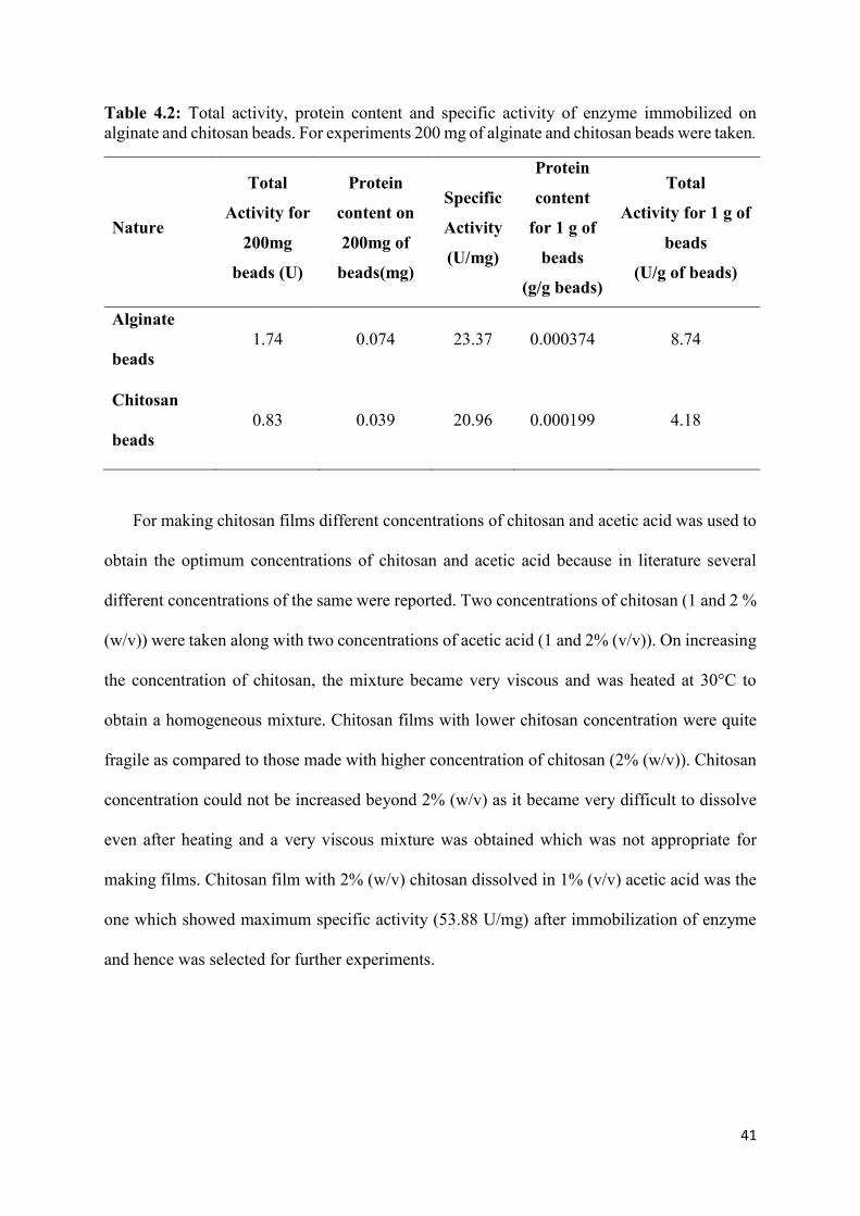
Table 4.2: Total activity, protein content and specific activity of enzyme immobilized on alginate and chitosan beads. For experiments 200 mg of alginate and chitosan beads were taken.
Nature
Total
Activity for
200mg
beads (U)
Protein
content on
200mg of
beads(mg)
Specific
Activity
(U/mg)
Protein
content
for 1 g of
beads
(g/g beads)
Total
Activity for 1 g of
beads
(U/g of beads)
Alginate
beads 1.74 0.074 23.37 0.000374 8.74
Chitosan
beads 0.83 0.039 20.96 0.000199 4.18
For making chitosan films different concentrations of chitosan and acetic acid was used to
obtain the optimum concentrations of chitosan and acetic acid because in literature several
different concentrations of the same were reported. Two concentrations of chitosan (1 and 2 %
(w/v)) were taken along with two concentrations of acetic acid (1 and 2% (v/v)). On increasing
the concentration of chitosan, the mixture became very viscous and was heated at 30°C to
obtain a homogeneous mixture. Chitosan films with lower chitosan concentration were quite
fragile as compared to those made with higher concentration of chitosan (2% (w/v)). Chitosan
concentration could not be increased beyond 2% (w/v) as it became very difficult to dissolve
even after heating and a very viscous mixture was obtained which was not appropriate for
making films. Chitosan film with 2% (w/v) chitosan dissolved in 1% (v/v) acetic acid was the
one which showed maximum specific activity (53.88 U/mg) after immobilization of enzyme
and hence was selected for further experiments.

42
Table 4.3: Optimization of chitosan concentration and acetic acid percentage for chitosan film preparation.
Chitosan concentration
(g)
Acetic acid
(%)
Specific Activity
(U/mg)
1 1 30.22
1 2 20.14
2 1 53.88
2 2 22.48
4.2.1. Effect of temperature on free and immobilized enzyme
Temperature is known to have a very significant effect on the activity of enzymes. Most of
the enzymes denature at higher temperature which is the reason why enzymes are being
immobilized. In industrial processes the temperatures are very high which are not favourable
for enzymes. Immobilization of enzymes has been proven to aid in the stability of enzymes at
higher temperatures. In this study the effect of temperature on both free and immobilized
enzyme was done to see which one is more effective for the enzyme to be used at a higher
temperature.
The effect of temperature on free enzyme, alginate beads, chitosan beads and chitosan film
were studied by incubating free and immobilized enzyme in enzyme solution (0.1 mg/ml) at
temperatures ranging from 25-60°C for an hour and then their activity was measured. Free
enzyme showed maximum activity at 30°C as shown in figure 4.2.
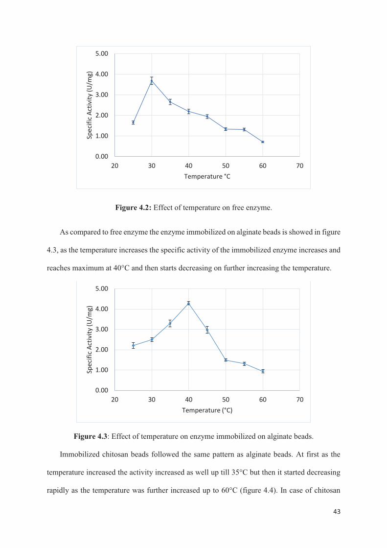
43
0.00
1.00
2.00
3.00
4.00
5.00
20 30 40 50 60 70
Spec
ific A
ctiv
ity (U
/mg)
Temperature °C
0.00
1.00
2.00
3.00
4.00
5.00
20 30 40 50 60 70
Spec
ific A
ctiv
ity (U
/mg)
Temperature (°C)

44
film, the enzyme activity was maximum at 35°C which was 5°C higher than free enzyme
(figure 4.5).
Figure 4.4: Effect of temperature on enzyme immobilized on chitosan beads.
Figure 4.5: Effect of temperature on enzyme immobilized on chitosan film.
Free enzyme showed higher activity at 30°C while alginate beads (40°C), chitosan beads
(35°C) and chitosan films (35°C) showed high activity at higher temperatures than free
enzyme, this difference in temperatures for free and immobilized enzyme indicates that at
1.00
3.00
5.00
7.00
9.00
20 30 40 50 60 70
Spec
ific
act
ibit
y (U
/mg)
Temperature
1.00
2.00
3.00
4.00
5.00
6.00
20 30 40 50 60 70
Spec
ific
Act
ivit
y (U
/mg)
Temperature (°C)

45
higher temperature the support protect the enzyme from denaturation. It has been reported that
that immobilization increases the rigidity of enzyme, which increases the stability towards
increasing temperatures compared to free enzymes in solution (Abdel-Naby 1993). The
decrease in activity after reaching the optimum activity may be due to denaturation of enzyme
at higher temperature which is in concurrence with earlier reported work (R. R. Yadav et al.
2012), (Vinoba et al. 2012). At higher temperatures the protein denatures because of
conformational changes and protein unfolding (Vinoba et al. 2012). Thus, it can be concluded
that at higher temperatures the immobilized enzymes are more stable than free enzyme.
Alginate beads show highest specific activity at 40°C and chitosan beads at 35°C. In case
of alginate beads the enzyme is entrapped in the beads while in case of chitosan the enzyme
has been adsorbed on the beads. Enzyme is more stable at higher temperature when
immobilized by entrapment than by physical adsorption. In case of physical adsorption, the
enzymes are released from the support at higher temperature. These results coincide with those
reported by Ohtakara et al, (1988) whose report suggest that immobilization of glucoamylase
on chitosan beads showed lesser stability on physical adsorption as compared to that of
entrapment or ionic bonding (Skjak-Braek et al. 1989).
4.2.2. Effect of pH on free and immobilized enzyme
One of the most enzyme activity altering parameter in an aqueous medium is pH. Change
in pH can alter the shape of protein which can lead to altered protein recognition or the
enzyme might lose its activity. pH is a measure of H+ ions and therefore a good indicator of
OH- ions. The charges on H+ and OH- ions interfere with the hydrogen and ionic bond that
hold together an enzyme, since they will be repelled or attracted by the charges created by the
bonds. This interference causes a change in the shape of the enzyme. Once the shape of
enzyme changes the substrate cannot bind to it. pH alterations not only change the shape of

46
the enzyme but also the charge on the substrate because of which the substrate cannot bind to
the active site and cannot undergo catalysis.
For determining the effect of pH on the activity of free and immobilized enzyme, they
were incubated for an hour in Tris-HCl buffer prepared at pH ranging from 5.5-10. The
specific activity of the free and immobilized enzyme was calculated to obtain the pH at which
each of them showed the highest activity.
From figure 4.6 it can be seen that as the pH of the buffer was increased the enzyme
activity also increased but after reaching the maximum activity (pH 8) it started decreasing.
Alginate beads also showed the same pattern of increase in activity with increase in pH, with
highest specific activity at pH 8 followed by a decreasing pattern (figure 4.7). According to
literature, for both free enzyme and enzyme immobilized on alginate beads the pH with
highest activity has been reported close to 8.5 which coincides with the results in this study
(R. R. Yadav et al. 2012).
Figure 4.6: Effect of pH on free enzyme.
0.00
1.00
2.00
3.00
4.00
5.00
5 5.5 6 6.5 7 7.5 8 8.5 9 9.5 10
Spec
ific
Act
ivit
y (U
/mg)
pH

47
Figure 4.7: Effect of pH on enzyme immobilized on alginate beads.
Chitosan beads showed maximum activity at pH 8.5 and for chitosan film the maximum
specific activity was obtained at pH 7.5 (figure 4.8 and 4.9).
Figure 4.8: Effect of pH on enzyme immobilized on chitosan beads.
0.00
1.00
2.00
3.00
4.00
5.00
5 5.5 6 6.5 7 7.5 8 8.5 9 9.5 10
Spec
ific
Act
ivit
y (U
/mg)
pH
1.00
2.00
3.00
4.00
5.00
6.00
7.00
8.00
5 5.5 6 6.5 7 7.5 8 8.5 9 9.5 10 10.5
Spec
ific
Act
ivit
y (U
/mg)
pH

48
Figure 4.9: Effect of pH on enzyme immobilized on chitosan film. Out of all the materials used for immobilization Chitosan beads showed the maximum
stability of enzyme at pH 8.5 as compared to that of free enzyme at pH 8, alginate beads at pH
8 and chitosan film at pH 7.5. This difference in the optimum pH for chitosan beads and
chitosan film is due to the different porosity and adsorption structure (Adarsh et al. 2007),
(Ouyang et al. 2014).
4.3. Sequestration of CO2 by free and immobilized CA
To demonstrate the feasibility of CO2 sequestration the biomimetic approach using CA
from plant domain was done. CA was added to CO2 saturated water containing calcium
chloride solution for enhanced precipitation of carbonate and bicarbonate salts. The
immobilized enzymes were used in place of free enzyme in the process to check sequestration
efficiency of immobilized enzymes. Table 4.4 shows the CaCO3 precipitate formed after
carbonation reaction.
0.00
1.00
2.00
3.00
4.00
5.00
6.00
7.00
8.00
5 5.5 6 6.5 7 7.5 8 8.5 9 9.5 10 10.5
spec
ific
act
ivit
y (u
/mg)
pH

49
Table 4.4: Precipitation catalysed by free and immobilized CA.
Enzyme
Nature
Total
Activity
(U)
Total
Protein
(mg)
Specific
Activity
(U/mg)
CaCO3
Precipitate
(g)
Free enzyme 8.61 1 172.3 0.089
Alginate
Beads
1.74 0.074 23.37 0.018
Chitosan beads 0.83 0.039 20.96 0.009
Chitosan film 0.29 0.017 17.58 0.005
The carbonation capacity of free enzyme was found to be 89 mg as compared to 18mg for
alginate beads, 9 mg for chitosan beads and 5 mg for chitosan film. Sharma and Bhattacharya
(2010) successfully demonstrated the sequestration of CO2 to CaCO3 using indigenous CA
from P. fragi, M. lylae, and M luteus. In another study, carbon composite beads were used for
CA immobilization from Bacillus Pumilus showed 19.22 mg precipitate while the free enzyme
gave 33.6 mg of precipitate (Prabhu et al. 2011). This shows that carbonate deposition was
lower in immobilized enzyme than in case of free enzyme. The lower carbonation rate in
immobilized enzyme can be due to lower accessibility of the active site to the substrate. The
amount of enzyme immobilized also plays a very important role in carbonation because in case
of immobilized enzyme the amount of enzyme that got immobilized on the substrate is very
low as compared to free enzyme. Hence, lower enzyme immobilization means lower number
of active sites for the substrate to bind and therefore lower carbonation.

50
Solubility of CO2 is 3.36 g per 1000 g of water at 0-4°C and 1 atmospheric pressure. Which
means 0.0336 g of CO2 gets dissolved in 10 g of H2O (CO2 saturated water). Stoichiometrically,
100 g of CaCO3 has 44 g of CO2. Theoretically, 0.0076 g of CaCO3 should have been formed
from 0.0336 g of CO2. But, from the actual experiment 0.089 g of CaCO3 was obtained. This
increased amount of CaCO3 could be the result of CO2 that entered the experimental setup
while it was opened to add CaCl2 solution and because the setup was not completely sealed.
4.4. Reusability of immobilized enzyme
For industrial applications of enzyme, reusability is an essential parameter since it can
reduce the cost of enzyme driven processes. The reusability of CA was evaluated for 4 cycles.
The reaction was carried out with in the same manner as CO2 sequestration but after every
cycle the immobilized enzyme was rinsed with Tris-Hcl buffer to neutralize the pH and to
remove any excess ions.
Table 4.5: Summary of precipitate of CaCO3 reaction for immobilized enzyme.
Number of cycles
CaCO3 Precipitate (mg)
Alginate beads Chitosan beads Chitosan film
1 18 9 6
2 12 7 4
3 8 5 3
4 6 3 2.5
Free enzyme could not be reused after one cycle as the enzyme cannot be separated from
the solution. The amount of CaCO3 precipitate formed was highest in case of alginate beads
(18 mg) than chitosan beads (9 mg) and chitosan film (2.5 mg). The decrease in precipitate
formation has been explained because of the leaching effect of enzyme which means that with
each cycle the enzyme on the immobilization material is leaching out (Wanjari et al. 2011).

51
4.5. Storage stability of free and immobilized enzyme
Stability of free and immobilized enzyme was determined by storing them for 4 weeks at
4°C. Samples were taken every week and enzyme activity was assayed.
Figure 4.10: Stability of free and immobilized enzyme over a period of 4 weeks. In case of free enzyme there was a sudden drop in relative activity of the enzyme in the
first week, but after that for the rest of the three weeks there was only 20% lost in activity in
total. Because of this advantageous shelf life observed, it can be concluded that CA obtained
from plants can be used in industries. This observation regarding the good stability of plant CA
enzyme coincides with what has been reported in literature (Pocker et al. 1973), (Bednár et al.
2016).
In case of immobilized enzymes, chitosan beads retained about 83% of its original activity
after 4 weeks while alginate beads retained 76.5% and chitosan film retained 80.75% of its
original activity.
455565758595
105
1 2 3 4REL
ATIV
E A
CTI
VIT
Y (%
)
WEEKS
Free enzyme Alginate beadsChitosan beads Chitosan film

52
Chapter 5 Conclusion

53
For this study, the extraction of carbonic anhydrase was done from a plant source namely
spinach leaves. The extraction and partial purification of carbonic anhydrase was done
successfully with total activity of 1759.64 Units.
The partially purified enzyme was then immobilized on sodium alginate beads, chitosan beads
and chitosan film to study the effectiveness of two immobilization techniques on two
supports/materials. The immobilization method was used because it is an easy and cheap
method to show better reusability and to preserve the stability of enzyme. Optimum
temperature and pH for both free and immobilized enzyme was studied. The optimum
temperature for immobilized enzyme was better than free enzyme. Free enzyme showed
optimum temperature at 30°C, alginate beads had optimum temperature of 40°C while chitosan
beads and film had an optimum temperature of 35°C both. The optimum pH for free enzyme
was 8 which is lower than that of chitosan beads which is 8.5. Alginate beads and chitosan film
showed optimum pH at 8 and 7.5 respectively. Chitosan beads showed higher relative activity
in case of optimum temperature and pH.
Transformation of CO2 to CaCO3 was carried out with the help of both free and immobilized
enzymes. Free enzyme produced 89 mg of precipitate per 8.6 U of enzyme activity. Reusability
of the immobilized enzymes was performed up till the 4 cycles. Both the free and immobilized
enzymes produced relatively same amount of precipitate per unit enzyme. Solubility of CO2 is
3.36 g per 1000 g of water at 0-4°C and 1 atmospheric pressure. Which means 0.0336 g of CO2
gets dissolved in 10 g of H2O (CO2 saturated water). Theoretically, 0.0076 g of CaCO3 should
have been formed from 0.0336 g of CO2. But, from the actual experiment 0.089 g of CaCO3
was obtained. This increased amount of CaCO3 could be the result of CO2 that entered the
experimental setup while it was opened to add CaCl2 solution and because the setup was not
completely sealed.

54
Stability of free and immobilized was also compared over a period of 4 weeks. The free enzyme
showed a relatively better stability than the enzymes extracted from microorganisms.
Immobilized enzymes showed better relative stability than free enzyme. Free enzyme retained
about 68% of its activity, while chitosan beads retained the maximum relative activity 83% as
compared to chitosan film (80.75%) and alginate beads (76.59%). This shows that the
immobilized enzyme can be more useful.

55
Chapter 6 Recommendations for future work

56
Recommendations for future work include:
Listed below are some are some suggestions for future work with Carbonic Anhydrase:
• Different plant sources can be utilized to check the CO2 sequestration purposes.
• Cross linking of enzyme can be done during immobilization to prevent desorption or
leaching of immobilized enzyme.
• Chemical methods of immobilization can be explored for stability and reuse of enzyme.
• Membrane immobilization of plant-based enzyme can be done for separation of CO2
from flue gases.

57
Chapter 7 References

58
Abdel-Naby, Mohamed A. 1993. “Immobilization OfAspergillus Niger NRC 107 Xylanase
and β-Xylosidase, and Properties of the Immobilized Enzymes.” Applied Biochemistry
and Biotechnology 38 (1–2). Humana Press: 69–81.
https://doi.org/10.1007/BF02916413.
Abu-Khader, Mazen M. 2006. “Recent Progress in CO 2 Capture/Sequestration: A Review.”
Energy Sources, Part A: Recovery, Utilization, and Environmental Effects 28 (14):
1261–79. https://doi.org/10.1080/009083190933825.
Adarsh, and G Madhu. 2007. “A Comparative Study on Metal Adsorption Properties of
Different Forms of Chitosan.” International Journal of Innovative Research in Science,
Engineering and Technology (An ISO. Vol. 3297. www.ijirset.com.
Adler, L, J Brundell, S O Falkbring, and P O Nyman. 1972. “Carbonic Anhydrase from
Neisseria Sicca, Strain 6021. I. Bacterial Growth and Purification of the Enzyme.”
Biochimica et Biophysica Acta 284 (1): 298–310.
http://www.ncbi.nlm.nih.gov/pubmed/4627452.
Aggarwal, Mayank, Christopher D. Boone, Bhargav Kondeti, and Robert McKenna. 2013.
“Structural Annotation of Human Carbonic Anhydrases.” Journal of Enzyme Inhibition
and Medicinal Chemistry 28 (2). Taylor & Francis: 267–77.
https://doi.org/10.3109/14756366.2012.737323.
Alber, B E, and J G Ferry. 1994. “A Carbonic Anhydrase from the Archaeon Methanosarcina
Thermophila.” Proceedings of the National Academy of Sciences of the United States of
America 91 (15): 6909–13. http://www.ncbi.nlm.nih.gov/pubmed/8041719.
Alissandratos, Apostolos, and Christopher J Easton. 2015. “Biocatalysis for the Application
of CO 2 as a Chemical Feedstock.” Beilstein Journal of Organic Chemistry 11 (1).
Beilstein-Institut: 2370–87. https://doi.org/10.3762/bjoc.11.259.
Amin, Mohamad Hassan, and Suresh K Bhargava. 2018. “A Mini-Review on CO 2
Reforming of Methane.” https://doi.org/10.31031/PPS.2018.02.000532.
Amornvadee Veawab, * and Paitoon Tontiwachwuthikul, and Amit Chakma. 1999.
“Corrosion Behavior of Carbon Steel in the CO2 Absorption Process Using Aqueous

59
Amine Solutions.” American Chemical Society . https://doi.org/10.1021/IE9901630.
B Krajewska. 2004. “Application of Chitin-and Chitosan-Based Materials for Enzyme
Immobilizations: A Review.” Elsevier.
https://www.sciencedirect.com/science/article/pii/S0141022904001231.
Barton, Emily E, David M Rampulla, and Andrew B Bocarsly. 2008. “Selective Solar-Driven
Reduction of CO 2 to Methanol Using a Catalyzed.” J. Am. Chem. Soc 130: 6342–44.
https://doi.org/10.1021/ja0776327.
Battle, M., M.L. Bender, P.P. Tans, J.W.C. White, J.T. Ellis, T. Conway, and R.J. Francey.
2000. “Global Carbon Sinks and Their Variability Inferred from Atmospheric [O.Sub.2]
and [[Delta].Sup.13]C.” Science 287 (5462). American Association for the
Advancement of Science: 2467–2467.
http://go.galegroup.com/ps/i.do?p=AONE&sw=w&issn=00368075&v=2.1&it=r&id=G
ALE%7CA61793513&sid=googleScholar&linkaccess=fulltext&authCount=1&u=ocul_
lakehead&selfRedirect=true.
Bednár, András, Nándor Nemestóthy, Péter Bakonyi, László Fülöp, Guangyin Zhen, Xueqin
Lu, Takuro Kobayashi, Gopalakrishnan Kumar, Kaiqin Xu, and Katalin Bélafi-Bakó.
2016. “Enzymatically-Boosted Ionic Liquid Gas Separation Membranes Using Carbonic
Anhydrase of Biomass Origin.” Chemical Engineering Journal 303 (November).
Elsevier: 621–26. https://doi.org/10.1016/J.CEJ.2016.05.148.
Bhattacharya, Sumana, Marc Schiavone, Subhra Chakrabarti, and Sanjoy K. Bhattacharya.
2003. “CO2 Hydration by Immobilized Carbonic Anhydrase.” Biotechnology and
Applied Biochemistry 38 (2). Wiley-Blackwell: 111.
https://doi.org/10.1042/BA20030060.
Boone, Christopher D., Sonika Gill, Andrew Habibzadegan, and Robert McKenna. 2013.
“Carbonic Anhydrase: An Efficient Enzyme with Possible Global Implications.”
International Journal of Chemical Engineering 2013: 22–27.
https://doi.org/10.1155/2013/813931.
Boone, Christopher, Andrew Habibzadegan, Sonika Gill, and Robert McKenna. 2013.
“Carbonic Anhydrases and Their Biotechnological Applications.” Biomolecules 3 (4).
Multidisciplinary Digital Publishing Institute: 553–62.
https://doi.org/10.3390/biom3030553.

60
Brena, Beatriz, Paula González-Pombo, and Francisco Batista-Viera. 2013. “Immobilization
of Enzymes: A Literature Survey.” Methods in Molecular Biology 1051.
https://doi.org/10.1007/978-1-62703-550-7_2.
Datta, Sumitra, L. Rene Christena, and Yamuna Rani Sriramulu Rajaram. 2013. “Enzyme
Immobilization: An Overview on Techniques and Support Materials.” 3 Biotech 3 (1).
Springer Berlin Heidelberg: 1–9. https://doi.org/10.1007/s13205-012-0071-7.
Dhanwantri, K, P Sharma, S Mehta, and P Prakash. 2014. “Carbon Sequestration , Its
Methods and Significance.” In Environmental Sustainability: Concepts, Principles,
Evidences and Innovations, edited by Prof. (Dr.) Govind Chandra Mishra, 1st ed., 151–
57. New delhi: EXCELLENT PUBLISHING HOUSE.
Elleuche, S., and S. Poggeler. 2010. “Carbonic Anhydrases in Fungi.” Microbiology 156 (1):
23–29. https://doi.org/10.1099/mic.0.032581-0.
Eriksson, M, J Karlsson, Z Ramazanov, P Gardeström, and G Samuelsson. 1996. “Discovery
of an Algal Mitochondrial Carbonic Anhydrase: Molecular Cloning and
Characterization of a Low-CO2-Induced Polypeptide in Chlamydomonas Reinhardtii.”
Proceedings of the National Academy of Sciences of the United States of America 93
(21): 12031–34. http://www.ncbi.nlm.nih.gov/pubmed/8876257.
Fiore, Anna Di, Vincenzo Alterio, Simona M. Monti, Giuseppina De Simone, and Katia
D’Ambrosio. 2015. “Thermostable Carbonic Anhydrases in Biotechnological
Applications.” International Journal of Molecular Sciences 16 (7): 15456–80.
https://doi.org/10.3390/ijms160715456.
Frommer, WB. 2010. “CO2mmon Sense.” Science.Sciencemag.Org.
http://science.sciencemag.org/content/327/5963/275.short.
Fukuzawa, H, S Fujiwara, Y Yamamoto, M L Dionisio-Sese, and S Miyachi. 1990. “CDNA
Cloning, Sequence, and Expression of Carbonic Anhydrase in Chlamydomonas
Reinhardtii: Regulation by Environmental CO2 Concentration.” Proceedings of the
National Academy of Sciences of the United States of America 87 (11): 4383–87.
http://www.ncbi.nlm.nih.gov/pubmed/2112252.
Ganai, Bashir Ahmad. 2017. “Carbonic Anhydrase : Mechanism , Structure and Importance
in Higher Plants” 7 (August): 17–23.
61
Ge, J, RM Cowan, C Tu, … ML McGregor - Life Support &, and Undefined 2002. 2002.
“Enzyme-Based CO2 Capture for Advanced Life Support.” Ingentaconnect.Com.
https://www.ingentaconnect.com/content/cog/lsbs/2002/00000008/f0020003/art00007.
Gill, Steven R., Paula J. Fedorka-Cray, Rodney K. Tweten, and Bayard P. Sleeper. 1984.
“Purification and Properties of the Carbonic Anhydrase of Rhodospirillum Rubrum.”
Archives of Microbiology 138 (2). Springer-Verlag: 113–18.
https://doi.org/10.1007/BF00413010.
Gonz, Javier M, and S Zo. 2014. “Carbonic Anhydrases in Industrial Applications” 75
(September 2014). https://doi.org/10.1007/978-94-007-7359-2.
Goodsell, D.S. 2004. “Carbonic Anhydrase.” RCSB Protein Data Bank, January.
https://doi.org/10.2210/rcsb_pdb/mom_2004_1.
Harish Prashanth, K.V., and R.N. Tharanathan. 2007. “Chitin/Chitosan: Modifications and
Their Unlimited Application Potential—an Overview.” Trends in Food Science &
Technology 18 (3). Elsevier: 117–31. https://doi.org/10.1016/J.TIFS.2006.10.022.
Hewett-Emmett, David. 2000. “Evolution and Distribution of the Carbonic Anhydrase Gene
Families.” In The Carbonic Anhydrases, 29–76. Basel: Birkhäuser Basel.
https://doi.org/10.1007/978-3-0348-8446-4_3.
Homaei, Ahmad Abolpour, Reyhaneh Sariri, Fabio Vianello, and Roberto Stevanato. 2013.
“Enzyme Immobilization: An Update.” Journal of Chemical Biology 6 (4). Springer:
185–205. https://doi.org/10.1007/s12154-013-0102-9.
Ingall, Ellery D., Julia M. Diaz, Amelia F. Longo, Michelle Oakes, Lydia Finney, Stefan
Vogt, Barry Lai, Patricia L. Yager, Benjamin S. Twining, and Jay A. Brandes. 2013.
“Role of Biogenic Silica in the Removal of Iron from the Antarctic Seas.” Nature
Communications 4 (June). https://doi.org/10.1038/ncomms2981.
Initiative, The Arabidopsis Genome. 2000. “Analysis of the Genome Sequence of the
Flowering Plant Arabidopsis Thaliana.” Nature 408 (6814). Nature Publishing Group:
796–815. https://doi.org/10.1038/35048692.
IPCC. 2005. “IPCC Special Report on Carbon Dioxide Capture and Storage, Prepared by
Working Group III of the Intergovernmental Panel on Climate Change [Metz, B., O.
Davidson, H. C. de Coninck, M. Loos, and L. A. Meyer (Eds.)].” Cambridge University

62
Press, Cambridge, United Kingdom and New York, NY, USA.
https://www.ipcc.ch/pdf/special-reports/srccs/srccs_wholereport.pdf.
Jansson, Christer, Stan D. Wullschleger, Udaya C. Kalluri, and Gerald A. Tuskan. 2010a.
“Phytosequestration: Carbon Biosequestration by Plants and the Prospects of Genetic
Engineering.” BioScience 60 (9): 685–96. https://doi.org/10.1525/bio.2010.60.9.6.
———. 2010b. “Phytosequestration: Carbon Biosequestration by Plants and the Prospects of
Genetic Engineering.” BioScience 60 (9). Oxford University Press: 685–96.
https://doi.org/10.1525/bio.2010.60.9.6.
Jansson, Christer, Stan D Wullschleger, Udaya C Kalluri, and Gerald A Tuskan. 2010c.
“Phytosequestration: Carbon Biosequestration by Plants and the Prospects of Genetic
Engineering.” BioScience 60 (685). https://doi.org/10.1525/bio.2010.60.9.6.
Kaar, JL, HI Oh, AJ Russell, WJ Federspiel - Biomaterials, and Undefined 2007. 2007.
“Towards Improved Artificial Lungs through Biocatalysis.” Elsevier.
https://www.sciencedirect.com/science/article/pii/S0142961207002360.
Kargupta, Wriju, Anuradda Ganesh, and Suparna Mukherji. 2015. “Estimation of Carbon
Dioxide Sequestration Potential of Microalgae Grown in a Batch Photobioreactor.”
Bioresource Technology 180 (March). Elsevier: 370–75.
https://doi.org/10.1016/J.BIORTECH.2015.01.017.
Kimmel, J. D., D. T. Arazawa, S.-H. Ye, V. Shankarraman, W. R. Wagner, and W. J.
Federspiel. 2013. “Carbonic Anhydrase Immobilized on Hollow Fiber Membranes
Using Glutaraldehyde Activated Chitosan for Artificial Lung Applications.” Journal of
Materials Science: Materials in Medicine 24 (11). Springer US: 2611–21.
https://doi.org/10.1007/s10856-013-5006-2.
Klara, Scott M, Rameshwar D Srivastava, and Howard G McIlvried. 2003. “Integrated
Collaborative Technology Development Program for CO2 Sequestration in Geologic
Formations––United States Department of Energy R&D.” Energy Conversion and
Management 44 (17). Pergamon: 2699–2712. https://doi.org/10.1016/S0196-
8904(03)00042-6.
Kovscek, Ar. 2004. “Geologic Storage of Carbon Dioxide and Enhanced Oil Recovery. II.
Cooptimization of Storage and Recovery.”

63
https://doi.org/10.1016/j.enconman.2004.09.009.
Kumar, R. Siva Sai, and James G. Ferry. 2014. “Prokaryotic Carbonic Anhydrases of Earth’s
Environment.” In Sub-Cellular Biochemistry, 75:77–87. https://doi.org/10.1007/978-94-
007-7359-2_5.
Kupriyanova, E., N. Pronina, and D. Los. 2017. “Carbonic Anhydrase — a Universal
Enzyme of the Carbon-Based Life.” Photosynthetica 55 (1): 3–19.
https://doi.org/10.1007/s11099-017-0685-4.
Lal, R. 2004. “Soil Carbon Sequestration Impacts on Global Climate Change and Food
Security.” Science (New York, N.Y.) 304 (5677). American Association for the
Advancement of Science: 1623–27. https://doi.org/10.1126/science.1097396.
Lal, Rattan. 2008. “Carbon Sequestration.” Philosophical Transactions of the Royal Society
of London. Series B, Biological Sciences 363 (1492). The Royal Society: 815–30.
https://doi.org/10.1098/rstb.2007.2185.
Lamb, David, Peter D Erskine, and John A Parrotta. 2005. “Restoration of Degraded Tropical
Forest Landscapes.” Science (New York, N.Y.) 310 (5754). American Association for the
Advancement of Science: 1628–32. https://doi.org/10.1126/science.1111773.
Lampitt, R.S, E.P Achterberg, T.R Anderson, J.A Hughes, M.D Iglesias-Rodriguez, B.A
Kelly-Gerreyn, M Lucas, et al. 2008. “Ocean Fertilization: A Potential Means of
Geoengineering?” Philosophical Transactions of the Royal Society A: Mathematical,
Physical and Engineering Sciences 366 (1882): 3919–45.
https://doi.org/10.1098/rsta.2008.0139.
Lane, T. W., and F. M. M. Morel. 2000. “A Biological Function for Cadmium in Marine
Diatoms.” Proceedings of the National Academy of Sciences 97 (9): 4627–31.
https://doi.org/10.1073/pnas.090091397.
Larjavaara, Markku. 2008. “A Review on Benefits and Disadvantages of Tree Diversity.” The
Open Forest Science Journal 1: 24–26.
http://www.lerf.eco.br/img/publicacoes/2008_2611 A Review on Benefits and
Disadvantages of Tree Diversity.pdf.
Lee JF, Thirumavalavan M, and Lee JF. 2015. “A Short Review on Chitosan Membrane for
Biomolecules Immobilization.” Journal of Molecular and Genetic Medicine 09 (03).

64
OMICS International: 1–5. https://doi.org/10.4172/1747-0862.1000178.
Lehtonen, J, B Shen, M Vihinen, … A Casini - Journal of Biological, and Undefined 2003.
2003. “Characterization of CA XIII, a Novel Member of the Carbonic Anhydrase
Isozyme Family.” ASBMB.
http://www.jbc.org/content/early/2003/11/04/jbc.M308984200.full.pdf.
Lindskog, S. 1997. “Structure and Mechanism of Carbonic Anhydrase.” Elsevier.
https://www.sciencedirect.com/science/article/pii/S0163725896001982.
Lindskog, Sven, and Per Olof Nyman. 1964. “Metal-Binding Properties of Human
Erythrocyte Carbonic Anhydrases.” Biochimica et Biophysica Acta (BBA) - Specialized
Section on Enzymological Subjects 85 (3). Elsevier: 462–74.
https://doi.org/10.1016/0926-6569(64)90310-4.
Liu, Li, Amit Chakma, and Xianshe Feng. 2004. “Preparation of Hollow Fiber Poly(Ether
Block Amide)/Polysulfone Composite Membranes for Separation of Carbon Dioxide
from Nitrogen.” Chemical Engineering Journal 105 (1–2). Elsevier: 43–51.
https://doi.org/10.1016/J.CEJ.2004.08.005.
Lowry, Oliver H, Rose J Randall, and A Lewis. 1951. “The Folin by Oliver.” Readings 193
(1): 265–75. https://doi.org/10.1016/0304-3894(92)87011-4.
MacAuley, Sheridan R., Sabrina A. Zimmerman, Ethel E. Apolinario, Caryn Evilia, Ya-Ming
Hou, James G. Ferry, and Kevin R. Sowers. 2009. “The Archetype γ-Class Carbonic
Anhydrase (Cam) Contains Iron When Synthesized in Vivo †.” Biochemistry 48 (5).
American Chemical Society: 817–19. https://doi.org/10.1021/bi802246s.
Madhumati Mondal, Saumyakanti Khanra. O.N. Towari, K. Gayen, G.N. Halder. 2016. “Role
of Carbonic Anhydrase on the Way to Biological Carbon Capture through Microalgae-A
Mini Review.” Environmental Progress & Sustainable Energy 35 (6): 1605–15.
https://doi.org/10.1002/ep.
Magalhães, Júlia M.C.S., and Adélio A.S.C. Machado. 1998. “Urea Potentiometric Biosensor
Based on Urease Immobilized on Chitosan Membranes.” Talanta 47 (1): 183–91.
https://doi.org/10.1016/S0039-9140(98)00066-6.
Marcus, E. A., A. P. Moshfegh, G. Sachs, and D. R. Scott. 2005. “The Periplasmic -Carbonic
Anhydrase Activity of Helicobacter Pylori Is Essential for Acid Acclimation.” Journal

65
of Bacteriology 187 (2): 729–38. https://doi.org/10.1128/JB.187.2.729-738.2005.
Marianne K., Gornall A. 1978. “Carbonic Anhydrase from Spinach Leaves.” The Journal of
Biological Chemistry 253 (3): 679–85.
Matear, Richard J., and Bronwyn Elliott. 2004. “Enhancement of Oceanic Uptake of
Anthropogenic CO 2 by Macronutrient Fertilization.” Journal of Geophysical Research
109 (C4): C04001. https://doi.org/10.1029/2000JC000321.
Mazzotti, Marco, and Switzerland Italy. 2007. “Mineral Carbonation and Industrial Uses of
Carbon Dioxide 319 7 Mineral Carbonation and Industrial Uses of Carbon Dioxide.”
IPCC Special Report on Carbon Dioxide Capture and Storage.
https://www.ipcc.ch/pdf/special-reports/srccs/srccs_chapter7.pdf.
Metting, F. Blaine, Jeffrey L. Smith, Jeffrey S. Amthor, and R. Cesar Izaurralde. 2001.
“Science Needs and New Technology for Increasing Soil Carbon Sequestration.”
Climatic Change 51 (1). Kluwer Academic Publishers: 11–34.
https://doi.org/10.1023/A:1017509224801.
Mirjafari, Parissa, Koorosh Asghari, and Nader Mahinpey. 2007. “Investigating the
Application of Enzyme Carbonic Anhydrase for CO 2 Sequestration Purposes.”
Industrial and Engineering Chemistry Research 46 (3): 921–26.
https://doi.org/10.1021/ie060287u.
Mitsuhashi, S, T Mizushima, E Yamashita, M Yamamoto, T Kumasaka, H Moriyama, T
Ueki, S Miyachi, and T Tsukihara. 2000. “X-Ray Structure of Beta-Carbonic Anhydrase
from the Red Alga, Porphyridium Purpureum, Reveals a Novel Catalytic Site for CO(2)
Hydration.” The Journal of Biological Chemistry 275 (8): 5521–26.
http://www.ncbi.nlm.nih.gov/pubmed/10681531.
Morais, Michele Greque de, and Jorge Alberto Vieira Costa. 2007. “Carbon Dioxide Fixation
by Chlorella Kessleri, C. Vulgaris, Scenedesmus Obliquus and Spirulina Sp. Cultivated
in Flasks and Vertical Tubular Photobioreactors.” Biotechnology Letters 29 (9). Springer
Netherlands: 1349–52. https://doi.org/10.1007/s10529-007-9394-6.
Moroney, J. V., S. G. Bartlett, and G. Samuelsson. 2001. “Carbonic Anhydrases in Plants and
Algae.” Plant, Cell and Environment 24 (2). Blackwell Science Ltd: 141–53.
https://doi.org/10.1111/j.1365-3040.2001.00669.x.

66
Neish, A C. 1939. “Studies on Chloroplasts: Their Chemical Composition and the
Distribution of Certain Metabolites between the Chloroplasts and the Remainder of the
Leaf.” The Biochemical Journal 33 (3). Portland Press Ltd: 300–308.
http://www.ncbi.nlm.nih.gov/pubmed/16746912.
Nishimori, Isao, Tomoko Minakuchi, Saburo Onishi, Daniela Vullo, Andrea Scozzafava, and
Claudiu T. Supuran. 2007. “Carbonic Anhydrase Inhibitors. DNA Cloning,
Characterization, and Inhibition Studies of the Human Secretory Isoform VI, a New
Target for Sulfonamide and Sulfamate Inhibitors.” Journal of Medicinal Chemistry 50
(2): 381–88. https://doi.org/10.1021/jm0612057.
Nishimori, Isao, Daniela Vullo, Alessio Innocenti, Andrea Scozzafava, Antonio
Mastrolorenzo, and Claudiu T. Supuran. 2005. “Carbonic Anhydrase Inhibitors. The
Mitochondrial Isozyme VB as a New Target for Sulfonamide and Sulfamate Inhibitors.”
Journal of Medicinal Chemistry 48 (24): 7860–66. https://doi.org/10.1021/jm050483n.
Nogia, Panchsheela, Gurpreet Kaur Sidhu, Rajesh Mehrotra, and Sandhya Mehrotra. 2016.
“Capturing Atmospheric Carbon: Biological and Nonbiological Methods.” International
Journal of Low-Carbon Technologies 11 (2): 266–74.
https://doi.org/10.1093/ijlct/ctt077.
O’Connor, William K., David C. Dahlin, David N. Nilsen, G.E. Rush, Richard P. Walters,
and Paul C. Turner. 2001. “Carbon Dioxide Sequestration by Direct Mineral
Carbonation: Results from Recent Studies and Current Status.” National Energy
Technology Laboratory, U.S. Department of Energy.
https://www.osti.gov/biblio/897125.
Oelkers, E. H., S. R. Gislason, and J. Matter. 2008. “Mineral Carbonation of CO2.” Elements
4 (5). GeoScienceWorld: 333–37. https://doi.org/10.2113/gselements.4.5.333.
Okabe, K, SY Yang, M Tsuzuki, S Miyachi - Plant Science Letters, and Undefined 1984.
1984. “Carbonic Anhydrase: Its Content in Spinach Leaves and Its Taxonomic Diversity
Studied with Anti-Spinach Leaf Carbonic Anhydrase Antibody.” Elsevier.
https://www.sciencedirect.com/science/article/pii/030442118490004X.
Oren, Ram, David S. Ellsworth, Kurt H. Johnsen, Nathan Phillips, Brent E. Ewers, Chris
Maier, Karina V.R. Schäfer, et al. 2001. “Soil Fertility Limits Carbon Sequestration by
Forest Ecosystems in a CO2-Enriched Atmosphere.” Nature 411 (6836). Nature

67
Publishing Group: 469–72. https://doi.org/10.1038/35078064.
Ouyang, An, and Ji Liang. 2014. “Tailoring the Adsorption Rate of Porous Chitosan and
Chitosan-Carbon Nanotube Core-Shell Beads †.” https://doi.org/10.1039/c4ra04131c.
Oviya, M., V. Sukumaran, and Sib Sankar Giri. 2013. “Immobilization and Characterization
of Carbonic Anhydrase Purified from E. Coli MO1 and Its Influence on
CO2sequestration.” World Journal of Microbiology and Biotechnology 29 (10): 1813–
20. https://doi.org/10.1007/s11274-013-1343-z.
Ozdemir, E. 2009. “Biomimetic CO2 Sequestration: 1. Immobilization of Carbonic
Anhydrase within Polyurethane Foam.” ACS Publications.
https://pubs.acs.org/doi/abs/10.1021/ef9005725.
Ozdemir, Ekrem. 2009a. “Biomimetic CO 2 Sequestration: 1. Immobilization of Carbonic
Anhydrase within Polyurethane Foam.” Energy & Fuels 23 (11): 5725–30.
https://doi.org/10.1021/ef9005725.
———. 2009b. “Biomimetic CO 2 Sequestration: 1. Immobilization of Carbonic Anhydrase
within Polyurethane Foam.” Energy & Fuels 23 (11): 5725–30.
https://doi.org/10.1021/ef9005725.
Pachauri, Rajendra K, Leo Meyer, Jean-Pascal Van Ypersele, Sander Brinkman, Line Van
Kesteren, Noëmie Leprince-Ringuet, and Fijke Van Boxmeer. 2014. Climate Change
2014 Synthesis Report The Core Writing Team Core Writing Team Technical Support
Unit for the Synthesis Report. Russian Federation), Hoesung Lee (Republic of Korea)
Scott B. Power (Australia) N.H. Ravindranath (India). IPCC.
http://www.ipcc.ch/pdf/assessment-report/ar5/syr/SYR_AR5_FINAL_full_wcover.pdf.
Park, H, PJ McGinn, and FMM Morel. 2008. “Expression of Cadmium Carbonic Anhydrase
of Diatoms in Seawater.” Aquatic Microbial Ecology 51 (2): 183–93.
https://doi.org/10.3354/ame01192.
Paustian, K., J. Six, E.T. Elliott, and H. W. Hunt. 2000. “Management Options for Reducing
CO2 Emissions from Agricultural Soils.” Biogeochemistry 48 (1). Kluwer Academic
Publishers: 147–63. https://doi.org/10.1023/A:1006271331703.
Pierre, Alain C. 2012. “Enzymatic Carbon Dioxide Capture.” ISRN Chemical Engineering
2012: 1–22. https://doi.org/10.5402/2012/753687.

68
Pocker, Y., and Joan S.Y. Ng. 1973. “Plant Carbonic Anhydrase. Properties and Carbon
Dioxide Hydration Kinetics.” Biochemistry 12 (25): 5127–34.
https://doi.org/10.1021/bi00749a016.
Prabhu, Chandan, Aarti Valechha, Snehal Wanjari, Nitin Labhsetwar, Swati Kotwal, T.
Satyanarayanan, and Sadhana Rayalu. 2011. “Carbon Composite Beads for
Immobilization of Carbonic Anhydrase.” Journal of Molecular Catalysis B: Enzymatic
71 (1–2): 71–78. https://doi.org/10.1016/j.molcatb.2011.03.011.
Prete, Sonia Del, Viviana De Luca, Andrea Scozzafava, Vincenzo Carginale, Claudiu T.
Supuran, and Clemente Capasso. 2014. “Biochemical Properties of a New α-Carbonic
Anhydrase from the Human Pathogenic Bacterium, Vibrio Cholerae.” Journal of
Enzyme Inhibition and Medicinal Chemistry 29 (1): 23–27.
https://doi.org/10.3109/14756366.2012.747197.
Ramanan, R, and K Kannan. 2009. “Bio-Sequestration of Carbon Dioxide Using Carbonic
Anhydrase Enzyme Purified from Citrobacter Freundii.” Springer.
https://idp.springer.com/authorize/casa?redirect_uri=https://link.springer.com/article/10.
1007/s11274-009-9975-
8&casa_token=ABweLeni3wgAAAAA:JdZrJ3ptdNEcVpAZwRTCb8n9W2ZBiWRi-
DxXj4uR2_hZRbYkowS1DiEf6xDA2xFnR4WG2GUcRXvctqxR.
Ramanan, Rishiram, Krishnamurthi Kannan, Nadimuthu Vinayagamoorthy, Kunga Mohan
Ramkumar, Saravana Devi Sivanesan, and Tapan Chakrabarti. 2009a. “Purification and
Characterization of a Novel Plant-Type Carbonic Anhydrase from Bacillus Subtilis.”
Biotechnology and Bioprocess Engineering 14 (1): 32–37.
https://doi.org/10.1007/s12257-008-0099-z.
———. 2009b. “Purification and Characterization of a Novel Plant-Type Carbonic
Anhydrase from Bacillus Subtilis.” Biotechnology and Bioprocess Engineering 14 (1).
The Korean Society for Biotechnology and Bioengineering: 32–37.
https://doi.org/10.1007/s12257-008-0099-z.
Rathnam, CKM, VSR Das - Zeitschrift für Pflanzenphysiologie, and Undefined 1975. 1975.
“Aspartate-Type C-4 Photosynthetic Carbon Metabolism in Leaves of Eleusine
CoracanaGaertn.” Elsevier.
https://www.sciencedirect.com/science/article/pii/S0044328X75801486.

69
Reichle, Dave, John Houghton, Sally Benson, John Clarke, Roger Dahlman, Doe George
Hendrey, Howard Herzog, et al. 1999. “Carbon Sequestration DRAFT (February
1999).” https://www.netl.doe.gov/publications/press/1999/seqrpt.pdf.
Ren, Xiaoling, Jizhong Ren, Hui Li, Shichao Feng, and Maicun Deng. 2012. “Poly (Amide-6-
b-Ethylene Oxide) Multilayer Composite Membrane for Carbon Dioxide Separation.”
International Journal of Greenhouse Gas Control 8 (May). Elsevier: 111–20.
https://doi.org/10.1016/J.IJGGC.2012.01.017.
Riebeek, Holli. 2011. “The Carbon Cycle : Feature Articles,” June. NASA Earth Observatory.
https://earthobservatory.nasa.gov/Features/CarbonCycle/.
Roberts, Samantha B., Todd W. Lane, and Francois M. M. Morel. 1997. “CARBONIC
ANHYDRASE IN THE MARINE DIATOM THALASSIOSIRA WEISSFLOGII
(BACILLARIOPHYCEAE)1.” Journal of Phycology 33 (5). Blackwell Publishing Ltd:
845–50. https://doi.org/10.1111/j.0022-3646.1997.00845.x.
Rudenko, N. N., L. K. Ignatova, T. P. Fedorchuk, and B. N. Ivanov. 2015. “Carbonic
Anhydrases in Photosynthetic Cells of Higher Plants.” Biochemistry (Moscow) 80 (6):
674–87. https://doi.org/10.1134/S0006297915060048.
S. J. Metz, and M. H. V. Mulder, and M. Wessling*. 2004. “Gas-Permeation Properties of
Poly(Ethylene Oxide) Poly(Butylene Terephthalate) Block Copolymers.” American
Chemical Society . https://doi.org/10.1021/MA049847W.
Satav, Sunita S., Shreedhar Bhat, and S. Thayumanavan. 2010. “Feedback Regulated Drug
Delivery Vehicles: Carbon Dioxide Responsive Cationic Hydrogels for Antidote
Release.” Biomacromolecules 11 (7): 1735–40. https://doi.org/10.1021/bm1005454.
Sharma, Anjana, Abhishek Bhattacharya, and Ankita Shrivastava. 2011. “Biomimetic
CO2sequestration Using Purified Carbonic Anhydrase from Indigenous Bacterial Strains
Immobilized on Biopolymeric Materials.” Enzyme and Microbial Technology 48 (4–5):
416–26. https://doi.org/10.1016/j.enzmictec.2011.02.001.
Sharma, Anjana, Abhishek Bhattacharya, and Shyamlata Singh. 2009. “Purification and
Characterization of an Extracellular Carbonic Anhydrase from Pseudomonas Fragi.”
Process Biochemistry 44 (11). Elsevier: 1293–97.
https://doi.org/10.1016/J.PROCBIO.2009.07.022.

70
Sharma, J, R Mahajan, and VK Gupta. 2010. “Comparison and Suitability of Gel Matrix for
Entrapping Higher Content of Enzymes for Commercial Applications.” Indian Journal
of Pharmaceutical Sciences 72 (2). OMICS International: 223.
https://doi.org/10.4103/0250-474X.65010.
Shazia Faridi Satyanarayana T. 2015. “Applicability of Carbonic Anhydrases in Mitigating
Global Warming and Development of Useful Products from CO2.” Climate Change and
Environmental Sustainability 3(2) (December): 77–92.
Sheppard, Michael. 2018. “Carbon Capture and Sequestration.” Society of Petroleum
Engineers. http://www.spe.org/industry/docs/carbonsequestration.pdf.
Simsek-Ege, Fatma Arzum, Gillian M. Bond, and John Stringer. 2002. “Matrix Molecular
Weight Cut-off for Encapsulation of Carbonic Anhydrase in Polyelectrolyte Beads.”
Journal of Biomaterials Science, Polymer Edition 13 (11): 1175–87.
https://doi.org/10.1163/156856202320892948.
Sipilä, J., S. Teir, and R. Zevenhoven. 2008. “Carbon Dioxide Sequestration by Mineral
Carbonation Literature Review Update 2005–2007.”
Skjak-Braek, Gudmund., Thorleif. Anthonsen, Paul A. Sandford, and Norway) International
Conference on Chitin and Chitosan (4th : 1988 : Trondheim. 1989. Chitin and Chitosan :
Sources, Chemistry, Biochemistry, Physical Properties, and Applications. London ;
Elsevier Applied Science,. https://searchworks.stanford.edu/view/1351905.
Smith, K S, and J G Ferry. 2000. “Prokaryotic Carbonic Anhydrases.” FEMS Microbiology
Reviews 24 (4): 335–66. http://www.ncbi.nlm.nih.gov/pubmed/10978542.
Soltes-Rak, E, M E Mulligan, and J R Coleman. 1997. “Identification and Characterization of
a Gene Encoding a Vertebrate-Type Carbonic Anhydrase in Cyanobacteria.” Journal of
Bacteriology 179 (3). American Society for Microbiology (ASM): 769–74.
http://www.ncbi.nlm.nih.gov/pubmed/9006032.
Supuran, Claudiu T. 2008a. “Carbonic Anhydrases: Novel Therapeutic Applications for
Inhibitors and Activators.” Nature Reviews Drug Discovery 7 (2): 168–81.
https://doi.org/10.1038/nrd2467.
———. 2008b. “Carbonic Anhydrases: Novel Therapeutic Applications for Inhibitors and
Activators.” Nature Reviews Drug Discovery 7 (2). Nature Publishing Group: 168–81.

71
https://doi.org/10.1038/nrd2467.
———. 2013. “Carbonic Anhydrases: From Biomedical Applications of the Inhibitors and
Activators to Biotechnological Use for CO2capture.” Journal of Enzyme Inhibition and
Medicinal Chemistry 28 (2): 229–30. https://doi.org/10.3109/14756366.2013.761876.
Supuran, Claudiu T., Andrea Scozzafava, and Angela Casini. 2003. “Carbonic Anhydrase
Inhibitors.” Medicinal Research Reviews 23 (2). Wiley-Blackwell: 146–89.
https://doi.org/10.1002/med.10025.
Supuran, Claudiu T., Andrea Scozzafava, and Janet. Conway. 2004. Carbonic Anhydrase : Its
Inhibitors and Activators. CRC Press.
The Late U Meldrum, BY N, and F J W Roughton. 1933. “CARBONIC ANHYDRASE. ITS
PREPARATION AND PROPERTIES.” The Physiological and Biochemical
Laboratories, Cambridge.
https://www.ncbi.nlm.nih.gov/pmc/articles/PMC1394121/pdf/jphysiol01616-0010.pdf.
Thiry, A, JM Dogne, B Masereel, CT Supuran - Trends in pharmacological, and undefined
2006. n.d. “Targeting Tumor-Associated Carbonic Anhydrase IX in Cancer Therapy.”
Elsevier. Accessed August 2, 2018.
https://www.sciencedirect.com/science/article/pii/S0165614706002203.
Thompson, Richard B., and Eric R. Jones. 1993. “Enzyme-Based Fiber Optic Zinc
Biosensor.” Analytical Chemistry 65 (6). American Chemical Society: 730–34.
https://doi.org/10.1021/ac00054a013.
Tripp, Brian C., Kerry Smith, and James G. Ferry. 2001. “Carbonic Anhydrase: New Insights
for an Ancient Enzyme.” Journal of Biological Chemistry 276 (52): 48615–18.
https://doi.org/10.1074/jbc.R100045200.
Tsuzuki, M, S Miyachi, GE Edwards - Plant and cell Physiology, and Undefined 1985. 1985.
“Localization of Carbonic Anhydrase in Mesophyll Cells of Terrestrial C3 Plants in
Relation to CO2 Assimilation.” Academic.Oup.Com.
https://academic.oup.com/pcp/article-abstract/26/5/881/1856766.
Vinoba, Mari, Margandan Bhagiyalakshmi, Soon Kwan Jeong, Yeo I I Yoon, and Sung Chan
Nam. 2012. “Immobilization of Carbonic Anhydrase on Spherical SBA-15 for
Hydration and Sequestration of CO 2.” Colloids and Surfaces B:

72
Biointerfaces 90 (1): 91–96. https://doi.org/10.1016/j.colsurfb.2011.10.001.
Vullo, D, J Voipio, A Innocenti, C Rivera, … H Ranki - Bioorganic & medicinal, and
undefined 2005. n.d. “Carbonic Anhydrase Inhibitors. Inhibition of the Human Cytosolic
Isozyme VII with Aromatic and Heterocyclic Sulfonamides.” Elsevier. Accessed August
2, 2018. https://www.sciencedirect.com/science/article/pii/S0960894X04015136.
Walker, Tara L., Chris Collet, and Saul Purton. 2005. “ALGAL TRANSGENICS IN THE
GENOMIC ERA1.” Journal of Phycology 41 (6). Blackwell Science Inc: 1077–93.
https://doi.org/10.1111/j.1529-8817.2005.00133.x.
Wanjari, Snehal, Chandan Prabhu, Renu Yadav, T. Satyanarayana, Nitin Labhsetwar, and
Sadhana Rayalu. 2011. “Immobilization of Carbonic Anhydrase on Chitosan Beads for
Enhanced Carbonation Reaction.” Process Biochemistry 46 (4). Elsevier Ltd: 1010–18.
https://doi.org/10.1016/j.procbio.2011.01.023.
Warrier, Rekha R, Suriyanarayanan Lalitha, and Chellapan Savitha. 2014. “A Modified
Assay of Carbonic Anhydrase Activity in Tree Species.” BBR - Biochemistry and
Biotechnology Reports 3 (1): 48. https://doi.org/10.5433/2316-5200.2014v3n1p48.
Whittington, A, A Waheed, and Baebara U. 2001. “Crystal Structure of the Dimeric
Extracellular Domain of Human Carbonic Anhydrase XII, a Bitopic Membrane Protein
Overexpressed in Certain Cancer Tumor Cells.” National Acad Sciences.
http://www.pnas.org/content/98/17/9545.short.
Whittingtons, DA, JH Grubb, A Waheed, … GN Shah - Journal of Biological, and Undefined
2004. 2004. “Expression, Assay, and Structure of the Extracellular Domain of Murine
Carbonic Anhydrase XIV IMPLICATIONS FOR SELECTIVE INHIBITION OF
MEMBRANE.” ASBMB. http://www.jbc.org/content/279/8/7223.short.
Wong, Tuck Seng. 2014. “Carbon Dioxide Capture and Utilization Using Biological
Systems: Opportunities and Challenges.” Journal of Bioprocessing & Biotechniques 04
(03). https://doi.org/10.4172/2155-9821.1000155.
Yadav, Raju R., Kannan Krishnamurthi, Sandeep N. Mudliar, S. Saravana Devi, Pravin K.
Naoghare, Amit Bafana, and Tapan Chakrabarti. 2014. “Carbonic Anhydrase Mediated
Carbon Dioxide Sequestration: Promises, Challenges and Future Prospects.” Journal of
Basic Microbiology 54 (6): 472–81. https://doi.org/10.1002/jobm.201300849.

73
Yadav, Raju R., Sandeep N. Mudliar, Ajam Y. Shekh, Abhay B. Fulke, Sivanesan Saravana
Devi, Kannan Krishnamurthi, Asha Juwarkar, and Tapan Chakrabarti. 2012.
“Immobilization of Carbonic Anhydrase in Alginate and Its Influence on Transformation
of CO2to Calcite.” Process Biochemistry 47 (4): 585–90.
https://doi.org/10.1016/j.procbio.2011.12.017.
Yadav, Renu, Snehal Wanjari, Chandan Prabhu, Vivek Kumar, Nitin Labhsetwar, T.
Satyanarayanan, Swati Kotwal, and Sadhana Rayalu. 2010. “Immobilized Carbonic
Anhydrase for the Biomimetic Carbonation Reaction.” Energy & Fuels 24 (11).
American Chemical Society: 6198–6207. https://doi.org/10.1021/ef100750y.
Yamasaki, Akihiro. 2003. “An Overview of CO2 Mitigation Options for Global Warming-
Emphasizing CO2 Sequestration Options.” JOURNAL OF CHEMICAL ENGINEERING
OF JAPAN 36 (4): 361–75. https://doi.org/10.1252/jcej.36.361.
Yang, Hongqun, Zhenghe Xu, Maohong Fan, Rajender Gupta, Rachid B Slimane, Alan E
Bland, and Ian Wright. 2008. “Progress in Carbon Dioxide Separation and Capture: A
Review.” Journal of Environmental Sciences 20 (1). Elsevier: 14–27.
https://doi.org/10.1016/S1001-0742(08)60002-9.
Yeates, Todd O., Cheryl A. Kerfeld, Sabine Heinhorst, Gordon C. Cannon, and Jessup M.
Shively. 2008. “Protein-Based Organelles in Bacteria: Carboxysomes and Related
Microcompartments.” Nature Reviews Microbiology 6 (9). Nature Publishing Group:
681–91. https://doi.org/10.1038/nrmicro1913.
Yee, Donald, and François M. M. Morel. 1996. “In Vivo Substitution of Zinc by Cobalt in
Carbonic Anhydrase of a Marine Diatom.” Limnology and Oceanography 41 (3): 573–
77. https://doi.org/10.4319/lo.1996.41.3.0573.
Yuan, Q, S Ouyang, A Wang, W Zhu, … R Maiti - Plant, and Undefined 2005. 2005. “The
Institute for Genomic Research Osa1 Rice Genome Annotation Database.” Am Soc Plant
Biol. http://www.plantphysiol.org/content/138/1/18.short.
Zeng, Ning. 2008. “Carbon Sequestration via Wood Burial.” Carbon Balance and
Management 3 (1). Nature Publishing Group: 1. https://doi.org/10.1186/1750-0680-3-1.