case presentation - suny downstate medical · pdf filecase presentation zchief complaint: ......
TRANSCRIPT

Case PresentationCase Presentation
Karoline Nowillo, MDLong Island College Hospital

Case PresentationCase Presentation
Chief complaint: xx year old woman with a two week history of progressively worsening shortness of breath and epigastric painPast medical history: high impact motor vehicle accident 3 years prior with pelvic fractures and pelvic wall hematoma, hypertension, gastritisPast surgical history: tubal ligationMedications: accupril, prevacid, allegra

Case PresentationCase Presentation
Vital signs: RR 40 28 98% O2 Sat on RA T 97.2 BP 110/77 HR 111 BMI 27
Physical exam:General in distress Head NCATChest decreased breath sounds on left, tachypnicCardiac S1 S2 tachycardicAbdomen softExtremities within normal
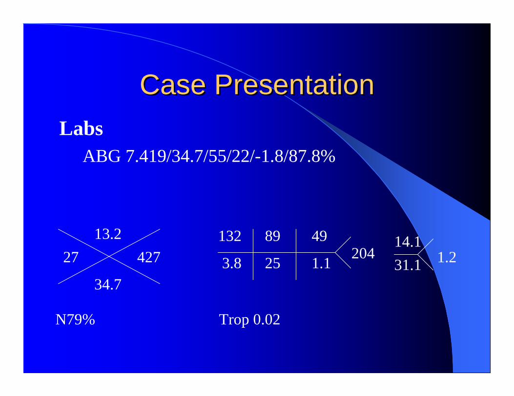
Case PresentationCase PresentationLabs
ABG 7.419/34.7/55/22/-1.8/87.8%
Trop 0.02
2713.2
34.7
427
N79%
14.131.1 1.2
132 89 49
3.8 25 1.1 204

Chest XChest X--rayray
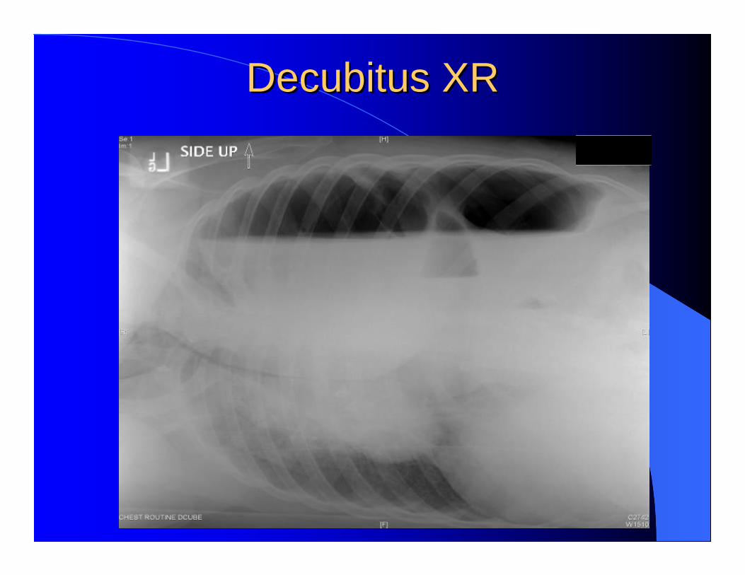
Decubitus XRDecubitus XR

Left Tube ThoracostomyLeft Tube Thoracostomy
Left Tube ThoracostomyLeft Tube Thoracostomy
Almost immediate relief of symptoms. Approximately one liter of foul smelling fluid drained.Culture klebsiella pneumonia, streptococcus viridans, moderate yeast.Admit to thoracic surgery service.Started on zosyn, fluconazole.

Upper GI Study Upper GI Study

Laparotomy: Gastric PerforationLaparotomy: Gastric Perforation

Left Lobe of LiverLeft Lobe of Liver

Repair of Diaphragmatic Repair of Diaphragmatic DefectDefect
ThoracotomyThoracotomy
Patient placed in right decubitus positionLeft sided posterolateral thoracotomyEvacuation of debris, clots and hernia sacLeft chest tube insertedReapproximation of chest wall and muscle

Gross SpecimensGross Specimens
liver fundus spleen

Chest XR Chest XR PrePre--operativeoperative PostPost--operative operative
PostPost--operative Courseoperative CoursePOD 0 patient remained in recovery room POD 1 extubated, given pneumovax and transferred to SICUPOD 2 transferred to surgical floorPOD 4 started clears and advanced to regular dietPOD 8 and 10 intubated, bronchoscopy and positive pressure ventilation in an attempt to expand the left lower lobePOD 13 extubated and returned to surgical floorPOD 18 chest tube removedPOD 19 discharged home
FollowFollow--upupAt one month post op patient offers no complaints. Physical exam:General no acute distressHead NCATChest decreased air entry left baseAbdomen benignSkin scars maturingReturn for follow up in one year.
Traumatic Traumatic Diaphragmatic HerniasDiaphragmatic Hernias
Diagnosis and Management Diagnosis and Management

Formed between 3rd and 8th week of gestation
Central tendon originates from transverse septum
Lateral portions from pleuroperitoneal folds
Fusion eliminates communication between thorax and abdomen
Diaphragm EmbryologyDiaphragm Embryology

Nerves: right phrenic and left phrenic.
Arterial supply: pericardiophrenic, small direct branches from the abdominal aorta.
Venous drainage: follows arterial supply.
Insertion: anteriorly at xyphoid process, laterally to the lower 6 ribs, posteriorly L1 to L3 vertebral bodies, dome rises as high as 4th intercostal space.
Function: generates tidal volume & intrapleural pressures between -5 to -10 cmH2O.
Anatomy and PhysiologyAnatomy and Physiology

Diaphragmatic Hernias: Diaphragmatic Hernias: NontraumaticNontraumatic
1. Sternocostal foramina of Morgagni
2. Esophageal hiatus
3. Lumbocostal foramina of Bochdalek

Traumatic Diaphragmatic InjuryTraumatic Diaphragmatic Injury
Incidence– 0.8 to 5% of all abdominal injuries– up to 5% in patients with multiple traumas
More common in penetrating trauma v. blunt75% affect left hemidiaphragm,23% right hemidiaphragm &2% bilateral in clinical practice13.7% mortality rate
Mattox, KL et al. Trauma. 5th edition. 2004.Shah, R, et al. Traumatic rupture of the diaphragm. Annals of Thoracic Surgery. 1995; 60: 1444-1449.
Clinical PresentationClinical PresentationMarked respiratory distress.Decreased breath sounds.Decreased function of diaphragm, compression of lungs, shifting of mediastinum.Hemodynamic compromise.

Clinical FeaturesClinical Features
Diagnosis begins with a high index of suspicion.Associated injuries: pelvic fracture, splenic and hepatic injuries, thoracic aortic tears.Spontaneous closure of a rupture does not occur.Delayed herniation may present with strangulation or dyspnea.

DiagnosisDiagnosis
Chest X ray– Abdominal contents in thorax– Nasogastric tube in thorax– Elevated hemidiaphragm– Shift of mediastinum– Blunting of costophrenic angle– Reported 13-62% diagnostic
inaccuracy
Eren, S. Diaphragmatic hernia: diagnostic approaches with reviewof literature. European Journal of Radiology. 2005 Jun; 54(3): 448-459.

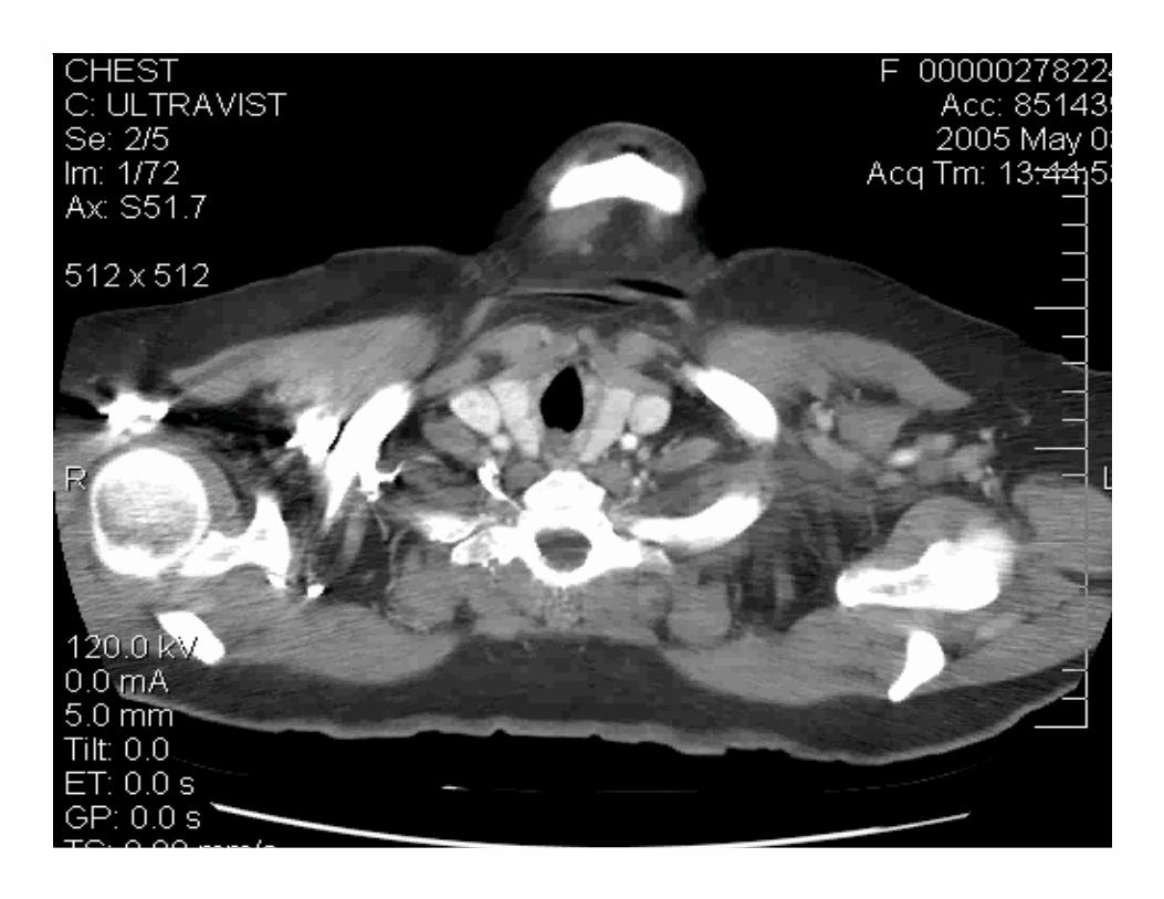
DiagnosisDiagnosisCT scan– May miss injury if no herniation.
– Coronal and sagittal reconstructions most
effective.
– Shows associated complications: strangulation,
hemothorax.
Contrast study- barium– Diagnose gastrointestinal herniation.
MRI– Use on selected patients: late presentation, diagnostic doubt.
– Clearly reveals attachment sites.
– Logistically problematic in acute setting.
Eren, S. Diaphragmatic hernia: diagnostic approaches with review of literature. European Journal of Radiology. 2005 Jun; 54(3): 448-459.

DiagnosisDiagnosisDiagnostic peritoneal lavage– Penetrating and blunt trauma.
– Sensitivity approaches 100% when rbc >10,000/mm3.
– Rbc of 1000/mm3 yield negative explorations of about 20%.
– Specificity is low.
Laparoscopy– May be used for diagnosis and repair.
– Reported sensitivity 87.5% in prospective trial for penetrating injuries.
– Awaiting long term outcomes.
Friese, RS et al. Laparoscopy is sufficient to exclude occult diaphragm injury after penetrating abdominal trauma. Journal of Trauma. 2005 Apr; 58(4): 789-92.
Spann, JC et al. Evaluation of VATS in the diagnosis of diaphragmatic injuries. American Journal of Surgery. 1995 Dec; 170 (6): 628-30.

DiagnosisDiagnosisThoracoscopy– In presence of pneumothorax or hemothorax
– May have diagnostic accuracy comparable to laparotomy
Exploratory laparotomy – Performed in patients with suspected injury
– High rate of negative findings
Ultrasound/ doppler– Shows diaphragmatic discontinuity, herniated organs
– Reveals associated abdominal organ pathologies
– Herniation of vessels from omentum and abdominal organs converging into hernia sac
Blaivas, M. et al. Bedside emergency ultrasonographic diagnosis of diaphragmatic rupture in blunt abdominal trauma. American journal of emergency medicine. 2004; 22(7): 601-604.
Eren, S. Diaphragmatic hernia: diagnostic approaches with review of literature. European Journal of Radiology. 2005 Jun; 54(3): 448-459.

Management Traumatic Management Traumatic HerniaHernia
Diagnosis is indication for repair.Acute injuries may be repaired during exploratory laparotomy.Chronic post-traumatic hernias are best done through chest or combined thorascopic/ abdominal approach.Number 1 monofilament permanent suture.Polypropylene mesh for defects >25cm2.Laparoscopic repair reported.
Thoman, DS, et al. Laparoscopic diaphragmatic hernia repair. Surgical endoscopy (2002) 16: 1345-1349.


ConclusionsConclusions
Diaphragmatic injuries may be misdiagnosed or missed on initial evaluation of the trauma patient.Delayed diagnosis carries a high morbidity due to the risk of incarceration or strangulation.High index of suspicion is required to diagnose these injuries.Transabdominal route in acute rupture is preferred.Chronic herniation should be approached through the chest, with a laparotomy when indicated.