case report double threaded screw fixation for...
TRANSCRIPT
Case ReportDouble Threaded Screw Fixation for Bilateral StressFracture of the Medial Malleolus
Ryo Kanto,1 Shigeo Fukunishi,1 Takatoshi Morooka,1 Daisuke Seino,1
Takayuki Takashima,2 Shinichi Yoshiya,1 and Juichi Tanaka1
1 Department of Orthopaedic Surgery, Hyogo College of Medicine, 1-1 Mukogawa-cho, Nishinomiya, Hyogo 663-8501, Japan2 Takashima Clinic, 1-13-22 Sawaraginishi, Ibaraki, Osaka 567-0868, Japan
Correspondence should be addressed to Ryo Kanto; [email protected]
Received 31 October 2013; Accepted 18 December 2013; Published 28 January 2014
Academic Editors: P. Carpintero, Y.-J. Chen, M. Massobrio, and D. Saragaglia
Copyright © 2014 Ryo Kanto et al. This is an open access article distributed under the Creative Commons Attribution License,which permits unrestricted use, distribution, and reproduction in any medium, provided the original work is properly cited.
An 18-year-old college basketball player presented with continued ankle pain. A radiographic examination showed bilateral medialmalleolus stress fractures. Considering the prolonged history and refractory nature of this injury, surgerywas adopted as a treatmentoption. At surgery, the fracture site was percutaneously fixed using two cannulated double threaded screws. Surgery for each sidewas sequentially performed two months apart. Prompt bony healing was attained after surgery, and the patient could return to hisprevious sports level six months after the first surgery without subsequent recurrence.
1. Introduction
Repetitive loading during regular strenuous sports activitymay cause stress fractures necessitating interruption of play.Its incidence in athletes has been reported to be around 2.0%[1–3]. The most common location is on the posteromedial-concave side of the tibial shaft [1], while stress fractures ofmedial malleolus are extremely rare. Shelbourne described6 patients with stress fracture of this type in 1988 [4].All of the patients were involved in running or jumpingactivities such as basketball, long distance running, andfootball. The typical clinical sequence of this stress fractureis gradual onset of pain and discomfort over the medialmalleolus followed by prolonged symptoms.The radiologicalappearance is characterized by a vertically oriented fissureoriginating from the tibial plafond and medial malleolusjunction or an obliquely arched radiolucent line through themedial malleolus; however, routine radiographs often appearnormal at initial presentation. Therefore, for patients withclinical features suggestive of this stress fracture, bone scanor MRI may be considered for early diagnosis [5–7].
Since this fracture is mostly encountered among highlevel athletes, prompt diagnosis with aggressive interventionis critical to enable early return to original sports activity.
The basic treatment option for stress fractures, in general,is conservative measure consisting of cessation of runningand jumping activities; however, stress fracture of the medialmalleolus is often complicated with delayed healing or recur-rence necessitating surgery.
In this case report, we present a case of a college basketballplayer, who sustained bilateral medial malleolus stress frac-tures and sequentially underwent surgical treatment usingdouble threaded screws.Hewas able to successfully go back tothe original sports activity following the bilateral surgeries. Inprevious literatures, bilateral medial malleolus stress fracturehas been reported in only one paper (in Germany) by Steckelet al. [8].
2. A Case Report
An 18-year-old college basketball player presented with dis-comfort and pain in his left ankle. He noted the symptomsapproximately a month before his initial visit to our hospitalwithout a history of preceding ankle injury. He had playedbasketball during his junior high and high school period andjust started his career with the first season of the Division Icollege basketball league.
Hindawi Publishing CorporationCase Reports in OrthopedicsVolume 2014, Article ID 729035, 4 pageshttp://dx.doi.org/10.1155/2014/729035
2 Case Reports in Orthopedics
(a) (b)
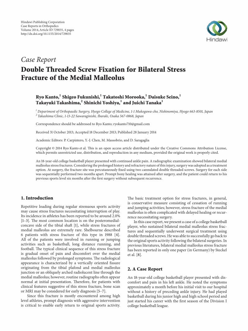
Figure 1: (a) T1-weighted MR image demonstrates a vertical linear lesion of low signal intensity originating from the superomedial corner ofthe ankle. (b) STIR (short T1 inversion recovery) image reveals bone marrow edema surrounding the linear lesion.
(a) (b)
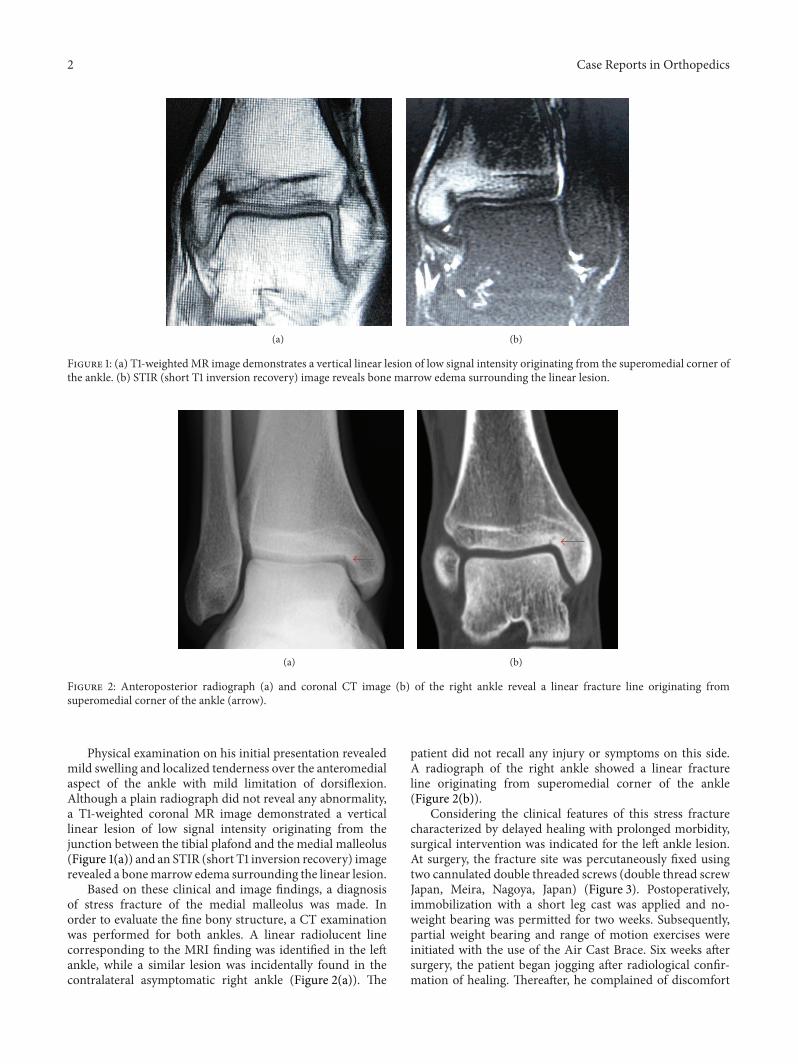
Figure 2: Anteroposterior radiograph (a) and coronal CT image (b) of the right ankle reveal a linear fracture line originating fromsuperomedial corner of the ankle (arrow).
Physical examination on his initial presentation revealedmild swelling and localized tenderness over the anteromedialaspect of the ankle with mild limitation of dorsiflexion.Although a plain radiograph did not reveal any abnormality,a T1-weighted coronal MR image demonstrated a verticallinear lesion of low signal intensity originating from thejunction between the tibial plafond and the medial malleolus(Figure 1(a)) and an STIR (short T1 inversion recovery) imagerevealed a bonemarrow edema surrounding the linear lesion.
Based on these clinical and image findings, a diagnosisof stress fracture of the medial malleolus was made. Inorder to evaluate the fine bony structure, a CT examinationwas performed for both ankles. A linear radiolucent linecorresponding to the MRI finding was identified in the leftankle, while a similar lesion was incidentally found in thecontralateral asymptomatic right ankle (Figure 2(a)). The
patient did not recall any injury or symptoms on this side.A radiograph of the right ankle showed a linear fractureline originating from superomedial corner of the ankle(Figure 2(b)).
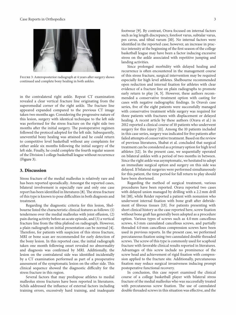
Considering the clinical features of this stress fracturecharacterized by delayed healing with prolonged morbidity,surgical intervention was indicated for the left ankle lesion.At surgery, the fracture site was percutaneously fixed usingtwo cannulated double threaded screws (double thread screwJapan, Meira, Nagoya, Japan) (Figure 3). Postoperatively,immobilization with a short leg cast was applied and no-weight bearing was permitted for two weeks. Subsequently,partial weight bearing and range of motion exercises wereinitiated with the use of the Air Cast Brace. Six weeks aftersurgery, the patient began jogging after radiological confir-mation of healing. Thereafter, he complained of discomfort
Case Reports in Orthopedics 3
Figure 3: Anteroposterior radiograph at 4 years after surgery showscontinued and complete bony healing in both ankles.
in the contralateral right ankle. Repeat CT examinationrevealed a clear vertical fracture line originating from thesuperomedial corner of the right ankle. The fracture lineappeared expanded compared to the previous CT imagetaken two months ago. Considering the progressive nature ofthis lesion, surgery with identical technique to the left sidewas performed for the stress fracture on the right side twomonths after the initial surgery. The postoperative regimenfollowed the protocol adopted for the left side. Subsequently,uneventful bony healing was attained and he could returnto competitive level basketball without any complaints foreither ankle six months following the initial surgery of theleft side. Finally, he could complete the fourth regular seasonof the Division I college basketball league without recurrence(Figure 3).
3. Discussion
Stress fracture of the medial malleolus is relatively rare andhas been reported sporadically. Amongst the reported cases,bilateral involvement is especially rare and only one casereport has been identified in literatures [8].The stress fractureof this type is known to pose difficulties in both diagnosis andtreatment.
Regarding the diagnostic criteria for this lesion, Shel-bourne listed the characteristic clinical features as follows: (1)tenderness over the medial malleolus with joint effusion, (2)pain during activity before an acute episode, and (3) a verticalfracture line from the tibia plafond on radiograph. However,a plain radiograph on initial presentation can be normal [4].Therefore, for patients with suspicion of this stress fracture,MRI or bone scan are recommended for early detection ofthe bony lesion. In this reported case, the initial radiographtaken one month following onset revealed no abnormalityand diagnosis was confirmed by MRI. Additionally, thelesion on the contralateral side was identified incidentallyby a CT examination performed as part of a preoperativeassessment of the symptomatic lesion on the other side. Thisclinical sequence showed the diagnostic difficulty for thestress fracture in this region.
Several factors that may predispose athletes to medialmalleolus stress fractures have been reported in literatures.Schils addressed the influence of external factors includingtraining errors, excessively hard training, and inadequate
footwear [9]. By contrast, Orava focused on internal factorssuch as leg length discrepancy, forefoot varus, subtalar varus,pes cavus, and tibial varum [10]. No internal factors wereidentified in the reported case; however, an increase in prac-tice intensity at the beginning of the first season of the collegebasketball league may have been a factor inducing excessivestress on the ankle associated with repetitive jumping andlanding activities.
Since prolonged morbidity with delayed healing andrecurrence is often encountered in the management courseof this stress fracture, surgical intervention may be requiredespecially for high level athletes. Shelbourne recommendedopen reduction and internal fixation for athletes with clearevidence of a fracture line on plain radiographs to promoteearly return to play [4, 5]. However, these authors recom-mended a conservative treatment option with casting forcases with negative radiographic findings. In Orava’s caseseries, five of the eight patients were successfully managedwith conservative treatment while surgery was required forthree patients with fractures with displacement or delayedhealing. A recent article by these authors (Orava et al.) in2012 reported a clinical course of 10 patients who underwentsurgery for this injury [11]. Among the 10 patients includedin this case series, surgery was indicated for five patients afterfailed attempts of conservative treatment. Based on the reviewof previous literatures, Shabat et al. concluded that surgicaltreatment can be considered as a primary option for high levelathletes [12]. In the present case, we sequentially operatedon bilateral ankles with a period of two months in between.Since the right ankle was asymptomatic, we hesitated to adoptan immediate surgical option and surgery on this side wasdelayed. If bilateral surgeries were performed simultaneouslyfor this patient, the time period for full return to play shouldhave been shortened.
Regarding the method of surgical treatment, variousprocedures have been reported. Orava reported two caseswith delayed union managed by drilling with a 2.2mm drillbit [10], while Reider reported a patient with nonunion whounderwent internal fixation with bone graft after debride-ment of fibrous tissues [13]. For patients presenting withshort clinical history as the case reported here, screw fixationwithout bone graft has generally been adopted as a procedureoption. Various types of screws such as 4.0mm cancellousscrews, 6.5mm cannulated cancellous screws, and partiallythreaded 4.0mm cancellous compression screws have beenused in previous reports. In the present case, we performedpercutaneous fixation using two cannulated double threadedscrews.The screw of this type is commonly used for scaphoidfracture with favorable clinical results reported in literatures.Advantages of this screw include no prominence of thescrew head and achievement of rigid fixation with compres-sion applied to the fracture site. Additionally, percutaneousfixation may reduce surgical invasiveness inducing promptpostoperative functional recovery.
In conclusion, this case report examined the clinicalcourse of a college basketball player with bilateral stressfracture of the medial malleolus who was successfully treatedwith percutaneous screw fixation. The use of cannulateddouble threaded screws in this situation was effective, and the
4 Case Reports in Orthopedics
patient could return to full sports activity sixmonths after thefirst surgery and continued to play at the competitive level forthe subsequent years without recurrence.
Conflict of Interests
The authors declare that there is no conflict of interestsregarding the publication of this paper.
References
[1] J. Iwamoto and T. Takeda, “Stress fractures in athletes: reviewof 196 cases,” Journal of Orthopaedic Science, vol. 8, no. 3, pp.273–278, 2003.
[2] B. Goldberg and C. Pecora, “Stress fractures: a risk of increasedtraining in freshmen,” Physician and Sportsmedicine, vol. 22, no.3, pp. 68–76, 1994.
[3] P. S. Sherbondy and W. J. Sebastianelli, “Stress fractures of themedial malleolus and distal fibula,” Clinics in Sports Medicine,vol. 25, no. 1, pp. 129–137, 2006.
[4] K. D. Shelbourne, D. A. Fisher, A. C. Rettig, and J. R.McCarroll,“Stress fractures of the medial malleolus,”TheAmerican Journalof Sports Medicine, vol. 16, no. 1, pp. 60–63, 1988.
[5] J. Brockwell, Y. Yeung, and J. F. Griffith, “Stress fractures of thefoot and ankle,” SportsMedicine and Arthroscopy Review, vol. 17,no. 3, pp. 149–159, 2009.
[6] K. Okada, S. Senma, E. Abe, K. Sato, and S. Minato, “Stressfractures of the medial malleolus: a case report,” Foot and AnkleInternational, vol. 16, no. 1, pp. 49–52, 1995.
[7] M. Ariyoshi, K. Nagata, K. Hiraoka, K. Sonoda, R. Hori, andA. Inoue, “Stress fracture of the medial malleolus,” KurumeMedical Journal, vol. 44, no. 3, pp. 233–236, 1997.
[8] H. Steckel, H. M. Klinger, M. H. Baums, and W. Schultz,“Beidseitige stressfraktur des malleolus medialis,” SportverletzSportschaden, vol. 19, no. 1, pp. 41–45, 2005.
[9] J. P. Schils, J. T. Andrish, D. W. Piraino, G. H. Belhobek, B. J.Richmond, and J. A. Bergfeld, “Medial malleolar stress fracturesin seven patients,” Radiology, vol. 185, pp. 219–221, 1992.
[10] S. Orava, J. Karpakka, S. Taimela, A. Hulkko, J. Permi, and U.Kujala, “Stress fracture of the medial malleolus,”The Journal ofBone and Joint Surgery A, vol. 77, no. 3, pp. 362–365, 1995.
[11] L. Lempainen, E. Liimatainen, J. Heikkila et al., “Medialmalleolar stress fracture in athletes: diagnosis and operativetreatment,” Scandinavian Journal of Surgery, vol. 101, no. 4, pp.261–264, 2012.
[12] S. Shabat, K. B. Sampson, G. Mann et al., “Stress fractures ofthe medial malleolus—review of the literature and report of a15-year-old elite gymnast,” Foot and Ankle International, vol. 23,no. 7, pp. 647–650, 2002.
[13] B. Reider, R. Falconiero, J. Yurkofsky, and J. C. Hughston,“Nonunion of a medial malleolus stress fracture: a case report,”The American Journal of Sports Medicine, vol. 21, no. 3, pp. 478–481, 1993.

Submit your manuscripts athttp://www.hindawi.com
Stem CellsInternational
Hindawi Publishing Corporationhttp://www.hindawi.com Volume 2014
Hindawi Publishing Corporationhttp://www.hindawi.com Volume 2014
MEDIATORSINFLAMMATION
of
Hindawi Publishing Corporationhttp://www.hindawi.com Volume 2014
Behavioural Neurology
EndocrinologyInternational Journal of
Hindawi Publishing Corporationhttp://www.hindawi.com Volume 2014
Hindawi Publishing Corporationhttp://www.hindawi.com Volume 2014
Disease Markers
Hindawi Publishing Corporationhttp://www.hindawi.com Volume 2014
BioMed Research International
OncologyJournal of
Hindawi Publishing Corporationhttp://www.hindawi.com Volume 2014
Hindawi Publishing Corporationhttp://www.hindawi.com Volume 2014
Oxidative Medicine and Cellular Longevity
Hindawi Publishing Corporationhttp://www.hindawi.com Volume 2014
PPAR Research
The Scientific World JournalHindawi Publishing Corporation http://www.hindawi.com Volume 2014
Immunology ResearchHindawi Publishing Corporationhttp://www.hindawi.com Volume 2014
Journal of
ObesityJournal of
Hindawi Publishing Corporationhttp://www.hindawi.com Volume 2014
Hindawi Publishing Corporationhttp://www.hindawi.com Volume 2014
Computational and Mathematical Methods in Medicine
OphthalmologyJournal of
Hindawi Publishing Corporationhttp://www.hindawi.com Volume 2014
Diabetes ResearchJournal of
Hindawi Publishing Corporationhttp://www.hindawi.com Volume 2014
Hindawi Publishing Corporationhttp://www.hindawi.com Volume 2014
Research and TreatmentAIDS
Hindawi Publishing Corporationhttp://www.hindawi.com Volume 2014
Gastroenterology Research and Practice
Hindawi Publishing Corporationhttp://www.hindawi.com Volume 2014
Parkinson’s Disease
Evidence-Based Complementary and Alternative Medicine
Volume 2014Hindawi Publishing Corporationhttp://www.hindawi.com