case report pediatric masked mastoiditis associated with...
TRANSCRIPT

Case ReportPediatric Masked Mastoiditis Associated withMultiple Intracranial Complications
Charalampos Voudouris,1 Ioannis Psarommatis,1 Ioannis Nikas,2
Dimitrios Kafouris,1 and Konstantina Chrysouli1
1ENT Department, “P. & A. Kyriakou” Children’s Hospital of Athens, Athens, Greece2Imaging Department, “Agia Sophia” Children’s Hospital of Athens, Athens, Greece
Correspondence should be addressed to Ioannis Psarommatis; [email protected]
Received 30 April 2015; Accepted 15 June 2015
Academic Editor: Wolfgang Issing
Copyright © 2015 Charalampos Voudouris et al.This is an open access article distributed under the Creative CommonsAttributionLicense, which permits unrestricted use, distribution, and reproduction in anymedium, provided the originalwork is properly cited.
Masked mastoiditis is a distinct form of mastoiditis with little or no symptomatology, characterized by its potential to generatesevere otogenic complications. Therefore, suspected masked mastoiditis should be diagnosed and treated without delay. This studyreports a rare case of masked mastoiditis, manifested by multiple intracranial complications in an immunocompetent girl. Thechild exhibited headache and neurological symptomatology. Imaging studies revealed an epidural and a large cerebellar abscessand the patient was immediately treated with a triple antibiotic therapy. Mastoid surgery and drainage of the epidural abscess tookplace after the stabilization of the patient’s neurologic status, on the 3rd hospitalization day. The cerebellar abscess was treated bycraniectomy and ultrasound-guided needle aspiration in the 3rd week of hospitalization.The girl was finally discharged in excellentcondition. Two years later, she is still in good health, without otological or neurological sequelae. Maskedmastoiditis is an insidiousdisease which requires increased clinical awareness and adequate imaging. Should clinical and/or radiological findings be positive,mastoidectomy must follow in order to prevent severe otogenic complications that can be triggered by masked mastoiditis.
1. Introduction
Masked mastoiditis is a rare yet distinct clinical entity,reported by many authors in the spectrum of intratemporalcomplications of otitis media [1, 2]. It may be definedas a suppurative subclinical process of the mastoid cavity,involving both the mucosa and the bony structures ofthe mastoid air-cell system. An incompletely healed acuteotitis media and the obstruction of the aditus-ad-antrumby mucosal edema and granulation tissue are believed torepresent the underlying pathogenetic mechanism. Underthese circumstances, masked mastoiditis can potentiallyresult in an effusion-freemiddle ear cavity, as the latter drainsthrough the eustachian tube, combinedwith an active chronicinflammation within the blocked mastoid air-cell system[3]. The mastoid inflammation may last weeks, months, oreven years before healing occurs—with or without surgicalintervention—or its complications become symptomatic [4].
We report on an unusual case of pediatric maskedmastoiditis presented with central nervous system (CNS)
symptomatology due to multiple intracranial suppurativecomplications. Hopefully, this report will provide a betterinsight to clinicians in order to better understand the clinicalbehaviour and the aggressiveness of this disease, so thatnew awareness leads to improved diagnostic and therapeuticapproach.
2. Case Report
An 8-year-old girl with no past medical history was takento the emergency department with disequilibrium and gaitinstability, confusion, dysarthria, and vomiting, all mani-fested within the last 24 hours. An episode of acute otitismedia on her right ear two weeks before treated with an oralantibiotic (amoxicillin 90mg/kg/24 h for 10 days), along witha one-week history of vague occipitoparietal headache, wasmentioned by her parents.
During her physical examination, spontaneous nystag-mus, pathologic smooth pursuit eyemovement, adiadochoki-nesia, and positive Romberg test were observed. The child
Hindawi Publishing CorporationCase Reports in OtolaryngologyVolume 2015, Article ID 897239, 4 pageshttp://dx.doi.org/10.1155/2015/897239

2 Case Reports in Otolaryngology
(a) (b)
Figure 1: Coronal (a) and axial (b) MRI sections with contrast on admission, showing a large cerebellar abscess on the right hemisphere(short arrows) coexisting with an epidural abscess of the posterior fossa (long arrows) and inflammatory tissue within the mastoid cavity((a), black arrowhead). The abscesses exert mass effect against the middle-line structures, causing their displacement to the left ((a), whitearrowheads).
developed fever up to 39∘C in the first days after thediagnosis of acute otitis media, but she had no fever inthe last week. Otoscopy revealed a near normal tympanicmembrane, showing only minor redness with a clear lightingreflex. Type A tympanograms were recorded on both ears.Neurologic and neurosurgical consultations followed and thegirl was admitted to the pediatric department and referred toimaging.
Brain magnetic resonance imaging (MRI) revealed animpressive in size abscess cavity, localized at the rightcerebellar hemisphere and extending towards the cerebello-pontine angle. A second, epidural abscess, localized in thesuperior-posterior part of petrous bone was also observed(Figure 1). The right mastoid was poorly developed andopaque, indicating that it was occupied with inflammatorycontent. Magnetic resonance venography yielded no find-ings of thrombophlebitis. Finally, temporal bone computedtomography (CT) without contrast was performed to providethe anatomic relations of the abscesses with the bony struc-tures. Apart from that, CT scans also disclosed the weakenedpoint of the posterior tegmen of the right mastoid, fromwhere the abscess had probably arisen (Figure 2).
The patient was immediately given triple intravenousantibiotic therapy (ceftriaxone, vancomycin, and clin-damycin) and dexamethasone. Mastoid surgery and drainageof the epidural abscess took place after the stabilization of thepatient’s neurologic status, on the 3rd day of the girl’s hospi-talization. Myringotomy revealed an effusion free middle ear.Mastoid cells and antrumwere full of edematousmucosa andinflammatory granulation tissue, causing a substantial blockat the level of aditus. Most of this tissue was removed andan unobstructed communication between middle ear andantrumwas established.While drilling at the posterosuperior
part of the mastoid tegmen, the epidural abscess was cutopen and the purulent exudate filled the surgical field.Specimens were taken for microbiological cultures. Thecommunication between the two compartments (epiduraland mastoid) was further opened to a size of approximately1/2 cm2. Mastoidectomy was concluded by closing in layersand leaving a rubber drain within the mastoid cavity.
Drainage of the cerebellar abscess through a craniectomyand ultrasound-guided needle aspiration and instillation ofantibiotic solution were successfully performed by the neu-rosurgical team on the 3rd week of hospitalisation. Culturesfrom both abscesses were negative and the patient remainedunder the initial empiric antibiotic treatment. Repeat aspi-ration of the cerebellar abscess was not required. The girlrecovered and was finally discharged from the hospital inexcellent condition (Figure 3). During a two year follow-up,the patient showed no otological or neurological sequelae andshe continues to be in good health.
3. Discussion
There are several reports of masked mastoiditis in the litera-ture, most of which describe single intracranial or intratem-poral complications [3, 5–8]. In contrast, only one case ofmasked mastoiditis manifested with multiple intracranialcomplications has been reported so far [3].
In the case described above were diagnosed two separateintracranial complications (epidural and brain abscesses) thatendangered the life of the girl. Notably, these life-threateningcomplications happened in an immunocompetent child, whodid not belong to any of the previously reported high riskpopulations for developing masked mastoiditis [3, 9].
Case Reports in Otolaryngology 3
(a) (b)
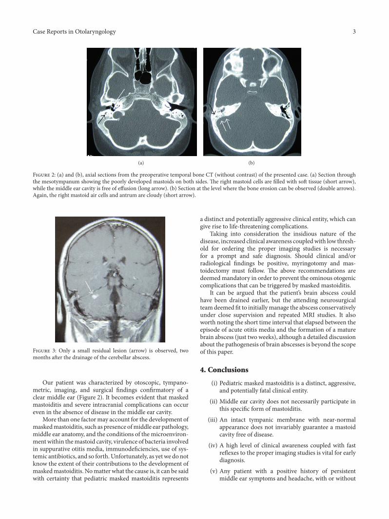
Figure 2: (a) and (b), axial sections from the preoperative temporal bone CT (without contrast) of the presented case. (a) Section throughthe mesotympanum showing the poorly developed mastoids on both sides. The right mastoid cells are filled with soft tissue (short arrow),while the middle ear cavity is free of effusion (long arrow). (b) Section at the level where the bone erosion can be observed (double arrows).Again, the right mastoid air cells and antrum are cloudy (short arrow).
Figure 3: Only a small residual lesion (arrow) is observed, twomonths after the drainage of the cerebellar abscess.
Our patient was characterized by otoscopic, tympano-metric, imaging, and surgical findings confirmatory of aclear middle ear (Figure 2). It becomes evident that maskedmastoiditis and severe intracranial complications can occureven in the absence of disease in the middle ear cavity.
More than one factormay account for the development ofmaskedmastoiditis, such as presence ofmiddle ear pathology,middle ear anatomy, and the conditions of the microenviron-ment within themastoid cavity, virulence of bacteria involvedin suppurative otitis media, immunodeficiencies, use of sys-temic antibiotics, and so forth.Unfortunately, as yet we do notknow the extent of their contributions to the development ofmaskedmastoiditis. Nomatter what the cause is, it can be saidwith certainty that pediatric masked mastoiditis represents
a distinct and potentially aggressive clinical entity, which cangive rise to life-threatening complications.
Taking into consideration the insidious nature of thedisease, increased clinical awareness coupledwith low thresh-old for ordering the proper imaging studies is necessaryfor a prompt and safe diagnosis. Should clinical and/orradiological findings be positive, myringotomy and mas-toidectomy must follow. The above recommendations aredeemedmandatory in order to prevent the ominous otogeniccomplications that can be triggered by masked mastoiditis.
It can be argued that the patient’s brain abscess couldhave been drained earlier, but the attending neurosurgicalteamdeemed fit to initiallymanage the abscess conservativelyunder close supervision and repeated MRI studies. It alsoworth noting the short time interval that elapsed between theepisode of acute otitis media and the formation of a maturebrain abscess (just two weeks), although a detailed discussionabout the pathogenesis of brain abscesses is beyond the scopeof this paper.
4. Conclusions
(i) Pediatric masked mastoiditis is a distinct, aggressive,and potentially fatal clinical entity.
(ii) Middle ear cavity does not necessarily participate inthis specific form of mastoiditis.
(iii) An intact tympanic membrane with near-normalappearance does not invariably guarantee a mastoidcavity free of disease.
(iv) A high level of clinical awareness coupled with fastreflexes to the proper imaging studies is vital for earlydiagnosis.
(v) Any patient with a positive history of persistentmiddle ear symptoms and headache, with or without

4 Case Reports in Otolaryngology
otorrhea, should have anENTexamination. Ifmaskedmastoiditis cannot otherwise be excluded, a CT scan-ning should be considered.
(vi) If clinical and/or imaging findings are positive, mid-dle ear exploration and mastoidectomy must follow.
Ethical Approval
This studywas approved by the Institutional Ethical Commit-tee. The parents consented to the publication of this case.
Conflict of Interests
The authors declare that there is no conflict of interestsregarding the publication of this paper.
References
[1] S. R. Mawson, Diseases of the Ear, Williams & Wilkins, Balti-more, Md, USA, 1963.
[2] C. Blustone and J. Klein, “Intratemporal complications andsequelae of otitis media,” in Pediatric Otolaryngology, C. Blue-stone, S. E. Stool, C.M. Alper et al., Eds., pp. 687–763, Saunders,Philadelphia, Pa, USA, 4th edition, 2003.
[3] G. R. Holt and G. A. Gates, “Maskedmastoiditis,” Laryngoscope,vol. 93, no. 8, pp. 1034–1037, 1983.
[4] A. S. Moody and D. E. Brackmann, “Chronic mastoiditis,” inAdvanced Therapy of Otitis Media, C. M. Alper, C. Bluestone,and M. Casselbrant, Eds., pp. 330–333, BC Decker, Hamilton,Canada, 2004.
[5] D. P. Martin-Hirsch, S. Habashi, R. Page, and A. E. Hinton,“Latent mastoiditis: no room for complacency,” Journal ofLaryngology and Otology, vol. 105, no. 9, pp. 767–768, 1991.
[6] C. R. Pearson, D. K. Riden, R. J. N. Garth, and M. R.Thomas, “Two eases of lateral sinus thrombosis presenting withextracranial head and neck abscesses,” Journal of Laryngologyand Otology, vol. 108, no. 9, pp. 779–782, 1994.
[7] T. Fukuda, H. Sugie, M. Ito, and T. Kikawada, “Bilateralfacial palsy caused by bilateral masked mastoiditis,” PediatricNeurology, vol. 18, no. 4, pp. 351–353, 1998.
[8] F. Tovi and A. Leiberman, “Silent mastoiditis and bilateralsimultaneous facial palsy,” International Journal of PediatricOtorhinolaryngology, vol. 5, no. 3, pp. 303–307, 1983.
[9] R. Badrawy, A. Abou Bieh, and A. Taha, “Masked diabeticmastoiditis,” Journal of Laryngology and Otology, vol. 89, no. 8,pp. 815–821, 1975.

Submit your manuscripts athttp://www.hindawi.com
Stem CellsInternational
Hindawi Publishing Corporationhttp://www.hindawi.com Volume 2014
Hindawi Publishing Corporationhttp://www.hindawi.com Volume 2014
MEDIATORSINFLAMMATION
of
Hindawi Publishing Corporationhttp://www.hindawi.com Volume 2014
Behavioural Neurology
EndocrinologyInternational Journal of
Hindawi Publishing Corporationhttp://www.hindawi.com Volume 2014
Hindawi Publishing Corporationhttp://www.hindawi.com Volume 2014
Disease Markers
Hindawi Publishing Corporationhttp://www.hindawi.com Volume 2014
BioMed Research International
OncologyJournal of
Hindawi Publishing Corporationhttp://www.hindawi.com Volume 2014
Hindawi Publishing Corporationhttp://www.hindawi.com Volume 2014
Oxidative Medicine and Cellular Longevity
Hindawi Publishing Corporationhttp://www.hindawi.com Volume 2014
PPAR Research
The Scientific World JournalHindawi Publishing Corporation http://www.hindawi.com Volume 2014
Immunology ResearchHindawi Publishing Corporationhttp://www.hindawi.com Volume 2014
Journal of
ObesityJournal of
Hindawi Publishing Corporationhttp://www.hindawi.com Volume 2014
Hindawi Publishing Corporationhttp://www.hindawi.com Volume 2014
Computational and Mathematical Methods in Medicine
OphthalmologyJournal of
Hindawi Publishing Corporationhttp://www.hindawi.com Volume 2014
Diabetes ResearchJournal of
Hindawi Publishing Corporationhttp://www.hindawi.com Volume 2014
Hindawi Publishing Corporationhttp://www.hindawi.com Volume 2014
Research and TreatmentAIDS
Hindawi Publishing Corporationhttp://www.hindawi.com Volume 2014
Gastroenterology Research and Practice
Hindawi Publishing Corporationhttp://www.hindawi.com Volume 2014
Parkinson’s Disease
Evidence-Based Complementary and Alternative Medicine
Volume 2014Hindawi Publishing Corporationhttp://www.hindawi.com