catecholamine-induced changes in the splanchnic
TRANSCRIPT

� CLINICAL CONCEPTS AND COMMENTARY
Richard B Weiskopf, M.D., Editor
Anesthesiology 2004; 100:434–9 © 2004 American Society of Anesthesiologists, Inc. Lippincott Williams & Wilkins, Inc.
Catecholamine-induced Changes in the SplanchnicCirculation Affecting Systemic HemodynamicsSimon Gelman, M.D., Ph.D.*, Phillip S. Mushlin, M.D., Ph.D.†
THIS article focuses on the effects of catecholamines onthe splanchnic circulation that influence systemic hemo-dynamics (particularly venous return and cardiac out-put) under normal physiologic conditions. Because of itsrequired brevity, this article could not address otherimportant hemodynamic effects of catecholamines, suchas those that result from metabolic alterations, effects onthe circulatory system that do not involve the splanchnicorgans, and those that accompany major pathophysio-logic states, such as sepsis or congestive heart failure.
Anatomy and Blood Supply
The splanchnic system receives nearly 25% of the car-diac output through three large arteries (fig. 1): theceliac artery (which typically has three major branches:hepatic, splenic, and gastric) and the superior and inferiormesenteric arteries. Roughly one fourth of the splanchnicarterial flow goes directly to the liver via the hepatic artery;the remaining three fourths reaches the liver after perfus-ing the preportal organs. The preportal veins anastomoseto form the portal vein. The portal vein and hepatic arteryenter the liver at its hilum and ramify into progressivelysmaller vessels before emptying into the hepatic sinusoids.Postsinusoidal blood flows through venules, sublobular andlobular veins, and the hepatic veins, which drain into theinferior vena cava.
The hepatic artery and the arteries of the preportalsplanchnic organs have mean pressures of approximately90 mmHg. The portal venous pressure is 7–10 mmHg,which is only slightly higher than the pressure in thesinusoids (fig. 2). Most of the intrahepatic vascular resis-tance is distal to the sinusoids1,2; possible locations of thisresistance include one or more of the following sites: thesublobular veins, upstream to the larger veins, or at thejunction of the hepatic veins and inferior vena cava.2
Distribution and Function of AdrenergicReceptors
Although knowledge of the various adrenoceptor sub-types has expanded dramatically during the past decade,much uncertainty remains about the distribution and func-tional importance of these receptors in the splanchnicvasculature.3 Pure �-adrenergic agonists (e.g., phenyleph-rine) constrict hepatic arterial smooth muscle, increasearterial resistance, and reduce hepatic arterial blood flow(table 1). Pure �-adrenergic agonists (e.g., isoproterenol)dilate hepatic arterioles, decrease vascular resistance, andincrease flow through the hepatic artery; these effects areblocked by nonselective �-adrenergic antagonists (e.g., pro-pranolol) but not by selective �1-antagonists (e.g., atenolol).Therefore, the hepatic artery contains �-adrenergic and�2-adrenergic receptors.4 The arterial supply of the prepor-tal splanchnic organs is densely populated with �1-, �2-,and �2-adrenergic receptors.4
Preportal (intestinal) capacitance vasculatures haveboth �1- and �2-adrenergic receptors but lack �2 recep-tors.5 The portal vein contains �-adrenergic but not�2-adrenergic receptors.4 Capacitance vessels of the liver(including sinusoids) have �-adrenergic receptors, andhepatic veins contain both �- and �2-adrenergic recep-tors.4 In the splanchnic venous system overall, �1- and�2-adrenergic receptor stimulation leads to venocon-striction, which decreases venous capacitance and in-creases venous resistance, whereas �2 receptor activa-tion decreases hepatic venous resistance. It seems thatthe density of �-adrenergic receptors is the highest inthe splanchnic (preportal) arteries.
The Splanchnic Blood Reservoir
Normovolemic healthy male adults have a blood volumeof approximately 70 ml/kg body weight. Splanchnic organsconstitute 10% of the body weight, but they contain 25% ofthe total blood volume.1,6 Nearly two thirds of the splanch-nic blood (i.e., � 800 ml) can be autotransfused into thesystemic circulation within seconds (table 2). The liver andintestines each provide between 300 and 400 ml of theblood; the spleen only contributes approximately 100 ml,but the hematocrit of this blood often approaches 75%.Therefore, the splanchnic vasculature serves as an impor-tant blood reservoir for the circulatory system.
* Leroy D. Vandam/Benjamin G. Covino Distinguished Professor of Anaesthe-sia, † Associate Professor of Anaesthesia.
Received from the Department of Anesthesiology, Perioperative and PainMedicine, Harvard Medical School, Boston, Massachusetts. Submitted for publi-cation December 19, 2002. Accepted for publication August 22, 2003. Supportwas provided solely from institutional and/or departmental sources.
Address reprint requests to Dr. Gelman: Department of Anesthesiology, Peri-operative and Pain Medicine, Brigham and Women’s Hospital, 75 Francis Street,Boston, Massachusetts 02115. Address electronic mail to: [email protected].
The illustrations for this section are prepared by Dimitri Karetnikov, 7 Ten-nyson Drive, Plainsboro, New Jersey 08536.
Anesthesiology, V 100, No 2, Feb 2004 434

Regulation of the Splanchnic Reservoir Volume
The capacity of catecholamines to increase cardiacoutput is dependent, in part, on the compliance, capac-itance, and blood volume of the splanchnic vasculature.With normovolemia, the volume of blood in the splanch-nic capacitance vessels varies in a manner that is approx-imately linearly related to the transmural pressure in thisvasculature.7–9 That is, under physiologic conditions, achange in splanchnic arterial flow leads to a proportionalchange in the pressure within splanchnic capacitancevessels.7 When arterial flow decreases, the blood volumeand therefore the pressure within the veins decrease.These veins recoil around the decreasing pressure, mitigat-ing the reduction in pressure. This phenomenon, referredto as elastic recoil, maintains the intramural pressure at alevel sufficient to provide a driving force for expulsion ofintravenous volume to the systemic circulation.
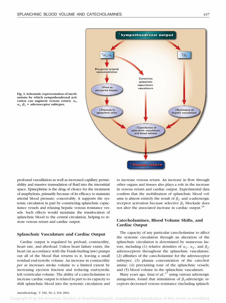
Sympathoadrenal stimulation causes sympatheticnerve terminals and the adrenal medulla to release cat-echolamines, which play a major role in regulating thetone of the arterial resistance vessels and venous capac-itance vessels (fig. 3). Catecholamines can cause splanch-nic arterial constriction or relaxation; the net effect de-pends on the specific catecholamines involved, theirconcentrations at adrenergic receptors, and the densitiesof the adrenoceptor subtypes within the vasculature. Themajor effect of catecholamines on splanchnic capacitancevessels is venoconstriction, which increases the pressure incapacitance vessels. This mechanism can actively expelsplanchnic blood into the systemic circulation, even whensplanchnic arterial flow has been markedly reduced.
Active (venoconstriction) and passive (elastic recoil)mechanisms work in concert to shift splanchnic bloodvolume to the systemic circulation. Both mechanisms make
Fig. 1. Schematic representation of thesplanchnic circulation.
Fig. 2. Diagrammatic representation ofthe splanchnic vasculature. Splanchnicarteries represent all arterial vessels ofthe preportal organs; splanchnic veinsrepresent the pooled venous blood fromall these organs. The distribution of adre-noceptor subtypes (�1, �2, �2) and ap-proximate intravascular pressures areshown for corresponding segments ofthe splanchnic vasculature.
435SPLANCHNIC BLOOD VOLUME AND CATECHOLAMINES
Anesthesiology, V 100, No 2, Feb 2004

equal contributions to the volume shift during activation ofthe sympathetic nervous system.10 These processes ac-count for the 35% decrease in splanchnic blood volumeassociated with mild exercise in humans.11 In animals sub-jected to moderate hemorrhage (8–9 ml/kg), the splanch-nic vessels deliver 5 ml/kg to the systemic circulation,effectively compensating for 60% of the withdrawnblood.12 Studies in dogs indicate that electrical stimulationof the splanchnic (sympathetic) nerves can rapidly de-crease splanchnic arterial flow while triggering a brief butsubstantial outflow of blood from the splanchnic vascula-ture.13 The maximum volume expelled (15 ml/kg) wasnearly 66% of the total splanchnic blood volume, and oc-curred in 12–15 s.13
Intrahepatic vascular resistance can affect the volume
of the splanchnic blood reservoir. By decreasing or in-creasing this resistance, catecholamines may either facil-itate or impede the shift of blood from splanchnic ca-pacitance vessels to the systemic circulation.14
Alterations in hepatic venous resistance usually contrib-ute less to such shifts than the degree of venoconstric-tion and venous recoil within the splanchnic organs.However, large increases in hepatic venous resistance cancause blood to pool within the splanchnic system and canlead to systemic hypovolemia. Histamine-related anaphylac-tic reactions in animal experiments provide a dramaticexample of this phenomenon.15 Clearly, the mechanism ofthe severe arterial hypotension that occurs during anaphy-lactic shock is complex; pathophysiologic events include
Table 1.
Table 2.
436 S. GELMAN AND P. S. MUSHLIN
Anesthesiology, V 100, No 2, Feb 2004
profound vasodilation as well as increased capillary perme-ability and massive transudation of fluid into the interstitialspace. Epinephrine is the drug of choice for the treatmentof anaphylaxis, primarily because of its efficacy to maintainarterial blood pressure; conceivably, it supports the sys-temic circulation in part by constricting splanchnic capac-itance vessels and relaxing hepatic venous resistance ves-sels. Such effects would maximize the translocation ofsplanchnic blood to the central circulation, helping to re-store venous return and cardiac output.
Splanchnic Vasculature and Cardiac Output
Cardiac output is regulated by preload, contractility,heart rate, and afterload. Unless heart failure exists, theheart (in accordance with the Frank-Starling law) pumpsout all of the blood that returns to it, leaving a smallresidual end-systolic volume. An increase in contractilityper se increases stroke volume to a limited extent byincreasing ejection fraction and reducing end-systolicleft ventricular volume. The ability of a catecholamine toincrease cardiac output is related in part to its capacity toshift splanchnic blood into the systemic circulation and
to increase venous return. An increase in flow throughother organs and tissues also plays a role in the increasein venous return and cardiac output. Experimental dataconfirm that the mobilization of splanchnic blood vol-ume is almost entirely the result of �2- and �-adrenergicreceptor activation because selective �1 blockade doesnot alter the associated increase in cardiac output.16
Catecholamines, Blood Volume Shifts, andCardiac Output
The capacity of any particular catecholamine to affectthe systemic circulation through an alteration of thesplanchnic circulation is determined by numerous fac-tors, including (1) relative densities of �1-, �2-, and �2-adrenoceptors throughout the splanchnic vasculature;(2) affinities of the catecholamine for the adrenoceptorsubtypes; (3) plasma concentration of the catechol-amine; (4) preexisting tone of the splanchnic vessels;and (5) blood volume in the splanchnic vasculature.
Many years ago, Imai et al.,17 using various adrenergicantagonists, found that stimulation of �2-adrenergic re-ceptors decreased venous resistance (including splanch-
Fig. 3. Schematic representation of mech-anisms by which sympathoadrenal acti-vation can augment venous return. �1,�2, �2 � adrenoceptor subtypes.
437SPLANCHNIC BLOOD VOLUME AND CATECHOLAMINES
Anesthesiology, V 100, No 2, Feb 2004
nic venous resistance) and increased venous return,whereas activation of �-adrenergic receptors increasedvenous resistance and decreased venous return. Activa-tion of �2 adrenoceptors almost invariably enhancesvenous return (by increasing arterial flow and decreasinghepatic venous resistance); the situation with � adreno-ceptors is more complex—an increase or a decrease mayoccur. Generally, � agonists increase venous return un-der normovolemic conditions, but they decrease it whenused at high doses or in the presence of severe hypovole-mia. Although the decrease could result from an �-adreno-ceptor–mediated increase in hepatic venous resistance, it ismore likely the result of removing a portion of the vascu-lature from the systemic circulation by arterial vasoconstric-tion. The initial response to an � agonist is usually anincrease in venous return. However, if the splanchnic res-ervoir is depleted, further �-adrenergic stimulation will nolonger increase venous return and may indeed decrease it.
Epinephrine has a high affinity for all splanchnic ad-renergic receptors, but it seems to exert greater effectson �1- and �2-adrenergic receptors than on �2-adrenergicreceptors. In the splanchnic system, norepinephrine ex-erts pronounced effects on both �1 and �2 receptors buthas very little if any effect on �2-adrenergic receptors.Selective �2-adrenergic agonists can increase venous re-turn by one third, entirely by increasing splanchnicblood flow and decreasing splanchnic venous resistanceby more than 40%.18
The effect of dopamine on splanchnic circulation is com-plex, and the information in the literature is controversial.Dopamine, at low doses, stimulates dopaminergic-1 anddopaminergic-2 receptors and produces vasodilation. Athigher doses, dopamine (directly and via conversion tonorepinephrine) activates both �1 and �2 adrenoceptors.Infused at relatively low doses, dopamine consistentlycauses an increase in portal blood flow; this effect may berelated to the stimulation of �2-adrenergic receptors withinthe preportal arteries because phenoxybenzamine (an �-ad-renergic antagonist) augments the increase, whereas pro-pranolol (a �-adrenergic antagonist) abolishes it.19
Dopamine can cause hepatic arterial blood flow todecrease, increase, or remain constant.20,21 The mecha-nism of the decrease is unclear; it may result from theeffect of dopamine on �-adrenergic receptors, particu-larly at relatively high doses, or from the hepatic arterialbuffer response,22 which mediates the reciprocal rela-tion between portal flow and hepatic arterial blood flow(i.e., an increase in the former causes the latter to de-crease) when lower doses are used.
The dose-related effects of dopamine on the hepaticoxygen supply–demand relation have been studied in ani-mals. At the low end of the dose range, dopamine causes aparallel increase in hepatic oxygen supply and consump-tion; however, higher doses of dopamine decrease the ratioof oxygen supply to consumption.20 Dopamine-inducedincreases in mesenteric blood flow have been associated
with decreases in oxygen extraction,23 nutritive bloodflow, and capillary density in the intestines.24 Therefore,dopamine may divert blood flow away from splanchnicmucosa and predispose to mucosal ischemia.25,26
More than 25 yr ago, Marino et al.,27 using a cardiopul-monary bypass model, noted that phenylephrine and do-pamine increased the blood volume in the bypass reservoir,indicating that both drugs decrease venous capacitance. Insuch experiments, changes in bypass reservoir volume areinversely related to the changes in venous capacitance.Studies in the cardiopulmonary bypass model have alsoshown that dopamine produces dose-dependent decreasesin venous capacitance during spinal anesthesia.28 Experi-ments using selective receptor antagonists have demon-strated that both �- and �-adrenergic receptor stimulationcontribute to dopamine- and norepinephrine-induced in-creases in venous return.29 Epinephrine and norepineph-rine can produce equivalent reductions of splanchnic (in-cluding hepatic) blood volume.30
Clinical Implications for Hypovolemia
Hypovolemia leads to an activation of sympatheticnervous system and an increase in circulating cat-echolamines. As a result, blood is translocated fromblood reservoirs (primarily splanchnic capacitance ves-sels) of the body into the systemic circulation, whichpartially compensates for the decreases in circulatingblood volume. In a recent study in dogs, hemorrhagesufficient to decrease mean aortic pressure by 50% wasassociated with a 50% reduction in cardiac output andnearly a 90% reduction in intestinal blood volume.31
Although the induction of moderate hypervolemia orhypovolemia did not substantially affect cardiac output,it markedly altered the intestinal blood volume.31 Whenintestinal blood volumes ranged between 95% and 135%of the baseline values, cardiac output remained constant.Outside this range, constriction or dilation of intestinalvessels caused large increases or decreases in venouspressure and cardiac output.31 Therefore, these observa-tions confirm the notion that splanchnic venous vascu-lature moderates changes in cardiac output during acutevolume loading and hemorrhage, thereby maintainingcardiac output relatively constant over a wide range oftotal vascular blood volume.6,12,31,32
The effects of catecholamines administered during hy-povolemia depend on the volume of blood in thesplanchnic reservoir. When hypovolemia is severe, thehomeostatic mechanisms involved in blood pressure andcardiac output regulation have already emptied thesplanchnic reservoir. That is, the capacity of exogenouscatecholamines to improve systemic hemodynamics bymodulating the splanchnic circulation decreases pro-gressively in the setting of increasing sympathoadrenaloutflow and increases in the plasma concentrations ofendogenous catecholamines and other vasoconstrictors
438 S. GELMAN AND P. S. MUSHLIN
Anesthesiology, V 100, No 2, Feb 2004

(e.g., angiotensin II, vasopressin/antidiuretic hormone,endothelin-1). After depletion of the splanchnic reser-voir, large doses of catecholamines may still be able toincrease blood pressure by further elevating arterial re-sistance in splanchnic and other vascular beds. Such effectsmight be essential for maintaining perfusion pressure andblood flow to the heart and brain. However, the intensevasoconstriction can be detrimental to the splanchnic or-gans by producing severe ischemic injury, which couldsubsequently lead to multiorgan failure. Therefore, whentreating hypovolemia-induced hypotension, the use of ex-ogenous catecholamines should be limited to a brief periodand should not be viewed as a substitute for the immediatereplacement of blood volume.
Summary
Nearly 25% of the total blood volume in humans re-sides within the splanchnic venous vasculature. Inhealthy normovolemic adults, sympathoadrenal stimula-tion can almost instantaneously transfuse approximately2 units of whole blood from the splanchnic to the sys-temic circulation (table 2). �-Adrenoceptor stimulationactively expels blood from splanchnic capacitance ves-sels, producing a rapid increase in venous return.33–35
This volume mobilization occurs because of active veno-constriction as well as passive elastic recoil of thesplanchnic veins secondary to decreased arterial inflow.The initial increase in venous return may be counter-acted by other �-adrenergic effects, such as an increasein hepatic venous resistance (which impedes expulsionof blood from the splanchnic to the central circulation)and a significant decrease in splanchnic arterial flow(which pharmacologically removes a portion of the sys-temic circulation). The degree to which an �-adrenergicagonist affects venous return and cardiac output is there-fore dependent on many factors, including baseline myocar-dial contractility, blood volume, and sympathetic tone.
Pure �-adrenergic agonists (e.g., isoproterenol) aug-ment cardiac output primarily by increasing venous re-turn, which results from increases in splanchnic bloodflow due to lowered resistances in splanchnic arterialvessels and hepatic veins. In general, a drug that stimu-lates both �- and �-adrenergic receptors would be ex-pected to more effectively maintain systemic hemody-namics than one that activates either �- or �-adrenergicreceptors. When simultaneously stimulated, �- and �-ad-renergic receptors act in concert to maximally shiftblood from the splanchnic vasculature into the systemiccirculation by producing vasoconstriction, decreasingsplanchnic vascular capacitance, and decreasing (or min-imizing the increase in) intrahepatic vascular resistance.
References1. Greenway C, Lautt W: Hepatic circulation, Handbook of Physiology, The
Gastrointestinal System, Motility and Circulation. Edited by Wood J. Bethesda,American Physiology Society, 1989, pp 1519–64
2. Maass-Moreno R, Rothe CF: Contribution of the large hepatic veins topostsinusoidal vascular resistance. Am J Physiol 1992; 262:G14–22
3. Guimaraes S, Moura D: Vascular adrenoceptors: An update. Pharmacol Rev2001; 53:319–56
4. Richardson PD, Withrington PG: Physiological regulation of the hepaticcirculation. Annu Rev Physiol 1982; 44:57–69
5. Patel P, Bose D, Greenway C: Effects of prazosin and phenoxybenzamine onalpha- and beta-receptor-mediated responses in intestinal resistance and capaci-tance vessels. J Cardiovasc Pharmacol 1981; 3:1050–9
6. Greenway CV: Role of splanchnic venous system in overall cardiovascularhomeostasis. Fed Proc 1983; 42:1678–84
7. Greenway CV, Seaman KL, Innes IR: Norepinephrine on venous complianceand unstressed volume in cat liver. Am J Physiol 1985; 248:H468–76
8. Jacobsohn E, Chorn R, O’Connor M: The role of the vasculature in regulat-ing venous return and cardiac output: Historical and graphical approach. Can JAnaesth 1997; 44:849–67
9. Magder S, De Varennes B: Clinical death and the measurement of stressedvascular volume. Crit Care Med 1998; 26:1061–4
10. Shepherd JT: Reflex control of the venous system, Venous Problems.Edited by Bergan JJ, Yao JST. Chicago, Year Book, 1978, pp 5–23
11. Wade O, Combes B, Childs A, Wheeler H, Cournand A, Bradley S: Theeffect of exercise on the splanchnic blood flow and splanchnic blood volume innormal man. Clin Sci 1956; 15:457–63
12. Donald D: Splanchnic circulation, Handbook of Physiology, section 2. Editedby Shepherd J, Abboud F. Bethesda, American Physiological Society, 1983, pp219–40
13. Brooksby GA, Donald DE: Dynamic changes in splanchnic blood flow andblood volume in dogs during activation of sympathetic nerves. Circ Res 1971;29:227–38
14. Rutlen DL, Supple EW, Powell WJ Jr: Adrenergic regulation of total systemicdistensibility: Venous distensibility effects of norepinephrine and isoproterenol be-fore and after selective adrenergic blockade. Am J Cardiol 1981; 47:579–88
15. Bennett TD, MacAnespie CL, Rothe CF: Active hepatic capacitance re-sponses to neural and humoral stimuli in dogs. Am J Physiol 1982; 242:H1000–9
16. Chang PI, Rutlen DL: Effects of beta-adrenergic agonists on splanchnicvascular volume and cardiac output. Am J Physiol 1991; 261:H1499–507
17. Imai Y, Satoh K, Taira N: Role of the peripheral vasculature in changes invenous return caused by isoproterenol, norepinephrine, and methoxamine inanesthetized dogs. Circ Res 1978; 43:553–61
18. Green JF: Mechanism of action of isoproterenol on venous return. Am JPhysiol 1977; 232:H152–6
19. Richardson PD, Withrington PG: Responses of the canine hepatic arterialand portal venous vascular beds to dopamine. Eur J Pharmacol 1978; 48:337–49
20. Roytblat L, Gelman S, Bradley EL, Henderson T, Parks D: Dopamine andhepatic oxygen supply-demand relationship. Can J Physiol Pharmacol 1990;68:1165–9
21. Kato M, Nimura Y, Miyachi M, Kitagawa Y, Watanabe T, Kawabata Y,Akiyama H: Intravenous catecholamines alter hepatic blood flow in consciousdogs with experimental hepatic denervation. J Surg Res 1996; 66:179–84
22. Lautt WW: Intrinsic regulation of hepatic blood flow. Can J Physiol Phar-macol 1996; 74:223–33
23. Giraud GD, MacCannell KL: Decreased nutrient blood flow during dopa-mine- and epinephrine-induced intestinal vasodilation. J Pharmacol Exp Ther1984; 230:214–20
24. Pawlik W, Mailman D, Shanbour LL, Jacobson ED: Dopamine effects on theintestinal circulation. Am Heart J 1976; 91:325–31
25. Marik PE, Haupt MT: Splanchnic circulation and the dopexamine paradox.Crit Care Med 1999; 27:2302–3
26. Segal JM, Phang PT, Walley KR: Low-dose dopamine hastens onset of gutischemia in a porcine model of hemorrhagic shock. J Appl Physiol 1992; 73:1159–64
27. Marino R, Romagnoli A, Keats A: Selective venoconstriction by dopamine incomparison with isoproterenol and phenylephrine. ANESTHESIOLOGY 1975; 43:570–2
28. Butterworth JF, Austin JC, Johnson MD, Berrizbeitia LD, Dance GR,Howard G, Cohn LH: Effect of total spinal anesthesia on arterial and venousresponses to dopamine and dobutamine. Anesth Analg 1987; 66:209–14
29. Van Maanen EF, Banning JW, Roebel LE, Morgan JP: Dopamine andnorepinephrine increase venous return by stimulating alpha and beta adrenocep-tors in the dog. J Cardiovasc Pharmacol 1988; 11:627–34
30. Greenway CV, Lautt WW: Effects of infusions of catecholamines, angio-tensin, vasopressin and histamine on hepatic blood volume in the anaesthetizedcat. Br J Pharmacol 1972; 44:177–84
31. Scott-Douglas NW, Robinson VJ, Smiseth OA, Wright CI, Manyari DE,Smith ER, Tyberg JV: Effects of acute volume loading and hemorrhage onintestinal vascular capacitance: A mechanism whereby capacitance modulatescardiac output. Can J Cardiol 2002; 18:515–22
32. Greenway CV, Lautt WW: Blood volume, the venous system, preload, andcardiac output. Can J Physiol Pharmacol 1986; 64:383–7
33. Rutlen DL, Supple EW, Powell WJ Jr: The role of the liver in the adrenergicregulation of blood flow from the splanchnic to the central circulation. Yale J BiolMed 1979; 52:99–106
34. Rothe CF: Control of capacitance vessels, Physiology of the IntestinalCirculation. Edited by Shepherd AP, Granger DN. New York, Raven, 1984, p 73
35. Perry MA, Ardell JL, Barrowman JA, Kvietys PR: Physiology of the splanch-nic circulation, Pathophysiology of the Splanchnic Circulation. Edited by KvietysPR, Barrowman JA, Granger DN. Boca Raton, CRC, 1987, pp 2–56
439SPLANCHNIC BLOOD VOLUME AND CATECHOLAMINES
Anesthesiology, V 100, No 2, Feb 2004