mast cell-mediated splanchnic cholestatic inflammation
TRANSCRIPT

ARTICLE IN PRESS+ModelCLINRE-1237; No. of Pages 14
Clinics and Research in Hepatology and Gastroenterology (2019) xxx, xxx—xxx
Available online at
ScienceDirectwww.sciencedirect.com
ORIGINAL ARTICLE
Mast cell-mediated splanchnic cholestaticinflammation
María-Ángeles Allera,∗, Vicente Martínezb, Ana Ariasc,Maria-Paz Navad, Valentín Cuervas-Monsc, Patri Vergarab,e,Jaime Ariasa
a Department of Surgery, School of Medicine, Complutense University of Madrid, 28040 Madrid, Spainb Department of Cell Biology, Physiology and Immunology, Veterinary School, Autonoma University ofBarcelona, 08193 Cerdanyola del Vallès, Spainc Department of Medicine, Autonoma University of Madrid, Hospital Puerta de Hierro. 28222 Madrid, Spaind Department of Genetic, Physiology and Microbiology, Complutense University of Madrid, 28040 Madrid,Spaine Centro de Investigación Biomédica en Red de Enfermedades Hepáticas y Digestivas (CIBERehd), Institutode Salud Carlos III, Barcelona, Spain
KEYWORDSCholestasis;Mast cells;Portal hypertension;Ascites
SummaryIntroduction: Splanchnic mast cells increase in chronic liver and in acute-on-chronic liver dis-eases. We administered Ketotifen, a mast cell stabilizer, and measured the mast cells in thesplanchnic organs of cholestatic rats.Material and Methods: These groups were studied: sham-operated rats (S; n = 15), untreatedmicrosurgical cholestasic rats (C; n = 20) and rats treated with Ketotifen: early (SK-e; n = 20 andCKe; n = 18), and late (SK-l; n = 15 and CK-l; n = 14).Results: The cholestatic rats showed systemic and splanchnic impairments, such as ascites,portal hypertension, and biliary proliferation and fibrosis. The rats also showed a splanchnic
Please cite this article in press as: Aller M-Á, et al. Mast cell-mediated splanchnic cholestatic inflammation. Clin ResHepatol Gastroenterol (2019), https://doi.org/10.1016/j.clinre.2019.02.001
increase of TNF-�, IL-1� and MCP-1, and a reduction of IL-4, IL-10 and antioxidants. An increaseof VEGF in the ileum and mesenteric lymphatic complex was associated with a liver reduction ofTGF-�1. Ketotifen reduces the degree of hepatic insufficiency and the splanchnic inflammatorymediators, as well as VEGF and TGF-ß1 levels. Ketotifen also reduces the connective tissue
Abbreviations: ALT, alanine aminotransferase; AP, alkaline phosphatase; AST, aspartate aminotransferase; FGF, fibroblastic growth factor;BW, body weight; CTMCs, connective tissue mast cells; HDL-C, high density lipoprotein; LDL-C, low density lipoproteins; LW, liver weight;MMCs, mucosal mast cells; MCP-1, monocyte chemoattractant protein-1; NEFAs, non-esterified fatty acids; PP, Portal vein pressure; RMCP-II,rat mast cell proteinases 2; SW, spleen weight; TB, total bilirubin; TGF-�1, Transforming growth factor beta 1; Treg, regulatory T-cell; VLDL,very low density lipoproteins; VCU, villus-crypt units; VEGF, vascular endothelium growth factor.
∗ Corresponding author at: Department of Surgery, School of Medicine, Complutense University of Madrid, Ramón y Cajal s.n. 28040 Madrid,Spain
E-mail addresses: [email protected] (M.-Á. Aller), [email protected] (V. Martínez), [email protected] (A. Arias),[email protected] (M.-P. Nava), [email protected] (V. Cuervas-Mons), [email protected] (P. Vergara), [email protected](J. Arias).
https://doi.org/10.1016/j.clinre.2019.02.0012210-7401/© 2019 Elsevier Masson SAS. All rights reserved.

ARTICLE IN PRESS+ModelCLINRE-1237; No. of Pages 14
2 M.-Á. Aller et al.
mast cells in the mesenteric lymphatic complex of cholestatic rats, while increases the hepaticmucosal mast cells.Conclusions: In cholestatic rats, Ketotifen improves liver function and ascites, and also reducespro-inflammatory mediators in the splanchnic area. The decrease in connective tissue mast cellsin the mesenteric lymphatic complex due to the administration of Ketotifen would lead to theimprovement of the inflammatory splanchnic response, and consequently the abovementionedcomplications.© 2019 Elsevier Masson SAS. All rights reserved.
I
Hatwbub
atesm(imbpgpa
a2pfdc
M
WfTpEpwf
E
Ton
caosrKteapoitww
K
Ftamcado
S
R(dst([fap
Pm
ntroduction
epatic dysfunction related to fibrosis or cirrhosis wouldggravate the grade of splanchnic and systemic inflamma-ion characteristic of portal hypertension. As a result, thisould increase the incidence of complications [1,2]. It haseen recognized that splanchnic mast cells are presentnder normal and pathological liver-related conditions inoth humans and experimental animals [3—9].
It was suggested that mast cells, involved in both innatend adaptive immunity [10,11], could be correlated withhe evolution of experimental portal hypertension [2] andxperimental hepatic chronic disease [1,12]. This is the rea-on why we have studied the hepatic-spleen-intestinal andesenteric lymph node distribution of mucosal mast cells
MMCs) and connective tissue mast cells (CTMCs) in an exper-mental model of chronic liver insufficiency secondary toicrosurgical obstructive cholestasis in the rat [13]. It cane accepted that this experimental model evolves in twohases. A first or ‘‘compensated’’ phase and a rapidly pro-ressive ‘‘decompensated’’ phase, from the sixth week ofostoperative evolution, resulting in the development ofscites [14].
Since Ketotifen is a mast cell stabilizer drug [15], wedministered it to the obstructive cholestatic rats, either4 hours before the intervention or during the postoperativeeriod (8 weeks) to prevent decompensation of the rats, orrom the sixth week of p.o. through the eighth week to treatecompensation and evaluate its effect on the potentialhanges in the splanchnic mast cell distribution.
aterials and methods
e used male Wistar rats, weighing between 230 and 270 grom the Vivarium of the Complutense University of Madrid.he experimental procedures employed in this study wereerformed in accordance with the Ethical Guidelines fromuropean Community Council Directive (86/609/EEC) andublished in Spanish Royal Decree 53/2013. All proceduresere approved by the Complutense University Animal Wel-
are Committee.
xperimental design
Please cite this article in press as: Aller M-Á, et al. Mast celHepatol Gastroenterol (2019), https://doi.org/10.1016/j.clinr
he animals were randomly divided into six groups: sham-perated (S; n = 15), rats with microsurgical cholestasis (C;
= 20), early sham-operated (SK-e; n = 20) and microsurgi-
Twcv
al cholestatic rats (CK-e; n = 18); both of these groups weredministered Ketotifen early on, starting 24 hours before theperation until they were sacrificed 8 weeks post-operation;ham-operated (SK-l; n = 15) and microsurgical cholestaticats (CK-l; n = 14): both of these groups were administeredetotifen late, starting the sixth week after the opera-ion until they were sacrificed 8 weeks post operation. Inssence, early administration of Ketotifen means that thenimals receive Ketotifen throughout the preoperative andostoperative periods. On the contrary, late administrationf Ketotifen means that the rats only receive Ketotifen dur-ng the last two weeks of the postoperative period. Allhe animals were sacrificed by anaesthesia overdose at 8eeks post operation. Body (BW), liver (LW) and spleen (SW)eights were determined.
etotifen treatment
or the stabilization of the mast cells, Ketotifen was addedo the drinking water. The compound was dissolved in watert an initial concentration of 0.1 mg/mL, allowing an esti-ated dosage of 10 mg/kg per day (based on a mean water
onsumption of 30 mL/day). During the treatment period,nimals were housed individually. The amount of waterrunk by each rat was monitored daily and the concentrationf Ketotifen adjusted to ensure the desired dosage [16].
urgical procedure
ats were anesthetized with Ketamine Hydrochloride100 mg/kg) and Xylazine (12 mg/kg) i.m. In S-rats, the bileuct and its lobular branches were dissected. The micro-urgical cholestatic rats underwent an extrahepatic biliaryract resection using a binocular operatory microscopeZeiss, OPMI 1-FR, Madrid, Spain), as previously described13]. Buprenorphine (0.05 mg/Kg/8 h s.c.) was administeredor analgesia the first day of the postoperative period, andntibiotic and vitamin K were administered during all the.o., as previously described [13,14].
ortosystemic collateral circulation studyethod
he splenorenal, gastroesophageal, and colorectal areas,here collateral venous circulation normally develops, were
l-mediated splanchnic cholestatic inflammation. Clin Rese.2019.02.001
arefully studied for the presence of increased collateraleins [17].

IN+Model
Ip
ImpcUdsocMteueaoo
S
Tavadn
R
TttacP(A(ct(cii(lrcinfbT
ARTICLECLINRE-1237; No. of Pages 14
Mast cell-mediated splanchnic cholestatic inflammation
Portal vein pressure measurements
Portal vein pressure (PP) was measured by inserting a fluid-filled 20-gauge needle into the splenic parenchyma [18].Previous studies have demonstrated the excellent correla-tion between splenic pulp pressure and PP [19].
Gross mesenteric venous vasculopathy study
Three grades of mesenteric venous vasculopathy were con-sidered, as previously described; grade 0: normal; grade 1:after the Pringle maneuver, and grade 2: spontaneous [6,20].
Serum biochemical test
Blood samples were drawn by puncture of the infra-hepatic inferior vena cava. The serum was frozen at−40 ◦C until total bilirubin (TB), alkaline phosphatase (AP),aspartate aminotransferase (AST), alanine aminotransferase(ALT), albumin and Cholesterol, Triglicerides, high den-sity lipoprotein (HDL-C), low density lipoproteins (LDL-C),very low-density lipoproteins (VLDL) and non-esterified fattyacids (NEFAs) were assayed in an autoanalizer.
TNF-�, IL-1�, IL-4, and IL-10 splanchnic levels
Samples from the distal ileum (1 cm from the ileo-caecaljunction), the liver, the spleen, and the mesenteric lymphcomplex were quickly taken, frozen on dry ice, andtransferred to 5 mL polypropylene tubes (Falcon; BectonDickinson, Lincoln Park, NJ) containing lysis buffer (4 ◦C)at a ratio of 10 mL buffer/L g of wet tissue. Lysis bufferwas 1 mmol/L phenylmethylsulfonyl fluoride (PMSF; SigmaChemical Company, Madrid, Spain), and 1 mg/mL pepstatin A(Sigma Chemical Company), aprotinin (Sigma Chemical Com-pany), anti-pain (Sigma Chemical Company), and leupeptinin 13 phosphate-buffered saline solution with pH 7.2 (Bioflu-ids, Rockville, MD) containing 0.05% sodium azide (SigmaChemical Company). The samples were homogenized for30 s with an electrical homogenizer (Polytron; BrinkmannInstruments, Westminster, NY) at maximum speed, andimmediately frozen in liquid nitrogen at —80 ◦C. After thaw-ing at 4 ◦C, the aggregates were pelleted at 3000 g (4 ◦C).Finally, the in these homogenates were assayed the quan-titative levels of rat TNF-�, IL-1�, IL-4, and IL-10, in sixanimals of each group (sham-operated and cholestatic rats)using commercially available enzyme linked immunosorbentassay specific kits (BioNOVA Cientifica Ltd. Madrid, Spain).
Histopathologic examinations
Five �m-thick samples of the middle liver lobe were stainedwith hematoxylin and eosin (H&E) and Sirius red. An image
Please cite this article in press as: Aller M-Á, et al. Mast celHepatol Gastroenterol (2019), https://doi.org/10.1016/j.clinr
analysis system was used to assess fibrosis grade of thehepatic tissue from the sections stained with Sirius red com-pared to the total area of hepatic tissue by using Leica Q Winsoftware [21].
w(
o
PRESS3
mmunohistochemistry for rat mast cellroteinases 2 and connective mast cell counts
mmunodetection of RMCP-II, specific marker of mucosalast cells (MMC) in the rats, was carried out onaraformaldehyde-fixed sections using a specific rat mono-lonal antibody (1:500; Moredun Animal Health, Edinburgh,K) [22]. The connective tissue mast cells (CTMC) wereetected by toluidine blue staining protocol [23]. Positivelytained mast cells were counted in three to five sectionsf ileum per animal. Seven to ten well oriented villus-rypt units (VCU) were examined per ileum section. MucosalC and connective tissue MC counting was normalized for
he surface area of liver, lymphatic nodes and spleen, asvaluated in digital images. Cell counting was performedsing an Axioskop 40 microscope [Carl Zeiss, Jena, Germany;quipped with a digital camera, Zeiss AxioCam MRm; imagenalysis software: Zeiss Axiovision Release 4.8.1]. Analysesf all morphological data were performed blinded to preventbserver bias [23].
tatistical analysis
he results are expressed as the mean ± the standard devi-tion. Student’s t test for independent data or analysis ofariance (ANOVA) and the Duncan test were used whenppropriate for comparison of the variables between theifferent groups studied. The results were considered sig-ificant if P < 0.05.
esults
he cholestatic rats displayed ascites, increased por-al pressure (P < 0.001), mesenteric venous vasculopa-hy and portosystemic collateral circulation with hep-tosplenomegaly (P < 0.001), testicular atrophy (P < 0.01),holesterol (P < 0.001), triglycerides (P < 0.05) and LDL;
< 0.001) and VLDL; P < 0.001) (Table 1), hyperbilirubinemiaP < 0.001), increased AP (P < 0.001), bile acids (P < 0.001),STe (P < 0.001) (Table 2). On the contrary, hypoproteinemiaP < 0.001) and hypoalbuminemia (P < 0.001) and reducedoncentration of HDL; P < 0.05) (Table 3) were shown inhese rats. Antioxidants mediators reduced in the ileumP < 0.01), the liver, and in the mesenteric lymph nodes in theholestatic rats (Table 4), whereas IL-1� was reduced in theleum (P < 0.05) and mesenteric lymph nodes, but increasedn the liver (P < 0.05) (Table 5). IL-10 increased in the ileumP < 0.05); IL-4 increased in the ileum (P < 0.05) and in theiver; IL-13 and IL-17A increased in the ileum, but wereeduced in the mesenteric lymph nodes (Table 4). Mono-yte chemoattractant protein-1 (MCP-1) increased in theleum (P < 0.05), and was reduced in the mesenteric lymphodes and liver (P < 0.01) (Table 5). Transforming growthactor beta 1 (TGF-�1) increased in the ileum and liver,ut was reduced in the mesenteric lymph nodes (Table 2).he cholestatic rats showed hepatic necrosis associated
l-mediated splanchnic cholestatic inflammation. Clin Rese.2019.02.001
ith epithelial biliary proliferation and fibrosis (P < 0.001)Fig. 1).
The treatment with Ketotifen during the entire post-perative period of the rats with cholestasis reduced ascites,

ARTICLE IN PRESS+ModelCLINRE-1237; No. of Pages 14
4 M.-Á. Aller et al.
Table 1 Increase in body weight (�BW), liver weight (LW), spleen weight (SW), testes weight (TW), portal pressure (PP) andascites in sham-operated rats (S), in microsurgical cholestatic (C) rats, in sham-operated (SK-e) and microsurgical rats (CK-e),which were early administered Ketotifen and in sham-operated (SK-l) and microsurgical rats (CK-l) that were late administeredKetotifen.
Groups � BW (g) LW (g) SW (g) TW (g) PP (mmHg) ASCITES (ml)
S n = 15 153’66 ± 20’06 13’83 ± 2’27 0’87 ± 0’09 3’64 ± 0’28 8’25 ± 2’33 -------------C n = 20 51’82 ± 31’26a 26’01 ± 5’22a 2’03 ± 0’41a 2’90 ± 0’55a 13’43 ± 2’33a 6’88 ± 5’35
SK-e n = 20 113’48 ± 34’64a, b 12’13 ± 0’87b 0’72 ± 0’09b 3’56 ± 0’16oo 10’29 ± 2’12o** -------------CK-e n = 18 39’81 ± 24’65a, c 17’90 ± 3’88c, d** 1’57 ± 0’39a, c, d 2’23 ± 0’85a, coo 11’28 ± 5’80a 3’71 ± 2’70SK-l n = 15 161’00 ± 39’28 13’41 ± 1’37 0’84 ± 0’15 3’78 ± 0’37 7’02 ± 1’83 -------------CK-l n = 14 87’08 ± 37’05++ 28’22 ± 5’23d 2’11 ± 0’32d 3’10 ± 0’4d 15’04 ± 2’63d 4’01 ± 3’18
Mean ± SD. Mean ± SD. ***P < 0’001. Statistically significant value in regards to S; ooo: statistically significant value in relation to C;���P < 0’001: statistically significant value in relation to SK-e; +++P < 0’001: statistically significant value in relation to SK-l.
a P < 0’001. Statistically significant value in regards to S.b Statistically significant value in relation to C.
bbatt(l
soalisaprai1(c(
paa
S
Mdias
i(t(p
fiwtc
Ctiamt(tir
dtttt((
ecwreocatt
D
c P < 0’001: statistically significant value in relation to SK-e.d P < 0’001: Statistically significant value in relation to SK-l.
ody weight, the hepatosplenomegaly, total and directilirubin and bile acid concentrations whereas HDL levelsnd total protein were increased (Tables 1, 2 and 3). Keto-ifen also reduced the levels of MCP-1 in the liver, TNF-� inhe ileum and liver (P < 0.05), IL-17 A and IL-1� in the ileumP < 0.05) whereas concentrations of IL-4 and IL-10 in theiver and IL-13 in the ileum increased (Table 5).
Rats with cholestasis treated with Ketotifen starting atix weeks until they were sacrificed at eight weeks post-peration showed an increase in body weight (P < 0.01), and
reduction in ascites, splenomegaly (P < 0.001), testicu-ar atrophy (P < 0.01) (Table 1), collateral circulation, andntestinal mesenteric vasculopathy. Ketotifen reduced theerum index of AST/ALT (P < 0.001), LDH (P < 0.01) and cre-tinine (P < 0.001) (Table 2); while (P < 0.05) HDL and totalrotein levels increased (Tables 1, 2 and 3). Ketotifen alsoeduced (P < 0.001) the levels of MCP-1 in the liver, ileum,nd mesenteric lymph nodes, and reduced TNF-� in theleum and mesenteric lymph nodes, and IL-4, IL-13, and IL-7 A in the liver (P < 0.01) (Table 5). Lastly, Ketotifen reducedP < 0.001) VEGF in all the splanchnic organs studied, whileoncentrations of TGF-�1 in the liver (P < 0.05) and ileumTable 1) increased.
Ketotifen administered during the entire post-operativeeriod increased (P < 0.05) liver fibrosis, whereas Ketotifendministered from only week six to eight did not produceny effect (Fig. 1).
planchnic mast cell density
ucosal (MMC) and connective tissue mast cells (CTMC) wereetermined in liver, spleen, mesenteric lymph nodes andleum. The concentration of connective tissue mast cellsnd most importantly, mucosal mast cells, change in theplanchnic area in the cholestatic rats (Figs. 2−4).
CTMC in the mesenteric lymphatic complex showed anncrease in relation to SKe (P < 0.05 ) and SKl (P < 0.01)
Please cite this article in press as: Aller M-Á, et al. Mast celHepatol Gastroenterol (2019), https://doi.org/10.1016/j.clinr
Fig. 2a). Ketotifen-reduced CTMC in the lymph nodes ofhe cholestatic rats ketotifen administered continuouslypre- and postoperatory; CKe) as in a late period of theostoperatory (CKl), did not show statistically significant dif-
Mpc
erences (Fig. 2a). MMC in the mesenteric lymph nodes alsoncreased in the cholestatic rats and both types of treatmentith Ketotifen produced a reduction, although this reduc-
ion is not statistically significant in regards to the controlholestatic rats (Fig. 2a).
In the liver, no statistically significant differences ofTMC were found between the animals treated with Keto-ifen and the cholestatic rats without treatment. This results paradoxical because cholestasis in these animals causedn increase (P < 0.05) compared to the sham-operated ani-als (Fig. 2b). Similar results were found in the MMC liver. In
his case, the animals with cholestasis showed an increaseP < 0.05) when compared to sham-operated animals andhose treated with Ketotifen. Neither of the two admin-stration methods showed statistically significant values inegards to the cholestatic rats (Fig. 2b).
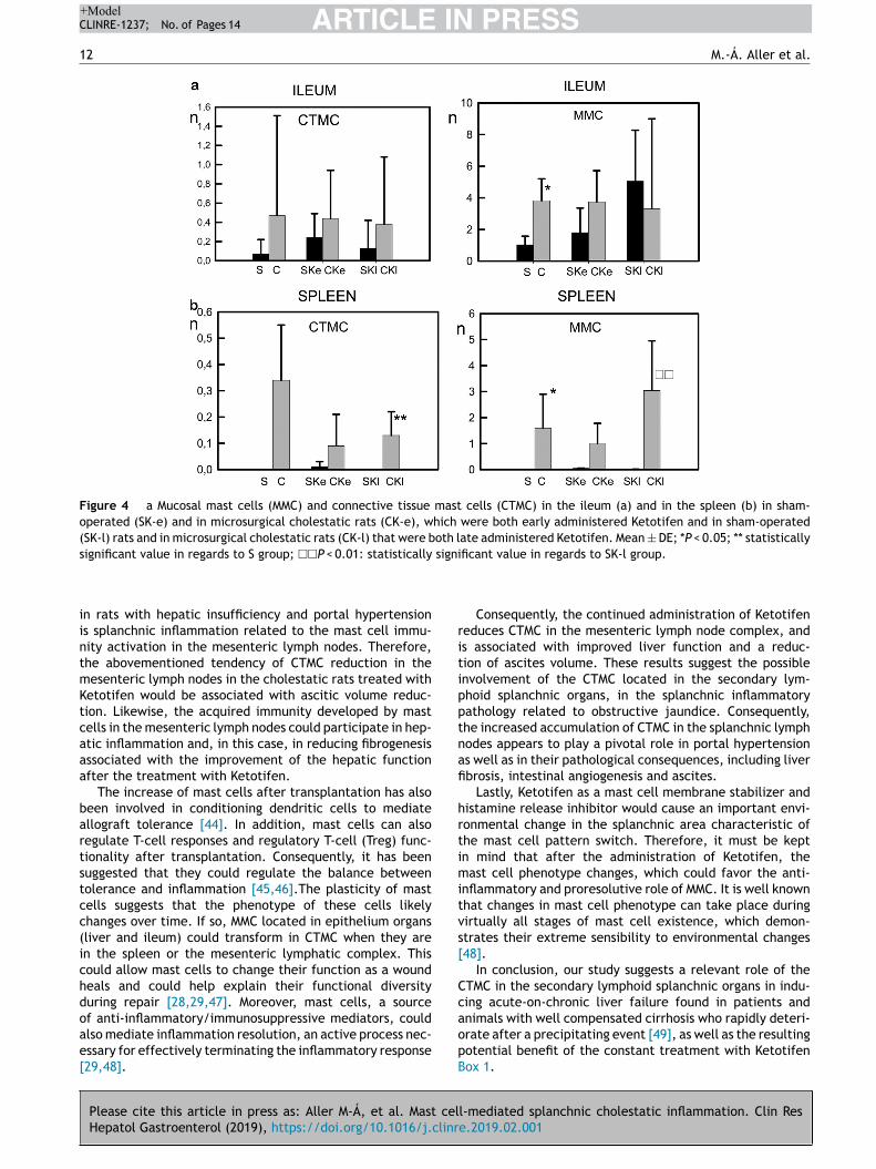
The administration of Ketotifen to the cholestatic ratsid not change CTMC concentration in the ileum comparedo the treated cholestatic rats (Figs. 4a and 3). Furthermore,he density of MMC in the ileum did not change when Keto-ifen was administered to the rats with cholestasis (C), evenhough cholestasis is a factor associated with the increaseP < 0.05) of MMC in relation to the sham-operated animalsFigs. 3 and 4a).
In the spleen, CTMC increased in the cholestatic rats,ither those treated or not treated with Ketotifen whenompared to the sham-operated rats. This CTMC increaseas only statistically significant (P < 0.01) in the cholestatic
ats that were administered Ketotifen during the late potop-rative period (CKl) (Figs. 3 and 4b). In addition, the valuesf MMC showed an increase in the spleen of the cholestaticontrol rats (C) and in those treated with Ketotifen (CKend CKl) in regards to the sham-operated animals. However,his increase was only statistically significant (P < 0.05) inhe control cholestatic rats (C) (Figs. 3 and 4b).
iscussion
l-mediated splanchnic cholestatic inflammation. Clin Rese.2019.02.001
ast cells in rodents are divided into two types based onrotease expression patterns: the connective tissue mastells (CTMCs) and the mucosal mast cells (MMCs) [24]. MMCs,
Please cite
this article
in press
as: Aller
M-Á,
et al.
Mast
cell-mediated
splanchnic cholestatic
inflam
mation.
Clin Res
Hepatol
Gastroenterol
(2019), https://doi.org/10.1016/j.clinre.2019.02.001
AR
TIC
LE
IN P
RE
SS
+Model
CLINRE-1237;
N
o. of
Pages 14
Mast
cell-mediated
splanchnic cholestatic
inflam
mation
5
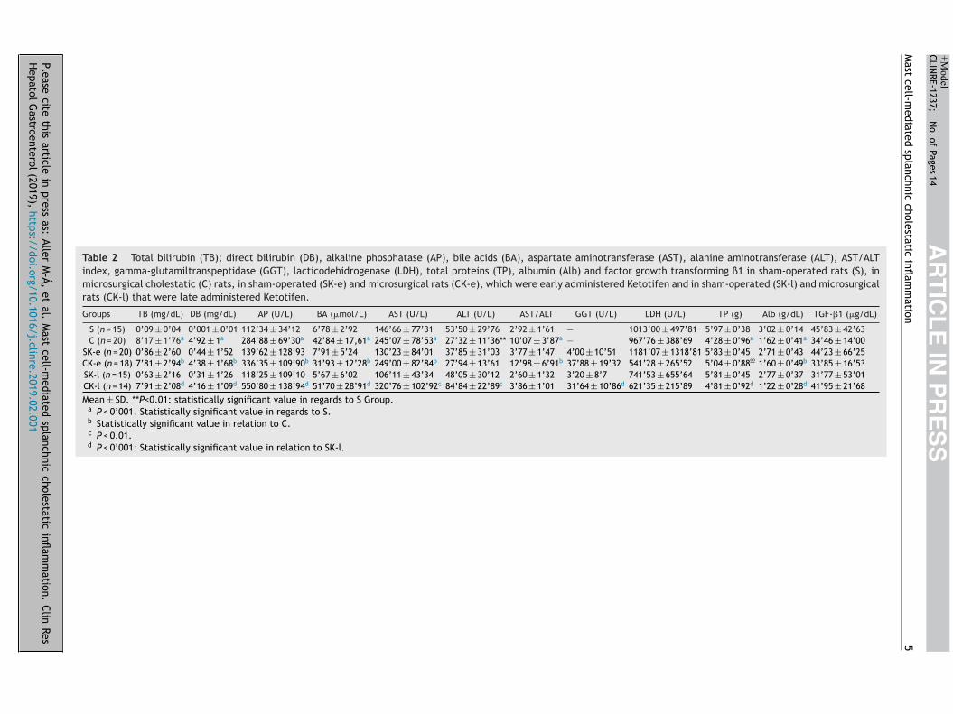
Table 2 Total bilirubin (TB); direct bilirubin (DB), alkaline phosphatase (AP), bile acids (BA), aspartate aminotransferase (AST), alanine aminotransferase (ALT), AST/ALTindex, gamma-glutamiltranspeptidase (GGT), lacticodehidrogenase (LDH), total proteins (TP), albumin (Alb) and factor growth transforming ß1 in sham-operated rats (S), inmicrosurgical cholestatic (C) rats, in sham-operated (SK-e) and microsurgical rats (CK-e), which were early administered Ketotifen and in sham-operated (SK-l) and microsurgicalrats (CK-l) that were late administered Ketotifen.
Groups TB (mg/dL) DB (mg/dL) AP (U/L) BA (�mol/L) AST (U/L) ALT (U/L) AST/ALT GGT (U/L) LDH (U/L) TP (g) Alb (g/dL) TGF-�1 (�g/dL)
S (n = 15) 0’09 ± 0’04 0’001 ± 0’01 112’34 ± 34’12 6’78 ± 2’92 146’66 ± 77’31 53’50 ± 29’76 2’92 ± 1’61 — 1013’00 ± 497’81 5’97 ± 0’38 3’02 ± 0’14 45’83 ± 42’63C (n = 20) 8’17 ± 1’76a 4’92 ± 1a 284’88 ± 69’30a 42’84 ± 17,61a 245’07 ± 78’53a 27’32 ± 11’36** 10’07 ± 3’87a — 967’76 ± 388’69 4’28 ± 0’96a 1’62 ± 0’41a 34’46 ± 14’00
SK-e (n = 20) 0’86 ± 2’60 0’44 ± 1’52 139’62 ± 128’93 7’91 ± 5’24 130’23 ± 84’01 37’85 ± 31’03 3’77 ± 1’47 4’00 ± 10’51 1181’07 ± 1318’81 5’83 ± 0’45 2’71 ± 0’43 44’23 ± 66’25CK-e (n = 18) 7’81 ± 2’94b 4’38 ± 1’68b 336’35 ± 109’90b 31’93 ± 12’28b 249’00 ± 82’84b 27’94 ± 13’61 12’98 ± 6’91b 37’88 ± 19’32 541’28 ± 265’52 5’04 ± 0’88oo 1’60 ± 0’49b 33’85 ± 16’53SK-l (n = 15) 0’63 ± 2’16 0’31 ± 1’26 118’25 ± 109’10 5’67 ± 6’02 106’11 ± 43’34 48’05 ± 30’12 2’60 ± 1’32 3’20 ± 8’7 741’53 ± 655’64 5’81 ± 0’45 2’77 ± 0’37 31’77 ± 53’01CK-l (n = 14) 7’91 ± 2’08d 4’16 ± 1’09d 550’80 ± 138’94d 51’70 ± 28’91d 320’76 ± 102’92c 84’84 ± 22’89c 3’86 ± 1’01 31’64 ± 10’86d 621’35 ± 215’89 4’81 ± 0’92d 1’22 ± 0’28d 41’95 ± 21’68
Mean ± SD. **P<0.01: statistically significant value in regards to S Group.a P < 0’001. Statistically significant value in regards to S.b Statistically significant value in relation to C.c P < 0.01.d P < 0’001: Statistically significant value in relation to SK-l.

Please cite
this article
in press
as: Aller
M-Á,
et al.
Mast
cell-mediated
splanchnic cholestatic
inflam
mation.
Clin Res
Hepatol
Gastroenterol
(2019), https://doi.org/10.1016/j.clinre.2019.02.001
AR
TIC
LE
IN P
RE
SS
+Model
CLINRE-1237;
N
o. of
Pages 14
6
M.-Á.
Aller et
al.
Table 3 Cholesterol, Triglicerides, high density lipoprotein (HDL-C), low density lipoproteins (LDL-C), very low density lipoproteins (VLDL) and non-esterified fatty acids(NEFAs) in sham-operated rats (S), in microsurgical cholestatic (C) rats, in sham-operated (SK-e) and microsurgical rats (CK-e), which were early administered Ketotifen and insham-operated (SK-l) and microsurgical rats (CK-l) that were late administered Ketotifen.
Groups Cholesterol (mg/dl) Triglicerides (mg/dl) HDL-C (mmol/L) LDL-C (mmol/L) VLDL-C (mmol/L) NEFAs (mmol/L)
S (n = 15) 63’30 ± 10’19 161’33 ± 53’53 0’78 ± 0’11 0’27 ± 0’05 0’57 ± 0’22 1’39 ± 0’88C◦ ◦ (n = 20) 143’31 ± 65’47c 157’69 ± 53’72 0’65 ± 0’16a 2’69 ± 1’43c 0’35 ± 0’21 0’61 ± 0’20b
SK-e (n = 20) 70’30 ± 232’13 120’07 ± 43053 0’81 ± 0’11 0’52 ± 0’85 0’48 ± 0’27 0’64 ± 0’22CK-e (n = 18) 158’30 ± 75’93e 165’58 ± 75’94d 0’92 ± 0’23d 0’48 ± 0’27 0’42 ± 0’36 0’49 ± 0’17SK-L (n = 15) 62’12 ± 26’72 145’32 ± 43’25 0’79 ± 0’13 0’44 ± 0’71 0’38 ± 0’16 0’65 ± 0’32CK-l (n = 14) 151’57 ± 72’84f 170’83 ± 93’68 0’89 ± 0’22 2’83 ± 1’4f 0’25 ± 0’44 0’53 ± 0’16
Mean ± SD.◦ ◦ P < 0.01.
a P < 0.05.b P < 001.c P < 0’001: Statistically significant value in regards to S.d P < 0.05.e P < 0.001: Statistically significant value in relation to C.f P < 0’001: Statistically significant value in relation to SK-l.

Please cite
this article
in press
as: Aller
M-Á,
et al.
Mast
cell-mediated
splanchnic cholestatic
inflam
mation.
Clin Res
Hepatol
Gastroenterol
(2019), https://doi.org/10.1016/j.clinre.2019.02.001
AR
TIC
LE
IN P
RE
SS
+Model
CLINRE-1237;
N
o. of
Pages 14
Mast
cell-mediated
splanchnic cholestatic
inflam
mation
7
Table 4 Catalase Glutation-transferase and Glutation peroxidase in the ileum, the mesenteric lymph nodes and the liver in in sham-operated rats (S), in microsurgicalcholestatic (C) rats, in sham-operated (SK-e) and microsurgical rats (CK-e), which were early administered Ketotifen and in sham-operated (SK-l) and microsurgical rats (CK-l)that were late administered Ketotifen.
Catalase (�mol/mg) Glutation-transferase (�mol/mg) Glutation peroxidase (�mol/mg)
Groups Ileum Mesenteric nodes Liver Ileum Mesenteric nodes Liver Ileum Mesenteric nodes Liver
S (n = 6) 1’05 ± 0’40 2’24 ± 0’50 3’18 ± 0’08 3326’70 ± 1025’33 718’35 ± 135’08 4882’69 ± 474’24 1000’57 ± 136’63 876’13 ± 40’45 5450’40 ± 346’41C (n = 6) 1’97 ± 0’44 1’04 ± 0’11 2’84 ± 0’28 1925’40 ± 479’40a 321’49 ± 16’54a 1681’90 ± 144’69b 799’72 ± 128’67 962’91 ± 5’50 2750’67 ± 126’49
SK-e (n = 6) 2’23 ± 0’53 3’73 ± 0’05 3’60 ± 0’30 2823’05 ± 479’48 1137’17 ± 4’59 5459’24 ± 349’26 1062’91 ± 100’15 972’92 ± 55’87 7309’65 ± 308’37CK-e (n = 6) 1’34 ± 0’26 0’97 ± 0’06d 2’56 ± 0’20c 2306’16 ± 213’07 318’09 ± 7’89d 1622’26 ± 80’87e 597’66 ± 63’27d 1085’72 ± 67’32 2759’15 ± 309’96e
SK-l (n = 6) 2’84 ± 0’81 2’67 ± 0’57 3’40 ± 0’86 2584’49 ± 982’12 577’33 ± 296’83 6429’42 ± 858’52 1130’83 ± 239’25 987’90 ± 342’24 2258’26 ± 1652’68CK-l (n = 6) 0’60 ± 0’43f 1’49 ± 0’39 3’40 ± 0’38 1921’82 ± 519’61 268’39 1439’36 ± 376’84e 865’95 ± 474’97d 529’73 ± 265’47c 2534’17 ± 178’26
Mean ± SD.a P < 001.b P < 0’001. Statistically significant value in regards to S.c P < 0.05.d P < 0.01.e P < 0.001: Statistically significant value in relation to C.f P < 0’01: Statistically significant value in relation to SK-l.

Please cite
this article
in press
as: Aller
M-Á,
et al.
Mast
cell-mediated
splanchnic cholestatic
inflam
mation.
Clin Res
Hepatol
Gastroenterol
(2019), https://doi.org/10.1016/j.clinre.2019.02.001
AR
TIC
LE
IN P
RE
SS
+Model
CLINRE-1237;
N
o. of
Pages 14
8
M.-Á.
Aller et
al.
Table 5 Tumoral necrosis Factor alpha (TNF-�), Interleukin-1ß (IL-1ß), Monocyte chemoattractant protein-1 (MCP-1), Interleukin-13 (IL-13), Interleukin-17A (IL-17A), Inter-leukin 4 (IL-4) and Interleukin 10 (IL-10) in the ileum, the mesenteric lymph nodes and the liver in in sham-operated rats (S), in microsurgical cholestatic (C) rats, in sham-operated(SK-e) and microsurgical rats (CK-e), which were early administered Ketotifen and in sham-operated (SK-l) and microsurgical rats (CK-l) that were late administered Ketotifen.
Groups TNF-� (pg/100 mg) IL-1� (pg/100 mg) MPC-1
Ileum Mesenteric nodes Liver Ileum Mesenteric nodes Liver Ileum Mesenteric nodes Liver
S (n = 6) 130’24 ± 37’76 129’83 ± 22’48 505’56 ± 65’19 266’02 ± 42’53 431’85 ± 110’88 173’20 ± 30’16 7’30 ± 2’59 29’65 ± 2’22 9’75 ± 1’01C (n = 6) 192’27 ± 161’47 141’34 ± 59’21 352’00 ± 255’13 150’87 ± 44’57* 436’10 ± 63’39 249’71 ± 96’64 17’22 ± 6’24* 16’23 ± 4’18 31’83 ± 16’78
SK-e (n = 6) 40’14 ± 35’60 138’29 ± 32’01 268’82 ± 77’06 154’82 ± 86’66 502’68 ± 79’99 103’73 ± 71’07 6’56 ± 2’80 184’63 ± 26’66 5’70 ± 0’95CK-e (n = 6) 36’57 ± 10’78 69’84 ± 17’01oo 125’99 ± 76’08o 170’00 ± 20’53 266’65 ± 81’12 204’58 ± 104’51o 26’93 ± 19’39 94’12 ± 13’06o 26’93 ± 8’56oo
SK-l (n = 6) 85’97 ± 20’46 157’66 ± 51’13 2256’32 ± 157’36 431’66 ± 115’12 483’82 ± 194’92oo 884’76 ± 54’86 72’69 ± 30’07 44’07 ± 19’83 235’24 ± 40’10CK-l (n = 6) 299’64 ± 32’59++ 76’05 ± 13’34+++ 852’35 ± 505’56++ 140’29 ± 17’35+ 631’04 ± 194’60 495’26 ± 153’06+ 59’10 ± 34’16 110’66 ± 95’03+ 276’61 ± 150’23
Groups IL-13 (pg/100 mg) IL-17A (pg/100 mg) IL-4 (pg/100 mL)
Ileum Mesenteric nodes Liver Ileum Mesenteric nodes Liver Ileum Mesenteric nodes Liver
S (n = 6) 12’16 ± 2’88 44’62 ± 22’61 20’79 ± 4’96 15’29 ± 4’73 65’72 ± 30’31 33’10 ± 8’77 15’87 ± 7’36 40’77 ± 0’86 53’28 ± 7’56C (n = 6) 18’71 ± 8’88 24’78 ± 10’19 24’20 ± 11’25 27’83 ± 16’83 37’91 ± 13’47 40’67 ± 21’68 28’05 ± 13’02* 40’55 ± 12’39 47’58 ± 15’03
SK-e (n = 6) 8’02 ± 1’53 51’23 ± 21’13 27’14 ± 7’45 12’73 ± 5’77 70’22 ± 28’35 58’03 ± 12’01 13’82 ± 6’29 42’89 ± 1’02 18’15 ± 8’20CK-e (n = 6) 15’09 ± 3’35o 87’63 ± 4’65o 21’97 ± 7’91 29’52 ± 6’78o 99’10 ± 17’88o 78’11 ± 20’73o 28’60 ± 9’65o 76’56 ± 3’23oo 60’81 ± 5’42ooo
SK-l (n = 6) 20’19 ± 7’48 40’41 ± 24’85 110’26 ± 28’93 25’56 ± 9’87 50’69 ± 42’16 174’14 ± 28’28 0’12 ± 0’01 157’66 ± 51’13 277’77 ± 11’42CK-l (n = 6) 35’49 ± 23’09 26’19 ± 46’97 37’38 ± 20’38+++ 40’78 ± 28’23 78’84 ± 77’08 53’44 ± 26’06++ 15’22 ± 18’59++ 76’05 ± 19’34+ 97’22 ± 25’95++
Mean ± SD. * P < 005; Statistically significant value in regards to S, ◦ P < 0.05; ◦ ◦ P < 0.01; oooP < 0.001: Statistically significant value in relation to C; +P < 0.05;++P < 0.01; +++P < 0.001:Statistically significant value in relation to SK-l.

Please cite this article in press as: Aller M-Á, et al. Mast cell-mediated splanchnic cholestatic inflammation. Clin ResHepatol Gastroenterol (2019), https://doi.org/10.1016/j.clinre.2019.02.001
ARTICLE IN PRESS+ModelCLINRE-1237; No. of Pages 14
Mast cell-mediated splanchnic cholestatic inflammation 9
Figure 1 a: Microscopical Liver image in a sham-operated rat (S; H&E); b: in one microsurgical cholestatic (C; H&E) rat, in whicha severe fibrosis and biliary proliferation is shown (H&E); c: in a sham-operated (SK-e) (Sirius red stain) and d. one microsurgicalrat (CK-e) (Sirius red stain), which were both early administered Ketotifen (Sirius red stain); d: in one sham-operated (SK-l)rat and e: in one microsurgical cholestatic rats (CK-l) that were both late administered Ketotifen(Sirius red stain) and f) liverareas occupied by fibrosis, determined by spectrophotometry after Sirius red stain in the above mentioned groups. Mean ± DE;***P < 0.001: statistically significant value in regards to S group; ���P < 0.001: statistically significant value in regards to SK-egroup; ���P < 0.001: statistically significant value in regards to SK-l group.

Please cite this article in press as: Aller M-Á, et al. Mast cell-mediated splanchnic cholestatic inflammation. Clin ResHepatol Gastroenterol (2019), https://doi.org/10.1016/j.clinre.2019.02.001
ARTICLE IN PRESS+ModelCLINRE-1237; No. of Pages 14
10 M.-Á. Aller et al.
Figure 2 Mucosal mast cells (MMC) and connective tissue mast cells (CTMC) In the mesenteric lymphatic complex (a) and in theliver (b) in sham-operated (SK-e) and in microsurgical cholestatic rats (CK-e), which were both early administered Ketotifen andin sham-operated (SK-l) rats and in microsurgical cholestatic rats (CK-l) that were both late administered Ketotifen. Mean ± DE;*P < 0.05: statistically significant value in regards to S group; O P < 0.05: statistically significant value in regards to SK-e group;�P < 0.05; ��P < 0.01: statistically significant value in regards to SK-l group.
Figure 3 Connective Tissue mast cells [CTMC] [A] were identified after stained with Toluidin blue as granulated violet/blue cellsin the lymphatic nodes of cholestatic rats and [B] in and in microsurgical cholestatic rats (CK-e), which were early administeredKetotifen. Mucosal mast cells [c] were identified as rMCP-II green immunopositive cells in the liver of cholestatic rats, and [D] inthe liver of microsurgical cholestatic rats (CK-e), which were early administered Ketotifen.

IN+Model
a[pdifteeiito(dTmt
epotiemsr
ciTtt[[
maittfemrtdrramiio(
a
ARTICLECLINRE-1237; No. of Pages 14
Mast cell-mediated splanchnic cholestatic inflammation
located in mucosal compartments, express chymase (mousemast cell proteases mMCP-1and mMCP-2). Connective tis-sue type mast cells, located in the skin and blood vessel,express chymase, tryptase and carboxypeptidase A [25]. Theincrease of both populations of MCs in the liver and of MMCin the small bowel of rats with obstructive cholestasis couldindicate that they are involved in the impairments devel-oped by these two organs.
Therefore, in this experimental model, it could beconsidered that two overlapping pathological conditionsparticipate: hepatic insufficiency caused by cholestasis,which in produces fibrosis and portal hypertension. There-fore, Ketotifen administration would act differentely on oneor the other pathological conditions.
In the liver, the inflammatory response against cholestasisis considered a wound-like inflammatory liver response [26],i.e. portal fibrosis and proliferation of the biliary epithelialcells [27], and could be mediated by mast cells. It is wellknown that mast cells are involved in the fibrosis relatedwound-healing process [28—30]. Moreover, there is a pos-itive correlation between mast cell number/activity andscar formation mediated by fibroblasts [28,30]. Althoughdata indicates that mast cells and their mediators are ben-eficial [31—33] in the different phases of wound healing,they can also have detrimental roles, especially when CTMCare chronically activated [29]. There is evidence that mastcells are involved in various hepatobiliary disorders, suchas chronic liver diseases and biliary/cholestatic diseases[34,35]. This suggests that they are at least involved ininflammation and periportal fibrosis [12,35—37]. In addi-tion, mast cells produce cytokines and growth factors thatcan promote excessive biliary epithelial cells prolifera-tion [29]. This would therefore explain why a reductionof fibrosis is produced in the cholestatic rats after Keto-tifen is administered. Since the number of mast cells inthe Ketotifen-treated cholestatic rats and those not treatedwith Ketotifen does not differ, the reduction of fibrosiscould be attributed to extra-hepatic factors. Therefore,the existence of porto-systemic collateral circulation wouldreduce the effectiveness of this drug since it was admin-istered orally. Moreover, oral administration during statesof portal hypertension would not be the most appropri-ate use of this drug to successfully reach the hepaticcirculation and stabilize the mast cells, thus reducing itsprofibrogenic activity. Furthermore, in cholestasis, the hep-atic accumulation of bilirubin, a potent anti-oxidant andanti-inflammatory substance [14,26], could be a cause offibrogenisis limitation. Consequently, we must rememberthat the effects of Ketotifen when it reaches the liverby systemic recirculation through the hepatic artery mod-ifies the mast cell phenotype and, therefore achieves ananti-fibrogenic action without changing the amount of mastcells.
On the contrary, the effectiveness of Ketotifen admin-istered orally would be greater for the intestine sufferingportal hypertension. The increase of portal pressure alsospreads to the post-capillar venules of the splanchnicvenous system and can produce gastrointestinal impair-
Please cite this article in press as: Aller M-Á, et al. Mast celHepatol Gastroenterol (2019), https://doi.org/10.1016/j.clinr
ments by mechanotransduction [38]. In particular, theexcessive mechanical energy acting over the gastrointestinalvenous vascular wall would not only affect the endothe-lium through mechanotransduction, but also those cells that
ipnr
PRESS11
re in its neightborhood, like the gastrointestinal mast cells39]. Thus, the increase of mast cells in cholestatic rats withortal hypertension could be related to the mechanotrans-uctor stimulus caused by portal hypertension. If so, thentestinal inflammatory activation of the mast cells wouldavor the production and release of proinflammatory media-ors through an array of mechanisms, such as degranulation,xosomes/ extracellular vesicles, tunneling nanotubes, andxtracellular trap formation [40]. The mast cells then turnnto mediators of the portal hypertension pathology affect-ng the organism. The excess of intestinal MMC mast cells,hrough the production and release of angiogenic (VEGF) andther growth factors, such as basic fibroblastic growth factorFGF2) [3,11], in the cholestatic rats, could be related to theevelopment of hypertensive portal intestinal vasculopathy.hus, the impact of the portal hypertension mediated byast cells would induce angiogenesis in the gastrointestinal
ract [38].Hence, it could be considered that the mast cells in the
xtrahepatic cholestatic rat collaborate in the proliferationhase of splanchnic wound healing and in the distributionf the two main components that form granulation tissuehroughout the portal system. In other words, fibrogenesisn the liver with increased deposition of collagen. In thisxperimental model of acute-on-chronic liver failure, theast cells would therefore participate in the inflammatory
planchnic response involved in the wound healing gut-liveresponse.
Likewise, due to the close relationship that the mastells have with neurons, they would produce neurogenicnflammation and portal hypertensive encephalopathy [39].he mediators released by the mast cells that bypasshe liver and reach the systemic circulation throughhe portosystemic collateral vessels would also take part17,26] in the development of this type of encephalopathy39].
It is known that after antigen stimulus, mast cellsigrate to secondary lymphoid organs and can present
ntigens to the naïve T cells contributing to the adaptivemmune response [41,42]. Thus, the increased density ofhe CTMC in the mesenteric lymph nodes found in long-erm cholestatic rats could be the result of their migrationrom the hepatic-intestinal inflamed tissues. So, CTMC couldffectively induce an adaptive immune response and couldodulate the development of the splanchnic inflammatory
esponse in rats with biliary fibrosis and hypertensive por-al intestinal vasculopathy. The tendency for mast cells toecrease in the mesenteric lymph nodes in the cholestaticats treated with Ketotifen (Fig. 4a) would suggest theireduced ability to activate the acquired immunity. If so, thedministration of Ketotifen, which reduces the number ofast cells in the mesenteric lymph nodes, would lessen the
ntestinal inflammatory activity mediated by the acquiredmmunity and, therefore would lower the intestinal levelsf proinflammatory mediators, such as TNF-�, IL-17 e IL-1�Table 5).
In addition, the mast cells, given that they release medi-tors that produce vasodilation and vascular permeability,
l-mediated splanchnic cholestatic inflammation. Clin Rese.2019.02.001
ncluding histamine and serotonin [10], would thereforearticipate in the acute-on-chronic inflammatory splanch-ic response causing ascites [43]. However, it cannot beuled out that one of the mechanisms producing ascites
ARTICLE IN PRESS+ModelCLINRE-1237; No. of Pages 14
12 M.-Á. Aller et al.
Figure 4 a Mucosal mast cells (MMC) and connective tissue mast cells (CTMC) in the ileum (a) and in the spleen (b) in sham-operated (SK-e) and in microsurgical cholestatic rats (CK-e), which were both early administered Ketotifen and in sham-operated(SK-l) rats and in microsurgical cholestatic rats (CK-l) that were both late administered Ketotifen. Mean ± DE; *P < 0.05; ** statisticallys signi
iintmKtcaaa
bartstcc(ichdoae[
ritipptnafi
hrtimitvs[
Cc
ignificant value in regards to S group; ��P < 0.01: statistically
n rats with hepatic insufficiency and portal hypertensions splanchnic inflammation related to the mast cell immu-ity activation in the mesenteric lymph nodes. Therefore,he abovementioned tendency of CTMC reduction in theesenteric lymph nodes in the cholestatic rats treated withetotifen would be associated with ascitic volume reduc-ion. Likewise, the acquired immunity developed by mastells in the mesenteric lymph nodes could participate in hep-tic inflammation and, in this case, in reducing fibrogenesisssociated with the improvement of the hepatic functionfter the treatment with Ketotifen.
The increase of mast cells after transplantation has alsoeen involved in conditioning dendritic cells to mediatellograft tolerance [44]. In addition, mast cells can alsoegulate T-cell responses and regulatory T-cell (Treg) func-ionality after transplantation. Consequently, it has beenuggested that they could regulate the balance betweenolerance and inflammation [45,46].The plasticity of mastells suggests that the phenotype of these cells likelyhanges over time. If so, MMC located in epithelium organsliver and ileum) could transform in CTMC when they aren the spleen or the mesenteric lymphatic complex. Thisould allow mast cells to change their function as a woundeals and could help explain their functional diversityuring repair [28,29,47]. Moreover, mast cells, a source
Please cite this article in press as: Aller M-Á, et al. Mast celHepatol Gastroenterol (2019), https://doi.org/10.1016/j.clinr
f anti-inflammatory/immunosuppressive mediators, couldlso mediate inflammation resolution, an active process nec-ssary for effectively terminating the inflammatory response29,48].
aopB
ficant value in regards to SK-l group.
Consequently, the continued administration of Ketotifeneduces CTMC in the mesenteric lymph node complex, ands associated with improved liver function and a reduc-ion of ascites volume. These results suggest the possiblenvolvement of the CTMC located in the secondary lym-hoid splanchnic organs, in the splanchnic inflammatoryathology related to obstructive jaundice. Consequently,he increased accumulation of CTMC in the splanchnic lymphodes appears to play a pivotal role in portal hypertensions well as in their pathological consequences, including liverbrosis, intestinal angiogenesis and ascites.
Lastly, Ketotifen as a mast cell membrane stabilizer andistamine release inhibitor would cause an important envi-onmental change in the splanchnic area characteristic ofhe mast cell pattern switch. Therefore, it must be keptn mind that after the administration of Ketotifen, theast cell phenotype changes, which could favor the anti-
nflammatory and proresolutive role of MMC. It is well knownhat changes in mast cell phenotype can take place duringirtually all stages of mast cell existence, which demon-trates their extreme sensibility to environmental changes48].
In conclusion, our study suggests a relevant role of theTMC in the secondary lymphoid splanchnic organs in indu-ing acute-on-chronic liver failure found in patients and
l-mediated splanchnic cholestatic inflammation. Clin Rese.2019.02.001
nimals with well compensated cirrhosis who rapidly deteri-rate after a precipitating event [49], as well as the resultingotential benefit of the constant treatment with Ketotifenox 1.

ARTICLE IN+ModelCLINRE-1237; No. of Pages 14
Mast cell-mediated splanchnic cholestatic inflammation
Box 1: Mast cells and experimental cholestatic liverdisease.
Mast cells are a type of inflammatory cells involvedin the splanchnic area, including the liver and the gas-trointestinal tract in chronic liver diseases.
Mast cells are involved in the splanchnic inflamma-tory response related to the liver diseases through anacquired immune response that is reduced by Ketotifenadministration.
The modulation of the mast cell inflammatory func-tions in chronic liver diseases would reduce the severity
[
[
[
[
[
[
[
[
[
[
[
of their associated complications, including hepaticfibrosis and ascites
Ethical approval
The experimental procedures employed in this study werein accordance with the Ethical Guidelines from EuropeanCommunity Council Directive (86/609/EEC) and publishedin Spanish Royal Decree 53/2013. All procedures wereapproved by the Complutense University Animal WelfareCommittee.
Funding
This study was supported with a grant from the MutuaMadrilena Automovilista, Ref. No. AP6977/2009.
Authors’ contribution
M.A.A., J.A. and P.V. designed the study and wrote themanuscript, A.A., V.M. and VC.M. analyzed and interpretedthe data. All authors gave approval of the submitted versionof the manuscript.
Disclosure of interest
The authors declare that they have no competing interest.
References
[1] Aller MA, Arias JL, Cruz A, Arias J. Inflammation: a way tounderstanding the evolution of portal hypertension. Theor BiolMed Model 2007;4:44.
[2] Aller MA, Arias JL, Arias J. The mast cell integratesthe splanchnic and systemic inflammatory responsein portal hypertension. J Transl Med 2007;5:44,http://dx.doi.org/10.1186/1742-4682-4-44.
[3] Diez-Arias JA, Aller MA, Palma MD, Arias JL, Muniz E, SánchezM, Arias J. Increased duodenal mucosa infiltration by mastcells in rats with portal hypertension. Dig Surg 2001;18:34—40,http://dx.doi.org/10.1159/000050094.
[4] Grizzi F, Franceschini B, Barbieri B, Gagliano N, Arosio B,Chiriva-Internati M, Annoni G, Dioguardi N. Mast cell density:a quantitative index of acute liver inflammation. Anal Quant
Please cite this article in press as: Aller M-Á, et al. Mast celHepatol Gastroenterol (2019), https://doi.org/10.1016/j.clinr
Cytol Histol 2002;24:63—9 [PMID: 12026051].[5] Grizzi F, Franceschini B, Gagliano N, Moscheni C, Annoni
G, Vergani C, Hermonat PL, Chiriva-Internati M, DioguardiN. Mast cell density, hepatic stellate cell activation and
PRESS13
TGF-beta1 transcripts in the aging Sprague-Dawley rat dur-ing early acute liver injury. Toxicol Pathol 2003;31:173—8,http://dx.doi.org/10.1080/01926230390183643.
[6] Prieto I, Aller MA, Santamaría L, Nava MP, Madero R,Pérez-Robledo JP, Arias J. Prehepatic portal hyper-tension produces increased mast cell density inthe small bowel and in mesenteric lymph nodes inthe rat. J Gastroenterol Hepatol 2005;20:1025—31,http://dx.doi.org/10.1111/j.1440-1746.2005.03831.x.
[7] Franceschini B, Ceva-Grimaldi G, Russo C, Dioguardi N,Grizzi F. The complex functions of mast cells in chronichuman liver diseases. Dig Dis Sci 2006;51:2248—56,http://dx.doi.org/10.1007/s10620-006-9082-8.
[8] Moquillaza LM, Aller MA, Nava MP, Santamaría L, VergaraP, Arias J. Partial hepatectomy, partial portal vein stenosisand mesenteric lymphadenectomy increase splanchnic mastcell infiltration in the rat. Acta Histochem 2010;112:372—82,http://dx.doi.org/10.1016/j.acthis.2009.03.002.
[9] Frizzi F, Di Caro G, Laghi L, Hermonat P, Mazzola P, Nguyen DD,Radhi S, Figueroa JA, Cobos E, Annoni G, Chiriva-Internati M.Mast cells and the liver aging process. Immun Ageing 2013;10:9,http://dx.doi.org/10.1186/1742-4933-10-9.
10] Galli SJ, Nakae S, Tsai M. Mast cells in the developmentof adaptive immune responses. Nat Immunol 2005;6:135—42,http://dx.doi.org/10.1038/ni1158.
11] Wernersson S, Pejler G. Mast cell secretory granules:armed for battle. Nat Rev Immunol 2014;14:478—94,http://dx.doi.org/10.1038/nri3690.
12] Aller MA, Ortega L, Sánchez-Patán F, Anchuelo R, Sánchez-Patán F, Anchuelo R, Cruz A, Losada M, Arias J. Microsurgicalextrahepatic cholestasis in the rat: a histopathological liverstudy. The Open Pathol J 2008;2:71—7.
13] Aller MA, Lorente L, Alonso S, Arias J. A model of cholestasis inthe rat, using a microsurgical technique. Scand J Gastroenterol1993;28:10—4 [PMID: 8430270].
14] Aller MA, Prieto I, Cruz A, Losada M, Arias JI, García-DomínguezJ, Argudo S, Arias JL, Arias J Extrahepatic Cholestasis. AllerMA, Arias J, Microsurgery in Liver Research. Bentham Scientific;2009; 137-156.
15] Sastre E, Caracuel L, Xavier FE, Balfagón G, Blanco-Rivero J.Opposite effect of mast cell stabilizers ketotifen and tranilaston the vasoconstrictor response to electrical field stimu-lation in rat mesenteric artery. PLoS One 2013;8:e73232.,http://dx.doi.org/10.1371/journal.pone.0073232.
16] Fernández-Blanco JA, Hollenberg MD, Martínez V, Ver-gara P. PAR-2-mediated control of barrier functionand motility differs between early and late phasesof postinfectious gut dysfunction in the rat. Am JPhysiol Gastrointest Liver Physiol 2013;304:G390—400,http://dx.doi.org/10.1152/ajpgi.00387.2012.
17] Dieguez B, Aller MA, Nava MP, Palma MD, Arias JL, LópezL, Arias J. Chronic portal hypertension in the rat by tripleportal stenosing ligation. J Invest Surg 2002;15:329—36,http://dx.doi.org/10.1080/08941930290086146.
18] Castaneda B, Dubernardi-Venon W, Bandi JC, Andreu V,Pérez-del-Pulgar S, Moitinho E, Pizcueta P, Bosch J.The role of portal pressure in the severity of bleed-ing in portal hypertensive rats. Hepatology 2000;31:581—6,http://dx.doi.org/10.1002/hep.510310306.
19] Kravetz D, Sikuler E, Groszmann R. Splanchnic and sys-temic hemodynamics in portal hypertensive rats duringhemorrhage and blood volume restitution. Gastroenterology1986;90:1232—40 [PMID: 3956942].
20] Palma MD, Aller MA, Vara E, Nava MP, Garcia C,
l-mediated splanchnic cholestatic inflammation. Clin Rese.2019.02.001
Arias-Diaz J, Balibrea JL, Arias J. Portal hypertensionproduces an evolutive hepato-intestinal pro- and anti-inflammatory response in the rat. Cytokine 2005;31:213—26,http://dx.doi.org/10.1016/j.cyto.2005.04.008.

IN+ModelC
1
[
[
[
[
[
[
[
[
[
[
[
[
[
[
[
[
[
[
[
[
[
[
[
[
[
[
[
[
[Gines P, Kim WR, Kamath PS. World Gastroenterology Organi-
ARTICLELINRE-1237; No. of Pages 14
4
21] Standish RA, Cholongitas E, Dhillon A, BurroughsAK, Dhillon AP. An appraisal of the histopathologi-cal assessment of liver fibrosis. Gut 2006;55:569—78,http://dx.doi.org/10.1136/gut.2005.084475.
22] Sanchez-Patan F, Anchuelo R, Corcuera MT, Casado I, Gómez-Aguado F, Aller MA, Cruz A, Alonso MJ, Arias J. Biliary fibrosis inmicrosurgical extrahepatic cholestasis in the rat. Microsurgery2008;28:361—6, http://dx.doi.org/10.1002/micr.20495.
23] Serna H, Porras M, Vergara P. Mast cell stabilizer ketotifen[4-(1-methyl-4-piperidylidene)-4h-benzo [4, 5] cyclo-hepta [1,2-b] thiophen-10(9H)-one fumarate] preventsmucosal mast cell hyperplasia and intestinal dysmotil-ity in experimental Trichinella spiralis inflammationin the rat. J Pharmacol Exp Ther 2006;319:1104—11,http://dx.doi.org/10.1124/jpet.106.104620.
24] Enerbäck L, Norrby K. The mast cells. Curr Top Pathol1989;79:169—204 [PMID:2644084].
25] Gurish MF, Austen KF. Developmental origin and functionalspecialization of mast cell subsets. Immunity 2012;37:25—33,http://dx.doi.org/10.1016/j.immuni.2012.07.003.
26] Aller MA, Arias JL, García-Domínguez J, Arias JI, Durán M, AriasJ. Experimental obstructive cholestasis: the wound-like inflam-matory liver response. Fibrogenesis Tissue Repair 2008;1:6,http://dx.doi.org/10.1186/1755-1536-1-6.
27] Aller MA, Arias JL, Prieto I, Losada M, Arias J. Bileduct ligation: step-by-step to cholangiocyte inflammatorytumorigenesis. Eur J Gastroenterol Hepatol 2010;22:651—61,http://dx.doi.org/10.1097/MEG.0b013e32832e0a2f.
28] Wulff BC, Wilgus TA. Mast cell activity in the healing wound:more than meets the eye? Exp Dermatol 2013;22:507—10,http://dx.doi.org/10.1111/exd.12169.
29] Douaiher J, Succar J, Lancerotto L, Gurish MF, OrgillDP, Hamilton MJ, Krilis SA, Stevens RL. Develop-ment of mast cells and importance of their tryptaseand chymase serine proteases in inflammation andwound healing. Adv Immunol 2014;122:211—52,http://dx.doi.org/10.1016/B978-0-12-800267-4.00006-7.
30] Wilgus TA, Wulff BC. The importance of mast cells in dermalscarring. Adv Wound Care (New Rochelle) 2014;3:356—65.
31] Aller MA, Martinez V, Corcuera MT, Benito J, Traver E,Gómez-Aguado F, Vergara P, Arias J. Liver impairmentafter portacaval shunt in the rat: the loss of protec-tive role of mast cells? Acta Histochem 2012;114:301—10,http://dx.doi.org/10.1016/j.acthis.2011.06.011.
32] Yoo JM, Yang JH, Kim YS, et al. Inhibitory effects ofviscum coloratum extract on IgE/antigen-activated mastcells and mast cell-derived inflammatory mediator-activated chondrocytes. Molecules 2016;22:E37,http://dx.doi.org/10.1097/MIB.0000000000000892.
33] Girolamo F, Coppola C, Ribatti D. Immunoregulatory effectof mast cells influenced by microbes in neurodegen-erative diseases. Brain Behav Immun 2017;65:68—89,http://dx.doi.org/10.1016/j.bbi.2017.06.017.
34] Farrell DJ, Hines JE, Walls AF, Kelly PJ, Bennett MK, Burt AD.
Please cite this article in press as: Aller M-Á, et al. Mast celHepatol Gastroenterol (2019), https://doi.org/10.1016/j.clinr
Intrahepatic mast cells in chronic liver diseases. Hepatology1995;22(4 Pt 1):1175—81 [PMID: 7557869].
35] Armbrust T, Batusic D, Ringe B, Ramadori G. Mast cells distribu-tion in human liver disease and experimental rat liver fibrosis.
PRESSM.-Á. Aller et al.
Indications for mast cell participation in development of liverfibrosis. J Hepatol 1997;26:1042—54 [PMID:9186835].
36] Yamashiro M, Kouda W, Kono N, Tsuneyama K, Matsui O,Nakanuma Y. Distribution of intrahepatic mast cells in var-ious hepatobiliary disorders. An immunohistochemical study.Virchows Arch 1998;433:471—9 [PMID: 9849863].
37] Matsunaga Y, Terada T. Mast cell subpopulations in chronicinflammatory hepatobiliary diseases. Liver 2000;20:152—6[PMID: 10847484].
38] Viggiano TR, Gostout CJ. Portal hypertensive intestinalvasculopathy: a review of the clinical, endoscopic, andhistopathologic features. Am J Gastroenterol 1992;87:944—54[PMID: 1642217].
39] Aller MA, Arias N, Blanco-Rivero J, Arias JL, AriasJ. Hepatic encephalopathy: Sometimes more por-tal than hepatic. J Gastroenterol Hepatol 2018,http://dx.doi.org/10.1111/jgh.14514 [Epub ahead of print].
40] Gupta K, Harvima IT. Mast cell-neural interactions con-tribute to pain and itch. Immunol Rev 2018;282:168—87,http://dx.doi.org/10.1111/imr.12622.
41] Wang HW, Tedla N, Lloyd AR, Wakefield D, McNeil PH. Mast cellactivation and migration to lymph nodes during induction ofan immune response in mice. J Clin Invest 1998;102:1617—26,http://dx.doi.org/10.1172/JCI3704.
42] Elieh Ali Komi D, Grauwet K. Role of mast cells inregulation of T Cell Responses in experimental and clin-ical settings. Clin Rev Allergy Immunol 2018;54:432—45,http://dx.doi.org/10.1007/s12016-017-8646-z.
43] Aller MA, Prieto I, Argudo S, de Vicente F, Santamaría L, deMiguel MP, Arias JL, Arias J. The interstitial lymphatic peri-toneal mesothelium axis in portal hypertensive ascites: whenin danger, go back to the sea. Int J Inflam 2010;2010:148689,http://dx.doi.org/10.4061/2010/148689.
44] de Vries VC, Pino-Lagos K, Nowak EC, et al.Mast cell condition dendritic cell to mediateallograft tolerance. Immunity 2011;35:550—61,http://dx.doi.org/10.4049/jimmunol.1202567.
45] de Vries VC, Le Mercier I, Nowak EC, Noelle RJ. Stu-dyng mast cells in peripheral tolerance by using a skintransplantation model. Methods Mol Biol 2015;1220:461—86,http://dx.doi.org/10.1007/978-1-4939-1568-2 28.
46] Kurashima Y, Kiyono H. New era for mucosal mast cells:their roles in inflammation allergic immune responseand adjuvant development. Exper Mol Med 2014;46:e83,http://dx.doi.org/10.1038/emm.2014.7.
47] Galli SJ, Tsai M. Mast cell: versatile regulatorsof inflammation tissue remodeling, host defenseand homeostasis. J Dermatol Sci 2008;49:7—19,http://dx.doi.org/10.1016/j.jdermsci.2007.09.009.
48] Da Silva EZM, Jamur MC, Oliver C. Mast cell function: A newvision of an old cell. J Histochem Cytochem 2014;62:698—738,http://dx.doi.org/10.1369/0022155414545334.
49] Jalan R, Yuraydin C, Bajaj JS, Acharya SK, Arroyo V, Lin HC,
l-mediated splanchnic cholestatic inflammation. Clin Rese.2019.02.001
zation Working Party. Toward an improved definition of acute-on-chronic liver failure. Gastroenterology 2014;147:4—10,http://dx.doi.org/10.1053/j.gastro.2014.05.005.