cdc42 is a rho gtpase family member which can … insulin signaling to glucose transport in 3t3-l1...
TRANSCRIPT

1
Cdc42 is a Rho GTPase Family Member Which can
Mediate Insulin Signaling to Glucose Transport in 3T3-
L1 Adipocytes
ISAO USUI§, TAKESHI IMAMURA§, JIE HUANG§, HIROAKI SATOH§, AND
JERROLD M. OLEFSKY§‡¶
§ The Department of Medicine, Division of Endocrinology and Metabolism,
University of California, San Diego, La Jolla, California, 92093-0673
‡ San Diego Veterans Administration Medical Research Service and the Whittier
Diabetes Institute, La Jolla, California, 92037
¶ To whom correspondence should be addressed:
Jerrold M. Olefsky, MD
Dept. of Medicine (0673), University of California, San Diego
9500 Gilman Drive, La Jolla, CA 92093-0673
Phone : 858-534-6651
Fax : 858-534-6653
e-mail : [email protected]
Running title; cdc42 in insulin-induced glucose transport
Copyright 2003 by The American Society for Biochemistry and Molecular Biology, Inc.
JBC Papers in Press. Published on February 3, 2003 as Manuscript M208904200 by guest on M
ay 28, 2018http://w
ww
.jbc.org/D
ownloaded from
2
Abstract
We investigated the role of cdc42, a Rho GTPase family member, in
insulin-induced glucose transport in 3T3-L1 adipocytes. Microinjection of anti-
cdc42 antibody or cdc42 siRNA led to decreased insulin- and constitutively
active Gq (Q209L; CA-Gq)-induced GLUT4 translocation. Adenovirus mediated
expression of constitutively active cdc42 (V12; CA-cdc42) stimulated 2-DOG
uptake to 56% of the maximal insulin response, and this was blocked by
treatment with the PI3-kinase inhibitor, wortmannin or LY294002. Both insulin
and CA-Gq expression caused an increase in cdc42 activity, showing that cdc42
is activated by insulin and is downstream of Gαq/11 in this activation pathway.
Immunoprecipitation experiments showed that insulin enhanced a direct
association of cdc42 and p85, and both insulin treatment and CA-cdc42
expression stimulated PI3-kinase activity in immunoprecipitates with anti-cdc42
antibody. Furthermore, the effects of insulin, CA-Gq, and CA-cdc42 on GLUT4
translocation or 2-DOG uptake were inhibited by microinjection of anti-PKCλ
antibody or overexpression of a kinase-deficient PKCλ construct. In summary,
(1) Activated cdc42 can mediate insulin-stimulated GLUT4 translocation and
glucose transport in a PI3-kinase dependent manner. (2) Insulin treatment, and
constitutively active Gq expression, can enhance cdc42 activity state, as well as
the association of cdc42 with activated PI3-kinase. (3) PKCλ inhibition blocks
CA-cdc42, CA-Gq and insulin-stimulated GLUT4 translocation. (4) Taken
together, these data indicate that cdc42 can mediate insulin signaling to GLUT4
translocation and lies downstream of Gαq/11 and upstream of PI3-kinase and
PKCλ in this stimulatory pathway.
by guest on May 28, 2018
http://ww
w.jbc.org/
Dow
nloaded from

3
Introduction
Insulin stimulates glucose transport in skeletal muscle and adipose tissue
by inducing the translocation of the GLUT4 glucose transporter from an
intracellular pool to the plasma membrane (1,2). Although the signaling pathways
and the dynamics of GLUT4 movement have been intensively studied, the precise
mechanisms of GLUT4 translocation remain incompletely understood. Insulin
initiates its signal transduction cascade by activating the insulin receptor tyrosine
kinase, leading to phosphorylation of phosphoprotein substrates, activation of
PI3-kinase and stimulation of glucose transport (3-6). Phosphorylation of IRS-1
which binds to and activates PI3-kinase is one mechanism by which insulin
stimulates PI3-kinase to mediate glucose transport. However, several reports
using various approaches indicate that IRS-1 is not necessarily essential for
transport stimulation and that other pathways exist (7-12). Thus, we have shown
that the activated insulin receptor can also phosphorylate the heterotrimeric
protein component Gαq/11, leading to activation of PI3-kinase and glucose
transport stimulation (6). Others have also found that Gαq/11 can stimulate
glucose transport, but have failed to observe PI3-kinase dependency of this effect
(13-15).
More recently, it has been recognized that a separate PI3-kinase
independent pathway is initiated by insulin stimulation which must complement
the PI3-kinase dependent pathway to achieve full GLUT4 translocation (16,17).
This PI3-kinase independent pathway involves insulin mediated localization of
CAP/Cbl complexes to membrane rafts with subsequent recruitment of CrkII/C3G
to these structures, leading to activation of the small Rho family GTPase, TC10.
Insulin-stimulated cortical actin remodeling and polymerization is necessary for
by guest on May 28, 2018
http://ww
w.jbc.org/
Dow
nloaded from

4
the final steps of GLUT4 movement to the plasma membrane, and it has been
proposed that TC10 interacts with N-WASP in an insulin dependent manner to
mediate cortical actin polymerization (18).
Cdc42, another member of the Rho GTPase family, has 69% homology and
86% similarity to TC10 (16). Insulin can stimulate cdc42 activity and cdc42 can
facilitate actin rearrangement (19). Furthermore, bradykinin, a GPCR agonist
which couples into Gαq/11, can stimulate glucose transport and actin remodeling
in 3T3-L1 adipocytes (20). Since it has been shown that cdc42 can mediate the
effects of bradykinin-stimulated Gαq/11 on actin remodeling (21), we
hypothesized that cdc42 might also mediate signals to GLUT4 translocation. In
the current study, we show a novel role for cdc42 as a downstream activator of
Gαq/11 which can mediate GLUT4 translocation in a PI3-kinase dependent
manner in 3T3-L1 adipocytes.
by guest on May 28, 2018
http://ww
w.jbc.org/
Dow
nloaded from
5
Experimental Procedures
Materials- Mouse monoclonal anti-cdc42 and anti-p85 antibodies (N-
SH2), cdc42 assay kit and protein A agarose were purchased from Upstate
Biotechnology Inc. (Lake Placid, NY). Mouse monoclonal anti-phosphotyrosine
(PY20) antibody was from Transduction Laboratories (Lexington, KY). Rabbit
polyclonal anti-GLUT4 antibody was purchased from Chemicon International
Inc. (Temecula, CA). Rabbit polyclonal anti-Gαq/11, anti-p110α, and anti-cdc42
(P1) antibodies, goat polyclonal anti-cdc42 (C20) antibody, mouse monoclonal
anti-cdc42 antibody (B8), and horseradish peroxidase-linked anti-rabbit and -
mouse antibodies were from Santa Cruz Biotechnology (Santa Cruz, CA). Sheep
IgG and fluorescein isothiocyanate (FITC)-conjugated and tetramethyl rhodamine
isothiocyanate (TRITC)-conjugated anti-rabbit and anti-mouse IgG antibodies
were from Jackson Immunoresearch Laboratories Inc. (West Grove, PA).
Dulbecco’s modified Eagle's medium (DMEM) and fetal bovine serum (FBS) were
purchased from Life Technologies (Grand Island, NY). All radioisotopes were
from ICN (Costa Mesa, CA). All other reagents were purchased from Sigma
Chemical Co. (St. Louis, MO). The GTPase deficient (constitutively active)
Q209L mutant Gαq expression vector and recombinant adenoviruses were
described elsewhere (6). Adenoviruses encoding constitutively active and kinase
deficient PKCλ were kindly provided by Dr. Wataru Ogawa (Kobe University,
Japan), and adenoviruses encoding constitutively active cdc42 and dominant
negative cdc42 were kindly provided by Dr. James R. Bamburg (Colorado State
University, CO).
Cell culture and cell treatment- 3T3-L1 cells were cultured and
differentiated as described previously (6). Differentiated 3T3-L1 adipocytes were
by guest on May 28, 2018
http://ww
w.jbc.org/
Dow
nloaded from
6
incubated with 100 µM LY294002, 100 nM or 300 nM wortmannin, 50 µM
PD98059 or 0.1% dimethyl sulfoxide vehicle for 1h or 4h before each assay. For
adenovirus infection, 3T3-L1 adipocytes were transduced for 16 h in DME high
glucose medium with 5% heat inactivated serum with the following multiplicity of
infection (m.o.i.) , with either the recombinant adenovirus of wild type-Gαq (40
m.o.i.), constitutively active mutant-Gαq (Q209L) (40 m.o.i.), constitutively active
cdc42 (V12) (40 or 80 m.o.i.), dominant negative cdc42 (N17) (80 m.o.i.),
constitutively active PI3-kinase (P110-CAAX) (40m.o.i.), constitutively active
PKCλ (80 m.o.i.) or kinase deficient PKCλ (10 to 100 m.o.i.), or a control
recombinant adenovirus of GFP. The total amount of adenovirus was adjusted to
the same m.o.i. with control adenovirus in each experiment. Transduced cells
were incubated for 48 or 60 h at 37°C in 10% CO2 and DME high glucose
medium with 10% heat inactivated serum, followed by incubation in the
starvation media required for the assays. The efficiency of adenovirus mediated
gene transfer was above 90% as measured by histocytochemical staining of LacZ
infected cells with β-galactosidase, as we reported previously (6).
2-Deoxyglucose uptake- The procedure for glucose uptake was
described previously (22) with some modifications. After 60 h of adenovirus
infection, 3T3-L1 adipocytes were serum starved for 3h, and the cells were
stimulated with 0.5 nM or 17 nM insulin in KRP-Hepes buffer (10 mM Hepes [pH
7.4], 131.2 mM NaCl, 4.7 mM KCl, 1.2 mM MgSO4, 2.5 mM CaCl2, 2.5 mM
NaH2PO4) for 30 min at 37°C. The procedure for stimulation by osmotic shock
was the same as described previously (23). Glucose uptake was determined in
triplicate at each point after the addition of 2-[3H]deoxyglucose (0.1 µCi, final
concentration 0.1 mM) in KRP-Hepes buffer for 5 min at 37°C.
by guest on May 28, 2018
http://ww
w.jbc.org/
Dow
nloaded from

7
Microinjection of antibodies and siRNAs- Microinjection was carried
out using a semiautomatic Eppendorf microinjection system. Antibodies for
microinjection were concentrated and dissolved at 5 mg/ml in microinjection
buffer containing 5 mM sodium phosphate (pH 7.2), 100 mM KCl, and were
injected into the cytoplasm. Five mg/ml sheep IgG was injected into the control
cells. SiRNA for cdc42 (guuauccacagacagaugutt), SMRT
(cgagaguucucgcuggacutt) and insulin receptor (tataccatgaattccagcaactt) were
purchased from Dharmacon. SiRNAs were dissolved at 5 µM in microinjection
buffer.
Immunostaining and immunofluorescence microscopy-
Immunostaining of GLUT4 was performed essentially as described (6). 3T3-L1
adipocytes were stimulated with insulin for 20 min at 37°C and were fixed in
3.7% formaldehyde in PBS (phosphate-buffered saline) for 10 min at room
temperature. Following washing, the cells were permeabilized with 0.1% Triton
X-100 in PBS for 10 min and blocked with 2% FCS in PBS for 10 min. The cells
were then incubated with anti-GLUT4 antibody in PBS with 2% FCS overnight
at 4 °C. After washing, GLUT4 and injected IgG were detected by incubation
with TRITC-conjugated donkey anti-rabbit IgG antibody and FITC-conjugated
donkey anti-mouse or anti-sheep antibody, respectively, followed by observation
under an immunofluorescence microscope. In all counting experiments, the
observer was blinded to the experimental condition of each coverslip.
Western blotting- Serum-starved 3T3-L1 cells were stimulated with 17
nM insulin at 37 °C for various time periods as indicated in each experiment. The
cells were lysed in solubilizing buffer containing 20 mM Tris, 1 mM EDTA, 140
mM NaCl, 1% Nonidet P-40 (NP-40), 1 mM Na3VO4, 1 mM PMSF, and 10 mM
by guest on May 28, 2018
http://ww
w.jbc.org/
Dow
nloaded from

8
NaF, pH 7.5 for 15 min at 4°C. The cell lysates were centrifuged to remove
insoluble materials. For western blot analysis, whole cell lysates (20-50 µg
protein) were denatured by boiling in Laemmli sample buffer containing 100 mM
dithiothreitol and resolved by SDS-PAGE. Gels were transferred to
polyvinylidene difluoride (PVDF) membrane (Immobilon-P, Millipore, Bedford,
MA) using Transblot apparatus (Bio-rad, Hercules, CA). For immunoblotting,
membranes were blocked and probed with specific antibodies. Blots were then
incubated with horseradish peroxidase-linked secondary antibodies followed by
chemiluminescence detection, according to the manufacturer’s instructions
(Pierce Chemical Co., Rockford, IL).
PI3-kinase assay- After 48h of adenovirus infection, 3T3-L1 adipocytes
were starved for 16h and stimulated with insulin (17 nM) for 10 min, washed once
with ice-cold PBS, lysed, and subjected to immunoprecipitation (300 to 500 µg
total protein) with anti-cdc42 or anti-p110α antibody for 4 h at 4°C.
Immunocomplexes were precipitated with protein A-plus agarose (Upstate
Biotechnology Inc.; Lake Placid, NY). The immunoprecipitates were washed
with the following buffer: (i) PBS, containing 1% Nonidet-P40, 100 µM sodium
orthovanadate, pH 7.4; (ii) 100 mM Tris, 0.5 M LiCl, 100 µM sodium
orthovanadate, pH 7.4; and (iii) 10 mM Tris, 100 mM NaCl, 100 µM sodium
orthovanadate, pH 7.4. Immunoprecipitates were washed mildly (once with each
washing buffer) only in the experiment using LY294002, while they were
washed more strictly (twice with each buffer) in the other experiments, because
LY294002 was a reversible inhibitor. The washed immunocomplexes were
incubated with phosphatidylinositol for 5 min and then with [γ-32P]ATP (3000 Ci
/mmol) for 5 min at room temperature. Reactions were stopped with 20 µl of 8 N
by guest on May 28, 2018
http://ww
w.jbc.org/
Dow
nloaded from

9
HCl, mixed with 160 µl of CHCl3 : methanol (1:1). Samples were centrifuged and
the lower organic phase was applied to a silica gel thin-layer chromatography
(TLC) plate which had been coated with 1 % potassium oxalate. TLC plates were
developed in CHCl3:CH3OH:H2O:NH4OH (60:47:11.3:2), dried, and exposed to an
X-ray film. PI3-kinase activity was quantitated by scanning the film using NIH
Image.
cdc42 assay- Cdc42 activity was measured according to the
manufacturer’s instructions (Upstate Biotechnology Inc.; Lake Placid, NY). After
48h of adenovirus infections, 3T3-L1 adipocytes were starved for 16h and
stimulated with 17 nM insulin for indicated time periods, washed once with ice-
cold PBS and lysed with lysis buffer containing 25 mM HEPES (pH 7.5), 150 mM
NaCl, 1% Igepal CA-630, 10 mM MgCl2, 1 mM EDTA, 10% glycerol, 1 mM
Na3VO4, 10 µg/ml aprotinin, 10 µg/ml leupeptin, and 25 mM NaF for 15 min at 4
°C. Insoluble materials were removed by centrifugation. For a negative control,
cell lysate was incubated with 1 mM GDP for 15 min at 30 °C. Five µg PAK-1
agarose beads, which specifically bound to active cdc42 (24), were added to the
cell lysates and incubated for 1h at 4°C. Agarose beads were washed with lysis
buffer three times, boiled in 2X Laemmli sample buffer. Samples were resolved by
SDS-PAGE and immunoblotted with anti-cdc42 antibody.
by guest on May 28, 2018
http://ww
w.jbc.org/
Dow
nloaded from

10
Results
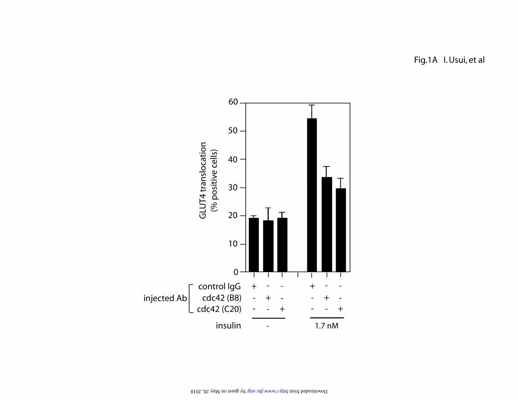
cdc42 plays a role in insulin-induced GLUT4 translocation and 2-DOG
uptake. To evaluate the role of cdc42 in insulin stimulated GLUT4
translocation, we conducted single cell microinjection studies using mouse
monoclonal (B8) or goat polyclonal (C20) anti-cdc42 antibodies, followed by
immunofluorescence staining with GLUT4 antibody in 3T3-L1 adipocytes (Fig.
1A). In the basal state, most of the cells display GLUT4 staining in the perinuclear
region, and after insulin stimulation there is a marked increase in the proportion of
cells demonstrating GLUT4 localization at the plasma membrane as a
circumferential ring, as previously described(6). Microinjection of either mouse
monoclonal or goat polyclonal anti-cdc42 antibody decreased 1.7 nM insulin-
stimulated GLUT4 translocation by 60-75%. To further demonstrate the
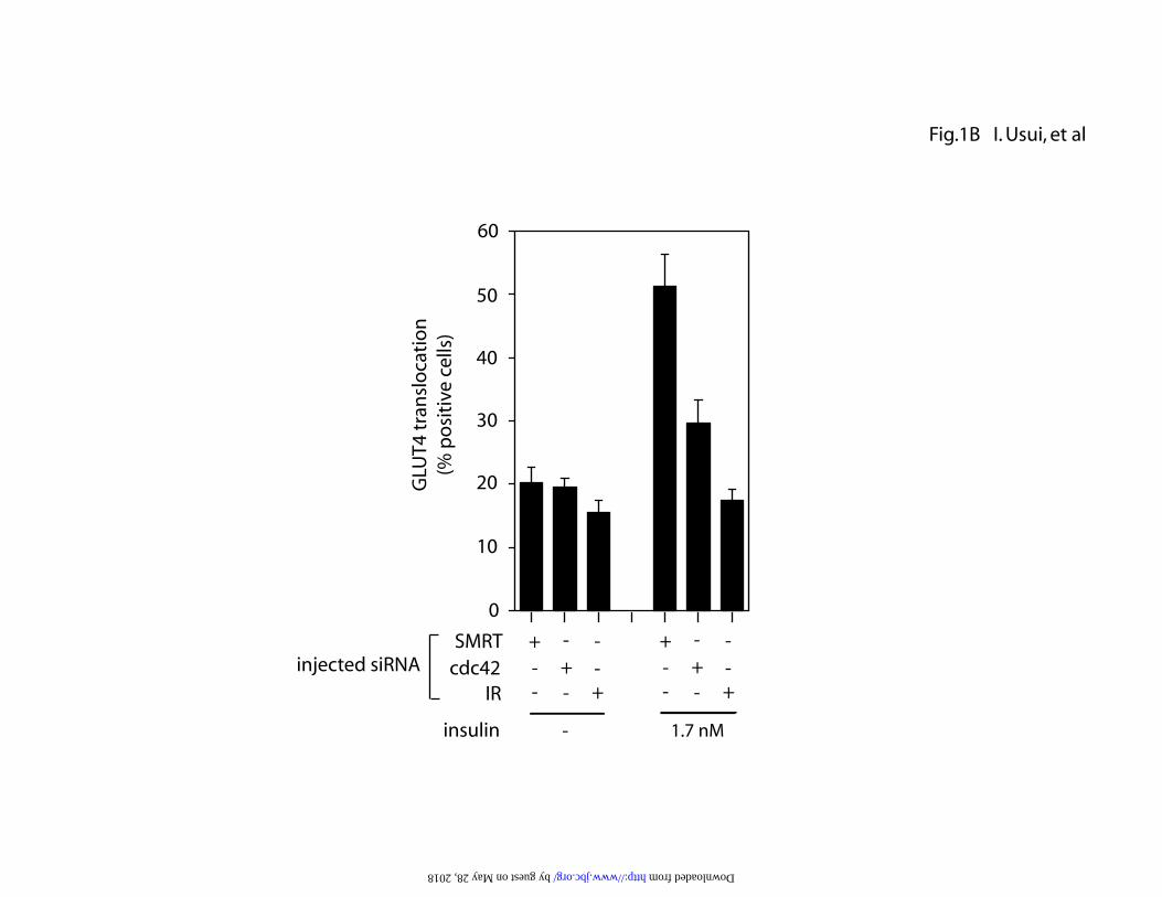
importance of cdc42 for insulin-stimulated GLUT4 translocation, we utilized
siRNA to knock down cdc42 expression followed by measurement of GLUT4
translocation (Fig.1B). SiRNA directed against cdc42 was microinjected into the
cytoplasm of 3T3-L1 adipocytes, and 72 h later, GLUT4 translocation was
measured. As seen in Fig.1B, cdc42 siRNA led to a 65 % decrease in
insulin-induced GLUT4 translocation. As a positive control, siRNA against the
insulin receptor was injected, which completely abolished insulin stimulation. As
a negative control, siRNA against SMRT, a transcription co-regulating molecule,
was also injected and had no effect.
To further examine the role of cdc42 in insulin-induced glucose transport,
we next measured 2-deoxyglucose (2-DOG) uptake in 3T3-L1 adipocytes
infected with adenovirus vectors containing either constitutively active cdc42
(V12; CA-cdc42) or dominant negative cdc42 (N17, DN-cdc42). As shown in
by guest on May 28, 2018
http://ww
w.jbc.org/
Dow
nloaded from

11
Fig. 1C, infection of CA-cdc42 at 80 m.o.i. increased 2-DOG uptake to 61 % of
the maximal insulin response in the basal state and further enhanced 2-DOG
uptake stimulated by submaximal insulin (0.5 nM). Interestingly, adenovirus
mediated expression of DN-cdc42 was without effect on insulin stimulation of
glucose transport. Taken together, these results suggest that cdc42 plays an
important role in insulin signaling leading to GLUT4 translocation.
cdc42 is downstream of G q/11 in the insulin-GLUT4 translocation signaling
pathway. As we recently reported (6), an adenovirus encoding
constitutively active Gq (CA-Gq; Q209L) stimulated GLUT4 translocation to
60% of the maximal insulin response, showing a role for Gαq/11 in this insulin
action. In order to examine whether cdc42 is downstream of Gαq/11, we
microinjected mouse monoclonal anti-cdc42 antibody (B8) into cells infected
with CA-Gq or CA-cdc42, and similar to the results with insulin- or CA-cdc42-
stimulation, anti-cdc42 antibody injection inhibited CA-Gq-induced GLUT4
translocation (Fig. 2). To further assess the relative loci of Gαq/11 and cdc42 in
this pathway, we microinjected anti-Gαq/11 antibody into cells stimulated by CA-
cdc42, CA-Gq or insulin. Anti-Gαq/11 antibody injection inhibited insulin- or
CA-Gq-induced GLUT4 translocation, but did not inhibit the effects of CA-cdc42
(Fig. 2). These data provide further evidence that cdc42 is downstream of
Gαq/11 in mediating insulin-stimulated GLUT4 translocation.
Constitutively active cdc42 stimulates 2-DOG uptake in a PI3-kinase
dependent manner. We next examined the effects of the PI3-kinase
inhibitors, wortmannin and LY294002, on CA-cdc42-induced 2-DOG uptake
by guest on May 28, 2018
http://ww
w.jbc.org/
Dow
nloaded from

12
(Fig. 3A). In the absence of insulin, expression of CA-cdc42 increased 2-DOG
uptake in a dose responsive manner with 40 and 80 m.o.i. stimulating to 39% and
56% of the maximal insulin response, respectively. Incubation of the cells with
100 nM wortmannin or 100 µM LY294002 for 1 h did not alter 2-DOG uptake
stimulated by CA-cdc42 expression, while it completely inhibited insulin-
stimulated glucose uptake. However, when the cells were incubated with these
inhibitors for 4 h, not only insulin-induced, but also CA-cdc42-induced 2-DOG
uptake was inhibited to basal levels. As reported previously (6), overexpression
of CA-Gq enhanced 2-DOG uptake, and this was also inhibited by incubation
with 100 nM wortmannin or 100 µM LY294002 for 4 h, but not for 1 h. As
expected, incubation with a MEK1 inhibitor, PD98059, for 4 h did not affect 2-
DOG uptake stimulated by insulin, CA-cdc42 or CA-Gq expression (Fig. 3A).
These results suggest that activated cdc42 and Gαq/11 can stimulate glucose
uptake in a PI3-kinase dependent manner in 3T3-L1 adipocytes.
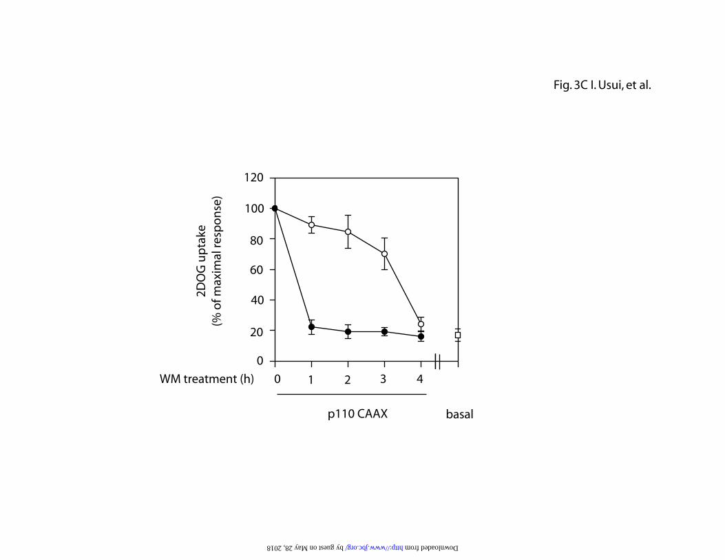
Since the lower doses of wortmannin and LY294002 took 4 h to reach
their maximal effects, we further examined the time course and dose dependency
of PI3-kinase inhibition on 2-DOG uptake in cells expressing CA-cdc42 as well as
constitutively active PI3-kinase (p110-CAAX). As seen in Figure 3B, 48 h after
expression of CA-cdc42, cells were treated with 100 or 300 nM wortmannin for
1-4 h and 2-DOG uptake was measured. The CA-cdc42-induced increase in
glucose transport was maximally inhibited at 300 nM at the first time point,
whereas, at the lower dose (100 nM), transport was inhibited in a gradual, time
dependent manner reaching maximal inhibition by 4 h. Constitutively active PI3-
kinase (p110-CAAX) expression is known to stimulate 2-DOG uptake in 3T3-L1
cells, and the same inhibitory experiments were performed in p110-CAAX
by guest on May 28, 2018
http://ww
w.jbc.org/
Dow
nloaded from

13
expressing cells with essentially identical results (Fig. 3C). These data indicate
that in cells manifesting chronic PI3-kinase activation, there is a time and dose
dependent effect of these inhibitors to suppress glucose transport. This is in
contrast to acute effects of insulin to stimulate transport, which can be inhibited
rapidly at low or high dose of PI3-kinase inhibitors.
To exclude the possibility that the decrease in CA-cdc42- or CA-Gq-
induced 2-DOG uptake after 4 h treatment with the PI3-kinase inhibitors might be
due to the toxicity or the non-specific effects on general trafficking systems, 2-
DOG uptake stimulated by osmotic shock was measured after 4 h treatment with
the PI3-kinase inhibitors. Osmotic shock stimulates glucose transport
predominantly through a PI3-kinase independent mechanism, and, as seen in Fig.
3D, pretreatment with 100 nM wortmannin or 100 µM LY294002 for 4 h did not
inhibit osmotic shock-induced glucose transport.
Insulin and constitutively active G q stimulate cdc42 activity. Since
our data indicate that glucose uptake induced by CA-cdc42 and CA-Gq involve
common mechanisms, we further examined the relationship between cdc42 and
Gαq/11 before and after insulin stimulation. We measured the time course of
insulin-induced cdc42 activation using GST-PAK1 as a substrate, which
specifically binds to active cdc42 (24). As shown in Fig.4A, insulin rapidly
stimulated cdc42 activity with a maximal response at 1 min, returning to basal
levels, thereafter. We recently showed that insulin treatment led to tyrosine
phosphorylation of Gαq/11 and this could activate Gαq/11 as a positive signaling
molecule (6). Interestingly, the time course of insulin-stimulated tyrosine
phosphorylation of Gαq/11 was comparable to the time course of cdc42
by guest on May 28, 2018
http://ww
w.jbc.org/
Dow
nloaded from

14
activation (Fig. 4A). Cdc42 activity was also measured in 3T3-L1 adipocytes
after adenovirus expression of wild type or constitutively active Gq, before and
after insulin stimulation for 1 min (Fig. 4B). Expression of CA-Gq stimulated
cdc42 activity in the basal state and enhanced the effect of insulin compared to
control cells. These data show that cdc42 is activated by insulin and Gαq/11,
further suggesting the concept that cdc42 is downstream of Gαq/11 in the insulin
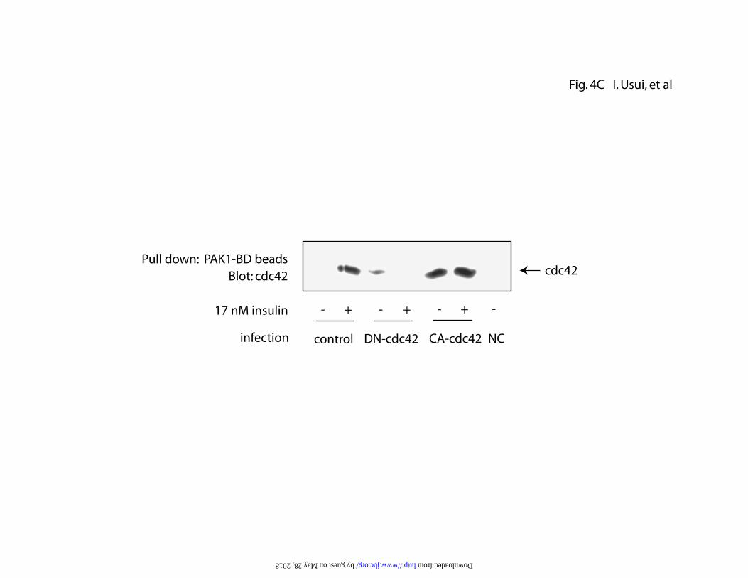
signaling cascade. We also assessed the activity of DN-cdc42 using this method.
As can be seen in Fig. 4C, expression of CA-cdc42 resulted in increased cdc42
activity in the absence of insulin, whereas, expression of DN-cdc42 at 80 m.o.i.
inhibited the effect of insulin to activate cdc42.
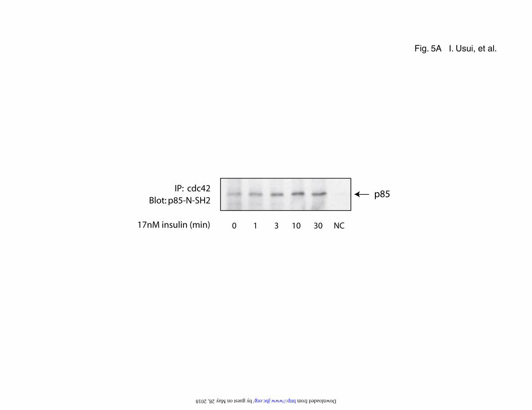
Cdc42 stimulates PI3-kinase activity. Since it has been shown in other systems
that cdc42 can bind to the p85 regulatory subunit of PI3-kinase and stimulate
PI3-kinase activity (25,26), we examined the relationship between cdc42 and
PI3-kinase in 3T3-L1 adipocytes. Fig. 5A shows the co precipitation of p85 in
anti-cdc42 immunoprecipitates after insulin stimulation. Association of p85 with
cdc42 was barely detected in the basal state and was increased by insulin
stimulation, with a maximal association at 10 min. Next, we directly measured PI3-
kinase activity associated with cdc42 (Fig. 5B, C). Insulin increased the PI3-
kinase activity in anti-cdc42 immunoprecipitates by 2.8 fold with a maximal
response by 10 min. Expression of CA-cdc42 and CA-Gq (40 m.o.i.) stimulated
PI3-kinase activity in the absence of insulin by 4.4 fold and 4.2 fold, respectively.
Incubation of the cells with 100 nM wortmannin for 4 h inhibited PI3-kinase
activity stimulated by either CA-Gq, CA-cdc42, or insulin (Fig. 5B), while 1 h
by guest on May 28, 2018
http://ww
w.jbc.org/
Dow
nloaded from

15
treatment with wortmannin inhibited only insulin stimulation (data not shown).
Comparable results were obtained when 100 µM LY294002 was used (Fig. 5C).
As with the glucose transport data presented in Figure 3, inhibition of
cdc42 associated PI3-kinase activity was not complete until 4 h after treatment
with 100 nM wortmannin or 100 µM LY294002. To study the dose response
and time course of this inhibition, CA-cdc42 and p110-CAAX expressing cells
were treated with wortmannin at 100 or 300 nM for up to 4 h, followed by
measurement of cdc42 associated PI3-kinase activity. As seen in Figure 5D and
5E, 300 nM led to maximal inhibition at the first time point studied, whereas, the
inhibition was more gradual, reaching a maximal effect at 4 h at the low dose (100
nM) of wortmannin. These results are completely consistent with the dose
response and time course of 2-DOG inhibition shown in Figure 3.
Constitutively active cdc42 stimulates GLUT4 translocation and 2-DOG
uptake in a PKC -dependent manner. It has been reported that PKCλ is a
component of the insulin signaling cascade, and plays an important role in insulin-
induced GLUT4 translocation downstream of PI3-kinase (27,28). Since we
recently showed that PKCλ was also required for CA-Gq-induced GLUT4
translocation (6), we determined whether PKCλ was a participant in the cdc42
pathway leading to GLUT4 translocation and glucose uptake. First, we measured
the effect of anti- PKCλ antibody microinjection on GLUT4 translocation
stimulated by insulin, CA-cdc42, or CA-Gq. Anti- PKCλ antibody injection
completely blocked CA-cdc42-stimulated GLUT4 translocation, similar to the
results with insulin stimulation or CA-Gq expression. Adenoviral mediated
by guest on May 28, 2018
http://ww
w.jbc.org/
Dow
nloaded from

16
expression of constitutively active PKCλ stimulated GLUT4 translocation to the
same extent as insulin, further arguing that PKCλ is a participant in the insulin
signaling pathway leading to GLUT4 translocation (Fig.6A). Next, we examined
the effect of a kinase-deficient mutant of PKCλ (K273E; KD-PKCλ) on insulin- or
CA-cdc42-induced 2-DOG uptake (Fig.6B). Adenoviral gene transfer of this
mutant PKCλ resulted in a dose-dependent inhibition of 2-DOG uptake
stimulated by either 17 nM insulin or CA-cdc42 expression. Taken together,
these results further argue that both Gαq/11 and cdc42 mediate insulin-stimulated
GLUT4 translocation and glucose transport by a common signaling pathway in
which Gαq/11 lies upstream of cdc42 and that both are upstream of PKCλ.
by guest on May 28, 2018
http://ww
w.jbc.org/
Dow
nloaded from

17
Discussion
One of the major actions of insulin is to stimulate GLUT4 translocation in
order to increase glucose uptake into target cells. This is accomplished by a
complicated, multi-step signaling pathway, which remains incompletely
understood. In the current study, we have shown an important role for cdc42, a
Rho GTPase family member, in this process. We show that insulin stimulates
cdc42 activity and that interfering with cdc42 function by microinjection of anti-
cdc42 antibody or siRNA into 3T3-L1 adipocytes inhibits insulin-stimulated
GLUT4 translocation. Furthermore, we show that cdc42 is downstream of
another insulin stimulated activator of glucose transport, Gαq/11, and that the
effects of cdc42 to facilitate GLUT4 translocation are mediated through PI3-
kinase. Taken together, these results describe a novel role for cdc42 as a
signaling molecule within the insulin action cascade ultimately mediating GLUT4
translocation.
TC10 is a small Rho family GTPase which participates in insulin
stimulation of glucose transport and GLUT4 translocation (17). It has been
suggested that TC10 helps mediate cortical actin polymerization, possibly
through interaction with N-WASP (18). Cdc42 is also a member of the Rho
GTPase family, with a high degree of homology (69%) to TC10 (17), and a role for
cdc42 in mediating actin rearrangement has been suggested (19). With respect to
bradykinin-stimulated actin remodeling, data exist placing cdc42 downstream of
Gαq/11 in this process(21). Since bradykinin can also stimulate glucose transport
under certain conditions(20), and since we have previously demonstrated an
important role for Gαq/11 as a mediator of insulin-stimulated GLUT4
translocation, we postulated a role for cdc42 in insulin-stimulated glucose
by guest on May 28, 2018
http://ww
w.jbc.org/
Dow
nloaded from

18
transport. The current studies fully support this idea and also argue strongly that
cdc42 lies downstream of Gαq/11 in this pathway. Thus, insulin stimulates
Gαq/11 as well as cdc42, and both of these events are necessary for full
stimulation of GLUT4 translocation. Adenoviral mediated expression of CA-
cdc42 increased GLUT4 translocation and glucose transport independent of
insulin, and these effects of CA-cdc42 were attenuated by inhibitors of PI3-
kinase. Moreover, microinjection of cdc42 antibody inhibits insulin- and CA-Gq-
stimulated GLUT4 translocation, and CA-Gq expression stimulates cdc42 activity.
Furthermore, microinjection of Gαq/11 antibody inhibits insulin-, but not CA-
cdc42-stimulated GLUT4 translocation. These results place cdc42 downstream of
Gαq/11 in this insulin stimulatory cascade. Lastly, insulin stimulates PKCλ
activity in a PI3-kinase dependent manner (6,27,28), and microinjection of anti-
PKCλ antibody blocks insulin, CA-Gq-, and CA-cdc42-stimulated GLUT4
translocation, indicating that all of these molecules participate in a common
signaling pathway. These results do not argue in anyway against a role for TC10
as a mediator of GLUT4 translocation, and it is quite possible that both of these
small Rho family GTPase proteins participate and may comprise parallel or
redundant steps to mediate this important biologic effect of insulin.
It should be noted that some recent studies have shown that transfection
of dominant negative cdc42 (DN-cdc42) does not affect insulin induced glucose
transport into 3T3-L1 adipocytes(17), and, at first approximation, this is not
consistent with our above described results. However, in our own study, we also
find that expression of DN-cdc42 did not inhibit insulin-stimulated glucose
transport and this result may have to do with the specific nature of this dominant
negative construct. The DN-cdc42 used in the current studies, as well as in the
by guest on May 28, 2018
http://ww
w.jbc.org/
Dow
nloaded from
19
previous reports (17), contains a point mutation impairing its ability to
phosphorylate PAK1, one of the target molecules of cdc42. Indeed, we
demonstrate that DN-cdc42 inhibits insulin stimulated cdc42 activity towards
PAK1. However, cdc42 may have multiple target molecules (19), and it is
unknown whether this mutant has a dominant negative effect on actions of
cdc42 other than PAK1 phosphorylation. Thus, DN-cdc42 inhibits the ability of
endogenous cdc42 to bind to GST-PAK1 beads, but does not inhibit insulin-
stimulated glucose transport or insulin-stimulated cdc42-associated PI3-kinase
activity (data not shown). These results raise the possibility that cdc42 may
stimulate PI3-kinase and glucose uptake through a mechanism independent from
its ability to interact with PAK1, and this could provide an explanation for why
DN-cdc42 did not inhibit insulin induced glucose transport in the studies by
Chiang et al.(17), or in our own experiments.
Based on numerous studies, it is quite clear that activation of PI3-kinase
is a necessary step for stimulation of GLUT4 translocation and glucose transport
(29-32). However, the connection between the activated insulin receptor and
PI3-kinase stimulation is somewhat more involved. IRS-1 is a major substrate of
the insulin receptor and tyrosine phosphorylated IRS-1 can bind to the SH2
domain of the p85 regulatory subunit of PI3-kinase, resulting in PI3-kinase
enzymatic activation (3). However, a number of studies, using different
approaches, have indicated that IRS-1 is not strictly essential for glucose
transport stimulation and that other signaling pathways may be involved (7-12).
For example, inhibition of IRS-1 activity in 3T3-L1 adipocytes does not impair
insulin-stimulated glucose transport (9), and IRS-1 knockout animals are only
mildly insulin resistant and adipocytes from these animals show only a partial
by guest on May 28, 2018
http://ww
w.jbc.org/
Dow
nloaded from
20
defect in insulin-stimulated glucose transport (10,11). Additionally, when IRS-1 is
activated independently of insulin, glucose transport is not stimulated (12).
Furthermore, we have previously shown that insulin can cause tyrosine
phosphorylation of Gαq/11 and that microinjection of Gαq/11 inhibitory reagents
into 3T3-L1 adipocytes blocks insulin stimulated glucose transport (6). We also
found that constitutively active Gαq stimulates glucose transport by itself in a
PI3-kinase dependent manner (6). Additionally, other ligands such as bradykinin
and ET-1, which receptors couple into Gαq/11, can also stimulate GLUT4
translocation (33). Some of these studies have shown that the effects of Gαq/11
are PI3-kinase dependent (6,33), whereas, others have been unable to
demonstrate this linkage(13-15). In the current study, we find that adenovirus
mediated expression of CA-Gq leads to activation of cdc42 and that CA-Gq as
well as CA-cdc42 can stimulate glucose transport. These stimulatory effects are
inhibited by 1h treatment with 300 nM wortmannin, while 100 nM wortmannin
took 4 h to reach full inhibitory effect. Since these constitutively active proteins
were expressed in cells through adenovirus mediated gene transfer, and the
assays were performed 48 hours after infection, CA-Gq and CA-cdc42 had
considerable time to exert their effects. Since both wortmannin and LY294002
inhibit ATP binding to PI3-kinase (34), in the case of constitutively active
preactivation, the ATP binding site of PI3-kinase is already occupied by
endogenous ATP before the treatment with the inhibitors. It is possible that
higher concentrations of inhibitors or longer term treatments are necessary to
replace ATP under these conditions. The dose response and time course studies
for inhibition of 2-DOG and PI3-kinase activity are fully consistent with this idea
(Fig. 3B, C, Fig. 5D, E). This may explain why some of the earlier studies were
by guest on May 28, 2018
http://ww
w.jbc.org/
Dow
nloaded from
21
unable to show PI3-kinase dependency of Gαq/11 effects. Another possibility is
that insulin and cdc42 could utilize somewhat different PI3-kinase isoforms to
mediate their effects and that different isoforms of PI3-kinase could have different
sensitivity to inhibtion by wortmannin. In any event, under the current
experimental conditions, the effects of CA-cdc42 as well as CA-Gq are clearly
inhibited by wortmannin and LY294002.
Supporting this line of reasoning, and consistent with our current results, it
has already been reported that some of the biologic actions of cdc42 are mediated
through activation of PI3-kinase. For example, cdc42, as well as Rac1, modifies
actin organization leading to increased motility and invasiveness in epithelial cells
(35). The activation of PAK1 and JNK, both of which are known target
molecules of cdc42, is not required for these functions, but PI3-kinase activation
is necessary (35). It has also been suggested that the Bcr homology domain (BH
domain) of p85 directly interacts with cdc42 and Rac1 (26,36), and it is quite
possible that the biological actions of cdc42 which require PI3-kinase activation
are mediated through the interaction of cdc42 with the BH domain of p85.
In summary, we have demonstrated an important role for cdc42 as a novel
signaling molecule in the insulin action pathway leading to GLUT4 translocation
and stimulation of glucose transport. We find that cdc42 is downstream of
Gαq/11 in this signaling system and lies upstream of PI3-kinase and PKCλ. Our
experiments show that the various components of this pathway are necessary for
full and efficient insulin stimulation of glucose transport in 3T3-L1 adipocytes.
Since stimulation of glucose transport is a major action of insulin, and since insulin
resistance to glucose transport stimulation is a central component of a number of
disease states such as Type II diabetes, obesity, etc., it is possible that functional
by guest on May 28, 2018
http://ww
w.jbc.org/
Dow
nloaded from

22
abnormalities in this pathway may participate in the mechanisms of insulin
resistance in pathophysiologic conditions in man.
by guest on May 28, 2018
http://ww
w.jbc.org/
Dow
nloaded from

23
Acknowledgments
We would like to thank Ms. Elizabeth Hansen for editorial assistance.
This work was supported by a research grant from the National Institutes of
Health (DK 33651), the Veterans Administration San Diego Health Care System,
Research Service, and the Whittier Institute for Diabetes. Dr. Isao Usui is
supported through an American Diabetes Association Mentor-based Fellowship
Award.
by guest on May 28, 2018
http://ww
w.jbc.org/
Dow
nloaded from
24
Figure legends
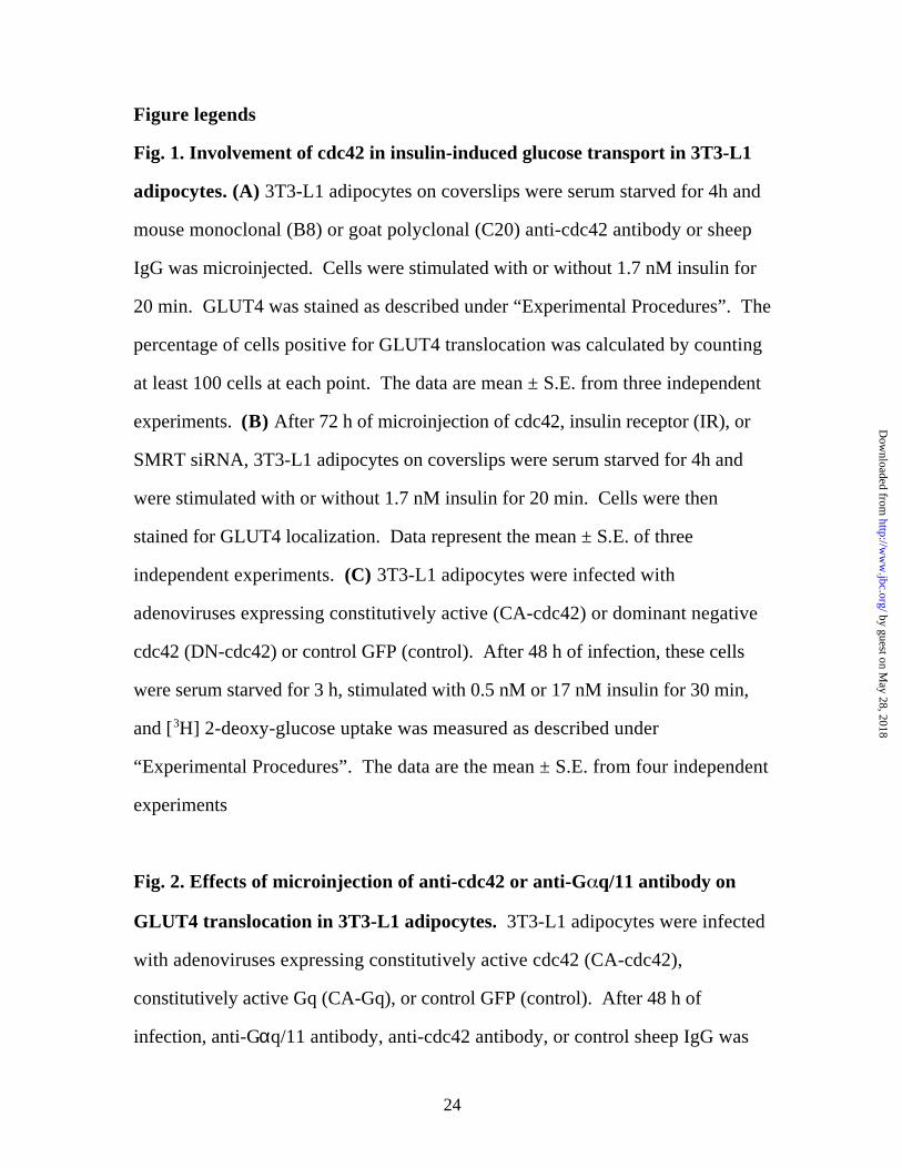
Fig. 1. Involvement of cdc42 in insulin-induced glucose transport in 3T3-L1
adipocytes. (A) 3T3-L1 adipocytes on coverslips were serum starved for 4h and
mouse monoclonal (B8) or goat polyclonal (C20) anti-cdc42 antibody or sheep
IgG was microinjected. Cells were stimulated with or without 1.7 nM insulin for
20 min. GLUT4 was stained as described under “Experimental Procedures”. The
percentage of cells positive for GLUT4 translocation was calculated by counting
at least 100 cells at each point. The data are mean ± S.E. from three independent
experiments. (B) After 72 h of microinjection of cdc42, insulin receptor (IR), or
SMRT siRNA, 3T3-L1 adipocytes on coverslips were serum starved for 4h and
were stimulated with or without 1.7 nM insulin for 20 min. Cells were then
stained for GLUT4 localization. Data represent the mean ± S.E. of three
independent experiments. (C) 3T3-L1 adipocytes were infected with
adenoviruses expressing constitutively active (CA-cdc42) or dominant negative
cdc42 (DN-cdc42) or control GFP (control). After 48 h of infection, these cells
were serum starved for 3 h, stimulated with 0.5 nM or 17 nM insulin for 30 min,
and [3H] 2-deoxy-glucose uptake was measured as described under
“Experimental Procedures”. The data are the mean ± S.E. from four independent
experiments
Fig. 2. Effects of microinjection of anti-cdc42 or anti-G q/11 antibody on
GLUT4 translocation in 3T3-L1 adipocytes. 3T3-L1 adipocytes were infected
with adenoviruses expressing constitutively active cdc42 (CA-cdc42),
constitutively active Gq (CA-Gq), or control GFP (control). After 48 h of
infection, anti-Gαq/11 antibody, anti-cdc42 antibody, or control sheep IgG was
by guest on May 28, 2018
http://ww
w.jbc.org/
Dow
nloaded from

25
microinjected into the cells on coverslips. Cells were serum starved for 4h and
stimulated with 1.7 nM insulin for 20 min. GLUT4 in the cells was stained as
described under “Experimental Procedures”. The percentage of cells positive for
GLUT4 translocation was calculated by counting at least 100 cells at each point.
The data are mean ± S.E. from three independent experiments.
Fig. 3. Effects of PI3-kinase inhibitors on constitutively active cdc42-,
constitutively active Gq-, insulin-, or osmotic shock-induced 2-DOG uptake
into 3T3-L1 adipocytes. (A) 3T3-L1 adipocytes were infected with
adenoviruses expressing constitutively active cdc42 (CA-cdc42) or
constitutively active Gq (CA-Gq) or control GFP (control) with indicated m.o.i.
After 48h of infection, cells were serum starved and incubated with 100 µM
LY294002 (L), 100 nM wortmannin (W), 50 µM PD98059 (P) or 0.1% DMSO (D)
for 1h or 4 h. Some cells were stimulated by 17 nM insulin for 30 min (insulin).
[3H] 2-deoxy-glucose uptake was measured as described under “Experimental
Procedures”. (B, C) 3T3-L1 adipocytes were infected with adenoviruses
expressing CA-cdc42 (B) or constitutively active PI3-kinase (p110-CAAX) (C).
After 48 h of infection, cells were serum starved and incubated with 100 nM
(open circle) or 300 nM wortmannin (closed circle) for the indicated time periods.
[3H] 2-deoxy-glucose uptake was measured as described under “Experimental
Procedures”. Open square stands for [3H] 2-deoxy-glucose uptake in basal
without virus infection. (D) 3T3-L1 adipocytes were serum starved and
incubated with 100 µM LY294002 (L), 100 nM wortmannin (W), or 0.1% DMSO
(D) for 4 h. They were incubated with 600 mM sorbitol or 17 nM insulin for 30
min, and [3H] 2-deoxy-glucose uptake was measured as described under
by guest on May 28, 2018
http://ww
w.jbc.org/
Dow
nloaded from

26
“Experimental Procedures”. The data are the mean ± S.E. from four (A) or three
(B-D) independent experiments.
Fig. 4. Insulin- or constitutively active Gq-induced cdc42 activity (A) 3T3-L1
adipocytes were serum-starved for 16h, stimulated with 17 nM insulin for the
indicated time periods. Cdc42 activities were measured by mixing the cell lysates
with GST-PAK1 beads which specifically recognize active cdc42. Samples were
analysed by Western blotting using anti-cdc42 antibody. The activity was
quantitated by scanning the film using NIH Image. Tyrosine phosphorylated Gq
was detected by immunoprecipitating with or without (negative control; NC)
anti-phosphotyrosine antibody (PY20) and Western blotting using anti-Gq
antibody as described under “Experimental Procedures”. Representative blots
are shown from five (cdc42) or three (Gq) independent experiments. (B) 3T3-L1
adipocytes were infected with adenoviruses expressing wild type Gq (WT-Gq),
constitutively active Gq (CA-Gq) or control GFP (C). After 48 h of infection,
3T3-L1 adipocytes were serum starved for 16 h, stimulated with or without insulin
for 1 min, and lysed. Cdc42 activity was analysed as described above. These
experiments were repeated twice. (C) 3T3-L1 adipocytes were infected with
adenoviruses expressing dominant negative cdc42 (DN-cdc42), constitutive
active cdc42 (CA-cdc42) or control GFP (control). After 48 h of infection, 3T3-
L1 adipocytes were serum starved for 16 h, stimulated with or without insulin for
1 min, and lysed. Cdc42 activity was analysed as described above. A sample for
a negative control (NC) was prepared as described under “Experimental
Procedures”. Representative blot is shown from three independent experiments.
by guest on May 28, 2018
http://ww
w.jbc.org/
Dow
nloaded from
27
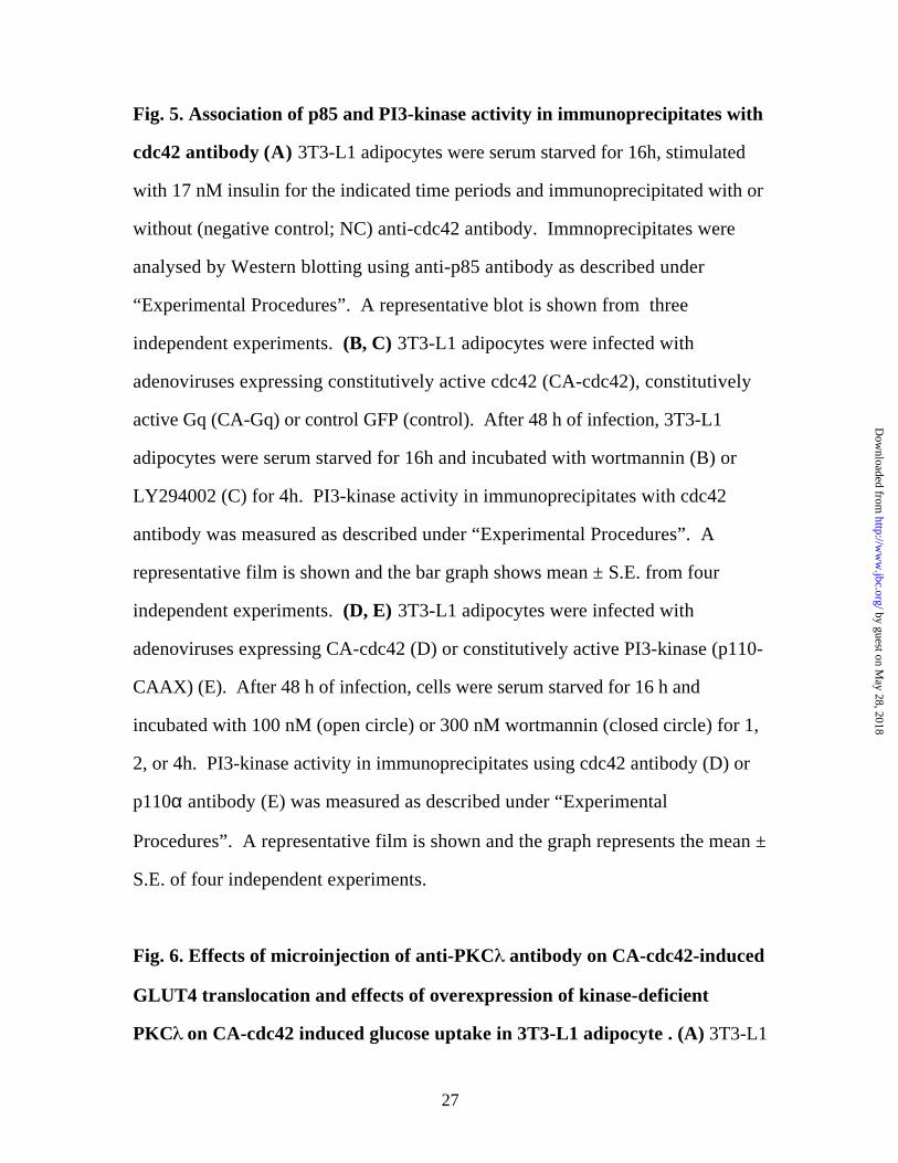
Fig. 5. Association of p85 and PI3-kinase activity in immunoprecipitates with
cdc42 antibody (A) 3T3-L1 adipocytes were serum starved for 16h, stimulated
with 17 nM insulin for the indicated time periods and immunoprecipitated with or
without (negative control; NC) anti-cdc42 antibody. Immnoprecipitates were
analysed by Western blotting using anti-p85 antibody as described under
“Experimental Procedures”. A representative blot is shown from three
independent experiments. (B, C) 3T3-L1 adipocytes were infected with
adenoviruses expressing constitutively active cdc42 (CA-cdc42), constitutively
active Gq (CA-Gq) or control GFP (control). After 48 h of infection, 3T3-L1
adipocytes were serum starved for 16h and incubated with wortmannin (B) or
LY294002 (C) for 4h. PI3-kinase activity in immunoprecipitates with cdc42
antibody was measured as described under “Experimental Procedures”. A
representative film is shown and the bar graph shows mean ± S.E. from four
independent experiments. (D, E) 3T3-L1 adipocytes were infected with
adenoviruses expressing CA-cdc42 (D) or constitutively active PI3-kinase (p110-
CAAX) (E). After 48 h of infection, cells were serum starved for 16 h and
incubated with 100 nM (open circle) or 300 nM wortmannin (closed circle) for 1,
2, or 4h. PI3-kinase activity in immunoprecipitates using cdc42 antibody (D) or
p110α antibody (E) was measured as described under “Experimental
Procedures”. A representative film is shown and the graph represents the mean ±
S.E. of four independent experiments.
Fig. 6. Effects of microinjection of anti-PKC antibody on CA-cdc42-induced
GLUT4 translocation and effects of overexpression of kinase-deficient
PKC on CA-cdc42 induced glucose uptake in 3T3-L1 adipocyte . (A) 3T3-L1
by guest on May 28, 2018
http://ww
w.jbc.org/
Dow
nloaded from

28
adipocytes were infected with adenoviruses expressing constitutively active
cdc42 (CA-cdc42), constitutively active Gq (CA-Gq), constitutively active PKCλ
(CA-PKCλ), or control GFP (control). After 48 h of infection, anti-cdc42
antibody (+) or sheep IgG (-) was microinjected into the cells on coverslips. Cells
were serum starved for 4h and incubated with 1.7 nM insulin for 20 min. GLUT4
in the cells was stained as described under “Experimental Procedures”. The
percentage of cells positive for GLUT4 translocation was calculated by counting
at least 100 cells at each point. The data are mean ± S.E. from three independent
experiments. (B) 3T3-L1 adipocytes were infected with adenovirus expressing
kinase-deficient PKCλ (KD-PKCλ) with the indicated m.o.i.. Adenoviruses
expressing constitutively active cdc42 (CA-cdc42) with 40 m.o.i. or control
adenovirus (control) were also co-infected to adjust the total amount of
adenovirus. After 48 h of infection, these cells were serum starved for 3 h,
stimulated by 17 nM insulin for 30 min, and [3H] 2-deoxy-glucose uptake was
measured as described under “Experimental Procedures”. The data are mean ±
S.E. from three independent experiments.
by guest on May 28, 2018
http://ww
w.jbc.org/
Dow
nloaded from

29
Reference
1. Birnbaum, M. J. (1992) Int Rev Cytol 137, 239-97
2. James, D. E., Piper, R. C., and Slot, J. W. (1993) J Cell Sci 104 ( Pt 3),
607-12
3. Czech, M. P., and Corvera, S. (1999) J Biol Chem 274(4), 1865-8
4. Holman, G. D., and Kasuga, M. (1997) Diabetologia 40(9), 991-1003
5. Pessin, J. E., and Saltiel, A. R. (2000) J Clin Invest 106(2), 165-9
6. Imamura, T., Vollenweider, P., Egawa, K., Clodi, M., Ishibashi, K.,
Nakashima, N., Ugi, S., Adams, J. W., Brown, J. H., and Olefsky, J. M.
(1999) Mol Cell Biol 19(10), 6765-74
7. Sakoda, H., Ogihara, T., Anai, M., Funaki, M., Inukai, K., Katagiri, H.,
Fukushima, Y., Onishi, Y., Ono, H., Fujishiro, M., Kikuchi, M., Oka, Y., and
Asano, T. (2000) Diabetes 49(10), 1700-8.
8. Kanoh, Y., Bandyopadhyay, G., Sajan, M. P., Standaert, M. L., and Farese,
R. V. (2001) Endocrinology 142(4), 1595-605.
9. Sharma, P. M., Egawa, K., Gustafson, T. A., Martin, J. L., and Olefsky, J. M.
(1997) Mol Cell Biol 17(12), 7386-97.
10. Araki, E., Lipes, M. A., Patti, M. E., Bruning, J. C., Haag, B., 3rd, Johnson,
R. S., and Kahn, C. R. (1994) Nature 372(6502), 186-90
11. Tamemoto, H., Kadowaki, T., Tobe, K., Yagi, T., Sakura, H., Hayakawa, T.,
Terauchi, Y., Ueki, K., Kaburagi, Y., Satoh, S., and et al. (1994) Nature
372(6502), 182-6
12. Guilherme, A., and Czech, M. P. (1998) J Biol Chem 273(50), 33119-22.
by guest on May 28, 2018
http://ww
w.jbc.org/
Dow
nloaded from

30
13. Bose, A., Cherniack, A. D., Langille, S. E., Nicoloro, S. M., Buxton, J. M.,
Park, J. G., Chawla, A., and Czech, M. P. (2001) Mol Cell Biol 21(15),
5262-75
14. Lawrence, J. T., and Birnbaum, M. J. (2001) Mol Cell Biol 21(15), 5276-
85
15. Kanzaki, M., Watson, R. T., Artemyev, N. O., and Pessin, J. E. (2000) J
Biol Chem 275(10), 7167-75.
16. Baumann, C. A., Ribon, V., Kanzaki, M., Thurmond, D. C., Mora, S.,
Shigematsu, S., Bickel, P. E., Pessin, J. E., and Saltiel, A. R. (2000) Nature
407(6801), 202-7
17. Chiang, S. H., Baumann, C. A., Kanzaki, M., Thurmond, D. C., Watson, R.
T., Neudauer, C. L., Macara, I. G., Pessin, J. E., and Saltiel, A. R. (2001)
Nature 410(6831), 944-8
18. Jiang, Z. Y., Chawla, A., Bose, A., Way, M., and Czech, M. P. (2002) J
Biol Chem 277(1), 509-15
19. Mackay, D. J., and Hall, A. (1998) J Biol Chem 273(33), 20685-8
20. Kishi, K., Muromoto, N., Nakaya, Y., Miyata, I., Hagi, A., Hayashi, H., and
Ebina, Y. (1998) Diabetes 47(4), 550-8.
21. Kozma, R., Ahmed, S., Best, A., and Lim, L. (1995) Mol Cell Biol 15(4),
1942-52.
22. Takano, A., Usui, I., Haruta, T., Kawahara, J., Uno, T., Iwata, M., and
Kobayashi, M. (2001) Mol Cell Biol 21(15), 5050-62
23. Janez, A., Worrall, D. S., Imamura, T., Sharma, P. M., and Olefsky, J. M.
(2000) J Biol Chem 275(35), 26870-6
by guest on May 28, 2018
http://ww
w.jbc.org/
Dow
nloaded from

31
24. Benard, V., Bohl, B. P., and Bokoch, G. M. (1999) J Biol Chem 274(19),
13198-204.
25. Zheng, Y., Bagrodia, S., and Cerione, R. A. (1994) J Biol Chem 269(29),
18727-30
26. Musacchio, A., Cantley, L. C., and Harrison, S. C. (1996) Proc Natl Acad
Sci U S A 93(25), 14373-8
27. Wada, T., Sasaoka, T., Funaki, M., Hori, H., Murakami, S., Ishiki, M., Haruta,
T., Asano, T., Ogawa, W., Ishihara, H., and Kobayashi, M. (2001) Mol Cell
Biol 21(5), 1633-46.
28. Kotani, K., Ogawa, W., Matsumoto, M., Kitamura, T., Sakaue, H., Hino, Y.,
Miyake, K., Sano, W., Akimoto, K., Ohno, S., and Kasuga, M. (1998) Mol
Cell Biol 18(12), 6971-82.
29. Haruta, T., Morris, A. J., Rose, D. W., Nelson, J. G., Mueckler, M., and
Olefsky, J. M. (1995) J Biol Chem 270(47), 27991-4
30. Hara, K., Yonezawa, K., Sakaue, H., Ando, A., Kotani, K., Kitamura, T.,
Kitamura, Y., Ueda, H., Stephens, L., Jackson, T. R., and et al. (1994) Proc
Natl Acad Sci U S A 91(16), 7415-9
31. Egawa, K., Sharma, P. M., Nakashima, N., Huang, Y., Huver, E., Boss, G. R.,
and Olefsky, J. M. (1999) J Biol Chem 274(20), 14306-14
32. Cheatham, B., Vlahos, C. J., Cheatham, L., Wang, L., Blenis, J., and Kahn,
C. R. (1994) Mol Cell Biol 14(7), 4902-11
33. Imamura, T., Ishibashi, K., Dalle, S., Ugi, S., and Olefsky, J. M. (1999) J Biol
Chem 274(47), 33691-5
34. Walker, E. H., Pacold, M. E., Perisic, O., Stephens, L., Hawkins, P. T.,
Wymann, M. P., and Williams, R. L. (2000) Mol Cell 6(4), 909-19.
by guest on May 28, 2018
http://ww
w.jbc.org/
Dow
nloaded from

32
35. Keely, P. J., Westwick, J. K., Whitehead, I. P., Der, C. J., and Parise, L. V.
(1997) Nature 390(6660), 632-6
36. Tolias, K. F., Cantley, L. C., and Carpenter, C. L. (1995) J Biol Chem
270(30), 17656-9
by guest on May 28, 2018
http://ww
w.jbc.org/
Dow
nloaded from
40
20
0
GLU
T4 t
ran
slo
cati
on
(% p
osi
tive
cel
ls)
- +
60
-
50
30
10
Fig.1A I. Usui, et al
insulin 1.7 nM
cdc42 (B8)cdc42 (C20)
control IgG- ++ - -
-- - +
- ++ - -
--
injected Ab
by guest on May 28, 2018 http://www.jbc.org/ Downloaded from
40
20
0
GLU
T4 t
ran
slo
cati
on
(% p
osi
tive
cel
ls)
- +
60
-
50
30
10
Fig.1B I. Usui, et al
insulin 1.7 nM
cdc42IR
SMRT- ++ - -
-- - +
- ++ - -
--
injected siRNA
by guest on May 28, 2018 http://www.jbc.org/ Downloaded from

100
75
50
25
0
125
2-D
OG
up
take
(% o
f in
sulin
sti
mu
lati
on
)
insulin (nM) 0 0.5 17 0 0.5 17
control CA-cdc42
0 0.5 17
DN-cdc42infection
Fig. 1C I. Usui, et al
by guest on May 28, 2018 http://www.jbc.org/ Downloaded from

40
20
0
GLU
T4 t
ran
slo
cati
on
(% p
osi
tive
cel
ls)
- + - + - +
60
insulin
CA-Gq
50
30
10
infection control
Fig.2 I. Usui, et al
control
1.7nM- -
CA-cdc42
+ +
-
- - - -
-
- -
-
+
- - +
- - +
-
Gαq/11
cdc42 (B8)
control IgG + - -
injected Ab
+ - - + - - + - -
by guest on May 28, 2018 http://www.jbc.org/ Downloaded from

100
80
60
40
20
0
D P L W D P L W L W D P L W L W D P L W L W D P L W L W
4h 1h4h 4h 1h4h 4h 1h4h 4h 1h4h 4h
CA-cdc4240m.o.i.
CA-cdc4280m.o.i.
CA-Gq40m.o.i.
control 40m.o.i.
control 40m.o.i.
insulin
2-D
OG
up
take
(% o
f in
sulin
sti
mu
lati
on
)
treatment
infection
(time)
Fig. 3A I. Usui, et al
- 17nM- - -
by guest on May 28, 2018 http://www.jbc.org/ Downloaded from

1 2 3 4
100
80
60
40
20
2DO
G u
pta
ke(%
of m
axim
al re
spo
nse
)
120
0WM treatment (h)
0
CA-cdc42 basal
Fig. 3B I. Usui, et al.
by guest on May 28, 2018 http://www.jbc.org/ Downloaded from
1 2 3 4
100
80
60
40
20
2DO
G u
pta
ke(%
of m
axim
al re
spo
nse
)
120
0WM treatment (h)
0
p110 CAAX
Fig. 3C I. Usui, et al.
basal
by guest on May 28, 2018 http://www.jbc.org/ Downloaded from

D L W
2-D
OG
up
take
(% o
f in
sulin
sti
mu
lati
on
)
treatment D L W D L W
17 nMinsulin
600mMsorbitol
125
100
75
50
25
0
stimulation -
Fig. 3D I. Usui, et al
by guest on May 28, 2018 http://www.jbc.org/ Downloaded from

0 insulin (min)
Pull down: PAK1-BD beadsBlot: cdc42
IP: PY20Blot: Gαq/11
1 3 10 30
0
cdc4
2 ac
tivi
ty(%
of m
axim
al in
sulin
eff
ect)
0 insulin (min) 1 3 10 30
cdc42
Gαq/11
NC
25
50
75
100
125
Fig. 4A I. Usui, et al
by guest on May 28, 2018 http://www.jbc.org/ Downloaded from

17 nM insulin(1 min)
Pull down: PAK1-BD beadsBlot: cdc42
NC
- -infection
cdc42
C WT-Gq
CA-Gq
- C WT-Gq
CA-Gq
stimulation -
Fig. 4B I. Usui, et al
by guest on May 28, 2018 http://www.jbc.org/ Downloaded from
17 nM insulin
Pull down: PAK1-BD beadsBlot: cdc42 cdc42
NC
- + - + - +
control DN-cdc42
-
infection CA-cdc42
Fig. 4C I. Usui, et al
by guest on May 28, 2018 http://www.jbc.org/ Downloaded from
0 1 3 10 30 NC17nM insulin (min)
IP: cdc42Blot: p85-N-SH2
p85
Fig. 5A I. Usui, et al.
by guest on May 28, 2018 http://www.jbc.org/ Downloaded from

1
2
3
4
5
6
0
PI3-
kin
ase
acti
vity
(arb
itra
ry u
nit
)
PI3P
insulin
infection
- +- +- +-
CA-cdc42 controlcontrol CA-Gq
100 nM wortmannin
Fig. 5B I. Usui, et al
- 17 nM - -
by guest on May 28, 2018 http://www.jbc.org/ Downloaded from

1
2
3
4
5
6
0
PI3-
kin
ase
acti
vity
(arb
itra
ry u
nit
)
PI3P
infection
- +- +- +-100µM LY294002
CA-cdc42 controlcontrol CA-Gq
Fig. 5C I. Usui, et al
17 nM insulin - - -
by guest on May 28, 2018 http://www.jbc.org/ Downloaded from

1 2 3 4
100
80
60
40
20
PI3-
kin
ase
acti
vity
(% o
f max
imal
resp
on
se)
120
0WM treatment (h)
0
CA-cdc42
Fig. 5D I. Usui, et al.
1 2 40 1 2 4WM treatment (h)
100 nM 300 nM
by guest on May 28, 2018 http://www.jbc.org/ Downloaded from

1 2 3 4
100
80
60
40
20
PI3-
kin
ase
acti
vity
(% o
f max
imal
resp
on
se)
120
0WM treatment (h)0
p110 CAAX
1 2 40 1 2 4WM treatment (h)
100 nM 300 nM
Fig. 5E I. Usui, et al.
by guest on May 28, 2018 http://www.jbc.org/ Downloaded from

40
20
0
GLU
T4 t
ran
slo
cati
on
(% p
osi
tive
cel
ls)
- + - + - + - +
60
CA-Gq
50
30
10
70
CA-cdc42control controlinfection
Fig.6A I. Usui, et al
insulin - --1.7nM
control IgGinjected Ab
PKCλ
-+ -+ -+ -+ --
CA-PKCλ
-
by guest on May 28, 2018 http://www.jbc.org/ Downloaded from

100
75
50
25
0
125
2-D
OG
up
take
(% o
f in
sulin
sti
mu
lati
on
)
control CA-cdc42 control
KD-PKCλ (m.o.i.) 0 10 40 100 0 10 40 100 0 10 40 100
infection
Fig. 6B I. Usui, et al
17 nMinsulin - -
by guest on May 28, 2018 http://www.jbc.org/ Downloaded from

Isao Usui, Takeshi Imamura, Jie Huang, Hiroaki Satoh and Jerrold M. Olefskytransport in 3T3-L1 adipocytes
Cdc42 is a Rho GTPase family member which can mediate insulin signaling to glucose
published online February 3, 2003J. Biol. Chem.
10.1074/jbc.M208904200Access the most updated version of this article at doi:
Alerts:
When a correction for this article is posted•
When this article is cited•
to choose from all of JBC's e-mail alertsClick here
by guest on May 28, 2018
http://ww
w.jbc.org/
Dow
nloaded from