ch 6: a tour of the cell
TRANSCRIPT

LECTURE PRESENTATIONSFor CAMPBELL BIOLOGY, NINTH EDITION
Jane B. Reece, Lisa A. Urry, Michael L. Cain, Steven A. Wasserman, Peter V. Minorsky, Robert B. Jackson
© 2011 Pearson Education, Inc.
Lectures byErin Barley
Kathleen Fitzpatrick
A Tour of the Cell
Chapter 6

Overview: The Fundamental Units of Life
• All organisms are made of cells• The cell is the simplest collection of matter
that can be alive• Cell structure is correlated to cellular function• All cells are related by their descent from earlier
cells
© 2011 Pearson Education, Inc.
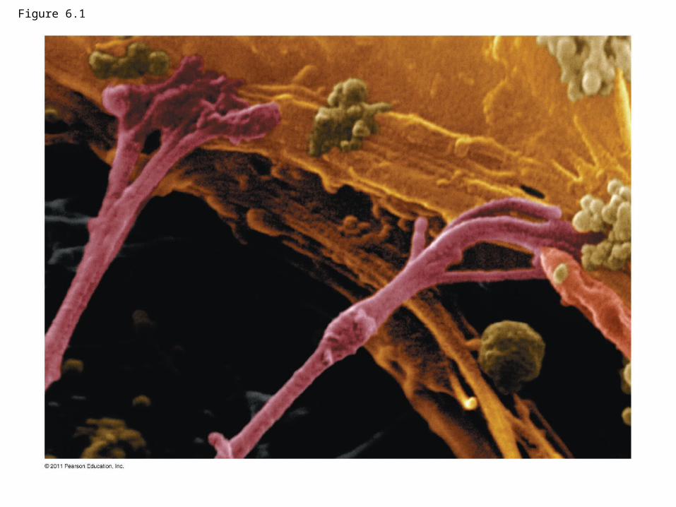
Figure 6.1

Concept 6.1: Biologists use microscopes and the tools of biochemistry to study cells
• Though usually too small to be seen by the unaided eye, cells can be complex
© 2011 Pearson Education, Inc.

Microscopy
• Scientists use microscopes to visualize cells too small to see with the naked eye
• In a light microscope (LM), visible light is passed through a specimen and then through glass lenses
• Lenses refract (bend) the light, so that the image is magnified
© 2011 Pearson Education, Inc.
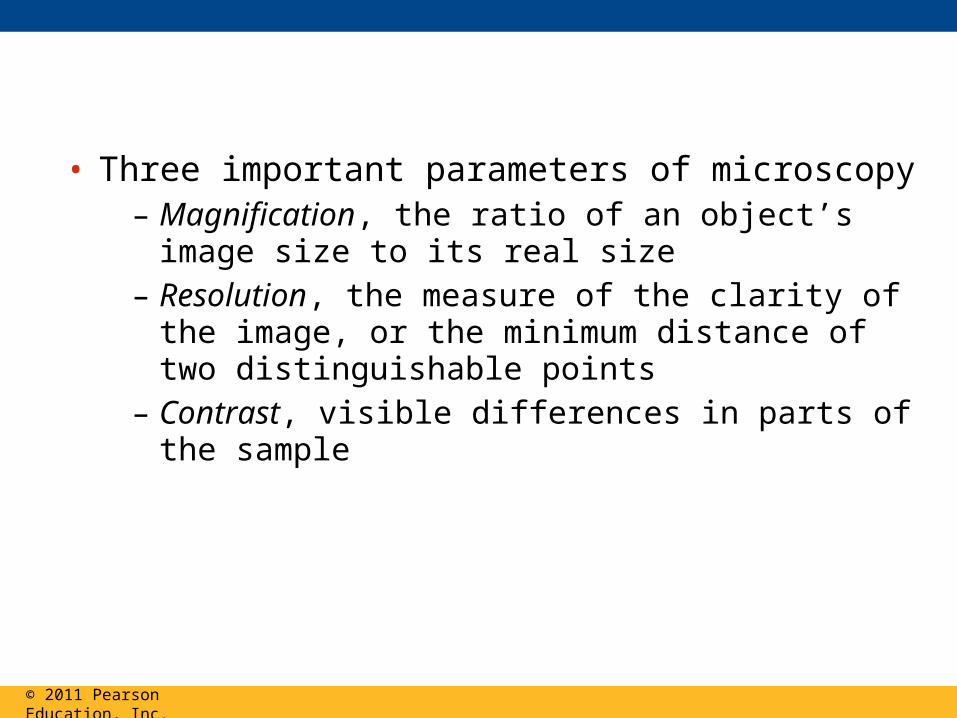
• Three important parameters of microscopy– Magnification, the ratio of an object’s image size
to its real size– Resolution, the measure of the clarity of the
image, or the minimum distance of two distinguishable points
– Contrast, visible differences in parts of the sample
© 2011 Pearson Education, Inc.

Figure 6.2 10 m
1 m
0.1 m
1 cm
1 mm
100 m
10 m
1 m
100 nm
10 nm
1 nm
0.1 nm Atoms
Small molecules
Lipids
Proteins
Ribosomes
VirusesSmallest bacteria
Mitochondrion
Most bacteriaNucleus
Most plant andanimal cells
Human egg
Frog egg
Chicken egg
Length of somenerve andmuscle cells
Human height
Un
aid
ed e
ye
Lig
ht
mic
rosc
op
y
Ele
ctro
n m
icro
sco
py
Super-resolution
microscopy

10 m
1 m
0.1 m
1 cm
1 mm
100 mHuman egg
Frog egg
Chicken egg
Length of somenerve andmuscle cells
Human height
Un
aid
ed
ey
e
Figure 6.2a

Figure 6.2b
1 mm
100 m
10 m
1 m
100 nm
10 nm
1 nm
0.1 nm Atoms
Small molecules
Lipids
Proteins
Ribosomes
VirusesSmallest bacteria
MitochondrionMost bacteriaNucleus
Most plant andanimal cells
Human egg
Lig
ht
mic
rosc
op
y
Ele
ctro
n m
icro
sco
py
Super-resolution
microscopy
1 cm
Frog egg

Brightfield(unstained specimen)
Brightfield(stained specimen)
50
m
Confocal
Differential-interference-contrast (Nomarski)
Fluorescence
10 m
Deconvolution
Super-resolution
Scanning electronmicroscopy (SEM)
Transmission electronmicroscopy (TEM)
Cross sectionof cilium
Longitudinal sectionof cilium
Cilia
Electron Microscopy (EM)
1
m1
0
m5
0
m
2 m
2 m
Light Microscopy (LM)
Phase-contrast
Figure 6.3

Figure 6.3a
(unstained specimen)
50
m
Brightfield

Figure 6.3b
Brightfield(stained specimen)

Figure 6.3c
Phase-contrast

Figure 6.3d
Differential-interference-contrast (Nomarski)

Figure 6.3e
Fluorescence
10 m

Figure 6.3f
Confocal
50
m

Figure 6.3fa
Confocal
50
m

Figure 6.3fb
Confocal
50
m
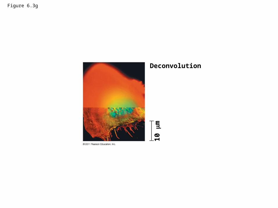
Figure 6.3g
Deconvolution
10
m

Figure 6.3h
Super-resolution
1 m

Figure 6.3ha
Super-resolution
1 m

Figure 6.3hb
Super-resolution
1 m
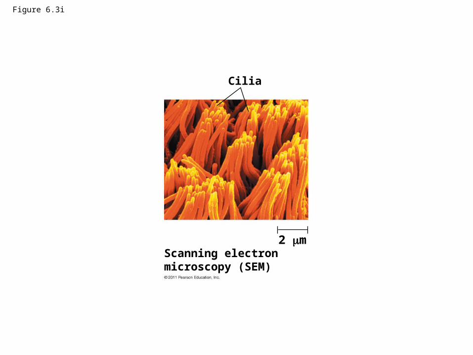
Figure 6.3i
Cilia
2 mScanning electronmicroscopy (SEM)
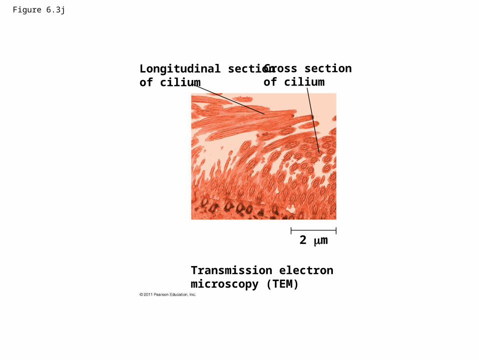
Figure 6.3j
Longitudinal sectionof cilium
Cross sectionof cilium
2 m
Transmission electronmicroscopy (TEM)

• LMs can magnify effectively to about 1,000 times the size of the actual specimen
• Various techniques enhance contrast and enable cell components to be stained or labeled
• Most subcellular structures, including organelles (membrane-enclosed compartments), are too small to be resolved by an LM
© 2011 Pearson Education, Inc.
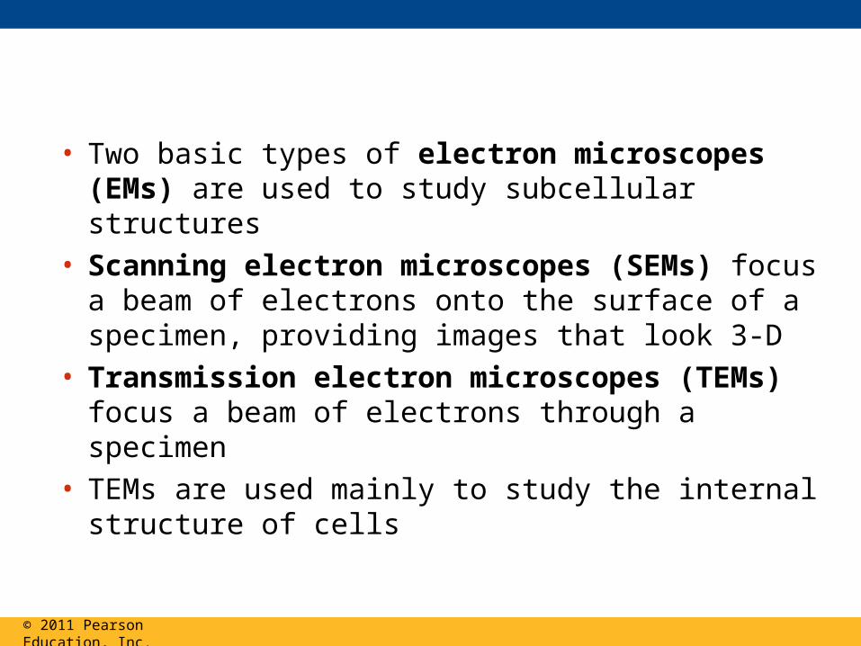
• Two basic types of electron microscopes (EMs) are used to study subcellular structures
• Scanning electron microscopes (SEMs) focus a beam of electrons onto the surface of a specimen, providing images that look 3-D
• Transmission electron microscopes (TEMs) focus a beam of electrons through a specimen
• TEMs are used mainly to study the internal structure of cells
© 2011 Pearson Education, Inc.

• Recent advances in light microscopy– Confocal microscopy and deconvolution
microscopy provide sharper images of three-dimensional tissues and cells
– New techniques for labeling cells improve resolution
© 2011 Pearson Education, Inc.

Cell Fractionation
• Cell fractionation takes cells apart and separates the major organelles from one another
• Centrifuges fractionate cells into their component parts
• Cell fractionation enables scientists to determine the functions of organelles
• Biochemistry and cytology help correlate cell function with structure
© 2011 Pearson Education, Inc.

Figure 6.4 TECHNIQUE
Homogenization
Tissuecells
Homogenate
Centrifugation
Differentialcentrifugation
Centrifuged at1,000 g
(1,000 times theforce of gravity)
for 10 min Supernatantpoured intonext tube
20,000 g20 min
80,000 g60 min
Pellet rich innuclei andcellular debris
150,000 g3 hr
Pellet rich inmitochondria(and chloro-plasts if cellsare from a plant)
Pellet rich in“microsomes”(pieces of plasmamembranes andcells’ internalmembranes) Pellet rich in
ribosomes
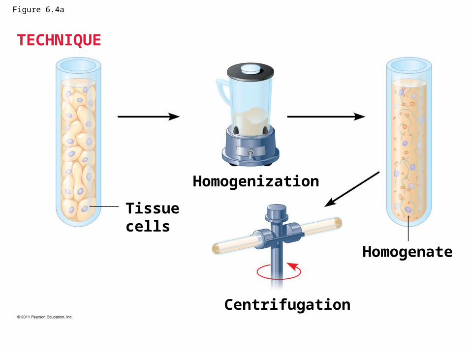
Figure 6.4a
TECHNIQUE
Homogenization
Tissuecells
Homogenate
Centrifugation

Differentialcentrifugation
Centrifuged at1,000 g
(1,000 times theforce of gravity)
for 10 min Supernatantpoured intonext tube
20 min
60 minPellet rich innuclei andcellular debris
3 hr
Pellet rich inmitochondria(and chloro-plasts if cellsare from a plant)
Pellet rich in“microsomes”
Pellet rich inribosomes
20,000 g
80,000 g
150,000 g
TECHNIQUE (cont.)Figure 6.4b
Concept 6.2: Eukaryotic cells have internal membranes that compartmentalize their functions
• The basic structural and functional unit of every organism is one of two types of cells: prokaryotic or eukaryotic
• Only organisms of the domains Bacteria and Archaea consist of prokaryotic cells
• Protists, fungi, animals, and plants all consist of eukaryotic cells
© 2011 Pearson Education, Inc.

Comparing Prokaryotic and Eukaryotic Cells
• Basic features of all cells – Plasma membrane
– Semifluid substance called cytosol
– Chromosomes (carry genes)
– Ribosomes (make proteins)
© 2011 Pearson Education, Inc.
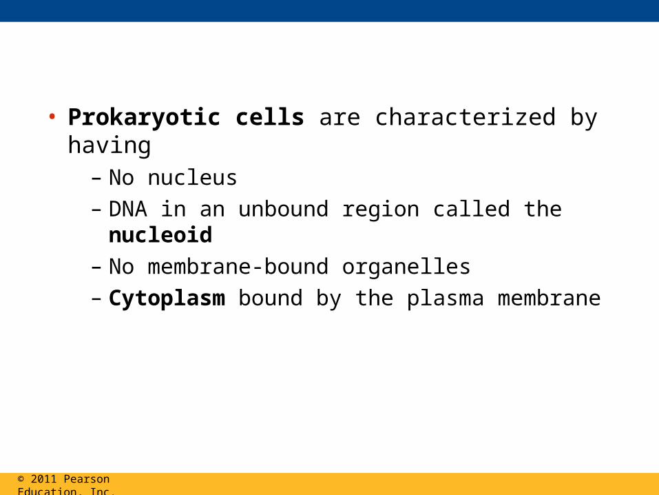
• Prokaryotic cells are characterized by having– No nucleus
– DNA in an unbound region called the nucleoid
– No membrane-bound organelles
– Cytoplasm bound by the plasma membrane
© 2011 Pearson Education, Inc.

Fimbriae
Bacterialchromosome
A typicalrod-shapedbacterium
(a)
Nucleoid
Ribosomes
Plasmamembrane
Cell wall
Capsule
Flagella A thin sectionthrough thebacterium Bacilluscoagulans (TEM)
(b)
0.5 m
Figure 6.5
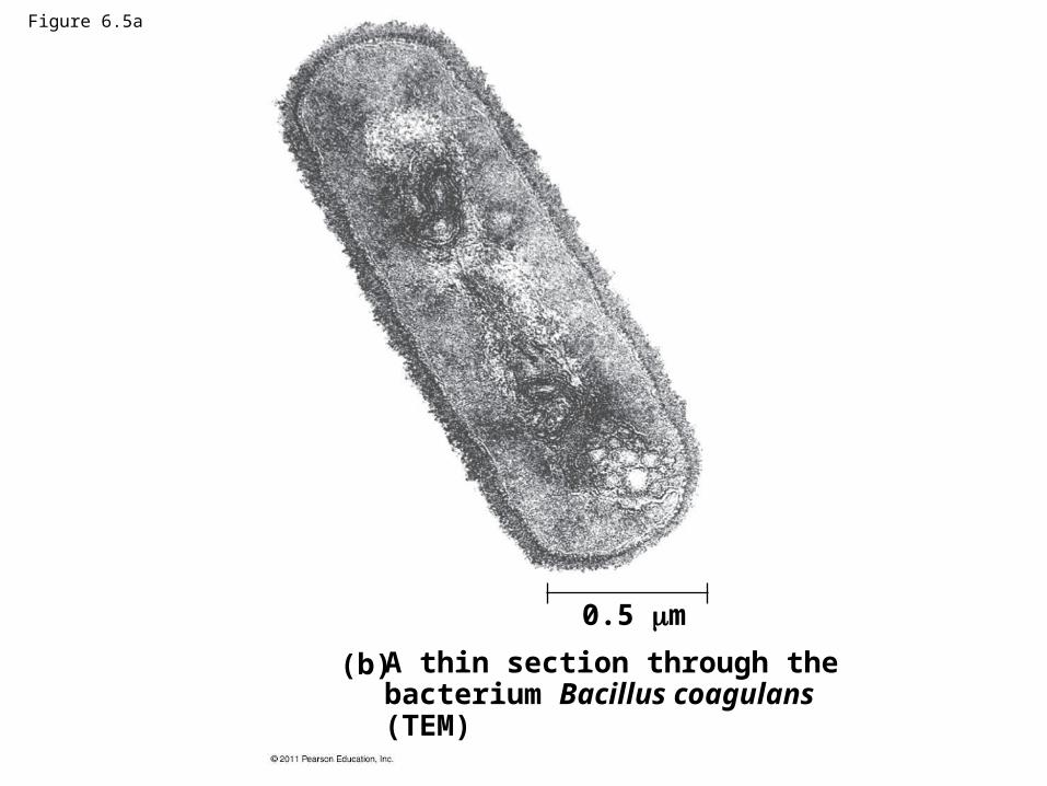
Figure 6.5a
A thin section through thebacterium Bacillus coagulans (TEM)
(b)
0.5 m

• Eukaryotic cells are characterized by having– DNA in a nucleus that is bounded by a
membranous nuclear envelope
– Membrane-bound organelles
– Cytoplasm in the region between the plasma membrane and nucleus
• Eukaryotic cells are generally much larger than prokaryotic cells
© 2011 Pearson Education, Inc.
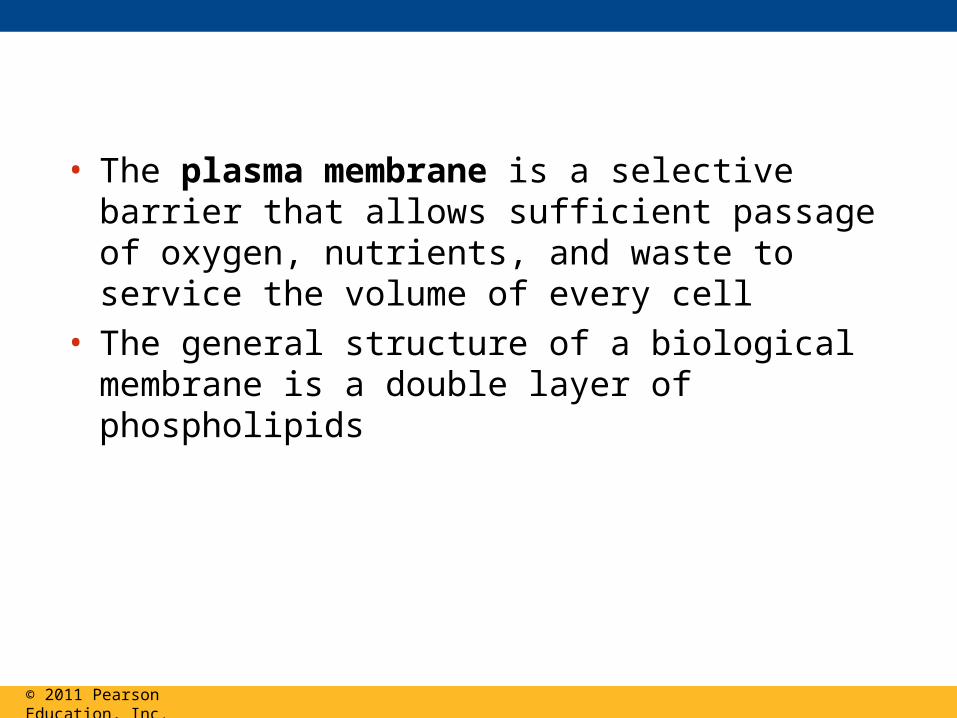
• The plasma membrane is a selective barrier that allows sufficient passage of oxygen, nutrients, and waste to service the volume of every cell
• The general structure of a biological membrane is a double layer of phospholipids
© 2011 Pearson Education, Inc.

Figure 6.6
Outside of cell
Inside of cell0.1 m
(a) TEM of a plasmamembrane
Hydrophilicregion
Hydrophobicregion
Hydrophilicregion
Carbohydrate side chains
ProteinsPhospholipid
(b) Structure of the plasma membrane

Figure 6.6a
Outside of cell
Inside of cell0.1 m
(a) TEM of a plasma membrane

• Metabolic requirements set upper limits on the size of cells
• The surface area to volume ratio of a cell is critical
• As the surface area increases by a factor of n2, the volume increases by a factor of n3
• Small cells have a greater surface area relative to volume
© 2011 Pearson Education, Inc.
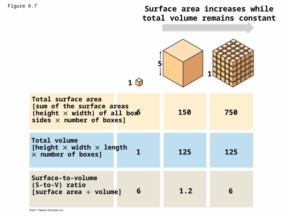
Surface area increases whiletotal volume remains constant
Total surface area[sum of the surface areas(height width) of all boxsides number of boxes]
Total volume[height width length number of boxes]
Surface-to-volume(S-to-V) ratio[surface area volume]
1
5
6 150 750
1
1251251
1.26 6
Figure 6.7
A Panoramic View of the Eukaryotic Cell
• A eukaryotic cell has internal membranes that partition the cell into organelles
• Plant and animal cells have most of the same organelles
© 2011 Pearson Education, Inc.
BioFlix: Tour of an Animal Cell
BioFlix: Tour of a Plant Cell

Figure 6.8a
ENDOPLASMIC RETICULUM (ER)
RoughER
SmoothER
Nuclearenvelope
Nucleolus
Chromatin
Plasmamembrane
Ribosomes
Golgi apparatus
LysosomeMitochondrion
Peroxisome
Microvilli
MicrotubulesIntermediate filaments
Microfilaments
Centrosome
CYTOSKELETON:
Flagellum NUCLEUS

Figure 6.8b
Animal Cells
Cell
NucleusNucleolus
Human cells from liningof uterus (colorized TEM)
Yeast cells budding(colorized SEM)
10
m
Fungal Cells
5 m
Parentcell
Buds
1 m
Cell wall
Vacuole
Nucleus
Mitochondrion
A single yeast cell(colorized TEM)
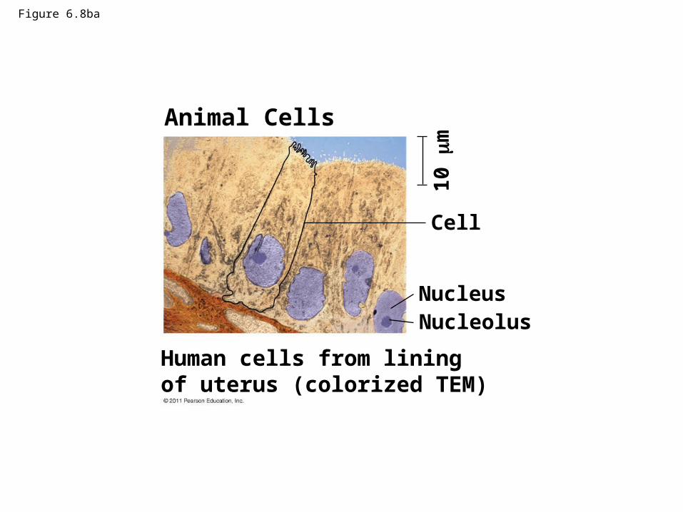
Figure 6.8ba
Animal Cells
Cell
NucleusNucleolus
Human cells from liningof uterus (colorized TEM)
10
m
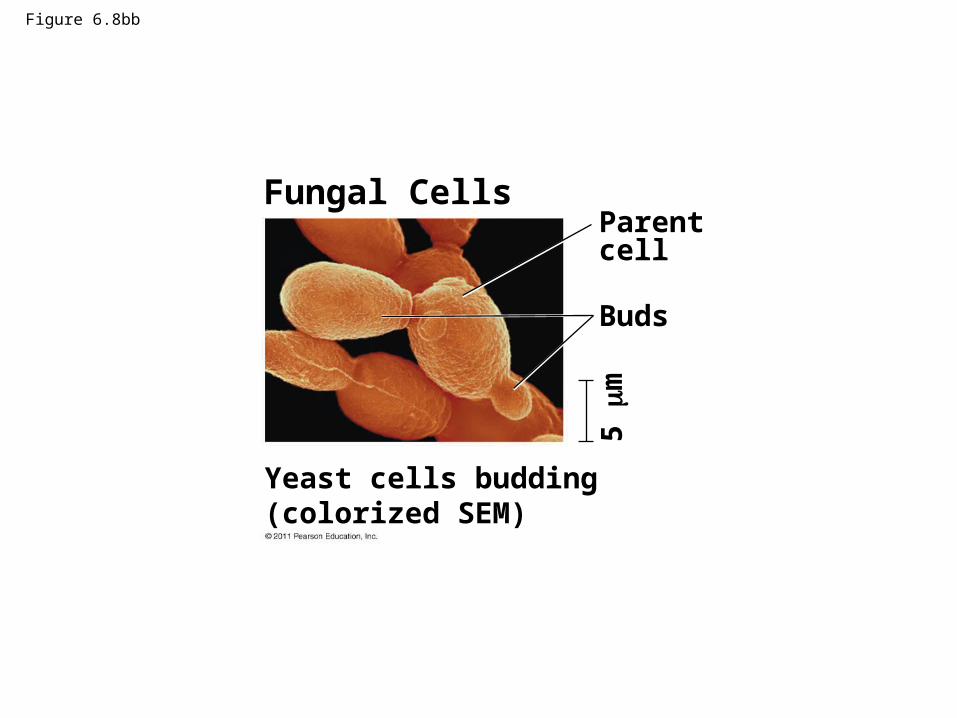
Figure 6.8bb
Yeast cells budding(colorized SEM)
Fungal Cells
5 m
Parentcell
Buds

Figure 6.8bc
1 m
Cell wall
Vacuole
Nucleus
Mitochondrion
A single yeast cell(colorized TEM)
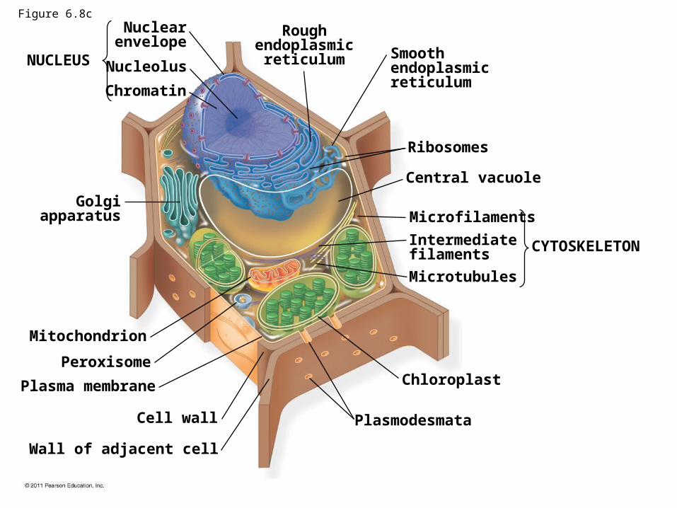
NUCLEUS
Nuclearenvelope
Nucleolus
Chromatin
Golgiapparatus
Mitochondrion
Peroxisome
Plasma membrane
Cell wall
Wall of adjacent cell
Plasmodesmata
Chloroplast
Microtubules
Intermediatefilaments
Microfilaments
CYTOSKELETON
Central vacuole
Ribosomes
Smoothendoplasmicreticulum
Roughendoplasmic
reticulum
Figure 6.8c

Figure 6.8d
Plant Cells
Cells from duckweed(colorized TEM)
Cell
5 m
Cell wall
Chloroplast
Nucleus
Nucleolus
8 m
Protistan Cells
1 m
Chlamydomonas(colorized SEM)
Chlamydomonas(colorized TEM)
Flagella
Nucleus
Nucleolus
Vacuole
Chloroplast
Cell wall
Mitochondrion

Figure 6.8da
Plant Cells
Cells from duckweed(colorized TEM)
Cell5
m
Cell wall
Chloroplast
Nucleus
Nucleolus
Mitochondrion

Figure 6.8db
8 m
Protistan Cells
Chlamydomonas(colorized SEM)
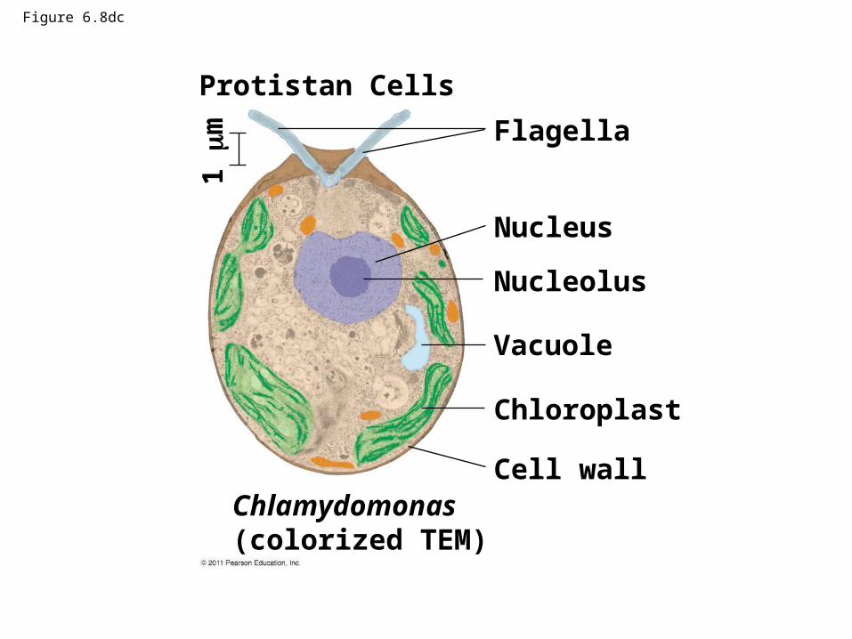
Figure 6.8dc
1 m
Chlamydomonas(colorized TEM)
Flagella
Nucleus
Nucleolus
Vacuole
Chloroplast
Cell wall
Protistan Cells

Concept 6.3: The eukaryotic cell’s genetic instructions are housed in the nucleus and carried out by the ribosomes
• The nucleus contains most of the DNA in a eukaryotic cell
• Ribosomes use the information from the DNA to make proteins
© 2011 Pearson Education, Inc.

The Nucleus: Information Central
• The nucleus contains most of the cell’s genes and is usually the most conspicuous organelle
• The nuclear envelope encloses the nucleus, separating it from the cytoplasm
• The nuclear membrane is a double membrane; each membrane consists of a lipid bilayer
© 2011 Pearson Education, Inc.
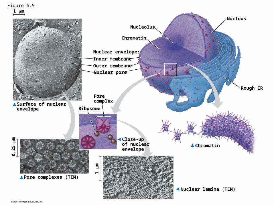
Nucleus
Rough ER
Nucleolus
Chromatin
Nuclear envelope:
Inner membrane
Outer membrane
Nuclear pore
Ribosome
Porecomplex
Close-upof nuclearenvelope
Surface of nuclearenvelope
Pore complexes (TEM)
0.2
5
m
1
m
Nuclear lamina (TEM)
Chromatin
1 mFigure 6.9

Nucleus
Rough ER
Nucleolus
Chromatin
Nuclear envelope:
Inner membrane
Outer membrane
Nuclear pore
Chromatin
Ribosome
Porecomplex
Close-upof nuclearenvelope
Figure 6.9a

Figure 6.9b
Nuclear envelope:
Inner membrane
Outer membrane
Nuclear pore
Surface of nuclearenvelope
1 m

Figure 6.9c
Pore complexes (TEM)
0.2
5
m

Figure 6.9d
1
m
Nuclear lamina (TEM)

• Pores regulate the entry and exit of molecules from the nucleus
• The shape of the nucleus is maintained by the nuclear lamina, which is composed of protein
© 2011 Pearson Education, Inc.

• In the nucleus, DNA is organized into discrete units called chromosomes
• Each chromosome is composed of a single DNA molecule associated with proteins
• The DNA and proteins of chromosomes are together called chromatin
• Chromatin condenses to form discrete chromosomes as a cell prepares to divide
• The nucleolus is located within the nucleus and is the site of ribosomal RNA (rRNA) synthesis
© 2011 Pearson Education, Inc.

Ribosomes: Protein Factories
• Ribosomes are particles made of ribosomal RNA and protein
• Ribosomes carry out protein synthesis in two locations
– In the cytosol (free ribosomes)
– On the outside of the endoplasmic reticulum or the nuclear envelope (bound ribosomes)
© 2011 Pearson Education, Inc.

Figure 6.10
0.25 m
Free ribosomes in cytosol
Endoplasmic reticulum (ER)
Ribosomes bound to ER
Largesubunit
Smallsubunit
Diagram of a ribosomeTEM showing ER andribosomes

Figure 6.10a
0.25 m
Free ribosomes in cytosol
Endoplasmic reticulum (ER)
Ribosomes bound to ER
TEM showing ER andribosomes

Concept 6.4: The endomembrane system regulates protein traffic and performs metabolic functions in the cell
• Components of the endomembrane system– Nuclear envelope– Endoplasmic reticulum– Golgi apparatus– Lysosomes– Vacuoles– Plasma membrane
• These components are either continuous or connected via transfer by vesicles
© 2011 Pearson Education, Inc.

The Endoplasmic Reticulum: Biosynthetic Factory
• The endoplasmic reticulum (ER) accounts for more than half of the total membrane in many eukaryotic cells
• The ER membrane is continuous with the nuclear envelope
• There are two distinct regions of ER– Smooth ER, which lacks ribosomes
– Rough ER, surface is studded with ribosomes
© 2011 Pearson Education, Inc.

Figure 6.11 Smooth ER
Rough ER
ER lumen
CisternaeRibosomes
Smooth ERTransport vesicle
Transitional ER
Rough ER 200 nm
Nuclearenvelope

Figure 6.11a
Smooth ER
Rough ER
CisternaeRibosomes
Transport vesicle
Transitional ER
Nuclearenvelope
ER lumen
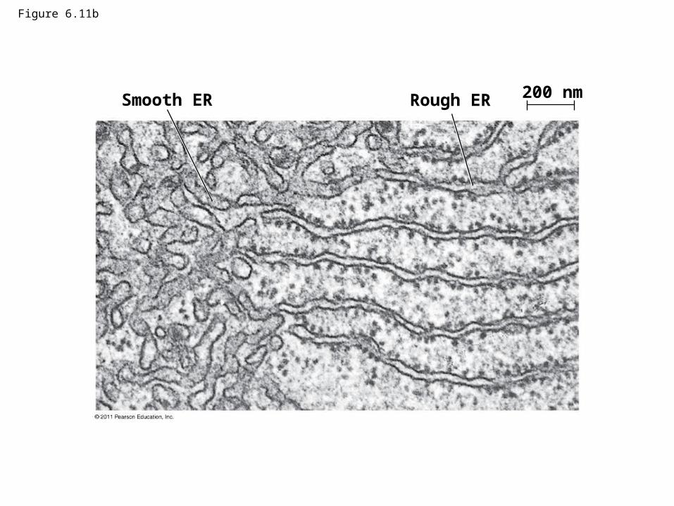
Figure 6.11b
Smooth ER Rough ER 200 nm

Functions of Smooth ER
• The smooth ER– Synthesizes lipids
– Metabolizes carbohydrates
– Detoxifies drugs and poisons
– Stores calcium ions
© 2011 Pearson Education, Inc.

Functions of Rough ER
• The rough ER– Has bound ribosomes, which secrete
glycoproteins (proteins covalently bonded to carbohydrates)
– Distributes transport vesicles, proteins surrounded by membranes
– Is a membrane factory for the cell
© 2011 Pearson Education, Inc.

• The Golgi apparatus consists of flattened membranous sacs called cisternae
• Functions of the Golgi apparatus– Modifies products of the ER
– Manufactures certain macromolecules
– Sorts and packages materials into transport vesicles
The Golgi Apparatus: Shipping and Receiving Center
© 2011 Pearson Education, Inc.
Figure 6.12
cis face(“receiving” side ofGolgi apparatus)
trans face(“shipping” side ofGolgi apparatus)
0.1 m
TEM of Golgi apparatus
Cisternae

Figure 6.12a
TEM of Golgi apparatus
0.1 m

Lysosomes: Digestive Compartments
• A lysosome is a membranous sac of hydrolytic enzymes that can digest macromolecules
• Lysosomal enzymes can hydrolyze proteins, fats, polysaccharides, and nucleic acids
• Lysosomal enzymes work best in the acidic environment inside the lysosome
© 2011 Pearson Education, Inc.
Animation: Lysosome Formation

• Some types of cell can engulf another cell by phagocytosis; this forms a food vacuole
• A lysosome fuses with the food vacuole and digests the molecules
• Lysosomes also use enzymes to recycle the cell’s own organelles and macromolecules, a process called autophagy
© 2011 Pearson Education, Inc.

Figure 6.13
Nucleus
Lysosome
1 m
Digestiveenzymes
Digestion
Food vacuole
LysosomePlasma membrane
(a) Phagocytosis
Vesicle containingtwo damagedorganelles
1 m
Mitochondrionfragment
Peroxisomefragment
(b) Autophagy
Peroxisome
VesicleMitochondrion
Lysosome
Digestion

Figure 6.13a
Nucleus
Lysosome
1 m
Digestiveenzymes
Digestion
Food vacuole
LysosomePlasma membrane
(a) Phagocytosis

Figure 6.13aa
Nucleus
Lysosome
1 m

Figure 6.13b Vesicle containingtwo damagedorganelles
1 m
Mitochondrionfragment
Peroxisomefragment
Peroxisome
VesicleMitochondrion
Lysosome
Digestion
(b) Autophagy
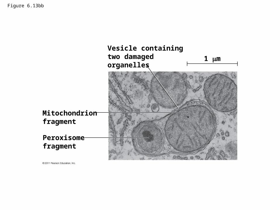
Figure 6.13bb
Vesicle containingtwo damagedorganelles
1 m
Mitochondrionfragment
Peroxisomefragment

Vacuoles: Diverse Maintenance Compartments
• A plant cell or fungal cell may have one or several vacuoles, derived from endoplasmic reticulum and Golgi apparatus
© 2011 Pearson Education, Inc.

• Food vacuoles are formed by phagocytosis• Contractile vacuoles, found in many freshwater
protists, pump excess water out of cells• Central vacuoles, found in many mature plant
cells, hold organic compounds and water
© 2011 Pearson Education, Inc.
Video: Paramecium Vacuole

Figure 6.14
Central vacuole
Cytosol
Nucleus
Cell wall
Chloroplast
Centralvacuole
5 m

Figure 6.14a
Cytosol
Nucleus
Cell wall
Chloroplast
Centralvacuole
5 m

The Endomembrane System: A Review
• The endomembrane system is a complex and dynamic player in the cell’s compartmental organization
© 2011 Pearson Education, Inc.
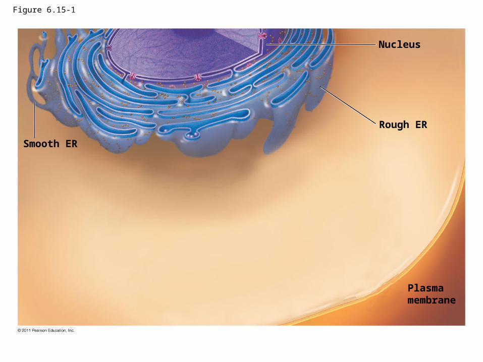
Figure 6.15-1
Smooth ER
Nucleus
Rough ER
Plasmamembrane
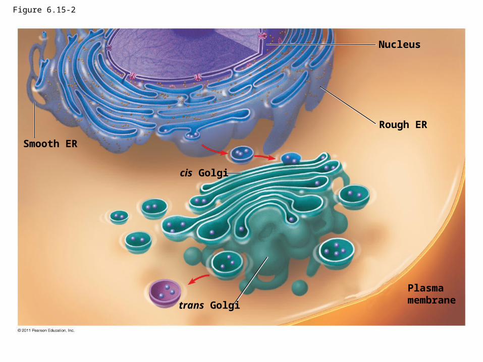
Figure 6.15-2
Smooth ER
Nucleus
Rough ER
Plasmamembrane
cis Golgi
trans Golgi

Figure 6.15-3
Smooth ER
Nucleus
Rough ER
Plasmamembrane
cis Golgi
trans Golgi

Concept 6.5: Mitochondria and chloroplasts change energy from one form to another
• Mitochondria are the sites of cellular respiration, a metabolic process that uses oxygen to generate ATP
• Chloroplasts, found in plants and algae, are the sites of photosynthesis
• Peroxisomes are oxidative organelles
© 2011 Pearson Education, Inc.

• Mitochondria and chloroplasts have similarities with bacteria
– Enveloped by a double membrane
– Contain free ribosomes and circular DNA molecules
– Grow and reproduce somewhat independently in cells
© 2011 Pearson Education, Inc.
The Evolutionary Origins of Mitochondria and Chloroplasts

• The Endosymbiont theory – An early ancestor of eukaryotic cells engulfed
a nonphotosynthetic prokaryotic cell, which formed an endosymbiont relationship with its host
– The host cell and endosymbiont merged into a single organism, a eukaryotic cell with a mitochondrion
– At least one of these cells may have taken up a photosynthetic prokaryote, becoming the ancestor of cells that contain chloroplasts
© 2011 Pearson Education, Inc.
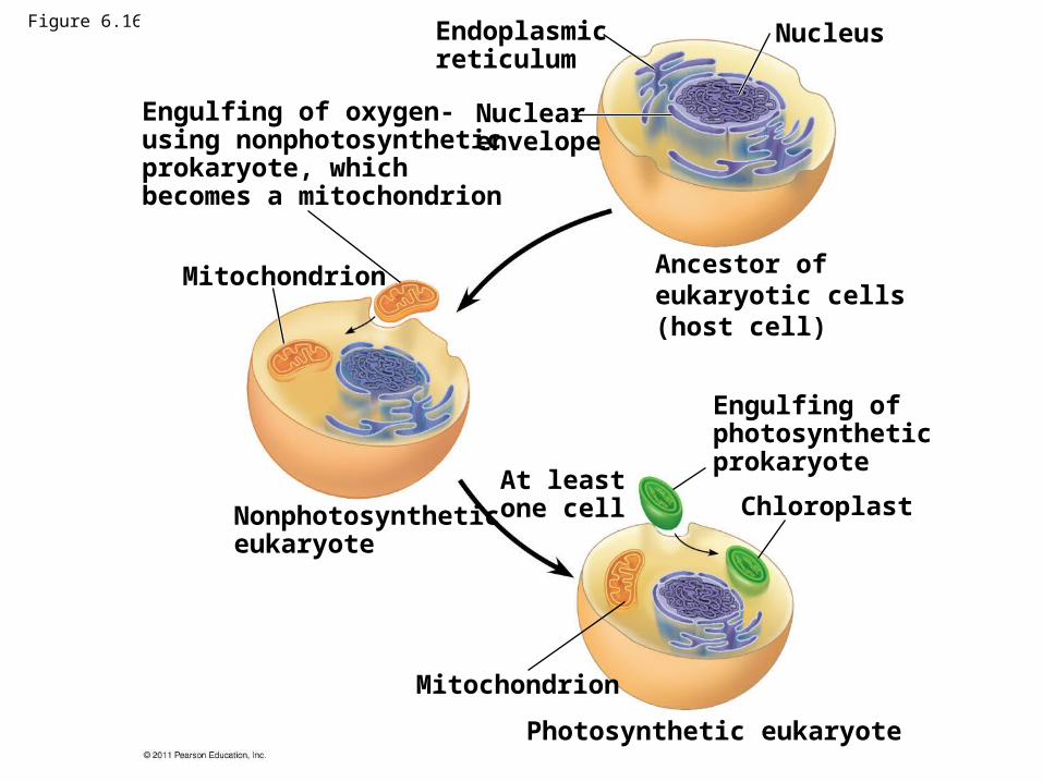
Figure 6.16NucleusEndoplasmic
reticulum
Nuclear envelope
Ancestor ofeukaryotic cells(host cell)
Engulfing of oxygen-using nonphotosyntheticprokaryote, whichbecomes a mitochondrion
Mitochondrion
Nonphotosyntheticeukaryote
Mitochondrion
At leastone cell
Photosynthetic eukaryote
Engulfing ofphotosyntheticprokaryote
Chloroplast

Mitochondria: Chemical Energy Conversion
• Mitochondria are in nearly all eukaryotic cells• They have a smooth outer membrane and an
inner membrane folded into cristae• The inner membrane creates two compartments:
intermembrane space and mitochondrial matrix• Some metabolic steps of cellular respiration are
catalyzed in the mitochondrial matrix• Cristae present a large surface area for enzymes
that synthesize ATP
© 2011 Pearson Education, Inc.
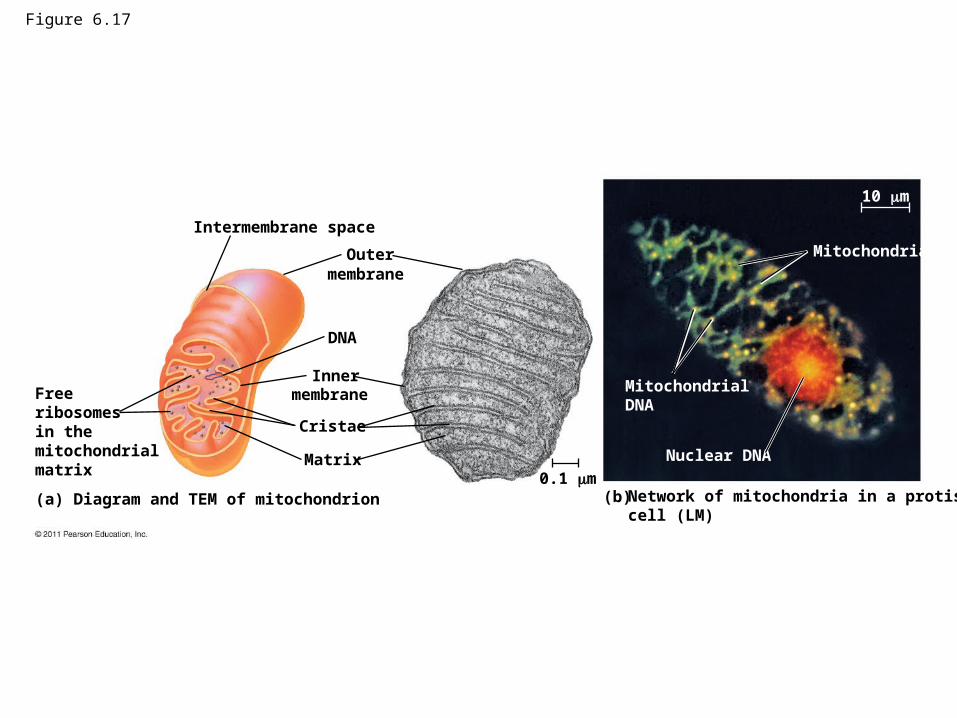
Figure 6.17
Intermembrane space
Outermembrane
DNA
Innermembrane
Cristae
Matrix
Freeribosomesin themitochondrialmatrix
(a) Diagram and TEM of mitochondrion (b) Network of mitochondria in a protistcell (LM)
0.1 m
MitochondrialDNA
Nuclear DNA
Mitochondria
10 m

Figure 6.17a
Intermembrane space
Outer
DNA
Innermembrane
Cristae
Matrix
Freeribosomesin themitochondrialmatrix
(a) Diagram and TEM of mitochondrion0.1 m
membrane

Figure 6.17aa
Outermembrane
Innermembrane
Cristae
Matrix0.1 m
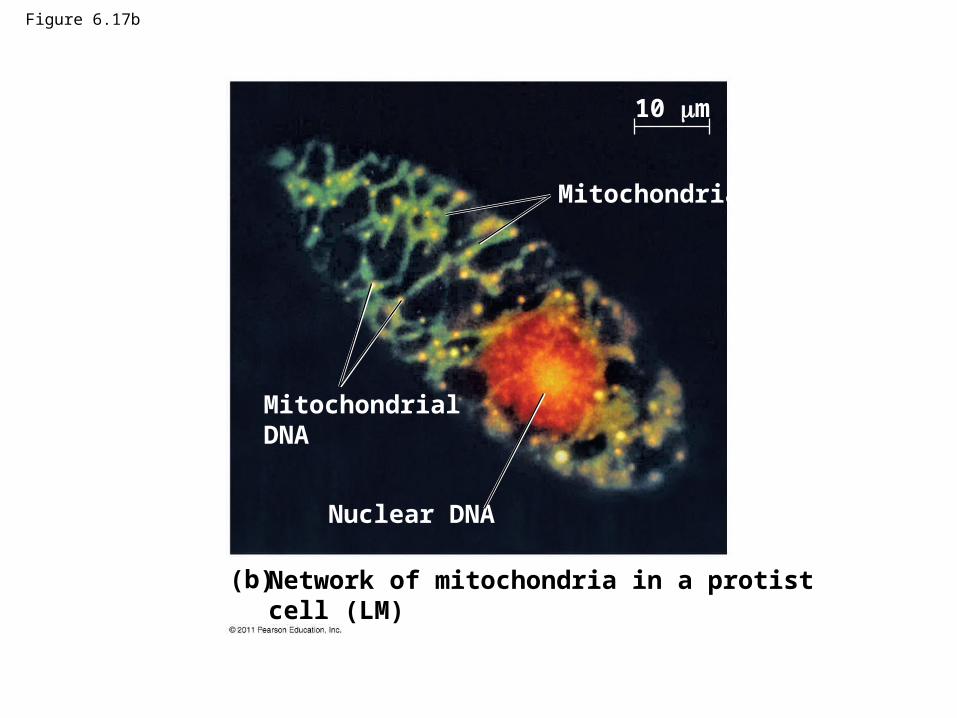
Figure 6.17b
(b) Network of mitochondria in a protistcell (LM)
MitochondrialDNA
Nuclear DNA
Mitochondria
10 m
Chloroplasts: Capture of Light Energy
• Chloroplasts contain the green pigment chlorophyll, as well as enzymes and other molecules that function in photosynthesis
• Chloroplasts are found in leaves and other green organs of plants and in algae
© 2011 Pearson Education, Inc.

• Chloroplast structure includes– Thylakoids, membranous sacs, stacked to
form a granum– Stroma, the internal fluid
• The chloroplast is one of a group of plant organelles, called plastids
© 2011 Pearson Education, Inc.

Figure 6.18
RibosomesStroma
Inner and outermembranes
Granum
1 mIntermembrane spaceThylakoid
(a) Diagram and TEM of chloroplast (b) Chloroplasts in an algal cell
Chloroplasts(red)
50 m
DNA

Figure 6.18a
RibosomesStroma
Inner and outermembranes
Granum
1 mIntermembrane spaceThylakoid
(a) Diagram and TEM of chloroplast
DNA

Figure 6.18aa
Stroma
Inner and outermembranes
Granum
1 m
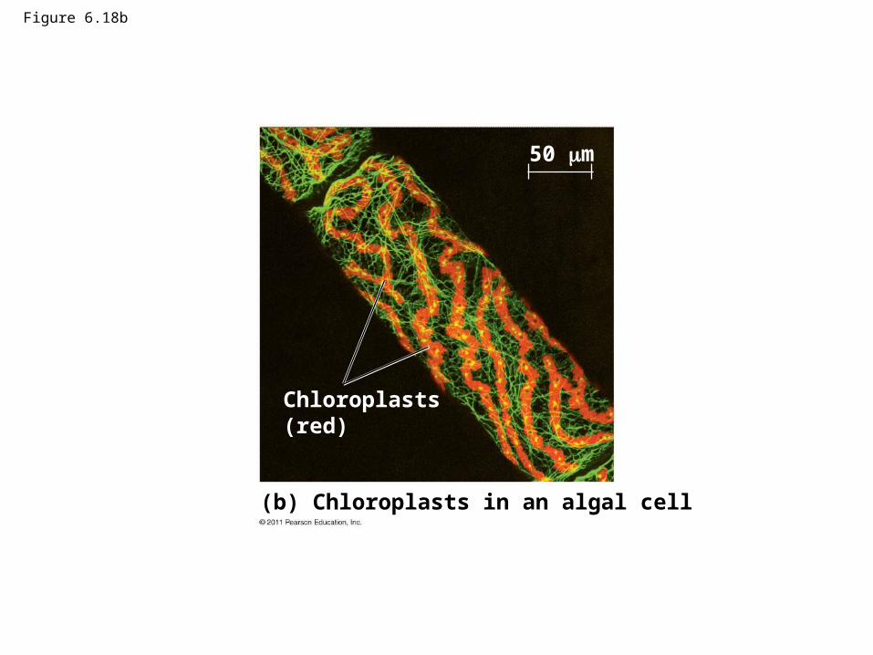
Figure 6.18b
(b) Chloroplasts in an algal cell
Chloroplasts(red)
50 m

Peroxisomes: Oxidation
• Peroxisomes are specialized metabolic compartments bounded by a single membrane
• Peroxisomes produce hydrogen peroxide and convert it to water
• Peroxisomes perform reactions with many different functions
• How peroxisomes are related to other organelles is still unknown
© 2011 Pearson Education, Inc.

Figure 6.19
ChloroplastPeroxisome
Mitochondrion
1 m

Concept 6.6: The cytoskeleton is a network of fibers that organizes structures and activities in the cell
• The cytoskeleton is a network of fibers extending throughout the cytoplasm
• It organizes the cell’s structures and activities, anchoring many organelles
• It is composed of three types of molecular structures
– Microtubules– Microfilaments– Intermediate filaments
© 2011 Pearson Education, Inc.
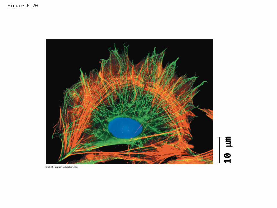
Figure 6.20
10
m
Roles of the Cytoskeleton: Support and Motility
• The cytoskeleton helps to support the cell and maintain its shape
• It interacts with motor proteins to produce motility
• Inside the cell, vesicles can travel along “monorails” provided by the cytoskeleton
• Recent evidence suggests that the cytoskeleton may help regulate biochemical activities
© 2011 Pearson Education, Inc.

Figure 6.21
ATPVesicle
(a)
Motor protein(ATP powered)
Microtubuleof cytoskeleton
Receptor formotor protein
0.25 m VesiclesMicrotubule
(b)

Figure 6.21a
0.25 m VesiclesMicrotubule
(b)

Components of the Cytoskeleton
• Three main types of fibers make up the cytoskeleton
– Microtubules are the thickest of the three components of the cytoskeleton
– Microfilaments, also called actin filaments, are the thinnest components
– Intermediate filaments are fibers with diameters in a middle range
© 2011 Pearson Education, Inc.

Column of tubulin dimers
Tubulin dimer
25 nm
Actin subunit
7 nm
Keratin proteins
812 nm
Fibrous subunit (keratinscoiled together)
10 m 10 m 5 m
Table 6.1

Tubulin dimer
25 nm
Column of tubulin dimers
10 m
Table 6.1a

Table 6.1aa
10 m

10 m
Actin subunit
7 nm
Table 6.1b

Table 6.1bb
10 m

5 m
Keratin proteins
Fibrous subunit (keratinscoiled together)
812 nm
Table 6.1c
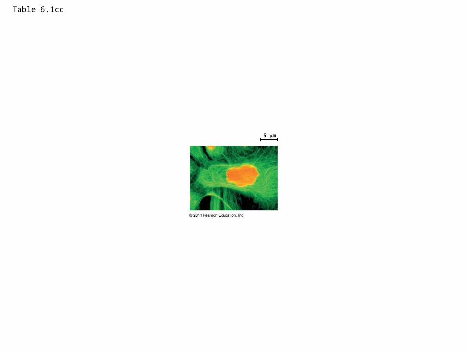
Table 6.1cc
5 m

Microtubules
• Microtubules are hollow rods about 25 nm in diameter and about 200 nm to 25 microns long
• Functions of microtubules– Shaping the cell
– Guiding movement of organelles
– Separating chromosomes during cell division
© 2011 Pearson Education, Inc.
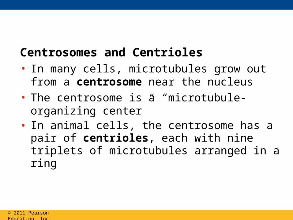
Centrosomes and Centrioles• In many cells, microtubules grow out from a
centrosome near the nucleus• The centrosome is a “microtubule-organizing
center”• In animal cells, the centrosome has a pair of
centrioles, each with nine triplets of microtubules arranged in a ring
© 2011 Pearson Education, Inc.
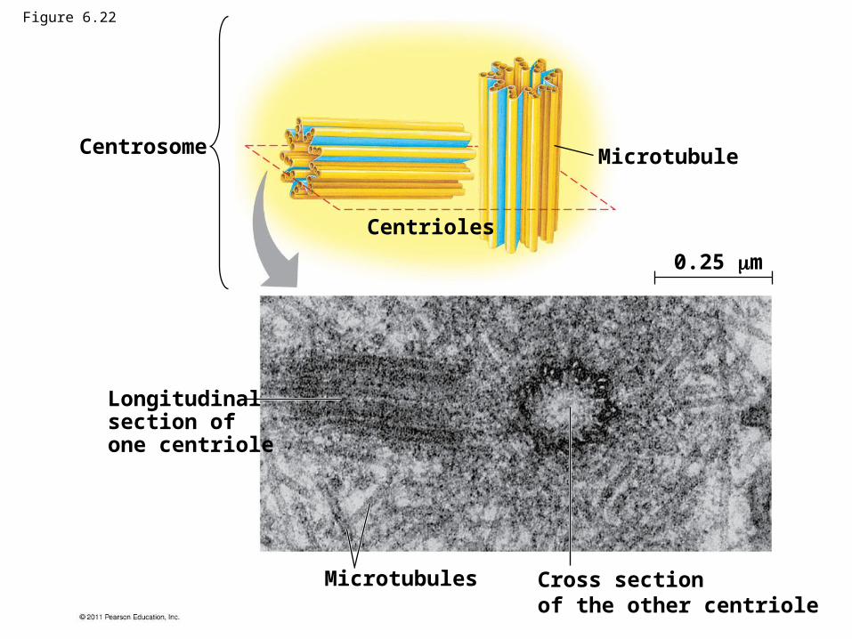
Centrosome
Longitudinalsection ofone centriole
Centrioles
Microtubule
0.25 m
Microtubules Cross sectionof the other centriole
Figure 6.22
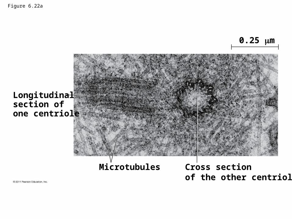
Figure 6.22a
Longitudinalsection ofone centriole
0.25 m
Microtubules Cross sectionof the other centriole
Cilia and Flagella• Microtubules control the beating of cilia and
flagella, locomotor appendages of some cells• Cilia and flagella differ in their beating patterns
© 2011 Pearson Education, Inc.
Video: Chlamydomonas
Video: Paramecium Cilia

Direction of swimming
(b) Motion of cilia
Direction of organism’s movement
Power stroke Recovery stroke
(a) Motion of flagella5 m
15 m
Figure 6.23

Figure 6.23a
5 m
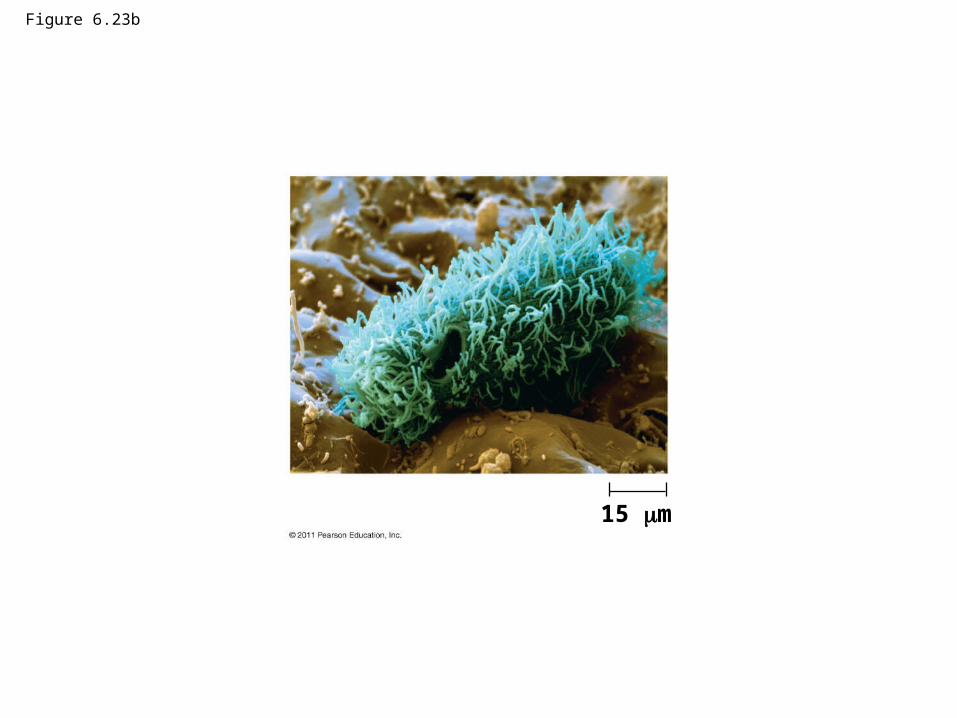
Figure 6.23b
15 m

• Cilia and flagella share a common structure– A core of microtubules sheathed by the plasma
membrane
– A basal body that anchors the cilium or flagellum
– A motor protein called dynein, which drives the bending movements of a cilium or flagellum
© 2011 Pearson Education, Inc.
Animation: Cilia and Flagella

Microtubules
Plasmamembrane
Basal body
Longitudinal sectionof motile cilium
(a)0.5 m 0.1 m
0.1 m
(b) Cross section ofmotile cilium
Outer microtubuledoubletDynein proteins
CentralmicrotubuleRadialspoke
Cross-linkingproteins betweenouter doublets
Plasma membrane
Triplet
(c) Cross section ofbasal body
Figure 6.24
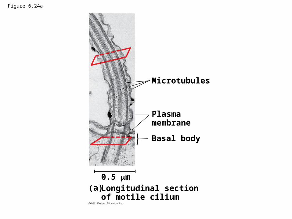
Figure 6.24a
Microtubules
Plasmamembrane
Basal body
Longitudinal sectionof motile cilium
0.5 m(a)

Figure 6.24b
0.1 m
(b) Cross section ofmotile cilium
Outer microtubuledoublet
Dynein proteins
Centralmicrotubule
Radialspoke
Cross-linkingproteins betweenouter doublets
Plasma membrane

Figure 6.24ba
0.1 m
(b) Cross section ofmotile cilium
Outer microtubuledoublet
Dynein proteins
Centralmicrotubule
Radialspoke
Cross-linkingproteins betweenouter doublets

Figure 6.24c
0.1 m
Triplet
(c) Cross section ofbasal body

Figure 6.24ca
0.1 m
Triplet
(c) Cross section ofbasal body

• How dynein “walking” moves flagella and cilia− Dynein arms alternately grab, move, and release
the outer microtubules
– Protein cross-links limit sliding
– Forces exerted by dynein arms cause doublets to curve, bending the cilium or flagellum
© 2011 Pearson Education, Inc.

Figure 6.25 Microtubuledoublets
Dynein protein
ATP
(a) Effect of unrestrained dynein movement
Cross-linking proteinsbetween outer doublets
ATP
Anchoragein cell
(b) Effect of cross-linking proteins
(c) Wavelike motion
1
2
3

Microtubuledoublets
Dynein protein
ATP
(a) Effect of unrestrained dynein movement
Figure 6.25a

Figure 6.25b
Cross-linking proteinsbetween outer doublets
ATP
Anchoragein cell
(b) Effect of cross-linking proteins (c) Wavelike motion
31
2
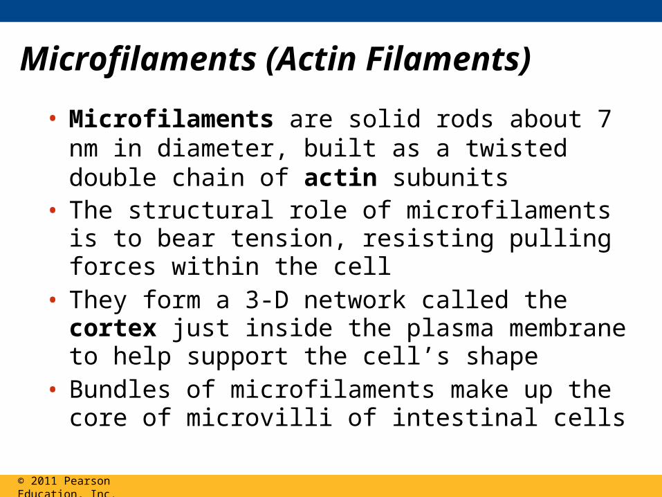
Microfilaments (Actin Filaments)
• Microfilaments are solid rods about 7 nm in diameter, built as a twisted double chain of actin subunits
• The structural role of microfilaments is to bear tension, resisting pulling forces within the cell
• They form a 3-D network called the cortex just inside the plasma membrane to help support the cell’s shape
• Bundles of microfilaments make up the core of microvilli of intestinal cells
© 2011 Pearson Education, Inc.
Figure 6.26
Microvillus
Plasma membrane
Microfilaments (actinfilaments)
Intermediate filaments
0.25 m
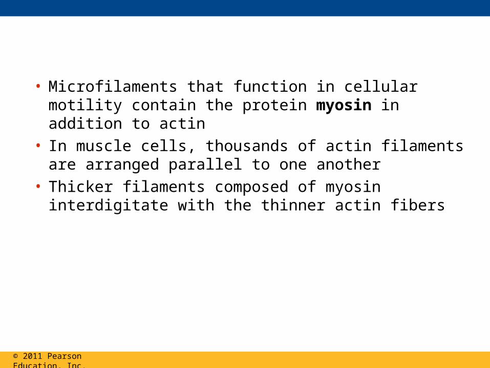
• Microfilaments that function in cellular motility contain the protein myosin in addition to actin
• In muscle cells, thousands of actin filaments are arranged parallel to one another
• Thicker filaments composed of myosin interdigitate with the thinner actin fibers
© 2011 Pearson Education, Inc.

Figure 6.27
Muscle cell
Actinfilament
Myosin
Myosin
filament
head
(a) Myosin motors in muscle cell contraction
0.5 m
100 m
Cortex (outer cytoplasm):gel with actin network
Inner cytoplasm: solwith actin subunits
(b) Amoeboid movement
Extendingpseudopodium
30 m(c) Cytoplasmic streaming in plant cells
Chloroplast

Figure 6.27a
Muscle cell
Actinfilament
Myosin
Myosinfilament
(a) Myosin motors in muscle cell contraction
0.5 m
head
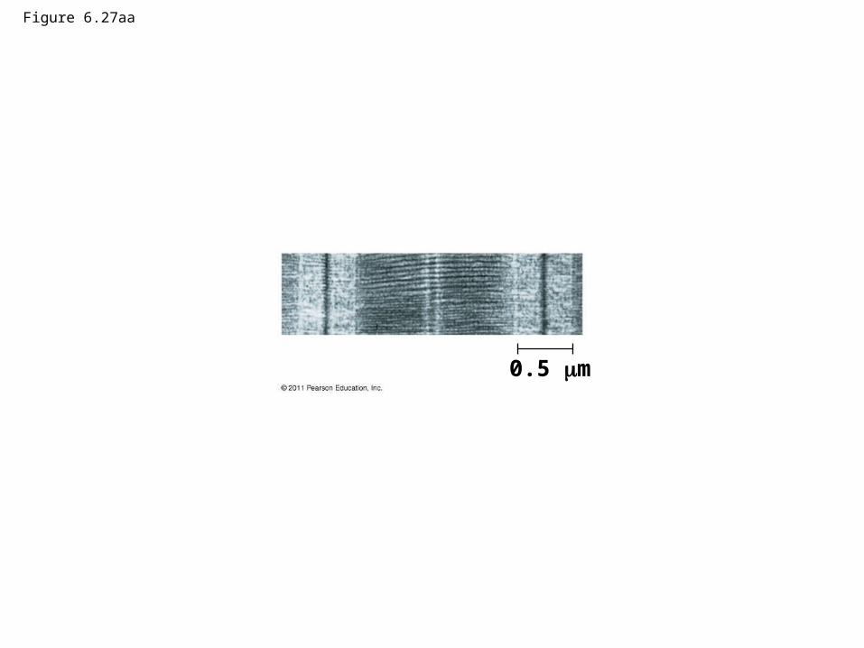
Figure 6.27aa
0.5 m

Figure 6.27b
100 m
Cortex (outer cytoplasm):gel with actin network
Inner cytoplasm: solwith actin subunits
(b) Amoeboid movement
Extendingpseudopodium
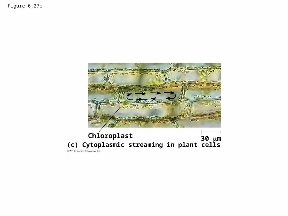
Figure 6.27c
30 m(c) Cytoplasmic streaming in plant cells
Chloroplast

• Localized contraction brought about by actin and myosin also drives amoeboid movement
• Pseudopodia (cellular extensions) extend and contract through the reversible assembly and contraction of actin subunits into microfilaments
© 2011 Pearson Education, Inc.

• Cytoplasmic streaming is a circular flow of cytoplasm within cells
• This streaming speeds distribution of materials within the cell
• In plant cells, actin-myosin interactions and sol-gel transformations drive cytoplasmic streaming
© 2011 Pearson Education, Inc.
Video: Cytoplasmic Streaming

Intermediate Filaments
• Intermediate filaments range in diameter from 8–12 nanometers, larger than microfilaments but smaller than microtubules
• They support cell shape and fix organelles in place
• Intermediate filaments are more permanent cytoskeleton fixtures than the other two classes
© 2011 Pearson Education, Inc.

Concept 6.7: Extracellular components and connections between cells help coordinate cellular activities
• Most cells synthesize and secrete materials that are external to the plasma membrane
• These extracellular structures include– Cell walls of plants
– The extracellular matrix (ECM) of animal cells
– Intercellular junctions
© 2011 Pearson Education, Inc.

Cell Walls of Plants
• The cell wall is an extracellular structure that distinguishes plant cells from animal cells
• Prokaryotes, fungi, and some protists also have cell walls
• The cell wall protects the plant cell, maintains its shape, and prevents excessive uptake of water
• Plant cell walls are made of cellulose fibers embedded in other polysaccharides and protein
© 2011 Pearson Education, Inc.
• Plant cell walls may have multiple layers– Primary cell wall: relatively thin and flexible– Middle lamella: thin layer between primary walls
of adjacent cells– Secondary cell wall (in some cells): added
between the plasma membrane and the primary cell wall
• Plasmodesmata are channels between adjacent plant cells
© 2011 Pearson Education, Inc.
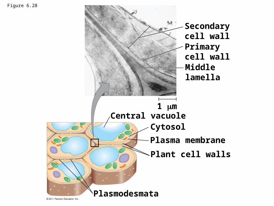
Secondarycell wallPrimarycell wallMiddlelamella
Central vacuoleCytosol
Plasma membrane
Plant cell walls
Plasmodesmata
1 m
Figure 6.28

Figure 6.28a
Secondarycell wall
Primarycell wall
Middlelamella
1 m

Figure 6.29
RESULTS 10 m
Distribution of cellulosesynthase over time
Distribution ofmicrotubulesover time

Figure 6.29a
10 m
Distribution of cellulosesynthase over time

Figure 6.29b
10 m
Distribution ofmicrotubulesover time

The Extracellular Matrix (ECM) of Animal Cells
• Animal cells lack cell walls but are covered by an elaborate extracellular matrix (ECM)
• The ECM is made up of glycoproteins such as collagen, proteoglycans, and fibronectin
• ECM proteins bind to receptor proteins in the plasma membrane called integrins
© 2011 Pearson Education, Inc.

Figure 6.30
EXTRACELLULAR FLUIDCollagen
Fibronectin
Plasmamembrane
Micro-filaments
CYTOPLASM
Integrins
Proteoglycancomplex
Polysaccharidemolecule
Carbo-hydrates
Coreprotein
Proteoglycanmolecule
Proteoglycan complex

Figure 6.30a
EXTRACELLULAR FLUIDCollagen
Fibronectin
Plasmamembrane
Micro-filaments
CYTOPLASM
Integrins
Proteoglycancomplex

Figure 6.30b
Polysaccharidemolecule
Carbohydrates
Coreprotein
Proteoglycanmolecule
Proteoglycan complex

• Functions of the ECM– Support
– Adhesion
– Movement
– Regulation
© 2011 Pearson Education, Inc.

Cell Junctions
• Neighboring cells in tissues, organs, or organ systems often adhere, interact, and communicate through direct physical contact
• Intercellular junctions facilitate this contact• There are several types of intercellular junctions
– Plasmodesmata– Tight junctions– Desmosomes– Gap junctions
© 2011 Pearson Education, Inc.
Plasmodesmata in Plant Cells
• Plasmodesmata are channels that perforate plant cell walls
• Through plasmodesmata, water and small solutes (and sometimes proteins and RNA) can pass from cell to cell
© 2011 Pearson Education, Inc.

Figure 6.31
Interiorof cell
Interiorof cell
0.5 m Plasmodesmata Plasma membranes
Cell walls

Tight Junctions, Desmosomes, and Gap Junctions in Animal Cells
• At tight junctions, membranes of neighboring cells are pressed together, preventing leakage of extracellular fluid
• Desmosomes (anchoring junctions) fasten cells together into strong sheets
• Gap junctions (communicating junctions) provide cytoplasmic channels between adjacent cells
© 2011 Pearson Education, Inc.

© 2011 Pearson Education, Inc.
Animation: Tight Junctions
Animation: Desmosomes
Animation: Gap Junctions

Figure 6.32
Tight junctions preventfluid from movingacross a layer of cells
Tight junction
Tight junction
TEM0.5 m
TEM1 m
TE
M0.1 m
ExtracellularmatrixPlasma membranes
of adjacent cells
Spacebetween cells
Ions or smallmolecules
Desmosome
Intermediatefilaments
Gapjunction

Tight junctions preventfluid from movingacross a layer of cells
Extracellularmatrix
Plasma membranesof adjacent cells
Spacebetween cells
Ions or smallmolecules
Desmosome
Intermediatefilaments
Tight junction
Gapjunction
Figure 6.32a

Figure 6.32b
Tight junction
TEM0.5 m

Figure 6.32c
TEM1 m

Figure 6.32d
TE
M
0.1 m

The Cell: A Living Unit Greater Than the Sum of Its Parts
• Cells rely on the integration of structures and organelles in order to function
• For example, a macrophage’s ability to destroy bacteria involves the whole cell, coordinating components such as the cytoskeleton, lysosomes, and plasma membrane
© 2011 Pearson Education, Inc.
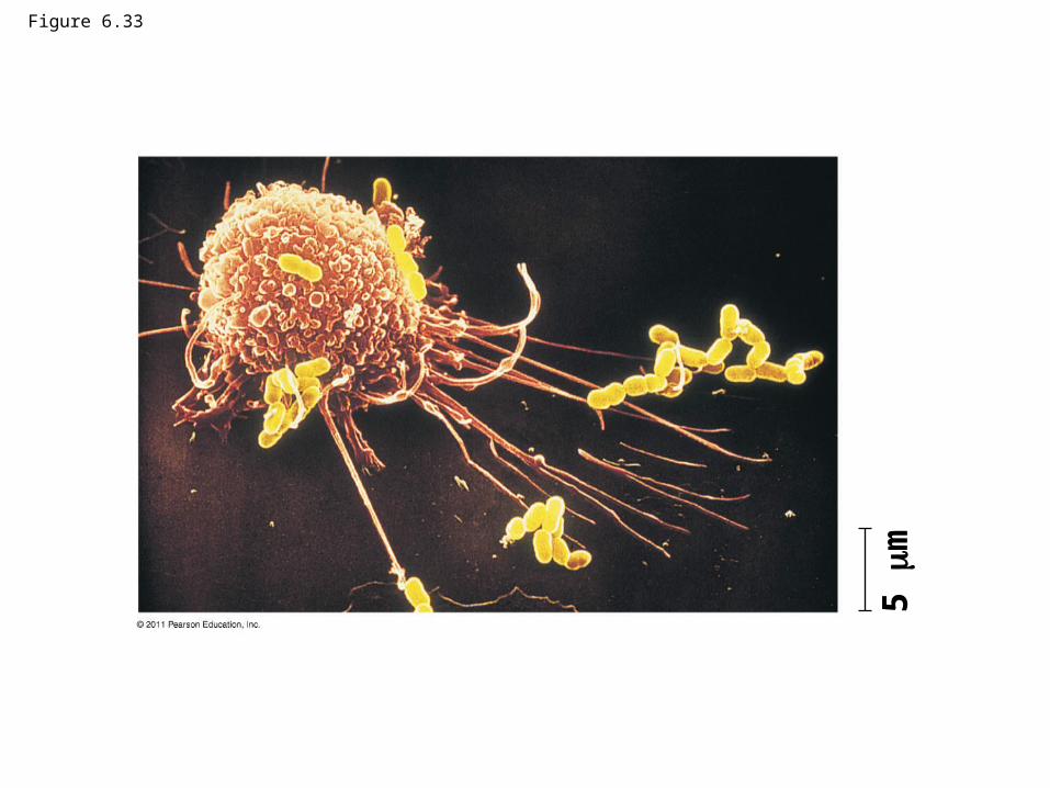
Figure 6.33
5 m
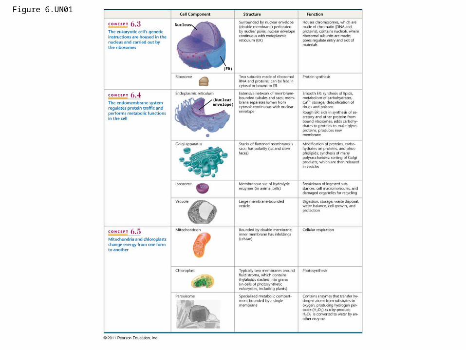
Figure 6.UN01
Nucleus
(ER)
(Nuclearenvelope)

Figure 6.UN01a
Nucleus
(ER)

Figure 6.UN01b
(Nuclearenvelope)

Figure 6.UN01c

Figure 6.UN02

Figure 6.UN03

Figure 6.UN04