changes in gut microbiota control inflammation in obese...
TRANSCRIPT
Changes in gut microbiota control inflammation inobese mice through a mechanism involving GLP-2-driven improvement of gut permeability
P D Cani,1 S Possemiers,2 T Van de Wiele,2 Y Guiot,3 A Everard,1 O Rottier,1 L Geurts,1
D Naslain,1,4 A Neyrinck,1 D M Lambert,4 G G Muccioli,5 N M Delzenne1
See Commentary, p 1044
c A supplementary table andthree files of supplementarydata are published online only athttp://gut.bmj.com/content/vol58/issue8
1 Unit of Pharmacokinetics,Metabolism, Nutrition andToxicology, Louvain DrugResearch Institute, Universitecatholique de Louvain, Brussels,Belgium; 2 Laboratory ofMicrobial Ecology andTechnology, Faculty ofBioscience Engineering, GhentUniversity, Gent, Belgium;3 Department of Pathology,Universite catholique de Louvain,Brussels, Belgium; 4 MedicinalChemistry and RadiopharmacyUnit, Louvain Drug ResearchInstitute, Universite catholiquede Louvain, Brussels, Belgium;5 Laboratory of Chemical andPhysico-chemical Analysis ofDrugs (CHAM), Louvain DrugResearch Institute, Universitecatholique de Louvain, Brussels,Belgium
Correspondence to:Dr P D Cani, UCL, Unit PMNT-7369, Av E Mounier, 73/69,B-1200 Brussels, Belgium;[email protected]; orProfessor NM Delzenne, UCL,Unit PMNT-7369, Av R Mounier,73/69, B-1200 Brussels,Belgium;[email protected]
Revised 4 February 2009Accepted 6 February 2009Published Online First24 February 2009
This paper is freely availableonline under the BMJ Journalsunlocked scheme, see http://gut.bmj.com/info/unlocked.dtl
ABSTRACTBackground and aims: Obese and diabetic mice displayenhanced intestinal permeability and metabolic endotox-aemia that participate in the occurrence of metabolicdisorders. Our recent data support the idea that aselective increase of Bifidobacterium spp. reduces theimpact of high-fat diet-induced metabolic endotoxaemiaand inflammatory disorders. Here, we hypothesised thatprebiotic modulation of gut microbiota lowers intestinalpermeability, by a mechanism involving glucagon-likepeptide-2 (GLP-2) thereby improving inflammation andmetabolic disorders during obesity and diabetes.Methods: Study 1: ob/ob mice (Ob-CT) were treated witheither prebiotic (Ob-Pre) or non-prebiotic carbohydrates ascontrol (Ob-Cell). Study 2: Ob-CT and Ob-Pre mice weretreated with GLP-2 antagonist or saline. Study 3: Ob-CTmice were treated with a GLP-2 agonist or saline. Weassessed changes in the gut microbiota, intestinalpermeability, gut peptides, intestinal epithelial tight-junctionproteins ZO-1 and occludin (qPCR and immunohistochem-istry), hepatic and systemic inflammation.Results: Prebiotic-treated mice exhibited a lower plasmalipopolysaccharide (LPS) and cytokines, and a decreasedhepatic expression of inflammatory and oxidative stressmarkers. This decreased inflammatory tone was asso-ciated with a lower intestinal permeability and improvedtight-junction integrity compared to controls. Prebioticincreased the endogenous intestinotrophic proglucagon-derived peptide (GLP-2) production whereas the GLP-2antagonist abolished most of the prebiotic effects. Finally,pharmacological GLP-2 treatment decreased gut perme-ability, systemic and hepatic inflammatory phenotypeassociated with obesity to a similar extent as thatobserved following prebiotic-induced changes in gutmicrobiota.Conclusion: We found that a selective gut microbiotachange controls and increases endogenous GLP-2production, and consequently improves gut barrierfunctions by a GLP-2-dependent mechanism, contributingto the improvement of gut barrier functions during obesityand diabetes.
Obesity is typically associated with a cluster ofseveral metabolic disorders, characterised by a low-grade inflammation.1 2 Evidence that the gutmicrobiota composition can be different betweenhealthy and obese and/or type 2 diabetic patientshas led to the investigation of this environmentalelement as a key factor in the pathophysiology ofmetabolic diseases.3–5
We previously reported that the gut microbiotais involved in high-fat diet-induced metabolic
endotoxaemia, adipose tissue inflammation andmetabolic disorders.6–9 The link between high-fatdiet-induced inflammation, oxidative stress, meta-bolic disorders, and gut microbiota, could belipopolysaccharide (LPS)-dependent.7 10–14 Severalpieces of clinical and experimental data haveconfirmed that LPS significantly contributes tothe development of obesity-related inflammatoryliver diseases such as non-alcoholic fatty liverdisease and non-alcoholic steatohepatitis.15–17 Highplasma LPS levels could result from an increasedproduction of endotoxin upon changes in the gutmicrobiota.7 8 Normally, the intestinal epitheliumacts as a continuous barrier to avoid LPS transloca-tion; yet some endogenous or exogenous eventsmay alter this protective function. Among theelements promoting a leaky gut, and thus anincreased plasmatic LPS level, are alcohol consump-tion,15 18–22 immobilisation stress,23 24 and radiation25
have been proposed. In addition, we have recentlyshown that the modulation of gut bacteriafollowing a high-fat diet strongly increases intest-inal permeability, by reducing the expression ofgenes coding for two tight junction proteins ZO-1and occludin.6 We previously reported that gutbacteria are clearly involved in these events sinceobese and high-fat fed-diabetic mice treated withan antibiotic recovered normal intestinal epithelialintegrity.6 Also, recent data have shown that obeseand diabetic mice display enhanced intestinalpermeability, and are characterised by a metabolicendotoxaemia and a low-grade inflammation.6 7 26
Furthermore, high-fat feeding changes gut micro-biota6–8 27 towards a decreased number of bifido-bacteria,6–8 a group of bacteria which has beenshown to reduce intestinal LPS levels in mice andto improve the mucosal barrier function.28–32
Besides, we have shown that feeding mice withprebiotics increased the number of intestinalbifidobacteria and reduced the impact of high-fatdiet-induced metabolic endotoxaemia and inflam-matory disorders.8 33
Importantly, however, the mechanisms linkingprebiotic-induced changes in gut microbiota, meta-bolic endotoxaemia and the improvement ofobesity-related hepatic and metabolic disordersare still unknown.
By using these experiments, we have tested thehypothesis that the control of gut permeabilitythrough the selective modulation of gut microbiotaby prebiotics participates in the improvement ofmetabolic diseases in ob/ob mice. Novel mechan-isms involving the influence of gut fermentation
Inflammatory bowel disease
Gut 2009;58:1091–1103. doi:10.1136/gut.2008.165886 1091
on 27 June 2018 by guest. Protected by copyright.
http://gut.bmj.com
/G
ut: first published as 10.1136/gut.2008.165886 on 24 February 2009. D
ownloaded from

on specific proglucagon-derived peptides – namely, glucagon-like peptide-2 (GLP-2) – are proposed.
MATERIALS AND METHODSAnimalsExperiment 1Six-week-old ob/ob (n = 10/group) mice (C57BL/6 background;Jackson Laboratory, Bar Harbor, Maine, USA) were housed in acontrolled environment (12 h daylight cycle, lights off at 18.00hours) in groups of 2 mice/cage, and kept with free access tofood and water. The mice were fed a control diet (Ob-CT) (A04,Villemoisson sur Orge, France), or a control diet containing amix of a fermentable dietary fibre (oligofructose) (Ob-Pre)(Orafti, Tienen, Belgium),33 or a diet containing a mix a non-fermentable dietary fibre (microcrystalline cellulose) (Ob-Cell)(Vivapur Microcrystalline cellulose; J. Retten Maier 38 Sohne,Weissenborn, Germany). Dietary fibres were added in aproportion of 9:1 (weight of control diet:weight of fibres).
Experiment 2Six-week-old ob/ob (n = 8/group) mice (C57BL/6 background;Jackson Laboratory) were housed in a controlled environment(12 h daylight cycle, lights off at 18.00 hours) in groups of2 mice/cage, and kept with free access to food and water. Tostudy the significance of GLP-2 in this model, mice were injectedsubcutaneously twice daily for 4 weeks with 2.5 mg/kg of GLP-2receptor antagonist GLP-2 (3–33) (Eurogentec, Verviers,Belgium) as described34–36 or saline. The mice were fed a controldiet and injected with saline or GLP-2 antagonist (Ob-CT andOb-Ant, respectively), or fed the prebiotic diet and injected withsaline or GLP-2 antagonist (Ob-Pre and Ob-Pre-Ant, respec-tively).
Experiment 3Six-week-old ob/ob (n = 6/group) mice (C57BL/6 background;Jackson Laboratory) were housed in a controlled environment(12 h daylight cycle, lights off at 1800 hours) in groups of2 mice/cage, and kept with free access to food and water. Themice were fed the same control diet as described in experiment1. The mice were separated into two groups and injectedsubcutaneously twice daily for 12 days with 25 mg GLP-2 (1–33)(Bachem, Bubendorf, Switzerland) (Ob-GLP-2), or saline (Ob-CT). GLP-2 doses were based on previous studies describing thephysiological effects (ie, the intestinotrophic properties) of thepeptides.35 37–39
Tissue samplingMice were anaesthetised (ketamine/xylazine, intraperineally,100 and 10 mg/kg, respectively) after a 5 h period of fasting, andblood samples and tissues were harvested for further analysis.Mice were killed by cervical dislocation. Liver, caecum (full andempty), muscles (vastus lateralis), and adipose tissues (epididy-mal, subcutaneous and visceral) were precisely dissected andweighed. The intestinal segments (jejunum, colon) wereimmersed in liquid nitrogen, and stored at 280uC, for furtheranalysis.
Microbial analysis of the caecal content of selected miceMetagenomic DNA was extracted from the caecal content ofrandomly selected mice (5/group), using the QIAamp DNA stoolmini kit (Qiagen, Venlo, Netherlands) according to the manu-facturer’s instructions. Denaturing gradient gel electrophoresis(DGGE) on total bacteria, bifidobacteria and lactobacilli were
performed to study the qualitative effect of the treatment on thestructure and composition of the intestinal microbial commu-nity.40 DGGE with a 45–60% denaturant gradient were used toseparate the polymerase chain reaction (PCR) products obtainedwith a nested approach for the 16S rRNA genes of bifidobacteria(primers BIF164f-BIF662r) and lactobacilli (SGLAB0158f-SGLAB0667). The first PCR round was followed by a secondamplification with primers 338F-GC and 518R. The latter primerswere also used to amplify the 16S rDNA of all bacteria on totalextracted DNA. The DGGE patterns obtained were subsequentlyanalysed using the Bionumerics software version 2.0 (AppliedMaths, Sint-Martens-Latem, Belgium).41 In brief, the calculationof the similarities was based on the Pearson (product–moment)correlation coefficient. Clustering analysis was performed usingthe unweighted pair group method with arithmetic meanclustering algorithm (UPGMA) to calculate the dendrograms ofeach DGGE gel and a combination of all gels. The latter wasperformed on a created composite dataset. Multidimensionalscaling (MDS) analysis was used to reduce the different data ofthe complex DGGE patterns of one sample to one point in a three-dimensional space. MDS was based on the combined informationfrom the distance matrices of each DGGE, obtained usingsimilarity coefficients (Pearson correlation).
Quantitative PCR (qPCR) for total bacteria (using primersPRBA338f and P518r) and specific for bifidobacteria, lactobacillior the Eubacterium rectale/Clostridium coccoides grp. was per-formed to study the quantitative effect of the treatment on thecomposition of the intestinal microbial community as reportedby Possemiers et al.42
Real-time qPCRTotal RNA from tissues was prepared using the TriPure reagent(Roche, Basel, Switzerland) as described.33 cDNA was synthe-sised using a reverse transcription kit (Promega, Madison,Wisconsin, USA) from 1 mg of total RNA. qPCR was performedwith a STEP one PLUS instrument and software (AppliedBiosystems, Foster City, California, USA), as described.6 Primersequences for the targeted mouse genes are presented insupplemental table 1.
Immunofluorescence analysis of occludin and ZO-1Jejunum segments were immediately removed, washed withPBS, mounted in embedding medium (Tissue-Tek, Sakura,Netherlands), and stored at 280uC until use. Cryosections(5 mm) were fixed in acetone at 220uC for 5 min for occludinand fixed in ethanol for 30 min at room temperature and inacetone at 220uC for 5 min for ZO-1. Non-specific backgroundwas blocked by incubation with 10% bovine serum albumin(BSA) in Tris-buffered saline (TBS) and 0.3% Triton X-100(30 min at room temperature). Sections were incubated withrabbit anti-occludin or rabbit anti-ZO-1 (1:400 for ZO-1 and1:100 for occludin staining; Zymed Laboratories, San Francisco,California, USA) for 2 h. Sections were washed three times for10 min in TBS and probed with goat anti-rabbit fluoresceinisothiocyante (FITC)-conjugated antibodies (1:50, Zymax;Zymed Laboratories). Slides were washed three times for10 min in TBS and mounted in mounting medium(Vectashield; Vector Laboratories, Burlingame, California,USA). Sections were visualised on a fluorescence microscopeusing a 640 objective, and images were stored digitally withLeica software. As a control, slides were incubated with serialdilutions of the primary antibody to signal extinction. Twonegative controls were used: slides incubated with irrelevant
Inflammatory bowel disease
1092 Gut 2009;58:1091–1103. doi:10.1136/gut.2008.165886
on 27 June 2018 by guest. Protected by copyright.
http://gut.bmj.com
/G
ut: first published as 10.1136/gut.2008.165886 on 24 February 2009. D
ownloaded from

antibody or without primary antibody. All the stainings wereperformed in duplicate in non-serial distant sections, and analysedin a double-blind manner by two different investigators.
Intestinal permeability in vivoThis measure is based on the intestinal permeability towards4000 Da fluorescent dextran–FITC (DX-4000–FITC) (FD4000;Sigma-Aldrich, St. Louis, Missouri, USA) as described.6 43 Briefly,mice that had fasted for 6 h were given DX-4000–FITC bygavage (500 mg/kg body weight, 125 mg/ml). After 1 h and 4 h,120 ml of blood was collected from the tip of the tail vein. Theblood was centrifuged at 4uC, 12 000 g for 3 min. Plasma wasdiluted in an equal volume of PBS (pH 7.4) and analysed for DX-4000–FITC concentration with a fluorescence spectrophot-ometer (HTS-7000 Plus-plate-reader; Perkin Elmer, Wellesley,Massachusetts, USA) at an excitation wavelength of 485 nmand emission wavelength of 535 nm. Standard curves wereobtained by diluting FITC–dextran in non-treated plasmadiluted with PBS (1:3 v/v).
Biochemical analysesPlasma LPS concentration was determined by using a kit basedupon a Limulus amoebocyte extract (LAL kit endpoint-QCL1000; Cambrex BioScience, Walkersville, Maryland, USA),samples were diluted 1/40 to 1/100 and heated for 20 cycles of10 min at 68uC and 10 min at 4uC. An internal control for LPSrecovery was included in the calculation. Plasma cytokines(interleukin (IL) 1a, IL1b, tumour necrosis factor (TNF) a, IL6,monocyte chemoattractant protein (MCP)-1, macrophageinflammatory protein (MIP)-1a, IL10, interferon (INF) c, IL15,IL18) and gut hormones (GLP-1 (active), GIP (total), amylin(active), pancreatic polypeptide) were respectively determinedin duplicate by using a Bio-Plex Multiplex kit (Bio-Rad,Nazareth, Belgium), or a mouse gut hormones panel(LincoPlex; Millipore, Brussels, Belgium), and measured byusing Luminex technology (Bio-Rad Bioplex; Bio-Rad) followingthe manufacturer’s instructions, an EIA kit (GLP-2 EIA kit)(Yanaihara Institute, Shizuoka, Japan) was used to quantifyGLP-2.
Statistical analysisResults are presented as mean with the SEM. The statisticalsignificance of differences was analysed by one-way ANOVAfollowed by post hoc Bonferroni’s multiple comparison test orKruskal–Wallis for non-parametric data followed by Dunn’smultiple comparison test. Data with different superscript lettersare significantly different p,0.05, according to the post hocANOVA statistical analysis. Comparisons between GLP-2-treated mice and control mice were performed using thetwo-tailed Student t test. Multiple correlation analyses wereassessed by the Pearson’s test using GraphPad Prism version 5.00
for windows. Results were considered statistically significantwhen p,0.05.
RESULTSPrebiotic treatment induces changes in the gut microbiota of ob/ob miceFeeding ob/ob mice with the prebiotic carbohydrates (Ob-Pre)induced significant changes in the gut microbiota of the caecum,with a higher total bacteria count, Lactobacillus spp.,Bifidobacterium spp., and the C coccoides–E rectale cluster(table 1), as compared to the control mice (Ob-CT and Ob-Cell).
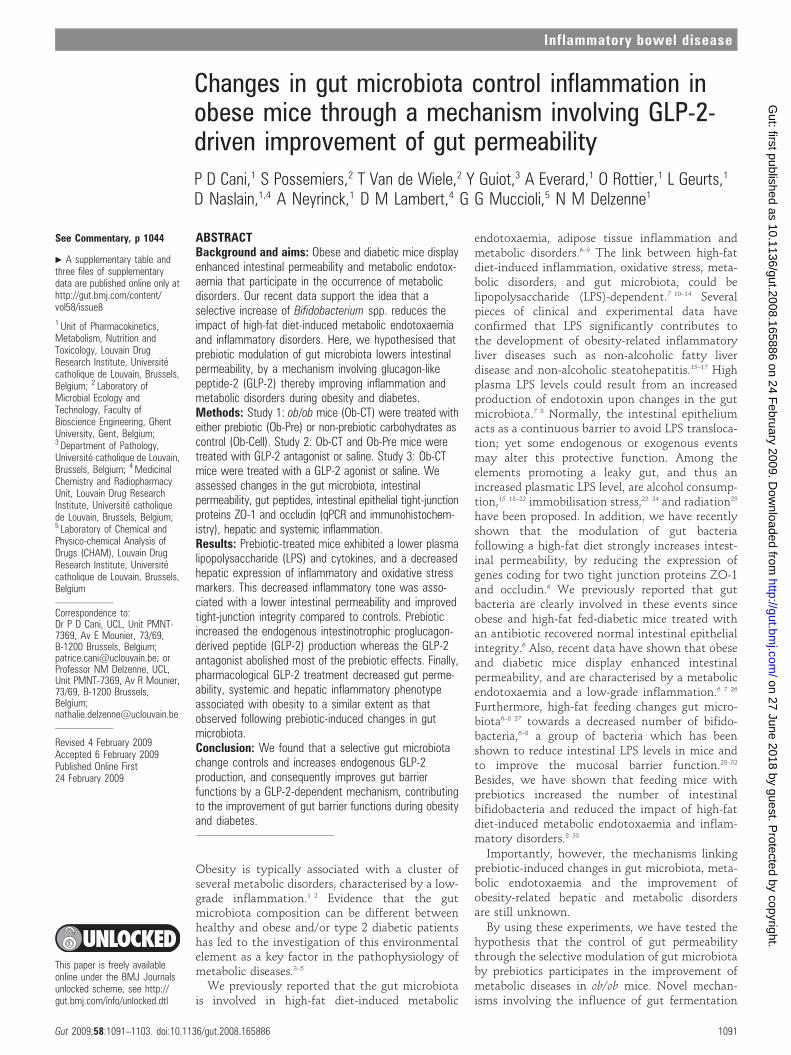
Changes in the microbial community composition were alsoobserved upon DGGE analysis of the caecal content of thedifferent treatment groups (fig 1A–C). Clustering of the DGGEfingerprints for total bacteria and for the specific groups ofbifidobacteria and lactobacilli, indicated, for all fingerprints, aseparate cluster of the Ob-Pre mice. Secondary clustering wasobserved between the Ob-CT and Ob-Cell mice. Finally, multi-dimensional scaling (MDS) analysis, performed on the compositedata set derived from the combination of all the DGGE analyses ofthis study (total bacteria, lactobacilli and bifidobacteria), alsoindicated a specific clustering pattern, depending on the treatmentof the ob/ob mice. (fig 1D). As the distance between 2 data pointsin the three-dimensional MDS plot is a visual representation ofthe difference in the microbial community composition of thecaecum of the different mice, MDS analysis provides the finalconfirmation that the prebiotic treatment induced importantchanges in the gut microbiota of the Ob-Pre mice compared to thecontrol groups.
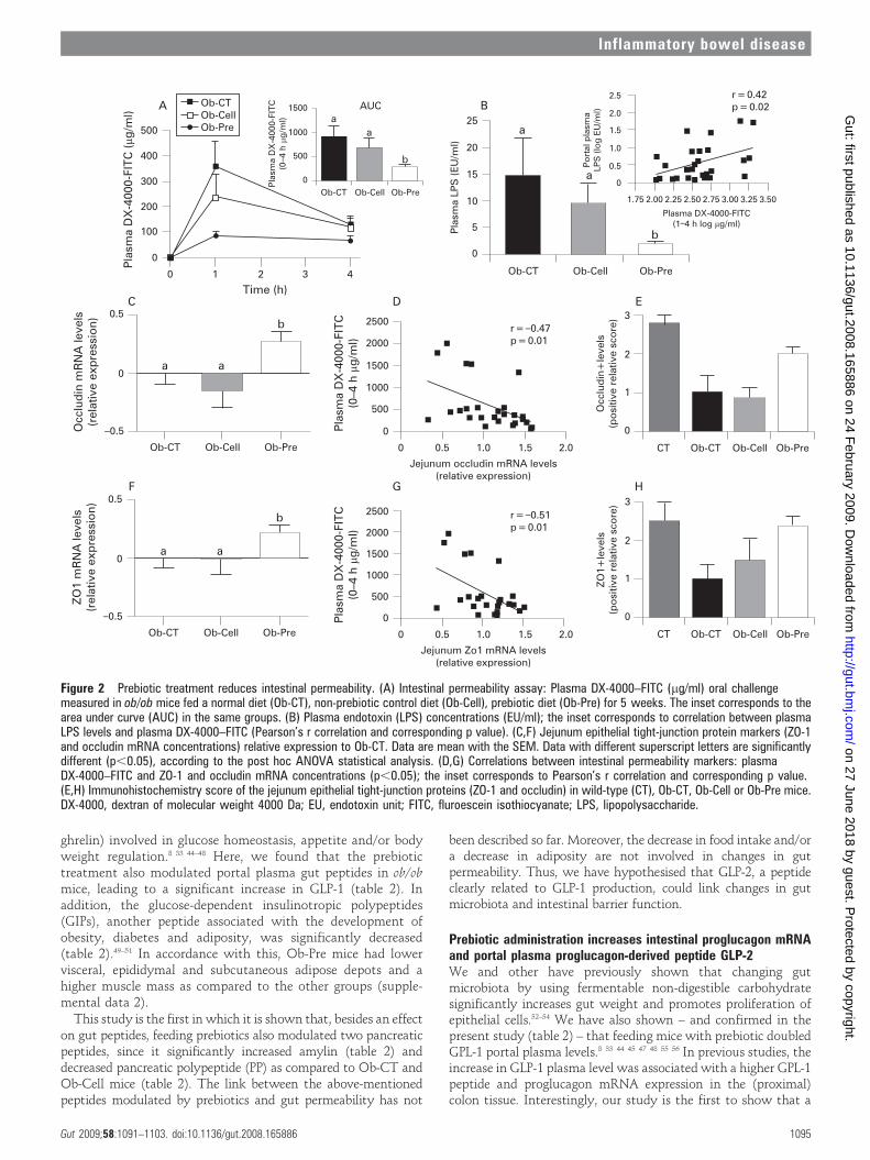
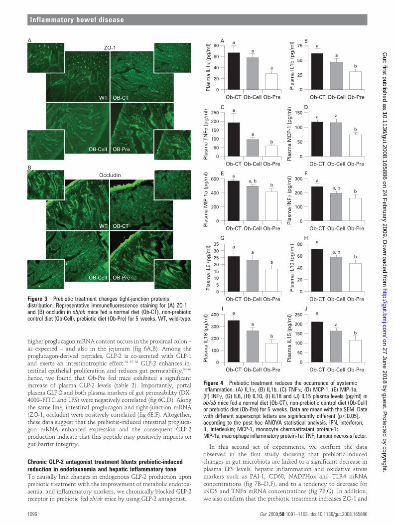
Changes in the gut microbiota upon prebiotic administrationreduce intestinal permeabilityOb-Pre fed mice exhibited a 3-fold lower plasma DX-4000–FITCarea under the curve (fig 2A) as compared to Ob-CT and Ob-Cell mice. In accordance with the in vivo assessment ofintestinal permeability, LPS levels were significantly lower inOb-Pre mice plasma samples, as compared to the other groups(fig 2B). Moreover, plasma DX-4000–FITC and portal plasmaLPS levels were positively and significantly correlated (fig 2B,inset), further confirming the relation between intestinalpermeability and the development of metabolic endotoxaemia.Gut permeability is controlled by several specific tight-junctionproteins. Among these, ZO-1 and occludin have been proposedas key markers of tight-junction integrity.26 The prebiotictreatment increased ZO-1 and occludin mRNA in the jejunumsegment (fig 2C,F). As previously suggested, a representativeimmunofluorescence assay performed on intestinal sections ofwild-type C57BL/6J mice demonstrated an intact network ofZO-1 and occludin proteins which were predominantly localisedalong the apical cellular border.26 In strong contrast, ZO-1 andoccludin staining appears to be decreased and discontinuous inOb-CT tissues (fig 3A,B). Furthermore, immunohistochemical
Table 1 Prebiotic-associated changes in gut microbiota
Bacterial content Ob-CT Ob-Cell Ob-Pre
Total bacteria 5.74a (2.05) 4.30b (0.38) 8.07c (1.05)
Bifidobacterium spp 4.65a (1.58) 3.38b (0.37) 6.35c (1.10)
Lactobacillus spp 4.93a (1.76) 3.79a (0.38) 7.16b (1.40)
Clostridium coccoides–Eubacterium rectale cluster 3.65a (1.37) 2.72a (0.20) 6.41b (1.27)
Results are given as the log DNA copies/caecal content (SD).Data are mean with the SD.Data with different superscript letters are significantly different p,0.05, according to the post hoc ANOVA statistical analysis.CT, Cell and Pre, respectively, refer to the selected ob/ob mice fed a normal-diet, non-prebiotic control diet or prebiotic diet.
Inflammatory bowel disease
Gut 2009;58:1091–1103. doi:10.1136/gut.2008.165886 1093
on 27 June 2018 by guest. Protected by copyright.
http://gut.bmj.com
/G
ut: first published as 10.1136/gut.2008.165886 on 24 February 2009. D
ownloaded from
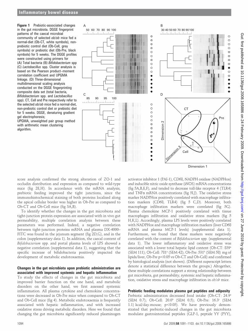
score analysis confirmed the strong alteration of ZO-1 andoccludin distribution and expression as compared to wild-typemice (fig 2E,H). In accordance with the mRNA analysis,prebiotic feeding improved the tight junctions, since theimmunohistochemical staining of both proteins localised alongthe apical cellular border was higher in Ob-Pre as compared toOb-CT and Ob-Cell mice (fig 3A,B).
To identify whether the changes in the gut microbiota andtight-junction protein expression are associated with in vivo gutpermeability, multiple correlation analysis between theseparameters was performed. Indeed, a negative correlationbetween tight-junction proteins mRNA and plasma DX-4000–FITC was found in the jejunum segment (fig 2D,G), and in thecolon (supplementary data 1). In addition, the caecal content ofBifidobacterium spp. and portal plasma levels of LPS showed anegative correlation (supplemental data 1), suggesting that thespecific increase of bifidobacteria positively impacted thedevelopment of metabolic endotoxaemia.
Changes in the gut microbiota upon prebiotic administration areassociated with improved systemic and hepatic inflammationTo study the effects of changes in the gut microbiota andimproved barrier function on the one hand, and metabolicdisorders on the other hand, we first assessed systemicinflammation. All plasma cytokine and chemokine concentra-tions were decreased in Ob-Pre mice when compared to Ob-CTand Ob-Cell mice (fig 4). Metabolic endotoxaemia is frequentlyassociated with hepatic inflammation, and with increasedoxidative stress driving metabolic disorders. Here we found thatchanging the gut microbiota significantly reduced plasminogen
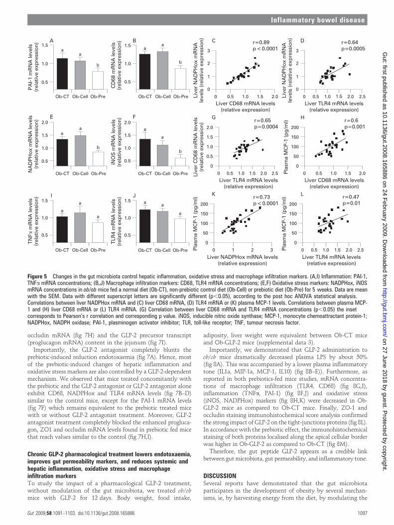
activator inhibitor 1 (PAI-1), CD68, NADPH oxidase (NADPHox)and inducible nitric oxide synthase (iNOS) mRNA concentrations(fig 5A,B,E,F), and tended to decrease toll-like receptor 4 (TLR4)and TNFa mRNA concentrations (fig 5I,J). The oxidative stressmarker NADPHox positively correlated with macrophage infiltra-tion markers (CD68, TLR4) (fig 5 C,D). Moreover, bothmacrophage infiltration markers were correlated (fig 5G).Plasma chemokine MCP-1 positively correlated with tissuemacrophages infiltration and oxidative stress markers (fig 5H,K,L). Accordingly, plasma LPS levels were positively correlatedwith NADPHox and macrophage infiltration markers (liver CD68mRNA and plasma MCP-1 levels) (supplemental data 1).Furthermore, we found that these markers were negativelycorrelated with the content of Bifidobacterium spp. (supplementaldata 1). The lower inflammatory and oxidative stress wasassociated with a lower total hepatic lipid content (Ob-CT: 839a
(SEM 111); Ob-Cell: 781a (SEM 42); Ob-Pre: 531b (SEM 58) mg oflipids/liver, Ob-Pre p,0.05 vs Ob-CT and Ob-Cell) and confirmedby histological analysis (not shown). (Different superscript lettersdenote a statistical difference between the groups.) Altogether,these multiple correlations support a strong relationship betweengut microbiota, gut permeability, systemic and hepatic inflamma-tion, oxidative stress and macrophage infiltration in ob/ob mice.
Prebiotic feeding modulates plasma gut peptides and adiposityPrebiotic administration decreased food intake (Ob-CT: 24.9a
(SEM 0.7); Ob-Cell: 20.0b (SEM 0.5); Ob-Pre: 16.3c (SEM1.1) kcal/day.mouse; p,0.05). We have previously demon-strated that prebiotic-induced changes in the gut microbiotamodulate gastrointestinal peptides (GLP-1, peptide YY (PYY),
Figure 1 Prebiotic-associated changesin the gut microbiota. DGGE fingerprintpatterns of the caecal microbialcommunity of selected ob/ob mice fed anormal-diet (Ob-CT, white symbols), non-prebiotic control diet (Ob-Cell, greysymbols) or prebiotic diet (Ob-Pre, blacksymbols) for 5 weeks. The DGGE profileswere constructed using primers for(A) Total bacteria (B) Bifidobacterium spp(C) Lactobacillus spp. Cluster analysis isbased on the Pearson product–momentcorrelation coefficient and UPGMAlinkage. (D) Three-dimensionalmultidimensional scaling analysisconducted on the DGGE fingerprintingcomposite data set (total bacteria,Bifidobacterium spp. and Lactobacillusspp). CT, Cell and Pre respectively refer tothe selected ob/ob mice fed a normal-diet,non-prebiotic control diet or prebiotic dietfor 5 weeks. DGGE, denaturing gradientgel electrophoresis;UPGMA, unweighted pair group methodwith arithmetic mean clusteringalgorithm.
Inflammatory bowel disease
1094 Gut 2009;58:1091–1103. doi:10.1136/gut.2008.165886
on 27 June 2018 by guest. Protected by copyright.
http://gut.bmj.com
/G
ut: first published as 10.1136/gut.2008.165886 on 24 February 2009. D
ownloaded from
ghrelin) involved in glucose homeostasis, appetite and/or bodyweight regulation.8 33 44–48 Here, we found that the prebiotictreatment also modulated portal plasma gut peptides in ob/obmice, leading to a significant increase in GLP-1 (table 2). Inaddition, the glucose-dependent insulinotropic polypeptides(GIPs), another peptide associated with the development ofobesity, diabetes and adiposity, was significantly decreased(table 2).49–51 In accordance with this, Ob-Pre mice had lowervisceral, epididymal and subcutaneous adipose depots and ahigher muscle mass as compared to the other groups (supple-mental data 2).
This study is the first in which it is shown that, besides an effecton gut peptides, feeding prebiotics also modulated two pancreaticpeptides, since it significantly increased amylin (table 2) anddecreased pancreatic polypeptide (PP) as compared to Ob-CT andOb-Cell mice (table 2). The link between the above-mentionedpeptides modulated by prebiotics and gut permeability has not
been described so far. Moreover, the decrease in food intake and/ora decrease in adiposity are not involved in changes in gutpermeability. Thus, we have hypothesised that GLP-2, a peptideclearly related to GLP-1 production, could link changes in gutmicrobiota and intestinal barrier function.
Prebiotic administration increases intestinal proglucagon mRNAand portal plasma proglucagon-derived peptide GLP-2We and other have previously shown that changing gutmicrobiota by using fermentable non-digestible carbohydratesignificantly increases gut weight and promotes proliferation ofepithelial cells.52–54 We have also shown – and confirmed in thepresent study (table 2) – that feeding mice with prebiotic doubledGPL-1 portal plasma levels.8 33 44 45 47 48 55 56 In previous studies, theincrease in GLP-1 plasma level was associated with a higher GPL-1peptide and proglucagon mRNA expression in the (proximal)colon tissue. Interestingly, our study is the first to show that a
Figure 2 Prebiotic treatment reduces intestinal permeability. (A) Intestinal permeability assay: Plasma DX-4000–FITC (mg/ml) oral challengemeasured in ob/ob mice fed a normal diet (Ob-CT), non-prebiotic control diet (Ob-Cell), prebiotic diet (Ob-Pre) for 5 weeks. The inset corresponds to thearea under curve (AUC) in the same groups. (B) Plasma endotoxin (LPS) concentrations (EU/ml); the inset corresponds to correlation between plasmaLPS levels and plasma DX-4000–FITC (Pearson’s r correlation and corresponding p value). (C,F) Jejunum epithelial tight-junction protein markers (ZO-1and occludin mRNA concentrations) relative expression to Ob-CT. Data are mean with the SEM. Data with different superscript letters are significantlydifferent (p,0.05), according to the post hoc ANOVA statistical analysis. (D,G) Correlations between intestinal permeability markers: plasmaDX-4000–FITC and ZO-1 and occludin mRNA concentrations (p,0.05); the inset corresponds to Pearson’s r correlation and corresponding p value.(E,H) Immunohistochemistry score of the jejunum epithelial tight-junction proteins (ZO-1 and occludin) in wild-type (CT), Ob-CT, Ob-Cell or Ob-Pre mice.DX-4000, dextran of molecular weight 4000 Da; EU, endotoxin unit; FITC, fluroescein isothiocyanate; LPS, lipopolysaccharide.
Inflammatory bowel disease
Gut 2009;58:1091–1103. doi:10.1136/gut.2008.165886 1095
on 27 June 2018 by guest. Protected by copyright.
http://gut.bmj.com
/G
ut: first published as 10.1136/gut.2008.165886 on 24 February 2009. D
ownloaded from
higher proglucagon mRNA content occurs in the proximal colon –as expected – and also in the jejunum (fig 6A,B). Among theproglucagon-derived peptides, GLP-2 is co-secreted with GLP-1and exerts an intestinotrophic effect.39 57 58 GLP-2 enhances in-testinal epithelial proliferation and reduces gut permeability,59–62
hence, we found that Ob-Pre fed mice exhibited a significantincrease of plasma GLP-2 levels (table 2). Importantly, portalplasma GLP-2 and both plasma markers of gut permeability (DX-4000–FITC and LPS) were negatively correlated (fig 6C,D). Alongthe same line, intestinal proglucagon and tight-junction mRNA(ZO-1, occludin) were positively correlated (fig 6E,F). Altogether,these data suggest that the prebiotic-induced intestinal progluca-gon mRNA enhanced expression and the consequent GLP-2production indicate that this peptide may positively impacts ongut barrier integrity.
Chronic GLP-2 antagonist treatment blunts prebiotic-inducedreduction in endotoxaemia and hepatic inflammatory toneTo causally link changes in endogenous GLP-2 production uponprebiotic treatment with the improvement of metabolic endotox-aemia, and inflammatory markers, we chronically blocked GLP-2receptor in prebiotic fed ob/ob mice by using GLP-2 antagonist.
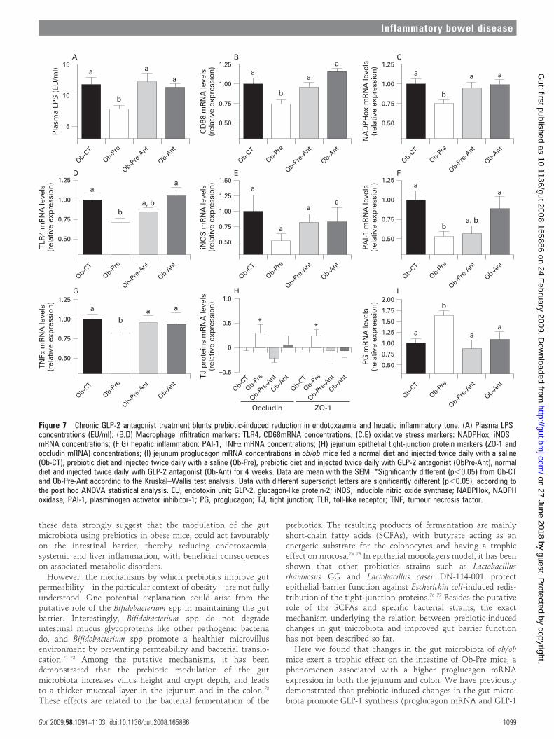
In this second set of experiments, we confirm the dataobserved in the first study showing that prebiotic-inducedchanges in gut microbiota are linked to a significant decrease inplasma LPS levels, hepatic inflammation and oxidative stressmarkers such as PAI-1, CD68, NADPHox and TLR4 mRNAconcentrations (fig 7B–D,F), and to a tendency to decrease foriNOS and TNFa mRNA concentrations (fig 7E,G). In addition,we also confirm that the prebiotic treatment increases ZO-1 and
Figure 3 Prebiotic treatment changes tight-junction proteinsdistribution. Representative immunofluorescence staining for (A) ZO-1and (B) occludin in ob/ob mice fed a normal diet (Ob-CT), non-prebioticcontrol diet (Ob-Cell), prebiotic diet (Ob-Pre) for 5 weeks. WT, wild-type.
Figure 4 Prebiotic treatment reduces the occurrence of systemicinflammation. (A) IL1a, (B) IL1b, (C) TNFa, (D) MCP-1, (E) MIP-1a,(F) INFc, (G) IL6, (H) IL10, (I) IL18 and (J) IL15 plasma levels (pg/ml) inob/ob mice fed a normal diet (Ob-CT), non-prebiotic control diet (Ob-Cell)or prebiotic diet (Ob-Pre) for 5 weeks. Data are mean with the SEM. Datawith different superscript letters are significantly different (p,0.05),according to the post hoc ANOVA statistical analysis. IFN, interferon;IL, interleukin; MCP-1, monocyte chemoattratant protein-1;MIP-1a, macrophage inflammatory protein-1a; TNF, tumour necrosis factor.
Inflammatory bowel disease
1096 Gut 2009;58:1091–1103. doi:10.1136/gut.2008.165886
on 27 June 2018 by guest. Protected by copyright.
http://gut.bmj.com
/G
ut: first published as 10.1136/gut.2008.165886 on 24 February 2009. D
ownloaded from
occludin mRNA (fig 7H) and the GLP-2 precursor transcript(proglucagon mRNA) content in the jejunum (fig 7I).
Importantly, the GLP-2 antagonist completely blunts theprebiotic-induced reduction endotoxaemia (fig 7A). Hence, mostof the prebiotic-induced changes of hepatic inflammation andoxidative stress markers are also controlled by a GLP-2-dependentmechanism. We observed that mice treated concomitantly withthe prebiotic and the GLP-2 antagonist or GLP-2 antagonist aloneexhibit CD68, NADPHox and TLR4 mRNA levels (fig 7B–D)similar to the control mice, except for the PAI-1 mRNA levels(fig 7F) which remains equivalent to the prebiotic treated micewith or without GLP-2 antagonist treatment. Moreover, GLP-2antagonist treatment completely blocked the enhanced progluca-gon, ZO1 and occludin mRNA levels found in prebiotic fed micethat reach values similar to the control (fig 7H,I).
Chronic GLP-2 pharmacological treatment lowers endotoxaemia,improves gut permeability markers, and reduces systemic andhepatic inflammation, oxidative stress and macrophageinfiltration markersTo study the impact of a pharmacological GLP-2 treatment,without modulation of the gut microbiota, we treated ob/obmice with GLP-2 for 12 days. Body weight, food intake,
adiposity, liver weight were equivalent between Ob-CT miceand Ob-GLP-2 mice (supplemental data 3).
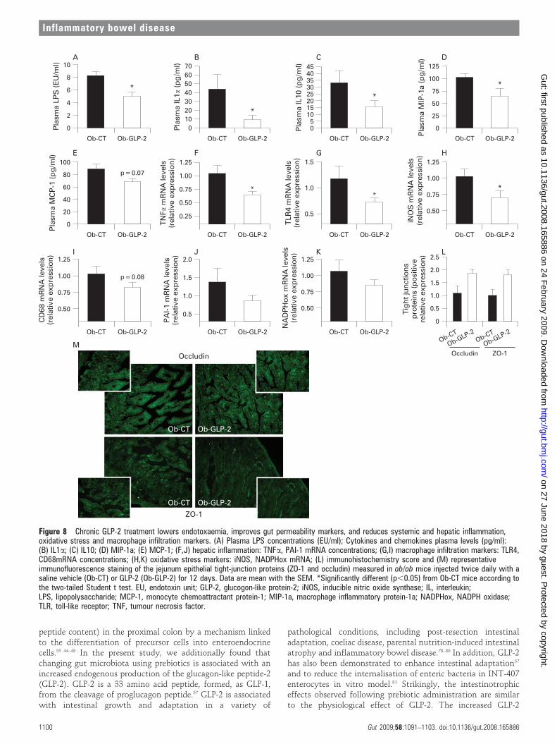
Importantly, we demonstrated that GLP-2 administration toob/ob mice dramatically decreased plasma LPS by about 50%(fig 8A). This was accompanied by a lower plasma inflammatorytone (IL1a, MIP-1a, MCP-1, IL10) (fig 8B–E)). Furthermore, asreported in both prebiotics-fed mice studies, mRNA concentra-tions of macrophage infiltration (TLR4, CD68) (fig 8G,I),inflammation (TNFa, PAI-1) (fig 8F,J) and oxidative stress(iNOS, NADPHox) markers (fig 8H,K) were decreased in Ob-GLP-2 mice as compared to Ob-CT mice. Finally, ZO-1 andoccludin staining immunohistochemical score analysis confirmedthe strong impact of GLP-2 on the tight-junctions proteins (fig 8L).In accordance with the prebiotic effect, the immunohistochemicalstaining of both proteins localised along the apical cellular borderwas higher in Ob-GLP-2 as compared to Ob-CT (fig 8M).
Therefore, the gut peptide GLP-2 appears as a credible linkbetween gut microbiota, gut permeability, and inflammatory tone.
DISCUSSIONSeveral reports have demonstrated that the gut microbiotaparticipates in the development of obesity by several mechan-isms, ie, by harvesting energy from the diet, by modulating the
Figure 5 Changes in the gut microbiota control hepatic inflammation, oxidative stress and macrophage infiltration markers. (A,I) Inflammation: PAI-1,TNFa mRNA concentrations; (B,J) Macrophage infiltration markers: CD68, TLR4 mRNA concentrations; (E,F) Oxidative stress markers: NADPHox, iNOSmRNA concentrations in ob/ob mice fed a normal diet (Ob-CT), non-prebiotic control diet (Ob-Cell) or prebiotic diet (Ob-Pre) for 5 weeks. Data are meanwith the SEM. Data with different superscript letters are significantly different (p,0.05), according to the post hoc ANOVA statistical analysis.Correlations between liver NADPHox mRNA and (C) liver CD68 mRNA, (D) TLR4 mRNA or (K) plasma MCP-1 levels. Correlations between plasma MCP-1 and (H) liver CD68 mRNA or (L) TLR4 mRNA. (G) Correlation between liver CD68 mRNA and TLR4 mRNA concentrations (p,0.05) the insetcorresponds to Pearson’s r correlation and corresponding p value. iNOS, inducible nitric oxide synthase; MCP-1, monocyte chemoattractant protein-1;NADPHox, NADPH oxidase; PAI-1, plasminogen activator inhibitor; TLR, toll-like receptor; TNF, tumour necrosis factor.
Inflammatory bowel disease
Gut 2009;58:1091–1103. doi:10.1136/gut.2008.165886 1097
on 27 June 2018 by guest. Protected by copyright.
http://gut.bmj.com
/G
ut: first published as 10.1136/gut.2008.165886 on 24 February 2009. D
ownloaded from

synthesis of gut peptides involved in energy homeostasis, and/orby regulating fat storage.63 We have recently demonstrated thatmice fed a high-fat diet were characterised by an increase in gutpermeability and metabolic endotoxaemia. The inflammatoryphenotype of ob/ob mice originates from the gut microbiota,since changing the gut microbiota using antibiotics lowersendotoxaemia-induced inflammation and metabolic disor-ders.6 64 65 Recently, it has been proposed that this higherendotoxaemia in ob/ob mice and db/db mice was dependent ona disruption of the key tight-junction proteins, ZO-1 andoccludin.26 Here we confirm that obese mice exhibit an alteredgut barrier, characterised by a disruption of tight-junctionproteins. Among the mechanisms involved in this phenomenon,excessive TNFa production was proposed to help propagate theextension of a local or systemic inflammation and thisinflammatory process per se would trigger the alteration ofboth junctional proteins.24 Our data show that prebiotic-fedmice have a lower level of several plasma cytokines well-known
to promote tight-junction disruption such as TNFa, IL1b, IL1a,IL6, INFc.66–69 In addition, we also found a significant decrease ofthe chemokines MCP-1 and MIP-1a.
In the present study, we demonstrated, in two separate set ofexperiments, that changing gut microbiota of ob/ob mice infavour of the Bifidobacterium spp. is associated with a significantimprovement of gut permeability measured in vivo; thisphenomenon was linked to an increase in tight-junctionmRNA expression and proteins distribution. The improvedgut barrier observed in prebiotic-fed mice was correlated to thelower portal plasma LPS levels and inflammatory tone (ie,decrease in circulating cytokines). The leakage of gut micro-biota-derived LPS into the portal blood is a well-establishedmechanism of metabolic endotoxaemia that triggers liverinflammation and oxidative stress.26 70 In the present studies,we found that lowering systemic inflammation by prebiotics issignificantly correlated with a strong decrease in markers ofoxidative and inflammatory stress in the liver tissue. Altogether,
Table 2 Changes in the gut microbiota upon administration of prebiotics impacts on portal plasma gutpeptides
Gut peptide Ob-CT Ob-Cell Ob-Pre
GLP-1 (active) (pmol/l) 7.65a (1.52) 7.71a (1.62) 21.15b (6.71)
GIP (total) (pg/ml) 207.91a (37.76) 180.71a (27.53) 114.02b (16.92)
Pancreatic polypeptide (pg/ml) 68.07a (8.45) 75.94a (6.56) 50.05b (2.67)
Amylin (active) (pg/ml) 358.87a (36.12) 364.99a (45.07) 504.90b (58.34)
GLP-2 (ng/ml) 0.56a (0.06) 0.62a (0.04) 0.75b (0.05)
Data are mean (SEM).Data with different superscript letters are significantly different at p,0.05, according to the post hoc ANOVA statistical analysis.CT, Cell and Pre, respectively, refer to the selected ob/ob mice, fed a normal diet, non-prebiotic control diet or prebiotic diet.GIP, glucose-dependent insulinotropic polypeptide; GLP, glucogon-like peptide.
Figure 6 Prebiotic administration increases intestinal proglucagon mRNA and correlates with intestinal permeability markers. (A) Jejunumproglucagon mRNA concentrations; (B) proximal colon proglucagon mRNA concentrations in ob/ob mice fed a normal diet (Ob-CT), non-prebioticcontrol diet (Ob-Cell), prebiotic diet (Ob-Pre) for 5 weeks. Data are mean with the SEM. Data with different superscript letters are significantly different(p,0.05), according to the post hoc ANOVA statistical analysis. Correlations between plasma GLP-2 levels and (C) plasma LPS plasma; or (D) DX-4000–FITC plasma levels. Correlations between proglucagon mRNA and (E) jejunum ZO-1 or (F) occludin mRNA concentrations; the inset correspondsto Pearson’s r correlation and corresponding p value. DX 4000, dextran of molecular weight 4000 Da; FITC, fluorescein isothiocyanate;GLP-2, glucogon-like protein-2; LPS, lipopolysaccharide.
Inflammatory bowel disease
1098 Gut 2009;58:1091–1103. doi:10.1136/gut.2008.165886
on 27 June 2018 by guest. Protected by copyright.
http://gut.bmj.com
/G
ut: first published as 10.1136/gut.2008.165886 on 24 February 2009. D
ownloaded from
these data strongly suggest that the modulation of the gutmicrobiota using prebiotics in obese mice, could act favourablyon the intestinal barrier, thereby reducing endotoxaemia,systemic and liver inflammation, with beneficial consequenceson associated metabolic disorders.
However, the mechanisms by which prebiotics improve gutpermeability – in the particular context of obesity – are not fullyunderstood. One potential explanation could arise from theputative role of the Bifidobacterium spp in maintaining the gutbarrier. Interestingly, Bifidobacterium spp do not degradeintestinal mucus glycoproteins like other pathogenic bacteriado, and Bifidobacterium spp promote a healthier microvillusenvironment by preventing permeability and bacterial translo-cation.71 72 Among the putative mechanisms, it has beendemonstrated that the prebiotic modulation of the gutmicrobiota increases villus height and crypt depth, and leadsto a thicker mucosal layer in the jejunum and in the colon.73
These effects are related to the bacterial fermentation of the
prebiotics. The resulting products of fermentation are mainlyshort-chain fatty acids (SCFAs), with butyrate acting as anenergetic substrate for the colonocytes and having a trophiceffect on mucosa.74 75 In epithelial monolayers model, it has beenshown that other probiotics strains such as Lactobacillusrhamnosus GG and Lactobacillus casei DN-114-001 protectepithelial barrier function against Escherichia coli-induced redis-tribution of the tight-junction proteins.76 77 Besides the putativerole of the SCFAs and specific bacterial strains, the exactmechanism underlying the relation between prebiotic-inducedchanges in gut microbiota and improved gut barrier functionhas not been described so far.
Here we found that changes in the gut microbiota of ob/obmice exert a trophic effect on the intestine of Ob-Pre mice, aphenomenon associated with a higher proglucagon mRNAexpression in both the jejunum and colon. We have previouslydemonstrated that prebiotic-induced changes in the gut micro-biota promote GLP-1 synthesis (proglucagon mRNA and GLP-1
Figure 7 Chronic GLP-2 antagonist treatment blunts prebiotic-induced reduction in endotoxaemia and hepatic inflammatory tone. (A) Plasma LPSconcentrations (EU/ml); (B,D) Macrophage infiltration markers: TLR4, CD68mRNA concentrations; (C,E) oxidative stress markers: NADPHox, iNOSmRNA concentrations; (F,G) hepatic inflammation: PAI-1, TNFa mRNA concentrations; (H) jejunum epithelial tight-junction protein markers (ZO-1 andoccludin mRNA) concentrations; (I) jejunum proglucagon mRNA concentrations in ob/ob mice fed a normal diet and injected twice daily with a saline(Ob-CT), prebiotic diet and injected twice daily with a saline (Ob-Pre), prebiotic diet and injected twice daily with GLP-2 antagonist (ObPre-Ant), normaldiet and injected twice daily with GLP-2 antagonist (Ob-Ant) for 4 weeks. Data are mean with the SEM. *Significantly different (p,0.05) from Ob-CTand Ob-Pre-Ant according to the Kruskal–Wallis test analysis. Data with different superscript letters are significantly different (p,0.05), according tothe post hoc ANOVA statistical analysis. EU, endotoxin unit; GLP-2, glucagon-like protein-2; iNOS, inducible nitric oxide synthase; NADPHox, NADPHoxidase; PAI-1, plasminogen activator inhibitor-1; PG, proglucagon; TJ, tight junction; TLR, toll-like receptor; TNF, tumour necrosis factor.
Inflammatory bowel disease
Gut 2009;58:1091–1103. doi:10.1136/gut.2008.165886 1099
on 27 June 2018 by guest. Protected by copyright.
http://gut.bmj.com
/G
ut: first published as 10.1136/gut.2008.165886 on 24 February 2009. D
ownloaded from
peptide content) in the proximal colon by a mechanism linkedto the differentiation of precursor cells into enteroendocrinecells.33 44–48 In the present study, we additionally found thatchanging gut microbiota using prebiotics is associated with anincreased endogenous production of the glucagon-like peptide-2(GLP-2). GLP-2 is a 33 amino acid peptide, formed, as GLP-1,from the cleavage of proglucagon peptide.57 GLP-2 is associatedwith intestinal growth and adaptation in a variety of
pathological conditions, including post-resection intestinaladaptation, coeliac disease, parental nutrition-induced intestinalatrophy and inflammatory bowel disease.78–80 In addition, GLP-2has also been demonstrated to enhance intestinal adaptation57
and to reduce the internalisation of enteric bacteria in INT-407enterocytes in vitro model.81 Strikingly, the intestinotrophiceffects observed following prebiotic administration are similarto the physiological effect of GLP-2. The increased GLP-2
Figure 8 Chronic GLP-2 treatment lowers endotoxaemia, improves gut permeability markers, and reduces systemic and hepatic inflammation,oxidative stress and macrophage infiltration markers. (A) Plasma LPS concentrations (EU/ml); Cytokines and chemokines plasma levels (pg/ml):(B) IL1a; (C) IL10; (D) MIP-1a; (E) MCP-1; (F,J) hepatic inflammation: TNFa, PAI-1 mRNA concentrations; (G,I) macrophage infiltration markers: TLR4,CD68mRNA concentrations; (H,K) oxidative stress markers: iNOS, NADPHox mRNA; (L) immunohistochemistry score and (M) representativeimmunofluorescence staining of the jejunum epithelial tight-junction proteins (ZO-1 and occludin) measured in ob/ob mice injected twice daily with asaline vehicle (Ob-CT) or GLP-2 (Ob-GLP-2) for 12 days. Data are mean with the SEM. *Significantly different (p,0.05) from Ob-CT mice according tothe two-tailed Student t test. EU, endotoxin unit; GLP-2, glucogon-like protein-2; iNOS, inducible nitric oxide synthase; IL, interleukin;LPS, lipopolysaccharide; MCP-1, monocyte chemoattractant protein-1; MIP-1a, macrophage inflammatory protein-1a; NADPHox, NADPH oxidase;TLR, toll-like receptor; TNF, tumour necrosis factor.
Inflammatory bowel disease
1100 Gut 2009;58:1091–1103. doi:10.1136/gut.2008.165886
on 27 June 2018 by guest. Protected by copyright.
http://gut.bmj.com
/G
ut: first published as 10.1136/gut.2008.165886 on 24 February 2009. D
ownloaded from

production is in accordance with our earlier study, where wefound that changing the gut microbiota of rats fed a high-fatdiet following a prebiotic treatment, doubled intestinal GLP-2concentrations.46 Besides, here we found that the higherendogenous GLP-2 production is associated with an improve-ment of the mucosal barrier function, leading to improved tightjunctions and decreased plasma LPS concentrations and there-fore blunted inflammatory and oxidative stresses. A recentreport demonstrates that a chronic treatment with GLP-2protects rats against the ischaemia–reperfusion-induced endo-toxaemia and inflammation, by an unknown mechanism.82
Our first experiment strongly suggests that the lowermetabolic endotoxaemia and the consequent low-grade inflam-mation observed upon prebiotic feeding could be a mechanisminvolving an enhanced endogenous GLP-2 production. Indeed,we found that Ob-Pre fed mice exhibited a significant increaseof plasma GLP-2 levels and that this parameter negativelycorrelates with markers of gut permeability. Along the sameline, we found a positive correlation between intestinalproglucagon and tight-junction proteins mRNA (ZO-1, occlu-din). Altogether, these data led us to postulate that theprebiotic-induced intestinal proglucagon mRNA expressionand the consequent GLP-2 production may positively impacton gut barrier integrity. Therefore, to investigate this hypoth-esis, we performed a second set of experiments where wetreated mice concomitantly with the prebiotic and a GLP-2antagonist. First, we confirm the improvement of inflammatorymarkers following the prebiotic treatment; second, and moreimportantly, we show that the GLP-2 antagonist completelyblocks the major features of the prebiotic treatment. Hence, inabsence of GLP-2 signalling, the prebiotic treatment failed toreduce plasma LPS levels, and most of the hepatic inflammatoryand oxidative stress markers remained equivalent to the controlmice. In addition, the GLP-2 antagonist blocks the positiveeffects of the prebiotic treatment on both intestinal tight-junction proteins and proglucagon mRNA. Altogether, both setof experiments strongly support that specific changes in gutmicrobiota improve gut permeability and inflammatory tone bya GLP-2 dependent mechanism.
To support the putative therapeutic effect of GLP-2 in thecontext of a low-grade inflammation associated with obesity,and in order to delineate the protective role of the GLP-2independently of the modulation of the gut microbiota, wechronically treated mice with GLP-2. This third set ofexperiment clearly shows that GLP-2 treatment alone signifi-cantly lowers inflammatory tone in obese mice. We found thatGLP-2 treated mice exhibited a lower metabolic endotoxaemia,and a lower systemic and hepatic inflammatory tone. Thesefeatures were associated with a strong positive effect of GLP-2on the tight-junction proteins distribution. This set of experi-ments demonstrates that GLP-2 can be useful to treat thealtered gut barrier observed in obesity and diabetes, andtherefore could lower the inflammatory tone associated withthese diseases. It would be interesting to see whether themagnitude of those effects is similar in other models such asdiet-induced obesity and diabetes (ie, non-leptin-deficientmodels).
While the molecular and cellular mechanisms underlying theeffect of GLP-2 on the tight junctions and the gut barrierfunction remained to define, several hypotheses can bepostulated. GLP-2 increases the rate of crypt cell proliferation,villus elongation and reduces apoptosis, leading to improvedbarrier function.83 It has been proposed that insulin-like growthfactor (IGF)-I acts as an essential target of GLP-2. Recent data
have shown that mice lacking IGF-I were resistant to GLP-2-induced intestinal growth, crypt–villus height, and crypt cellproliferation. Furthermore, GLP-2 administration increasesintestinal IGF-I secretion in vitro and enhances intestinalIGF-I mRNA transcript levels both in vitro and in vivo.84 Inaddition to this potential mechanism, data suggest that thedownstream molecular mechanism by which GLP-2 receptoractivation controls barrier function could be related to theactivation of the b-catenin signalling pathway.39 Nonetheless,further studies are required to fully characterise the mechanismsthrough which GLP-2 improves gut barrier function.
Together, using the complementary approaches of the specificmodulation of the gut microbiota, and of the pharmacologicalinhibition or activation of the GLP-2 receptor, our findingsstrongly suggest that GLP-2 participates to the modulation ofthe gut barrier function and the consequent systemic andhepatic inflammatory phenotype associated with obesity.
In addition, these data demonstrate that a selective modula-tion of the gut microbiota improves intestinal permeability andinflammatory markers during obesity and diabetes, via anunexpected mechanism such as higher GLP-2 endogenousproduction.
Thus, we propose that a selective gut microbiota modulationcontrols and increases endogenous production of the intestino-trophic proglucagon derived peptides GLP-2, and consequentlyimproves gut barrier functions by a GLP-2-dependent mechan-ism. These new findings demonstrate the usefulness ofdeveloping specific therapeutic strategies using GLP-2 to tacklemetabolic endotoxaemia and disorders associated with obesityand diabetes, and that gut microbiota modulation could be aninteresting tool in this context.
Acknowledgement: We thank RM Goebbels for assistance with the histology.
Funding: PDC is Postdoctoral Researcher from the FRS-FNRS (Fonds de la RechercheScientifique) Belgium. NMD and PDC are recipients of FSR and FRSM subsidies (Fondsspeciaux de recherches, UCL, Belgium; Fonds de la recherche scientifique medicale,Belgium). SP and TVDW are Postdoctoral Researchers from the Research Foundation –Flanders (Fonds voor Wetenschappelijk Onderzoek (FWO) – Vlaanderen).
Competing interests: None.
Ethics approval: Animal experiments were approved by the local ethics committeeand housing conditions were as specified by the Belgian Law of 14 November 1993 onthe protection of laboratory animals (agreement nu LA 1230314).
REFERENCES1. Alberti KG, Zimmet P, Shaw J. The metabolic syndrome – a new worldwide
definition. Lancet 2005;366:1059–62.2. Matarese G, Mantzoros C, La CA. Leptin and adipocytokines: bridging the gap
between immunity and atherosclerosis. Curr Pharm Des 2007;13:3676–80.3. Backhed F, Manchester JK, Semenkovich CF, et al. Mechanisms underlying the
resistance to diet-induced obesity in germ-free mice. Proc Natl Acad Sci U S A2007;104:979–84.
4. Ley RE, Backhed F, Turnbaugh P, et al. Obesity alters gut microbial ecology. Proc NatlAcad Sci U S A 2005;102:11070–5.
5. Backhed F, Ding H, Wang T, et al. The gut microbiota as an environmental factorthat regulates fat storage. Proc Natl Acad Sci U S A 2004;101:15718–23.
6. Cani PD, Bibiloni R, Knauf C, et al. Changes in gut microbiota control metabolicendotoxemia-induced inflammation in high-fat diet-induced obesity and diabetes inmice. Diabetes 2008;57:1470–81.
7. Cani PD, Amar J, Iglesias MA, et al. Metabolic endotoxemia initiates obesity andinsulin resistance. Diabetes 2007;56:1761–72.
8. Cani PD, Neyrinck AM, Fava F, et al. Selective increases of bifidobacteria in gutmicroflora improve high-fat-diet-induced diabetes in mice through a mechanismassociated with endotoxaemia. Diabetologia 2007;50:2374–83.
9. Cai D, Yuan M, Frantz DF, et al. Local and systemic insulin resistance resulting fromhepatic activation of IKK-beta and NF-kappaB. Nat Med 2005;11:183–90.
10. Shi H, Kokoeva MV, Inouye K, et al. TLR4 links innate immunity and fatty acid-induced insulin resistance. J Clin Invest 2006;116:3015–25.
Inflammatory bowel disease
Gut 2009;58:1091–1103. doi:10.1136/gut.2008.165886 1101
on 27 June 2018 by guest. Protected by copyright.
http://gut.bmj.com
/G
ut: first published as 10.1136/gut.2008.165886 on 24 February 2009. D
ownloaded from
11. Poggi M, Bastelica D, Gual P, et al. C3H/HeJ mice carrying a toll-like receptor 4mutation are protected against the development of insulin resistance in white adiposetissue in response to a high-fat diet. Diabetologia 2007;50:1267–76.
12. Song MJ, Kim KH, Yoon JM, et al. Activation of Toll-like receptor 4 is associated withinsulin resistance in adipocytes. Biochem Biophys Res Commun 2006;346:739–45.
13. Suganami T, Mieda T, Itoh M, et al. Attenuation of obesity-induced adipose tissueinflammation in C3H/HeJ mice carrying a Toll-like receptor 4 mutation. BiochemBiophys Res Commun 2007;354:45–9.
14. Tsukumo DM, Carvalho-Filho MA, Carvalheira JB, et al. Loss-of-function mutation inToll-like receptor 4 prevents diet-induced obesity and insulin resistance. Diabetes2007;56:1986–98.
15. Adachi Y, Moore LE, Bradford BU, et al. Antibiotics prevent liver injury in ratsfollowing long-term exposure to ethanol. Gastroenterology 1995;108:218–24.
16. Lichtman SN, Keku J, Schwab JH, et al. Hepatic injury associated with small bowelbacterial overgrowth in rats is prevented by metronidazole and tetracycline.Gastroenterology 1991;100:513–9.
17. Lichtman SN, Sartor RB, Keku J, et al. Hepatic inflammation in rats with experimentalsmall intestinal bacterial overgrowth. Gastroenterology 1990;98:414–23.
18. Nanji AA, Khettry U, Sadrzadeh SM, et al. Severity of liver injury in experimentalalcoholic liver disease. Correlation with plasma endotoxin, prostaglandin E2,leukotriene B4, and thromboxane B2. Am J Pathol 1993;142:367–73.
19. Nishida J, Ekataksin W, McDonnell D, et al. Ethanol exacerbates hepaticmicrovascular dysfunction, endotoxemia, and lethality in septic mice. Shock1994;1:413–8.
20. Enomoto N, Ikejima K, Yamashina S, et al. Kupffer cell sensitization by alcoholinvolves increased permeability to gut-derived endotoxin. Alcohol Clin Exp Res2001;25(6 Suppl):51S–4S.
21. Enomoto N, Ikejima K, Bradford B, et al. Alcohol causes both tolerance andsensitization of rat Kupffer cells via mechanisms dependent on endotoxin.Gastroenterology 1998;115:443–51.
22. Rivera CA, Bradford BU, Seabra V, et al. Role of endotoxin in the hypermetabolicstate after acute ethanol exposure. Am J Physiol 1998;275(6 Pt 1):G1252–8.
23. Rivera CA, Tcharmtchi MH, Mendoza L, et al. Endotoxemia and hepatic injury in arodent model of hindlimb unloading. J Appl Physiol 2003;95:1656–63.
24. Mazzon E, Cuzzocrea S. Role of TNF-alpha in ileum tight junction alteration in mousemodel of restraint stress. Am J Physiol Gastrointest Liver Physiol 2008;294:G1268–80.
25. Paulos CM, Wrzesinski C, Kaiser A, et al. Microbial translocation augments thefunction of adoptively transferred self/tumor-specific CD8 T cells via TLR4 signaling.J Clin Invest 2007;117:2197–204.
26. Brun P, Castagliuolo I, Leo VD, et al. Increased intestinal permeability in obese mice:new evidence in the pathogenesis of nonalcoholic steatohepatitis. Am J PhysiolGastrointest Liver Physiol 2007;292:G518–25.
27. Turnbaugh PJ, Backhed F, Fulton L, et al. Diet-induced obesity is linked to markedbut reversible alterations in the mouse distal gut microbiome. Cell Host Microbe2008;3:213–23.
28. Wang Z, Xiao G, Yao Y, et al. The role of bifidobacteria in gut barrier function afterthermal injury in rats. J Trauma 2006;61:650–7.
29. Griffiths EA, Duffy LC, Schanbacher FL, et al. In vivo effects of bifidobacteria andlactoferrin on gut endotoxin concentration and mucosal immunity in Balb/c mice. DigDis Sci 2004;49:579–89.
30. Wang ZT, Yao YM, Xiao GX, et al. Risk factors of development of gut-derived bacterialtranslocation in thermally injured rats. World J Gastroenterol 2004;10:1619–24.
31. Commane DM, Shortt CT, Silvi S, et al. Effects of fermentation products of pro- andprebiotics on trans-epithelial electrical resistance in an in vitro model of the colon.Nutr Cancer 2005;51:102–9.
32. Ruan X, Shi H, Xia G, et al. Encapsulated Bifidobacteria reduced bacterialtranslocation in rats following hemorrhagic shock and resuscitation. Nutrition2007;23:754–61.
33. Cani PD, Knauf C, Iglesias MA, et al. Improvement of glucose tolerance and hepaticinsulin sensitivity by oligofructose requires a functional glucagon-like peptide 1receptor. Diabetes 2006;55:1484–90.
34. Shin ED, Estall JL, Izzo A, et al. Mucosal adaptation to enteral nutrients is dependenton the physiologic actions of glucagon-like peptide-2 in mice. Gastroenterology2005;128:1340–53.
35. Thulesen J, Knudsen LB, Hartmann B, et al. The truncated metabolite GLP-2 (3-33)interacts with the GLP-2 receptor as a partial agonist. Regul Pept 2002;103:9–15.
36. Nelson DW, Murali SG, Liu X, et al. Insulin-like growth factor I and glucagon-likepeptide-2 responses to fasting followed by controlled or ad libitum refeeding in rats.Am J Physiol Regul Integr Comp Physiol 2008;294:R1175–84.
37. Thulesen J, Hartmann B, Hare KJ, et al. Glucagon-like peptide 2 (GLP-2) acceleratesthe growth of colonic neoplasms in mice. Gut 2004;53:1145–50.
38. Kitchen PA, FitzGerald AJ, Goodlad RA, et al. Glucagon-like peptide-2 increasessucrase-isomaltase but not caudal-related homeobox protein-2 gene expression.Am J Physiol Gastrointest Liver Physiol 2000;278:G425–8.
39. Dube PE, Rowland KJ, Brubaker PL. Glucagon-like peptide-2 activates beta-cateninsignaling in the mouse intestinal crypt: role of insulin-like growth factor-I.Endocrinology 2008;149:291–301.
40. Van de Wiele T, Boon N, Possemiers S, et al. Prebiotic effects of chicory inulin in thesimulator of the human intestinal microbial ecosystem. FEMS Microbiol Ecol2004;51:143–53.
41. Boon N, De Windt W, Verstraete W, et al. Evaluation of nested PCR–DGGE(denaturing gradient gel electrophoresis) with group-specific 16S rRNA primers for
the analysis of bacterial communities from different wastewater treatment plants.Fems Microbiol Ecol 2002;39:101–12.
42. Possemiers S, Bolca S, Grootaert C, et al. The prenylflavonoid isoxanthohumol fromhops (Humulus lupulus L.) is activated into the potent phytoestrogen 8-prenylnaringenin in vitro and in the human intestine. J Nutr 2006;136:1862–7.
43. Wang Q, Fang CH, Hasselgren PO. Intestinal permeability is reduced and IL-10 levelsare increased in septic IL-6 knockout mice. Am J Physiol Regul Integr Comp Physiol2001;281:R1013–23.
44. Cani PD, Hoste S, Guiot Y, et al. Dietary non-digestible carbohydrates promote L-celldifferentiation in the proximal colon of rats. Br J Nutr 2007;98:32–7.
45. Delzenne NM, Cani PD, Neyrinck AM. Modulation of glucagon-like peptide 1 andenergy metabolism by inulin and oligofructose: experimental data. J Nutr2007;137:2547S–51S.
46. Cani PD, Neyrinck AM, Maton N, et al. Oligofructose promotes satiety in rats fed ahigh-fat diet: involvement of glucagon-like peptide-1. Obes Res 2005;13:1000–7.
47. Cani PD, Daubioul CA, Reusens B, et al. Involvement of endogenous glucagon-likepeptide-1(7-36) amide on glycaemia-lowering effect of oligofructose instreptozotocin-treated rats. J Endocrinol 2005;185:457–65.
48. Cani PD, Dewever C, Delzenne NM. Inulin-type fructans modulate gastrointestinalpeptides involved in appetite regulation (glucagon-like peptide-1 and ghrelin) in rats.Br J Nutr 2004;92:521–6.
49. McClean PL, Irwin N, Cassidy RS, et al. GIP receptor antagonism reverses obesity,insulin resistance, and associated metabolic disturbances induced in mice byprolonged consumption of high-fat diet. Am J Physiol Endocrinol Metab2007;293:E1746–55.
50. Gault VA, McClean PL, Cassidy RS, et al. Chemical gastric inhibitory polypeptidereceptor antagonism protects against obesity, insulin resistance, glucose intoleranceand associated disturbances in mice fed high-fat and cafeteria diets. Diabetologia2007;50:1752–62.
51. Althage MC, Ford EL, Wang S, et al. Targeted ablation of GIP-producing cells intransgenic mice reduces obesity and insulin resistance induced by a high fat diet.J Biol Chem 2008;283:18365–76.
52. Reimer RA, McBurney MI. Dietary fiber modulates intestinal proglucagon messengerribonucleic acid and postprandial secretion of glucagon-like peptide-1 and insulin inrats. Endocrinology 1996;137:3948–56.
53. Delzenne NM, Kok N, Deloyer P, et al. Dietary fructans modulate polyamineconcentration in the cecum of rats. J Nutr 2000;130:2456–60.
54. Kok NN, Morgan LM, Williams CM, et al. Insulin, glucagon-like peptide 1, glucose-dependent insulinotropic polypeptide and insulin-like growth factor I as putative mediatorsof the hypolipidemic effect of oligofructose in rats. J Nutr 1998;128:1099–103.
55. Urias-Silvas JE, Cani PD, Delmee E, et al. Physiological effects of dietary fructansextracted from Agave tequilana Gto. and Dasylirion spp. Br J Nutr 2008;99:254–61.
56. Delmee E, Cani PD, Gual G, et al. Relation between colonic proglucagon expressionand metabolic response to oligofructose in high fat diet-fed mice. Life Sci2006;79:1007–13.
57. Dube PE, Brubaker PL. Frontiers in glucagon-like peptide-2: multiple actions, multiplemediators. Am J Physiol Endocrinol Metab 2007;293:E460–5.
58. Drucker DJ, Erlich P, Asa SL, et al. Induction of intestinal epithelial proliferation byglucagon-like peptide 2. Proc Natl Acad Sci U S A 1996;93:7911–6.
59. Jasleen J, Ashley SW, Shimoda N, et al. Glucagon-like peptide 2 stimulatesintestinal epithelial proliferation in vitro. Dig Dis Sci 2002;47:1135–40.
60. Cameron HL, Perdue MH. Stress impairs murine intestinal barrier function:improvement by glucagon-like peptide-2. J Pharmacol Exp Ther 2005;314:214–20.
61. Cameron HL, Yang PC, Perdue MH. Glucagon-like peptide-2-enhanced barrierfunction reduces pathophysiology in a model of food allergy. Am J Physiol GastrointestLiver Physiol 2003;284:G905–12.
62. Benjamin MA, McKay DM, Yang PC, et al. Glucagon-like peptide-2 enhancesintestinal epithelial barrier function of both transcellular and paracellular pathways inthe mouse. Gut 2000;47:112–9.
63. Cani PD, Delzenne NM. Gut microflora as a target for energy and metabolichomeostasis. Curr Opin Clin Nutr Metab Care 2007;10:729–34.
64. Bigorgne AE, Bouchet-Delbos L, Naveau S, et al. Obesity-induced lymphocytehyperresponsiveness to chemokines: a new mechanism of fatty liver inflammation inobese mice. Gastroenterology 2008;134:1459–69.
65. Membrez M, Blancher F, Jaquet M, et al. Gut microbiota modulation withnorfloxacin and ampicillin enhances glucose tolerance in mice. FASEB J2008;22:2416–26.
66. Yang R, Han X, Uchiyama T, et al. IL-6 is essential for development of gut barrierdysfunction after hemorrhagic shock and resuscitation in mice. Am J PhysiolGastrointest Liver Physiol 2003;285:G621–9.
67. Adams RB, Planchon SM, Roche JK. IFN-gamma modulation of epithelial barrierfunction. Time course, reversibility, and site of cytokine binding. J Immunol1993;150:2356–63.
68. Coyne CB, Vanhook MK, Gambling TM, et al. Regulation of airway tight junctions byproinflammatory cytokines. Mol Biol Cell 2002;13:3218–34.
69. Nusrat A, Turner JR, Madara JL. Molecular physiology and pathophysiology of tightjunctions. IV. Regulation of tight junctions by extracellular stimuli: nutrients, cytokines,and immune cells. Am J Physiol Gastrointest Liver Physiol 2000;279:G851–7.
70. Sakaguchi S, Furusawa S. Oxidative stress and septic shock: metabolic aspects ofoxygen-derived free radicals generated in the liver during endotoxemia. FEMSImmunol Med Microbiol 2006;47:167–77.
Inflammatory bowel disease
1102 Gut 2009;58:1091–1103. doi:10.1136/gut.2008.165886
on 27 June 2018 by guest. Protected by copyright.
http://gut.bmj.com
/G
ut: first published as 10.1136/gut.2008.165886 on 24 February 2009. D
ownloaded from
71. Caplan MS, Miller-Catchpole R, Kaup S, et al. Bifidobacterial supplementationreduces the incidence of necrotizing enterocolitis in a neonatal rat model.Gastroenterology 1999;117:577–83.
72. Ruseler-van Embden JG, van Lieshout LM, Gosselink MJ, et al. Inability ofLactobacillus casei strain GG, L. acidophilus, and Bifidobacterium bifidum todegrade intestinal mucus glycoproteins. Scand J Gastroenterol1995;30:675–80.
73. Kleessen B, Hartmann L, Blaut M. Fructans in the diet cause alterations of intestinalmucosal architecture, released mucins and mucosa-associated bifidobacteria ingnotobiotic rats. Br J Nutr 2003;89:597–606.
74. Bartholome AL, Albin DM, Baker DH, et al. Supplementation of total parenteralnutrition with butyrate acutely increases structural aspects of intestinal adaptationafter an 80% jejunoileal resection in neonatal piglets. JPEN J Parenter Enteral Nutr2004;28:210–22.
75. Tappenden KA, Drozdowski LA, Thomson AB, et al. Short-chain fatty acid-supplemented total parenteral nutrition alters intestinal structure, glucose transporter2 (GLUT2) mRNA and protein, and proglucagon mRNA abundance in normal rats.Am J Clin Nutr 1998;68:118–25.
76. Johnson-Henry KC, Donato KA, Shen-Tu G, et al. Lactobacillus rhamnosus strain GGprevents enterohemorrhagic Escherichia coli O157:H7-induced changes in epithelialbarrier function. Infect Immun 2008;76:1340–8.
77. Parassol N, Freitas M, Thoreux K, et al. Lactobacillus casei DN-114 001 inhibits theincrease in paracellular permeability of enteropathogenic Escherichia coli-infected T84cells. Res Microbiol 2005;156:256–62.
78. Martin GR, Wallace LE, Hartmann B, et al. Nutrient-stimulated GLP-2 release andcrypt cell proliferation in experimental short bowel syndrome. Am J PhysiolGastrointest Liver Physiol 2005;288:G431–8.
79. Thulesen J, Hartmann B, Kissow H, et al. Intestinal growth adaptation andglucagon-like peptide 2 in rats with ileal–jejunal transposition or small bowelresection. Dig Dis Sci 2001;46:379–88.
80. Jeppesen PB, Hartmann B, Thulesen J, et al. Glucagon-like peptide 2 improvesnutrient absorption and nutritional status in short-bowel patients with no colon.Gastroenterology 2001;120:806–15.
81. Chiba M, Sanada Y, Kawano S, et al. Glicentin inhibits internalization of entericbacteria by cultured INT-407 enterocytes. Pediatr Surg Int 2007;23:551–4.
82. Zhang W, Zhu W, Zhang J, et al. Protective effects of glucagon-like peptide 2 onintestinal ischemia-reperfusion rats. Microsurgery 2008;28:285–90.
83. Tsai CH, Hill M, Asa SL, et al. Intestinal growth-promoting properties of glucagon-likepeptide-2 in mice. Am J Physiol 1997;273(1 Pt 1):E77–84.
84. Dube PE, Forse CL, Bahrami J, et al. The essential role of insulin-like growth factor-1in the intestinal tropic effects of glucagon-like peptide-2 in mice. Gastroenterology2006;131:589–605.
A gastrointestinal cause for heartfailure
CLINICAL PRESENTATIONA 67-year-old man presented with worsening shortness ofbreath on exertion and peripheral oedema of 3 monthsduration. He had evidence of heart failure including raisedjugular venous pressure, gallop rhythm and bilateral pedaloedema. A non-tender liver was palpable about 2–3 cm belowthe costal margin in the mid-clavicular line. Laboratoryinvestigations revealed iron deficiency anaemia (haemoglobin6.1 g/dl, mean corpuscular volume 71 fl) and a minimally raisedalkaline phosphatase of 130 IU/l (normal range, 25–120 IU/l).His serum ferritin was 10 ng/ml (normal range, 15–250 ng/ml).
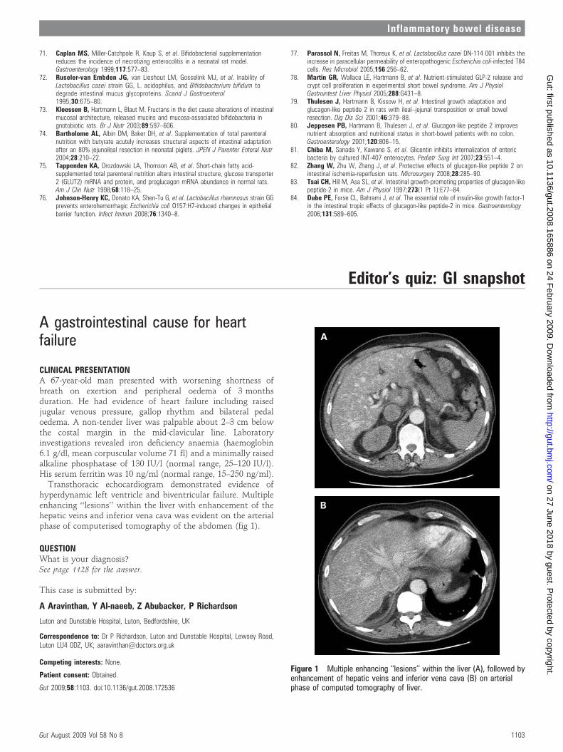
Transthoracic echocardiogram demonstrated evidence ofhyperdynamic left ventricle and biventricular failure. Multipleenhancing ‘‘lesions’’ within the liver with enhancement of thehepatic veins and inferior vena cava was evident on the arterialphase of computerised tomography of the abdomen (fig 1).
QUESTIONWhat is your diagnosis?See page 1128 for the answer.
This case is submitted by:
A Aravinthan, Y Al-naeeb, Z Abubacker, P Richardson
Luton and Dunstable Hospital, Luton, Bedfordshire, UK
Correspondence to: Dr P Richardson, Luton and Dunstable Hospital, Lewsey Road,Luton LU4 0DZ, UK; [email protected]
Competing interests: None.
Patient consent: Obtained.
Gut 2009;58:1103. doi:10.1136/gut.2008.172536
Figure 1 Multiple enhancing ‘‘lesions’’ within the liver (A), followed byenhancement of hepatic veins and inferior vena cava (B) on arterialphase of computed tomography of liver.
Editor’s quiz: GI snapshot
Inflammatory bowel disease
Gut August 2009 Vol 58 No 8 1103
on 27 June 2018 by guest. Protected by copyright.
http://gut.bmj.com
/G
ut: first published as 10.1136/gut.2008.165886 on 24 February 2009. D
ownloaded from