case report: radiation-induced small bowel “web”...
TRANSCRIPT

Case report: Radiation-induced small bowel “web” formation is associated with acquired microvascular dysfunction.
Ossama A. Hatoum 1, David G. Binion 2, Shane A. Philips 1, Colm O’Loughlin 2,
Richard A. Komorowski 3, David D. Gutterman 1#, Mary F. Otterson 4#
Division of Cardiovascular Medicine1, Division of Gastroenterology and Hepatology2, Department of Pathology3, Department of Surgery4, Froedtert Memorial Lutheran Hospital, and Zablocki VA Medical Center#, Medical College of Wisconsin, Milwaukee, WI, USA. Short title: Radiation induced microvascular dysfunction and intestinal webs Abbreviations used in this paper: reactive oxygen species (ROS), nitric oxide (NO), acetylcholine (Ach), dichlorofluorescein diacetate (DCF-DA), hydro-ethidene (HE), centigray (cGy), endothelial derived hyperpolarizing factor (EDHF), superoxide (O2
·-), megavoltage (MV), tumor-node-metastasis staging system (TNM). Correspondence to: Ossama A. Hatoum, M.D, MS, Division of Cardiovascular Medicine, Medical College of Wisconsin, Milwaukee, WI 53226, +1(414) 456-5626, Fax +1(414) 456 6572, e-Mail: [email protected] Key words: Radiation therapy, radiation toxicity, small bowel obstruction, microvascular dysfunction. Word count body: 2857 Abstract: 264
1
Gut Online First, published on August 26, 2005 as 10.1136/gut.2005.073734
Copyright Article author (or their employer) 2005. Produced by BMJ Publishing Group Ltd (& BSG) under licence.
on 3 Septem
ber 2018 by guest. Protected by copyright.
http://gut.bmj.com
/G
ut: first published as 10.1136/gut.2005.073734 on 26 August 2005. D
ownloaded from

Abstract: Background & Aims: Radiation therapy of abdominal and pelvic solid tumors results in late intestinal toxicity of a severe nature in approximately 5% of cases. These manifestations may include ischemia and stricture formation, which may present as “webs”. These webs are likely to play a role in the pathogenesis of recurrent bowel obstruction. The mechanisms of microvascular injury to the bowel in the setting of radiation have not been defined. We hypothesized that microvascular dysfunction with impaired vasodilation to acetylcholine (Ach) would be an acquired pathophysiologic abnormality in radiation and “web” formation. Methods: A 40-year-old patient treated with radiation, two years prior, for an anal squamous cell cancer presented with recurrent small bowel obstruction. “Webs” in the distal ileum were detected using wireless capsule endoscopy, after small bowel barium radiographs failed to demonstrate a lesion. Following resection, freshly isolated 50 – 150 micron diameter arterioles from the “web” and adjacent normal-caliber bowel were analyzed with histology and microvessel physiologic studies. Results: After constriction (30-50%) with endothelin, dilation to graded doses of Ach (10-9-10–4M) was observed in vessels dissected from the stricture and the adjacent normal-caliber area. Ach dilation was reduced in vessels from “web” (mean diameter (MD) = 7 ± 2%, n=3, p=<0.01 compared to the adjacent unaffected bowel MD = 85 ± 5%). Dihydroethidine and dichlorofluorescein diacetate (DCF-DA) intravital staining demonstrated increased reactive oxygen species (ROS) production in microvessels from “web” compared to adjacent normal-caliber bowel. Histology from the strictured bowel demonstrated narrowing of the arterial lumen due to intimal and muscularis propria fibrosis, with endothelial preservation. Conclusions: External radiation is associated with acquired microvascular endothelial dysfunction and “web” formation in the small bowel. Introduction Small intestine “webs” are increasingly appreciated to play a role in a spectrum of small bowel pathology, including occult gastrointestinal bleeding, enteropathy and intermittent partial small bowel obstruction. The prevalence of small bowel webs is not known, but autopsy series suggest that specific adult populations may demonstrate stricture related lesions in 1.5 – 9% of individuals (1-3). Late radiation injury of the small intestine may take years, with a median of about 8-12 months before the injury becomes apparent (4;5). The clinical signs and symptoms of chronic radiation intestinal injury may be multifactorial in etiology. The chief problems that may require major medical or surgical intervention are intestinal obstruction, severe bleeding, fistula formation, and intractable diarrhea (4-6). The radiologic picture of late radiation injury is of fibrosis and ischemia. These lesions are frequently missed in standard endoscopy and contrast studies of the upper and lower gastrointestinal tract. Capsule endoscopy is a new method that permits the endoscopic examination of the mucosa of the entire small bowel. The advent of wireless capsule endoscopy has increased diagnostic yield in patients with negative contrast
2
on 3 Septem
ber 2018 by guest. Protected by copyright.
http://gut.bmj.com
/G
ut: first published as 10.1136/gut.2005.073734 on 26 August 2005. D
ownloaded from

studies. Using this technique, mucosal “webs” may be found in review of the video study or by capsule retention. In this paper, we describe a case of radiation ileal “web”, diagnosed by capsule endoscopy, and its association with local, acquired microvascular dysfunction and impaired vasodilator capacity. Case report A 40-year-old female, with a medical history significant for external beam radiation treatment of a T2N0M0 squamous cell anal cancer was evaluated for symptoms of small bowel obstruction. She had received anal canal/inguinal node irradiation of 3060 cGy in 17 daily fractions (18MV x-rays) with reduced field anal irradiation 1980 cGy (18MV x-rays) and 1980 cGy bilateral inguinal node irradiation (9MeV electrons) in 11 fractions, completed 18 months prior. This brought the tumor dose to 5040 cGy. The patient received concurrent chemotherapy. Within 12 months of the radiation treatment, she experienced small bowel obstruction due to stricture formation and rectal sphincter damage requiring terminal ileal resection, primary anastomosis, and diverting sigmoid colostomy. During this procedure the remaining small intestine was carefully assessed for the presence of occult strictures using intraoperative passage and withdrawal of a balloon catheter inflated to a diameter of 2 to 2.5 cm through an enterotomy (7). Six months later, she again experienced symptoms of partial small bowel obstruction, which led to the current admission. Radiographic studies (plain radiographs, CT, small bowel barium radiographs) and endoscopic studies (upper endoscopy, stoma colonoscopy) were nondiagnostic. Wireless capsule enteroscopy (Figure 1A) resulted in capsule retention (Figure 1B) at an ileal web stricture (Figure 1C); felt to be the cause of her symptoms. The patient underwent exploratory laparotomy, during which two webs in a 25 cm ileal segment were identified and resected. The resected tissues, including the “webs” and the adjacent normal caliber intestine, were used for physiologic studies and pathologic analysis. Material and Methods The Medical College of Wisconsin’s Institutional Review Board approved all protocols. Mucosal gut arterioles were dissected from the full thickness specimen from both strictured and normal caliber small bowel. Following resection, tissue samples were preserved as reported previously (8). Microvessels physiology studies: Videomicroscopy was performed as previously reported (8). Briefly, isolated microvessels from the strictured and normal caliber small intestine were carefully dissected from the submucosal surface of the bowel tissue and transferred to a 20 ml organ chamber containing Krebs solution of the following composition (in mmol/L): NaCl 118, KCl 4.7, CaCl2 2.5, KH2PO4 1.2, MgSO4 1.2, NaHCO3 20, Na2EDTA 0.026, and glucose 11, pH 7.4. Each end of the arteriole was secured to a separate glass micropipette (25-50 µm internal diameter) filled with Krebs buffer (9) and transferred to the stage of an inverted microscope (CK2, Olympus) coupled to a CCD camera (WV-BL200, Panasonic) and video micrometer (VIA-100K, Boeckeler Instruments, Inc.).
3
on 3 Septem
ber 2018 by guest. Protected by copyright.
http://gut.bmj.com
/G
ut: first published as 10.1136/gut.2005.073734 on 26 August 2005. D
ownloaded from

Internal vascular diameters were measured throughout the experiment with a manually adjusted video-microscope as described previously (9). Micropipettes were connected to a hydrostatic reservoir at a final pressure of 60 mmHg. The chamber solution was continuously re-circulated at 30 ml/min, aerated with 20% O2, 5% CO2, and 75% N2, and warmed to 37o C by an external heat changer. All pharmacological agents were added to the external bathing solution. After a 60-min stabilization period, vessels were constricted to 30 - 50% of maximal diameter (at 60 mmHg) with administration of endothelin-1 (10-10 to 5x10⁻¹⁰ M). Vascular diameter responses to cumulative logarithmic increases in the concentration of Ach (10-9 to 10-4 M) in the external bathing media were examined. At the end of each experiment, passive vessel diameter was determined by adding papaverine (10-4 M). The addition of pharmacological agents produced less than 1% change in the volume of the circulating bath. All chemicals were obtained from Sigma Chemical Co. (St. Louis, MO). Fluorescence Detection of ROS: The cell-permeable dye dihydroethidine (HE, Molecular Probes, Eugene, OR)
was used to evaluate the production of superoxide (O2·-). In the presence of
superoxide, HE is oxidized to ethidium bromide, which remains trapped within the cell (10). Ethidium bromide fluorescence was excited by light at a 488-nm wavelength with an emission spectrum of 620 nm, and examined under confocal microscopy equipped with a krypton/argon laser fluorescence microscope (Nikon Eclipse, TE 200). A freshly isolated artery from normal-caliber gut was used as a control to adjust for laser settings. Those settings were maintained constant throughout the remained of the experiment. The fluorescence intensity/vascular area ratio of the central portion of the vessel was normalized to that obtained from the control vessel using the public domain NIH Image program (developed at the U.S. National Institutes of Health and available on the Internet at http://rsb.info.nih.gov/nih-image/). The ratio of the fluorescence intensity between experimental vessel and control vessel was compared. To evaluate the in situ production of all ROS (superoxide, hydrogen peroxide, and peroxynitrite), vessels were loaded with 5 X 10⁻⁶ M DCF-DA. DCF-DA oxidizes rapidly to the highly fluorescent 2’,7’-dichlorofluorescein in the presence of ROS (8;10) and was visualized on a fluorescence microscope. Captured digital images were analyzed as described above. Tissue histology and immunostaining: Paraffin embedded sections of the strictured and normal caliber intestine were examined under light microscopy with H & E staining. Presence of an intact endothelial monolayer was assessed using CD 34 antibody immunodetection (DakoCytomation, Carpinteria, CA, USA) with horseradish peroxide resolution. Materials: Endothelin-1 (Peninsula Laboratories, Inc, San Carlos, CA) was prepared in saline with 1% bovine serum albumin. Other agents were prepared in distilled water. Final molar concentrations of agents in the organ chambers are
4
on 3 Septem
ber 2018 by guest. Protected by copyright.
http://gut.bmj.com
/G
ut: first published as 10.1136/gut.2005.073734 on 26 August 2005. D
ownloaded from

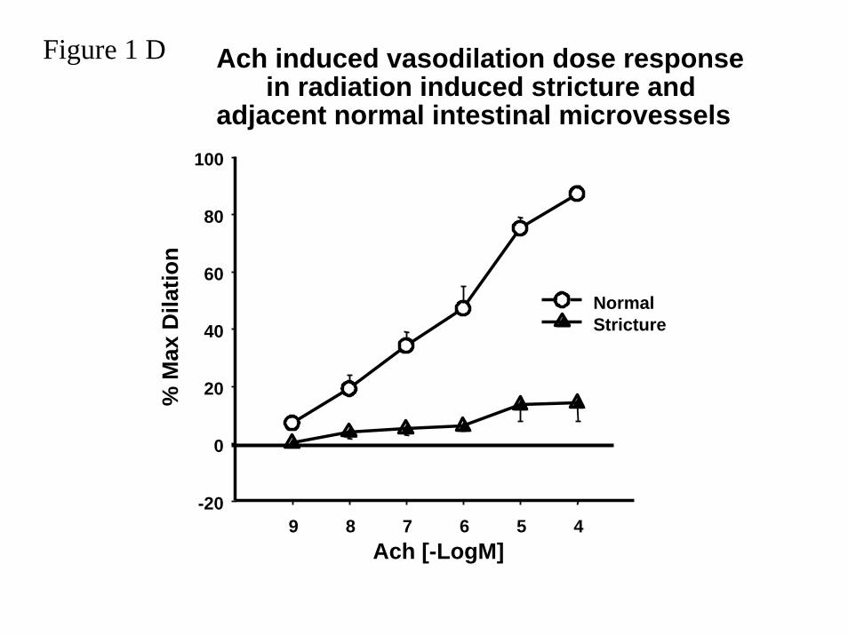
reported. None of the pharmacological antagonists produced significant changes in baseline microvessels diameter. Results Microvessels from the stricture and macroscopic intervening normal areas of small bowel were isolated and studied using physiologic assessment for endothelium dependent vasorelaxation in response to Ach (10-9-10–4M) following partial constriction with endothelin-1 using previously published protocols (8). Ach-induced vasodilation was reduced in vessels from two areas of intestinal web strictures (Max diameter (MD) = 7 ± 2%, n = 3) compared to the adjacent normal-caliber bowel microvessels (MD = 85 ± 5%, p = <0.01) (Figure 1D). Maximal dilation to papaverine was similar in vessels from the area of the web (86 ± 4.6%) compared to remote regions (89 ± 5.2%). Dihydroethidine and dichlorofluorescein diacetate (DCF-DA) staining were used to assess intravital production of ROS (8;10). Increased ROS were seen in microvessels isolated from previously irradiated area containing the stricture compared to vessels from the adjacent, preserved bowel (Figure 1E). Histology of the intestinal strictures demonstrated narrowing of the arteriolar lumen due to intimal thickening and muscularis propria fibrosis, which was not seen in the adjacent, preserved bowel (Figure 1 F). Immunostaining for CD34 demonstrated preserved endothelial anatomy in the strictured microvessels (Figure 1G)(11;12).
Discussion
We report the association of a local, acquired microvascular dysfunction with impaired vasodilator capacity and elevated ROS production from microvessels localized to the area of stricture formation and not from vessels isolated from adjacent histologically normal bowel segments. This association of microvascular dysfunction in areas of stricture formation in a patient with recurrent small bowel obstruction following radiation therapy is a novel insight into the pathophysiology of radiation enteritis. This patient presented with a two-year history of gastrointestinal dysfunction following successful radiation therapy for anal squamous cell carcinoma. The patient presented with two episodes of small bowel obstruction requiring resection within eighteen months of treatment. We speculate that the vascular dysfunction was localized to the endothelium since only impaired dilation to Ach (an endothelial-dependent vasodilator) and not to papaverine (an endothelial-independent vasodilator) was affected.
Radiation therapy is an effective modality for pelvic tumors, but may be associated with gastrointestinal complications. Radiation-induced complications in tissues have been carefully calculated defining dose-effect relationships (Nominal Standard Dose/Tolerance Dose). The minimal and maximal tolerance doses (TD5/5 and TD50/5) are 4500-6500 cGy, respectively, for human small bowel injury, describing 5 – 50% rates of injury within 5 years following exposure (13). Chronic radiation injury to the intestine contributes to fibrosis and vascular insufficiency (chronic ischemia) (4;14;15). The rates of radiation induced bowel adverse reactions range between 5-25% (4). Grossly,
5
on 3 Septem
ber 2018 by guest. Protected by copyright.
http://gut.bmj.com
/G
ut: first published as 10.1136/gut.2005.073734 on 26 August 2005. D
ownloaded from

the intestine with chronic radiation injury has been described as fibrotic with adhesions and a mottled appearance (4). The mucosa can appear pale and telangiectatic. Consistent with this report, the area of radiation injury is multifocal with relatively normal-appearing segments of bowel located immediately adjacent to the web. Taken together this line of evidence suggests radiation induced bowel injury is a localized vascular phenomenon (4;15). The most common portions of the intestine involved reflect the clinical application of radiation therapy and include cecum, terminal ileum, rectum, and distal sigmoid (16;17).
Vascular endothelium plays an essential role in the regulation of vascular tone and tissue perfusion through the release of nitric oxide (NO), prostacyclin, endothelial derived hyperpolarizing factor (EDHF), in response to physiological stimuli including Ach (8). Microvascular dysfunction has been identified in human disease states, including diabetes mellitus, hyperlipidemia and inflammatory bowel disease, and is characterized by impaired endothelium-dependent vasodilation (8). Interestingly, the chronic microscopic radiation induced changes are localized to submucosa. In contrast, the pathological effects of acute radiation induced injury are observed in the mucosal layer (15). The normal submucosa is characterized by atypical fibroblast and collagen proliferation. Small arteries show hyalinized thickening of the wall with intimal foam cell proliferation and recanalized fibrin thrombi within the lumen. The present study demonstrates that radiation induced bowel injury is also associated with an acquired local microvascular dysfunction which manifests as impaired vasodilator capacity. We hypothesize that this local microvascular dysfunction initiated by chronic elevations in ROS production following external beam ionizing radiation predisposes the bowel to ischemia and ultimately web formation. In addition, these data suggest that ROS formation is a therapeutic target in long-term radiation effects on bowel, potentially preventing the impaired perfusion and subsequent web formation. Further studies investigating the short and long-term effect of therapeutic radiation (and chemotherapy) on the human gut microvasculature and associated bowel complications are warranted.
6
on 3 Septem
ber 2018 by guest. Protected by copyright.
http://gut.bmj.com
/G
ut: first published as 10.1136/gut.2005.073734 on 26 August 2005. D
ownloaded from

Figure Legend: Microvascular dysfunction is associated with radiation enteritis induced intestinal strictures. Wireless capsule endoscopic appearance of radiated non-stenotic ileum (A); Abdominal radiograph demonstrating capsule retention (B); Capsule endoscopic appearance of an ileal “web” following radiation therapy (C); Ach-induced dilation of microvessels from radiation induced intestinal stricture and adjacent normal caliber intestine (D); ROS production from intestinal arterioles isolated from “web” and normal caliber bowel (E); Histology of radiation induced stricture (F); Endothelial staining with CD34 immunolocalization in the radiation induced intestinal web demonstrating preservation of endothelial layer in stricture microvessels (G). Consent from the patient regarding the publication of this case report and the images were obtained. Licence for Publication and Royalty Form: The Corresponding Author has the right to grant on behalf of all authors and does grant on behalf of all authors, an exclusive license on a worldwide basis to the BMJ Publishing Group Ltd and its Licensees to permit this article (if accepted) to be published in Gut editions and any other BMJPGL products to exploit all subsidiary rights, as set out in our licence (http://gut.bmjjournals.com/misc/ifora/licenceform.shtml)."
Beyond Conflict of Interest: As the corresponding author, I declare no competing interests in this manuscript.
Patient Images: Consent from the patient regarding the publication of this case report and the images was obtained.
Acknowledgments: We thank Professor John E. Moulder for his expertise in reviewing this manuscript and Heather Brandenburg for expert secretarial assistance.
Grants: This study was supported by a Career Development Award Grant from Crohn’s Colitis Foundation of America (OAH) and Alvin and Marion Birnschein Foundation (MFO+DGB).
7
on 3 Septem
ber 2018 by guest. Protected by copyright.
http://gut.bmj.com
/G
ut: first published as 10.1136/gut.2005.073734 on 26 August 2005. D
ownloaded from
Reference List (1) Allison MC, Howatson AG, Torrance CJ et al. Gastrointestinal damage
associated with the use of nonsteroidal antiinflammatory drugs. N Engl J Med 1992;327(11):749-54.
(2) Bjarnason I, Hayllar J, MacPherson AJ et al. Side effects of nonsteroidal anti-inflammatory drugs on the small and large intestine in humans. Gastroenterology 1993;104(6):1832-47.
(3) Lang J, Price AB, Levi AJ et al. Diaphragm disease: pathology of disease of the small intestine induced by non-steroidal anti-inflammatory drugs. J Clin Pathol 1988;41(5):516-26.
(4) Coia LR, Myerson RJ, Tepper JE. Late effects of radiation therapy on the gastrointestinal tract. Int J Radiat Oncol Biol Phys 1995;31(5):1213-36.
(5) Cox JD, Byhardt RW, Wilson JF et al. Complications of radiation therapy and factors in their prevention. World J Surg 1986;10(2):171-88.
(6) Lucarotti ME, Mountford RA, Bartolo DC. Surgical management of intestinal radiation injury. Dis Colon Rectum 1991;34(10):865-9.
(7) Otterson MF, Lundeen SJ, Spinelli KS et al. Radiographic underestimation of small bowel stricturing Crohn's disease: a comparison with surgical findings. Surgery 2004;136(4):854-60.
(8) Hatoum OA, Binion DG, Otterson MF et al. Acquired microvascular dysfunction in inflammatory bowel disease: Loss of nitric oxide-mediated vasodilation. Gastroenterology 2003;125(1):58-69.
(9) Miura H, Gutterman DD. Human coronary arteriolar dilation to arachidonic acid depends on cytochrome P-450 monooxygenase and Ca2+-activated K+ channels. Circ Res 1998;83(5):501-7.
(10) Benov L, Sztejnberg L, Fridovich I. Critical evaluation of the use of hydroethidine as a measure of superoxide anion radical. Free Radic Biol Med 1998;25(7):826-31.
(11) Hristov M, Erl W, Weber PC. Endothelial progenitor cells: mobilization, differentiation, and homing. Arterioscler Thromb Vasc Biol 2003;23(7):1185-9.
(12) Hristov M, Erl W, Weber PC. Endothelial progenitor cells: isolation and characterization. Trends Cardiovasc Med 2003;13(5):201-6.
(13) Rubin P, Casarett GW. Concepts of Clinical Radiation Pathology. In: Dalrymple GV, Gaulden ME, Kollmorgen GM, Vogel HH, editors. Medical Radiation Biology. Philadelphia: W.B. Saunders Company, 1973: 160-89.
8
on 3 Septem
ber 2018 by guest. Protected by copyright.
http://gut.bmj.com
/G
ut: first published as 10.1136/gut.2005.073734 on 26 August 2005. D
ownloaded from

(14) Berthrong M. Pathologic changes secondary to radiation. World J Surg 1986;10(2):155-70.
(15) Fajardo LF, Berthrong M. Vascular lesions following radiation. Pathol Annu 1988;23 Pt 1:297-330.
(16) BRICK IB. Effects of million volt irradiation on the gastrointestinal tract. AMA Arch Intern Med 1955;96(1):26-31.
(17) Green N, Iba G, Smith WR. Measures to minimize small intestine injury in the irradiated pelvis. Cancer 1975;35(6):1633-40.
9
on 3 Septem
ber 2018 by guest. Protected by copyright.
http://gut.bmj.com
/G
ut: first published as 10.1136/gut.2005.073734 on 26 August 2005. D
ownloaded from

Figure 1 A
on 3 September 2018 by guest. Protected by copyright. http://gut.bmj.com/ Gut: first published as 10.1136/gut.2005.073734 on 26 August 2005. Downloaded from

Figure 1 B
on 3 September 2018 by guest. Protected by copyright. http://gut.bmj.com/ Gut: first published as 10.1136/gut.2005.073734 on 26 August 2005. Downloaded from

Figure 1 C
on 3 September 2018 by guest. Protected by copyright. http://gut.bmj.com/ Gut: first published as 10.1136/gut.2005.073734 on 26 August 2005. Downloaded from
Ach induced vasodilation dose responsein radiation induced stricture and
adjacent normal intestinal microvessels
Ach [-LogM]9 8 7 6 5 4
% M
ax D
ilatio
n
-20
0
20
40
60
80
100
NormalStricture
Figure 1 D
on 3 September 2018 by guest. Protected by copyright. http://gut.bmj.com/ Gut: first published as 10.1136/gut.2005.073734 on 26 August 2005. Downloaded from

Relative production of ROS in normal and strictured intestinal microvessles
in radiation enteritis
Reactive Oxygen Species (ROS) production from isolated microvessels
O2.- production
stricture
H2O2 production
Arb
itrar
y U
nits
0.0
0.5
1.0
1.5
2.0
Figure 1 E
stricturenormal normal
on 3 September 2018 by guest. Protected by copyright. http://gut.bmj.com/ Gut: first published as 10.1136/gut.2005.073734 on 26 August 2005. Downloaded from

Figure 1 F
on 3 September 2018 by guest. Protected by copyright. http://gut.bmj.com/ Gut: first published as 10.1136/gut.2005.073734 on 26 August 2005. Downloaded from
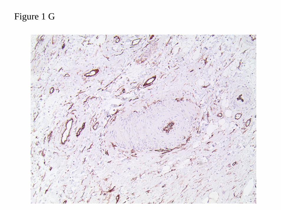
Figure 1 G
on 3 September 2018 by guest. Protected by copyright. http://gut.bmj.com/ Gut: first published as 10.1136/gut.2005.073734 on 26 August 2005. Downloaded from