radiological investigation the small …gut.bmj.com/content/gutjnl/1/4/366.full.pdf · into the...
TRANSCRIPT

Gut, 1960, 1, 366.
RADIOLOGICAL INVESTIGATION OFTHE SMALL INTESTINE BY SMALL BOWEL
ENEMA TECHNIQUEBY
F. PYGOTT, D. F. STREET, M. F. SHELLSHEAR, and C. J. RHODES
From the Central Middlesex Hospital, London
This paper records some experience with the Scott Harden technique of small intestinalintubation for performing small bowel enemas. It is particularly recommended for searchingfor obstructive or infiltrative lesions of the small intestine.
A completely satisfactory demonstration of theentire small bowel by the usual barium follow-through series, or one of its many modifications, isoften not attained in the normal subject, anddifficulties are intensified in the presence ofpathology. Individual loops of bowel may fill andempty rapidly, and, however short the intervalsbetween examinations, these loops may not beshown effectively. Gross delay in the onward pas-sage of barium may frustrate the efforts of theradiologist to show the exact site and probablenature of an obstructing lesion, or several intestinalloops may overlap and so obscure a lesion; this isparticularly true of ileal loops lying in the pelvis.Since the radiologist is unable to affect the degreeof filling attained in the region under immediatescrutiny, the significance of local constrictions ordilatations observed at any one examination in theseries is obscure and it may not be possible to detectthe changes later. Local destructive processes in themucous membrane may be quite unrecognizable.
Dissatisfaction with the barium follow-throughexamination led Schatzki (1943) to adopt the smallbowel enema technique of duodenal intubation,using a small rubber tube and controlled filling ofthe small intestine, that had been recommendedearlier by Pesquera (1929). The disadvantages ofthis method are the uncertain delay during whichthe tube is awaiting its passage from the stomachinto the duodenum and the high failure rate becausethe tube cannot be passed in reasonable time.Immediate transgastric intubation with a flexiblepolythene tube does not impose delay and the suc-cess rate is much higher. The technique that we haveused is essentially that described by Scott Harden(1960) with minor modifications, though some earlycases were examined by a single tube method.
DESCRIPTION OF THE METHODThe equipment required consists of a flexible, but
semi-rigid, polythene tube, 4 mm. inside diameter(5.8 mm. outside diameter) about 1 metre long, throughwhich a second polythene tube of 15 mm. insidediameter, (2.7 mm. outside diameter), about 14 metreslong, can slide easily. The distal end of each tubehas a small brass ring or ferrule bonded to it sothat its position can be identified radiologically.We have found that a short, tightly fitting 3- or 4-in.length of flexible polythene tube sheathed over the distalend of the outer semi-rigid tube so that it projects2 or 3 in. beyond the tip is an advantage. The tubing issterilized by boiling and care is taken to avoid coilingor pinching the tubes in removing them from thesterilizer. A connexion is arranged with the proximalend of the inner tube so that a suspension of "micro-paque", 1: 1 part of tap water, can be injected throughit from a 50 ml. syringe.The patient is prepared as for a barium meal examina-
tion. The object of the examination and the method areclearly explained, Nearly all patients have had a bariummeal beforehand and abnormalities of the stomach andduodenal cap should be excluded before this techniqueis employed. The patient is then given a tablet ofamethocaine to suck and the fauces are lightly sprayedwith 2% xylocaine.The tubing has been carefully examined beforehand
for patency and free movement of the inner on the outeris assured. The inner tube is passed down the outer untilits distal end is just short of that of the outer. Anassistant holds the tubing while the radiologist passesthe distal end gently through the patient's mouth overthe epiglottis into the hypopharynx. The tube shouldprogress easily with gentle pressure once the cricopharyn-geus has been passed. Progress is continued until asense of elastic resistance is felt, due to the tube impactingagainst the greater curve of the stomach. The tube isthen withdrawn slightly and the patient is placed supineon the x-ray couch. We use a Marconi type 5-in. imageamplifier for the examination, and take under and over
366
on 27 August 2018 by guest. P
rotected by copyright.http://gut.bm
j.com/
Gut: first published as 10.1136/gut.1.4.366 on 1 D
ecember 1960. D
ownloaded from

NEW METHODS FOR DIAGNOSIS AND RESEARCH 367
couch large films as required. (The actual screeningfactors used are 90-95 kV. with 0.3 mA.) When radio-logical examination has identified the position of thelower ends of the tubes, then the inner tube is carefullyadvanced through the outer until it engages the pylorusand is slipped through it as far as possible. Coiling of theinner tube within the stomach can often be avoided byrotating the patient to the right and by carefully ad-vancing the outer tube over the inner immediately thelatter shows any tendency to turn upwards. In a veryfew cases the duodenum cannot be intubated in spite ofevery effort, usually in grossly distorted stomachs witha severe cup and spill shape. As with all practical pro- Al
cedures increased experience brings increased facility andfewer failures. In the 21 months we have been using thetechnique we have attempted 73 examinations with fourcomplete failures, one more patient refused all co-operation before trying, and in one more intubation waseffected but the examination was not entirely satisfactory.The 67 patients in whom the examination was satisfactorygive a success rate of, at worst, 90%, and this may wellbe improved upon.When the tip of the inner tube has been introduced
into the duodenum a little of the barium suspension issteadily injected under radiological control. Back flowinto the stomach does not normally occur even if the tip FIG. 1.-Normal appearances ofjejunum and upper ileumis only in the duodenal cap. An early film is taken to with small bowel enema technique. The position of theshow the duodenal loop and the uppermost loops of lower end of the inner tube is arrowed, the lower endjejunum filled. Successive injections of barium sus- of the outer is at the cardia.pension and tap water are made as required and theprogress of the contrast medium observed, controlledfilling of the intestine is attainable, and in the normalsubject the caecum is reached in 15 to 20 minutes. Inthe abnormal case irregularities in the lumen can beexamined continuously with different degrees of dis-tension and the area can be re-examined by washing itthrough with water and refilling with barium.Delay in forward flow indicates the presence of an
obstructive lesion ahead and discretion must be usedin avoiding proximal overfilling; nevertheless, withpatience it is nearly always possible to identify the exact ._site of any kinking or narrowing of the lumen in a waythat is rarely possible during a follow-through examina-tion. In infiltrative lesions, such as Crohn's disease, itis possible to form a much more accurate impressionof the extent of involvement as the rigidity of the un-constricted bowel can be assessed because its tubularstiffening can be recognized since filling can be madecomplete. Also local areas of narrowing can berecognized and their existence confirmed.
ILLUSTRATIVE CASESFigs. 1 and 2 show the appearance of the small bowel
in a normal subject, as demonstrated by this method._
CASE L.-A woman aged 29 years gave a history ef11 years' diarrhoea, the bowels opening seven to 20 timesdaily, and periodic pain in the right iliac fossa. Theonset of symptoms followed appendicectomy for pain in FIG. 2.-The whole of the small bowel, except the duo-the right iliac fossa. Her stools were watery, green at denal loop, is shown. The proximal colon is filled. Thetimes, and less often had blood in them. On examination lower end of the inner tube is in the duodenal cap; therea small mass was felt in the right iliac fossa. is some back leak into the stomach.
on 27 August 2018 by guest. P
rotected by copyright.http://gut.bm
j.com/
Gut: first published as 10.1136/gut.1.4.366 on 1 D
ecember 1960. D
ownloaded from
368 F. PYGOTT, D. F. STREET, M. F. SHELLSHEAR, AND C. J. RHODES
_* L sright psoas abscess was present. (There was no radio-_I b iB __logical evidence of lumbar or dorsal spine disease). Many
calcified and partially calcified glands were present.The distal ileum and proximal colon were resected withgreat difficulty, and the descending duodenum was un-avoidably opened in separating the ascending colon from
_ it. Unfortunately, the patient died eight days later.Necropsy confirmed the absence of any focus of
disease in the spine and the psoas abscess was due todirect extension from the caecal lesion. Histology of theresected specimen showed dense fibrotic changes but noevidence of any active tuberculous lesion; granulomatousand partially calcified tuberculous disease was present inthe lymph glands.
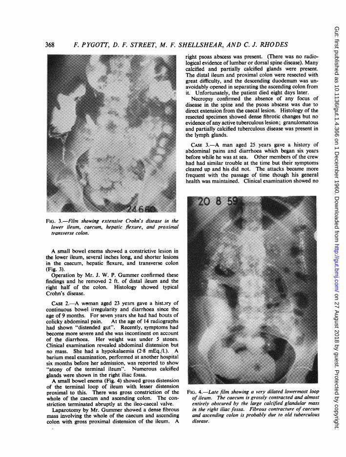
-~~~~~~~~~~~~~~~~~~~~~~~~~~~~~~~~~~~~~~~~~~~~~~~~~~~M. .....''. CASE 3.-A man aged 25 years gave a history of-M. : :abdominal pains and diarrhoea which began six years
before while he was at sea. Other members of the crewhad had similar trouble at the time but their symptoms
/ ~~~~~cleared up and his did not. The attacks became morer * jqgfrequent with the passage of time though his general
health was maintained Clinical examination showed no
FiG. 3-Film showing extensive Crohn's disease in the -.lower ileum, caecutm, hepatic flexure, and proximaltransverse colon.
A small bowel enema showed a constrictive lesion inthe lower ileum, several inches long, and shorter lesionsin the caecum, hepatic fiexure, and transverse colon(Fig. 3).
Operation by Mr. J. W. P. Gummer confirmed thesefindings and he removed 2 ft. of distal ileum and theright half of the colon. Histology showed typicalCrohn's disease.
CASE 2.-A woman aged 23 years gave a history ofcontinuous bowel irregularity and diarrhoea since theage of 9 months. For seven years she had had bouts ofcolicky abdominal pain. At the age of 14 radiographshad shown "distended gut". Recently, symptoms hadbecome more severe and she was incontinent on accountof the diarrhoea. Her weight was under 5 stones.Clinical examination revealed abdominal distension butno mass. She had a hypokalaemia (2-8 mEq./l.). Abarium meal examination, performed at another hospitalsix months before her admission, was reported to show -"atony of the terminal il-um". Numerous calcifiedglands were shown in the right iliac fossa.A small bowel enema (Fig. 4) showed gross distension
of the terminal loop of ileum with lesser distensionproximal to this. There was gross constriction of the FiG. 4. Late film showing a very dilated lowermost loopwhole of the caecum and ascending colon. The con- of ileum. The caecum is grossly contracted and almoststriction terminated abruptly at the ileo-caecal valve. entirely obscured by the large calcified glandular massLaparotomy by Mr. Gummer showed a dense fibrous in the right iliac fossa. Fibrous contracture of caecum
mass involving the whole of the caecum and ascending and ascending colon is probably due to old tuberculouscolon with gross proximal distension of the ileum. A disease.
1
on 27 August 2018 by guest. P
rotected by copyright.http://gut.bm
j.com/
Gut: first published as 10.1136/gut.1.4.366 on 1 D
ecember 1960. D
ownloaded from

NEW METHODS FOR DIAGNOSIS IN RESEARCH
FIGs. 5 AND 6.-Film (single tube method) showing multiple strictures in lower ileum. Histologically,necrotizing ileitis or ? Crohn's disease.
Fig. 5 shows the normality of the upper small bowel, and Fig. 6 five or six of the strictures.
abnormality except for tenderness in the right iliac fossa.Repeated barium meals had shown some duodenal capdeformity. A small bowel enema (Figs. 5 and 6) showednumerous very localizAd constrictions in the lower ileumwith intervening normal bowel. The mucosal patternwas locally destroyed. Laparotomy by Mr. Gummershowed numerous areas of stricture in the lower ileumassociated with regional glandular enlargement. Oneof the areas was excised and histology revealed simplemucosal ulceration with necrotic areas and granulations.Dr. R. Drury reported that the appearances, though nottypical, could be due to Crohn's disease. Ultimately,4 ft. of ileum had to be resected and a right hemi-colectomy be performed at a subsequent operation aftera spontaneous perforation at one of the sites ofconstriction.
APPLICATIONS OF THE METHODSuccess in this investigation depends on full
cooperation on the part of the patient, who must besufficiently ambulant to be able to submit to asomewhat rigorous examination. Many will havehad one or several barium meal examinations andwe like to have the results of these available. Ab-normalities in the stomach and duodenal cap must8
have been excluded and films showing the outlineof the stomach help in indicating the probable easeor difficulty of intubation.
Since the examination is more anatomical thanphysiological in character we prefer not to employit as a primary examination in patients presentingwith steatorrhoea, though we do not hesitate to useit secondarily in such patients. In spite of whathas just been said, characteristic steatorrhoea-typeappearances are produced when the method isapplied to patients with an idiopathic or pancreaticsteatorrhoea.The method is very valuable in patients presenting
with abdominal pain and diarrhoea in whom anobstructive or constrictive lesion of the small bowelis likely to be found. In patients who are knownto have Crohn's disease and who have had one ormore operations, sometimes of unknown nature, itis most helpful in giving the surgeon as accuratean impression as possible of the state of affairs hemay have to face before he undertakes a difficultlaparotomy. The presence of "blind loops" may bemore than suspected. In investigating the smallbowel for the source of occult bleeding it is, in our
369
on 27 August 2018 by guest. P
rotected by copyright.http://gut.bm
j.com/
Gut: first published as 10.1136/gut.1.4.366 on 1 D
ecember 1960. D
ownloaded from

370 F. PYGOTT, D. F. STREET, M. F. SHELLSHEAR, AND C. J. RHODES
opinion, the most effective radiological method.In conclusion, we recommend this investigation
when it can be used with care and discretion inthe search for obstructive or infiltrative lesions ofthe small bowel or where occult bleeding is likelyto be coming from a lesion below the duodenal cap.We would stress that it is not to be undertaken as aroutine but its application is most advantageous inthe circumstances indicated.
We are obliged to our clinical colleagues, especiallyDr. F. Avery Jones and Mr. J. W. P. Gummer, for accessto their findings and to Dr. R. Drury for his pathologicalreports on the cases quoted.
REFERENCESHarden, W. G. Scott (1960). In Modern Trends in Diagnostic
Radiology, (Third Series), ed. Dr. J. W. McLaren, p. 84.Buttcrworths, London.
Pesquera, G. S. (1929). Amer. J. Roentgenol, 22, 254.Schatzki, R. (1943). Ibid., 50, 743.
on 27 August 2018 by guest. P
rotected by copyright.http://gut.bm
j.com/
Gut: first published as 10.1136/gut.1.4.366 on 1 D
ecember 1960. D
ownloaded from