chapter 50: neural signalling reflex arc monosynaptic e.g ... · chapter 50: neural signalling...
TRANSCRIPT

Chapter 50: Neural Signalling
Reflex Arc
Monosynaptic e.g., patella reflex
Polysynaptic - most common reflexes to pain heat, etc.

I. Input
A. All stimuli represent forms of energy
1. Sensation
a. involves converting energy into a change in the membrane potential of sensory receptors
b. action potentials reach the CNS via sensory neurons
2. Perception
a. The brain interprets sensations, giving the perception of stimuli
II. Receptors - Functions of sensory pathways involve sensory reception, transduction, transmission, and integration
A. Sensory receptors transduce stimulus energy and transmit signals to the nervous system
1. Conversion of stimulus energy (See 1 below) into a change in membrane potential.
a. Receptor potential: a sensory receptor’s version of a graded potential (see 2 below).
2. Transmission - the conduction of sensory impulses to the CNS.
a. Some sensory receptors must transmit chemical signals to sensory neurons.
b. The strength of the stimulus and receptor potential affects the amount of neurotransmitter
released by the sensory receptor
c. Some sensory receptors are sensory neurons (as in example below)
d. The intensity of the receptor potential affects the frequency of action potentials (See 4 below)
3. Integration - the processing of sensory information.
a. Begins at the sensory receptor.
1. Amplification is the strengthening of stimulus energy by cells in sensory pathways
2. Sensory adaptation is a decrease in responsiveness to continued stimulation
b. Brain interprets information = Perception (see 4 above)
B. types of sensory receptors
1. Based on location:
a. Exteroreceptors detect stimuli originating outside the body.
b. Interoreceptors detect stimuli originating inside the body.
2. Based on type of stimulus
a. Pain receptors – pain
b. Thermoreceptors – temperature
c. Mechanoreceptors
1. touch, pressure
2. baroreceptors
3. vibration
4. proprioception - stretch receptors
d. chemoreceptors
1. taste
2. smell
3 internal chemical conditions
a. pH
b. PO2
c. PCO2
e. Electomagnetic receptors

C. General senses
1. Body Surface – more than just the sense of touch!
a. free nerve endings
1. simplest receptors
2. no actual receptor – endings of sensory neuron stimulated directly
a. pain (nocioceptors)
b. some forms of mechanoreception – coils around hair follicle
b. encapsulated receptors
1. Meissner’s corpuscle (“Gentle Touch” above)
a. responds to vibrations of low frequency
2. bulb of Krause (“Cold” above) thermoreceptor
a. activated at temp below 20C (68F)
b. at 10C gives stinging sensation of freezing
3. Ruffini’s end-organ (“Heat” above)
a. touch pressure
b. temp above 45C(113F)
4. Pacinian corpuscles (“Strong Presure” above)
a. widely distributed in dermis of skin, moveable joints and some
internal organs
b. responds to pressure changes
D. Pain
1. free nerve endings send information to the thalamus alerting to damage
2. thalamus relays information to the cerebrum for interpretation
3. Somatic pain – receptors in skin, muscles, tendons and ligaments and joints
4. Visceral pain - receptors associated with internal organs relay information about abnormal
conditions within these organs
a. changes in chemistry
b. changes in pressure
c. changes in blood flow
d. muscle spasms
e. referred pain

D. Special Sensors – special; sense organs
1. taste (gustatation)
a. Chemoreception
1. Substances dissolved in water
b. In humans taste receptors located primarily on tongue
c. Taste Receptor (Taste Buds)
1. sit in an circular pit called the papilla
a. Embedded in lining
b. Connected to surface by pore
c. Receptor cells connected to sensory nerve
e. Info relayed to thalamus and on to cerebrum and Limbic System
2. types of taste receptors
a. salt
b. sweet
c. sour
d. bitter
e. umami (Japanese word that translates best as “savory”)
1. stimulates glutamate receptors
2. MSG
3. Taste –transduction of a “sweet” receptor
1.Sweet, , umami and bitter use same mechanism
2. G protein coupled receptors (GPCR) 1 each for sweet & umami; 30 different for bitter)

2. smell (olfaction)
a. Chemoreception
1. Particles that are air borne.
2. “cilia” of receptor cells protrude from the surface of the nasal epithelium
a. Actually terminal part of olfactory neuron
b. The binding of odor molecules to olfactory receptors initiate signal
transduction pathways involving a G-protein-signaling pathway and, often,
adenylyl (adenylate) cyclase and cyclic AMP
c. info relayed to olfactory bulb
d. from there to cerebrum by way of the olfactory nerve and Limbic System
3. Ear – hearing and balance
a. anatomy

1. external ear
a. pinna
b. external auditory meatus
c. external auditory canal
d. tympanic membrane or tympanum
e. ceruminous glands – cerumen = ear wax
2. middle ear
a. air filled
b. communicates with nasopharynx by means of the Eustacian tube
c. auditory ossicles – three (characteristic of mammals)
1. malleus (hammer) attached to tympanic membrane)
2. incus (anvil) – attached to malleus
3. stapes (stirrup) – attached to incus and oval window of cochlea of
inner ear
4. function – conduct and amplify sound waves from tympanic
membrane to oval window
5. evolutionary origin – jaw bones of ancestral fish
a. malleus from articular bone
b. incus from quadrate bone
c. stapes from hyomandibular bone
3. inner ear – location of receptors for hearing and equilibrium
a. three divisions – two deal with equilibrium the other with hearing
4. All three divisions work using a similar system for stimulus discrimination
a. Sensory cell termed the ‘hair cell”
b. located within a sac or tube
c. sac or tube filled with a fluid called endolymph (perilymph)
5. stimulus is a mechanical bending of the hair cells that results from the movement of
endolymph
6. Hair cells release an excitatory neurotransmitter even when hair cells not bending
7. Constant action potential produced in accompanying sensory( figure a below)
8. So how does the nervous system discriminate that the hair cell has been stimulated?
a. when hair cells bend, rate of release of neurotransmitter is altered (figures b and
c above)
b. It is necessary to discriminate the direction of the movement endolymph
(distortion of the hair cells).
1. distortion in one direction will increase the amount of excitatory
neurotransmitter (figure b above) released and, therefore, the frequency
of action potentials in the sensory neuron will increase.
2. distortion in the opposite direction will decrease the amount of
excitatory neurotransmitter (figure c above) released and, therefore, the
frequency of action potentials in the sensory neuron will decrease.

1. Equilibrium requires an input of information about body’s position relative to the
gravitational field and to the bodies velocity and acceleration (the original function of the ear)
a. vestibule – position and acceleration
1. contains two membranous sacs, the saccule and the utricle
2. hair cells clustered in oval shaped sacs
3. sacs contain crystals of calcium carbonate = otoliths
4. otoliths press on hair cells – respond to gravity – tell us what our
position is relative to gravitational field
5. when body accelerates otoliths move
b. semicircular canals – rotational movement and acceleration
1. three ducts –measure three planes
2. hair cells contained in clusters (cupula) which floats in endolymph
3. when fluid moves because of head movement in that plane, cupula \
distorted and this stimulates nerves
c. collectively called the vestibular apparatus

4. Hearing - cochlea
a. oval window – connected to stapes
b. round window
c. cochlea
1. long and coiled – “snail”
2. three subsections
a. vestibular duct (scala vestibuli)
b. tympanic duct (scala tympani)
c. cochlear duct (scala media
d. cochlear duct and tympanic duct and separated by
basilar membrane
3. hair cells (acoustical receptors) found in organ of Corti which
sits on basilar membrane within the cochlear duct.
4. tectorial membrane lies above hair cells closest to wall of
vestibular duct

d. hearing
1. sound is a vibration of air particles that travel in sine waves –
two measurements of sound waves
a. amplitude = loudness (decibels)
b. frequency (pitch)= wave cycles/second which is a
function of the distance between the peaks (or troughs)
adjacent waves (cycles/second or Hz)
2. sound transmission and reception
a. sound waves reach tympanic membrane
b. membrane vibrates at set frequency and amplitude
determined by the pitch and loudness of the sound
c. membrane causes ossicles to vibrate
1. muscles attached to stapes and malleus can
tighten membrane and ossicles to dampen
amplitude of very loud noises
d. when stapes vibrate, sets up a vibration in the oval
window of cochlea
e. vibration of oval window sends a pressure wave along
the endolymph (perilymph) that fills the vestibular and
tympanic ducts.
f. pressure wave distorts basilar membrane of organ of
Corti
g. distortion bends hair cells against tectorial membrane
stimulating the hair cells
1. first bending them down and causing an
increased neurotransmitter release
2. then membrane pulls hairs up and causing a
decreased release of neurotransmitter.
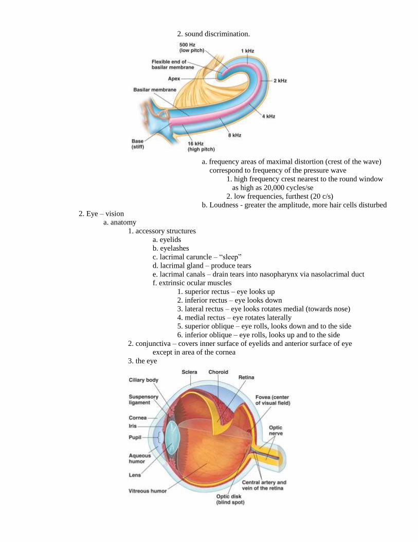
2. sound discrimination.
a. frequency areas of maximal distortion (crest of the wave)
correspond to frequency of the pressure wave
1. high frequency crest nearest to the round window
as high as 20,000 cycles/se
2. low frequencies, furthest (20 c/s)
b. Loudness - greater the amplitude, more hair cells disturbed
2. Eye – vision
a. anatomy
1. accessory structures
a. eyelids
b. eyelashes
c. lacrimal caruncle – “sleep”
d. lacrimal gland – produce tears
e. lacrimal canals – drain tears into nasopharynx via nasolacrimal duct
f. extrinsic ocular muscles
1. superior rectus – eye looks up
2. inferior rectus – eye looks down
3. lateral rectus – eye looks rotates medial (towards nose)
4. medial rectus – eye rotates laterally
5. superior oblique – eye rolls, looks down and to the side
6. inferior oblique – eye rolls, looks up and to the side
2. conjunctiva – covers inner surface of eyelids and anterior surface of eye
except in area of the cornea
3. the eye

a. mostly a hollow ball – wall consists of three layers
1. outer layer (fibrous tunic)
a. sclera – white of the eye
b. cornea – raised, transparent
2. middle layer (vascular tunic)
a. anteriorly behind cornea
1. iris – pigmented (melanin) – distribution of
pigments give iris its characteristic color
2. intrinsic eye muscles
a. pupilary constrictor muscle
b. pupilary dilator muscle
c. cilliary body
1. ciliary muscle
2. suspensory ligaments
3. lens
4. anterior cavity formed by space between
cornea and lens/ciliary body – filled with
fluid – aqueous humor
b. remainder of middle layer = choroid
1. contains extensive capillary network that delivers
oxygen and nutrients to retina
2. pigment layer
3. inner layer (sensory tunic) = retina; lies below choroid
a. neural retina – contains photoreceptors and
associated neurons
b. optical disc – blind spot – site of pathway for optic
nerve and blood vessels that supply blood to retina and
chorion
c. fovea – area of greatest visual acuity
4. main chamber of eye = posterior cavity – filled with vitreous
humor – thick and gelatinous
4. visual accomodation
a. lenses of eye

1. the cornea
a. light coming through is focused towards the interior
b. cornea’s shape is fixed (can’t be adjusted)
2. the lens
a. light from cornea then passes through lens
b. lens focuses light onto the retina
1. some vertebrates can move their lens back and
forth (like adjusting the lens of a camera
2. others like ourselves change the shape of their
lens (like changing lenses on a camera)
a. ciliary muscle relaxed – lens flattens
moving focal point farther back (distance)
b. ciliary muscle contracted – lens bulges
moving focal point forward (near vision)
3. image focused upside down on retina
4. myopia - focal point of image of distant objects in
front of retina even when lens rounded (nearsighted)
5. hyperopia – focal point beyond retina; must focus
just to see far objects (farsighted)
3. retina
a. cell types
1. photoreceptors
a. rods – black and white vision; dim light;
low acuity; approximately 125 million.
b. cones - color vision – bright light; high
acuity; approximately 6 million
2. neurons

Vertical Pathway a. bipolar cells – associated with photoreceptors
b. ganglion cells – associated with optic nerve
c. amacrine cells – interconnect across
bipolar/ganglion cell synapses
d. horizontal cells– interconnect across
Horizontal Pathway photoreceptor/bipolar cell synapses
1. along with amacrine cells responsible for lateral
integration/inhibition
3. interrelationship between receptors, bipolar and ganglion cells
a. usually not 1:1
1. 125 million photoreceptors:1 million ganglion cells
2. each bipolar cell communicates with many
receptor cells
3. each ganglion cell communicates with many
bipolar cells
b. exception - the fovea
1. area of closely packed cone cells (150,000/mm2)
2. 1:1 connection
3. area of greatest resolution – acuity
b. photoreception
1. cell must be able to detect light
a. visual pigments
1. combinations of rhodopsin derivatives
a. opsin – protein (variable) (4 types)
b. retinal – Vitamin A derivative
(common to all visual pigments)
2. located in stacks of membranous disks in rods
and cones

a. photoreceptors have voltage gated sodium channels
open in absence of light (-40mV potential)
b. neurotransmitter constantly being released
c. photon of light strikes retinal part of rhodopsin
d. activates opsin which acts as an enzyme
e. triggers transduction events that close sodium
channels
f. hyperpolarize receptor membrane (-70 mV potential)
g. neurotransmitter stops being released signaling
that receptor has absorbed light
h. rhodopsin molecule broken down into its opsin
and retinal = “bleaching”
i. rhodopsin molecule then has to be resynthesized

2. rods
a. rhodopsin sensitive to all wavelengths of light except red
b. this combined with the fact that many rods “wired”
together means that rods respond more easily to dim light
3. cones
a. rhodopsins of cones respond to only certain wavelengths of
light
b. three types of cones
1. blue cones (16%)
2. red cones (74%)
3. green cones (10%)
c. color blindness – one or more types of cones nonfunctional
d. processing visual information takes place at several levels
1. retina itself - receptive fields process certain fields
2. lateral geniculate nuclei
a. visual signals from one eye split at optic chiasm sending half their fibers to:
1. the lateral geniculate nucleus on the same side of the brain as the eye
2. the lateral geniculate nucleus on the opposite side of the brain (remember the
split brain studies)
3. lateral geniculate nuclei send information to:
a. visual cortex of cerebrum
1. each receives and sends a slightly different image
2. brain assemble images into an image conveying depth
3. visual cortex has a visual map like those seen in the somatosensory and
somatic motor cortices
a. unequal distribution 35X greater area devoted to fovea and area around
fovea (macula lutea - yellow spot with no rods)
b. image is upside down and backward
b. some fibers go to the brain stem
4. some fibers bypass the lateral geniculates to brain stem
a. superior colliculus - controls unconscious movements of eyes, head and neck in
response to visual stimuli
b. suprachiasmatic nucleus - sends fibers to hypothalamus and pineal gland to control
circadian rhythm
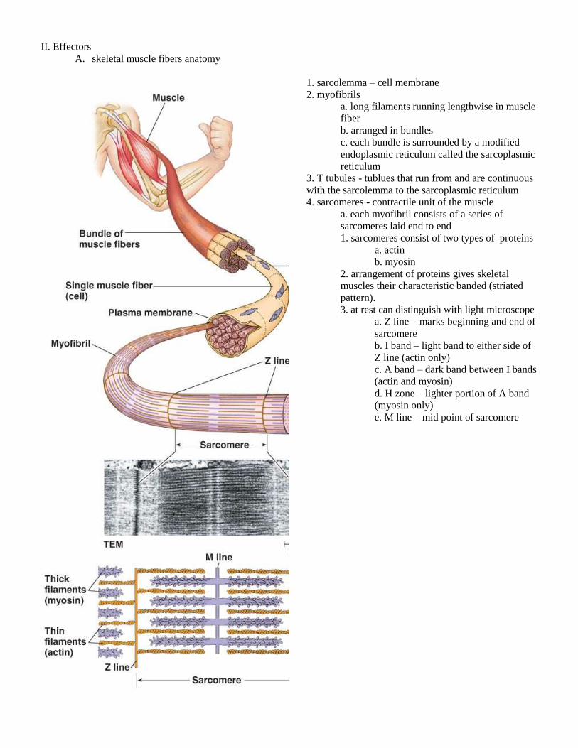
II. Effectors
A. skeletal muscle fibers anatomy
1. sarcolemma – cell membrane
2. myofibrils
a. long filaments running lengthwise in muscle
fiber
b. arranged in bundles
c. each bundle is surrounded by a modified
endoplasmic reticulum called the sarcoplasmic
reticulum
3. T tubules - tublues that run from and are continuous
with the sarcolemma to the sarcoplasmic reticulum
4. sarcomeres - contractile unit of the muscle
a. each myofibril consists of a series of
sarcomeres laid end to end
1. sarcomeres consist of two types of proteins
a. actin
b. myosin
2. arrangement of proteins gives skeletal
muscles their characteristic banded (striated
pattern).
3. at rest can distinguish with light microscope
a. Z line – marks beginning and end of
sarcomere
b. I band – light band to either side of
Z line (actin only)
c. A band – dark band between I bands
(actin and myosin)
d. H zone – lighter portion of A band
(myosin only)
e. M line – mid point of sarcomere

4. scientists observed that when a muscle contracts
a. the I band and H zones shrink
b. the A band remains the same width despite the change in the H zone
c. the width of the sacomere shortens resulting in:
1. a shorting of the myofibral resulting in:
a. a shortening of the myofibral bundle resulting in
1. a shortening of the muscle fiber resulting in
a. a shortening (contraction) of the muscle
B. mechanism of muscle contraction – the sliding filament model
1. closer look at thick myosin fibers
a. each fiber is a bundle of myosin molecules
b. each myosin molecule consists of two long protein chains and globular head
c. heads bind to ATP molecule converting it to ADP + P and storing the energy released

2. closer look at thin actin fibers
a. two chains of actin molecules wound around each other
b. each actin molecule is a globular protein that has a site to which globular myosin head
can bind
c. each actin fiber is surrounded by a thin protein filament called tropomyosin which covers
binding site of actin molecules preventing cross-bridge formation – no sarcomere contraction
b. tropomyosin, in turn, has attached at numerous sites along its length another protein
called troponin that has a binding site for calcium ions
d. when calcium ions bind to troponin, tropomyosin fibers pulled of myosin binding sites
of actin molecules allowing cross-bridge formation – contraction

3. actin ratcheted passed myosin as follows:
a. myosin head binds to actin molecule forming cross-bridge
b. ADP + P falls away
c. myosin head changes shape because of release of energy stored in head
1. ratchets actin fiber towards the M Line (middle of sarcomere)
d. new ATP molecule joins myosin head
1. releases head from actin
e. ATP converts to ADP + P
1. head returns to previous shape
f. head can bind again and repeat process
g. within the myosin fiber, individual heads are at various stages in the above series so at
any given time some cross-bridges are present preventing back sliding
4. turning series on and off
a. whole process dependant on whether or not calcium is present & if binding sites exposed

C. Nervous control
1. neuromuscular junction – axon releases acetylcholine
2. sarcolemma (plasma membrane) polarized – acetylcholine binds to receptors in sarcolemma
3. wave of depolarization runs across sarcolemma and down T-tubes
4. T-tubes in contact with sarcoplasmic reticulum – when depolarization reaches
sarcoplasmic reticulum, stored calcium ions released to myofibrils.
5. sets in motion contraction of sacromere as described earlier and muscle contracts
6. when polarity returns (i.e., no action potential arriving), calcium drawn back into S. R.
7. Myosin heads release and muscle relaxes.
8. individual muscle cells act on an all-or-none principle.

D. Motor units
1. a single motor neuron innervates several muslce fibers.
2. these fibers act as a unit.
3. when the motor neuron fires all the muscle fibers in the unit contract.
E. Drug interaction
1. Curare-binds to Acetylcholine receptors – paralyses
2. Botulotoxin – prevents release of Acetylcholine - paralyses
3. Organo-phosphate pesticides – prevent release of acetylcholinesterase – cannot stop muscle
stimulation tetanus – paralyses for opposite reason

F. Energy for muscle contractions provided as we have seen by ATP
1. Pathways that produce ATP (see figure above)
a. Creatine Phosphate pathway
1. very fast (direct)
2. does not require oxygen
3. does not produce lactic acid
b. Aerobic – cell respiration
1. efficient
2. requires oxygen
c Anaerobic – glycolysis & lactic acid fermentation
1. inefficient
2. does not require oxygen
3. produces lactic acid
H. Types of muscle fibers
1. Fast fibers
a. lag time very short (< .01 sec)
b. densely packed myofibrils (cells very thick)
c. large glycogen reserves
d. few mitochondria
e. little myoglobin (white in color)
f. great strength but fatigue quickly.
2. Slow fibers
a. lag time 3X longer to respond
b. less densely packed with myofibrils
c. use glucose, fatty acids and amino acids for fuel (little glycogen reserves)
d. lot’s of specialized mitochondria.
e. lots of myoglobin (red)
f. rich blood supply
g. fewer SR; calcium remains in cytosol longer
g. less strength but do not fatigue as quickly