characterization and inhibition of xylella … - cdfa... · characterization and inhibition of...
TRANSCRIPT
CHARACTERIZATION AND INHIBITION OF XYLELLA FASTIDIOSA PROTEINS SECRETED BY THE TYPE II SECRETION SYSTEM AND THEIR SECRETION MACHINERY
Principal Investigator Cooperator Cooperator Caroline Roper Bruce Kirkpatrick John Labavitch Dept. Plant Pathology & Microbiology Dept. of Plant Pathology Dept. of Plant Sciences University of California University of California University of California Riverside, CA 92521 Davis, CA 95616 Davis, CA 95616 [email protected] [email protected] [email protected] Cooperator Daniel Cosgrove Dept. of Biology Penn State University University Park, PA 16801 [email protected] Reporting Period: The results reported here are from work conducted July 2014 to July 2016. ABSTRACT The purpose of this study is to elucidate the contributions of host cell wall-degrading enzymes (CWDEs) produced by Xylella fastidiosa (Xf) to systemic colonization of grapevine, as well as the role of the Type II Secretion System (T2SS) in delivering these CWDEs into the xylem. Of the CWDEs predicted to be secreted by the T2SS, this project will focus on the endoglucanases (EGases) produced by Xf. We hypothesize that the T2SS secretes these EGases along with a polygalacturonase (PG), and that these enzymes collaborate to degrade the pit membranes that separate xylem vessels to facilitate the bacterium's systemic colonization of the grapevine via its xylem system. It has been previously reported that a purified PG and one of the Xf EGases are required to increase pore sizes of pit membranes in grapevine. Moreover, mutation of PG results in the loss of pathogenicity and movement for Xf. We also show that a loss of function in the T2SS results in a similar dramatic loss of pathogenicity. In addition, we are investigating the role of an EGase/expansin hybrid protein in pit membrane degradation. Ultimately, characterization of these EGases and the T2SS will help us to determine if they are suitable targets for Pierce's Disease management. LAYPERSON SUMMARY Xylella fastidiosa relies on degradation of the plant cell wall to move within the grapevine. This is accomplished by the cooperation of at least two classes of enzymes that target different components of the complex scaffold of the plant cell wall. A major goal of this research is to further elucidate the factors that lead to disassembly of the plant cell wall, thereby, allowing the bacteria to systemically colonize the plant. Systemic colonization is highly correlated with Pierce’s Disease development and preventing movement of the bacteria is critical to devising successful control strategies. We propose that characterizing and inhibiting Xf enzymes that facilitate movement throughout the plant and/or the secretion machinery responsible for delivering those Xf enzymes into the grapevines water pipes will provide a comprehensive approach to restriction of disease development. INTRODUCTION Xylella fastidiosa (Xf) is a xylem-limited bacterial pathogen that is the causal agent of Pierce’s Disease (PD) of grapevine (Hopkins and Purcell, 2002, Chatterjee et al., 2008a, Purcell and Hopkins, 1996). In order to systemically colonize the xylem, Xf must be able to move efficiently from one xylem vessel element to adjacent vessels. These xylem vessels are connected by pit membranes, which are porous primary cell wall interfaces that are composed of cellulose microfibrils embedded in a meshwork of pectin and hemicellulose (Buchanan, 2000, Sun et al., 2011). The pore sizes of these pit membranes range from 5 to 20 nm, and serve to prevent the movement of air embolisms and pathogens within the xylem (Mollenhauer & Hopkins, 1974, Buchanan, 2000). Indeed, these small pore sizes do prevent the passive movement of Xf between xylem vessels given that the size of the bacterium is 250-500 x 1,000-4,000 nm (Perez-Donoso et al., 2010, Mollenhauer & Hopkins, 1974). In order to move from one vessel to another, it has been shown through genomic and experimental evidence that Xf utilizes Cell Wall-Degrading Enzymes (CWDEs), including a polygalacturonase (PG) and at least one β-1,4 Endoglucanase (EGase), to break down the pit membrane's network (Roper et al., 2007, Perez-Donoso et al.,
2010). Furthermore, PG is necessary for pathogenicity in grape and has become a primary target for Xf inhibition studies (Roper et al, 2007). However, PG alone is not sufficient for pathogenicity in grape and Xf requires both PG and an EGase for pit membrane degradation (Perez-Donoso et al., 2010). Therefore, elucidating the role of EGases in pit membrane degradation is critical for understanding systemic movement within the xylem. The Xf genome contains two genes that encode canonical EGases: egl (PD2061) and engXCA2 (PD1851). A third annotated EGase, engXCA1 (PD 1856), putatively encodes a modular hybrid protein that contains both an EGase domain and an expansin domain (Simpson et al., 2000). Expansins are primarily plant proteins that function to non-enzymatically loosen the cell wall during development (e.g., cell elongation, fruit ripening). Recently, expansins have been found in several plant-associated bacteria, most of which have a significant xylem-dwelling phase in their lifestyle (Nikolaidis et al., 2014). It is predicted that these EGases and PG are delivered into the xylem by the Type II Secretion System (T2SS). Preliminary data demonstrate that X. fastidiosa with a deficient T2SS display a non-pathogenic phenotype similar to that of the Xf pglA mutant that is deficient in production of PG, suggesting that the T2SS is essential for Xf pathogenicity. Therefore, our central hypothesis is that Xf utilizes other CWDEs and an endoglucanase/expansin hybrid protein in concert with PG to breach the pit membranes and that the majority of these are secreted by the Type II Secretion System. We are determining the role that each of these components plays in pit membrane degradation and systemic movement, and subsequently if they are good candidates for potential inhibition to limit Pierce's Disease development. OBJECTIVES
1. Characterization of Xf host cell wall degrading enzymes and an endoglucanase/expansin protein. 2. Inhibition of Xf endoglucanases and the endoglucanase/expansin using endoglucanase-inhibiting proteins. 3. Characterization of the Xf Type II secretion system. 4. Inhibition of the Xf Type II secretion system.
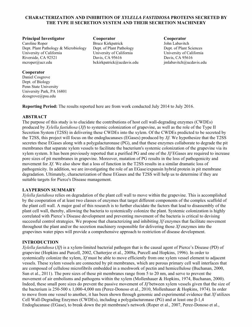
RESULTS AND DISCUSSION Objective 1: Characterization of Xf host cell wall degrading enzymes and an endoglucanase/expansin. It was previously determined that Xf EngXCA2, which is one of the four putative X. fastidiosa EGase-encoding genes is a major contributor to the pit membrane dissolution and the synergistic effects of the PG and the EngXCA2 were sufficient to increase pit membrane pore size (Perez-Donoso et al., 2010). Indeed, recombinant EngXCA2 was capable of digesting carboxymethyl cellulose (CMC) and xyloglucan (XyG) polymers, which both contain -1,4-linked glucan backbones and are representative of substrates Xf would likely encounter in grapevine primary cell walls (Roper, 2006; Perez-Donoso et al., 2010). Given the role EngXCA2 plays in pit membrane degradation, we hypothesize that other predicted EGases produced by Xf may impact pit membrane integrity as well. The egl gene is predicted to encode a -1,4 EGase belonging to the glycoside hydrolase family 5 as indicated in the CAZy (Carbohydrate Active Enzyme) database. Glycoside hydrolase family 5 proteins hydrolyze glycosidic bonds between two carbohydrates or a carbohydrate and non-carbohydrate moiety and have activities ranging from EGases to mannanases. Another gene annotated as an EGase is engXCA1, which encodes an EGase/expansin hybrid putatively involved in plant cell wall disassembly. This is of particular interest because expansins are primarily found in the plant kingdom and are non-enzymatic proteins that function to loosen the cell wall during plant growth without enzymatic digestion of the wall (Cosgrove, 2000). Expansins facilitate cell wall loosening by binding to their target polysaccharide and disrupting the weak bonds between the cellulosic glucan and the microfibril surface, allowing turgor pressure from within the cell to expand the cell wall (Cosgrove, 2000). Expansin-like proteins with similar structure and function were later found in a few bacterial species that associate with plants likely as a result of cross-kingdom horizontal gene transfer (Nikolaidis et al., 2014). These bacterial expansins are thought to enhance the activity of bacterial CWDEs by loosening the cell wall, thereby promoting wall breakdown, colonization and virulence. Interestingly, orthologs of at least one bacterial expansin (EXLX1) are found in several plant pathogens, including Xylella, Xanthomonas, Ralstonia and Erwinia species (Kerff et al., 2008, Georgelis et al., 2014). While these are phylogenetically diverse bacteria, they all share the commonality that they spend the majority of their lives in the xylem tissue of plants. It is hypothesized that they are involved in host colonization (Kerff et al., 2008). In the Xf pathosystem, they could potentially weaken the wall and more readily expose carbohydrate targets for digestion by the suite of other Xf CWDEs. Characterization of the Xf EGase/Expansin hybrid protein. The gene engXCA1 was cloned from the X. fastidiosa Temecula 1 genome into the pET200 Directional TOPO expression vector (Fig. 1A). The plasmid construct (pET200::engXCA1) was then transformed into the E. coli strain BL21 Star, and recombinant protein expression was induced with 1 mM IPTG for six hours at 37°C. The
bacterial cells were lysed using the B-PER lysis reagent containing lysozyme and DNaseI (ThermoFisher) and the lysate was run on an SDS-polyacrylamide gel (Fig. 1B). The lysate was analyzed by Western Blot using a monoclonal α-His-tag primary antibody and a polyclonal alkaline phosphatase (AP) secondary antibody (Fig. 1C). The Western Blot was developed using an AP development kit (Bio Rad), and the protein sequence was confirmed by Mass Spectrometry. Analysis of the soluble and insoluble lysate fractions determined that expression at 37°C did not favor soluble recombinant protein, so conditions were optimized to facilitate the presence of recombinant protein in the soluble fraction. The samples were incubated for four hours at either 25°C or 18°C in the presence of 0.1 mM IPTG, and GelQuant.NET software provided by biochemlabsolutions.com was used to calculate the band intensity for the soluble fraction relative to the insoluble fraction (Fig. 1D). After incubation at 25°C, the soluble fraction contained 7.3% of the total recombinant protein, while incubation at 18°C yielded 22.7% of the total recombinant protein in the soluble fraction. Using the optimized induction conditions (18°C, 0.1 mM IPTG, 4 hours) and increasing the total volume of bacterial cells from 10 ml to 40 ml allowed for the expression of a sufficient quantity of soluble recombinant EngXCA1 protein to proceed with protein purification. The recombinant protein was purified via column chromatography using Ni-NTA resin (ThermoFisher), following the product instructions. Elution of the protein
Figure 1: Expression of recombinant EngXCA1. A) engXCA1 was inserted into the pET200 TOPO vector downstream of the N-terminal His-tag. “CACC” nucleotides were added to the 5’-end of engXCA1 to ensure directionality for expression. B) E. coli was either induced with 1 mM IPTG, not induced with IPTG, or not transformed. Undiluted and 1:10-diluted samples for each treatment were subjected to SDS-PAGE, and stained with Coomassie Blue. The MW of recombinant EngXCA1 is 68 kDa. C) Western Blot of lysate samples subjected to SDS-PAGE. The membrane was probed with an α-His-tag 1° mAb and an AP-conjugated 2° pAb. D) Western Blot analysis of E. coli incubated for 4 hours with 0.1 mM IPTG at either 25°C or 18°C to determine optimal protein solubility conditions. Red arrows indicate recombinant protein bands.
A
B
C
D
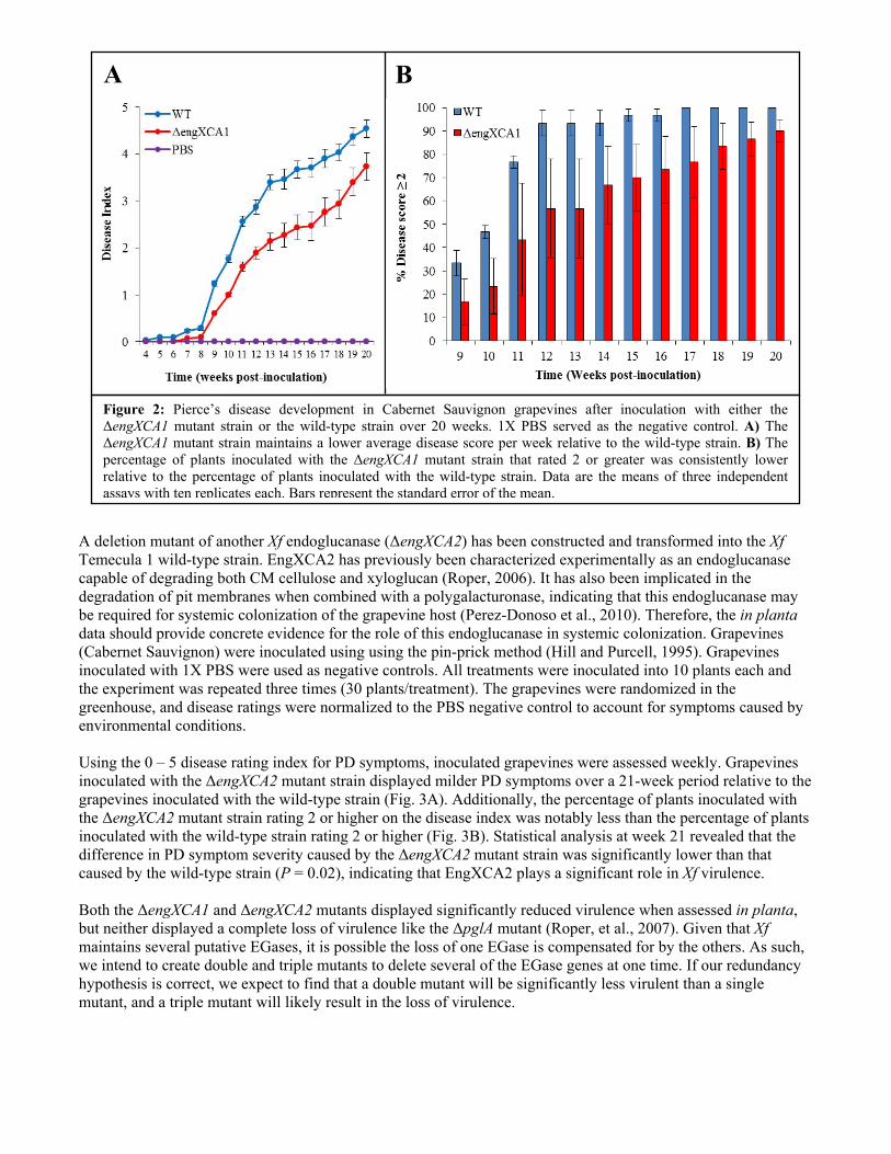
was monitored by absorbance readings at 280 nm. A sufficient quantity of recombinant protein was eluted from the column and subsequently dialyzed using a Slide-A-Lyzer cassette (ThermoFisher) in PBS. After concentrating the protein, an absorbance reading at 280 nm was taken to determine the concentration, only to discover that no protein remained after dialysis and subsequent concentration. Dialysis membranes are made of regenerated cellulose, thus, it is likely that the recombinant EGase may be binding to the membrane. Currently, alternative purification and dialysis methods are being pursued. Previous attempts to determine endoglucanase activity for EngXCA1 using a radial diffusion assay as outlined by Kasana et al. (2008) were inconclusive as the negative controls also showed zones of hydrolysis usually associated with endoglucanase activity. It is possible that the zones of hydrolysis in the negative controls are false positives for reasons outlined by Johnsen and Krause (2014). Due to these difficulties and the fact that radial diffusion assays are not very sensitive, we will be focusing our efforts on determining endoglucanase activity via reducing sugar assays instead. The reducing sugar assay, as outlined by Gross KC (1982), is more sensitive than the radial diffusion assay, and the methodology has already been used to show that another Xf endoglucanase, EngXCA2, has endoglucanase activity (Roper, 2006). Endoglucanase activity of EngXCA1 will be determined using the soluble fraction of the cell lysate in a reducing sugar assay (Gross, 1982). 0.02% CMC or XyG substrate will be dissolved in 0.1 M sodium acetate (pH 5.0). 3 ml of the substrate solution will be incubated with 0.5 ml of the cell lysate containing the recombinant endoglucanase. 0.5 ml aliquots will be taken at time = 0 and every hour for 6 hours, and the reactions will be stopped with 1 ml of 0.1 M sodium borate (pH 10.0). 200 µl of 1% 2-cyanoacetamide will be added and the samples will be boiled for 10 minutes. The absorbance for each sample will be determined spectrophotometrically at 276 nm. We will also assess expansin activity of the recombinant protein (i.e., its ability to promote the extension of plant tissues that are subjected to stress) in close collaboration with the Cosgrove Laboratory (Penn State University). The cell wall elongation assay will then be performed using an extensometer apparatus as described by Cosgrove, D. J. (1989), and expansin activity will be determined by measuring the extension of wall specimens over a 2-h period. Assessment of the biological contribution of the Xf EGase/Expansin and other Xf endoglucanases to pathogenicity and host colonization. To test the role of the Xf EGase/expansin in planta, we constructed a deletion mutant (ΔengXCA1) in the Xf Temecula 1 strain using established mutagenesis techniques and confirmed the mutant via PCR (Matsumoto et al., 2009). We mechanically inoculated the Temecula 1 wild-type and the ΔengXCA1 mutant into grapevine (Cabernet Sauvignon variety) using the pin-prick method (Hill and Purcell, 1995). Grapevines inoculated with 1X phosphate buffered saline (PBS) were used as negative controls. Both the wild-type and the ΔengXCA1 mutant were inoculated into 10 plants each and the experiment was repeated three times (30 plants/treatment). All plants in the experiment were randomized in the greenhouse, and disease ratings were normalized to the PBS negative control to account for symptoms caused by environmental conditions. Disease ratings for all plants were recorded using a scale of 0 – 5 where 0 = healthy, 5 = dead, and 1 – 4 are increasing degrees of leaf scorching as described by Guilhabert and Kirkpatrick (2005). Interestingly, the ΔengXCA1 mutant strain is less virulent than the wild-type parent strain (Fig. 2A). Statistical analysis using the Wilcoxon rank sum with continuity correction statistical test revealed that this difference in virulence between the wild-type and mutant strains at week 20 was statistically significant (P = 0.008). Furthermore, the percentage of plants inoculated with the ΔengXCA1 mutant strain rating 2 or higher on the disease index was significantly less than the percentage of plants inoculated with wild-type Xf rating 2 or higher over a 20-week period (Fig. 2B). This indicates that the onset of disease in plants inoculated with the ΔengXCA1 mutant is significantly delayed relative to plants inoculated with wild-type Xf. We have also constructed the engXCA1/engXCA1+ complement by inserting the engXCA1 gene and its native promoter into a neutral site in the Xf chromosome in the Xf ΔengXCA1 mutant strain (Matsumoto et al., 2009). Initial in planta experiments revealed that the engXCA1/engXCA1+ complement restored virulence to wild-type levels (data not shown), and repeat experiments will be done this year to confirm this result.
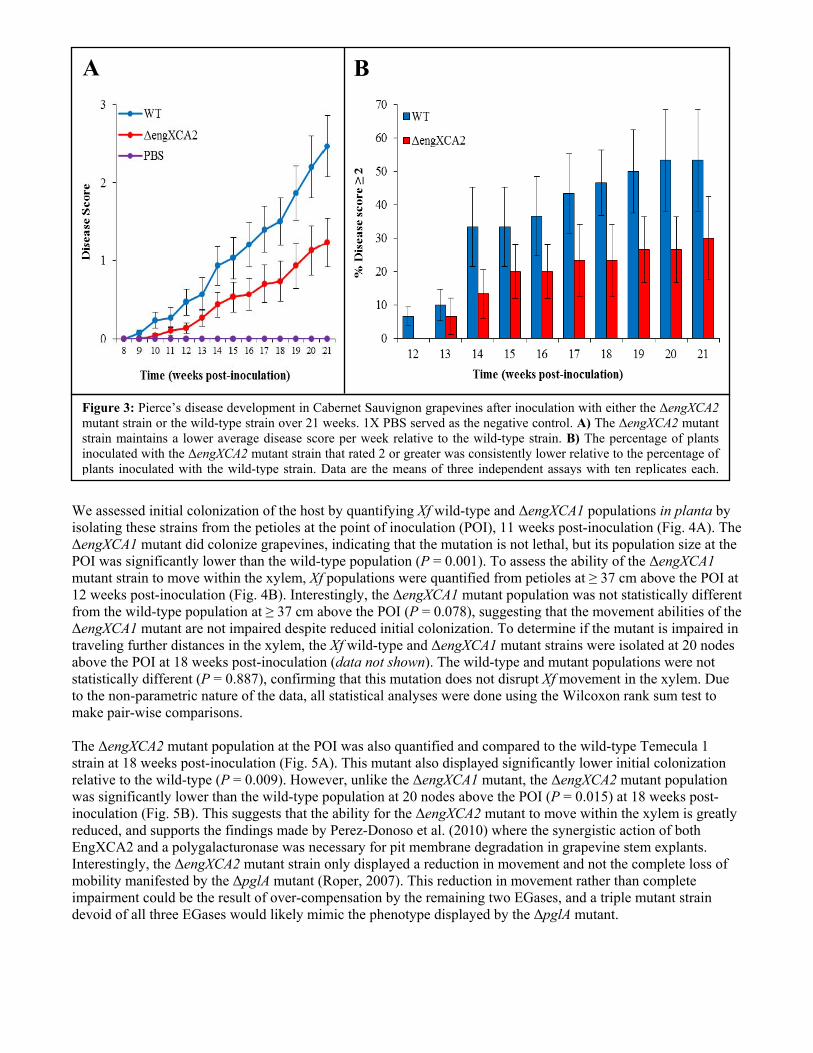
A deletion mutant of another Xf endoglucanase (ΔengXCA2) has been constructed and transformed into the Xf Temecula 1 wild-type strain. EngXCA2 has previously been characterized experimentally as an endoglucanase capable of degrading both CM cellulose and xyloglucan (Roper, 2006). It has also been implicated in the degradation of pit membranes when combined with a polygalacturonase, indicating that this endoglucanase may be required for systemic colonization of the grapevine host (Perez-Donoso et al., 2010). Therefore, the in planta data should provide concrete evidence for the role of this endoglucanase in systemic colonization. Grapevines (Cabernet Sauvignon) were inoculated using using the pin-prick method (Hill and Purcell, 1995). Grapevines inoculated with 1X PBS were used as negative controls. All treatments were inoculated into 10 plants each and the experiment was repeated three times (30 plants/treatment). The grapevines were randomized in the greenhouse, and disease ratings were normalized to the PBS negative control to account for symptoms caused by environmental conditions. Using the 0 – 5 disease rating index for PD symptoms, inoculated grapevines were assessed weekly. Grapevines inoculated with the ΔengXCA2 mutant strain displayed milder PD symptoms over a 21-week period relative to the grapevines inoculated with the wild-type strain (Fig. 3A). Additionally, the percentage of plants inoculated with the ΔengXCA2 mutant strain rating 2 or higher on the disease index was notably less than the percentage of plants inoculated with the wild-type strain rating 2 or higher (Fig. 3B). Statistical analysis at week 21 revealed that the difference in PD symptom severity caused by the ΔengXCA2 mutant strain was significantly lower than that caused by the wild-type strain (P = 0.02), indicating that EngXCA2 plays a significant role in Xf virulence. Both the ΔengXCA1 and ΔengXCA2 mutants displayed significantly reduced virulence when assessed in planta, but neither displayed a complete loss of virulence like the ΔpglA mutant (Roper, et al., 2007). Given that Xf maintains several putative EGases, it is possible the loss of one EGase is compensated for by the others. As such, we intend to create double and triple mutants to delete several of the EGase genes at one time. If our redundancy hypothesis is correct, we expect to find that a double mutant will be significantly less virulent than a single mutant, and a triple mutant will likely result in the loss of virulence.
A
Figure 2: Pierce’s disease development in Cabernet Sauvignon grapevines after inoculation with either the ΔengXCA1 mutant strain or the wild-type strain over 20 weeks. 1X PBS served as the negative control. A) The ΔengXCA1 mutant strain maintains a lower average disease score per week relative to the wild-type strain. B) The percentage of plants inoculated with the ΔengXCA1 mutant strain that rated 2 or greater was consistently lower relative to the percentage of plants inoculated with the wild-type strain. Data are the means of three independent assays with ten replicates each. Bars represent the standard error of the mean.
B
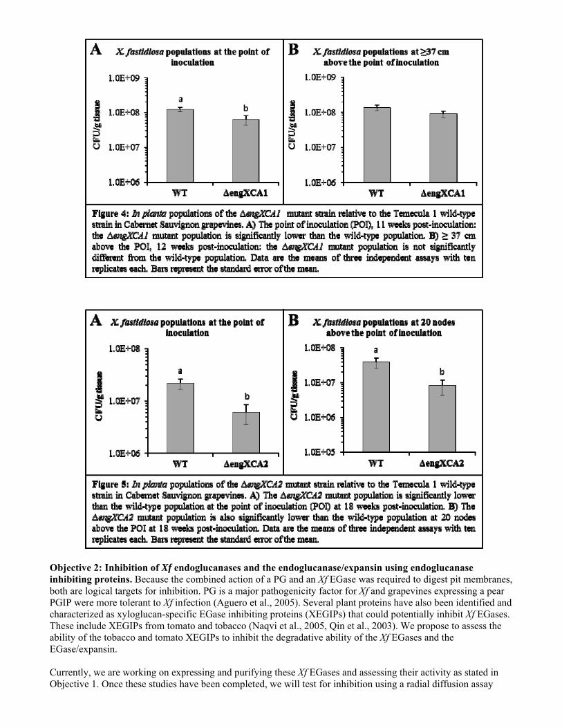
We assessed initial colonization of the host by quantifying Xf wild-type and ΔengXCA1 populations in planta by isolating these strains from the petioles at the point of inoculation (POI), 11 weeks post-inoculation (Fig. 4A). The ΔengXCA1 mutant did colonize grapevines, indicating that the mutation is not lethal, but its population size at the POI was significantly lower than the wild-type population (P = 0.001). To assess the ability of the ΔengXCA1 mutant strain to move within the xylem, Xf populations were quantified from petioles at ≥ 37 cm above the POI at 12 weeks post-inoculation (Fig. 4B). Interestingly, the ΔengXCA1 mutant population was not statistically different from the wild-type population at ≥ 37 cm above the POI (P = 0.078), suggesting that the movement abilities of the ΔengXCA1 mutant are not impaired despite reduced initial colonization. To determine if the mutant is impaired in traveling further distances in the xylem, the Xf wild-type and ΔengXCA1 mutant strains were isolated at 20 nodes above the POI at 18 weeks post-inoculation (data not shown). The wild-type and mutant populations were not statistically different (P = 0.887), confirming that this mutation does not disrupt Xf movement in the xylem. Due to the non-parametric nature of the data, all statistical analyses were done using the Wilcoxon rank sum test to make pair-wise comparisons. The ΔengXCA2 mutant population at the POI was also quantified and compared to the wild-type Temecula 1 strain at 18 weeks post-inoculation (Fig. 5A). This mutant also displayed significantly lower initial colonization relative to the wild-type (P = 0.009). However, unlike the ΔengXCA1 mutant, the ΔengXCA2 mutant population was significantly lower than the wild-type population at 20 nodes above the POI (P = 0.015) at 18 weeks post-inoculation (Fig. 5B). This suggests that the ability for the ΔengXCA2 mutant to move within the xylem is greatly reduced, and supports the findings made by Perez-Donoso et al. (2010) where the synergistic action of both EngXCA2 and a polygalacturonase was necessary for pit membrane degradation in grapevine stem explants. Interestingly, the ΔengXCA2 mutant strain only displayed a reduction in movement and not the complete loss of mobility manifested by the ΔpglA mutant (Roper, 2007). This reduction in movement rather than complete impairment could be the result of over-compensation by the remaining two EGases, and a triple mutant strain devoid of all three EGases would likely mimic the phenotype displayed by the ΔpglA mutant.
A B
Figure 3: Pierce’s disease development in Cabernet Sauvignon grapevines after inoculation with either the ΔengXCA2 mutant strain or the wild-type strain over 21 weeks. 1X PBS served as the negative control. A) The ΔengXCA2 mutant strain maintains a lower average disease score per week relative to the wild-type strain. B) The percentage of plants inoculated with the ΔengXCA2 mutant strain that rated 2 or greater was consistently lower relative to the percentage of plants inoculated with the wild-type strain. Data are the means of three independent assays with ten replicates each.
Objective 2: Inhibition of Xf endoglucanases and the endoglucanase/expansin using endoglucanase inhibiting proteins. Because the combined action of a PG and an Xf EGase was required to digest pit membranes, both are logical targets for inhibition. PG is a major pathogenicity factor for Xf and grapevines expressing a pear PGIP were more tolerant to Xf infection (Aguero et al., 2005). Several plant proteins have also been identified and characterized as xyloglucan-specific EGase inhibiting proteins (XEGIPs) that could potentially inhibit Xf EGases. These include XEGIPs from tomato and tobacco (Naqvi et al., 2005, Qin et al., 2003). We propose to assess the ability of the tobacco and tomato XEGIPs to inhibit the degradative ability of the Xf EGases and the EGase/expansin. Currently, we are working on expressing and purifying these Xf EGases and assessing their activity as stated in Objective 1. Once these studies have been completed, we will test for inhibition using a radial diffusion assay
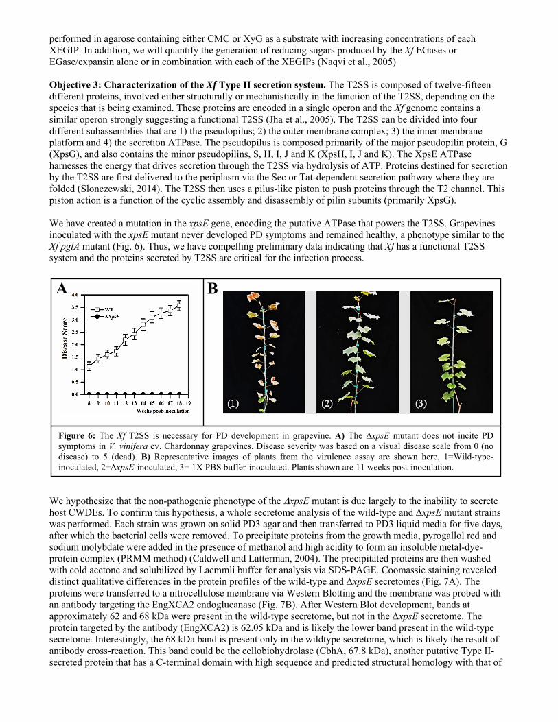
performed in agarose containing either CMC or XyG as a substrate with increasing concentrations of each XEGIP. In addition, we will quantify the generation of reducing sugars produced by the Xf EGases or EGase/expansin alone or in combination with each of the XEGIPs (Naqvi et al., 2005) Objective 3: Characterization of the Xf Type II secretion system. The T2SS is composed of twelve-fifteen different proteins, involved either structurally or mechanistically in the function of the T2SS, depending on the species that is being examined. These proteins are encoded in a single operon and the Xf genome contains a similar operon strongly suggesting a functional T2SS (Jha et al., 2005). The T2SS can be divided into four different subassemblies that are 1) the pseudopilus; 2) the outer membrane complex; 3) the inner membrane platform and 4) the secretion ATPase. The pseudopilus is composed primarily of the major pseudopilin protein, G (XpsG), and also contains the minor pseudopilins, S, H, I, J and K (XpsH, I, J and K). The XpsE ATPase harnesses the energy that drives secretion through the T2SS via hydrolysis of ATP. Proteins destined for secretion by the T2SS are first delivered to the periplasm via the Sec or Tat-dependent secretion pathway where they are folded (Slonczewski, 2014). The T2SS then uses a pilus-like piston to push proteins through the T2 channel. This piston action is a function of the cyclic assembly and disassembly of pilin subunits (primarily XpsG). We have created a mutation in the xpsE gene, encoding the putative ATPase that powers the T2SS. Grapevines inoculated with the xpsE mutant never developed PD symptoms and remained healthy, a phenotype similar to the Xf pglA mutant (Fig. 6). Thus, we have compelling preliminary data indicating that Xf has a functional T2SS system and the proteins secreted by T2SS are critical for the infection process. We hypothesize that the non-pathogenic phenotype of the xpsE mutant is due largely to the inability to secrete host CWDEs. To confirm this hypothesis, a whole secretome analysis of the wild-type and ΔxpsE mutant strains was performed. Each strain was grown on solid PD3 agar and then transferred to PD3 liquid media for five days, after which the bacterial cells were removed. To precipitate proteins from the growth media, pyrogallol red and sodium molybdate were added in the presence of methanol and high acidity to form an insoluble metal-dye-protein complex (PRMM method) (Caldwell and Latterman, 2004). The precipitated proteins are then washed with cold acetone and solubilized by Laemmli buffer for analysis via SDS-PAGE. Coomassie staining revealed distinct qualitative differences in the protein profiles of the wild-type and ΔxpsE secretomes (Fig. 7A). The proteins were transferred to a nitrocellulose membrane via Western Blotting and the membrane was probed with an antibody targeting the EngXCA2 endoglucanase (Fig. 7B). After Western Blot development, bands at approximately 62 and 68 kDa were present in the wild-type secretome, but not in the ΔxpsE secretome. The protein targeted by the antibody (EngXCA2) is 62.05 kDa and is likely the lower band present in the wild-type secretome. Interestingly, the 68 kDa band is present only in the wildtype secretome, which is likely the result of antibody cross-reaction. This band could be the cellobiohydrolase (CbhA, 67.8 kDa), another putative Type II-secreted protein that has a C-terminal domain with high sequence and predicted structural homology with that of
A B
Figure 6: The Xf T2SS is necessary for PD development in grapevine. A) The ∆xpsE mutant does not incite PD symptoms in V. vinifera cv. Chardonnay grapevines. Disease severity was based on a visual disease scale from 0 (no disease) to 5 (dead). B) Representative images of plants from the virulence assay are shown here, 1=Wild-type-inoculated, 2=∆xpsE-inoculated, 3= 1X PBS buffer-inoculated. Plants shown are 11 weeks post-inoculation.

EngXCA2. The antibody also appears to be binding specifically as several large bands were present on the Coomassie stained gel that were not present on the Western Blot.
To confirm the results attained by the Western Blot, the wild-type and ΔxpsE secretomes were analyzed by Matrix-assisted laser desorption/ionization (MALDI) mass spectrometry to determine Type II-secreted proteins. All proteins found in each secretome were checked for predicted secretion signals using SignalP4.1 and predicted subcellular localization using PSORTb3.0 to determine which proteins were secreted. Xf maintains three functional secretion systems (Types I, II, and V), and putative secreted proteins were then compared to known orthologs using NCBI BLAST to determine which secretion system they utilize. Of the five CWDEs predicted to be Type II-secreted, four were detected by mass spectrometry: EngXCA2, EngXCA1, Egl, and CbhA (Table 1). Both EngXCA1 and EngXCA2 were only detected in the wildtype secretome, indicating that they are dependent on the T2SS for secretion. Interestingly, Egl and CbhA were detected in both secretomes, and it is possible that these CWDEs may not rely solely on the T2SS for secretion. Instead, they may utilize alternate pathways such as outer membrane vesicles (OMVs) (Solé et al., 2015). This can be determined by removing OMVs from the media before protein precipitation to see if Egl and CbhA are present or absent in the ΔxpsE secretome. Table 1. Mass spectrometry analysis of putative Type II-secreted proteins
Presence in Secretome
PD Number Gene Function Wild-type ΔxpsE mutant
PD0529 cbhA β-1,4-cellobiohydrolase + +
PD1485 pglA Polygalacturonase - -
PD1851 engXCA2 β-1,4-endoglucanase + -
PD1856 engXCA1 β-1,4-endoglucanase + -
PD2061 egl β-1,4-endoglucanase + +
Wildtype ΔxpsE
75 kDa ─
50 kDa ─
37 kDa ─
CbhA
EngXCA2
Wildtype ΔxpsE
75 kDa ─
50 kDa ─
37 kDa ─
A B
Figure 7: Whole secretome analysis of the Xf wild-type Temecula 1 strain and the Xf ΔxpsE mutant strain. A) Coomassie staining shows distinct differences in the protein profiles of the wild-type and ΔxpsE mutant secretomes. B) Western Blot analysis using an antibody against EngXCA2 reveals its presence (62 kDa band) in the wild-type secretome only, indicating that it is Type II-secreted. Another band at 68 kDa is likely a cross-reaction with the cellobiohydrolase (CbhA).
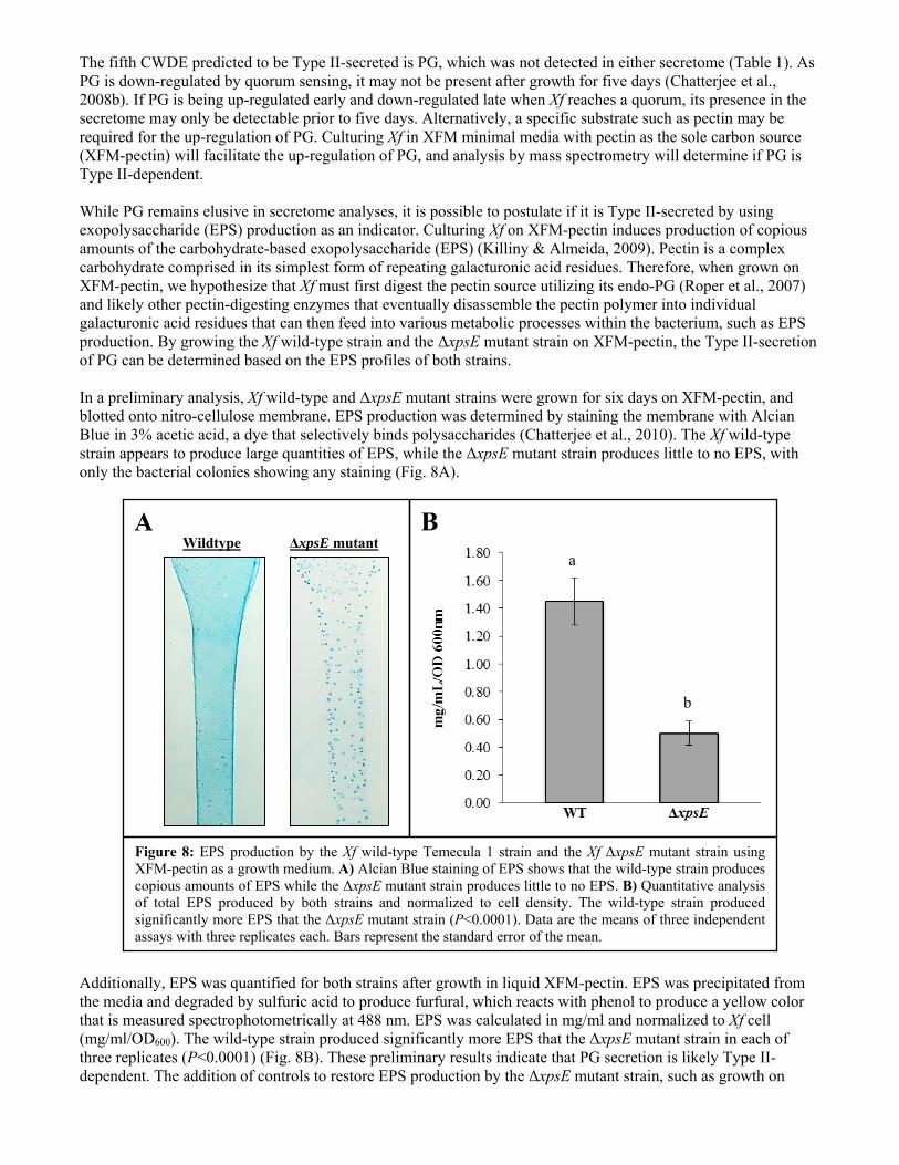
The fifth CWDE predicted to be Type II-secreted is PG, which was not detected in either secretome (Table 1). As PG is down-regulated by quorum sensing, it may not be present after growth for five days (Chatterjee et al., 2008b). If PG is being up-regulated early and down-regulated late when Xf reaches a quorum, its presence in the secretome may only be detectable prior to five days. Alternatively, a specific substrate such as pectin may be required for the up-regulation of PG. Culturing Xf in XFM minimal media with pectin as the sole carbon source (XFM-pectin) will facilitate the up-regulation of PG, and analysis by mass spectrometry will determine if PG is Type II-dependent. While PG remains elusive in secretome analyses, it is possible to postulate if it is Type II-secreted by using exopolysaccharide (EPS) production as an indicator. Culturing Xf on XFM-pectin induces production of copious amounts of the carbohydrate-based exopolysaccharide (EPS) (Killiny & Almeida, 2009). Pectin is a complex carbohydrate comprised in its simplest form of repeating galacturonic acid residues. Therefore, when grown on XFM-pectin, we hypothesize that Xf must first digest the pectin source utilizing its endo-PG (Roper et al., 2007) and likely other pectin-digesting enzymes that eventually disassemble the pectin polymer into individual galacturonic acid residues that can then feed into various metabolic processes within the bacterium, such as EPS production. By growing the Xf wild-type strain and the ΔxpsE mutant strain on XFM-pectin, the Type II-secretion of PG can be determined based on the EPS profiles of both strains. In a preliminary analysis, Xf wild-type and ΔxpsE mutant strains were grown for six days on XFM-pectin, and blotted onto nitro-cellulose membrane. EPS production was determined by staining the membrane with Alcian Blue in 3% acetic acid, a dye that selectively binds polysaccharides (Chatterjee et al., 2010). The Xf wild-type strain appears to produce large quantities of EPS, while the ΔxpsE mutant strain produces little to no EPS, with only the bacterial colonies showing any staining (Fig. 8A).
Additionally, EPS was quantified for both strains after growth in liquid XFM-pectin. EPS was precipitated from the media and degraded by sulfuric acid to produce furfural, which reacts with phenol to produce a yellow color that is measured spectrophotometrically at 488 nm. EPS was calculated in mg/ml and normalized to Xf cell (mg/ml/OD600). The wild-type strain produced significantly more EPS that the ΔxpsE mutant strain in each of three replicates (P<0.0001) (Fig. 8B). These preliminary results indicate that PG secretion is likely Type II-dependent. The addition of controls to restore EPS production by the ΔxpsE mutant strain, such as growth on
Wildtype ΔxpsE mutant
A B
Figure 8: EPS production by the Xf wild-type Temecula 1 strain and the Xf ΔxpsE mutant strain using XFM-pectin as a growth medium. A) Alcian Blue staining of EPS shows that the wild-type strain produces copious amounts of EPS while the ΔxpsE mutant strain produces little to no EPS. B) Quantitative analysis of total EPS produced by both strains and normalized to cell density. The wild-type strain produced significantly more EPS that the ΔxpsE mutant strain (P<0.0001). Data are the means of three independent assays with three replicates each. Bars represent the standard error of the mean.
a
b
XFM containing only glucose or galacturonic acid, will confirm that this result is based solely on the inability of the ΔxpsE mutant strain to secrete PG. Objective 4: Inhibition of the Xf Type II secretion system. Proteins destined for secretion by the T2SS are first exported to the periplasm by the Sec or Tat pathways. Xf appears to only possess the Sec-dependent secretion pathway. Disruption of the T2SS by small molecule inhibitors was demonstrated in Pseudomonas aeruginosa and Burkholderia pseudomallei, and could be used to inhibit the Xf Sec-dependent pathway (Moir et al., 2011). A chemical compound library will be screened for Sec-inhibitory molecules, including those compounds used by Moir et al. (2011). Inhibition of the Sec-dependent pathway will be confirmed by monitoring the secretion of a CWDE using a polyclonal antibody raised against EngXCA2 and analyzed via Western Blot and ELISA. CONCLUSIONS The goal of this research is to understand the roles each of the EGases produced by Xf has in pit membrane degradation, as well as the role of the T2SS in secreting these CWDEs. Preliminary results indicate that the EGase/expansin hybrid protein (EngXCA1) and a characterized endoglucanase (EngXCA2) play a role in virulence, and could possibly be elicitors of the host defense response. These studies will be repeated with the addition of respective complement strains to confirm these results. In addition, an Xf strain with a deficient T2SS (ΔxpsE) displayed a loss of virulence similar to that displayed by the PG-deficient mutant, lending credence to the hypothesis that the T2SS may be the secretory pathway for CWDEs that play a role in systemic colonization. Indeed, a whole secretome analysis revealed at least two endoglucanases are Type II-dependent. Despite its absence in the secretome analysis, preliminary experiments have indirectly linked the PG to the T2SS, though further work needs to be done to confirm that the PG is Type II-dependent. Taken together, it is clear that several of these CWDEs and the T2SS are virulence factors, and inhibition of the EGases and/or the T2SS will significantly reduce the ability of Xf to systemically colonize its grapevine host. REFERENCES CITED Aguero CB, Uratsu SL, Greve C, Powell, A. L.,Labavitch, J. M.,Meredith, C. P, Dandekar, A. M., 2005.
Evaluation of tolerance to Pierce's disease and Botrytis in transgenic plants of Vitis vinifera L. expressing the pear PGIP gene. Molecular Plant Pathology 6, 43-51.
Buchanan BB, Gruissem, W., and Jones, R.L. , 2000. Biochemistry and Molecular Biology of Plants. American Society of Plant Physiologists. Maryland. Chapter 2: The Cell Wall, 52-100.
Caldwell RB, Lattemann CT, 2004. Simple and reliable method to precipitate proteins from bacterial culture supernatant. Applied and Environmental Microbiology 70, 610-2.
Chatterjee S, Almeida RPP, Lindow S, 2008a. Living in two worlds: The plant and insect lifestyles of Xylella fastidiosa. Annual Review of Phytopathology 46, 243-71.
Chatterjee S, Wistrom C, Lindow SE, 2008b. A cell–cell signaling sensor is required for virulence and insect transmission of Xylella fastidiosa. Proceedings of the National Academy of Sciences 105, 2670-5.
Chatterjee S, Killiny N, Almeida RP, Lindow SE, 2010. Role of cyclic di-GMP in Xylella fastidiosa biofilm formation, plant virulence, and insect transmission. Molecular plant-microbe interactions 23, 1356-63.
Cosgrove DJ, 1989. Characterization of long term extension of isolated cell walls from growing cucumber hypocotyls. Planta 177, 121-30.
Cosgrove DJ, 2000. Loosening of plant cell walls by expansins. Nature 407, 321-6. Georgelis N, Nikolaidis N, Cosgrove DJ, 2014. Biochemical analysis of expansin-like proteins from microbes.
Carbohydr Polym 100, 17-23. Gross KC, 1982. A Rapid and Sensitive Spectrophotometric Method for Assaying Polygalacturonase Using 2-
Cyanoacetamide. Hortscience 17, 491-494. Guilhabert MR, Kirkpatrick BC, 2005. Identification of Xylella fastidiosa antivirulence genes: hemagglutinin
adhesins contribute to X. fastidiosa biofilm maturation and colonization and attenuate virulence. Molecular Plant-Microbe Interactions 18, 856-868.
Johnsen HR, Krause K, 2014. Cellulase Activity Screening Using Pure Carboxymethylcellulose: Application to Soluble Cellulolytic Samples and to Plant Tissue Prints. International Journal of Molecular Sciences 15, 830-838.
Hill BL, Purcell AH, 1995. Acquisition and retention of Xylella fastidiosa by an efficient vector, Graphocephala atropunctata. Phytopathology 85, 209-212.
Hopkins DL, Purcell AH, 2002. Xylella fastidiosa: Cause of Pierce's disease of grapevine and other emergent diseases. Plant Disease 86, 1056-66.
Jha G, Rajeshwari R, Sonti RV, 2005. Bacterial type two secretion system secreted proteins: double-edged swords for plant pathogens. Molecular Plant Microbe Interactions 18, 891-8.
Kasana RC, Salwan R, Dhar H, Dutt S, Gulati A, 2008. A rapid and easy method for the detection of microbial cellulases on agar plates using Gram’s Iodine. Current Microbiology 57, 503-507
Kerff F, Amoroso A, Herman R, Sauvage, E.,Petrella, S., Filee, P., Charlier, P., Joris, B., Tabuchi, A., Nikolaidis, N., Cosgrove, D. J., 2008. Crystal structure and activity of Bacillus subtilis YoaJ (EXLX1), a bacterial expansin that promotes root colonization. Proc Natl Acad Sci U S A 105, 16876-81.
Killiny N, Almeida RP, 2009. Host structural carbohydrate induces vector transmission of a bacterial plant pathogen. Proc Natl Acad Sci 106, 22416-20.
Matsumoto A, Young GM, Igo MM, 2009. Chromosome-Based Genetic Complementation System for Xylella fastidiosa. Applied and Environmental Microbiology 75, 1679-87.
Moir DT, Di M, Wong E, et al., 2011. Development and application of a cellular, gain-of-signal, bioluminescent reporter screen for inhibitors of type II secretion in Pseudomonas aeruginosa and Burkholderia pseudomallei. J Biomol Screen 16, 694-705.
Mollenhauer HH, Hopkins DL, 1974. Ultrastructural study of Pierce's disease bacterium in grape xylem tissue. J Bacteriol 119, 612-8.
Naqvi SM, Harper A, Carter C, Ren, G., Guirgis, A., York, W. S., Thornburg, R. W., 2005. Nectarin IV, a potent endoglucanase inhibitor secreted into the nectar of ornamental tobacco plants. Isolation, cloning, and characterization. Plant Physiol 139, 1389-400.
Nikolaidis N, Doran N, Cosgrove DJ, 2014. Plant expansins in bacteria and fungi: evolution by horizontal gene transfer and independent domain fusion. Molecular Biology and Evolution 31, 376-86.
Perez-Donoso AG, Sun Q, Roper MC, Greve LC, Kirkpatrick B, Labavitch JM, 2010. Cell Wall-Degrading Enzymes Enlarge the Pore Size of Intervessel Pit Membranes in Healthy and Xylella fastidiosa-Infected Grapevines. Plant Physiology 152, 1748-59.
Purcell AH, Hopkins DL, 1996. Fastidious xylem-limited bacterial plant pathogens. Annu Rev Phytopathol 34, 131-51.
Qin Q, Bergmann CW, Rose JK, et al., 2003. Characterization of a tomato protein that inhibits a xyloglucan-specific endoglucanase. Plant Journal 34, 327-38.
Roper MC, 2006. The characterization and role of Xylella fastidiosa plant cell wall degrading enzymes and exopolysaccharide in Pierce’s disease of grapevine. Ph.D. Thesis University of California, Davis, CA.
Roper MC, Greve LC, Warren JG, Labavitch JM, Kirkpatrick BC, 2007. Xylella fastidiosa requires polygalacturonase for colonization and pathogenicity in Vitis vinifera grapevines. Molecular Plant Microbe Interactions 20, 411-9.
Simpson AJ, Reinach FC, Arruda P, et al., 2000. The genome sequence of the plant pathogen Xylella fastidiosa. The Xylella fastidiosa Consortium of the Organization for Nucleotide Sequencing and Analysis. Nature 406, 151-9.
Slonczewski J.L. and Foster, J.W., 2014. Microbiology: An Evolving Science. W.W. Norton and Company, New York, NY.
Solé M, Scheibner F, Hoffmeister AK, Hartmann N, Hause G, Rother A, Jordan M, Lautier M, Arlat M, Büttner D, 2015. Xanthomonas campestris pv. vesicatoria secretes proteases and xylanases via the Xps type II secretion system and outer membrane vesicles. Journal of Bacteriology 197, 2879-93.
Sun Q., Greve LC, Labavitch JM 2011. Polysaccharide compositions of intervessel pit membranes contribute to Pierce's Disease resistance of grapevines. Plant Physiology 155, 1976-87.
FUNDING AGENCIES Funding for this project was provided by the CDFA Pierce’s Disease and Glassy-winged Sharpshooter Board.