characterization and reconstitution of enzymes and
TRANSCRIPT

CHARACTERIZATION AND RECONSTITUTION OF ENZYMES AND PATHWAYS IN THE BIOSYNTHETIC PRODUCTION OF NITROAROMATIC COMPOUNDS
BY
EMMANUEL MALOLES CHANCO
THESIS
Submitted in partial fulfillment of the requirements for the degree of Master of Science in Chemistry
in the Graduate College of the University of Illinois at Urbana-Champaign, 2012
Urbana, Illinois
Advisor:
Professor Huimin Zhao

ii
Abstract
Nitro-containing compounds are an important class of chemicals with high value in industry and
medicine. Several of these compounds are produced naturally in many different species; however,
several key aspects of their biosynthesis, and the discovery of further nitro compounds are topics of
active research. The prototypical enzyme for N-oxygenation of arylamines is the protein, AurF, from
Streptomyces thioletus. Partial characterization of AurF has already been performed, but in this thesis, a
more complete characterization of this enzyme is performed for eventual work in protein engineering.
The activity of the enzyme was increased more than 600-fold via optimizing reaction pH and
supplementing the chemical mediators reductants phenzazine methosulfate(PMS) and NADH. Alanine
scanning and substrate specificity studies were performed to identify key residues and mechanistic
information of the protein. The electron transfer mechanism of AurF was also studied, resulting in a
better understanding of the kinetics of each important step in the reaction mechanism. Finally, an in
vivo screening method for directed evolution was developed and validated. Screening was performed to
find mutants with activity towards meta-aminobenzoic acid. The resultant positive hits from the initial
screening are to be confirmed and characterized later.
In parallel, based on the bioinformatics search for AurF analogs, the mangotoxin gene cluster
from Pseudomonas syringae was identified as possibly making a nitroaromatic product. Product solation
and identification of this cluster’s gene product were attempted based on heterologous expression of
the cluster in E. coli, and fermentation studies in the native host. To that end, the gene cluster was
recloned in a variety of constructs using several methods such as DNA assembler, Golden Gate and
Gibson assembly. Expression of the pathway genes was observed in E. coli, while feeding studies
confirmed the production of nitroaromatic compounds from the cluster. A product detection method
based on reduction/diazotization was developed in order to find the putative product.

iii
Acknowledgements
The road to this thesis has been a difficult and often frustrating path. However, the way to this
thesis was never lonely, and most often pleasant due to the company I travelled with. Although I am a
scientist, I cannot begin to quantify how much people in the Zhao lab have helped me in my academic
journey. I cannot help but to be humbled by all their assistance and advice; my contributions and efforts
would be much less without them. First, I would like to thank Prof. Zhao for letting me have this exciting
ride. Although it did not end the way I thought it would, I am forever grateful for the chance to learn and
work with him and the people in the lab. Through this experience, I’ve found many things about myself,
the world and what it means to be a scientist. Several people have significantly helped me in my
academic journey; Carl Denard, Ryan Cobb, and Ning Sun come to mind quickly, but others in the lab
have gone above and beyond the call of duty to assist me, and to answer my often ridiculous questions
and problems with quiet patience and grace. I also took pride in the fact that I was able to support these
people when they needed me, and that I was part of their daily life, whether they consciously knew it or
not. I hope they consider me as someone they can call for help, as much as I can ask them for it.
Finally, I’d like to thank people who may not have helped me in my research, but have provided
for my well-being in different ways. My family, being so supportive and helpful through trying times.
Although they may not have understood what I did, they believed in me for what I was doing. A shout-
out goes to my cats Ammonia and Baking Soda for preventing my untimely descent into madness.
Finally, I would like to thank a very sweet girl, Jie Sun, for love, support and kindness, in times when I
thought I needed it, and in times that I thought I didn’t. If I could only repay these people for what they
had done for me, the world would not be enough for what they are worth.

iv
Table of Contents Chapter 1: Introduction ................................................................................................................................ 1
1.1. Nitro Products: An Opportunity for Synthetic Biology ................................................................. 1
1.2. Biological Mechanisms of Nitration and N-Oxygenation ............................................................. 2
1.3. Di-iron Arylamine N-Oxygenases .................................................................................................. 4
1.4. Project Overview .......................................................................................................................... 5
1.5. References .................................................................................................................................... 6
Chapter 2: Characterization of the N-oxygenase AurF ................................................................................. 8
2.1. Introduction .................................................................................................................................. 8
2.2. Protein Quantification, Reconstitution and pH Dependence ..................................................... 10
2.3. Kinetic Characterization .............................................................................................................. 13
2.4. Active-site Mutagenesis Studies ................................................................................................. 15
2.5. Substrate Specificity ................................................................................................................... 20
2.6. Kinetic Parameters of Alternate Substrates and Mutants.......................................................... 30
2.7. Electron Transport Mechanisms ................................................................................................. 31
2.8. Development of an In Vivo Assay for Directed Evolution ........................................................... 36
2.9. Conclusions ................................................................................................................................. 43
2.10. Materials and Methods .............................................................................................................. 44
2.11. References .................................................................................................................................. 48
Chapter 3: Reconstitution of the Mangotoxin Gene Cluster ...................................................................... 51
3.1. Introduction ................................................................................................................................ 51
3.2. Bioinformatics Characterization of the Mangotoxin (mgo) Gene Cluster .................................. 54
3.3. Cloning of the Mangotoxin Gene Cluster ................................................................................... 56
3.4. Expression of the Mangotoxin Gene Cluster .............................................................................. 59
3.5. Detection of Products and Intermediates .................................................................................. 62
3.6. Synthesis of Compounds ............................................................................................................ 70
3.7. Conclusions ................................................................................................................................. 72
3.8. Materials and Methods .............................................................................................................. 73
3.9. References .................................................................................................................................. 77
3.10. Tables and Supporting Information ............................................................................................ 80

1
Chapter 1: Introduction
1.1. Nitro Products: An Opportunity for Synthetic Biology
One of the goals of synthetic biology is to produce a given compound in high yield and purity
through biosynthetic processes.. 1,2Similar to the philosophy of total chemical synthesis, where an easily
available precursor is converted to a more desirable compound, the biological approach to create a
compound of interest is to introduce wild type or engineered enzymes to catalyze reactions of interest.
This has been applied successfully in many cases, such as in production of biofuels, chemicals and
natural products.3,4 A biology-based approach has several advantages, such as being able to use mild
reaction conditions, avoiding toxic byproducts and organic solvents, and being able to leverage the
exquisite specificity of enzymes to enforce desired stereochemistry. However, the toolkit for
chemosynthetic biology is limited by the reactions which can be found in nature. There is a significant
need for unique reactions and ways to install functional groups through biocatalysis. Thus, discovery of
new biosynthetic reactions and characterization of the involved enzymes are topics that are attracting
significant attention.
The formation of nitro compounds is one such reaction. Nitro compounds are industrially relevant as
feedstock chemicals, usually as precursors for other industrially important amines. However, the
industrial processes for making these compounds typically involve strong acids and harsh reaction
conditions, making cleanup and waste disposal difficult. As an example, the production of
trinitrotoluene involves progressively harsher conditions, as the nitro group is deactivating towards
further electrophilic substitution (Figure 1.1A).5 Contamination of chemical sites is a common
occurrence from these processes, eventually requiring expensive remediation procedures. 6 There are
significant health risks involved with these environments, as nitro compounds have been found to be
carcinogenic. 7 Although large-scale nitro compound production is a man-made phenomenon, bacteria

2
have evolved to consume and detoxify these compounds. Conversely, several different nitro compounds
are biosynthesized by a variety of species as antibiotics, antifungals, and other defensive compunds.7,8
Chloramphenicol, a common antibiotic which inhibits protein synthesis, was originally discovered in
Streptomyces venezuelae.9 Another Streptomyces species produces aureothin, a polyketide antifungal
compound which uses p-nitrobenzoic acid as a starter unit. 10,11 However, only recently has the detailed
information on the biosynthesis of these compounds been available. Thus, work in this thesis includes
characterizing enzymes and biosynthetic clusters involved in nitro compound biosynthesis.
1.2. Biological Mechanisms of Nitration and N-Oxygenation
A)
B)
C)
Figure 1.1. Methods of nitration industrially and biosynthetically. A) Synthesis of TNT by electrophilic aromatic substitution. Increasingly harsh conditions are necessary for additional nitration. B) Mechanism for electrophilic aromatic substitution. Formation of a nitronium ion allows for electrophilic addition to benzene. C) N-oxygenation of amines through successive two-electron oxidations.
Several mechanisms of nitro compound formation have been observed in nature. Electrophilic
substitution reactions, which operate by formation of nitric acid or by nitrogen radicals have been
observed in several compounds, and are the main mechanisms on which large-scale industrial nitro
compounds are made (Figure 1A and B).6,7,12 As the nitronium ion is highly reactive and produced under
3
conditions of oxidative stress, several health-related issues such as protein nitration, allergic reactions
and unwanted mutagenesis have been associated with this type of nitration. 6,12,13 Although this method
has the advantage of simplicity, it can be hard to direct the nitronium ion to react specifically at a given
position unless directed by an enzyme. An Antarctic psychotropic bacterium Salegentibacter sp. strain
T436 produces several nitroaromatic compounds, and it is thought the nitration proceeds via these
mechanisms.14
A second common mechanism of nitration involves oxidation of a preexisting amine to more
oxidized functionalities (Figure 1.1C). Hydroxylamine and nitroso compounds are intermediates toward
the production of nitroaromatic compounds. These reactions can be reversed to detoxify nitroaromatic
compounds, enabling several organisms such as Pseudomonas and Rhodococcus to survive in and
remediate contaminated soils. 7,15 Although these reactions do not proceed through a radical
mechanism, the hydroxylamine and nitroso intermediates are still very reactive, and can be toxic to the
host cells.
Several methods of N-oxygenation have been observed in nature. Commonly, metalloproteins
have been able to perform N-oxygenation. A variety of metals and functional moieties have been shown
to catalyze this reaction, from diiron monooxygenases (AurF, CmlI), copper-based tyrosinases, and even
P450-type proteins. 7,9,15–17 While the exact reaction mechanism differs depending on the identities of
the metal and prosthetic group, the mechanism used is still stepwise oxygenation of an amine precursor.
In contrast to metal-dependent hydroxylation, fully organic flavin-dependent monooxygenases have
also been observed to perform N-oxygenation. The synthesis of nitrosugars everninomicin and
rubradirin, the antifungal pyrrolnitrin and the N-oxide alkaloid senecionine begins from oxidation of a
free amine through formation of a hydroperoxo intermediate in the flavin, which proceeds to
hydroxylate their substrates, respectively. 18–20

4
1.3. Di-iron Arylamine N-Oxygenases
One class of N-oxygenases under significant study are the di-iron monooxygenases, best exemplified
by AurF. AurF is a member of the ferritin-like proteins such as ribonucleotide reductase and aldehyde
decarbonylase, which catalyze the oxidation of various substrates. 21,22 Currently, there are three unique
compounds known to involve AurF-type monooxygenases; aureothin and its derivatives,
chloramphenicol, and althiomicin (Figure 1.2). 9,10,23 In all these compounds, an aromatic ring or a
conjugated system contains an amine precursor, which is then oxygenated into either a nitroaromatic
compound, or an oxime. These proteins are similar in size (~320 aa) and share several conserved
residues involved in electron transfer and catalysis.9,23 These enzymes are thought to use reduced
ferredoxin as the source for their electrons, and could possibly be reconstituted in an E. coli host. 9,21,24
As there is not a lot of structural, mechanistic and biochemical characterization performed for these
proteins, much of their activity is still unknown. These factors make them currently unattractive as
biocatalysts, and thus significant protein engineering and directed evolution studies have yet to be
performed for this class of enzymes.
Figure 1.2: Products of gene clusters which contain di-iron N-oxygenase similar to AurF
Although all the previously mentioned compounds have been initially isolated in streptomyces
species, the genes encoding for these N-oxygenase enzymes have been found in several different
species.23,25 Pseudomonas, Rhodococcus and Burkholderia species contain several of these AurF-like
enzymes associated with either non-ribosomal peptide synthetases, or polyketide synthetases. It is not

5
surprising to see these compounds in these hosts, as these organisms can survive in soils contaminated
with polynitroaromatics such as TNT and munitions waste.6,7,26 In these organisms, several putative nitro
gene products have been isolated, but have not been linked to any gene clusters. Thus, linking the
production of these nitro compounds to their putative gene clusters could be potentially useful in
synthetic biology, first as a demonstration of product discovery, and second as as a means to
characterize and isolate useful biosynthetic transformations.
1.4. Project Overview
This thesis focuses on the characterization and reconstitution of an enzyme and a gene cluster which
have N-oxygenase activity. To that end, AurF, the prototypical enzyme of its class, has been fully
characterized using biochemical methods. Conditions were found which enabled it to work much faster
(>600x) compared to the previous reconstitution efforts. Additional work was performed to develop a
system for AurF directed evolution to accept other substrates of interest. This work paves the way for
future engineering of N-oxygenases with desired characteristics and substrate specificity.
Using the gene sequence of AurF as a probe leads to the second part of this thesis, focusing on the
reconstitution of a gene cluster from Pseudomonas syringae which encodes for a putative compound
mangotoxin. The goal of this project was to combine the Zhao group’s experience in synthetic biology
with the knowledge we have of enzymatic arylamine N-oxygenation to discover new compounds and
pathways. Cloning and heterologous expression of the genes in this cluster were performed, and
enzymatic activity was demonstrated in vivo. In total, the methodology, results and future work shown
in this thesis can lead to generalizable advances in the discovery and characterization of these enzymes
and gene clusters.

6
1.5. References
1. Liang, J., Luo, Y. & Zhao, H. Synthetic biology: putting synthesis into biology. Wiley Interdiscip Rev Syst Biol Med 3, 7–20 (2011).
2. Keasling, J. D. Synthetic biology for synthetic chemistry. ACS Chem. Biol. 3, 64–76 (2008). 3. Hanai, T., Atsumi, S. & Liao, J. C. Engineered Synthetic Pathway for Isopropanol Production in
Escherichia coli. Appl. Environ. Microbiol. 73, 7814–7818 (2007). 4. Ro, D.-K. et al. Production of the antimalarial drug precursor artemisinic acid in engineered yeast.
Nature 440, 940–943 (2006). 5. Agrawal, J. P. & Hodgson, R. Organic Chemistry of Explosives. (John Wiley & Sons: 2007). 6. Spain, J. C., Hughes, J. B. & Knackmuss, H.-J. Biodegradation of Nitroaromatic Compounds and
Explosives. (CRC Press: 2000). 7. Ju, K.-S. & Parales, R. E. Nitroaromatic Compounds, from Synthesis to Biodegradation. Microbiol.
Mol. Biol. Rev. 74, 250–272 (2010). 8. Parry, R., Nishino, S. & Spain, J. Naturally-occurring nitro compounds. Nat. Prod. Rep. 28, 152–167
(2010). 9. Lu, H., Chanco, E. & Zhao, H. CmlI is an N-oxygenase in the biosynthesis of chloramphenicol.
Tetrahedron 68, 7651–7654 (2012). 10. Hirata, Y., Nakata, H., Yamada, K., Okuhara, K. & Naito, T. The structure of aureothin, a nitro
compound obtained from Streptomyces thioluteus. Tetrahedron 14, 252–274 (1961). 11. Kawai, S., Kobayashi, K., Oshima, T. & Egami, F. Studies on the oxidation of p-aminobenzoate to p-
nitrobenzoate by Streptomyces thioluteus. Arch. Biochim. Biophys. 112, 537–543 (1965). 12. Radi, R. Nitric oxide, oxidants, and protein tyrosine nitration. Proceedings of the National Academy
of Sciences of the United States of America 101, 4003 –4008 (2004). 13. Grünewald, J. et al. Mechanistic studies of the immunochemical termination of self-tolerance with
unnatural amino acids. Proc Natl Acad Sci U S A 106, 4337–4342 (2009). 14. Schuhmann, I. et al. Nitro derivatives from the Arctic ice bacterium Salegentibacter sp. isolate
T436*. J. Antibiot. 62, 453–460 (2009). 15. Choi, Y. S., Zhang, H., Brunzelle, J. S., Nair, S. K. & Zhao, H. In vitro reconstitution and crystal
structure of p-aminobenzoate N-oxygenase (AurF) involved in aureothin biosynthesis. Proc. Natl. Acad. Sci. U.S.A 105, 6858 –6863 (2008).
16. Noguchi, A., Kitamura, T., Onaka, H., Horinouchi, S. & Ohnishi, Y. A copper-containing oxidase catalyzes C-nitrosation in nitrosobenzamide biosynthesis. Nat. Chem. Biol. 6, 641–643 (2010).
17. Dowers, T. S., Rock, D. A., Rock, D. A., Perkins, B. N. S. & Jones, J. P. An analysis of the regioselectivity of aromatic hydroxylation and N-oxygenation by cytochrome P450 enzymes. Drug Metab. Dispos. 32, 328–332 (2004).
18. Hu, Y., Al-Mestarihi, A., Grimes, C. L., Kahne, D. & Bachmann, B. O. A Unifying Nitrososynthase Involved in Nitrosugar Biosynthesis. J. Am. Chem. Soc. 130, 15756–15757 (2008).
19. Naumann, C., Hartmann, T. & Ober, D. Evolutionary recruitment of a flavin-dependent monooxygenase for the detoxification of host plant-acquired pyrrolizidine alkaloids in the alkaloid-defended arctiid moth Tyria jacobaeae. Proc Natl Acad Sci U S A 99, 6085–6090 (2002).
20. Lee, J. & Zhao, H. Mechanistic Studies on the Conversion of Arylamines into Arylnitro Compounds by Aminopyrrolnitrin Oxygenase: Identification of Intermediates and Kinetic Studies. Angew. Chem. 118, 638–641 (2006).
21. Fries, A., Bretschneider, T., Winkler, R. & Hertweck, C. A Ribonucleotide Reductase‐Like Electron Transfer System in the Nitroaryl‐Forming N‐Oxygenase AurF. ChemBioChem 12, 1832–1835 (2011).

7
22. Das, D., Eser, B. E., Han, J., Sciore, A. & Marsh, E. N. G. Oxygen‐Independent Decarbonylation of Aldehydes by Cyanobacterial Aldehyde Decarbonylase: A New Reaction of Diiron Enzymes. Angew. Chem. Int. Ed. 50, 7148–7152 (2011).
23. Cortina, N. S., Revermann, O., Krug, D. & Müller, R. Identification and Characterization of the Althiomycin Biosynthetic Gene Cluster in Myxococcus xanthus DK897. ChemBioChem 12, 1411–1416 (2011).
24. Zocher, G., Winkler, R., Hertweck, C. & Schulz, G. E. Structure and action of the N-oxygenase AurF from Streptomyces thioluteus. J. Mol. Biol 373, 65–74 (2007).
25. Platter, E., Lawson, M., Marsh, C. & Sazinsky, M. H. Characterization of a non-ribosomal peptide synthetase-associated diiron arylamine N-oxygenase from Pseudomonas syringae pv. phaseolicola. Arch. Biochim. Biophys. 508, 39–45 (2011).
26. Parales, R. E. Nitrobenzoates and Aminobenzoates Are Chemoattractants for Pseudomonas Strains. Appl Environ Microbiol 70, 285–292 (2004).

8
Chapter 2: Characterization of the N-oxygenase AurF
2.1. Introduction
The nitro group is a common functional group present in many industrially important
commodity chemicals, pharmaceuticals, and explosives. Nitro-containing compounds in the
environment are typically encountered as pollutants and xenobiotic compounds, and have been
implicated in several types of DNA damage and cancer; thus, biodegradation of these compounds is an
important area of research. However, the biosynthesis of naturally occurring nitro compounds is not as
well understood. Several important nitro-containing natural products such as stephanosporin,
pyrrolonitrin, and chloramphenicol, are produced by various organisms as antibiotics, signaling
molecules or other types of metabolites (Figure 2.1A).1,2 The mechanism for installation of nitro groups
in several compounds is known, such as the production of nitrotyrosine through free radical reactions of
peroxonitrate, and the N-oxygenation of pyrrolonitrin by the enzyme PrnD via hydroxylamine and
nitroso intermediates.1–3 Much of the interest in biosynthetic nitration is due to the gentle conditions
and benign wastes produced in comparison to the harsh conditions necessary for chemical nitration.
Additionally, enzyme-based nitration provides a way to precisely control regiospecificity, a task that can
be sometimes difficult in chemical synthesis. Therefore, characterization of enzymes which catalyze
nitro group formation is of interest for green chemistry, biochemical synthesis and in the development
of pharmaceuticals and niche chemicals.

9
Figure 2.1: (A) Nitro-containing compounds derived from natural sources. (B) N-oxygenation catalyzed by AurF proceeds
via hydroxylamine and possibly nitroso intermediates, with nitro product serving as the starter unit for biosynthesis of the
polyketide aureothin.
AurF is a diiron monooxygenase from Streptomyces thioletus that catalyzes the production of p-
nitrobenzoic acid (PNBA) from p-aminobenzoic acid (PABA) via p-hydroxylaminobenzoic acid (PHABA).
This precursor is used as a starter unit by S. thioletus for the synthesis of the polyketide antibiotic
aureothin. Like PrnD, N-oxygenation is the mechanism used by AurF (Figure 2.1B). Several properties of
the enzyme such as its regiospecificity, mild reaction conditions, heterologous expression and activity in
Escherichia coli, have made this enzyme attractive for biotechnology applications and as a complement
to chemical syntheses. Much work has been done by the Zhao, Bollinger, and Hertweck groups in
characterizing this enzyme such as determination of the active site metal and crystal structure,
elucidation of its mechanism, and progress in understanding its substrate specificity.2,4–11 However,
several pieces of data are contradictory, including substrate specificity of the enzyme as well as
mechanistic considerations. Additionally, much of the literature data comes from the use of a
manganese-enriched form of AurF which has a different active site structure compared to the diiron
enzyme.4,11,12 Finally, the low activity of AurF has also hampered its characterization. Thus, the work
done in this chapter is the characterization of AurF with optimized assay conditions, mutagenesis of

10
active site residues, and analysis of its substrate specificity. An additional focus in this chapter is the
development of a screening method to detect AurF mutants with activity towards desired unnatural
substrates. Previous work done in the Zhao lab focused on evolving AurF towards N-oxidation of 1,3,5-
triaminobenzene with 1,3-phenylenediamine as an intermediate evolutionary step.13 This inspired the
development of a new, generalizable screening method useful for N-oxygenase engineering. This
method was validated and tested with several substrates, with preliminary screening performed to test
the effectiveness of the assay.
2.2. Protein Quantification, Reconstitution and pH Dependence
A significant part of the work in this thesis was done with enzyme quantified via the Bradford protein
assay due to issues of convenience. However, we discovered that AurF quantified by the Bradford assay
gives approximately twice the enzyme concentration compared to the previously used commercial BCA
kit (Figure 2.2) using identical standards. This is somewhat unusual but not unexpected; it has been
reported in literature that the Bradford protein assay exhibits varying responses to different proteins,
and another report has shown that the nonheme diiron monooxygenase MiaE exhibits an
overestimation with the Bradford assay of a factor of 1.16.14,15 In this paper, we report all our specific
activity, kcat and kcat/Km multiplied by a correction factor of 2.05 (Figure 2.2) to be consistent with the
BCA assay used in previous AurF literature; however we report enzyme concentrations based on the
Bradford assay.4
Bradford Assay
(ug/mL)
BCA Assay
(ug/mL)
Bradford/BCA
Ratio
10X Dilution 782 ± 11 459 ± 5 1.70 ± 0.03
20X Dilution 499 ± 4 257 ± 4 1.94 ± 0.04
40X Dilution 247 ± 0 99 ± 5 2.50 ± 0.12
Averaged Ratio of Bradford / BCA = 2.05 + 0.41(SD)
Figure 2.2. Quantification of AurF using Bradford and BCA assays. Assay was repeated in duplicate. No significant
contamination can be observed after purification upon loading 10 (left) and 20 µL (right) of diluted enzyme.

11
Early reports from 1965 regarding AurF suggested that the pH optimum of the enzyme occurred
under neutral conditions. However, this involved an in vivo assay using whole cells of S. thioletus, the
results of which more likely reflect the pH dependence of the organism rather than actual enzymatic
activity.16 Under our assay conditions using PMS/NADH as a reduction system, the optimum pH of AurF
was determined to be pH 5.5 with significant activity seen between pH 4.5 and 6.5 (Figure 2.3A) .
However, when AurF was tested at pH 7.5 and 5.5 with either ferredoxin/ferredoxin-NADP+ reductase
(Fd/FDR) or H2O2 as reductants, the activity of AurF was fairly constant, and did not change as
significantly as in the case of PMS/NADH (Figure 2.4). pH 5.5 as an optimum is unusual, given that a
recent paper by Sazinsky et al. on an N-oxygenase (23% sequence identity to AurF) from Pseudomonas
A)
3 4 5 6 7 80.0
0.4
0.8
1.2
1.6
2.0
2.4
2.8
Specific
Activity (
µm
ol m
g-1 m
in-1)
pH
B)
0.0
0.2
0.4
0.6
0.8
1.0
1.2
1.4
1.6
1.8
2.0
AurF
H2O
2
Catalase
only
AurF
PMS/NADH
+ Catalase
W35F
Ferredoxin
W35F
PMS/NADH
AurF
Ferredoxin
Sp
ecific
Activity (
µm
ol m
g-1 m
in-1)
AurF
PMS/NADH
C)
Figure 2.3 A) pH dependence of AurF with 100 µM of PABA. B) Specific activity of AurF and W35F mutant using various
reductants using 250 µM of PABA. The effect of catalase on PMS/NADH was also examined. C) Reaction cycle for AurF.
Assuming the peroxide shunt (step B) is slow compared to step C, Ar-NHOH will not build up. However, if step A is relatively
fast compared to C, then accumulation of NHOH will occur.

12
syringae showed a pH optimum of 9.0-9.5, albeit using peroxide as a reductant.17 While the increased
activity of AurF using PMS/NADH at lower pH could simply be due to more favourable protonation /
redox state of PMS at that pH, the increased turnover seems to be related to the protonation of AurF’s
sidechain residues. Fitting the pH profile of AurF to an equation (see Experimental section) gives the pKa
of catalytically or structurally important residues, and it was found that these residues had pKas of 4.6
and 6.4, which likely correspond to an aspartate/glutamate and a histidine residue, respectively.
While the pH optimum of AurF at pH 5.5 using PMS/NADH is unmistakable, it is unlikely that this is
the pH at which the reaction occurs in vivo. Although Streptomyces species have been shown to be able
to grow and sporulate under low pH conditions depending on media conditions and strains, the internal
pH of the cell should be close to neutral or somewhat basic (pH 6.5-8.5).18,19 Additionally, required
accessory proteins such as ferredoxin may be show lesser or no activity at this lower pH (although this
was compensated for in vitro with the large amounts of ferredoxin and reductase used). Finally, a
related enzyme CmlI, which also catalyzes a similar N-oxidation in the chloramphenicol biosynthesis
pathway, does not show increased activity at this pH, and several other structurally-related diiron
enzymes have been found to have optimal pH values at physiological values.20–22. At this point, it is likely
pH 5.5 pH 7.50.00
0.05
0.10
0.15
0.20
0.25
1.0
1.5
2.0
Sp
ecific
Activity (m
ol m
g-1 m
in-1)
PMS/NADH
Ferredoxin
H2O
2
Figure 2.4. Activity of AurF using various electron mediators at pH 5.5 and 7.5 A concentration of 250 µM PABA was used
for the assay. Activity was measured in triplicate, with error bars indicating standard deviations.

13
that some combination of both enzymatic features of AurF and the redox properties of PMS that provide
this pH optimum.
2.3. Kinetic Characterization
AurF was characterized at pH 5.5 to have a Km of 14.7 ± 1.1 µM, a kcat of 97.4 ± 11.1 min-1 and a
kcat/Km of 6.6 ± 0.8 µM-1 min-1 (Figure 2.5). This is interesting in comparison to data reported in literature
at pH 7.5, with a Km of 8.89 ± 0.87 μM, kcat of 5.04 ± 0.22 min−1, and kcat/Km of 0.57 ± 0.03 min−1μM−1.4
The values for Km are similar at both pH suggesting no major changes in the active site architecture over
different pH, however the 19-fold increase in kcat for the pH 5.5 condition compared to the pH 7.5 data
suggests that a proton transfer is rate-limiting.
Several possibilities can account for this increase in kcat at lower pH. The proposed mechanism of
AurF as developed by Bollinger et al. requires the formation of a hydroperoxide bound to the diiron
center of the protein.6,8 The initial peroxo complex can be formed by reaction of oxygen and Fe2(II/II),
and subsequently be protonated to the hydroperoxo intermediate. In this mechanism, attack of the
aromatic amine on the hydroperoxo electrophile could enable the formation of the initial hydroxylamine
intermediate of PABA. Although this process could also proceed with nucleophilic attack on the peroxo
complex and subsequent protonation, the hydroperoxo species is a stronger electrophile, and would
more readily be attacked by the amine nucleophile. Another case is the possible protonation of active-
site carboxylates and histidines affecting either the reaction or electron transfer pathway. Several
aspartate, glutamate and histidine residues are found in the active site of AurF, and may perform some
catalytic roles relying on proton transfer. The pKas of these residues are approximately 4 and 6 for
aspartate/glutamate and histidine respectively, and their proximity to both the diiron cluster and the
active site cavity suggest their importance in the activity of the enzyme. Residue E196 binds to one of
the iron atoms as a unidentate ligand, and results from literature suggest that it may function as a
general base to abstract protons generated by the reaction.6,8 Recent modelling work by Jayapal and

14
Rajaraman has demonstrated that E196 is protonated in the oxidized and mix-valence forms of diiron
AurF.12 Alanine mutations of these residues have been shown to deactivate the enzyme, underlying their
importance.8,23
0 20 40 60 80 100
0.00
0.02
0.04
0.06
0.08
0.10
0.12
0.14
0.16
Ra
te (
µM
/s)
[PABA] (µM)
Km (µM) 14.7 ± 1.1
kcat (min-1) 97.4 ± 11.1
kcat/Km
(µM-1 min
-1)
6.6 ± 0.8
Figure 2.5. Michaelis-Menten Assay for AurF at pH 5.5. PABA concentrations were varied from 5-100 µM, with each datapoint measured in triplicate. Error bars indicate one standard deviation.
Although the N-oxygenation activity of AurF has been demonstrated in several labs, the specific
activity measured in our case is typically much higher compared to results from other labs. Notably, the
in vitro specific activity found in our conditions for the wildtype enzyme was 1.99 ± 0.27 and 0.148 ±
0.053 µmol mg-1 min-1 ,at pH 5.5 and 7.5 respectively with 100 µM of PABA, while the highest activity
seen in literature is 0.0032 µmol mg-1 min-1 at pH 7.5.4,11 We also assayed AurF using a peroxide-shunt
method at pH 5.5 and found a specific activity of 0.0162 ± 0.0043 µmol mg-1 min-1, indicating that while
the lower pH increases the activity of the enzyme (5-13X), the method for delivering reductive
equivalents to the enzyme provides significantly higher enhancements to activity (50-122X). We
previously attributed the differences in enzymatic activity between our results and results by Zocher et
al. to the metal in the active site, but it may also be the case that the source of the reducing equivalents
is more important.11 In experiments done by the Sazinsky and Hertweck groups, the source of electrons
used for the assay is hydrogen peroxide at physiological or basic pH, using a peroxide shunt-type

15
mechanism to enable catalysis by the active site. However, it has been demonstrated for the diiron
enzyme toluene 4-monooxygenase hydroxylase that the use of the peroxide shunt is ~600-fold slower
than the use of the native electron mediator proteins.24 The reaction of PMS and NADH has been
reported to produce superoxide and hydrogen peroxide, so to observe any contributions by a peroxide
shunt mechanism, 50 units of catalase were added to a typical reaction system.25,26 No difference in
activity was observed compared to reactions without catalase, indicating that the PMS/NADH system
proceeds via a different mechanism than the peroxide-based method (Figure 2.3C). It has also been
noted that the PMS/NADH system can perform hydroxylations of aromatic systems; however no such
activity was observed in our reactions.22,23
2.4. Active-site Mutagenesis Studies
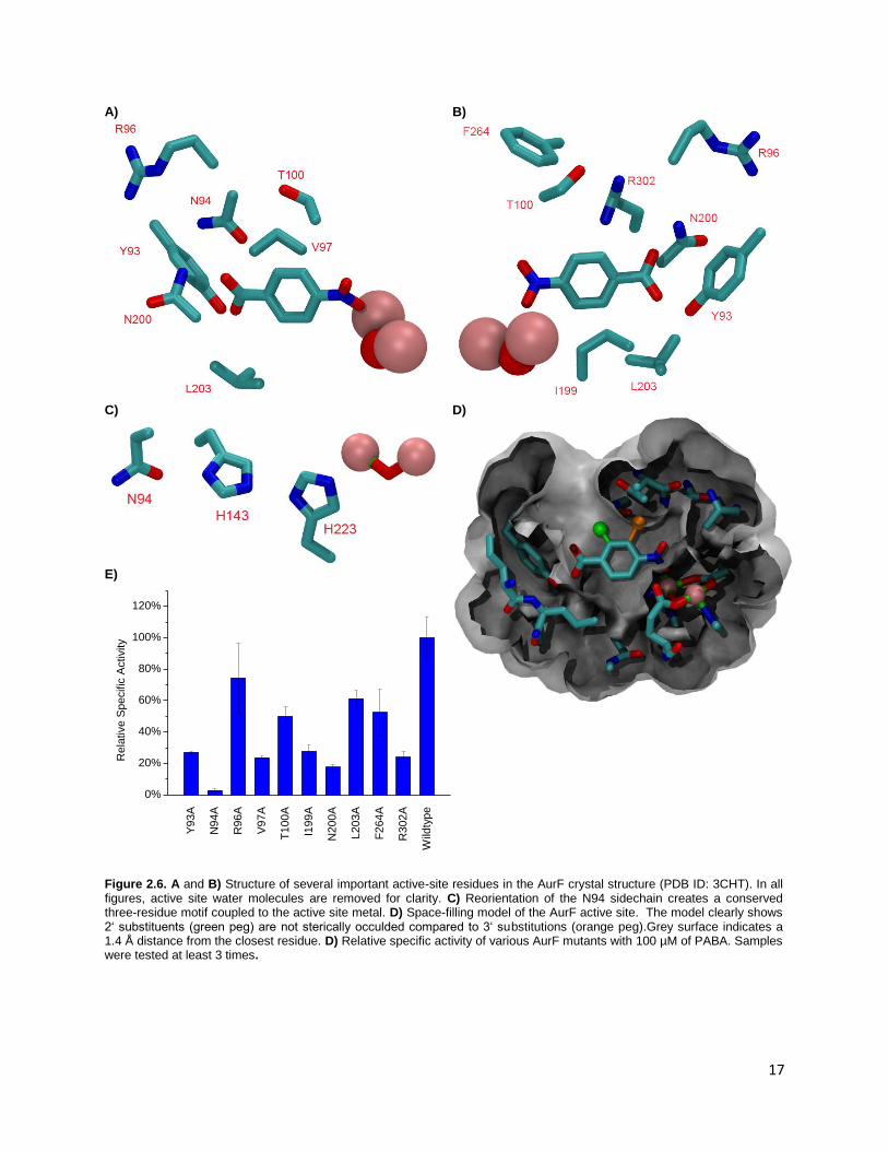
To determine which residues are important for catalysis or substrate recognition in AurF, alanine
scanning was performed on several different residues in the active site (Figure 2.6A-E) . In most cases,
important trends could be observed, while in a few others, the activity relationship could not be easily
defined. The crystal structure of diiron AurF gives several hints as to the structural role of important
active site residues (Figure 2.6 A and B). The minor difference in Km over a 2 pH unit range suggests that
the substrate recognition is dependent mostly on residues that do not change their ionization within the
pH range of the enzyme. The only protonatable groups that do not complex the active metals are
residues D135, D226, H143 and H223, and these four do not directly interact with the native substrate.
Alanine mutation of residues directly surrounding the substrate (Y93, V97, T100, N200, or R302)
decreases the activity of AurF to 50% or less of the wildtype. Y93, N200 and R302 are responsible for
recognition and binding the carboxylate end of the native substrate either directly or through water-
mediated contacts; however, none of these residues are completely essential for AurF activity,
indicating some redundancy in the carboxylate recognition (Figure 2.6A and B). Comparison with several
homologs shows that in general, residues with sidechains that can participate in hydrogen bonding are

16
favored at these positions (Table 2.1). Residues directly adjacent to the aromatic ring (V97 and T100)
also show similar reductions in activity. These residues delineate the side and the top of the active site
respectively, and mutation of these residues to alanine could allow the substrate to drift farther away
from the diiron center or alter the structure of the active site. However, it could also be argued that an
alanine mutation at these sites would not be a sufficiently drastic mutation from the previous mutation;
V97 is situated facing the aromatic ring, and one methyl group could concievably be enough to align the
substrate to the metal center. The counterpart of the T100 residue in a few AurF homologs is actually
alanine, (Table 2.1) which indicates that at best that T100A is a conservative mutation. The relatively
smaller decrease in AurF activity for this alanine mutant collaborates this fact. Thus, the decrease in
activity for these mutants is surprising considering the relatively minor steric changes from the wildtype
residues.
17
A)
B)
C)
D)
E)
Y9
3A
N9
4A
R9
6A
V97A
T1
00
A
I19
9A
N2
00
A
L203A
F2
64
A
R3
02
A
Wild
typ
e
0%
20%
40%
60%
80%
100%
120%
Re
lative
Sp
ecific
Activity
Figure 2.6. A and B) Structure of several important active-site residues in the AurF crystal structure (PDB ID: 3CHT). In all figures, active site water molecules are removed for clarity. C) Reorientation of the N94 sidechain creates a conserved three-residue motif coupled to the active site metal. D) Space-filling model of the AurF active site. The model clearly shows 2‘ substituents (green peg) are not sterically occulded compared to 3‘ substitutions (orange peg).Grey surface indicates a 1.4 Å distance from the closest residue. D) Relative specific activity of various AurF mutants with 100 µM of PABA. Samples were tested at least 3 times.
18
Table 2.1. Protein sequences of AurF and several related enzymes.
AurF ---------------MREEQPHLATTWAARGWVEEEGIGSATLGRLVRAWPRRAAVVNKA 45
Kribbella flavida DSM 17836 -----------------------MTTFAG-----EDTIGLATLKRLADTWPRRAAVRRDL 32
Haliangium ochraceum DSM 14365 ------------------------MQNAH-----EIKVNHQVIRGLANNWSKRCTIRSQK 31
Burkholderia glumae BGR1 ---------MIIDDLQCSDADGLVHLPGLPSFDPADEAENAVISRLAGNWHRRSTVKRD- 50
Rhodococcus opacus PD630 ---------MRDHTVAGASKDGHIQLAGLPPFDPRDPTENAVISRLVGNWHRRAAVKRA- 50
Micromonospora sp. ATCC 39149 ----------------------------------MTITNTKLLTQLNHAWPRRATVCDMA 26
Myxococcus xanthus DK 1622 MRRQTPPARLQTRNDSPLQLKADDGALVTQPLSGDSSAARRFLTRLGPHWARQAAVKNK- 59
Pseudomonas syringae pv. tomato -------------------------------------------MSFIDAWEGRATIRTR- 16
: * :.::
AurF DILDEWADYDTLVPDYPLEIVPFAEHPLFLAAEPHQRQRVLTGMWIGYNERVIATEQLIA 105
Kribbella flavida DSM 17836 AIEGQAGEYDATLPDYPAHLMPFAEHPDFLAATPEQRDRVMSGMWLGYNQRVIATESLIA 92
Haliangium ochraceum DSM 14365 YPFNADEQYDPNIPDYPHEMVPFWNHPRFESVSEDKKQMLLTWAWIVYNERTIAAEEYVA 91
Burkholderia glumae BGR1 -EPDLDELFDAGRADYPEAIVPFRDHPTWQAMPDALRSRLLSWAWIAYNRHTVLAEQRVA 109
Rhodococcus opacus PD630 -EPDVYALFDRDRPDFRDDMIPFRSHPIWQSLSDEMRSRLCSWGWVAYNRNTVLIEQRIA 109
Micromonospora sp. ATCC 39149 -KFRVEGEFDPTRADYPAALVPFFTHDAIQRLDDPLRQAILTWGWIGYNRRTVAAEDLVV 85
Myxococcus xanthus DK 1622 -EPSLTELLEPAKPDFAERLLPFRDHPTYQGLDEAMKRRVLSCGWLIYNERVVRVELDVV 118
Pseudomonas syringae pv. tomato ---PRRIVENDEKLIYPLSRQPLVLSDTFTRECAHLRDYALVQSLYKFINDVVIFETEIV 73
: : *: : : . .: * :.
AurF EPAFDLVMHGVFPGSDDPLIRKSVQQAIVDESFHTYMHMLAIDRTRELR-KISERPPQPE 164
Kribbella flavida DSM 17836 NPAFELVMQRVFPGSDDRVIQQTVQQSLVDESFHTYMHMMAVNRTCDLR-GIGERPKQPT 151
Haliangium ochraceum DSM 14365 NPAFGLIMHDRFPGCATIDYKNSIQQSLIDEHFHTFMHINGIHRTKVTR-NITSAPKFPY 150
Burkholderia glumae BGR1 NPAFALVMDGEFPGLGGQDMDIALAQAMVDEQYHTLMHINASALTRRKRGNPFPDSALPE 169
Rhodococcus opacus PD630 NPAFELVIAGEYPGIGGQQLELAVAQAMVDEQYHTLMHINGSAVTRRMRQREFSDRVLPD 169
Micromonospora sp. ATCC 39149 NPALNYLASEILAG-DDWVFTESVRQTLIDEHYHTLMHLSAIQRTRAQR-AITIDMELPM 143
Myxococcus xanthus DK 1622 NPVCNDVLLSKLPGATSQAARESMAQALVDEAYHILLVVRACRLTREQR--GLEDLRLPV 176
Pseudomonas syringae pv. tomato DKTARSIAKDNFAIRFPFACRYDAMTVVVDEDYHALVAMDFMQQTIALT--GIEPIQLPT 131
: . : . ::** :* : : * *
AurF LVTYRRLRRVLADMPEQWERDIAVLVWGAVAETCINALLALLARDATIQPMHSLITTLHL 224
Kribbella flavida DSM 17836 LITYRRLQAVLAEMTEQWQRDVAVLVWGAVAETCINALLGLIARDPGVQPMHSLITKLHL 211
Haliangium ochraceum DSM 14365 SVTYRRLLEAQSKVSDTWEKELLTLTFAIVSEISINAYLDLIADNPTIQPTHRLIAKLHA 210
Burkholderia glumae BGR1 SHTSRAHRRLRAHAAERWQRSLTTLAFATVSEISINAYLDLLADDHDIQVVNSTTAKLHN 229
Rhodococcus opacus PD630 SHITTIHQQHLDRCGERWERSLTTLAFATVAEISINAYLELLADDQEIQVVNSTTVKLHN 229
Micromonospora sp. ATCC 39149 SVTYRELEALKATLAEPWQRQLAAVVFATVAEISVNAFLDILADDETIQPQNRSVAQLHN 203
Myxococcus xanthus DK 1622 VGVVRRMHARQASCAEAWQRDLVQLMTGVATEMCISRYLSLLSTASEIQAFNRVTTALHQ 236
Pseudomonas syringae pv. tomato EIELSRAIPAALALAPEHLRSAVELICVAIAENTVTHDVAAFAKDDSVKQSIKGLMADHL 191
:. : :* :. : :: :: *
AurF RDETAHGSIVVEVVRELYARMNEQQRRALVRCLPIALEAFAEQDLS--ALLLELNAAGIR 282
Kribbella flavida DSM 17836 RDESAHGSVIVEVVKILYARMNQSQRDVLVRCLPPALESFGVQDPM--ALRIELRTAGIA 269
Haliangium ochraceum DSM 14365 HDENAHAYLLQEAGKSLYLEMNDKQRRIFLQTLPIALEAFLAHDYS--AWEVILDFLKID 268
Burkholderia glumae BGR1 RDEYCHASISDEMAKLVYDVLDPVKRRFFLDMLVAGLDAFVATDYS--TWEAIFRIEKVT 287
Rhodococcus opacus PD630 RDEYCHASISAEMLKQVYEALPTDRRRFLLDEIVAGLEAFVAPDFT--TWETIMAFEGIA 287
Micromonospora sp. ATCC 39149 RDEYAHSKTLGEISKVVFHRLNAKQKAFFIETLPVALRAFVAQDFS--MWEAILRQLGVP 261
Myxococcus xanthus DK 1622 QDEASHVDLFGTLARDVFNALEPVQQEFVREILPLPWLWFSEGEAD--VWRSVLLQLGIP 294
Pseudomonas syringae pv. tomato LDEGRHSGFWARLVRIYWHTAAEQDRECIARILPVFIAQYLTNDIQNGFDFTLIERLQVP 251
** * : : : . : : : : :
AurF GAEEIVGDLRSTAGGTRLVRDFSGARKMVEQLGLDDAVDFDFPERPDWSPHTPR----- 336
Kribbella flavida DSM 17836 KADDIVADVGRGGSGTKLVRDYSGTQRLVSELGLSGQVDFDFPDRPDWALTPPLDGREG 328
Haliangium ochraceum DSM 14365 GASEILADSRSSSTNTSLSRDYSGLKKMAEELDVVDKLEFDFGN--------------- 312
Burkholderia glumae BGR1 GWEKMLADVRAEKSGSRLVRDYSGLYSLMSDMNVLGDVDFDWGLAVTDK---------- 336
Rhodococcus opacus PD630 GWEKAAAEVREAQTGTHLVQDHSGVHTLLSEMDVLDQVHFGWGERTDRQR--------- 337
Micromonospora sp. ATCC 39149 NGAQIVRECRQPGSGNTLTRDYSGLHRLADELGILGQLDFEFAGTTRIAH--------- 311
Myxococcus xanthus DK 1622 RADVMMDDCIANKLLSANERAMTDAQKFSEALGIDN-IHWDRAAALL------------ 340
Pseudomonas syringae pv. tomato DPVRQALKAETLALSFPVNRHHPLIGNIMRFFKSSSMLDDPYVQRALAHYLPVQGSLQ- 309
. : . : : . :.
Protein sequences for AurF and AurF-like proteins from several organisms. Highlighted in red are residues tested by alanine
scanning. Residues marked with an asterisk are strictly conserved, while colons and periods indicate lesser degrees of
conservation. Analysis was performed using the ClustalW2 server at http://www.ebi.ac.uk/Tools/msa/clustalw2/

19
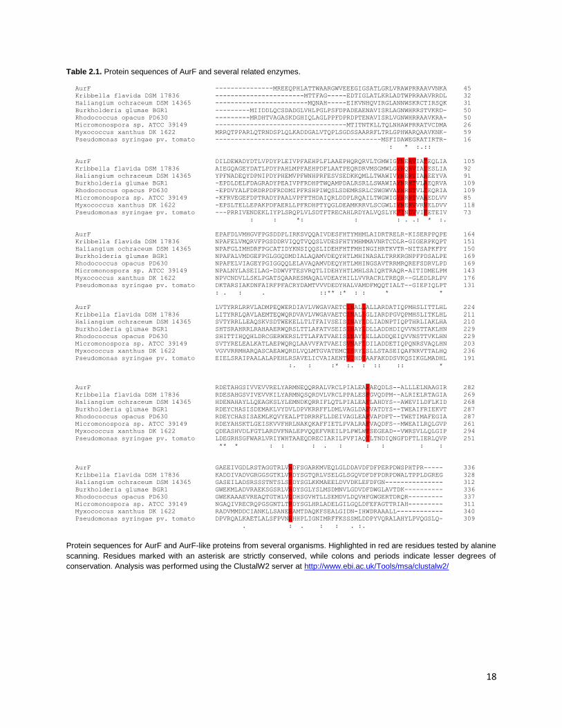
Modelling the rotamers of the N94 residue indicates that it may be involved in modulation of the
redox activity of the active metals (Figure 2.6C) by orienting residues H143 and H223. Similar Asp-His
couples mediate enzyme activity in peroxidases by modulating the pKa of the histidine residue.27,28 A
BLAST search revealed that N94 is very strongly conserved through several probable AurF-type enzymes
(Table 2.1), with the histidines are also being well-conserved. Substitution with alanine shows a sharp
decrease of activity, however activity was not completely abolished, indicating that this residue may
function in a structural role rather than as a modulator of electron transfer or redox. This is
corroborated by the the observation that mutation of this residue to alanine caused an extreme
decrease in solubility of the protein, suggestive of misfolding or instability. Similar results on AurF
insolubility have been observed for residues related to the electron transfer of AurF (W35A and
D135A).29 The activities of mutations R96A, I199A, L203A and F264 are more difficult to explain. The
R96A mutant was previously reported in literature not to have activity in vivo, but according to our
results, show around 75% activity.11 Zocher et al. rationalized that this arginine was important in the
carboxylate binding of AurF and by removal of this functionality, the enzyme would be deactivated. This
was supported by their crystal structure of the dimanganese version of AurF. However, in the diiron
version of AurF, R96 is oriented away from the active site and does not directly bind the substrate. That
is not to say that the residue plays no role in binding; it may assist in water-mediated contacts to the
substrate, or be flexible enough to bind directly as evidenced by the small drop (around 25%) in activity
compared to the wildtype. Residue I199 lines the cavity of the active site and separates it from the
exterior of the protein. Although somewhat far from the substrate (6.8 Å), this residue walls off two
water molecules that constrain the substrate. Mutation to alanine undermines the structure of the
active site by creating an opening accessible to bulk solvent. Strongly conserved residue L203 forms the
“floor” of the AurF active site, and mutating it to an alanine halves the activity of the enzyme. Similar to
V97, the most likely role of this residue is to restrict the substrate in the active site. While Y93 and N200

20
provide significant hydrogen bonding to PABA to restrain it in the binding pocket, L203 complements
this binding by providing a physical barrier below the substrate. Similar to R96, the positioning of F264 is
quite different in the diiron and dimanganese versions of AurF. In the dimanganese enzyme, F264 aligns
parallel to PABA and acts to align it to the active site, while in the diiron version, F264 is ~7 Å away from
the substrate and is perpendicular to the PABA aromatic ring. As this residue is located next to R302, the
F264A mutation allows the R302 residue to shift position, disrupting its water-mediated contacts with
the substrate. This leads to the 50% decrease in activity for this mutant.
2.5. Substrate Specificity
The substrate specificity of AurF was tested with a variety of different compounds (Figure 2.7 A and
B) at high substrate concentrations in order to saturate the enzyme. In general, our results show similar
trends to literature but with higher activities due to the optimized assay conditions.10. In certain cases,
significant exceptions were observed. The native substrate PABA (1) has a specific activity of 1.68 ± 0.25
µmol/ mg-1 min-1 at a concentration of 250 µM of substrate, indicating some slight substrate inhibition
compared to the data at 100 µM. From the data, compounds with substitutions at the 2’ position (2-6)
retained higher activity compared to the 3’ position (7-11). Notably, compound 2 showed nearly
identical activity to the native substrate. Steric and electrostatic effects are the probable reason for the
preference for 2’ substituents; approach of 3’ substituted compounds towards the metal center is
disfavored by steric clashes with the residues surrounding the active site (Figure 2.6D), while the 2’
position is more open. The 2’ position shows a clear preference for electronegative substituents and
several favorable hydrogen bonding interactions from Y93, R302, and N200 can provide additional
stabilization. Compound 9 has been reported to be turned over by AurF10, however reliable rates could
not be established as it spontaneously decomposed over the course of the assay, and putative
hydroxylamine and nitro products were not observed in LC-MS analysis even when larger amounts of
enzyme was added (data not shown). While compounds 7, 8, 10 and 29 show some activity and similar
21
sterics, it is not certain why 9 cannot be turned over, possibly due to oxidation or decay of the
compound being faster than AurF-catalyzed reaction.
Modification of the aromatic ring and the oxidizable amine provided interesting results. Replacement
of the benzene ring with a pyridine showed no activity due to decreased nucleophilicity of the pyridine
analog of PABA (13), while substitution of ring hydrogens with fluorine was accepted by AurF (12).
Hydroxylation of a secondary amine (14) proceeds slowly, while a benzylic amine 15 does not to react.
The result from 12 is surprising in the fact that the reaction proceeds well with the ring polarity
completely reversed, and it was previously reported that this could not occur due to the strong electron-
withdrawing effects of the fluorines.10 As fluorine is an isostere of hydrogen, PABA and 12 are similar in
size, and so the reduction in activity is not likely from steric factors. Activity towards 12 indicates that
electronic state of the substrate is a major factor in the catalysis by AurF, but the enzyme itself can
tolerate significantly different electronic states in its substrates. However, exaggerated electron-
withdrawing effects at the 2’ (6) or 3’ (11) position gives much decreased activity. As noted in literature
and as demonstrated here, the enzyme can tolerate a range of +I and –I functional groups in the
substrate.

22
A)
� B)
1 2 3 4 5 6 7 8 9 10 11 12 13 14 15 16 17 18 19 20 21 22 23 24 25 26 27 28 29 30
0%
10%
20%
30%
40%
50%
60%
70%
80%
90%
100%
110%
Activity R
ela
tive t
o P
AB
A
29
0.0%
0.1%
0.2%
0.3%
0.4%
0.5%
Activity R
ela
tive to P
AB
A
Figure 2.7. A) Compounds tested with AurF in this study. B) Relative specific activity compared to PABA (1). Samples were
tested in triplicate with rates from no-enzyme negative controls subtracted.
23
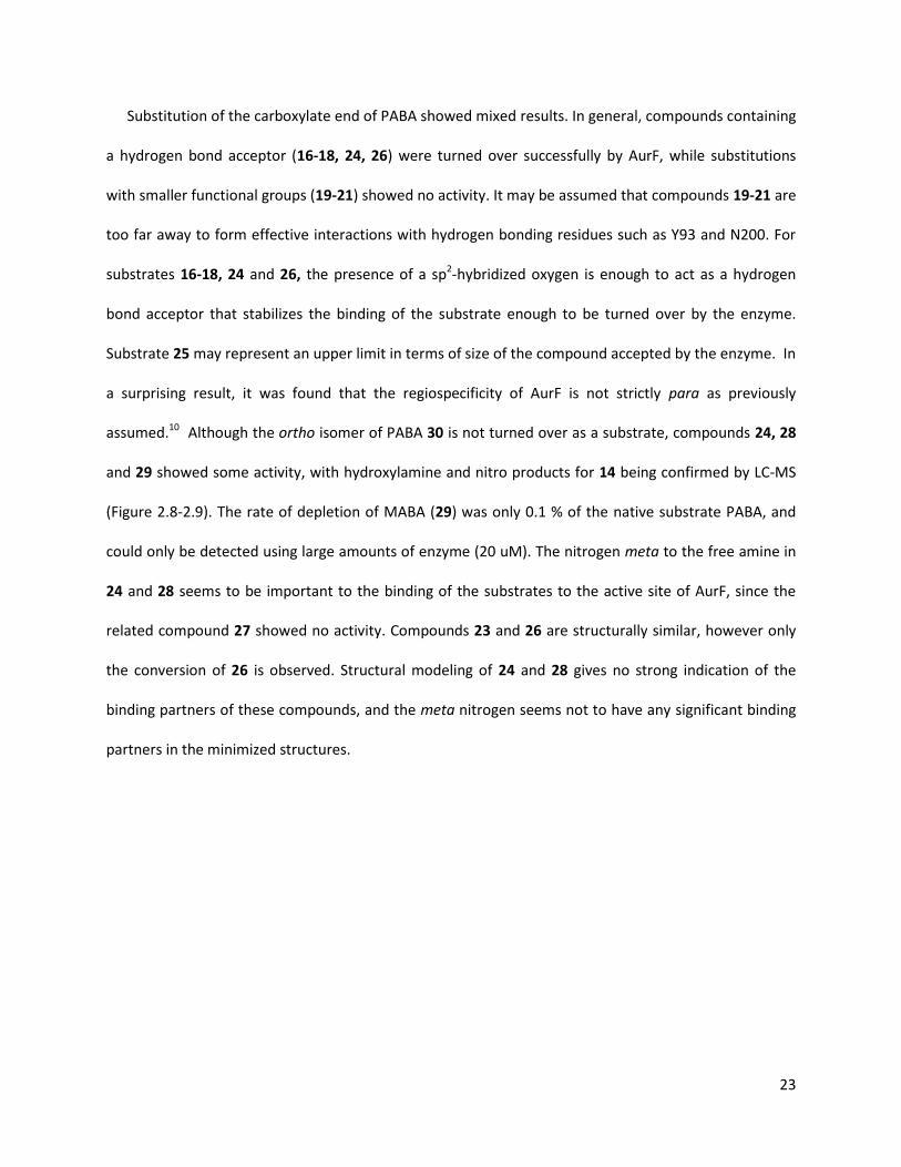
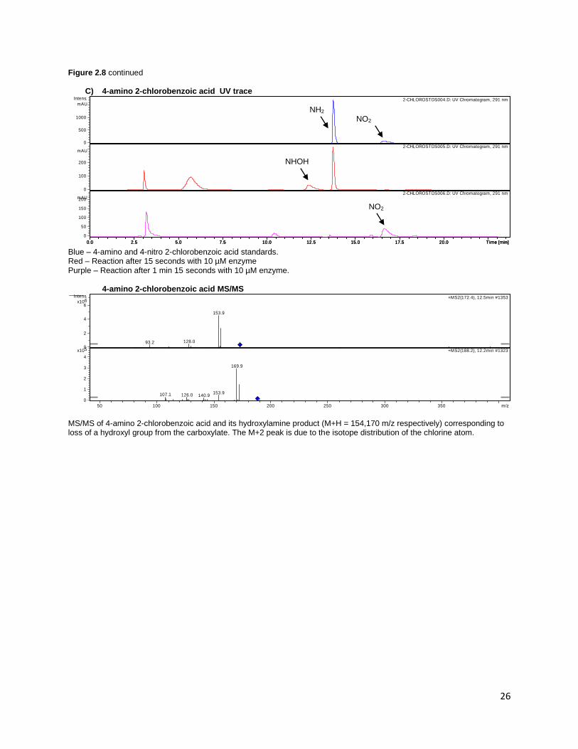
Substitution of the carboxylate end of PABA showed mixed results. In general, compounds containing
a hydrogen bond acceptor (16-18, 24, 26) were turned over successfully by AurF, while substitutions
with smaller functional groups (19-21) showed no activity. It may be assumed that compounds 19-21 are
too far away to form effective interactions with hydrogen bonding residues such as Y93 and N200. For
substrates 16-18, 24 and 26, the presence of a sp2-hybridized oxygen is enough to act as a hydrogen
bond acceptor that stabilizes the binding of the substrate enough to be turned over by the enzyme.
Substrate 25 may represent an upper limit in terms of size of the compound accepted by the enzyme. In
a surprising result, it was found that the regiospecificity of AurF is not strictly para as previously
assumed.10 Although the ortho isomer of PABA 30 is not turned over as a substrate, compounds 24, 28
and 29 showed some activity, with hydroxylamine and nitro products for 14 being confirmed by LC-MS
(Figure 2.8-2.9). The rate of depletion of MABA (29) was only 0.1 % of the native substrate PABA, and
could only be detected using large amounts of enzyme (20 uM). The nitrogen meta to the free amine in
24 and 28 seems to be important to the binding of the substrates to the active site of AurF, since the
related compound 27 showed no activity. Compounds 23 and 26 are structurally similar, however only
the conversion of 26 is observed. Structural modeling of 24 and 28 gives no strong indication of the
binding partners of these compounds, and the meta nitrogen seems not to have any significant binding
partners in the minimized structures.

24
Figure 2.8 LC-MS data demonstrating turnover of (A) 3-amino acetanilide 24, (B) 4-aminoacetophenone 17 and (C) 4-
amino-2-chlorobenzoic acid 3 to their corresponding hydroxylamine intermediates. The nitro compound could not be
observed on MS for 4-aminoacetophenone and 4-amino-2-chlorobenzoic due to the lack of ionizable groups, but could be
detected on UV.
A) 3-aminoacetanilide UV trace
Top – Standards (Left peak: 3-aminoacetanilide; right peak 3-nitroacetanilide) Bottom – 10 min reaction
3-aminoacetanilide MS/MS
MS/MS of amino, hydroxylamine, and nitro components of 3-aminoacetanilide (M+H = 151,167,181 m/z respectively). Major fragmentation peak in amino and nitro compounds corresponds to a loss of the acetyl group in LC-MS. Hydroxylamine major peak corresponds to loss of oxygen. 4-aminoacetophenone UV
SUBST-NEW000026.D: UV Chromatogram, 241 nm
0
200
400
600
Intens.
mAU
0.0 2.5 5.0 7.5 10.0 12.5 15.0 17.5 20.0 Time [min]
SUBST-NEW000023.D: UV Chromatogram, 241 nm
-50
0
50
100
mAU
0.0 2.5 5.0 7.5 10.0 12.5 15.0 17.5 20.0 Time [min]
89.3
110.1
133.0
+MS2(151.5), 4.6min #242
108.3
149.0+MS2(167.7), 8.5min #446
0
2
4
5x10
Intens.
0
1
2
6x10
50 100 150 200 250 300 350 m/z
93.3
139.0
+MS2(181.0), 15.3min #800
0.00
0.25
0.50
0.75
1.00
6x10
Intens.
50 100 150 200 250 300 350 m/z
NH2
NH2
NO2
NO2 NHOH
25
Figure 2.8 continued
B) 4-aminoacetophenone UV trace
Top – Standards (Left peak: 4-aminoacetophenone; right peak 4-nitroacetophenone). Bottom – 105 s reaction
4-aminoacetophenone MS/MS
MS/MS of 4-aminoacetophenone and its hydroxylamine product (M+H = 136,152, respectively). Peak at 94 m/z for the amino compound corresponds to formation of aniline by neutral loss of acetyl group. A similar m/z loss is seen in the hydroxylamine for the 110 m/z peak.
ACETOPH000011.D: UV Chromatogram, 291 nm
0
100
200
300
400
500
Intens.
mAU
0.0 2.5 5.0 7.5 10.0 12.5 15.0 17.5 20.0 Time [min]
ACETOPH000009.D: UV Chromatogram, 291 nm
0
50
100
150
200
250
mAU
0.0 2.5 5.0 7.5 10.0 12.5 15.0 17.5 20.0 Time [min]
94.2
391.3
+MS2(136.1), 14.0min #1280
82.3
92.1
110.0
134.9391.2
+MS2(152.0), 13.6min #12360
1
2
3
4
5
6x10
Intens.
0
2
4
6
5x10
50 100 150 200 250 300 350 m/z
NO2
NHOH
NH2
NO2
NH2
26
Figure 2.8 continued
C) 4-amino 2-chlorobenzoic acid UV trace
Blue – 4-amino and 4-nitro 2-chlorobenzoic acid standards. Red – Reaction after 15 seconds with 10 µM enzyme Purple – Reaction after 1 min 15 seconds with 10 µM enzyme.
4-amino 2-chlorobenzoic acid MS/MS
MS/MS of 4-amino 2-chlorobenzoic acid and its hydroxylamine product (M+H = 154,170 m/z respectively) corresponding to loss of a hydroxyl group from the carboxylate. The M+2 peak is due to the isotope distribution of the chlorine atom.
2-CHLOROSTDS004.D: UV Chromatogram, 291 nm
0
500
1000
Intens.
mAU
0.0 2.5 5.0 7.5 10.0 12.5 15.0 17.5 20.0 Time [min]
2-CHLOROSTDS005.D: UV Chromatogram, 291 nm
0
100
200
mAU
0.0 2.5 5.0 7.5 10.0 12.5 15.0 17.5 20.0 Time [min]
2-CHLOROSTDS006.D: UV Chromatogram, 291 nm
0
50
100
150
200mAU
0.0 2.5 5.0 7.5 10.0 12.5 15.0 17.5 20.0 Time [min]
93.2 128.0
153.9
+MS2(172.4), 12.5min #1353
107.1 126.0 140.9153.9
169.9
+MS2(188.2), 12.2min #13230
2
4
66x10
Intens.
0
1
2
3
4
5x10
50 100 150 200 250 300 350 m/z
NH2 NO2
NHOH
NO2

27
In a few cases, there have been discrepancies between our work and literature regarding in the
activity of AurF towards certain compounds. Compounds 16 and 17 have been shown by our work to be
turned over by in vitro AurF (Figure 2.7), while oxidation of compounds 19, 22 and 25 could not be
detected, but have previously been reported.10,17 As 16 and 17 share similar structural features (para-
oriented hydrogen bond acceptor) and comparable size with the native substrate, it would be unusual
that they would not be transformed into nitro products by AurF, and we are able to detect at least the
hydroxylamine intermediate on LC-MS. Although compound 22 is structurally and sterically quite similar
to the tested compounds, the amidine group in this compound is positively charged, which may cause
disfavorable electrostatic interactions with similarly charged residues R96 and R302. Therefore, it is
surprising that LC-MS results from other groups have shown that it can be turned over by wildtype AurF.
However, the nitro compound of 22 was only detected after reaction for 24 hours with H2O2, prolonged
conditions which we could not achieve using the PMS/NADH reaction system.5 From literature,
compound 25 has been reported to be slowly converted to its nitro product in whole-cell studies, but we
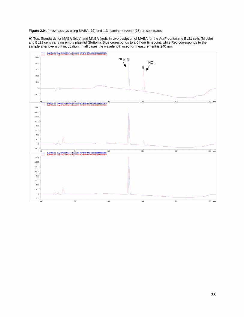
could not definitively detect any turnover on HPLC.30 The activity of AurF in vivo for some m-substituted
substrates was also determined (Figure 2.9). The partial conversion of MABA (29) to MNBA could be
observed on HPLC in an overnight incubation, collaborating results from the in vitro assay. 1,3
diaminobenzene (28) depletion was observed under similar conditions, but we could not definitively
confirm the production of either mono or di-nitro compounds. However, AurF-containing cells turned
the media from clear to brown overnight, while the media did not change color in the control cells,
indicating a reaction specifically catalyzed by AurF involving 28. In vivo reactions seemed to be more
sensitive than in vitro assays due to longer reaction conditions and large total amounts of AurF, but
overall activity may be complicated by the permeability of the substrates through the cell membrane.
28
Figure 2.9 . In vivo assays using MABA (29) and 1,3 diaminobenzene (28) as substrates. A) Top: Standards for MABA (blue) and MNBA (red). In vivo depletion of MABA for the AurF-containing BL21 cells (Middle) and BL21 cells carrying empty plasmid (Bottom). Blue corresponds to a 0 hour timepoint, while Red corresponds to the sample after overnight incubation. In all cases the wavelength used for measurement is 240 nm.
min0 5 10 15 20 25
mAU
-100
0
100
200
300
400
MWD1 D, Sig=240,8 Ref=off (LUIGI-ION-PAIR\04-30-11000002.D)
12.985
MWD1 D, Sig=240,8 Ref=off (LUIGI-ION-PAIR\04-30-11000003.D)
15.168
min0 5 10 15 20 25
mAU
-200
0
200
400
600
800
1000
1200
1400
MWD1 D, Sig=240,8 Ref=off (LUIGI-ION-PAIR\04-30-11000008.D) MWD1 D, Sig=240,8 Ref=off (LUIGI-ION-PAIR\04-30-11000034.D)
min0 5 10 15 20 25
mAU
-200
0
200
400
600
800
1000
1200
1400
MWD1 D, Sig=240,8 Ref=off (LUIGI-ION-PAIR\04-30-11000009.D) MWD1 D, Sig=240,8 Ref=off (LUIGI-ION-PAIR\04-30-11000035.D)
NH2 NO2

29
Figure 2.9 continued B) Top: Standards for 1,3 phenylene diamine (blue), 3-nitroaniline (red) and 1,3 dinitrobenzene (green). In vivo depletion of 1,3 phenylene diamine for the AurF-containing BL21 cells (middle) and BL21 cells carrying empty plasmid (bottom). Blue corresponds to a 0 hour timepoint, while Red corresponds to the sample after overnight incubation. The AurF-expressing 1,3 phenylene diamine media turned brown after overnight incubation, while the same media for the negative control remained white
min0 5 10 15 20 25
mAU
-200
0
200
400
600
800
1000
1200
1400
MWD1 D, Sig=240,8 Ref=off (LUIGI-ION-PAIR\04-30-11000004.D) MWD1 D, Sig=240,8 Ref=off (LUIGI-ION-PAIR\04-30-11000005.D) MWD1 D, Sig=240,8 Ref=off (LUIGI-ION-PAIR\04-30-11000006.D)
min0 5 10 15 20 25
mAU
0
500
1000
1500
2000
MWD1 D, Sig=240,8 Ref=off (LUIGI-ION-PAIR\04-30-11000011.D) MWD1 D, Sig=240,8 Ref=off (LUIGI-ION-PAIR\04-30-11000037.D)
min0 5 10 15 20 25
Norm.
0
500
1000
1500
2000
MWD1 D, Sig=240,8 Ref=off (LUIGI-ION-PAIR\04-30-11000012.D) MWD1 D, Sig=240,8 Ref=off (LUIGI-ION-PAIR\04-30-11000038.D)

30
2.6. Kinetic Parameters of Alternate Substrates and Mutants
Several substrates and mutants were considered for further analysis and characterization to
further understand the substrate specificity and mechanism of AurF. Aside from the native substrate,
three other compounds were chosen for kinetic analysis; 4-amino 2-chloro benzoic acid (3), 4-amino
tetrafluorobenzoic acid (12), and 4-amino acetophenone (17). In all cases, the largest contributor to the
lower activity compared to the native substrate is the much increased Km , since only a 40-60% decrease
in kcat is observed (Table 2.2). Unusually, this drop in kcat is similar in magnitude for all substrates and
mutants tested, suggesting some sort of baseline conversion rate for the substrates tested, which is
enhanced when the native substrate and enzyme are used. For compound 3, the bulk of the chloro
substituent can be accommodated by the entry pocket in the active site, and favorable interactions with
the carboxylate recognizing residues are maintained (Figure 2.6D). This causes relatively small changes
in the Km and kcat values compared to the native substrate, which is supported by the high activity seen
towards 2’ substituted compounds (Figure 2.7A). Compounds 12 and 17 are isosteres of 1 and show very
similar kinetic parameters to each other, however the basis for their kinetic parameters stem from
different sources. The substitution of the hydroxyl group in 1 to a methyl group in 17 increases the Km by
ten-fold, indicating that hydrogen bonding plays a major role in the stabilization and binding of the
substrate to the active site pocket. Electrostatics of the active site also plays a large role in substrate
binding, as evidenced by compound 12. In spite of the near-identical sterics of 1 and 12, the Km values
for 12 are perturbed significantly, due to the reversal of polarity of the aromatic ring by fluorine
substituents. However, the value for kcat for 12 and 17 are almost identical, indicating that electrostatic
effects do not strongly affect the rate of catalysis.
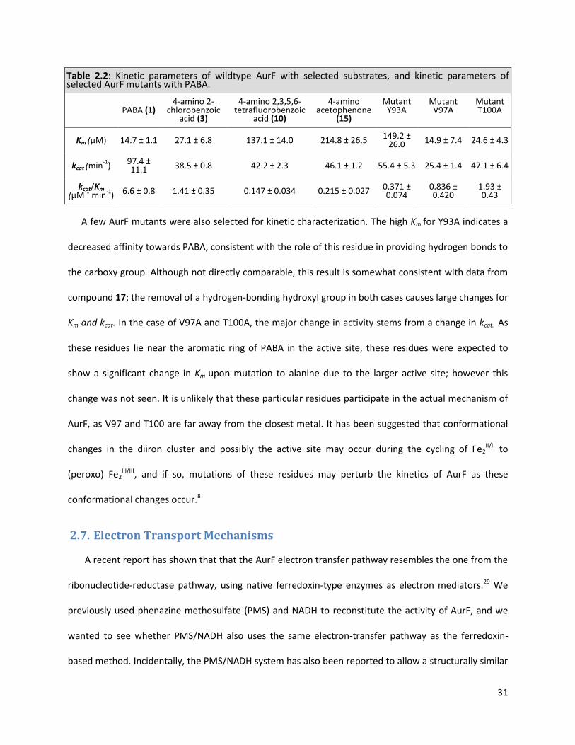
31
Table 2.2: Kinetic parameters of wildtype AurF with selected substrates, and kinetic parameters of selected AurF mutants with PABA.
PABA (1) 4-amino 2-
chlorobenzoic acid (3)
4-amino 2,3,5,6- tetrafluorobenzoic
acid (10)
4-amino acetophenone
(15)
Mutant Y93A
Mutant V97A
Mutant T100A
Km (µM) 14.7 ± 1.1 27.1 ± 6.8 137.1 ± 14.0 214.8 ± 26.5 149.2 ± 26.0 14.9 ± 7.4 24.6 ± 4.3
kcat (min-1
) 97.4 ± 11.1 38.5 ± 0.8 42.2 ± 2.3 46.1 ± 1.2 55.4 ± 5.3 25.4 ± 1.4 47.1 ± 6.4
kcat/Km (µM
-1 min
-1) 6.6 ± 0.8 1.41 ± 0.35 0.147 ± 0.034 0.215 ± 0.027 0.371 ±
0.074 0.836 ± 0.420
1.93 ± 0.43
A few AurF mutants were also selected for kinetic characterization. The high Km for Y93A indicates a
decreased affinity towards PABA, consistent with the role of this residue in providing hydrogen bonds to
the carboxy group. Although not directly comparable, this result is somewhat consistent with data from
compound 17; the removal of a hydrogen-bonding hydroxyl group in both cases causes large changes for
Km and kcat. In the case of V97A and T100A, the major change in activity stems from a change in kcat. As
these residues lie near the aromatic ring of PABA in the active site, these residues were expected to
show a significant change in Km upon mutation to alanine due to the larger active site; however this
change was not seen. It is unlikely that these particular residues participate in the actual mechanism of
AurF, as V97 and T100 are far away from the closest metal. It has been suggested that conformational
changes in the diiron cluster and possibly the active site may occur during the cycling of Fe2II/II to
(peroxo) Fe2III/III, and if so, mutations of these residues may perturb the kinetics of AurF as these
conformational changes occur.8
2.7. Electron Transport Mechanisms
A recent report has shown that that the AurF electron transfer pathway resembles the one from the
ribonucleotide-reductase pathway, using native ferredoxin-type enzymes as electron mediators.29 We
previously used phenazine methosulfate (PMS) and NADH to reconstitute the activity of AurF, and we
wanted to see whether PMS/NADH also uses the same electron-transfer pathway as the ferredoxin-
based method. Incidentally, the PMS/NADH system has also been reported to allow a structurally similar

32
non-heme diiron enzyme to turnover substrates faster than even ferredoxin-based methods.31 To that
end, we tested a few different mutants (R38A/G, W35A/F) based on docking studies of AurF with
ferredoxin. Out of the mutants tested, only W35F was soluble, and was assayed using both
ferredoxin/ferredoxin-NADP reductase and PMS/NADH. The W35F mutant has previously been reported
to be inactive in vivo due to disruption of the ferredoxin-AurF electron-transfer pathway, but active in
vitro via a peroxide-shunt mechanism.29 No activity was seen using the ferredoxin-based system, but
with the PMS/NADH system, activity comparable to the native enzyme was seen (Figure 2.3C). This
suggests that the PMS/NADH system reconstitutes the enzyme not through the complete ferredoxin
pathway, either bypassing W35, or through some different mechanism.
Table 2.3: Product Distribution of AurF using different reduction mechanisms
PABA (uM) PHABA (uM) PNBA (uM)
Ferredoxin (5 µM AurF) 191.0 ± 4.7 36.9 ±0.5 6.8 ±1.6
PMS/NADH (0.5 µM AurF) 214.0 ± 2.4 27.0 ±3.4 8.3 ±1.4
H2O2 (20 µM AurF) 245.6 ± 2.6 0.2 ± 0.02 5.3 ±0.3
Reaction conditions were 105 second reactions using 250 µM of PABA at pH 5.5. Reactions were performed in triplicate, except for Ferredoxin PHABA, which was only quantified in duplicate due to coelution issues.
Although the PMS/NADH system may not proceed through the native electron transfer pathways,
the product distribution of AurF using this system is similar to the native electron transfer pathways in
vitro, while the peroxide shunt mechanism provides a different product distribution. Accumulation of
the hydroxylamine intermediate is observed in vivo and the in vitro ferredoxin/NADPH-reductase and
PMS/NADH systems, while the primary product of the peroxide shunt is the final nitro product. (Table
2.3 and Figure 2.10). It has previously been observed that in vitro, the peroxide-shunt mechanism
produces slightly more of the nitro product than the hydroxylamine intermediate, while an in vivo
experiment showed that the PHABA accumulates in greater than 12-fold excess compared to the nitro
33
product.11 It may be the case that in the peroxide shunt mechanism, the initial conversion of PABA to
PHABA is a slow step; while in the other systems tested, the conversion of PHABA to PNBA is rate
limiting. This would explain the differences in product distribution for the peroxide shunt and the other
systems. Analogous to mechanisms found in other diiron enzymes, the peroxide shunt mechanism relies
on the formation of a µ-(peroxo)diferric intermediate from a µ-(oxo)diferric state, bypassing the
diferrous state (Figure 2.3C). 23,24,32 If the rate of µ-(peroxo)Fe2III/III generation is much slower through the
peroxide shunt mechanism (Step B in Figure 2C) than the Ar-NHOH → Ar-NO2 conversion (Step C), then
whatever hydroxylamine is generated is quickly converted to the nitro compound. Li et al. have shown
that conversion of PHABA to PNBA only requires the addition of oxygen to AurF, and does not require
the input of electrons to effect the reaction. However, if step A (the reaction pathway used by
ferredoxin and presumably PMS/NADH) is faster or goes at the same rate as step C, the accumulation of
the hydroxylamine will occur. It is unclear why the peroxide shunt is slower in AurF, but similar results
from T4MOH suggest H2O2 must lose its hydrogens before binding to the diiron center, or that binding of
the peroxide distorts the active site and slows the oxygenation reaction.24

34
Figure 2.10. Comparison of in vivo and in vitro (Ferredoxin/Ferredoxin-NADPH, PMS/NADH, or Hydrogen Peroxide) assays using PABA (1) as a substrate. In vivo trials used OD 10 cells and 1 mM of substrate, while in vitro trials used conditions seen in the methods section. In vivo - E. coli BL21 (DE3) 1 hr (1 mM, 25 µL injection)
In vitro - Ferredoxin pH 5.5 – 315 seconds
In vitro - Ferredoxin pH 7.5 – 315 seconds
min6 8 10 12 14 16
mAU
0
200
400
600
800
1000
1200
1400
1600
MWD1 C, Sig=290,8 Ref=off (LUIGI-ION-PAIR\08-20-11000005.D)
min6 8 10 12 14 16
mAU
-50
0
50
100
150
MWD1 C, Sig=290,8 Ref=off (LUIGI-ION-PAIR\08-20-11000016.D)
min6 8 10 12 14 16
mAU
-50
0
50
100
150
MWD1 C, Sig=290,8 Ref=off (LUIGI-ION-PAIR\08-20-11000022.D)
PHABA
PABA
PNBA

35
Figure 2.10 continued
In vitro - PMS/NADH pH 5.5 – 315 seconds
In vitro - H2O2 pH 5.5 – 315 seconds
In vitro - H2O2 pH 5.5 – Unquenched (~Overnight)
min6 8 10 12 14 16
mAU
-50
0
50
100
150
MWD1 C, Sig=290,8 Ref=off (LUIGI-ION-PAIR\08-20-11000029.D)
min6 8 10 12 14 16
mAU
-50
0
50
100
150
MWD1 C, Sig=290,8 Ref=off (LUIGI-ION-PAIR\08-20-11000042.D)
min6 8 10 12 14 16
mAU
0
50
100
150
MWD1 C, Sig=290,8 Ref=off (LUIGI-ION-PAIR\08-19-11000032.D)
10.472
11.137
PHABA
PABA PNBA
PABA PNBA
36
2.8. Development of an In Vivo Assay for Directed Evolution
Although AurF has been seen to have some promiscuity in the substrates it accepts, modifying its
substrate specificity is of significant interest. Previous attempts to modify its substrate specificity were
unsuccessful due to both a lack of a confirmatory in vitro assay, and a non-sensitive screening method. 13
Based on previous work, the ideal method of AurF screening would be to quantify the amount of
product present after several hours of incubation, and correlate that activity to a given mutation.
Although several methods have been utilized by a previous member in lab, screening methods suffered
from a lack of sensitivity and generalizability.13 To circumvent these issues, a method utilizing
diazotization-coupling of the arylamine precursor was developed.33 Diazotization has been used for
several decades as a method to detect either anilines or nitrites, with considerable sensitivity and
reproducibility. This method is used industrially to create dyes and color compounds, as a variety of
shades can be made by varying the composition of the diazotized amine and the coupling reagent.
Diazotization relies on the formation of a diazonium compound (Figure 2.11A) from an arylamine,
using sodium nitrite and a bronsted acid. This reactive compound is then coupled to a phenolic
compound in order to generate an azo dye, which can be easily quantified by absorbance measurements
or by visual inspection. Nitroaromatic compounds do not react under the diazotization conditions, and
the intermediates of the reaction (hydroxlamines and nitroso compounds) do not interfere as well.
Although the diazotization reaction reacts with all free amines, the stability of an alkyl diazo is much less
than that of an aromatic amine, and is quickly reacted away by solvent. Additionally, the color formation
only happens with the coupling of phenolic compounds to arylamines, increasing its accuracy. As there
are only a few arylamines (PABA, nucleotides) in high concentration in the cells, the reaction is fairly
specific. Color formation is fast, and the generated dyes are stable for days at room temperature. This
method has the disadvantage of being somewhat carcinogenic; formation of nitrogen radicals and nitric
oxide upon addition of acid to nitrite is a toxic prerequisite step in preparing the diazo intermediate.
37
Additionally, the coupling reaction requires several sequential time-sensitive steps, increasing the labor
of the screening method. However, the advantages of the diazotization-coupling approach outweigh
many of the disadvantages present in the assay, as it is the only one with the precision, stability and
sensitivity required for screening.
Previously in our lab, diazotization was tested and rejected as a screening method for AurF. This was
due to the use of thiamine as a coupling reagent, which gave a relatively weak signal, and the low
sensitivity and dynamic range of this method.13,34 However, improvements such as the use of
ammonium sulfamate to destroy any remaining nitric acid in solution, as well as using N-(1-
Naphthyl)ethylenediamine dihydrochloride (Bratton-Marshall Reagent) as a coupling reagent increase
the sensitivity of the assay to micromolar changes (Figure 2.11A).33 The proposed screening method is
illustrated in Figure 2.11B. A plasmid library containing mutants of Aurf (generated by saturation
mutagenesis or error-prone PCR) is transformed into BL21 (DE3) cells, colonies are picked, and later
regrown on a master 96 well plate. This plate is replicated depending on how many substrates are to be
tested. For each daughter plate, the cells are regrown and induced with IPTG overnight. The cell density
is then measured, and then cells are pelleted by centrifugation and resuspended in AurF in-vivo assay
buffer containing 200 µM of the substrate of interest. The cells are allowed to consume the substrate
for several hours, after which, 20-50 µL of the sample is transferred to a new 96-well plate and
diazotized.

38
Figure 2.11: A) Mechanism of diazotization reactions used in the Zhao lab for product detection. B) Screening method for detection of improved AurF mutants. A)
B)
Generate mutant library and transform into E.coli
Pick colonies and grow a master plate for the library
Replicate master plate and assay with substrate of interest
Diazotize and quantify substrate depletion
Centrifuge and transfer 20-50 uL to a development plate

39
Figure 2.12. Response of several compounds of interest to the diazotization method. All compounds with a free amine showed linear response to the coupling, with the exception of diamine compound G.
To validate the screening assay, several substrates were tested for their performance in the
diazotization procedure. As seen in Figure 2.12, diazotization of several arylamines could be visually
distinguished even at low concentrations (20 uM), with a linear standard curve from 0-200 µM.
Additionally, nitroaromatic products of the reactions did not produce any color change. The only
A B C D E F G H I J K
0
20
40
60
80
120
160
200
Compound
A C E G J
B D F H K
I
Su
bg
rate
Co
ncen
trati
on
µ
M

40
exception to this assay was the compounds p-phenylene diamine and its derivatives; no reaction to the
diazotization was observed for either the dinitro or diamino compound, but a normal reaction was
observed for the p-nitrophenylamine intermediate. Even under these results, diazotization-coupling
could provide a robust method of quantifying substrate depletion. The variation in response between
identical wells was approximately 5-10%, making it reasonable for assay conditions.
Optimum conditions were then established for the assay. As cells grown on 96-well plates do
not grow to high densities, the effect of cell concentration was tested to determine the depletion rate of
PABA as a baseline best-case scenario (Figure 2.13A). It was observed for flask-grown cells plated on 96-
well microplates, that having a higher OD600 during the assay increased the depletion of the substrate,
with the best performance seen between OD 2-4. No-cell controls and empty pTrc99a BL21 cells (OD 4)
did not show significant decreases in PABA concentrations, indicating the specificity of the assay
towards AurF activity. The well-to well variation for flask-grown cells was approximately 5%, indicating
the robustness of the detection method itself, however the cells grown on plates had significant
variations in OD600 and were difficult to control. It was noted that the most difficult part of the assay was
the cell growth, as in some cases cells did not grow well under the small volumes and limited aeration
conditions found in microplates. Additional issues such as shaking and moisture control also were
factors that could have influenced the uniformity of cell growth. In future experiments, the use of deep-
well plates would be an easier method to perform the assay; the larger culture volume would provide
greater cell density upon centrifugation, and the bigger wells would prevent spillage and cross-
contamination between wells.

41
Figure 2.13 A) Response of PABA diazotization for several different cell concentrations at various timepoints. B) Activity of various AurF mutants in vivo using the diazotization screening method. A)
B)
140
150
160
170
180
190
0 20 40 60 80 100 120
PA
BA
( µ
M)
Time (min)
Substrate Only
Neg Con
OD 4
OD 3
OD 2
OD 1.5OD 1
OD 0.5
0
0.2
0.4
0.6
0.8
1
1.2
1.4
1.6
1.8
0 60 120 180 240
A550
Time (min)
Y93A
R96A
N200A
L203A
Wildtype
Neg Control

42
To verify the screening method further, AurF mutants with known activity were tested with the
diazotization method (Figure 2.13B). Cells containing the wildtype AurF, an empty pTrc99a vector and
known mutants Y93A, R96A, N200A, L203A were subjected to the same screening method. It was found
that the in vitro activities of the mutants do not correspond to the screening method activity. This was
attributed to the solubility of the different enzymes; significant changes in solubility were observed for
the alanine mutants used, which influence their activity in vivo, but are invisible in vitro from soluble
purified protein. To increase expression of these proteins, AurF was also cloned into pMal-c2x in order
to create a maltose-binding protein – AurF fusion protein. Although the wildtype protein showed
identical activity to the MBP-tagged one in vitro, the results from the screening assay were inconclusive,
and repeats have yet to be performed.
As a final test of the AurF screening method, a series of single-site saturation mutagenesis
libraries which modify the residues Y93, R96 and T100 were tested on MABA (29) , while Y93 alone was
tested on 4-aminophenylalanine (25) as well . As shown earlier, MABA was chosen for to demonstrate
changing the regiospecificity of AurF, as the wildtype has some slight activity with this substrate.
Additionally, MABA is a good intermediate evolutionary step in evolving for trinitrobenzene compounds.
AurF has no activity towards 4-aminophenylalanine, but the compound can be biosynthesized, and has
potential as an unnatural amino acid. While no mutants were found to have increased activity towards
4-aminophenylalanine, a mutant Y93L was found to have 8% more activity than the wildtype towards
MABA in vivo after reconfirmation. However, in vitro reconfirmation of this mutant was not performed
due to the low increase in activity. Additionally, this particular mutant seems doubtful as a true mutant
due to two reasons; the leucine amino acid is overrepresented in the NNK degenerate codon and is
expected to be found in 3 / 20 colonies, but this mutant was found only once. Second, retesting the
Y93X library failed to recover this particular mutant again. Thus some other reason is responsible for this
anomalous result, possibly through host adaptation or cell density issues. Similar unknown failures in

43
engineering were also found in previous attempts to engineer AurF towards m-phenylenediamine ,
where results from the screening assay did not reflect large-scale in vivo results. 13 However, since only
single sites were mutated instead of performing randomized mutagenesis, it is possible (although
unlikely) that the sites chosen did not have significant influence in the substrate specificity. Similarily,
later efforts to rationally design AurF to accept MABA did not show improvements in activity, indicating
the difficulty in changing substrate specificity.
Several issues can account for the failure of the screening method to detect positive hits from
the saturation mutagenesis libraries. Although the screening assay works well according to control
plates and reactions, problems with the large amount of handling and growth in the 96-well plate
hampered the assay. Spillage and cell loss happened frequently with the manipulations necessary for
the assay. Additionally, comparisons between plates were difficult to follow; assays were self-consistent
within an individual plate, but not within two different plates due to differences in growth rates and
other factors. Automation of the assay by liquid handling systems could provide more reproducible
results. Finally, the solubility of AurF is problematic; it was found that AurF has a propensity to form
insoluble aggregates. Two approaches were used in literature to deal with this situation; fusion with
MBP to increase solubility, and cloning into pTrc99a to provide a slower, weaker cell induction.11,13
However, both approaches were tested, and no significant effects were observed in the screening
method. Future work will attempt to deal with these issues, such as coexpressing the enzyme with
chaperone proteins (GroEL/GroES), finding optimized growth conditions, and other solubility-increasing
directed evolution methods.35,36
2.9. Conclusions
As the prototypical aromatic N-oxygenase, characterization of AurF’s mechanism and substrate
specificity greatly widens the scope of this enzyme for possible engineering efforts. The unusual pH
optimum and kinetic parameters of the enzyme indicates that the reaction relies heavily on a proton-

44
transfer event, with kinetics, not binding being the limiting factor in the speed of the enzyme. Use of
PMS/NADH as a reductant was found to be much superior to the native Ferredoxin/Reductase
mechanisms, as well as the peroxide-based reductant. Based on the product distribution of AurF with
these different reductants, the native reaction mechanisms can be explained as a two-step mechanism;
an initial oxidation to produce the hydroxylamine intermediate, and a relatively slower step where the
intermediate is oxidized to the nitro compound directly.
AurF’s activity was probed in several ways. Characterization of AurF active-site mutants
highlighted the purpose of several residues in substrate specificity and catalysis, however certain
residues could not be easily explained. Substrate specificity was explored with a variety of compounds,
and it was found that the enzyme is not as sensitive to substitutions at the 2’ and benzyllic positions.
The main determinant of substrate acceptance was the sterics of the compound of interest, while a
general baseline rate was observed for many of the substrates observed. Finally, a screening method
was developed for directed evolution of arylamine N-oxygenases based on diazotization- coupling. This
method proved to be robust and accurate. However,issues of handling and cell growth/protein
expression variability hampered effective use of this assay. It is hoped that this characterization and
assay development could further the engineering of AurF towards being used as a unique biocatalyst.
2.10. Materials and Methods
Cloning and Library Creation
Restriction enzymes and polymerases used were from New England Biolabs. Plasmids containing
AurF used in this study were identical to previous constructs in our lab 4,9. AurF point mutants and NNK
codon saturation mutagenesis libraries were created by OE-PCR of primers containing the mutation of
interest. These inserts were digested by HindIII and NcoI, ligated into a similarly digested pTrc99a
plasmid, and electroporated into E. coli. Mutations were confirmed by sequencing. For MBP-based
45
constructs, AurF was subcloned from pTrc99a-AurF with EcoRI and HindIII as restriction sites, and ligated
into similarly digested pMal-C2x for expression.
Enzyme Purification
E. coli BL21 (DE3) cells containing a pTrc99a-6His-AurF plasmid were grown at 37°C in Terrific
Broth media and 100 ug/mL ampicillin. Cultures were induced at an OD600 of 0.6 with IPTG (final
concentration 100 µM) and grown overnight at 25°C. Cells were centrifuged and resuspended in a pH
7.4 buffer containing 20 mM NaH2PO4, 300 mM NaCl and 20 mM imidazole. The cells were then lysed
by sonication and protein purified via nickel affinity chromatography. Concentration and buffer
exchange was performed by diafiltration with a 10kDa Amicon filter, and aliquots of the enzyme was
stored at -80°C in a buffer containing 20 mM NaH2PO4 , 300 mM NaCl and 20% glycerol at pH 8.0.
Enzyme concentration was determined by the BioRad Quickstart Bradford assay using bovine serum
albumin as a standard. As a comparison, a Pierce BCA Protein assay kit was used with identical
standards as the Bradford assay. Purification of ferredoxin and ferredoxin-NADP reductase was as
previously reported.4
Enzymatic Assay
All reagents and compounds tested were obtained either from Sigma-Aldrich, TCI America,
Matrix Scientific or Acros Organics. The pH assay was performed at 20°C with a final volume of 1 mL in
the McIlvane buffer, using stock solutions of 40 mM NaH2PO4 and 20 mM citric acid (5X dilution of the
originally listed stock solutions).37 The final assay mixture contained 0.5 to 2 µM AurF, 75 µM PMS, 750
µM NADH and 100 µM of PABA in the McIlvane buffer. Addition of NADH initiated the reaction, with
timepoints taken every 30 seconds. Reactions were quenched by addition of 20 µL of 5%(v/v)
trifluoroacetic acid which gives a final pH of approximately 2. Depletion of substrate was used to
quantify reaction rate. Specific activity of the enzyme was fitted to the following equation to find the
pKa of the groups responsible for the pH dependence of the enzyme.38

46
110
10
10
10 2
1
max
pH
pka
pka
pH
Vv
For the rest of the assays, 20 mM MES buffer at pH 5.5 was used instead of the McIlvane buffer for
the sake of convenience. NADH and PMS concentrations used were identical to the previous reactions.
For kinetic determination of wildtype enzyme, 2.5-100 µM of PABA with 0.2 µM of enzyme was used,
taking timepoints every 30 seconds. Kinetic parameters were obtained by fitting the Michaelis-Menten
equation to the data using Origin 8 (OriginLab, Northampton, MA). For the alternate substrates assay, 2-
20 µM of AurF and 250 µM of substrate were used, with timepoints taken every 60 seconds for 3
minutes. Mutant and alternate substrate kinetic characterization followed their respective timepoints,
with substrate concentrations ranging from 15-400 µM. Negative controls containing no enzyme were
also tested, with rates from non-enzymatic depletion of substrate subtracted from the assayed data.
The peroxide-based assays were similar to the ones using the alternate substrates, using a final
concentration of 20 µM enzyme and 1.2% H2O2 , and omitting the NADH and PMS. Assays using
Ferredoxin/Ferredoxin-NADPH reductase used 5 µM AurF, 25 µM ferredoxin, 5 µM ferredoxin-NADPH
reductase and 750 µM NADPH.
In vivo AurF analysis
Cells were grown and induced in a manner similar to the cells used in enzymatic purification of AurF.
Cells were then centrifuged and resuspended in assay buffer to a final OD of 10.9 Substrate was added to
a final concentration of 500 µM to 25 mL of the resuspended cells, and timepoints were taken after
several hours. Cells containing a pTrc99a vector with no insert containing AurF were grown and tested in
an identical way and served as a negative control.
Screening Method
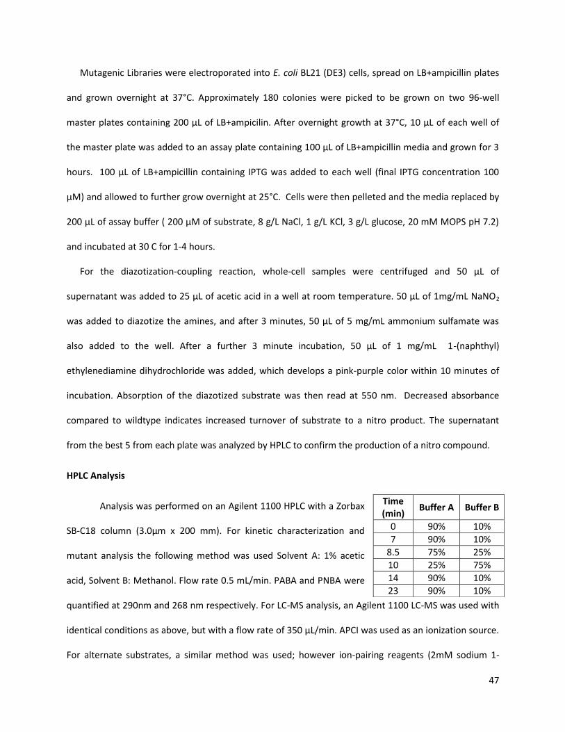
47
Mutagenic Libraries were electroporated into E. coli BL21 (DE3) cells, spread on LB+ampicillin plates
and grown overnight at 37°C. Approximately 180 colonies were picked to be grown on two 96-well
master plates containing 200 µL of LB+ampicilin. After overnight growth at 37°C, 10 µL of each well of
the master plate was added to an assay plate containing 100 µL of LB+ampicillin media and grown for 3
hours. 100 µL of LB+ampicillin containing IPTG was added to each well (final IPTG concentration 100
µM) and allowed to further grow overnight at 25°C. Cells were then pelleted and the media replaced by
200 µL of assay buffer ( 200 µM of substrate, 8 g/L NaCl, 1 g/L KCl, 3 g/L glucose, 20 mM MOPS pH 7.2)
and incubated at 30 C for 1-4 hours.
For the diazotization-coupling reaction, whole-cell samples were centrifuged and 50 µL of
supernatant was added to 25 µL of acetic acid in a well at room temperature. 50 µL of 1mg/mL NaNO2
was added to diazotize the amines, and after 3 minutes, 50 µL of 5 mg/mL ammonium sulfamate was
also added to the well. After a further 3 minute incubation, 50 µL of 1 mg/mL 1-(naphthyl)
ethylenediamine dihydrochloride was added, which develops a pink-purple color within 10 minutes of
incubation. Absorption of the diazotized substrate was then read at 550 nm. Decreased absorbance
compared to wildtype indicates increased turnover of substrate to a nitro product. The supernatant
from the best 5 from each plate was analyzed by HPLC to confirm the production of a nitro compound.
HPLC Analysis
Analysis was performed on an Agilent 1100 HPLC with a Zorbax
SB-C18 column (3.0µm x 200 mm). For kinetic characterization and
mutant analysis the following method was used Solvent A: 1% acetic
acid, Solvent B: Methanol. Flow rate 0.5 mL/min. PABA and PNBA were
quantified at 290nm and 268 nm respectively. For LC-MS analysis, an Agilent 1100 LC-MS was used with
identical conditions as above, but with a flow rate of 350 µL/min. APCI was used as an ionization source.
For alternate substrates, a similar method was used; however ion-pairing reagents (2mM sodium 1-
Time (min)
Buffer A Buffer B
0 90% 10%
7 90% 10%
8.5 75% 25%
10 25% 75%
14 90% 10%
23 90% 10%

48
heptane sulfonate) were added to each buffer, and a lower flow rate (0.4 mL/min) was used for good
separation of the compounds. Compounds were detected using wavelengths of 240, 250, 268, 290, 320,
and 380 nm and quantified via comparison to a standard curve.
Docking / Modelling Studies
Visualization and modelling studies were performed using VMD.39 For docking studies, PDB files
with structure IDs 3CHH (AurF) and 1QT9 (Ferredoxin) were submitted to the ClusPro server
(http://cluspro.bu.edu/), docked and scored.40
Synthesis of PHABA
PHABA was synthesized according to previously described literature,8 except that 1.6 equivalents of
Zn were used instead of 2 to prevent overreduction to PABA. PHABA was then purified via flash
chromatography on silica with ethyl acetate as a solvent. The compound was verified by LC/MS and
comparison to PHABA produced by AurF.
2.11. References
1. Ju, K.-S. & Parales, R. E. Nitroaromatic Compounds, from Synthesis to Biodegradation. Microbiol. Mol. Biol. Rev. 74, 250–272 (2010).
2. Winkler, R. & Hertweck, C. Biosynthesis of Nitro Compounds. ChemBioChem 8, 973–977 (2007). 3. Ferrer-Sueta, G. & Radi, R. Chemical Biology of Peroxynitrite: Kinetics, Diffusion, and Radicals. ACS
Chem. Biol. 4, 161–177 (2009). 4. Choi, Y. S., Zhang, H., Brunzelle, J. S., Nair, S. K. & Zhao, H. In vitro reconstitution and crystal
structure of p-aminobenzoate N-oxygenase (AurF) involved in aureothin biosynthesis. Proc. Natl. Acad. Sci. U.S.A 105, 6858–6863 (2008).
5. Fries, A., Winkler, R. & Hertweck, C. Structural and biochemical basis for the firm chemo- and regioselectivity of the nitro-forming N-oxygenase AurF. Chem. Commun. (Camb.) 46, 7760–7762 (2010).
6. Korboukh, V. K., Li, N., Barr, E. W., Bollinger, J. M. & Krebs, C. A long-lived, substrate-hydroxylating peroxodiiron(III/III) intermediate in the amine oxygenase, AurF, from Streptomyces thioluteus. J. Am. Chem. Soc 131, 13608–13609 (2009).
7. Krebs, C., Matthews, M. L., Jiang, W. & Bollinger, J. M. AurF from Streptomyces thioluteus and a possible new family of manganese/iron oxygenases. Biochemistry 46, 10413–10418 (2007).
8. Li, N., Korboukh, V. K., Krebs, C. & Bollinger, J. M. Four-electron oxidation of p-hydroxylaminobenzoate to p-nitrobenzoate by a peroxodiferric complex in AurF from Streptomyces thioluteus. Proc. Natl. Acad. Sci. U.S.A 107, 15722–15727 (2010).
49
9. Simurdiak, M., Lee, J. & Zhao, H. A new class of arylamine oxygenases: evidence that p-aminobenzoate N-oxygenase (AurF) is a di-iron enzyme and further mechanistic studies. ChemBioChem 7, 1169–1172 (2006).
10. Winkler, R., Richter, M. E. A., Knüpfer, U., Merten, D. & Hertweck, C. Regio- and Chemoselective Enzymatic N-Oxygenation In Vivo, In Vitro, and in Flow. Angew. Chem. Int. Ed. 45, 8016–8018 (2006).
11. Zocher, G., Winkler, R., Hertweck, C. & Schulz, G. E. Structure and action of the N-oxygenase AurF from Streptomyces thioluteus. J. Mol. Biol 373, 65–74 (2007).
12. Jayapal, P. & Rajaraman, G. On the controversy of metal ion composition on amine oxygenase (AurF): a computational investigation. Phys Chem Chem Phys 14, 9050–9053 (2012).
13. Simurdiak, M. Cloning, Expression, Partial Characterization and Engineering of a Novel N-Oxidase involved in Aureothin Biosynthesis. (2005).
14. Marion M., B. A rapid and sensitive method for the quantitation of microgram quantities of protein utilizing the principle of protein-dye binding. Anal. Biochem 72, 248–254 (1976).
15. Mathevon, C. et al. tRNA-modifying MiaE protein from Salmonella typhimurium is a nonheme diiron monooxygenase. Proc. Natl. Acad. Sci. U.S.A 104, 13295 –13300 (2007).
16. Kawai, S., Kobayashi, K., Oshima, T. & Egami, F. Studies on the oxidation of p-aminobenzoate to p-nitrobenzoate by Streptomyces thioluteus. Arch. Biochim. Biophys. 112, 537–543 (1965).
17. Platter, E., Lawson, M., Marsh, C. & Sazinsky, M. H. Characterization of a non-ribosomal peptide synthetase-associated diiron arylamine N-oxygenase from Pseudomonas syringae pv. phaseolicola. Arch. Biochim. Biophys. 508, 39–45 (2011).
18. Kontro, M., Lignell, U., Hirvonen, M. ‐R & Nevalainen, A. pH effects on 10 Streptomyces spp. growth and sporulation depend on nutrients. Lett. Appl. Microbiol. 41, 32–38 (2005).
19. Corvini, P. F. X. et al. Intracellular pH determination of pristinamycin-producing Streptomyces pristinaespiralis by image analysis. Microbiology 146, 2671 –2678 (2000).
20. Willing, A., Folmann, H. & Auling, G. Nucleotide and thioredoxin specificity of the manganese ribonucleotide reductase from Brevibucterium ammoniagenes. Eur. J. Biochem. 175, 167–173 (1988).
21. Luykx, D. M. A. ., Prenafeta-Boldú, F. X. & de Bont, J. A. . Toluene monooxygenase from the fungus Cladosporium sphaerospermum. Biochem. Biophys. Res. Commun. 312, 373–379 (2003).
22. Patel, R. N., Hou, C. T., Laskin, A. I. & Felix, A. Microbial Oxidation of Hydrocarbons: Properties of a Soluble Methane Monooxygenase from a Facultative Methane-Utilizing Organism, Methylobacterium sp. Strain CRL-26. Appl. Environ. Microbiol 44, 1130–1137 (1982).
23. Winkler, R. et al. A Binuclear Manganese Cluster That Catalyzes Radical‐Mediated N‐Oxygenation. Angew. Chem. Int. Ed. 46, 8605–8608 (2007).
24. Bailey, L. J. & Fox, B. G. Crystallographic and Catalytic Studies of the Peroxide-Shunt Reaction in a Diiron Hydroxylase. Biochemistry 48, 8932–8939 (2009).
25. Kumar, R. P., Ravindranath, S. D., Vaidyanathan, C. S. & Rao, N. A. Mechanism of hydroxylation of aromatic compounds: Evidence for the involvement of superoxide anion in a model system. Biochem. Biophys. Res. Commun. 48, 1049–1054 (1972).
26. Halliwell, B. Hydroxylation of aromatic compounds by reduced nicotinamide-adenine dinucleotide and phenazine methosulphate requires hydrogen peroxide and hydroxyl radicals, but not superoxide. Biochem J 167, 317–320 (1977).
27. Jakopitsch, C. et al. The catalytic role of the distal site asparagine‐histidine couple in catalase‐peroxidases. Eur. J. Biochem. 270, 1006–1013 (2003).
28. Schuller, D., Ban, N., Vanhuystee, R., Mcpherson, A. & Poulos, T. The crystal structure of peanut peroxidase. Structure 4, 311–321 (1996).
29. Fries, A., Bretschneider, T., Winkler, R. & Hertweck, C. A Ribonucleotide Reductase‐Like Electron Transfer System in the Nitroaryl‐Forming N‐Oxygenase AurF. ChemBioChem 12, 1832–1835 (2011).

50
30. Kawai, S., Oshima, T. & Egami, F. On the oxidation of p-aminobenzoate to p-nitrobenzoate by Streptomyces thioluteus. Biochim. Biophys. Acta 97, 391–393 (1965).
31. Das, D., Eser, B. E., Han, J., Sciore, A. & Marsh, E. N. G. Oxygen‐Independent Decarbonylation of Aldehydes by Cyanobacterial Aldehyde Decarbonylase: A New Reaction of Diiron Enzymes. Angew. Chem. Int. Ed. 50, 7148–7152 (2011).
32. Tinberg, C. E. & Lippard, S. J. Dioxygen Activation in Soluble Methane Monooxygenase. Acc. Chem. Res. 44, 280–288 (2011).
33. Bratton, A. C. & Marshall, E. K. A new coupling component for sulfanilamide determination. J. Biol. Chem. 128, 537 –550 (1939).
34. Kirch, E. & Bergeim, O. Determination of p-Aminobenzoic acid. J. Biol. Chem. 148, 445–450 (1943). 35. Nishihara, K., Kanemori, M., Kitagawa, M., Yanagi, H. & Yura, T. Chaperone Coexpression Plasmids:
Differential and Synergistic Roles of DnaK-DnaJ-GrpE and GroEL-GroES in Assisting Folding of an Allergen of Japanese Cedar Pollen, Cryj2, in Escherichia coli. Appl Environ Microbiol 64, 1694–1699 (1998).
36. Waldo, G. S. Genetic screens and directed evolution for protein solubility. Curr. Opin. Chem Bio 7, 33–38 (2003).
37. McIlvaine, T. C. A Buffer Solution for Colorimetric Comparison. J. Biol. Chem. 49, 183 (1921). 38. Copeland, R. A. Enzymes: a practical introduction to structure, mechanism, and data analysis. (John
Wiley and Sons: 2000). 39. Humphrey, W., Dalke, A. & Schulten, K. VMD: visual molecular dynamics. J Mol Graph 14, 33–38, 27–
28 (1996). 40. Comeau, S. R., Gatchell, D. W., Vajda, S. & Camacho, C. J. ClusPro: a fully automated algorithm for
protein-protein docking. Nucleic Acids Res. 32, W96–W99 (2004).

51
Chapter 3: Reconstitution of the Mangotoxin Gene Cluster
3.1. Introduction
The discovery of natural products from microorganisms is something of a challenge due to the varying
requirements of the host. The common top-down paradigm of natural product discovery involves
collecting significant amounts of a particular organism/biomass, performing chemical isolation of the
compounds of interest, and later scaling up to increase production. However slow growth, unknown
media and induction requirements, precursor availability and low productivity are challenges when
working with native hosts.1,2 Frequently, significant and expensive optimization of culture conditions are
required to produce significant amount of a compound, and in many cases, efforts to discover new
compounds are for naught. With the growth of genome sequencing technologies and freely available
gene databases, it has been found that even relatively well-characterized organisms contain cryptic
biosynthetic clusters which have unknown gene functions and products. Thus, there is significant
interest in a bottom-up approach; transferring gene pathways into tractable, well-characterized hosts
such as Escherichia coli, Streptomyces lividans and Sacchromyces cerevesiae. This has several
advantages; well-characterized promoters and genetic backgrounds could provide increased production
of compounds of interest, while careful host selection can provide easier separations and purifications
of these compounds. Additionally, genetic alterations of biosynthesis proteins such as polyketide
synthetases and non-ribosomal proteins and addition of alternative tailoring enzymes can alter the
product of a particular gene cluster in defined ways, increasing the range of possible products that can
be made.3,4 There are disadvantages, such as host incompatibility, lack of a resistance mechanism,
protein solubility and precursor availability, but overall, this synthetic biology-based approach can
leverage advances in genomics and pathway assembly to produce molecules and pathways never seen
in nature.

52
Figure 3.1. Nitroaromatic compounds isolated from Pseudomonas species.
The synthetic biology approach in reconstituting pathways also provides a way to study rare
enzymatic reactions and natural products. A few nitroaromatic compounds are known to be
biosynthetically produced, and even less is known about their biosynthesis and enzymatic activities.5–7
Thus, an initial thrust of this project was to identify gene clusters containing nitroaromatic compounds.
As a good assay for AurF was established, and with further work with CmlI confirming the validity of the
assay, it was thought to use AurF as a probe for clusters producing nitroaromatics.6,8 With the use of
modern cloning methods such as DNA assembler and Gibson Assembly, manipulations of the gene
cluster could be performed to find cryptic products.9,10 Thus, the reconstitution of a putative
nitroaromatic cluster from Pseudomonas syringae was chosen as a demonstration of this approach. This
approach is promising for several reasons. Pseudomonas species are known to be able to grow well on
xenobiotic nitro compounds such as TNT, nitrobenzene and nitrotoluene.5 Several naturally nitro
compounds have been isolated from a variety of Pseudomonas species with a variety of biological
activities (Figure 3.1). Finally, it has been reported that nitroaromatic compounds function as
chemoattractants for several Pseudomonas and Burkholderia species, indicating a biological role for
these compounds.11,12
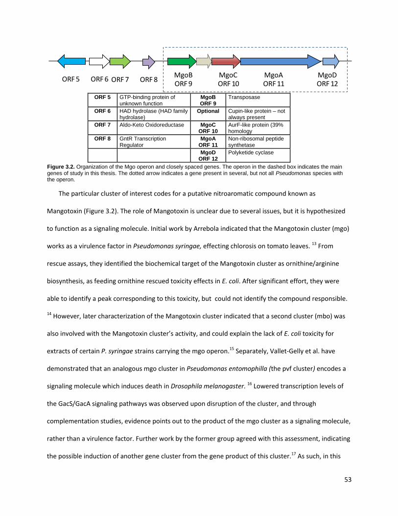
53
ORF 5 GTP-binding protein of
unknown function MgoB ORF 9
Transposase
ORF 6 HAD hydrolase (HAD family hydrolase)
Optional Cupin-like protein – not always present
ORF 7 Aldo-Keto Oxidoreductase MgoC ORF 10
AurF-like protein (39% homology
ORF 8 GntR Transcription Regulator
MgoA ORF 11
Non-ribosomal peptide synthetase
MgoD ORF 12
Polyketide cyclase
Figure 3.2. Organization of the Mgo operon and closely spaced genes. The operon in the dashed box indicates the main genes of study in this thesis. The dotted arrow indicates a gene present in several, but not all Pseudomonas species with the operon.
The particular cluster of interest codes for a putative nitroaromatic compound known as
Mangotoxin (Figure 3.2). The role of Mangotoxin is unclear due to several issues, but it is hypothesized
to function as a signaling molecule. Initial work by Arrebola indicated that the Mangotoxin cluster (mgo)
works as a virulence factor in Pseudomonas syringae, effecting chlorosis on tomato leaves. 13 From
rescue assays, they identified the biochemical target of the Mangotoxin cluster as ornithine/arginine
biosynthesis, as feeding ornithine rescued toxicity effects in E. coli. After significant effort, they were
able to identify a peak corresponding to this toxicity, but could not identify the compound responsible.
14 However, later characterization of the Mangotoxin cluster indicated that a second cluster (mbo) was
also involved with the Mangotoxin cluster’s activity, and could explain the lack of E. coli toxicity for
extracts of certain P. syringae strains carrying the mgo operon.15 Separately, Vallet-Gelly et al. have
demonstrated that an analogous mgo cluster in Pseudomonas entomophilla (the pvf cluster) encodes a
signaling molecule which induces death in Drosophila melanogaster. 16 Lowered transcription levels of
the GacS/GacA signaling pathways was observed upon disruption of the cluster, and through
complementation studies, evidence points out to the product of the mgo cluster as a signaling molecule,
rather than a virulence factor. Further work by the former group agreed with this assessment, indicating
the possible induction of another gene cluster from the gene product of this cluster.17 As such, in this
MgoDORF 12
MgoAORF 11
MgoCORF 10
MgoBORF 9
ORF 8ORF 7ORF 6ORF 5

54
report, Mangotoxin will refer to the putative product of the cluster, and not to any of its phenotypic
effects. Interestingly, a very strong inducer of the N-acylserine lactone system is a synthetic
nitroaromatic lactone which inhibits the LuxR signaling of gram-negative Vibrio fischeri.18 As other gram-
negative bacteria use similar quorum-sensing mechanisms, Mangotoxin’s putative structure may be
similar to these types of compounds (nitroaromatic lactone), giving some evidence to a possible
mechanism.
3.2. Bioinformatics Characterization of the Mangotoxin (mgo) Gene Cluster
To discover natural product clusters producing nitroaromatic compounds, AurF was used as a probe
sequence in sequence searches. This idea was validated by the identification of known N-oxygenases for
the chloramphenicol and althiomycin biosynthetic clusters from bioinformatics searching.8,19 BLAST
results of AurF showed the cluster with the most promise was the mgo cluster from several
Pseudomonas syringae hosts, which produces the cryptic compound Mangotoxin. However, as the
actual structure of Mangotoxin is unknown, the cluster itself has not been precisely delimited. According
to Arrebola et al., the Mangotoxin gene cluster (mgo) is composed of 3 genes encoding an AurF-like
protein, a non-ribosomal peptide synthase and a polyketide cyclase (Figure 3.2).14,17 These genes are
expressed in an operon structure, with an additional gene thought to be a transposase coexpressed
before the others. This motif is conserved in many other Pseudomonas and Burkholderia species, with
known nitroaromatic compounds such as pyrrolnitrin produced in these strains. 20 Only the last 3 genes
of this operon are required for observed activity, as shown by knockout experiments in literature.16,17
Although the Mangotoxin cluster’s boundaries have not been fully established, understanding the
putative product of this cluster requires analysis of each gene in the common operon. The mangotoxin
NRPS gene MgoA encodes for a single adenylation and reductase domain as well as one
phosphopantetheinylation site, indicating that the product is a single amino acid .21 Functionally, this is
similar to the NRPS CmlP found in the Streptomyces venezuelae chloramphenicol biosynthesis cluster,
55
with the exception that there is no reductase domain in CmlP. Although only 33% homology exists
between these proteins, they have similar sizes (1149 for MgoA vs 963) and the predicted substrate for
their adenylation domains is phenylalanine.22 In the case of CmlP, it is known that the actual substrate is
p-aminophenylalanine, and that several modifications of the substrate are performed as the substrate is
attached to the NRPS.23 The AurF-like gene MgoC shares only 39% homology to AurF, but shares
common structural features like the presence of a diiron binding site, and several conserved residues for
N-oxygenation. According to studies done by Sazinsky et al., MgoC functions similarly to AurF, producing
nitroaromatic compounds, with the greatest activity seen for 2-aminophenol derivatives.24 However, as
NRPS-like enzymes require the presence of an amino acid for coupling, the simple aromatic compounds
tested do not provide a method for interfacing with MgoA. Some limitations of the paper were the fact
that they did not test aminophenylalanine as a substrate, and their assay used peroxide to reconstitute
the enzyme, while in the case of AurF and CmlI, the best results were found for PMS/NADH as a
reduction system. 6,8
The two other genes in the mgo cluster are more difficult to understand. MgoB is ever-present in
the Pseudomonas species, but its function is unknown. BLAST results show that the closest known
function of this protein is as a transposase, which supports the findings of Arrebola et al. that only the
last 3 genes in the operon are important in the synthesis of Mangotoxin.17The final gene MgoD serves as
a putative polyketide cyclase or as an abscisic acid receptor, so its actual function is unknown. A similar
protein in E. coli YdgA serves to control the swarming behavior of this microorganism, but the
mechanism of this behavior is also unknown. Several other genes (ORF 5-8) close to the mgo cluster
have been reported to have no effect when they are disrupted, but conclusive evidence has not been
shown yet.
Knowing MgoC requires an arylamine substrate, and assuming the polyketide cyclase performs a
lactonization reaction, the likely product of the cluster is a compound similar to Obafluorin from

56
Pseudomonas fluoresens (Figure 3.1), as it contains all these chemical motifs. From feeding studies,
Obafluorin is derived from the condensation of p-aminophenylalanine and glycine, explaining the need
for an NRPS, as well as providing a likely substrate for the Aurf-like gene. Additionally, the lactone
present in Obafluorin could be obtained by reacting the product of the NRPS with the polyketide cyclase.
However, the Mangotoxin operon itself may not be complete; several variants of the operon encode an
additional cupin protein between MgoB and MgoC. Additionally, there is no means to attach the glycine
to the amino acid, as there is no additional condensation or adenylation domain in MgoA . Another set
of candidate compounds for the cluster’s production are nitropapuline and methyl (2,5-dihydroxy-3-
nitrophenyl) acetate(Figure 3.1). Both are attractive due to the fact that they were originally obtained
from P. syringae strains, functioning as phytotoxins and phytostimulants, respectively. 25,26 These
compounds seem to also be derived from aminophenylalanine, and the presence of the hydroxyl group
next to the nitro group coincides with the observations of Sazinsky et al. about preferred aminophenol
substrates for the enzyme.24 However, there is no role for the polyketide cyclase gene, and genes for the
methylation, deamination and hydroxylation of the compounds are missing. Thus, although possible
products produced by the Mangotoxin cluster can be identified, several factors complicate unambiguous
identification of the compound.
3.3. Cloning of the Mangotoxin Gene Cluster
A variety of cloning strategies were attempted to reconstruct the Mangotoxin cluster. The initial
approach was to clone a large portion of the neighboring genes and first try to overexpress them in the
native P. syringae host. It was assumed that cloning the genes into a multi-copy plasmid would drive the
overexpression of the target genes. The boundaries of the gene cluster were initially unknown as well,
thus this approach was a reasonable approach in forcing gene production. To this end, the DNA
assembler method was used to generate two constructs.3 First, a plasmid (hereforth referred to as
Mangotxin V1/Construct A) with approximately 15 kb of genomic DNA containing the putative

57
Mangotoxin genes was cloned and assembled via yeast-based homologous recombination. The
backbone for this construct was cloned and designed from several other fragments; a yeast helper
fragment cloned from pRS416 encoding with a CEN/ARS origin of replication and a URA3 gene for use as
a marker for uracil auxotrophy, an E. coli helper fragment from pTrc99a encoding the ampicillin
resistance and the BR322 origin of replication, and a Pseudomonas helper fragment from pUCP19
containing an origin enabling replication in Pseudomonas species (Figure 3.3A). The second construct to
be made (named Master Helper Plasmid), used the same backbone fragments as the first construct, but
had an additional LacI gene for repression of any lactose-inducible genes, as well as NotI and PacI
restriction sites, which are cut only once on the plasmid, and will be relatively rare as restriction sites
(Figure 3.3B). Thus, the Master Helper plasmid can be used as a shuttle vector for work in yeast, E. coli
and Pseudomonas species, and its propagation in all three hosts was verified. In both cases, the DNA
assembler method worked with high efficiencies to provide the correct construct.
After the successful cloning of these constructs, the logical step was to put promoters in front of
presumed gene clusters. After careful consideration, ORF 5-12 were recloned into operon-like
structures. As the DNA assembler method relies on homology to create correct constructs, the
promoters used needed to be different from each other. As it is known that Lac-based promoters
function both in E. coli and Pseudomonas species, the promoters pTrc and pTac were chosen to drive the
expression of the genes (Figure 3.3C). However, multiple attempts to construct the refactored pathway
failed, even after extensive troubleshooting. The reasons for the failure to construct the correctly
assembled pathways could not be precisely determined; however, several possibilities such as the high
homology between the pTrc and pTac promoters, the formation of a designed operon-like structure, the
use of a large number of fragments, or problems with homology regions could in combination affect the
efficiency of assembly.

58
Figure 3.3. Constructs used in this assay. Initial multi-copy overexpression plasmid A and master helper plasmid B were constructed via DNA assembler. Construct C with trc and tac promoters was designed, but could not be assembled. Construct D, E and F were constructed by Gibson Assembly, with construct D and F utilizing operons to express the mgo cluster. Construct E uses T7 promoters in front of each gene.

59
Upon the failure to make the correctly assembled constructs multiple times, alternative methods of
gene assembly were attempted. Several genes were individually cloned into pET28a and pTrc99a using
restriction/ligation to create the constructs. This provided the advantage of incorporating promoters
and terminators which could drive genes independently. One-pot Golden Gate assembly was attempted,
but with only partial success.27 Finally, Gibson assembly was used to construct several different versions
of the constructs of interest.9 Through this method, T7 and Trc-promoters were used to create
constructs in either operon or individual-gene forms respectively in either pET28 or pTrc99a-based
constructs (Figure 3.3D and F). In another construct, T7 promoters were placed in front of each gene in
the cluster via Gibson assembly (Figure 3.3E). Using those same primers, single-gene deletions were
constructed using the same strategies, but only the MgoA deletion (Del 11) was successfully confirmed.
3.4. Expression of the Mangotoxin Gene Cluster
Attempts were made to heterologously express the Mangotoxin cluster in E. coli. Single-gene
constructs cloned into pET28a were seen to express in good amounts in E. coli, with the exception of the
MgoA gene having much less expression than the others (Figure 3.4A). This was not unexpected, as the
gene was much larger than the other proteins in the cluster, and there may be a significant metabolic
burden associated with the NRPS production. All genes were seen to express in E. coli when the
heterologous T7 promoter was used in the operon, indicating the mRNA is expressed completely, with
no premature truncations. Additionally, the conserved operon structure showed that the Pseudomonas
ribosome-binding sites are compatible with the E. coli translation machinery. These results were very
promising, as they showed that E. coli could possibly operate as a good heterologous host for
production of the Mangotoxin gene product. However, the limitation of an operon-based construct is
the possible polar effect of the gene expression; later genes may have lower expression than initial
genes. This prompted the construction of a plasmid placing T7 promoters in front of every gene for
uniform expression (Figure 3.3E).

60
A)
B)
C)
Figure 3.4: Expression of the Mangotoxin genes. A) Expression of each gene cloned into pET28 in BL21 (DE3) cells. B) Expression of the operon based constructs. Left to right: Empty Plasmid, Construct F, and pET28a-T7 GoldenGate/Gibson operon. Arrows indicate the overexpressed genes. C) Expression of single-plasmid T7-promoter genes. Left to right: Negative Control, MgoA deletion, Construct E.
In the case where the T7 promoter drove the complete operon, the plasmid was found to be
stably maintained, with visible expression of all four proteins. However, cloning the T7 promoter before
every gene caused plasmid instability in certain strains. RecA deficient strains (DH5α or Top10) were
seen to maintain the plasmid well; however transformation of the plasmid into expression strains BL21
and its derivatives (BAP1) showed that recombination occurred, with the elimination of portions of the
plasmid (Figure 3.5). Thus, correct protein expression was not observed in construct E, while a similar
construct Del 11 (an identical plasmid except for MgoA deleted) at least showed expression of MgoD
(Figure 3.4C). Similar results were seen in several deletion plasmids with the repeated T7 promoter. The
cause of this was not conclusively determined, but there were several possibilities for this event. One
possible reason is the presence of several identical promoter regions, which are approximately 70 bp in
length. Since recombination is favored at highly homologous regions, identical stretches of DNA could
cause resection and modification of the plasmid.28 Contrary to this suggestion is previous precedent in
literature; it has been shown in the case of the heterologous expression of FR-900098 that multiple T7
promoters can be maintained in BL21 (DE3), and several other groups have created constructs
containing multiples of the same promoter on a single plasmid.29 Another possibility for the gene
Mgo
B
Mgo
C
Mgo
A
Mgo
D
250
150
100 75
50 37
25 20 15 10
250 150 100 75
50 37
25 20 15 10
170 135
40 33 24 17
100 72 55

61
instability is the possible toxicity of the Mangotoxin product. This is unlikely for a few reasons, such as
the lack of an inducer for the T7 promoter, as well as the putative role of the gene product as a signaling
molecule. However, since there is no T7 RNA polymerase in the cloning strains used, and the possibility
of leaky expression of the T7 promoter in BL21 (DE3) strains, this possibility cannot be discounted. To
avoid this problem in the future, each gene is planned to be cloned into Duet vectors and coexpressed
from separate plasmids simultaneously. The Duet vectors allow expression of two genes under T7
promoters in each plasmid, and since these vectors have different resistance markers and replication
origins, the Mangotoxin genes may possibly be expressed. Finally, a RecA-deficient host cell expressing
both the T7 RNA polymerase and the sfp pptase could be used in the future to retain plasmid stability
and expressivity. A possible strain to be used is the NovaBlue DE3 strain, which is designed for
expression of toxic genes under unstable plasmids. The pptase could then be supplied on the host
plasmid itself.
A)
B)
Figure 3.5: Analysis of recombination events for plasmids with multiple T7 promoters. A) Digestion of putative Construct E using EcoRV. #5 and #6 correspond to correctly assembled plasmids. These plasmids were obtained by Gibson Assembly and electroporation into Top10 cells. B) Plasmids #5 and #6 transformed into BAP1, plasmid extracted and redigested with EcoRV. The 1.2 kb band was lost in the resulting plasmid, and a ~500bp deletion was observed from the 3kb band.
The question of expression of the Mangotoxin genes in E. coli had been answered, however,
whether the native host expresses these genes was also investigated. RNA from 3-day old cultures was
isolated from the native P. syringae v. tomato hosts, and qPCR was performed to detect the expression.
Genes from ORF 5-ORF 12 (Figure 3.2) were tested, and the housekeeping gyrA gene was used as an
internal standard. However, the results from this assay were mixed. Most genes were transcribed at the
3-day timepoint, but MgoC and MgoA showed no amplification from either genomic DNA or cDNA,
#5 #6
5-A 5-B 6-A 6-B

62
indicating bad primers. Even worse, the internal standard also did not amplify. Thus, only a binary
analysis (gene expressed or not) was possible. However, these results were still useful; since the mgo
genes lie in an operon and transcription of the first and last genes MgoB and MgoD was detected, it can
be assumed that the intermediate genes MgoC and MgoA were also expressed. This is supported by
work by Arrebola et al. who by RT-PCR showed the mgo genes are expressed in an operon. Thus, it has
been shown that the operon itself is expressed natively, and the putative gene product is produced
naturally.
3.5. Detection of Products and Intermediates
As it was proven that the genes in the Mangotoxin cluster could be expressed in E. coli and that the
operon itself is expressed natively in the Pseudomonas host, the detection of the putative gene product
could be performed in two parallel approaches; heterologous reconstitution of the cluster, and product
extraction from the native host. Both approaches were tested in this project, through feeding assays and
media extraction protocols. Additionally, in vivo reconstitution of MgoC was also tested to narrow down
the possible precursors of the cluster.
As the precursors for Mangotoxin are unknown, a look into possible products from this cluster is
warranted. Nitroaromatic compounds isolated from Pseudomonas species suggest that the compound
is based on aminophenylalanine derivatives.25,26 Obafluorin has been shown to be produced from p-
aminophenylalanine and glycine, indicating that Pseudomonas species produces this arylamine.
However, E. coli does not natively produce aminophenylalanine, and although it is possible to produce
this compound heterologously, the construction of that particular pathway in the support pathway did
not succeed. Since aminophenylalanine biosynthesis branches off the shikimate/chorismate biosynthesis
pathways, a BLAST search of the signature p-aminophenylalanine biosynthesis genes was performed.
The CmlD gene from Streptomyces venezuelae encoding for a chorismate mutase was used as a
template for the search, as it is the committed step in synthesizing p-aminophenylalanine from 4-

63
amino4-deoxychorismate. Similar genes with 38% identity were found in several P. syringae species,
indicating the possibility of synthesizing a similar arylamine derivative. However, this number is very
low. Aside from the shikimate pathways, hydroxylated aminobenzoates have been obtained from TCA
cycle derivatives, raising the possibility that this is the possible precursor of the compound.30 If so, it
remains to be understood how this second pathway could be incorporated to make a phenylalanine
derivative.
Figure 3.6: Feeding studies with the pTrc99a-Gibson Construct (D) in BL21 (DE3) cells. A) Feeding with 4-aminophenylalanine . Top: Feeding of the construct with 4-aminophenylalanine. Encircled peaks indicate unique compounds produced only in the Gibson-assembled construct. Middle: Feeding of the empty plasmid negative control with 4-aminophenylalanine. Bottom: Identification of the nitrophenylalanine product by comparison of retention times.
min6 8 10 12 14 16 18
mAU
-150
-100
-50
0
50
100
150
MWD1 D, Sig=240,8 Ref=off (LUIGI-PSEUDOMONAS\06-27-12000033.D) MWD1 D, Sig=240,8 Ref=off (LUIGI-PSEUDOMONAS\06-27-12000034.D) MWD1 D, Sig=240,8 Ref=off (LUIGI-PSEUDOMONAS\06-27-12000035.D) MWD1 D, Sig=240,8 Ref=off (LUIGI-PSEUDOMONAS\06-27-12000036.D) MWD1 D, Sig=240,8 Ref=off (LUIGI-PSEUDOMONAS\06-27-12000037.D)
min6 8 10 12 14 16 18
mAU
-150
-100
-50
0
50
100
150
MWD1 D, Sig=240,8 Ref=off (LUIGI-PSEUDOMONAS\06-27-12000038.D) MWD1 D, Sig=240,8 Ref=off (LUIGI-PSEUDOMONAS\06-27-12000039.D) MWD1 D, Sig=240,8 Ref=off (LUIGI-PSEUDOMONAS\06-27-12000040.D) MWD1 D, Sig=240,8 Ref=off (LUIGI-PSEUDOMONAS\06-27-12000041.D) MWD1 D, Sig=240,8 Ref=off (LUIGI-PSEUDOMONAS\06-27-12000042.D)
min13.25 13.5 13.75 14 14.25 14.5 14.75 15 15.25
Norm.
0
500
1000
1500
2000
MWD1 A, Sig=268,8 Ref=off (LUIGI-PSEUDOMONAS\06-27-12000037.D) MWD1 A, Sig=268,8 Ref=off (LUIGI-PSEUDOMONAS\06-27-12000042.D) MWD1 A, Sig=268,8 Ref=off (LUIGI-PSEUDOMONAS\06-27-12000049.D)
4-Nitrophenylalanine Standard Gibson Plasmid Empty Plasmid

64
Figure 3.6. continued B) Feeding with PABA. Top: Feeding of the construct with PABA. Encircled peaks indicate the hydroxylamine (left) and nitro (right) compounds produced only in the Gibson-assembled construct. Bottom: Feeding of the empty plasmid negative control with PABA.
An initial feeding assay in E. coli using several aromatic amines was performed to check the
substrate specificity of the Mangotoxin cluster. The compounds 4-aminobenzyl alcohol, 4-
aminophenylalalnine, 4-aminosalicylic acid and 4-aminobenzoic acid (PABA) were used as feed
compounds. In the case of PABA and 4-aminophenylalanine, putative hydroxylamine and nitro
compounds were detected, and with the other compounds, new peaks were observed via HPLC (Figure
3.6). However, as the strain used was a BL21 (DE3) strain with no phosphopantetheine transferase gene,
it is believed that the MgoA remained in its apo-form, and thus no products from the NRPS were
expected. Thus, the new peaks are probably from the MgoBCD genes. Future work for this construct
involves repeating this feeding assay with the BAP1 strain to have a functional pptase.
The feeding assay confirmed the production of p-hydroxylaminophenylalanine fed from the
amino precursor. More tellingly, p-nitrophenylalanine was detected as well. This indicates that the
substrate specificity of this cluster is unlike AurF (which uses p-aminobenzoic acid as a precursor) and
min6 8 10 12 14 16 18
mAU
-100
-50
0
50
100
150
MWD1 C, Sig=290,8 Ref=off (LUIGI-PSEUDOMONAS\06-27-12000002.D) MWD1 C, Sig=290,8 Ref=off (LUIGI-PSEUDOMONAS\06-27-12000003.D) MWD1 C, Sig=290,8 Ref=off (LUIGI-PSEUDOMONAS\06-27-12000004.D) MWD1 C, Sig=290,8 Ref=off (LUIGI-PSEUDOMONAS\06-27-12000005.D) MWD1 C, Sig=290,8 Ref=off (LUIGI-PSEUDOMONAS\06-27-12000006.D)
min6 8 10 12 14 16 18
mAU
-100
-50
0
50
100
150
MWD1 C, Sig=290,8 Ref=off (LUIGI-PSEUDOMONAS\06-27-12000007.D) MWD1 C, Sig=290,8 Ref=off (LUIGI-PSEUDOMONAS\06-27-12000008.D) MWD1 C, Sig=290,8 Ref=off (LUIGI-PSEUDOMONAS\06-27-12000009.D) MWD1 C, Sig=290,8 Ref=off (LUIGI-PSEUDOMONAS\06-27-12000010.D) MWD1 C, Sig=290,8 Ref=off (LUIGI-PSEUDOMONAS\06-27-12000011.D)
65
more like CmlI (which uses amino-chloramphenicol as a substrate and also contains an NRPS in the gene
cluster). Definitive detection of the hydroxylamine and nitro derivatives of PABA indicated the generality
of this substrate for N-oxygenases, as seen for several other arylamine oxygenases. 6,8,24,31 Additionally,
the in vivo reconstitution of the N-oxygenase activity indicates that the ferredoxin-based electron
transfer pathway in E. coli also works for MgoC. Thus, electron transfer mechanisms devised in AurF
should be applicable to this analog.
Based on these results, in vitro reconstitution of MgoC in the cluster was also attempted. In
literature, this enzyme was reconstituted via a peroxide-based assay, with 2-aminophenol being the best
substrate. However, our findings from AurF suggest that the PMS/NADH system is far superior to this
method, and this was attempted. Six substrates (para, meta and ortho-aminobenzoic acids, P-
aminophenylalanine, p-aminolactic acid and 4-amino 3-hydroxybenzoic acid) were tested at two
different pH levels (pH 5.5 and 7.5). However, no depletion was observed of the substrate for any of the
compounds, even with 5 µM of enzyme. Work done by Sazinsky et al. showed that at least PABA is
turned over by the enzyme in vitro, but in our case, no activity was observed when their method was
attempted. This suggests that the particular purified protein was inactive, thus, the in vitro
reconstitution of MgoC has not been performed. Further repurification and assaying will be performed
to solve this issue.
Product detection from the native P. syringae v. tomato host was also attempted. Since the
Mangotoxin molecule is a small molecule which functions as a putative signaling molecule, the
production of the compound in the native host may be vanishingly small. This is collaborated by studies
with other Pseudomonas nitroaromatics, requiring growth of several liters of media to isolate low
milligram amounts of the compound. 25,26,32 As it was assumed that the product was a nitroaromatic
compound, the diazotization-coupling protocol established for AurF was tested to determine its
applicability for product detection(Figure 3.7). Briefly, reduction of the nitroaromatic compound

66
produces an arylamine compound. Treatment of this compound with sodium nitrite and acid produces a
diazonium compound, which then is coupled to an activated aromatic system (napthalenes or phenols)
to form a strongly colored compound. The detection limits of this method are in the micromolar range,
which was assumed to be sufficient for compound detection. This protocol was initially verified by
testing P. syringae grown for 7 days in Wooley’s media according to the protocol by Evidente et al.
25,33After acid extraction of 3 liters of media with ethyl acetate, the sample was concentrated and 100 µL
was subjected to reduction by Zn/HCl and the resulting amine was detected by diazotization. Control
samples of nitrophenylalalnine and the growth media were tested as well, and only significant color
development was observed in the positive nitrophenylalanine control and a weak purple color for the
organic extract (Figure 3.7B). Both the growth media and the no-diazotization negative controls showed
little color change, indicating that some nitroaromatic compound was indeed reduced and detected by
the diazotization protocol. Since the diazotized compound absorbs light at a relatively high wavelength,
it can be detected on HPLC or TLC, with its mass determined by LC-MS.
As the production of the compound was low, P. syringae was transformed with construct A
(Figure 3.3A) to try to overexpress the compound by having multiple copies of the gene cluster present.
This construct, and the native host were then grown for 5 days in M9, Wooley’s media and King’s Media
B, and then extracted with ethyl acetate under acidic conditions. A parallel extraction step using
Amberlite XAD resin was also attempted to gauge the effectiveness of the extraction method. However,
HPLC and LC-MS identification of the compound could not be established in any of the cases. Other
nitroaromatic compounds such as nitropapuline were not detected via LC-MS, which could parallel the
case seen for AurF, where spiked-in nitroaromatic standards could not be detected on LC-MS. The
diazotization protocol was not performed at this point, so arylamine precursors were not detected.
Compounding the difficulty was the fact that the only point of comparison was the native host itself; as
no gene knockouts of the cluster were created at this point, the only way to find the compound of
67
interest would be the overexpression of the gene cluster in the plasmid-based construct compared to
the wildtype. Some work was performed to try to delete the Mangotoxin cluster in the native host via
RecET-based recombineering, but as of this date the work has not been concluded.34
A final attempt to detect the compound in Pseudomonas media was attempted (Figure 3.7C and
D). Both the native host and construct A containing cells were grown in King’s Media B for 3 days, and
and extraction of the media with ethyl acetate was performed. An additional methanol extraction of
pelleted cells was performed to extract metabolites locked intracellularly. From these, small portions
were diazotized directly, or reduced first by Zn/HCl, and analyzed by HPLC. Upon visual analysis of this
sample, it was found that the King’s Media B gave a weak background signal, while extraction of both
the native cells and the construct A cells also gave a similar weak signal (Figure 3.7C). As the previously
used Wooley’s media is a defined minimal media, the lack of a diazotizable compound in the media is to
be expected, and since King’s Media B is a complex media, the presence of a diazotizable compound
from either the yeast extract or the peptone base could explain the weak signal. A strong signal could be
detected from the reduced and non-reduced wildtype media, and HPLC analysis confirmed that the
source of this signal was from diazotized PABA, possibly from the media, but more likely to be produced
intracellularly. An unknown diazotization product was also seen at low concentrations at 19 minutes,
which could possibly be the Mangotoxin product. As the assay itself is very sensitive, the low area
indicates the very small amount of this compound .Further work by HPLC and LC-MS will be performed
to confirm the identity of the new peak. Surprisingly, the diazotization protocol failed to detect any
strong nitroaromatic signal from Construct A, which was supposed to overexpress the cluster itself. The
reason for this anomaly is unclear; however, the growth of this construct was somewhat worse than
that of the wildtype (although the media was treated with Ampicillin to maintain the plasmid), and this
may have caused a difference in cell growth and metabolite production compared to the control.
Reconfirmation of the plasmid could not be performed via miniprep and digestion, due to the presence

68
of several other native plasmids in the P. syringae pv. tomato strain used. Thus, although there are
promising results in this assay, more work needs to be done to confirm the presence of a nitroaromatic
compound. Additionally, large scale growth / feeding experiments may need to be performed to isolate
enough compound for study.
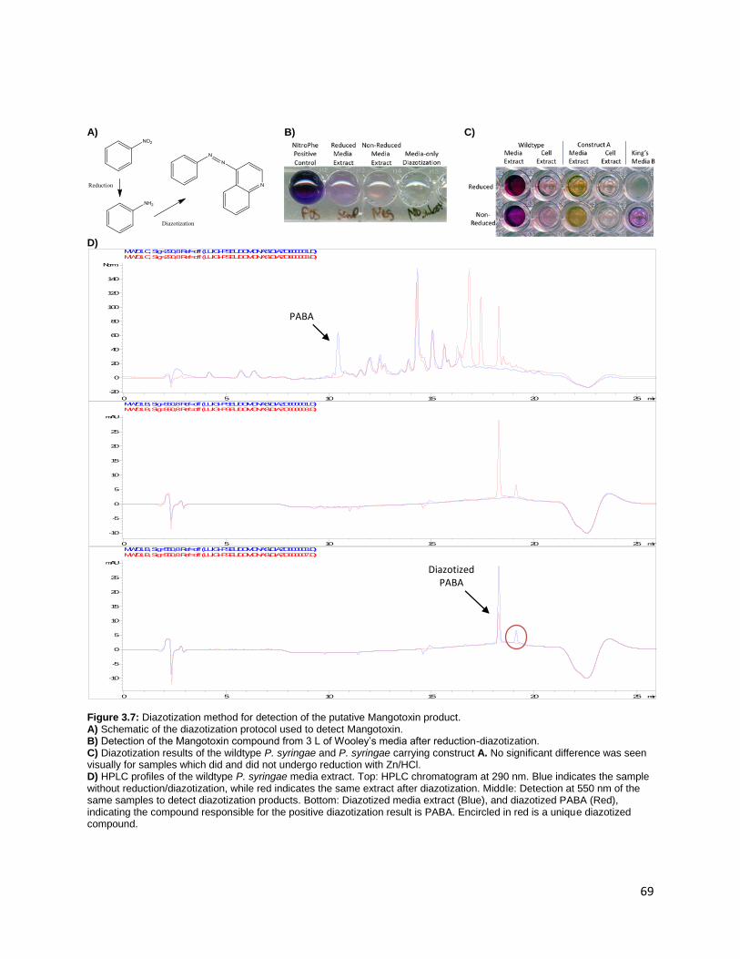
69
A)
B)
C)
D)
Figure 3.7: Diazotization method for detection of the putative Mangotoxin product. A) Schematic of the diazotization protocol used to detect Mangotoxin. B) Detection of the Mangotoxin compound from 3 L of Wooley’s media after reduction-diazotization. C) Diazotization results of the wildtype P. syringae and P. syringae carrying construct A. No significant difference was seen visually for samples which did and did not undergo reduction with Zn/HCl. D) HPLC profiles of the wildtype P. syringae media extract. Top: HPLC chromatogram at 290 nm. Blue indicates the sample without reduction/diazotization, while red indicates the same extract after diazotization. Middle: Detection at 550 nm of the same samples to detect diazotization products. Bottom: Diazotized media extract (Blue), and diazotized PABA (Red), indicating the compound responsible for the positive diazotization result is PABA. Encircled in red is a unique diazotized compound.
min0 5 10 15 20 25
Norm.
-20
0
20
40
60
80
100
120
140
MWD1 C, Sig=290,8 Ref=off (LUIGI-PSEUDOMONAS\DIAZO000001.D) MWD1 C, Sig=290,8 Ref=off (LUIGI-PSEUDOMONAS\DIAZO000003.D)
min0 5 10 15 20 25
mAU
-10
-5
0
5
10
15
20
25
MWD1 B, Sig=550,8 Ref=off (LUIGI-PSEUDOMONAS\DIAZO000001.D) MWD1 B, Sig=550,8 Ref=off (LUIGI-PSEUDOMONAS\DIAZO000003.D)
min0 5 10 15 20 25
mAU
-10
-5
0
5
10
15
20
25
MWD1 B, Sig=550,8 Ref=off (LUIGI-PSEUDOMONAS\DIAZO000003.D) MWD1 B, Sig=550,8 Ref=off (LUIGI-PSEUDOMONAS\DIAZO000007.D)
Diazotized PABA
PABA

70
3.6. Synthesis of Compounds
The nitro compounds produced by P. syringae share a motif where the nitro group is adjacent to a
hydroxyl group (Figure 3.1). This is collaborated by literature results showing that 2-aminophenol is one
of the best substrates for MgoC.24 Interestingly, this was thought to be unlikely, based on AurF substrate
specificity studies, as 3’ substituted aminobenzoates showed no activity with AurF due to steric issues.
Since this motif seems to be conserved, it may be that the substrate for the NRPS is a hydroxylated
aminophenylalanine. It was noted in literature that the positions for the hydroxyl and nitro moieties for
nitropapuline were difficult to assign, and it is possible that they may be switched on the benzene ring.
This would make the compound similar to the methyl (2,5-dihydroxy-3-nitrophenyl) acetate also isolated
from the same organism (Figure 3.8) . 25,26 However, as there are no other oxygenase or P450 genes
close to the cluster, the source of the biological hydroxylation is unknown. Thus, it was of interest to see
whether these aminohydroxyphenylalanines (AHP) could be used as a substrate for in vivo and in vitro
reactions.
Chemical synthesis of these compounds involved several laborious and time-consuming steps. 35
However, the main degradation products of pheomelanin, a compound derived from hair and
melanocytes, were exactly the AHP compounds required. As seen in Figure 3.8, a semisynthetic method
by Ito uses mushroom tyrosinase to first oxidize L-DOPA into dopaquinone. This compound then
condenses with cysteine to prepare the pheomelanin pigment. Reduction and hydrolysis of pheomelanin
produces the two isomers of AHP.36 Although the final yields for this process is low (~20 % according to
literature), the synthesis is relatively benign and straightforward, with results obtained within a few
days.
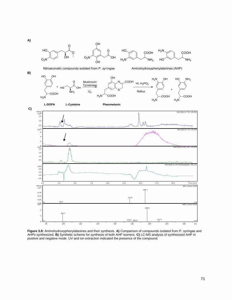
71
A)
B)
C)
Figure 3.8: Aminohydroxyphenylalanines and their synthesis. A) Comparison of compounds isolated from P. syringae and AHPs synthesized. B) Synthetic scheme for synthesis of both AHP isomers. C) LC-MS analysis of synthesized AHP in positive and negative mode. UV and ion extraction indicated the presence of the compound.
041-0201.D: TIC +All MS
041-0201.D: TIC -All MS
041-0201.D: EIC 197 +All MS
041-0201.D: UV Chromatogram, 291 nm
0.5
1.0
1.5
8x10
Intens.
0
2
4
6
7x10
0.5
1.0
1.5
2.0
7x10
-2.5
0.0
2.5
5.0
mAU
0.0 2.5 5.0 7.5 10.0 12.5 15.0 17.5 20.0 Time [min]
97.0
176.9
194.7
-MS, 4.1min #215
99.0
176.0 182.9
196.8
211.0
+MS, 4.1min #2160.00
0.25
0.50
0.75
7x10
Intens.
0
1
2
7x10
80 100 120 140 160 180 200 220 240 m/z
72
According to the protocol used, a yield of ~ 75 mg was expected for the pheomelanin given the
conditions we used for the synthesis. However, only 9 mg of product was obtained (12 % yield),
indicating that significant room for optimization remains. LC-MS analysis of the product indicated that
the synthesized compound was AHP, with some unknown byproducts (Figure 3.8C). As resolution of
each isomer could not be achieved, it was unknown how much of each isomer was present in the
sample, but as a compound 3-nitrotyrosine is commercially available and metal-mediated reduction of
that compound is straightforward. Thus, a pure standard can be obtained for at least one of the AHP
isomers. An initial feeding study was perfomed on this compound, however the previously mentioned
plasmid stability issues were encountered, and thus compound turnover was not established. Further
work such as scaling up the reaction to isolate more AHP is planned. After production of more of these
compounds, more feeding and enzymatic assays will be performed to validate these compounds as
substrates for the Mangotoxin gene cluster.
3.7. Conclusions
As demonstrated in this work, progress has been made towards the reconstitution and discovery of
the Mangotoxin gene product. Successful cloning of several constructs for expression in E. coli and
Pseudomonas were constructed, and heterologous expression of the operon has been demonstrated.
Although in vitro reconstitution of AurF-like protein MgoC failed, feeding studies demonstrating the in
vivo activity of MgoC suggest that its electron transfer mechanism is similar to AurF and other
structurally similar diiron monooxygenases. However, plasmid stability issues mar the heterologous
expression of the cluster in E. coli. These issues can be solved by judicious host and vector selection.
Parallel to the heterologous expression product detection experiments, a method was developed
based on reduction/diazotization-coupling in order to detect compound in the native host. This led to a
possible peak in the native host, which is produced in low level. The low production is characteristic of a
signaling/induction molecule, but it could also be due to other issues such as cluster protein expression
73
or substrate availability. For further feeding and enzymatic studies, aminohydroxyphenylalanine
compounds were prepared via chemoenzymatic means and characterized. In the end, this work
supports the efforts in discovering the identity and chemical characterization of the Mangotoxin
compound. However, further follow-up work is required for definitive identification and isolation of the
gene product of this cluster.
3.8. Materials and Methods
Materials
Unless otherwise noted, all reagents were obtained from either Sigma-Aldrich (St. Louis, MO),
Fisher Scientific (Pittsburg, PA), or Gold Biotechnology (St. Louis, MO).
Cloning and Standard DNA Manipulations
All oligonucleotides were obtained from Integrated DNA Technologies (Coralville, IA), while
restriction enzymes, DNA polymerases and other cloning-related enzymes were purchased from New
England Biolabs (Ipswitch, MA). Restriction/ligation cloning was performed according to standard
protocols. For antibiotic usage, 1000x stock solutions of ampicillin (100 mg/mL), kanamycin (50 mg/mL)
and gentamycin (10 mg/mL) were used. To isolate genomic DNA from P. syringae , the Wizard Genomic
DNA purification kit from Promega (Fitchberg, WI) was used. Plasmids were isolated from E. coli using
the QIAprep Spin Miniprep kit (Qiagen, Valencia, CA), while yeast plasmids were isolated using the
Zymoprep II kit (Zymo Research, Irvine, CA). Typical PCR conditions used 25 µL Buffer G (Epicentre
Technologies, Madison, WI), 50 pmol of each primer, 1 unit of Phusion DNA polymerase, and 0.5-1 µL of
template DNA. Annealing and elongation conditions were optimized based on the melting temperature
of the primers and the lengths of the PCR products desired. To create the T7 promoter-based
constructs, individual Mangotoxin genes were cloned from the genomic DNA of P. syringae by PCR with
appropriate restriction sites. PCR products were then purified and digested with the appropriate
74
restriction enzymes and ligated to similarly digested pET28a vectors (Novagen, Darmstadt, Germany) .
Similar methods were used to clone out Trc-promoter constructs using pTrc99a as a vector (Amersham
Biosciences, Piscataway, NJ). Both E. coli and P. syringae were transformed according to standard
electroporation protocols, using 0.2 cm cuvettes at a resistivity of 250 Ω and voltage of 2.5 kV. For E.
coli, electrocompetent cells were prepared according to common protocols. Similarly, P. syringae
competent cells were made using protocols developed by Schweizer et al.37
DNA Assembler
DNA assembler was performed according to the protocols found in literature.10 Briefly, primers
were designed to create PCR products with 40bp - 1kb overlaps to previous and succeeding fragments
and amplified using PCR. Around 100 ng of each PCR fragment was electroporated into the S.cerevesiae
strain HZ848 and recovered in 1 mL YPAD media. 30, 100 and 200 µL of cells were then replated on SC-
Ura plates and left to grow at 30 ⁰C for two days. Colonies were picked and yeast plasmid DNA was
isolated retransformed into E. coli DH5α for amplification. Plasmids were again isolated from E. coli by
miniprep and confirmed by digestion analysis and sequencing.
Gibson Assembly
The Gibson Assembly protocol was performed as found in literature. 9 50-100 ng of each
fragment was added to 15 µL of the Gibson premix. The assembly was incubated at 50C for 1 hour, and
0.5 µL of the product was retransformed into E. coli DH5α. Plasmids were isolated using the QIAprep
Spin Miniprep kit and confirmed by digestion analysis and sequencing. For expression studies, plasmids
were cloned into either BL21 (DE3) or BAP1. To create the T7-based operon by Gibson assembly, pET28a
was digested with HindIII and NcoI to linearize the vector. PCR was then used to amplify the fragments
from genomic DNA (for the operon based construct) or from the individually cloned pET28a-genes using
the appropriate primers.

75
In vivo assay for heterologous production of Mangotoxin
BL21 (DE3) or BAP1 cells containing plasmids of interest were grown in TB media at 37⁰C with
shaking at 250 rpm until their OD600 reached a value of 0.6-0.8. Cells were then induced with IPTG (0.5
mM final concentration) and left to grow overnight at 30⁰C under identical shaking conditions. Cultures
were then pelleted by centrifugation and washed twice with AurF assay buffer (25 mM MOPS, 8 g/L
NaCl, 1 g/L KCl and 2 g/L glucose, pH 7.2).38 The cells were then resuspended in the AurF assay buffer to
a final OD600 of 10 in 100 mL total volume. Substrates were added to a final concentration of 250 µM to
start the reaction. The cells were allowed to react the substrates at 30⁰C with timepoints taken every
hour for 4 hours. A final overnight timepoint was taken the next day.
Extraction of metabolites from resting cells
An overnight culture of E. coli was inoculated into 500 mL of Terrific Broth with the appropriate
antibiotic (Kanamycin or Ampicillin) and allowed to grow to saturation at 30C overnight. Cells were
collected by centrifugation. The remaining media was acidified by addition of concentrated HCl until the
pH reached approximately 1. The media was then extracted thrice with 200 mL of ethyl acetate, dried
with sodium sulfate, and concentrated by rotary evaporation. The extract was then reconstituted with 1
mL of methanol, and analyzed by HPLC. For P. syringae cultures, similar methods were used, except that
the innoculum culture was grown to saturation after 2 days of growth, and King’s Media B was used to
grow the culture for 3 days at 30C instead of overnight. Additionally, pelleted cells from the
Pseudomonas culture were extracted by resuspension with methanol and concentrated by rotary
evaporation.
Reduction/Diazotization/Coupling Protocol
To proceed with the reduction reaction, 100 µL of concentrated media or cell extract, 40 µL of
HCl and a small amount of Zn powder was added. This mixture was heated at 70C for 15-30 minutes to
complete the reduction. Bubbling and frothing indicated that the reaction was proceeding. The sample

76
was cooled to room temperature, centrifuged down and transferred to a new container. The
diazotization reaction was performed by adding 40 µL of 1 mg/mL NaNO2 was added to an acidic
solution of either the reduced compound, or a non-reduced sample. After 3 minutes of incubation, 40 µL
of 5 mg/mL ammonium sulfamate was added to destroy the remaining nitrous acid. After a further 3
minutes, the Bratton-Marshall reagent was added to develop the color of the compounds. Compounds
were then centrifuged to precipitate out contaminants and analyzed by HPLC or by microplate readers.
Synthesis of AHPs
L-DOPA (50 mg, 250 µmol) and L-Cysteine (45 mg, 375 µmol) were dissolved in 25 mL of 50 mM
sodium phosphate buffer at pH 6.8. Mushroom tyrosinase was added to a concentration of 500
units/mL, and oxygen was bubbled into the mixture for 4 hours at 25⁰C.36 The resulting pheomelanin
was then acidified with acetic acid at pH 3, and kept at 4⁰C overnight. The precipitate was then collected
by centrifugation and washed with 1% acetic acid twice, and once with acetone. The solid was then
dried with P2O5 and weighed. To hydrolyze the pheomelanin to AHP, the pheomelanin was dissolved in 5
mL of 57% HI with the addition of 100 mg of H3PO4. This was refluxed for 20 hours, evaporated and
resuspended in 5 mL of 0.1 M HCl. Centrifugation of the final solution provided the sample which was
verified by LC-MS analysis.
HPLC methods
An Agilent 1100 HPLC with a corresponding UV/Vis detector and
autosampler was used to detect and quantify changes in substrate and
product concentrations. Reverse-phase HPLC was performed using an
Agilent Zorbax SB-C18 column (250 mm x 3.5 mm) to separate
compounds, with buffer A being 1% acetic acid, and buffer B being
methanol. To enable better retention of polar products and substrates, the solvents all contained 2 mM
of sodium 1-heptanesulfonic acid as an ion-pairing reagent. The program used 0.4 mL/min as the flow
Time (min)
Buffer A Buffer B
0 90% 10%
7 90% 10%
8.5 75% 25%
10 25% 75%
14 90% 10%
23 90% 10%

77
rate, and used the above method to separate compounds of interest. Compounds were detected using
wavelengths of 240, 250, 268, 290 and 320 nm. Diazotized compounds were detected at 550 nm, as
products of this reaction have unique strong absorbances at this wavelength. For LC-MS analysis, an
Agilent 1100 LC-MS was used with identical conditions as above (with no ion-pairing reagents), but with
a flow rate of 350 µL/min. ESI was used as an ionization source.
3.9. References
1. Li, J. W.-H. & Vederas, J. C. Drug Discovery and Natural Products: End of an Era or an Endless
Frontier? Science 325, 161–165 (2009). 2. Wilkinson, B. & Micklefield, J. Mining and engineering natural-product biosynthetic pathways. Nat.
Chem. Biol. 3, 379–386 (2007). 3. Shao, Z., Luo, Y. & Zhao, H. Rapid characterization and engineering of natural product biosynthetic
pathways via DNA assembler. Mol. BioSyst. 7, 1056 (2011). 4. Zocher, G., Richter, M. E. A., Mueller, U. & Hertweck, C. Structural Fine-Tuning of a Multifunctional
Cytochrome P450 Monooxygenase. J. Am. Chem. Soc. 133, 2292–2302 (2011). 5. Ju, K.-S. & Parales, R. E. Nitroaromatic Compounds, from Synthesis to Biodegradation. Microbiol.
Mol. Biol. Rev. 74, 250–272 (2010). 6. Choi, Y. S., Zhang, H., Brunzelle, J. S., Nair, S. K. & Zhao, H. In vitro reconstitution and crystal
structure of p-aminobenzoate N-oxygenase (AurF) involved in aureothin biosynthesis. Proc. Natl. Acad. Sci. U.S.A 105, 6858 –6863 (2008).
7. Winkler, R. & Hertweck, C. Biosynthesis of Nitro Compounds. ChemBioChem 8, 973–977 (2007). 8. Lu, H., Chanco, E. & Zhao, H. CmlI is an N-oxygenase in the biosynthesis of chloramphenicol.
Tetrahedron 68, 7651–7654 (2012). 9. Gibson, D. G. Enzymatic assembly of overlapping DNA fragments. Meth. Enzymol. 498, 349–361
(2011). 10. Shao, Z., Zhao, H. & Zhao, H. DNA assembler, an in vivo genetic method for rapid construction of
biochemical pathways. Nucleic Acids Res 37, e16–e16 (2009). 11. Pandey, J. et al. Chemotaxis of Burkholderia sp. Strain SJ98 towards chloronitroaromatic compounds
that it can metabolise. BMC Microbiology 12, 19 (2012). 12. Parales, R. E. Nitrobenzoates and Aminobenzoates Are Chemoattractants for Pseudomonas Strains.
Appl Environ Microbiol 70, 285–292 (2004). 13. Arrebola, E. et al. Contribution of mangotoxin to the virulence and epiphytic fitness of Pseudomonas
syringae pv. syringae. Int. Microbiol. 12, 87–95 (2009). 14. Arrebola, E., Cazorla, F. M., Romero, D., Pérez-García, A. & de Vicente, A. A Nonribosomal Peptide
Synthetase Gene (mgoA) of Pseudomonas syringae pv. syringae Is Involved in Mangotoxin Biosynthesis and Is Required for Full Virulence. Mol. Plant Microbe Interact. 20, 500–509 (2007).
15. Carrión, V. J., Arrebola, E., Cazorla, F. M., Murillo, J. & de Vicente, A. The mbo operon is specific and essential for biosynthesis of mangotoxin in Pseudomonas syringae. PLoS ONE 7, e36709 (2012).

78
16. Vallet-Gely, I., Opota, O., Boniface, A., Novikov, A. & Lemaitre, B. A secondary metabolite acting as a signalling molecule controls Pseudomonas entomophila virulence. Cell. Microbiol. 12, 1666–1679 (2010).
17. Arrebola, E. et al. Characterisation of the mgo operon in Pseudomonas syringae pv. syringae µMAF0158 that is required for mangotoxin production. BMC Microbiol. 12, 10 (2012).
18. Geske, G. D., O’Neill, J. C. & Blackwell, H. E. N-Phenylacetanoyl-l-Homoserine Lactones Can Strongly Antagonize or Superagonize Quorum Sensing in Vibrio fischeri. ACS Chem Biol 2, 315–319 (2007).
19. Cortina, N. S., Revermann, O., Krug, D. & Müller, R. Identification and Characterization of the Althiomycin Biosynthetic Gene Cluster in Myxococcus xanthus DK897. ChemBioChem 12, 1411–1416 (2011).
20. Lee, J., Simurdiak, M. & Zhao, H. Reconstitution and Characterization of Aminopyrrolnitrin Oxygenase, a Rieske N-Oxygenase That Catalyzes Unusual Arylamine Oxidation. J. Biol. Chem. 280, 36719 –36727 (2005).
21. Marchler-Bauer, A. et al. CDD: a Conserved Domain Database for the functional annotation of proteins. Nucleic Acids Res 39, D225–D229 (2011).
22. Prieto, C., García-Estrada, C., Lorenzana, D. & Martín, J. F. NRPSsp: Non-Ribosomal Peptide Synthase substrate predictor. Bioinformatics (2011).doi:10.1093/bioinformatics/btr659
23. Pacholec, M., Sello, J. K., Walsh, C. T. & Thomas, M. G. Formation of an aminoacyl-S-enzyme intermediate is a key step in the biosynthesis of chloramphenicol. Org. Biomol. Chem 5, 1692–1694 (2007).
24. Platter, E., Lawson, M., Marsh, C. & Sazinsky, M. H. Characterization of a non-ribosomal peptide synthetase-associated diiron arylamine N-oxygenase from Pseudomonas syringae pv. phaseolicola. Arch. Biochim. Biophys. 508, 39–45 (2011).
25. Evidente, A., Iacobellis, N. S., Scopa, A. & Surico, G. Isolation of [beta]-phenyllactic acid related compounds from Pseudomonas syringae. Phytochem. 29, 1491–1497 (1990).
26. Evidente, A., Iacobellis, N. S., Impellizzeri, A. & Surico, G. Methyl (2,5-dihydroxy-3-nitrophenyl) acetate from Pseudomonas syringae pv. papulans. Phytochem. 31, 4105–4107 (1992).
27. Engler, C., Gruetzner, R., Kandzia, R. & Marillonnet, S. Golden gate shuffling: a one-pot DNA shuffling method based on type IIs restriction enzymes. PLoS ONE 4, e5553 (2009).
28. Friehs, K. Plasmid copy number and plasmid stability. Adv. Biochem. Eng. Biotechnol. 86, 47–82 (2004).
29. Johannes, T. W. et al. Deciphering the Late Biosynthetic Steps of Antimalarial Compound FR-900098. Chem Biol 17, 57 (2010).
30. Gould, S. J., Melville, C. R. & Cone, M. C. 3-Amino-4-hydroxybenzoic Acid Is Derived from the Tricarboxylic Acid Cycle Rather Than the Shikimic Acid Pathway1. J. Am. Chem. Soc. 118, 9228–9232 (1996).
31. Lee, J.-K., Ang, E.-L. & Zhao, H. Probing the Substrate Specificity of Aminopyrrolnitrin Oxygenase (PrnD) by Mutational Analysis. J Bacteriol 188, 6179–6183 (2006).
32. Wells, J. S., Trejo, W. H., Principe, P. A. & Sykes, R. B. Obafluorin, a novel beta-lactone produced by Pseudomonas fluorescens. Taxonomy, fermentation and biological properties. J. Antibiot. 37, 802–803 (1984).
33. Woolley, D. W., Pringle, R. B. & Braun, A. Isolation of the phytopathogenic toxin of Pseudomonas tabaci, an antagonist of methionine. J. Biol. Chem. 197, 409–417 (1952).
34. Swingle, B., Bao, Z., Markel, E., Chambers, A. & Cartinhour, S. Recombineering Using RecTE from Pseudomonas syringae. Appl. Environ. Microbiol. 76, 4960–4968 (2010).
35. Patil, D. G. & Chedekel, M. R. Synthesis and analysis of pheomelanin degradation products. 2. J. Org. Chem. 49, 997–1000 (1984).

79
36. Ito, S. Optimization of conditions for preparing synthetic pheomelanin. Pigment Cell Res. 2, 53–56 (1989).
37. Choi, K.-H., Kumar, A. & Schweizer, H. P. A 10-min method for preparation of highly electrocompetent Pseudomonas aeruginosa cells: Application for DNA fragment transfer between chromosomes and plasmid transformation. J. Microbiol. Methods 64, 391–397 (2006).
38. Simurdiak, M., Lee, J. & Zhao, H. A new class of arylamine oxygenases: evidence that p-aminobenzoate N-oxygenase (AurF) is a di-iron enzyme and further mechanistic studies. ChemBioChem 7, 1169–1172 (2006).
39. Pfeifer, B., Hu, Z., Licari, P. & Khosla, C. Process and Metabolic Strategies for Improved Production of Escherichia coli-Derived 6-Deoxyerythronolide B. Appl Environ Microbiol 68, 3287–3292 (2002).
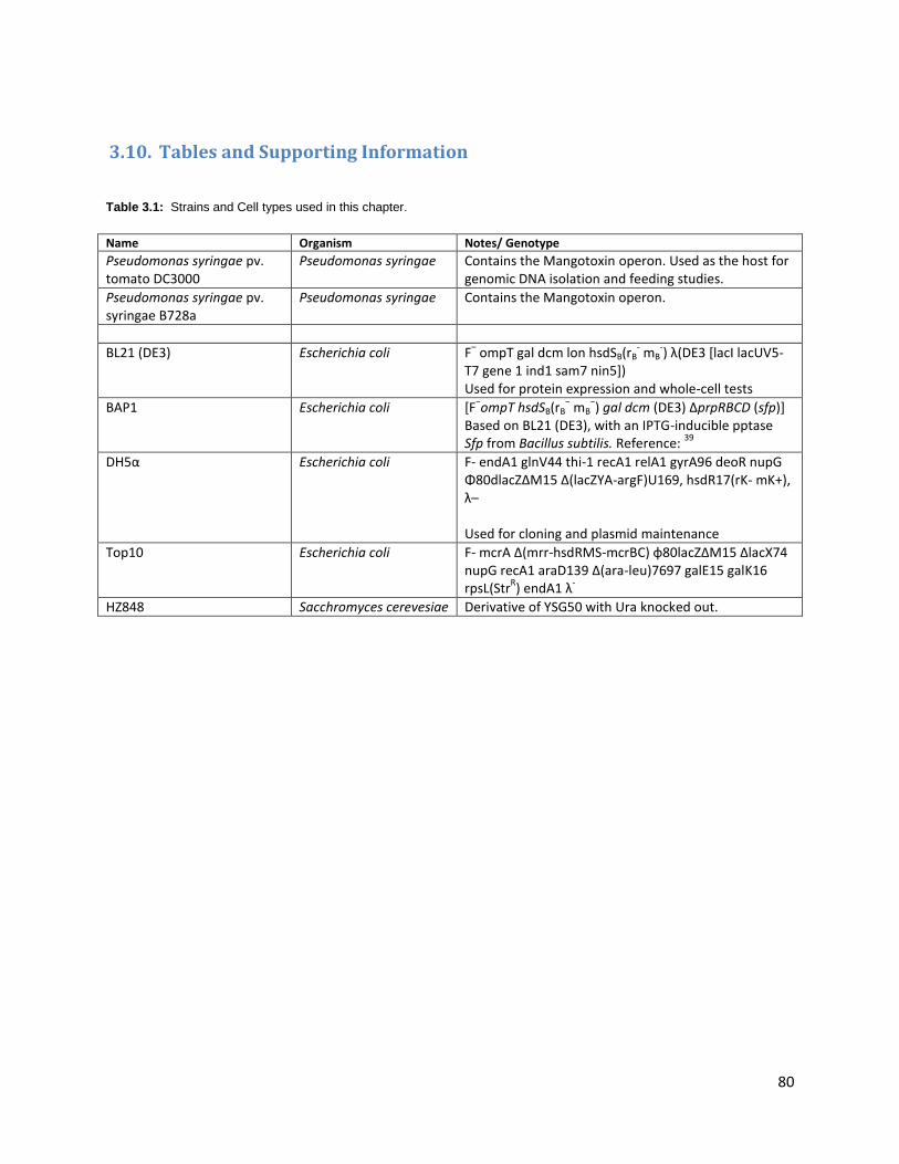
80
3.10. Tables and Supporting Information
Table 3.1: Strains and Cell types used in this chapter.
Name Organism Notes/ Genotype
Pseudomonas syringae pv. tomato DC3000
Pseudomonas syringae Contains the Mangotoxin operon. Used as the host for genomic DNA isolation and feeding studies.
Pseudomonas syringae pv. syringae B728a
Pseudomonas syringae Contains the Mangotoxin operon.
BL21 (DE3) Escherichia coli F– ompT gal dcm lon hsdSB(rB
- mB
-) λ(DE3 [lacI lacUV5-
T7 gene 1 ind1 sam7 nin5]) Used for protein expression and whole-cell tests
BAP1 Escherichia coli [F−ompT hsdSB(rB
− mB
−) gal dcm (DE3) ΔprpRBCD (sfp)]
Based on BL21 (DE3), with an IPTG-inducible pptase Sfp from Bacillus subtilis. Reference:
39
DH5α Escherichia coli F- endA1 glnV44 thi-1 recA1 relA1 gyrA96 deoR nupG Φ80dlacZΔM15 Δ(lacZYA-argF)U169, hsdR17(rK- mK+), λ– Used for cloning and plasmid maintenance
Top10 Escherichia coli F- mcrA Δ(mrr-hsdRMS-mcrBC) φ80lacZΔM15 ΔlacX74 nupG recA1 araD139 Δ(ara-leu)7697 galE15 galK16 rpsL(Str
R) endA1 λ
-
HZ848 Sacchromyces cerevesiae Derivative of YSG50 with Ura knocked out.

81
Table 3.2: Primers used for this study.
Name Sequence Template Comments
Primers used to construct Mangotoxin V1. Construct A
A - Reverse GGA CGA CCC AAG GAC CTG CA P. syringae Genomic DNA
B - Forward AGC GTG GTC AGG TAG CTG TC P. syringae Genomic DNA
B - Reverse GCC GCA GGG TGA GAA CGG CC P. syringae Genomic DNA
C - Forward GGA TGA GGC GTT CAT TGA TT P. syringae Genomic DNA
C - Reverse GAG CAC AAT CTC CAG GCG CT P. syringae Genomic DNA
D - Forward CAA CAT CAT GCG CTT CTT CA P. syringae Genomic DNA
Forward A GGC CTA GAG GCC GTG GCC ACC ACG GCC CGG CCT GCC TTT CAT GAG CAC AGC AGA TCA GAC
P. syringae Genomic DNA
Reverse D TTA TAG CAC GTG ATG AAA AGG ACC CAG GTG GCA CTT TTC GTC AGC CAA ACA GGC TGC GCA
P. syringae Genomic DNA
Yeast Forward Long
TGA CGG TGC GAT GAG TAA CGT GCG CAG CCT GTT TGG CTG ACG AAA AGT GCC ACC TGG GTC
pRS416 URA selection marker, CEN/ARS origin
Yeast Forward Short CGA AAA GTG CCA CCT GGG TC
pRS416 URA selection marker, CEN/ARS origin
Yeast Reverse TTT ATC AGG GTT ATT GTC TCA TGA GCG GAT ACA TAT TTG AAG TGA GTT TAG TAT ACA TGC
pRS416 URA selection marker, CEN/ARS origin
E. coli Ptrc Fragment Forward
AAA CTG TAT TAT AAG TAA ATG CAT GTA TAC TAA ACT CAC TTC AAA TAT GTA TCC GCT CAT
pTrc99a Amp Selection Marker, pBBR32 origin
E. coli Ptrc Fragment Reverse
CCT TGG CGT CCA ACC AGC GGC ACC AGC GGC GCC TGA GAG GCG CGT TGC TGG CGT TTT TCC
pTrc99a Amp Selection Marker, pBBR32 origin
Pseudomonas Helper Forward
CAG GGG GGC GGA GCC TAT GGA AAA ACG CCA GCA ACG CGC CTC TCA GGC GCC GCT GGT GCC
pUCP19
Pseudomonas Helper Reverse
TGT AAT TGC CAG CGC TTT GGG TCT GAT CTG CTG TGC TCA TGA AAG GCA GGC CGG GCC GTG
pUCP19
Primers used to clone individual genes from the Mangotoxin operon into pET28 and pTrc99a
NcoI / HindIII ORF 5-F TAG ATC CCA TGG CGA TGA ATA AAT CAG CAG
P. syringae Genomic DNA
Cloned into pTrc99a
NcoI / HindIII ORF 6-F TAG ATC CCA TGG CGA TGG ATG GCC TGC TGC
P. syringae Genomic DNA
Cloned into pTrc99a
NcoI / HindIII ORF 7-F TAG ATC CCA TGG CGA TGA AGT ATC GAA CCC
P. syringae Genomic DNA
Cloned into pTrc99a
EcoRI / XBaI ORF 8-F GAC CAT GGA ATT CAT GCG CAA ATC AGA TAG
P. syringae Genomic DNA
Cloned into pTrc99a
NcoI / HindIII ORF 9-F TAG ATC CCA TGG CGA TGT ATT GTG AGA AAA
P. syringae Genomic DNA
Used to clone MgoB into pTrc99a/pet28a
EcoRI / XBaI ORF 10-F GAC CAT GGA ATT CAT GTC GTT CAT CGA TGC
P. syringae Genomic DNA
Used to clone MgoC into pTrc99a
NcoI / XBaI ORF 11-F TAG ATC CCA TGG CGA TGA AGC GCC TGG AGA
P. syringae Genomic DNA
Used to clone MgoA into pTrc99a.
NcoI / HindIII ORF 12-F TAG ATC CCA TGG CGA TGA ACC ATA TTC ACA
P. syringae Genomic DNA
Used to clone MgoD into pET28.
NcoI / HindIII ORF 5-R ACA GCC AAG CTT TTA CTG GTT ACC CAG CAC
P. syringae Genomic DNA
Cloned into pTrc99a
NcoI / HindIII ORF 6-R ACA GCC AAG CTT TCA AAC GAG TTC AGG CAG
P. syringae Genomic DNA
Cloned into pTrc99a
NcoI / HindIII ORF 7-R ACA GCC AAG CTT TCA GCG AAC CGG ACG ACC
P. syringae Genomic DNA
Cloned into pTrc99a
EcoRI / XBaI ORF 8-R GTC GAC TCT AGA TCA GGC GGG GGT ATT CTT
P. syringae Genomic DNA
Cloned into pTrc99a

82
Table 3.2 continued NcoI / HindIII ORF 9-R ACA GCC AAG CTT CTA CGC ACT GGC CTG CAT
P. syringae Genomic DNA
Used to clone MgoB into pTrc99a/pet28a
EcoRI / XBaI ORF 10-R GTC GAC TCT AGA TCA TTG CAG CGA TCC CTG
P. syringae Genomic DNA
Used to clone MgoC into pTrc99a
NcoI / XBaI ORF 11-R GTC GAC TCT AGA TCA GAT GAA GTC GAT TTC
P. syringae Genomic DNA
Used to clone MgoA into pTrc99a.
NcoI / HindIII ORF 12-R ACA GCC AAG CTT TCA GCC AAA CAG GCT GCG
P. syringae Genomic DNA
Used to clone MgoD into pET28.
pEt28 TomatoNRPS Fwd NdeI GCG GCA GCC ATA TGA AGC GCC TGG AGA TT
P. syringae Genomic DNA
Used to clone MgoA into pET28.
pEt28 Fwd NdeI TomatoAurF GCG GCA GCC ATA TGT CGT TCA TCG ATG CG
P. syringae Genomic DNA
Used to clone MgoC into pET28.
pEt28 Rev EcoRI TomatoAurF GGA GCT CGA ATT CTC ATT GCA GCG ATC C
P. syringae Genomic DNA
Used to clone MgoC into pET28.
pEt28 TomatoNRPS Rev NotI TCG AGT GCG GCC GCT CAG ATG AAG TCG ATT
P. syringae Genomic DNA
Used to clone MgoA into pET28.
Primers used to construct the Master Helper Plasmid shuttle vector (also known as YEP – Yeast-E. coli-Pseudomonas) Construct B
Yeast/Ecoli forward
TCT GGC CTA GGC GGC CGC GAG ACT CTT AAT TAA CGA AAA GTG CCA CCT GGG TCC TTT TCA
Mangotoxin V1
Yeast/Ecoli Reverse
ACT CTG CGA CAT CGT ATA ACG TTA CTG GTT TCA CAT TCA CCA CCG CGT TGC TGG CGT TTT
Mangotoxin V1
LacI Forward AGG GGG GCG GAG CCT ATG GAA AAA CGC CAG CAA CGC GGT GGT GAA TGT GAA ACC AGT AAC
pTrc99a
LacI Reverse CCT TGG CGT CCA ACC AGC GGC ACC AGC GGC GCC TGA GAG GTC ACT GCC CGC TTT CCA GTC
pTrc99a
Pseudomonas Forward
CTG GCA CGA CAG GTT TCC CGA CTG GAA AGC GGG CAG TGA CCT CTC AGG CGC CGC TGG TGC
pUCP19
Pseudomonas Reverse
CCA GGT GGC ACT TTT CGT TAA TTA AGA GTC TCG CGG CCG CCT AGG CCA GAT CCA GCG GCA
pUCP19
Generic primers used for both the T7 and Trc based Gibson assemblies. Construct D and F
GibORF 9 Naked Rev
GCACTGGCCTGCATCAGCGG pET28a-ORF 9 Binds only at the end of the gene.
GibORF 10 Naked Rev
TCA TTG CAG CGA TCC CTG AAC CGG pET28a-ORF 10 Binds only at the end of the gene.
GibORF 11 Naked Rev
TCAGATGAAGTCGATTTCTTTCAGG pET28a-ORF 11 Binds only at the end of the gene.
Primers used to construct the Mangotoxin genes with individual T7 promoters by Gibson assembly. See Figure 3.3E for the vector map.
F ORF 9-10 Gibson
CCGCTGATGCAGGCCAGTGCGATGCGTCCGGCGTAGAGGATCGAGATCTC
pET28a-ORF 10 Contains the end of ORF 9 and the beginning of the T7 promoter
F ORF 10-11 Gibson
CCGGTTCAGGGATCG CTGCAATGAGATGCGTCCGGCGTAGAGGATCGAGATC
pET28a-ORF 11 Contains the end of ORF 10 and the beginning of the T7 promoter
F ORF 11-12 Gibson
CCTGAAAGAAATCGACTTCATCTGAGATGCGTCCGGCGTAGAGGATCGAGATCTC
pET28a-ORF 12 Contains the end of ORF 11 and the beginning of the T7 promoter
ORF 11 Gibson End Rev
GTGGTGGTGGTGCTCGAGTGCGGCCGCAAGCTTTCAGATGAAGTCGATTTCTTTCAGG
pET28a-ORF 11 Contains the end of ORF 11 and the beginning of the T7 terminator
ORF 10 Gibson End Forward
ATTTTGTTTAACTTTAAGAAGGAGATATACCATGGATGTCGTTCATCGATGCGTG
pET28a-ORF 10 Begins with the T7 Promoter from pET28a to construct the ORF 9 deletion
Primers for cloning individual Trc promoters before each gene.
GibTrc 9 start F GATCTGGCCTAGGCGGCCGCGAGACTCTTAATTAATTC TGG CAA ATA TTC TGA AAT GAG C
pTrc99a-ORF 9 Overlapping region between the beginning of Master Helper Plasmid and pTrc promoter.
GibTrc 9-10 F CCGCTGATGCAGGCCAGTGCTTC TGG CAA ATA TTC TGA AAT GAG C
pTrc99a-ORF 10 Contains the end of ORF 9 and the beginning of the Trc promoter
GibTrc 10-11 F CCG GTT CAG GGA TCG CTG CAA TGATTC TGG CAA ATA TTC TGA AAT GAG C
pTrc99a-ORF 11 Contains the end of ORF 9 and the beginning of the Trc promoter
GibTrc 11-12 F CCTGAAAGAAATCGACTTCATCTGATTC TGG CAA ATA TTC TGA AAT GAG C
pTrc99a-ORF 12 Contains the end of ORF 9 and the beginning of the Trc promoter

83
Table 3.2 continued
GibTrc 12 Rev AAAGGACCCAGGTGGCACTTTTCGTTAATTAATCAGCCAAACAGGCTGCGCACGTTAC
pTrc99a-ORF 12 Contains the end of ORF 12 and overlaps with Master Helper Plasmid.
Primers used for qPCR of the Mangotoxin genes – See Figure 3.2 for genes to be amplified.
Mangotoxin 1 Set 1 Forward TTC GGA ATC TTC GGA ATG GAG GCT
P. syringae cDNA
Mangotoxin 1 Set 1 Reverse AGG AGT AAT TGG CAG TCA GGG AGT
P. syringae cDNA
Mangotoxin 2 Set 1 Forward TTT AAG CTG ATG GTC AGG TCG GCA
P. syringae cDNA
Mangotoxin 2 Set 1 Reverse GCG CTT TCA TTC CGT ATT GGG ACA
P. syringae cDNA
Mangotoxin 3 Set 1 Forward ATC GTG GAG TTC CTT CTG CAC AGT
P. syringae cDNA
Mangotoxin 3 Set 1 Reverse TGG GCA CCT CAG GAC ATA CTG ATT
P. syringae cDNA
Mangotoxin 4 Set 1 Forward ATA CTC TTG CCT TGT GCT GCT CGT
P. syringae cDNA
Mangotoxin 4 Set 1 Reverse AGA CGC TGC TCA GGC TTG GAT TT
P. syringae cDNA
Mangotoxin 5 Set 1 Forward TGG AAA GCT CTT GAT TGG CCT TGC
P. syringae cDNA
Mangotoxin 5 Set 1 Reverse AGG TAT AGC GGT TGG CGT CAT TGT
P. syringae cDNA
Mangotoxin 6 Set 1 Forward TAC ACG GAG GTC ACA CAC CTG ATT
P. syringae cDNA
Mangotoxin 6 Set 1 Reverse CAG TTC AAG CGC CTT GAT CAC GTA
P. syringae cDNA
Mangotoxin 7 Set 1 Forward AAG CGA TTG CTG GAA ACC GAA TGG
P. syringae cDNA
Mangotoxin 7 Set 1 Reverse ACA CCC TTG TCC TGC ACG TAT TCT
P. syringae cDNA
Mangotoxin 8 Set 1 Forward GTT GTA CGC CAA GAC CCA CAA CAA
P. syringae cDNA
Mangotoxin 8 Set 1 Reverse ACT GCC AGT GGT CAT CCT GAG AAA
P. syringae cDNA
Mangotoxin 9 Set 1 Forward GCG CTT CAT CGA ACT GCA GAA ACA
P. syringae cDNA
Mangotoxin 9 Set 1 Reverse AAT GCT CTG GAA CAT CAG CGG GAT
P. syringae cDNA
Mangotoxin 10 Set 1 Forward AGA ACG GCT TCG ACT TTA CGC TGA
P. syringae cDNA
Mangotoxin 10 Set 1 Reverse GAA GAA GCG CAT GAT GTT GCC GAT
P. syringae cDNA
Mangotoxin 11 Set 3 Forward CAC ATC GTT GAA GCG CTG CTG AAT
P. syringae cDNA
Mangotoxin 11 Set 3 Reverse AAA CCT TCA CCC GAC TCA AGT CCA
P. syringae cDNA
Mangotoxin 12 Set 1 Forward TAT TCC GGC ATT GGA GAG CAC TGA
P. syringae cDNA
Mangotoxin 12 Set 1 Reverse ATC GCC ACA TTG CCA TCC TTG AAC
P. syringae cDNA
Gyrase A Set 2 Forward Primer TAT TTC CTG TCG CCA GAA CAG GCT
P. syringae cDNA
Gyrase A Set 2 Reverse Primer TTT CCT GGT ACT CAC CCA GCA GTT
P. syringae cDNA