chemistry and biochemistry of some biologically …
TRANSCRIPT

CHEMISTRY AND BIOCHEMISTRY OF SOMEBIOLOGICALLY ACTIVE BACTERIAL LIPIDS
E. LEDERER*
lnstitut de Biologie Physico-Chimique and Laboratoire deChimie Biologique de laFacultedes Sciences, Paris, France
INTRODUCTIONWork, which was begun in the author's laboratory in 1948, on the
chemistry of various lipid fractions of Mycobacteria and other acid-fastmicro-organisms, has led to the identification and elucidation of thechemistry of a number of compounds having unusual structures and interesting biological activities. The principal compounds studied are branchedchain high molecular weight fatty acids, gl ycolipids and peptido-glycolipids.Some information on biosynthetic mechanisms in these series has also beenobtained.
The infection of a healthy animal or human being by tubercle bacilligives rise to several pathological manifestations, such as: (i) formation oftubercles; (ii) establishment of a delayed type of hypersensitivity; (iii) lossof weight, haemorrhages, and death, due to the presence of a toxic factor;(iv) increased production of antibodies; (v) development of resistance to asecond infection.
We shall see that all of these pathological phenomena are due to particularlipid fractions of the tubercle bacillus. Besides these, we shall consider alsoa new category of glycolipids which are typical for certain strains of Mycobacteria (see Table 1).
Table 1. Correlation of biological activity and chemical structure
Pathological phenomenon
Formation of tuberclesDelayed hypersensitivityLoss of weight, haemorrhages, deathIncreased production of antibodies
Resistance to infection
Specific pathogenicity (?)
Chemical structure of the corresponding lipid
Branched-chain fatty acidsEsters of mycolic acids with carbohydratesCord factor (6,6'-dimycoloyl-trehalose)Wax D of human strains (a peptido-glyco
lipid)Phosphoglycolipids (?)
M ycoside A and B (glycolipids)Mycoside C (a peptide-glycolipid)
TUBERCLE FORMATION
Branched-chain fatty acidsBranched-chain fatty acids and their derivatives seem to be the principal
compounds producing the characteristic tubercles observed in tuberculosis.
* Present address: Institut de Chimie des Substances Naturelles, Gif-sur-Yvette, Seine etOise, France.
587

E.LEDERER
The excellent work of Anderson1, published between 1926 and 1946, hasshown that Mycobacteria contain several branched-chain fatty acids.One can distinguish two large groups: (i) compounds having only methylbranchings; (ii) compounds with longer branched chains.
Compounds with methyl side-chainsIn this group, we find compounds having only one methyl side-chain,
such as tuberculostearic acid (I) and phthiocerol (II) 2, and those havingseveral methyl branchings, such as phthienoic acid (III) 3 and mycocerosicacid (IV)4.
HsC-(CR 2)7-CH- (CR 2)8-CO O H
ICHa
(I)
HaC-(CH 2)n-CH- CH 2-CH-(CH 2)4-CH-CH-CH2-CH aI I I I
OH OH err, OCHa
(II)
(a) n = 20
(b) n = 22
HaC-(CH 2) 17-CH-CH2-CH-CH=C-COOH
I I ICHa CHa CHa
(III)
HaC-(CH 2) 19-CH-CH2-CH-CH2-CH-dH2-CH-COOH
I I I IC~ C~ C~ C~
(IV)
It seems that compounds having only one methyl branching, such as(I) and (II), are devoid of biological activity, whereas those with three ormore methyl branchings, such as (III) and (IV), are potent producers oftubercles (see the review by Asselineau"),
The structural determination of phthienoic and mycocerosic acids is dueto Polgar, Cason,]. and C. Asselineau, and S. and E. Stenhagen, and cannot be described in detail here (for recent reviews, see refs. 6 and 7).
Two different mechanisms of biosynthesis can be considered for the fattyacids with methyl side-chains: the" Birch mechanism "8, consisting of thefixation of a methyl group (from methionine) onto a polyacetic acid chain*
... Hofman and Liul have shown that the cyclopropane ring oflactobacillic acid is formedin vivo by the fixation of a one carbon unit derived from formate onto cis-vaccenic acid; thefixation of methyl groups on aliphatic chains by this mechanism does not seem to have beenobserved yet.
588
CHEMISTRY AND BIOCHEMISTRY OF SOME BACTERIAL LIPIDS
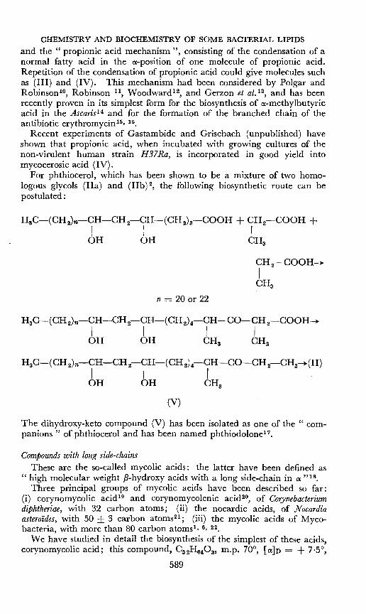
and the " propionic acid mechanism ", consisting of the condensation of anormal fatty acid in the ex-position of one molecule of propionic acid.Repetition of the condensation of propionic acid could give molecules suchas (III) and (IV). This mechanism had been considered by Polgar andRobinsori-", Robinson 11, Woodward P, and Gerzon et al.13 , and has beenrecently proven in its simplest form for the biosynthesis of o-methylbutyricacid in the Ascaris14 and for the formation of the branched chain of theantibiotic erythromycinP. 16.
Recent experiments of Gastambide and Grisebach (unpublished) haveshown that propionic acid, when incubated with growing cultures of thenon-virulent human strain H37Ra, is incorporated in good yield intomycocerosic acid (IV).
For phthiocerol, which has been shown to be a mixture of two homologous glycols (IIa) and (lIb) 2, the following biosynthetic route can bepostulated:
CH2-COOH~
ICHa
n = 20 or 22
HaC-(CH 2)n-CH-CH 2-CH-(CH 2)4-CH-CO-CH2-CHa~(II)
I I IOH OH CHa
(V)
The dihydroxy-keto compound (V) has been isolated as one of the" companions" of phthiocerol and has been named phthiodolone-".
Compounds with long side-chainsThese are the so-called mycolic acids: the latter have been defined as
" high molecular weight ,8-hydroxy acids with a long side-chain in 0: "18.
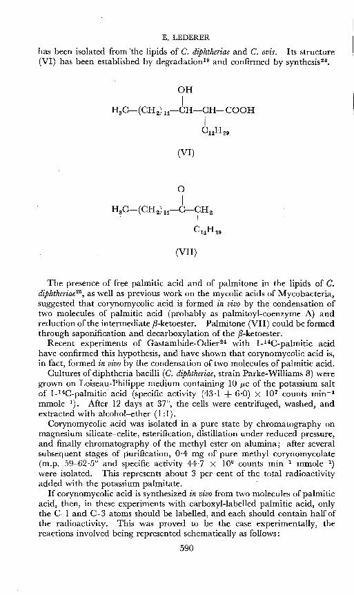
Three principal groups of mycolic acids have been described so far:(i) corynomycolic acid 19 and corynomycolenic acid 20, of Corynebacteriumdiphtheriae, with 32 carbon atoms; (ii) the nocardic acids, of Nocardiaasteroides, with 50 ± 3 carbon atomss- ; (iii) the mycolic acids of Mycobacteria, with more than 80 carbon atoms"- 6, 22.
We have studied in detail the biosynthesis of the simplest of these acids,corynomycolic acid; this compound, Ca2H640s, m.p. 70°, [o:]n = + 7'5°,
589
E. LEDERER
has been isolated from 'the lipids of C. diphtheriae and C. ovis. Its structure(VI) has been established by degradation 19 and confirmed by synthesist",
(VI)
(VII)
The presence of free palmitic acid and of palmitone in the lipids of C.diphtheriae 25, as well as previous work on the mycolic acids of Mycobacteria,suggested that corynomycolic acid is formed in vivo by the condensation oftwo molecules of palmitic acid (probably as palmitoyl-coenzyme A) andreduction of the intermediate ,B-ketoester. Palmitone (VII) could be formedthrough saponification and decarboxylation of the ,B-ketoester.
Recent experiments of Castambide-Odierv' with 1-l4C-palmitic acidhave confirmed this hypothesis, and have shown that corynomycolic acid is,in fact, formed in vivo by the condensation of two molecules of palmitic acid.
Cultures of diphtheria bacilli (C. diphtheriae, strain Parke-Williams 8) weregrown on Loiseau-Philippe medium containing 10 JLC of the potassium saltof 1-14C-palmitic acid (specific activity (43'1 ± 6'0) X 107 counts mirr!
mmolev-). After 12 days at 37°, the cells were centrifuged, washed, andextracted with alcohol-ether (1 :1).
Corynomycolic acid was isolated in a pure state by chromatography onmagnesium silicate-celite, esterification, distillation under reduced pressure,and finally chromatography of the methyl ester on alumina; after severalsubsequent stages of purification, 0-4 mg of pure methyl corynomycolate(m.p. 59-62'5° and specific activity 44·7 X 106 counts minr! mmole-:")were isolated. This represents about 3 per cent of the total radioactivityadded with the potassium palmitate.
If corynomycolic acid is synthesized in vivo from two molecules of palmiticacid, then, in these experiments with carboxyl-labelled palmitic acid, onlythe C-l and C-3 atoms should be labelled, and each should contain half ofthe radioactivity. This was proved to be the case experimentally, thereactions involved being represented schematically as follows:
590

CHEMISTRY AND BIOCHEMISTRY OF SOME BACTERIAL LIPIDS
HaC-(CH2)14-14COOR + CH 2-14COOR---+
IC14H 29
(VIa)
-}
(VIII) (VII)
(IX)
IHOI, ethanol
HaC-(CH 2) 14-NH-C2H 5
(XI)+
tHaC-(CH 2) 14-14COOH
(X)t NaNa• HaSO.
H aC-(CH 2) 14-NH 2 + 14C0 2
(XII)
The radioactive corynomycolic acid (VIa) was first diluted with authenticcorynomycolic acid to a specific activity of (33,0 ± 4,0) X 104 counts mg-1
min"! mmolev-, and then oxidized and decarboxylated in a chromic acidacetic acid mixture. The barium carbonate obtained had a specific activityof (16,3 ± 6,4) X 104 counts min-1 mmoler", and the palmitone (VII)obtained in a pure state (m.p. 79-80'5°) had the same specific activity( (16,3 ± 2,3) X 104) . This already shows that the carboxyl of the labelledcorynomycolic acid has half the radioactivity of the whole molecule.
The radioactive palmitone (VII) was then degraded in the following way:the oxime (VIII) was prepared and rearranged to the amide (IX); thiswas cleaved in acid medium and gave an inactive amine (XI) and palmitic
591

E.LEDERER
acid (X) having the same specific activity ( (12-5 ± 3'4) X 104) as thepalmitone; decarboxylation of this palmitic acid then gave again an inactive amine (XII) and barium carbonate having the same radioactivityas the initial sample of palmitone (VII).
This proves that, in the corynomycolic acid formed in vivo in the presenceof 1-l4C-palmitic acid, only the C-l and C-3 atoms are radioactive; thiscan only be explained by the condensation of two intact molecules ofpalmitic acid; otherwise, if the palmitic acid had undergone an initialdegradation, radioactivity would have been " smeared out" over the wholemolecule.
It can be surmised that one of the molecules of palmitoyl-CoA has to becarboxylated to tetradecylmalonyl-CoA. The detailed mechanism of thecondensation reaction has not yet been studied.
Corynomycolenic acid (XIII), the second Cs z-mycolic acid isolated fromC. diphiheriae'", is obviously formed from one molecule of palmitoleic acid,CHa-(CH2)s-CH=CH-(CHzh-COOH (which also exists in largequantities in the free state in the diphtheria bacillus), and one molecule ofpalmitic acid. Here, an intermediary ,8-keto-ester could give a ketone,~7-palmitenone,CHa-(CHz)s-CH=CH-(CH2)7-COClsHal' on saponification; this has also been isolated from the lipids orc. diphtheriae 2S•
OH
IHaC-(CH2)s-CH=CH-(CH2h-CH-CH-COOH
I(CH 2) l S
ICHa
(XIII)
The nocardic acids, CsoH960a ± 3CH 2, have been isolated recentlyfrom the pathogenic Actinomycete Nocardia asteroides, and the partialstructure (XIV) established (Michel et at. 21) ; they are probably synthesizedin vivo by the condensation of three molecules of long chain (C16 ?) acids ina similar way.
(XIV)
(a) n = 11
(b) n = 13
592

CHEMISTRY AND BIOCHEMISTRY OF SOME BACTERIAL LIPIDS
Mycolic acids are typical constituents ofMycobacteria, and were discoveredin 1938 by Lesuk and Anderson28. Detailed studies of their chemicalstructure, pursued in our laboratory principally by 1. Asselineau, haveyielded the following results: mycolic acids of human and bovine strains ofM. tuberculosis have the approximate formula CSSH17604 ± 5 CH2 and thegeneral structure (XV):
OH
IR-CH-CH-COOH
IC24H29
(XV)
where R is a radical containing about 60 carbon atoms with one oxygenfunction (OR, or OCHs or a carbonyl) and three chains.
Extensive degradative evidence has shown that in some of these mycolicacids the second oxygen function is either on C-5 or further away from thecarboxyl, and that C-4 or probably C-4 and C-6 carry side-chaines- 22.
As to the length of the various side chains, it is known that at least oneof these (besides that at C-o:) has 24 to 26 carbon atoms, and that the othersare shorter (C16 to CIS).
All experimental evidence is in agreement with formulae (XVII) and(XVIII), which could result from the condensation of four molecules oflong-chain fatty acids (2 X 26 + 2 X 18 = 88) by the mechanism whichwe have proven for corynomycolic acid. An appropriate reduction of theintermediate (quite hypothetical) triketo-compound (XVI) gives the twotypes of mycolic acids (XVII) and (XVIII). These structures are inagreement with all experimental findings'".
(XVI)
(XVII)
593
E.LEDERER
OH OH
I IC 25H51-CH-CH-CH2-CH-CH-CH-COOH
I I IC16H33 C 16H 33 C 24H49
(XVIII)
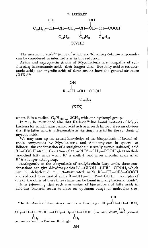
The mycolonic acids'" (some of which are 3-hydroxy-5-keto-compounds)can be considered as intermediates in this reduction.
Avian and saprophytic strains of Mycobacteria are incapable of synthesizing hexacosanoic acid; their longest chain free fatty acid is tetracosanoic acid; the mycolic acids of these strains have the general structure(XIX) 29:
OH
IR-CH-CH-COOH
IC 22H45
(XIX)
where R is a radical C5sH11 6 ± 3CH 2 with one hydroxyl group.It may be mentioned also that Karlsson" has found mutants of Myco
bacteria for which hexacosanoic acid acts as growth factor; it seems obviousthat this latter acid is indispensable as starting material for the synthesis ofmycolic acids.
We may sum up the actual knowledge of the biosynthesis of branchedchain compounds by Mycobacteria and Actinomycetes in general asfollows: the condensation of a straight-chain (usually even-numbered) acidR'-COOH on the C-a: atom of an acid R"-CH 2-COOH gives methylbranched fatty acids when R" is methyl, and gives mycolic acids whenR" is a longer alkyl group.
Analogously to the biosynthesis of straight-chain fatty acids, these condensations can give ,8-hydroxy-acids R'-CHOH-CHR"-COOH, whichcan be dehydrated to a,,8-unsaturated acids R'-CH=CR"-COOHand reduced to saturated acids R'-CH2-CHR"-COOH. Examples of
. one or the other of these three stages can be found in many bacteriallipids*.It is interesting that each mechanism of biosynthesis of fatty acids in
acid-fast bacteria seems to have an optimum range of molecular size:
* In the Ascaris all three stages have been found, e.g.:
CHa-CH=C-COOH and CHa-CH2-CH-COOH (SazI ICHa CHa
communication from Professor Bueding).
594
OHI
CHa-CH-CH-COOH,ICHa
and WeiP4, and personal

CHEMISTRY AND BIOCHEMISTRY OF SOME BACTERIAL LIPIDS
normal fatty acid synthesis stops at C26' branched acids with several methylgroups have 21 to 34 carbon atoms, whereas mycolic acids have 32 to 88carbon atoms.
Mycolic acids, as well as their esters with carbohydrates, are potentproducers of tubercles in vivo5 ; we shall see that glycolipids and peptidoglycolipids containing 50-88 per cent mycolic acid have other interestingbiological activities.
DELAYED TYPE OF HYPERSENSITIVITY
The injection of tuberculin, a protein (or mixture of proteins) secreted bythe tubercle bacillus into the culture filtrate, is not sufficient to produce thetypical" tuberculin hypersensitivity". Raffels! has shown that one canelicit this hypersensitivity by simultaneous injection of tuberculin and apurified wax fraction. It was later found that all esters of mycolic acidwith carbohydrates are actives>.
Amongst the lipids produced by tubercle bacilli, a particular wax fraction,called wax D, is also active in this respect. We shall say more about thisfraction later.
THE TOXIC FACTOR
The toxic factor has been called " cord factor", because it is found in" cord forming" organisms, i.e, virulent and attenuated strains which growin the culture medium in " serpentine cords "33. One intravenous injectionof 20p,g of natural cord factor kills mice within 10-20 days; haemorrhagesin the lung are the principal lesions observed.
Cord factor has been isolated in a pure state after repeated chromatographic purifications on magnesium silicate and silicic acid, and its structureas the 6,6'-dimycolate of trehalose (XX) (C186H366017 ± 10 CH 2) established by degradations", and by synthesis 35, 36, starting from trehalose andnatural mycolic acids.
595

E. LEDERER
Methylatedmycolic acid
Mycolic alcoholCssH1760a
" Cord factor ",..-----C186H366017----------.
alkaline I reductive cleavagehydrolysis with LiAIH,
r-----''-----,
tMycolic acid
C8sH17604
2,3,4,2',3',4'Hexamethyl trehalose
l-h~~~2,3,4-Tri-O-methyl
n-glucose
1acidhydrolysis
n-Glucose
TrehaloseC12H22011
I
1acetylation
Trehaloseocta-acetate
m.p.81-82°
oxidation
+K-Gluconate
m.p.180°
Simple, synthetic mycolic acids, such as (XXI) obtained by condensationof two molecules of methyl docosanoate, have also been esterified withtrehalose and have yielded" small cord factors", e.g. (XXII) (ClooH194015) 38which show approximately the same biological activity as natural cordfactor.
(XXI)
(XXII)
OHI
CH20-CO-CH-CH -(CH2hoCH3I . H OH
C20 H41o H H
o
596
CHEMISTRY AND BIOCHEMISTRY OF SOME BACTERIAL LIPIDS
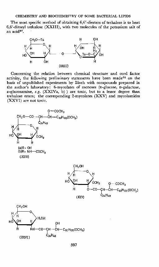
The most specific method of obtaining 6,6'-diesters of trehalose is to heat6,6'-ditosyl trehalose (XXIII), with two molecules of the potassium salt ofan acid3 ?
H OH
o
(XXIII)
H OH
H
Concerning the relation between chemical structure and cord factoractivity, the following preliminary statements have been made'" on thebasis of unpublished experiments by Bloch with compounds prepared inthe author's laboratory: 6-mycolates of monoses (n-glucose, n-galactose,n-glucosamine, e.g, (XXIVa, b») are toxic, but to a lesser degree thantrehalose esters; the corresponding 2-mycolates (XXV) and mycolamides(XXVI) are not toxic.
O-COCH 3
bCH20 co ~:H",~H-C6oH120(OCH3)
H H 0 H
HO OH H OCH3
H R(a)R= OH(b)R= NH-COCH3
(XXIV)
597

E.LEDERER
The 6,6'-dimycolates of trehalose seem more active than the 6-monoor 2,6,6'-trimycolates.
Acetylation of the ,B-hydroxyl of mycolic acid diminishes only slightlythe activity of cord factor; fully acetylated cord factor is inactive.
The influence of the structure of the acid esterifying trehalose can becharacterized as follows: even behenic (docosanoic) esters of trehalose areactive, but doses larger than 0·1 mg are necessary. The 6,6'-diester oftrehalose with the synthetic mycolic acid CuH880a (XXI) has about 50 percent of the activity of natural cord factor. Dehydration of the latter acidgives the unsaturated acid (XXVII):
(XXVII)
which, as the 6,6'-diester of trehalose, is inactive at 0·1 mg dose levels.Concerning the mechanism of action of cord factor, Kato et at. 40 have
found that injection of cord factor decreases the activity of most DPN-linkeddehydrogenases.
For reviews on cord factor, see Lederer'" and No1139•
INCREASED PRODUCTION OF ANTIBODIES
Freund's adjuvant, consisting of a water-in-oil emulsion containingkilled Mycobacteria in the oil phase and the antigen in the aqueous phase,has been widely used by immunologists for increasing antibody productionin animals. Freund41 has reviewed our knowledge of the mode of actionof this type of adjuvant.
After the preliminary experiments of White et at.42 with a " purified waxfraction ", we showed with White et ai. 43 that a particular wax fraction ofhuman strains of M. tuberculosis can effectively replace the whole bacilli inFreund's adjuvant mixture.
This wax fraction, which we call wax D5-7, is extracted from thebacilli with chloroform and can be separated from other wax fractions as aresult of its insolubility in boiling acetone.
The wax D fractions of human strains of tubercle bacilli are high-meltingdextrorotatory solids which represent 6-8 per cent of the dry weight ofvirulent strains, but only about 2 per cent of non-virulent strainsvs,
A recent study'" of the chemical structure of wax D has shown that it isa high molecular weight lipid, and that the composition and molecularweight varies from strain to strain. The best analysed wax D fraction,that of the human virulent strain " Brevannes ", has a molecular weight ofabout 54,000. On saponification, about 50 per cent of mycolic acidCSSH17604 is obtained (corresponding to approximately 22 molecules) and50 per cent of a water-soluble portion. This latter is a peptido-mucopolysaccharide and seems to be homogeneous on ultracentrifugation and byimmunological reactions.
598

CHEMISTRY AND BIOCHEMISTRY OF SOME BACTERIAL LlpIDS
The carbohydrate moiety of this peptido-mucopolysaccharide containsthe following sugars: D-arabinose, D-mannose, D-galactose, glucosamine andgalactosamine*; traces of muramic acid are present, probably as an impurity.
The peptide moiety of wax D of human strains is of particular interest,because the analogous wax D fractions of bovine and saprophytic strains,which are nitrogen-free glycolipids, are inactive as adjuvants; thus, thepeptide moiety seems to playa definite role in the adjuvant activity ofwax D43.
It had already been found several years ago that wax D fractions of allhuman strains investigated contain the same three amino-acids: mesodiaminopimelic acid, glutamic acid and alanine''.
The particular wax D of the strain" Brevannes " which was studied indetail contains two molecules of meso-diaminopimelic acid (DAP) , twomolecules of D-glutamic acid (Glu) and three molecules of alanine (Ala),one of which is D. The presence of several amino-acids of " unnatural"configuration is quite remarkable. A tentative structure for wax D of thehuman virulent strain" Brevannes " of M. tuberculosis has been proposed'"as follows:
Polysaccharide:'"170 molecules
2 meso-DAP of:
2 t-Ala amide I. . .1 glycosidic Arabinose ester mycolic acidsHexosamme Galactose ",22 molecules
1 n-Ala linkage r . ··1 linkage Mannose linkage- mol. wt. 27,0002 n-Glu GlucosamineGalactosaminemoL wt. 26,000
Quite recently, it has been shown that these seven molecules of aminoacids form a heptapeptide, and the sequence of amino-acids in this heptapeptide has been established as: meso-DAP.D-Ala.D-Glu.D-Glu.L-Ala.mesoDAP.L-Ala4 7•
From the products of the partial acid hydrolysis of the water-solublepart of wax D, a compound was isolated containing one molecule of mesoDAP, one of galactosamine and one of arabinose. Galactosamine wasfound to be linked to a carboxyl-group of meso-DAP. This shows thatthe heptapeptide is linked by a carboxyl from one of its meso-DAP molecules to galactosamine, which is itself joined by a glycosidic linkage witharabinose, one of the components of the polysaccharide moiety. A secondlinkage probably exists between one carboxyl-group of n-glutamic acid andarabinose-",
It should be mentioned that delipidated bacterial residues of human,bovine, avian and saprophytic strains are also active adjuvants; this can beexplained by the fact that these residues contain essentially cell-walls. It is
* The optical configuration of these amino-sugars has not yet been determined; however,Haworth et al.46 have found n-glucosamine in a " lipid-bound" polysaccharide of M. tuberculosis closely related to the polysaccharide of wax D.
599

E.LEDERER
known that mycobacterial cell-walls are mainly composed of three aminoacids (alanine, glutamic acid, and 0:, e-diaminopimelic acid) linked to apolysaccharide containing arabinose, mannose, galactose and muramicacid48. They thus have an overall composition very similar to that of thewater-soluble part of wax D of human strains.
The adjuvant activity of wax D of the human strain of M. tuberculosis isprobably due to its general chemical analogy with the cell-wall, and onemight consider the cellular reactions which result in the increase of antibodyproduction as a general reaction of the tissues of the higher organism tocontact with mycobacterial cell-walls.
DEVELOPMENT OF RESISTANCE TO INFECTION
Boquet and Negre49, 50 of the Pasteur Institute, Paris, have shown longago that methanol extraction of acetone-defatted tubercle bacilli yields anextract which possesses immunizing properties. They had called this crudefraction " antigene methylique ", and Macheboeuf et al.51 had shown thatphospholipids are the main antigenic components of this extract.
More recently, Weiss and DuboS52 have confirmed the findings of Boquetand Negre49, and have studied the preparation of active extracts and somebiological aspects of immunization.
CHzOCORICH-OCOR'
I ~OCHrO-P-OH
CH,OH /CH, 6 OH
vr-0~ /o0-~H~H~
H H H H H(XXVIII)
In view of the interesting biological properties of the crude phospholipidfraction, we have undertaken a more detailed study of its constituents.
It was already known from the work of Anderson! that mycobacterialphospholipids contain inositol and mannose. We have found with MrsVilkas that a phosphatidyl-inosito-dimannoside is the principal componentof these phospholipids, and we have proposed for this compound formula(XXVIII), in which inositol is linked to the C-I or C-3 atom of the mesoinositol, and in which the exact position of the 6-0-0:-D-mannopyranosidoo:-D-mannopyranose is not yet known'", The fatty acid molecules linked toglycerol are normal and methyl-branched 0 16 to OlD-fatty acids.
The phosphatidyl-inosito-dimannoside is accompanied by other phospholipids, amongst which a phosphatidyl-pentamannoside54 , 55 and a phosphatidyl-pentaglucoside'" are most remarkable.
600

CHEMISTRY AND BIOCHEMISTRY OF SOME BACTERIAL LIPIDS
In the course of recent immunization experiments conducted in collaboration with Bloch, Vilkas obtained fractions of phospholipids which produce(in 20 p.g doses) a distinct increase in survival time of mice; the mainconstituent of these high-melting, strongly dextrorotatory fractions seemsto be a phosphatidyl-inosito-trimannoside*.
THE MYCOSIDES, TYPE-SPECIFIC GLYCOLIPIDS
Smith et a1. 57 , 58, using the combined techniques of chromatography andinfra-red spectroscopy, were able to show that some lipid compounds arelimited in distribution to a single species of micro-organism; their presenceor absence may thus serve to distinguish between various species of Mycobacteria.
The distribution of these substances in the various species of Mycobacteriais as follows-": (i) phthiocerol dimycocerosate is present in the lipids of28 out of the 30 human strains studied; (ii) a compound, first called GB,is present in all 7 bovine strains studied; (iii) a compound, first called GA,is present in all 17 photochromogenic strains studied; (iv) a compound,called Jav, is present in 11 out of 13 avian strains, and also in 3 out of6 nonphotochromogenic strains.
As it was then showns" that compounds GA, GB and Jav are glycolipids,containing characteristic O-methylated 6-deoxyhexoses in glycosidic linkage,it was suggested that these compounds be called" mycosides "; a mycosideis defined as a " type specific glycolipid of mycobacterial origin "60.
Mycoside A (the former GA) is a nearly colourless solid, melting at 105°,[ex]n = -37°; it contains three different O-methylated 6-deoxyhexoses,which have been identified as 2-0-methylfucose, 2-0-methylrhamnose and2,4-di-O-methylrhamnose59• The lipid moiety of mycoside A is a di- or trimycocerosate of an aromatic alcohol with a typical ultra-violet light absorption spectrum (maxima at 222, 274 and 278 ixu: in hexane).
Mycoside B (the former GB) is a colourless wax melting at 25°, [a::]D =-22°; it has approximately the same ultra-violet light absorption maximaas mycoside A; the probable molecular formula is CS2H1520 10; mycoside Bcontains only one sugar, identified by MacLennan et ai.59 as 2-0-methylrhamnose.
The lipid moiety of mycoside B has the formula C75H14006 and is adiester of two molecules ofa branched-chain acid fraction ofmean molecularcomposition C22H4402, with a methoxylated phenolic triol C31H5604• Inmycoside B, the deoxyhexose is linked glycosidically to the one phenolichydroxyl-group of the lipid moiety (Demarteau-Ginsburg and Lederer,unpublished experiments).
Mycoside C differs from the other mycosides by its nitrogen content,which is due to the presence ofseveral amino-acids; mycoside C is a peptidoglycolipid; as a matter of fact, it is a mixture of closely-related compoundswhich can be separated by chromatography on silicic acid; three of thefractions thus obtained have been investigated in more detail (see Table 2)61.
The peptide portion of these three fractions contains three differentamino-acids linked in a pentapeptide: one molecule of n-phenylalanine
* More recently, the immunizing activity of this faction has been rather irregular.
601

E. LEDERER
Table 2. Composition of three different fractions of mycoside C
Fraction I/17I
Fraction II/7 Fraction III/7+8
Melting point ___2000
\
___2000 195-2000
[ex] D (in chloroform)I
-440 -340 -340
Probable molecular formulaI C 78Hl31N,,02S CSOH137N5024 C6sHulN5023
Hydroxyacid I C 2,tH,ts03±2CH2 C32HuOs±2CH2 C2oHss03±2CH2
Aceticacid 2 2 2Products (moles)of
hydrolysis Deoxyhexoses* 4 3 3(moles)
Amino-acids 1 D-Phe 1 D-Phe I-D-Phe2 n-allo-Thr 2 D-allo-Thr 2-D-allo-Thr(moles) 2 D-Ala 2-D-Ala 2-D-Ala
* The deoxyhexoses of these fractions have been identified by Macl.ennan'"; they are: 6-deoxytalose,3-0-methyl-6-deoxytalose and 3,4-di-O-methylrhamnose.
(Phe), two molecules of' n-allo-threonine (Thr) and two molecules ofn-alanine(Ala); the pentapeptide of fraction III/7 + 8 has the structure:
n-Phe.n-allo-Thr.n-Ala.n-allo-Thr.n-Ala.The unnatural configuration of all the constituent amino-acids, and thepresence of n-allo-threonine (which had not previously been isolated fromnatural sources), are remarkable features'",
These mycoside C preparations contain three different 6"deoxyhexoses:6-deoxytalose, 3-0-methyl-6-deoxytalose and 3,4-di-O"methylrhamnose(Macl.ennans"). It can be seen from Table 2 that fraction 1/17 containsfour molecules of deoxyhexoses, whereas the other two fractions containthree molecules.
The lipid moiety of these mycoside C preparations has not yet been fullycharacterized; it seems to be a mixture of saturated, and unsaturatedhydroxyacids, the approximate molecular formula of the saturated acidsvarying from C 20H400S to CsoH600a. Two O-acetyl groups are also presentin each of these fractions. For fraction III/7 + 8, the tentative structure(XXIX) has been proposed:
C6H5 CHs CHsI I I
CH2HCOAc CHa HCOAc CHa {6_deOXYtaloseI I I I I 3-0-methyl-6-
R-C-N-C-C-N-C-C-N-C-C-N-C-C-N-C-C-O- deoxytaloseII I I 1\ I I II I I II I I II I I II 3,4-di-O-methyl-6 H H 0 H H ° H H 0 H H 0 H H 0 rhamnose-------- -------- -------- '--" '--"
D-Phe n-Ala n-Ala
n-allo-Thr n-allo-ThrR-COOH = a fatty acid, mean mol. wt, 330
(XXIX)
602

CHEMISTRY AND BIOCHEMISTRY OF SOME BACTERIAL LIPIDS
In this structure, the terminal carboxyl group of n-alanine is not free butesterified with a hydroxyl group of one of the sugar molecules. As thethree mycoside C fractions studied all have two molecules of n-allo-threonineand two O-acetyl groups, it is assumed that the acetyl groups are esterifiedto the hydroxyl groups of allo-threonine.
Amongst the apparently type-specific lipids, we might also mention asugar-free peptido-lipid isolated from the pathogenic Actinomycete Nocardiaasteroides64 ; this fraction is a mixture of closely related compounds, m.p.215-217°; [a]n = + 44'5°, consisting of 30 per cent of a lipid moiety ofapproximate molecular formula Cs2H640 2' joined by an amide linkage toan oligopeptide containing the following six amino-acids: threonine,alanine, valine, isoleucine, leucine and proline. Two-thirds of the alanineand isoleucine are n-; here again the presence of n-amino-acids is noteworthy6 2.
CONCLUSION
In the course of a detailed study of the relations between biologicalactivity and chemical structure of bacterial lipids, we have determined thestructure, and mechanism ofbiosynthesis, ofhigh molecular weight branchedchain hydroxyacids. New types of glycolipids and peptido-glycolipidshave been isolated, and their chemical structures have been defined; theirinteresting biological activities have been characterized.
The simultaneous presence of several lipophilic and hydrophilic groupsin the same molecule confers to glyco- and peptido-glycolipids peculiarphysicochemical properties (such as emulsification, detergent action,etc. • . .) which might explain many of their biological activities and whichopen new fields of investigation.
Whereas n-amino acids had previously been known to exist only in antibiotics and in bacterial cell-walls, the work described above shows that theyare also frequent constituents of liposoluble bacterial products, such aspeptido- and peptido-glycolipids. This might indicate a close biochemicaland physiological relationship of these products with cell-wall.
The work of our group has beengreatly facilitated by large supplies ofbacteria, dueto the kindness of Drs J. Trifouel and J. Bretey (Institut Pasteur), by severalgrants from the "Fondation Waksman pour le Deoeloppemen; des Recherches microbiologiques en France" and, more recently, by Grant E 28-38 of the National Institutefor Allergy and Infectious Diseases (National Institute of Health, Bethesda, Md.,U.S.A.).
References
1 R. J. Anderson. Harvey Lectures, 35, 271 (1939)R. J. Anderson. Fortschr. Chern. org. Naturstoffi, 3, 145 (1939)R. J. Anderson. Chern. Reos., 29, 225 (1941)
:I H. Demarteau-Ginsburg, E. Lederer, R. Ryhage, S. Stallberg-Stenhagen and E. Stenhagen. Nature, 183, 1117 (1959)
I J. D. Chanley and N. Polgar. Nature, 166, 693 (1950)J. Cason and G. J. Fonken, J. BioI. Chem., 220, 391 (1952)c. Asselineau, J. Asselineau and S. Stallberg-Stenhagen, Acta Chern. Scand., 10, 478, 1035(1956)
P.A.C. 2-(3-4)-17 603

E. LEDERER
4 C. Asse1ineau, J. Asse1ineau, R. Ryhage, S. Stallberg-Stenhagen and E. Stenhagen.Acta Chem. Scand., 13, 822 (1959)
oJ. Asselineau. Progr. Explor. Tuberc., 5, 1 (1952)6 J. Asselineau and E. Lederer. In Lipid Metabolism (ed. K. Bloch), p. 337, Wiley,
New York (1960)7 E. Lederer. Farmaco (Pavia), 15, 44 (1960)
E. Lederer. Angew. Chem., 72, 372 (1960)8 A. J. Birch. Fortschr. Chem. org. Naturstojfe, 14, 186 (1957)9 K. Hofmann and T. Y. Liu. Biochim. et Biophys. Acta, 37, 364 (1960)
10 N. Polgar and R. Robinson. Chern. & Ind. (London), 1951, 68511 R. Robinson. "The Structural Relations of Natural Products ",1st Weizman Memorial
Lectures, p. 5 (1953)12 R. B. Woodward. Angeui. Chem., 68, 13 (1956)13 K. Gerzon, E. H. Flynn, M. V. Sigal, P. F. Wiley, R. Monahan and U. C. Quarck.
J. Am. Chem. Soc., 78, 6396 (1956)a J. J. Saz and A. Wei!. J. Bioi. Chem., 235, 914 (1960)15 H. Grisebach, H. Achenbach and U. C. Grisebach. Naturunssenschaften, 47, 206 (1960)16 J. W. Corcoran, T. Kaneda and J. C. Butte. Federation Proc., 19, 227 (1960)17 H. Demarteau-Ginsburg, A. Ginsburg and E. Lederer. Biochim. et Biophys. Acta, 12, 587
(1953)18 J. Asselineau and E. Lederer. Biochim. et Biophys. Acta, 7, 126 (1951)19 E. Lederer and ], Pud1es. Bull. soc. chim. bio!., 33, 1003 (1951)20 J. Pud1es and E. Lederer. Biochim.et Biophys. Acta, 11, 163 (1953)
J. Pudles and E. Lederer. Bull. soc. chim. France, 1954,91921 G. Michel, C. Bordet and E. Lederer. Compt, rend., 250, 3518 (1960)22 J. Asselineau and E. Lederer. Experimental Tuberculosis (Ciba Foundation Symposium),
p. 14, Churchill, London (1955)23 J. Polonsky and E. Lederer. Bull. soc. chim. France, 1954, 50424 M. Gastambide-Odier and E. Lederer. Nature, 184, 1563 (1959)
M. Gastambide-Odier and E. Lederer. Biochem. ..(., 333, 285 (1960)25 J. Pudles. Intern. Congo Microbiol., 6th Congr. Rome, 1, 99 (1953)26 A. Lesuk and R. J. Anderson. J. Biol. Chem., 126, 505 (1938)27 ]. Asselineau. Bull. soc. chim. France, 1960, 13528 A. Ginsburg and E. Lederer. Biochim. et Biophys. Acta, 9, 328 (1952)29 M. Barbier and E. Lederer. Biochim. et Biophys. Acta, 14, 246 (1954)30 j. L. Karlsson. J. Bacteriol., 72, 813 (1956)31 S. Raffel. Experientia, 6, 410 (1950)
S. Raffel. Immunity, Hypersensitivity, Serology, p. 531, Appleton-Century Crofts, NewYork (1953)
32 S. Raffel, J. Asselineau and E. Lederer. Experimental Tuberculosis (Ciba FoundationSymposium), p. 174, Churchill, London (1955)
33 H. Bloch. J. Exptl. Med., 91, 197 (1950)34 H. Noll, H. Bloch,]. Asselineau and E. Lederer. Biochim.et Biophys.Acta, 20, 299 (1956)35 T. Gendre and E. Lederer. Bull. soc. chim. France, 1956, 147836 ]. Polonsky, G. Ferreol, R. Toubiana and E. Lederer. Bull. soc. chim. France, 1956, 147137 G. Brochere-Ferreol andJ. Polonsky. Bull. soc. chim. France, 1958, 71438 E. Lederer. Festschr. Arthur Stoll, 1957, 38439 H. Noll. Advances in Tuberculosis Research, 7, 149 (1956)40 M. Kato, K. Miki, K. Matsunaga and Y. Yamamura. Am. Rev. Tuberc.,,77, 482 (1958)41 J. Freund. Advances in Tuberculosis Research, 7, 130 (1956)42 R. G. White, A. H. Coons and J. M. Connolly. J. Exptl. Med., 102,83 (1955)43 R. J. White, L. Bernstock, R. G. S. Johns and E. Lederer. Immunology, 1, 54 (1958)44 ]. Asselineau and E. Lederer. Compt. rend., 230, 142 (1950)45 J. Asselineau, H. Buc, P. Jolles and E. Lederer. Bull. soc. chim. biol., 40, 1953 (1958)46 H. Haworth, P. W. Kent and M. Stacey. J. Chern. Soc., 1948, 12247 P. ]olles, H. Cros and E. Lederer. Biochim. et Biophys. Acta, 43,559 (1960)48 C. S. Cummins and H. Harris. J. Gen. Microbiol., 18, 173 (1958)f.9 A. Boquet and L. Negre. Compt. rend. Soc. biol., 86, 581 (1922)50 L. Negre, Les Lipoides dans le Bacille Tuberculeux et le Tuberculose, Masson, Paris (1950)61 M. Macheboeuf, G. Levy and M. Faure. Bull. soc. chim. biol., 17, 1210 (1935)
604
CHEMISTRY AND BIOCHEMISTRY OF SOME BACTERIAL LIPIDS
62 D. W. Weiss and R. J. Dubas. J. Exptl. Med., 103, 73 (1956)63 E. Vilkas and E. Lederer. Bull. soc. chim. biol., 38, 111 (1956)
E. Vilkas and E. Lederer. Bull. soc. chim. biol, 42, 1013 (1960)54 M. C. Pangborn. Federation Proc., 17, 1133 (1958)65 S. Nojima. J. Biochem. (Tokyo), 46, 499, 607 (1959)66 E. Vilkas. Compt. rend., 248, 604 (1959),. Bull. soc. chim. biol., 42, 1005, (1960)67 D. W. Smith, W. K. Harrel and H. M. Randall. Am. Rev. Tuberc., 69, 505 (1954)68 D. W. Smith, H. M. Randall, A. P. MacLennan, R. K. Putney and S. V. Rao. J. Bacteriol.,
79, 217 (1960)69 A. P. MacLennan, D. W. Smith and H. M. Randall. Biochem. J., 74, 3P (1960)60 D. W. Smith, H. M. Randall, A. P. MacLennan and E. Lederer. Nature, 186, 887 (1960)61 P. Jolles, F. Bigler, T. Gendre and E. Lederer. Bull. soc. chim. biol., (1961) in press62 M. Ikawa, E. E. Snell and E. Lederer. Nature, 188, 658 (1960)63 A. P. MacLennan. Biochem, J., in press64 M. Guinand, G. Michel and E. Lederer. Compt. rend., 246, 848 (1958)
605