chromosome microarray testing (non-oncology conditions) · currently, most clinical applications of...
TRANSCRIPT

Chromosome Microarray Testing (Non-Oncology Conditions) Page 1 of 22
UnitedHealthcare Commercial Medical Policy Effective 04/01/2019
Proprietary Information of UnitedHealthcare. Copyright 2019 United HealthCare Services, Inc.
CHROMOSOME MICROARRAY TESTING (NON-ONCOLOGY CONDITIONS)
Policy Number: 2019T0559N Effective Date: April 1, 2019
Table of Contents Page
COVERAGE RATIONALE ............................................. 1 DEFINITIONS .......................................................... 2 APPLICABLE CODES ................................................. 2 DESCRIPTION OF SERVICES ...................................... 5 CLINICAL EVIDENCE ................................................ 5 U.S. FOOD AND DRUG ADMINISTRATION .................. 17 CENTERS FOR MEDICARE AND MEDICAID SERVICES .. 18 REFERENCES ......................................................... 18 POLICY HISTORY/REVISION INFORMATION ............... 22 INSTRUCTIONS FOR USE ........................................ 22 COVERAGE RATIONALE
Genome-wide comparative genomic hybridization microarray testing or single nucleotide polymorphism (SNP) chromosomal microarray analysis is proven and medically necessary for evaluating an embryo/fetus in the following cases:
Women undergoing invasive prenatal testing (i.e., amniocentesis, chorionic villus sampling or fetal tissue sampling) Intrauterine Fetal Demise or Stillbirth Genome-wide comparative genomic hybridization microarray testing or SNP chromosomal microarray
analysis is proven and medically necessary for evaluating individuals with one or more of the following: Multiple anomalies not specific to a well-delineated genetic syndrome and cannot be identified by a clinical
evaluation alone
Non-syndromic Developmental Delay/Intellectual Disability Autism spectrum disorder
Genome-wide comparative genomic hybridization microarray testing or SNP chromosomal microarray analysis are unproven and not medically necessary for all other populations and conditions including but not limited to the following: Preimplantation Genetic Testing (PGT) in embryos
Epilepsy There is insufficient evidence in the clinical literature demonstrating that genome-wide comparative genomic
hybridization microarray testing or SNP chromosomal microarray analysis has a role in clinical decision-making or has a beneficial effect on health outcomes for other indications such as PGT in embryos or epilepsy. Further studies are needed to determine the analytic validity, clinical validity, and clinical utility of this test for indications other than
those listed above as proven. Note: Genome-wide comparative genomic hybridization microarray testing or SNP chromosomal microarray analysis for the evaluation of cancer is addressed in the Medical Policy for Molecular Oncology Testing for Cancer Diagnosis
Prognosis, and Treatment Decisions. Genetic Counseling
Genetic counseling is strongly recommended prior to this test in order to inform persons being tested about the advantages and limitations of the test as applied to a unique person.
Related Commercial Policy
Molecular Oncology Testing for Cancer Diagnosis Prognosis, and Treatment Decisions
Community Plan Policy
Chromosome Microarray Testing (Non-Oncology
Conditions)
Medicare Advantage Coverage Summary
Genetic Testing
UnitedHealthcare® Commercial Medical Policy
Instructions for Use
Chromosome Microarray Testing (Non-Oncology Conditions) Page 2 of 22
UnitedHealthcare Commercial Medical Policy Effective 04/01/2019
Proprietary Information of UnitedHealthcare. Copyright 2019 United HealthCare Services, Inc.
DEFINITIONS
Developmental Delay: Developmental Delay may be used to describe children younger than 5 years of age who present with delays in the attainment of developmental milestones at the expected age (Moeschler and Shevell 2014). Intellectual Disability: Intellectual Disability may be used to describe persons 5 years of age and older (when
standardized measures of intelligence become reliable and valid) who exhibit deficits in intelligence (IQ), adaptive behavior, and systems of support (Moeschler and Shevell 2014).
Intrauterine Fetal Demise or Stillbirth: Fetal death at or after 20 weeks gestation (ACOG, 2009, reaffirmed 2016). Preimplantation Genetic Testing (PGT): A test performed to analyze the DNA from oocytes or embryos for human
leukocyte antigen (HLA)-typing or for determining genetic abnormalities. These include: PGT-A: For aneuploidy screening (formerly PGS) PGT-M: For monogenic/single gene defects (formerly single-gene PGD) PGT-SR: For chromosomal structural rearrangements (formerly chromosomal PGD)
(Zegers-Hochschild et al., 2017) Prenatal Diagnosis: A laboratory test performed on fetal DNA or chromosomes before birth to determine if a fetus
has a genetic or chromosomal disorder (American College of Obstetricians and Gynecologists 2016a). APPLICABLE CODES
The following list(s) of procedure and/or diagnosis codes is provided for reference purposes only and may not be all inclusive. Listing of a code in this policy does not imply that the service described by the code is a covered or non-covered health service. Benefit coverage for health services is determined by the member specific benefit plan
document and applicable laws that may require coverage for a specific service. The inclusion of a code does not imply any right to reimbursement or guarantee claim payment. Other Policies and Coverage Determination Guidelines may apply.
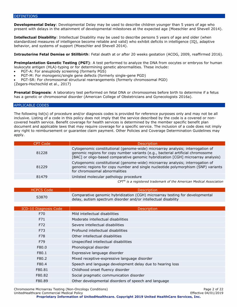
CPT Code Description
81228 Cytogenomic constitutional (genome-wide) microarray analysis; interrogation of genomic regions for copy number variants (e.g., bacterial artificial chromosome
[BAC] or oligo-based comparative genomic hybridization [CGH] microarray analysis)
81229 Cytogenomic constitutional (genome-wide) microarray analysis; interrogation of genomic regions for copy number and single nucleotide polymorphism (SNP) variants
for chromosomal abnormalities
81479 Unlisted molecular pathology procedure
CPT® is a registered trademark of the American Medical Association
HCPCS Code Description
S3870 Comparative genomic hybridization (CGH) microarray testing for developmental
delay, autism spectrum disorder and/or intellectual disability
ICD-10 Diagnosis Code Description
F70 Mild intellectual disabilities
F71 Moderate intellectual disabilities
F72 Severe intellectual disabilities
F73 Profound intellectual disabilities
F78 Other intellectual disabilities
F79 Unspecified intellectual disabilities
F80.0 Phonological disorder
F80.1 Expressive language disorder
F80.2 Mixed receptive-expressive language disorder
F80.4 Speech and language development delay due to hearing loss
F80.81 Childhood onset fluency disorder
F80.82 Social pragmatic communication disorder
F80.89 Other developmental disorders of speech and language
Chromosome Microarray Testing (Non-Oncology Conditions) Page 3 of 22
UnitedHealthcare Commercial Medical Policy Effective 04/01/2019
Proprietary Information of UnitedHealthcare. Copyright 2019 United HealthCare Services, Inc.
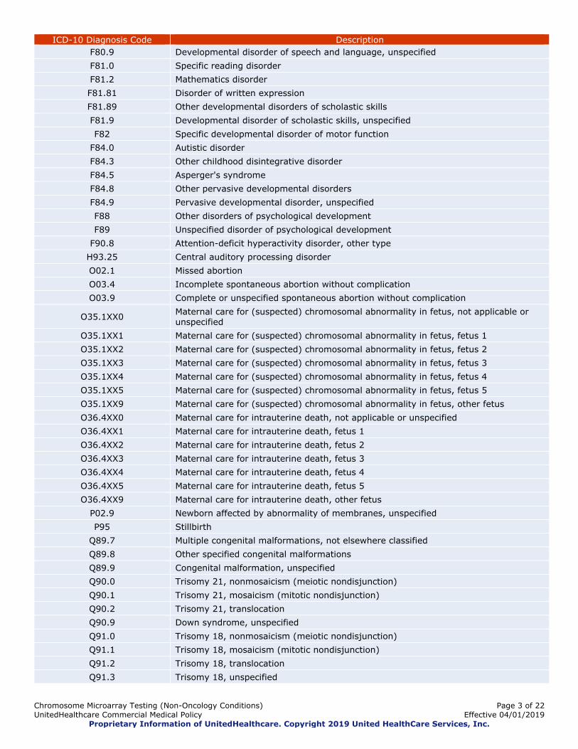
ICD-10 Diagnosis Code Description
F80.9 Developmental disorder of speech and language, unspecified
F81.0 Specific reading disorder
F81.2 Mathematics disorder
F81.81 Disorder of written expression
F81.89 Other developmental disorders of scholastic skills
F81.9 Developmental disorder of scholastic skills, unspecified
F82 Specific developmental disorder of motor function
F84.0 Autistic disorder
F84.3 Other childhood disintegrative disorder
F84.5 Asperger's syndrome
F84.8 Other pervasive developmental disorders
F84.9 Pervasive developmental disorder, unspecified
F88 Other disorders of psychological development
F89 Unspecified disorder of psychological development
F90.8 Attention-deficit hyperactivity disorder, other type
H93.25 Central auditory processing disorder
O02.1 Missed abortion
O03.4 Incomplete spontaneous abortion without complication
O03.9 Complete or unspecified spontaneous abortion without complication
O35.1XX0 Maternal care for (suspected) chromosomal abnormality in fetus, not applicable or unspecified
O35.1XX1 Maternal care for (suspected) chromosomal abnormality in fetus, fetus 1
O35.1XX2 Maternal care for (suspected) chromosomal abnormality in fetus, fetus 2
O35.1XX3 Maternal care for (suspected) chromosomal abnormality in fetus, fetus 3
O35.1XX4 Maternal care for (suspected) chromosomal abnormality in fetus, fetus 4
O35.1XX5 Maternal care for (suspected) chromosomal abnormality in fetus, fetus 5
O35.1XX9 Maternal care for (suspected) chromosomal abnormality in fetus, other fetus
O36.4XX0 Maternal care for intrauterine death, not applicable or unspecified
O36.4XX1 Maternal care for intrauterine death, fetus 1
O36.4XX2 Maternal care for intrauterine death, fetus 2
O36.4XX3 Maternal care for intrauterine death, fetus 3
O36.4XX4 Maternal care for intrauterine death, fetus 4
O36.4XX5 Maternal care for intrauterine death, fetus 5
O36.4XX9 Maternal care for intrauterine death, other fetus
P02.9 Newborn affected by abnormality of membranes, unspecified
P95 Stillbirth
Q89.7 Multiple congenital malformations, not elsewhere classified
Q89.8 Other specified congenital malformations
Q89.9 Congenital malformation, unspecified
Q90.0 Trisomy 21, nonmosaicism (meiotic nondisjunction)
Q90.1 Trisomy 21, mosaicism (mitotic nondisjunction)
Q90.2 Trisomy 21, translocation
Q90.9 Down syndrome, unspecified
Q91.0 Trisomy 18, nonmosaicism (meiotic nondisjunction)
Q91.1 Trisomy 18, mosaicism (mitotic nondisjunction)
Q91.2 Trisomy 18, translocation
Q91.3 Trisomy 18, unspecified

Chromosome Microarray Testing (Non-Oncology Conditions) Page 4 of 22
UnitedHealthcare Commercial Medical Policy Effective 04/01/2019
Proprietary Information of UnitedHealthcare. Copyright 2019 United HealthCare Services, Inc.
ICD-10 Diagnosis Code Description
Q91.4 Trisomy 13, nonmosaicism (meiotic nondisjunction)
Q91.5 Trisomy 13, mosaicism (mitotic nondisjunction)
Q91.6 Trisomy 13, translocation
Q91.7 Trisomy 13, unspecified
Q92.0 Whole chromosome trisomy, nonmosaicism (meiotic nondisjunction)
Q92.1 Whole chromosome trisomy, mosaicism (mitotic nondisjunction)
Q92.2 Partial trisomy
Q92.5 Duplications with other complex rearrangements
Q92.61 Marker chromosomes in normal individual
Q92.62 Marker chromosomes in abnormal individual
Q92.7 Triploidy and polyploidy
Q92.8 Other specified trisomies and partial trisomies of autosomes
Q92.9 Trisomy and partial trisomy of autosomes, unspecified
Q93.0 Whole chromosome monosomy, nonmosaicism (meiotic nondisjunction)
Q93.1 Whole chromosome monosomy, mosaicism (mitotic nondisjunction)
Q93.2 Chromosome replaced with ring, dicentric or isochromosome
Q93.3 Deletion of short arm of chromosome 4
Q93.4 Deletion of short arm of chromosome 5
Q93.7 Deletions with other complex rearrangements
Q93.51 Angelman syndrome
Q93.59 Other deletions of part of a chromosome
Q93.81 Velo-cardio-facial syndrome
Q93.82 Williams syndrome
Q93.88 Other microdeletions
Q93.89 Other deletions from the autosomes
Q93.9 Deletion from autosomes, unspecified
Q95.2 Balanced autosomal rearrangement in abnormal individual
Q95.3 Balanced sex/autosomal rearrangement in abnormal individual
Q99.8 Other specified chromosome abnormalities
Q99.9 Chromosomal abnormality, unspecified
R48.0 Dyslexia and alexia
R62.0 Delayed milestone in childhood
R62.50 Unspecified lack of expected normal physiological development in childhood
R62.51 Failure to thrive (child)
R62.59 Other lack of expected normal physiological development in childhood
R89.8 Other abnormal findings in specimens from other organs, systems and tissues
Z37.1 Single stillbirth
Z37.3 Twins, one liveborn and one stillborn
Z37.4 Twins, both stillborn
Z37.60 Multiple births, unspecified, some liveborn
Z37.61 Triplets, some liveborn
Z37.62 Quadruplets, some liveborn
Z37.63 Quintuplets, some liveborn
Z37.64 Sextuplets, some liveborn
Z37.69 Other multiple births, some liveborn
Z37.7 Other multiple births, all stillborn
Chromosome Microarray Testing (Non-Oncology Conditions) Page 5 of 22
UnitedHealthcare Commercial Medical Policy Effective 04/01/2019
Proprietary Information of UnitedHealthcare. Copyright 2019 United HealthCare Services, Inc.
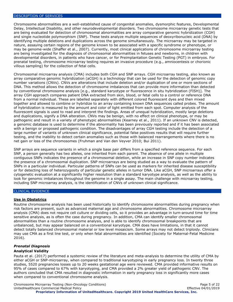
DESCRIPTION OF SERVICES
Chromosome abnormalities are a well-established cause of congenital anomalies, dysmorphic features, Developmental Delay, Intellectual Disability, and other neurodevelopmental disorders. Two chromosome microarray genetic tests that are being evaluated for detection of chromosomal abnormalities are array comparative genomic hybridization (CGH) and single nucleotide polymorphism (SNP). These tests analyze multiple sequences of deoxyribonucleic acid (DNA) by
identifying multiple deletions and duplications across the genome simultaneously. The microarray may be targeted in nature, assaying certain regions of the genome known to be associated with a specific syndrome or phenotype, or may be genome-wide (Shaffer et al., 2007). Currently, most clinical applications of chromosome microarray testing
are being investigated for the diagnosis of chromosomal abnormalities in fetuses and newborns, in children with developmental disorders, in patients who have cancer, or for Preimplantation Genetic Testing (PGT) in embryos. For prenatal testing, chromosome microarray testing requires an invasive procedure (e.g., amniocentesis or chorionic
villous sampling) for the collection of fetal cells. Chromosomal microarray analysis (CMA) includes both CGH and SNP arrays. CGH microarray testing, also known as array comparative genomic hybridization (aCGH) is a technology that can be used for the detection of genomic copy
number variations (CNVs). CNVs are alterations that include deletion and/or duplication of one or more sections of DNA. This method allows the detection of chromosome imbalances that can provide more information than detected by conventional chromosome analysis [e.g., standard karyotype or fluorescence in situ hybridization (FISH)]. The
array CGH approach compares patient DNA extracted from skin, blood, or fetal cells to a control or reference DNA from a normal individual. These are labelled separately with different colored fluorescent dyes and then mixed together and allowed to combine or hybridize to an array containing known DNA sequences called probes. The amount
of hybridization is measured by the amount and color of light emitted from each spot. Computer analysis of the fluorescent signals is used to read and interpret the findings. Areas of unequal hybridization, mostly large deletions and duplications, signify a DNA alteration. CNVs may be benign, with no effect on clinical phenotype, or may be pathogenic and result in a variety of phenotypic abnormalities (Kearney et al., 2011). If an unknown CNV is detected,
a genomic database is used to determine if the abnormality has been previously reported and if it has been associated with a benign or proposed pathogenic condition. The disadvantages of array CGH testing include the detection of a large number of variants of unknown clinical significance, potential false positives results that will require further
testing, and the inability to detect certain anomalies such as those with balanced rearrangements where there is no net gain or loss of the chromosomes (Fruhman and Van den Veyver 2010; Bui 2011).
SNP arrays are sequence variants in which a single base pair differs from a specified reference sequence. For each SNP, a person generally has two alleles, one inherited from each parent. The absence of one allele in multiple contiguous SNPs indicates the presence of a chromosomal deletion, while an increase in SNP copy number indicates
the presence of a chromosomal duplication. SNP microarrays are being studied as a way to evaluate the pattern of
SNPs in a particular individual. Particular patterns of SNPs can be used as markers for inherited disease susceptibility or for detecting loss of heterozygosity of particular genetic alleles in tumor DNA. Like aCGH, SNP microarrays offer a cytogenetic evaluation at a significantly higher resolution than a standard karyotype analysis, as well as the ability to
look for genomic imbalances throughout the genome in a single assay. The main challenge with microarray testing, including SNP microarray analysis, is the identification of CNVs of unknown clinical significance.
CLINICAL EVIDENCE Use in Obstetrics
Routine chromosome analysis has been used historically to identify chromosome abnormalities during pregnancy when risk factors are present, such as advanced maternal age and chromosome abnormalities. Chromosome microarray analysis (CMA) does not require cell culture or dividing cells, so it provides an advantage in turn-around time for time
sensitive analysis, as is often the case during pregnancy. In addition, CMA can identify smaller chromosomal abnormalities than a routine chromosome analysis, and is able to identify chromosomal breakpoints that are unbalanced, but may appear balanced on a conventional karyotype. CMA does have limitations, in that it cannot
detect totally balanced chromosomal material or low level mosaicism. Some arrays may not detect triploidy. Clinicians may use CMA as a first line test, or only when fetal abnormalities are identified (Society for Maternal-Fetal Medicine 2016).
Prenatal Diagnosis
Analytical Validity
Pauta et al. (2017) performed a systemic review of the literature and meta-analysis to determine the utility of CMA by either aCGH or SNP-microarray, when compared to traditional karyotyping in early pregnancy loss. In twenty three
studies, 5520 pregnancies losses up to 20 weeks gestational age were reviewed. CMA provided informative results on
95% of cases compared to 67% with karyotyping, and CMA provided a 2% greater yield of pathogenic CNV. The authors concluded that CMA resulted in diagnostic information in early pregnancy loss in significantly more cases
when compared to conventional chromosome analysis.

Chromosome Microarray Testing (Non-Oncology Conditions) Page 6 of 22
UnitedHealthcare Commercial Medical Policy Effective 04/01/2019
Proprietary Information of UnitedHealthcare. Copyright 2019 United HealthCare Services, Inc.
Clinical Validity
A variety of studies have evaluated the diagnostic yield of aCGH in prenatal cases, most often referred for advanced maternal age, abnormal ultrasound findings, abnormal maternal serum screening, a known chromosome abnormality
requiring further characterization, or a family history suggestive of a genetic syndrome or chromosome abnormality. Array CGH detects chromosomal imbalances in 1.3% to 10.2% of prenatal samples referred for abnormal ultrasound, advanced maternal age, abnormal maternal serum screening, family history, and/or parental anxiety (Fiorentino et al. 2011; Breman et al. 2012; Lee et al. 2012; Shaffer et al. 2012a; Wapner et al. 2012; Lovrecic et al. 2016). Several
studies reported that the diagnostic yield of aCGH in prenatal populations varied with the resolution of the platform
used, with the lowest rates (1.3% and 1.8%) among studies using a targeted array and the highest rates (7.6% and 10.2%) among studies that included arrays with genome-wide coverage (Shaffer et al. 2008; Breman et al. 2012;
Wapner et al. 2012). The available evidence also suggests that the diagnostic yield for aCGH in prenatal populations varies depending on the referral indication, with the highest rate of pathogenic CNVs most often being found in fetuses with multiple malformations identified by ultrasound, with a frequency ranging from 5.4% to 17% (Breman et
al. 2012; Lee et al. 2012; Shaffer et al. 2012a; Wapner et al. 2012; Lovrecic et al. 2016). Among studies reporting the rate of CNVs of unknown clinical significance, the rate ranged from 0% to 4.4% (Fiorentino et al. 2011; Breman et al. 2012; Lee et al. 2012; Shaffer et al. 2012a; Wapner et al. 2012).
Faas et al. (2012) evaluated the clinical and laboratory aspects of offering quantitative fluorescence (QF)-PCR followed by non-targeted whole genome 250K single-nucleotide polymorphism array analysis instead of routine karyotyping for prenatal diagnosis of fetuses with structural anomalies. Upon the detection of structural fetal anomalies, parents were
offered a choice between QF-PCR and 250K single-nucleotide polymorphism array analysis (QF/array) or QF-PCR and routine karyotyping (QF/karyo). Two hundred twenty fetal samples were included. In 153/220 cases (70%), QF/array analysis was requested. In 35/153 (23%), an abnormal QF-PCR result was found. The remaining samples were
analyzed by array, which revealed clinically relevant aberrations, including two known micro- deletions, in 5/118 cases. Inherited copy number variants were detected in 11/118 fetuses, copy number variants with uncertain clinical relevance in 3/118 and homozygous stretches in 2/118. In 67/220 (30%) of the fetuses, QF/karyo was requested: 23/67 (34%) were abnormal with QF-PCR, and in 3/67, an abnormal karyotype was found. The authors concluded
that even though QF/array does not reveal a high percentage of submicroscopic aberrations in fetuses with unselected structural anomalies, it is preferred over QF/karyo, since it provides a whole genome scan at high resolution, without additional tests needed and with a low chance on findings not related to the ultrasound anomalies.
Reddy et al. (2012) compared the results of karyotype and microarray analyses of samples obtained in stillbirths (fetal death at or after 20 weeks of gestation) after delivery. A single-nucleotide polymorphism array was used to detect
copy-number variants of at least 500 kb in placental or fetal tissue. Variants that were not identified in any of three databases of apparently unaffected persons were then classified into three groups: probably benign, clinical significance unknown, or pathogenic. In the analysis of samples from 532 stillbirths, microarray analysis yielded results more often than did karyotype analysis (87.4% vs. 70.5%) and provided better detection of genetic
abnormalities (aneuploidy or pathogenic copy-number variants, 8.3% vs. 5.8%). In this cohort, 443 were antepartum stillbirths, and CMA identified more abnormalities than karyotype alone, (8.8% vs. 6.5%) including 67 stillbirths with congenital anomalies (29.9% vs. 19.4%). As compared with karyotype analysis, microarray analysis provided a
relative increase in the diagnosis of genetic abnormalities of 41.9% in all stillbirths, 34.5% in antepartum stillbirths, and 53.8% in stillbirths with anomalies. The authors concluded that microarray analysis is more likely than karyotype analysis to provide a genetic diagnosis, primarily because of its success with nonviable tissue, and is especially
valuable in analyses of stillbirths (fetal death at or after 20 weeks of gestation) with congenital anomalies or in cases in which karyotype results cannot be obtained. In a systematic review, Grande et al. (2015) estimated the incremental yield of detecting copy number variants (CNVs)
by genomic microarray over karyotyping in fetuses with increased nuchal translucency (NT) diagnosed by first-trimester ultrasound. Seventeen studies met the inclusion criteria for analysis. Meta-analysis indicated an incremental yield of 5.0% for the detection of CNVs using microarray when pooling results. Stratified analysis of microarray results
demonstrated a 4.0% incremental yield in cases of isolated NT and 7.0% when other malformations were present. The pooled prevalence for variants of uncertain significance was 1%. The authors concluded that the use of genomic microarray provides a 5.0% incremental yield of detecting CNVs in fetuses with increased NT and normal karyotype.
Dhillon et al. (2014) evaluated whether CMA testing on the products of conception following miscarriage provides better diagnostic information compared with conventional karyotyping in a systematic review and meta-analysis that included 9 studies. There was agreement between CMA and karyotyping in 86.0% of cases. CMA detected 13%
additional chromosome abnormalities over conventional full karyotyping. In addition, traditional, full karyotyping detected 3% additional abnormalities over CMA. The incidence of a variant of unknown significance (VOUS) being
detected was 2%. The authors concluded that compared with karyotyping, there appears to be an increased detection
rate of chromosomal abnormalities when CMA is used to analyze the products of conception; however, some of these
Chromosome Microarray Testing (Non-Oncology Conditions) Page 7 of 22
UnitedHealthcare Commercial Medical Policy Effective 04/01/2019
Proprietary Information of UnitedHealthcare. Copyright 2019 United HealthCare Services, Inc.
abnormalities are VOUS, and this information should be provided when counseling women following miscarriage and when taking consent for the analysis of miscarriage products by CMA.
In a systematic review and meta-analysis, Hillman et al. (2011) evaluated whether array CGH testing in the prenatal population provides diagnostic information over that available from conventional karyotyping. Studies were selected if array CGH was used on prenatal samples or if array CGH was used on postnatal samples following termination of
pregnancy for structural abnormalities that were detected on an ultrasound scan. Of the 135 potential articles, 10 were included in this systematic review and eight were included in the meta-analysis. The pooled rate of extra information detected by array CGH when the prenatal karyotype was normal was meta-analyzed using a random-
effects model. The pooled rate of receiving an array CGH result of unknown significance was also meta-analyzed. Array CGH detected 3.6% additional genomic imbalances when conventional karyotyping was 'normal', regardless of referral indication. This increased to 5.2% more than karyotyping when the referral indication was a structural
malformation on ultrasound. The authors concluded that there appears to be an increased detection rate of chromosomal imbalances, compared with conventional karyotyping, when array CGH techniques are employed in the prenatal population. However, some are copy number imbalances that are not clinically significant. Therefore, maternal anxiety may be generated by an abnormal test result that has little clinical significance.
de Wit et al. (2014) conducted a systematic review to evaluate the diagnostic and prognostic value of genomic array testing in pregnancies with fetuses with a structural ultrasound anomaly (restricted to one anatomical system) and a normal karyotype. Combined data of the reviewed studies (n = 18) indicated that fetuses with an ultrasound anomaly restricted to one anatomical system (n = 2220) had a 3.1-7.9% chance of carrying a causative submicroscopic CNV,
depending on the anatomical system affected. This chance increased to 9.1% for fetuses with multiple ultrasound anomalies (n = 1139). According to the authors, this review indicates that 3.1-7.9% of fetuses with a structural
ultrasound anomaly restricted to one anatomical system and a normal karyotype will show a submicroscopic CNV, which explains its phenotype and provides information for fetal prognosis. The authors concluded that microarray has considerable diagnostic and prognostic value in these pregnancies.
Papoulidis et al. (2015) evaluated the diagnostic yield of comparative genomic hybridization microarrays (aCGH) and compare it with conventional karyotype analysis of standard >5-Mb resolution. A total of 1763 prenatal samples were
analyzed by aCGH (CytoChip Focus Constitutional microarrays, BlueGnome, Cambridge). The diagnostic yield of chromosomal abnormalities detected by aCGH was assessed, compared with conventional karyotype analysis. The result was pathogenic/unknown penetrance in 125 cases (7.1%), and a variant of unknown significance (VOUS) was detected in 13 cases (0.7%). Out of the 125 cases with abnormal findings, 110 were also detected by conventional
karyotype analysis. The aCGH increment in diagnostic yield was 0.9% (15/1763) and 1.6% when VOUS were included. Stratifying the sample according to indications for prenatal invasive testing, the highest values of diagnostic yield
increment were observed for patients positive for second-trimester sonographic markers (1.5%) and for the presence
of fetal structural anomalies (1.3%). In contrast, the incremental yield was marginal in patients with fetus with increased nuchal translucency (0.5%). The authors concluded that the routine implementation of aCGH offers an incremental yield over conventional karyotype analysis, which is also present in cases with 'milder' indications, further
supporting its use as a first-tier test. Srebniak et al. (2016) evaluated the diagnostic value of single-nucleotide polymorphism (SNP) array testing in 1033 fetuses with ultrasound anomalies by investigating the prevalence and genetic nature of pathogenic findings.
Pathogenic findings were classified into three categories: causative findings; unexpected diagnoses (UD); and susceptibility loci (SL) for neurodevelopmental disorders. After exclusion of trisomy 13, 18, 21, sex-chromosomal aneuploidy and triploidies, in 76/1033 (7.4%) fetuses a pathogenic chromosome abnormality was detected by
genomic SNP array: in 19/1033 cases (1.8%) a microscopically detectable abnormality was found and in 57/1033 (5.5%) fetuses a pathogenic submicroscopic chromosome abnormality was detected. 58% (n=44) of all these pathogenic chromosome abnormalities involved a causative finding, 35% (n=27) a SL for neurodevelopmental
disorder, and 6% (n=5) a UD of an early-onset untreatable disease. In 0.3% of parental samples an incidental pathogenic finding was encountered. According to the authors, these results confirm that a genomic array should be the preferred first-tier technique in fetuses with ultrasound anomalies.
Hillman et al. (2013) conducted a prospective cohort study of 243 women undergoing chromosomal microarray analysis (CMA) alongside karyotyping when a structural abnormality was detected on prenatal ultrasound. A systematic review of the literature was also performed. A total of 25 primary studies were included in the systematic
review. The cohort study found an excess detection rate of abnormalities by CMA of 4.1% over conventional karyotyping when the clinical indication for testing was an abnormal fetal ultrasound finding; this was lower than the detection rate of 10% (95% CI, 8-13%) by meta-analysis. The rate of detection for variants of unknown significance
(VOUS) was 2.1% (95% CI, 1.3-3.3%) when the indication for CMA was an abnormal scan finding. The VOUS
detection rate was lower (1.4%; 95% CI, 0.5-3.7%) when any indication for prenatal CMA was meta-analyzed. The authors concluded that there is a higher detection rate by CMA than by karyotyping not just in the case of abnormal

Chromosome Microarray Testing (Non-Oncology Conditions) Page 8 of 22
UnitedHealthcare Commercial Medical Policy Effective 04/01/2019
Proprietary Information of UnitedHealthcare. Copyright 2019 United HealthCare Services, Inc.
ultrasound findings but also in cases of other indications for invasive testing. According to the authors, it is likely that CMA will replace karyotyping in high-risk pregnancies.
Wapner et al. (2012) evaluated the accuracy, efficacy, and incremental yield of chromosomal microarray analysis as compared with karyotyping for routine prenatal diagnosis. Villus or amniotic fluid samples from women undergoing prenatal diagnosis at 29 centers were sent to a central karyotyping laboratory. Each sample was split in two; standard
karyotyping was performed on one portion and the other was sent to one of four laboratories for chromosomal microarray. A total of 4406 women were enrolled with indications for prenatal diagnosis for advanced maternal age (46.6%), abnormal result on Down's syndrome screening (18.8%), structural anomalies on ultrasonography (25.2%),
and other indications (9.4%). In 4340 (98.8%) of the fetal samples, microarray analysis was successful; 87.9% of samples could be used without tissue culture. Microarray analysis of the 4282 non-mosaic samples identified all the aneuploidies and unbalanced rearrangements identified on karyotyping but did not identify balanced translocations
and fetal triploidy. In samples with a normal karyotype, microarray analysis revealed clinically relevant deletions or duplications in 6.0% with a structural anomaly and in 1.7% of those whose indications were advanced maternal age or positive screening results. The authors concluded that for prenatal diagnostic testing, chromosomal microarray analysis identified additional, clinically significant cytogenetic information as compared with karyotyping and was
equally efficacious in identifying aneuploidies and unbalanced rearrangements but did not identify balanced translocations and triploidies. According to the authors, these data indicate a benefit to chromosomal microarray testing as a standard part of prenatal testing.
Rosenfeld et al. (2015) determined the frequency of clinically significant chromosomal abnormalities identified by chromosomal microarray in pregnancy losses at any gestational age and compared microarray performance with that
of traditional cytogenetic analysis when testing pregnancy losses. Among 535 fetal demise specimens of any gestational age, clinical microarray-based comparative genomic hybridization (aCGH) was performed successfully on 515, and a subset of 107 specimens underwent additional single nucleotide polymorphism (SNP) analysis. Overall, clinically significant abnormalities were identified in 12.8% (64/499) of specimens referred with normal or unknown
karyotypes. Detection rates were significantly higher with earlier gestational age. In the subset with normal karyotype, clinically significant abnormalities were identified in 6.9% (20/288). This detection rate did not vary significantly with gestational age, suggesting that, unlike aneuploidy, the contribution of submicroscopic chromosomal abnormalities to
fetal demise does not vary with gestational age. In the 107 specimens that underwent aCGH and SNP analysis, seven cases (6.5%) had abnormalities of potential clinical significance detected by the SNP component, including female triploidy. aCGH failed to yield fetal results in 8.3%, which is an improvement over traditional cytogenetic analysis of
fetal demise specimens. The authors concluded that both the provision of results in cases in which karyotype fails and the detection of abnormalities in the presence of a normal karyotype demonstrate the increased diagnostic utility of microarray in pregnancy loss. According to the authors, chromosomal microarray testing is a preferable, robust
method of analyzing cases of pregnancy loss to better delineate possible genetic etiologies, regardless of gestational
age. Clinical Utility
Shaffer et al. (2012b) evaluated the diagnostic utility of comparative genomic hybridization (CGH)-based microarrays for pregnancies with abnormal ultrasound findings. The authors conducted a retrospective analysis of 2858
pregnancies with abnormal ultrasounds and normal karyotypes (when performed) tested in their laboratory using CGH microarrays targeted to known chromosomal syndromes with later versions providing backbone coverage of the entire genome. Abnormalities were stratified according to organ system involvement. Detection rates for clinically significant
findings among these categories were calculated. Clinically significant genomic alterations were identified in cases with a single ultrasound anomaly (n = 99/1773, 5.6%), anomalies in two or more organ systems (n = 77/808, 9.5%), isolated growth abnormalities (n = 2/76, 2.6%), and soft markers (n = 2/77, 2.6%). The following anomalies in isolation or with additional anomalies had particularly high detection rates: holoprosencephaly (n = 9/85, 10.6%),
posterior fossa defects (n = 21/144, 14.6%), skeletal anomalies (n = 15/140, 10.7%), ventricular septal defect (n = 14/132, 10.6%), hypoplastic left heart (n = 11/68, 16.2%), and cleft lip/palate (n = 14/136, 10.3%). The authors concluded that microarray analysis identified clinically significant genomic alterations in 6.5% of cases with one or
more abnormal ultrasound findings; the majority was below the resolution of karyotyping. The authors stated that for most informed medical management, pregnancies with ultrasound anomalies undergoing invasive testing should be tested by microarray to identify all clinically significant copy number alterations (CNAs).
Brady et al. (2013) evaluated the clinical utility of chromosomal microarrays for prenatal diagnosis by a prospective study of fetuses with abnormalities detected on ultrasound. Patients referred for prenatal diagnosis due to ultrasound anomalies underwent analysis by array comparative genomic hybridization as the first-tier diagnostic test. A total of
383 prenatal samples underwent analysis by array comparative genomic hybridization. Array analysis revealed causal imbalances in a total of 9.6% of patients (n = 37). Submicroscopic copy-number variations were detected in 2.6% of
patients (n = 10/37), and arrays added valuable information over conventional karyotyping in 3.9% of patients (n =
15/37). Variants of uncertain significance were revealed in 1.6% of patients (n = 6/383). The authors concluded that there was added value of chromosomal microarrays for prenatal diagnosis in the presence of ultrasound anomalies.

Chromosome Microarray Testing (Non-Oncology Conditions) Page 9 of 22
UnitedHealthcare Commercial Medical Policy Effective 04/01/2019
Proprietary Information of UnitedHealthcare. Copyright 2019 United HealthCare Services, Inc.
Maya et al. (2010) described the clinical utility of aCGH in the prenatal testing of fetuses with an increased risk of a
chromosome disorder based on maternal age, maternal serum screening, ultrasound findings, or family history. The study involved a retrospective review of 269 cases, including 243 who had an amniocentesis and 16 who had a CVS (and 10 with an unknown specimen type). Of the 269 samples, 236 were tested with a BAC microarray and 19 were tested with an oligonucleotide microarray. Conventional cytogenetic testing identified 254 fetuses with a normal
karyotype and 15 with an abnormal karyotype of unknown clinical significance. Array-based CGH identified submicroscopic imbalances categorized as pathogenic in 3 of the 254 (1.2%) fetuses with a normal karyotype, and in 4 of the 15 (26.7%) fetuses with an abnormal karyotype. No duplications or deletions were identified in the remaining
11 (73.3%) fetuses with abnormal karyotypes. All 7 of the pregnancies with a genomic imbalance detected by aCGH were terminated based on these findings and all 262 with normal aCGH results were continued, suggesting that some patients may utilize this testing for reproductive decision making.
Professional Societies
American College of Obstetricians and Gynecologists (ACOG)
In a 2016 Committee Opinion on Microarrays and Next-Generation Sequencing Technology (American College of Obstetricians and Gynecologists, 2016a), ACOG and SMFM make the following recommendations and conclusions for
the use of chromosomal microarray analysis and newer genetic technologies in prenatal diagnosis (AGOG, Committee Opinion, 2016): Most genetic changes identified by chromosomal microarray analysis that typically are not identified on standard
karyotype are not associated with increasing maternal age; therefore, the use of this test can be considered for all women, regardless of age, who undergo prenatal diagnostic testing.
Prenatal chromosomal microarray analysis is recommended for a patient with a fetus with one or more major
structural abnormalities identified on ultrasonographic examination and who is undergoing invasive prenatal diagnosis. This test typically can replace the need for fetal karyotype.
In a patient with a structurally normal fetus who is undergoing invasive prenatal diagnostic testing, either fetal karyotyping or a chromosomal microarray analysis can be performed.
Chromosomal microarray analysis of fetal tissue (i.e., amniotic fluid, placenta, or products of conception) is recommended in the evaluation of intrauterine fetal death or stillbirth when further cytogenetic analysis is desired because of the test’s increased likelihood of obtaining results and improved detection of causative abnormalities.
Comprehensive patient pretest and posttest genetic counseling from an obstetrician–gynecologist or other health care provider with genetics expertise regarding the benefits, limitations, and results of chromosomal microarray analysis is essential. Chromosomal microarray analysis should not be ordered without informed consent, which
should include discussion of the potential to identify findings of uncertain significance, non-paternity,
consanguinity, and adult-onset disease. In a 2016 Practice Bulletin (American College of Obstetricians and Gynecologists, 2016b) on prenatal diagnostic
testing for genetic disorders, ACOG and the Society for Maternal-Fetal Medicine (SMFM) recommend the following based on good and consistent scientific evidence (Level A): Chromosome microarray analysis should be made available to any patient choosing to undergo invasive diagnostic
testing. Chromosome microarray analysis should be the primary test (replacing conventional karyotype) for patients
undergoing prenatal diagnosis for the indication of a fetal structural abnormality detected by ultrasound.
The 2016 Practice Bulletin further stated that prenatal diagnostic testing for genetic disorders makes the following recommendation based on limited or inconsistent scientific evidence (Level B): Chromosomal microarray analysis can be used to confirm an abnormal FISH test.
Society for Maternal-Fetal Medicine (SMFM)
In an SMFM Consult Series publication (2016) on the use of chromosomal microarray for prenatal diagnosis, SMFM makes the following recommendations: Chromosomal microarray analysis (CMA) should be offered when genetic analysis is performed in cases with fetal
structural anomalies and/or stillbirth and replaces the need for fetal karyotype in these cases (GRADE 1A) Providers should discuss the benefits and limitations of CMA and conventional karyotype with patients who are
considering amniocentesis and chorionic villus sampling (CVS) and that both options be available to women who choose to undergo diagnostic testing (GRADE 1B)
The use of CMA is not recommended as a first-line test to evaluate first trimester pregnancy losses due to limited data (GRADE 1C)
Pre- and post-test counseling should be performed by trained genetic counselors, geneticists or other providers
with expertise in the complexities of interpreting CMA results (Best practice).
Chromosome Microarray Testing (Non-Oncology Conditions) Page 10 of 22
UnitedHealthcare Commercial Medical Policy Effective 04/01/2019
Proprietary Information of UnitedHealthcare. Copyright 2019 United HealthCare Services, Inc.
Preimplantation Genetic Screening (PGS) and Preimplantation Genetic Diagnosis (PGD)
PGS is an analysis performed on an embryo prior to transfer to screen for aneuploidy, deletions and duplications of genomic material, generally referred to as copy number variations (CNVs). The term Preimplantation Genetic Diagnosis (PGD) is sometimes also used to refer to aneuploidy and CNV screening of embryos, but PGD primarily
refers to the analysis of single gene or other inherited disorders in an embryo. Use of this technology could potentially increase the success of infertility treatment, especially in women who have worse outcomes due to advanced maternal age, history of recurrent miscarriage, failed in vitro fertilization (IVF) (CDC, 2017) or a balanced chromosome translocation. In addition, it has been explored as a way to enable single embryo transfer (SET) rather than using
multiple embryos to increase the odds of having a successful pregnancy without the risk of multiparity. However,
there is limited evidence that the use of PGS improves fertility treatment outcomes. In the most recent update from the Centers of Disease Control on the status of assisted reproductive technologies (ART) in the US, PGS was used in
only 5% of 231,936 cycles tracked among 464 clinics. Analytical Validity
Treff et al. (2011a) studied various whole genome amplification (WGA) techniques in single cells, as accurately amplifying the genome is critical to accurately and successfully genotyping an individual cell. The authors compared three commercially available WGA kits; GenomePlex, REPLI-g, and GenomiPhi, and compared them based on the
purpose of the analysis. They specifically studied how well each produced whole genome DNA, and then how well various tests performed; SNP microarray copy number, ability to call individual SNPs, and molecular karyotyping. Human fibroblast cell lines from the Coriell Cell Repository with known karyotypes and genetic results were used.
GenomiPhi was able to amplify 88% of single cells yielding >250 ng of DNA, and REPLI-G and GenomePlex yielded >250 ng of DNA in 100% of single cells. Repli-G provided 88% of genome coverage, and GenomiPhi and GenomePlex provided 74% and 78% respectively. SNP copy number accuracy was 99% for GenomePlex, 95% for REPLI-G, and
62% for GenomiPhi. Chromosome ploidy accuracy was 99% for GenomePlex, 97% for REPLI-G, and 75% for GenomiPhi. The karyotyping diagnostic accuracy was 100% for GenomePlex, 83% for REPLI-G, and 0% for GenomiPhi. This study provides an example of the range of reliability of commercial WGA kits that may be used to predict the reproductive potential and chromosomal status of embryos.
Gutiérrez-Mateo et al. (2011) did an analysis of two aCGH approaches to determine the best method for evaluating single cells in preimplantation genetic screening, and compared aCGH to FISH. Group 1 had WGA with GenomePlex
and Group 2 with SurePlex. Results were compared to FISH to determine the error rate. 161 embryos were analyzed in Group 1, and 654 embryos in Group 2. Group 1 had an amplification failure of 11%, and Group 2 was 3%. A total of 66 Group 1 and 54 Group 2 embryos were disaggregated and the remaining cells were analyzed by FISH. The
discordance rate between aCGH in Group 1 was 9%, and Group 2 was 2%. Overall, 759 embryos had clear aCGH
results regardless if they were Group 1 or 2. 65% of the embryos were classified as abnormal. A comparison of FISH in 120 embryos found that 75% were abnormal, and 44% were mosaic. In reviewing aneuploid events in the cohort, aCGH identified 1615 aneuploid events. FISH would only have found 41% of these with nine probes and 54% with 12
probes. In embryos with two or more abnormalities, 12-chromosome FISH would have identified up to 87% of the abnormal embryos detected by CGH. Implantation rate across both groups was 42%. Overall the authors concluded that CGH will detect 42% more abnormalities and 13% more abnormal embryos as compared to 12-chromosome
standard FISH. Novik et al. (2014) published a comparison of fluorescence in situ hybridization (FISH) methods used to evaluate
chromosomal mosaicism in IVF embryos with CMA to determine the limits of mosaicism detection, accuracy, and mosaicism prevalence. Chromosomal mosaicism is higher in IVF created embryos than in other prenatal specimens, and may be found in 71-73% of human embryos. Low levels of mosaicism in prenatal specimens suggest selective pressure against mosaic embryos for ongoing pregnancy. Mosaicism has been reported in embryos evaluated by CMA
using trophoectoderm (TE) biopsies, but the effect of TE mosaicism on development, implantation and pregnancy outcome is unknown. To determine the limits of mosaicism detection, the authors mixed different ratios of amplified DNA from aneuploid and euploid cells, as well as tested clinical samples. Overall, they were able to identify the limit of
mosaicism detection with CMA at 25-37% for gains of DNA, and 37-50% for losses. They used the CMA technique developed to CMA was used to determine if an embryo was euploid, non-mosaic aneuploidy, or mosaic aneuploid. The diagnostic accuracy of the CMA test was assessed by FISH analysis on non-transferred embryos. In 47 embryos, 26
were considered to be non-mosaic aneuploid by CMA, and 100% were confirmed by FISH. In the mosaic category, 95% were confirmed by FISH. The single embryo not confirmed by FISH did have a discordant result with 7% of nuclei with an aneuploid FISH signal that was below the threshold to call the embryo abnormal. Embryos predicted to the euploid by CMA were not tested by FISH. The authors concluded that CMA testing can identify mosaicism in day
5/6 blastocysts, and that FISH confirms that the mosaicism is real and not likely a technical artifact. Capalbo et al. (2015) compared SNP based microarray screening, aCGH and qPCR techniques for screening embryos.
The authors conducted a prospective double blind observational study from Oct. 2012-Dec. 2013. TE biopsies were done on day 5-6. 45 patients were included who had indications of advanced maternal age, recurrent miscarriage or parental carrier of a balanced translocation. A total of 124 blastocysts underwent aCGH. Of these, 122 survived

Chromosome Microarray Testing (Non-Oncology Conditions) Page 11 of 22
UnitedHealthcare Commercial Medical Policy Effective 04/01/2019
Proprietary Information of UnitedHealthcare. Copyright 2019 United HealthCare Services, Inc.
warming and re-expansion and underwent TE biopsy and qPCR analysis. Two samples failed qPCR and were excluded. 82% of embryos showed the same diagnosis between aCGH and qPCR. 18% were discordant for at least one
chromosome. Discordant blastocysts were warmed and TE was biopsied again on 21 embryos that survived another rewarming and underwent a blinded SNP array analysis. A conclusive result was obtained in 18 of the 21. In 4 of these, the qPCR, aCGH and SNP array did not match and were considered mosaic aneuploid. Overall, when the data is viewed per chromosome, the aCGH and qPCR results were consistent in 99.9% of cases where both methods were
performed on TE biopsy from the same embryo. The SNP based reanalysis, however, showed a higher discordant rate between aCGH and qPCR. The authors concluded that TE biopsies can be a highly reliable and effective approach for PGS, and that until aCGH is studied for their clinical negative predictive value, this comparative study can only
demonstrate that aCGH results in a higher aneuploidy rate than other contemporary and better validated methods of chromosome screening.
Kurahashi et al. (2015) conducted a comprehensive review of the literature regarding the analytical validity of CMA for PGS. The authors reported that while oligonucleotide arrays (CMA) are the standard for clinical analysis of individuals with developmental delay and congenital anomalies, the need to use a single cell and then perform WGA when using CMA for PGS may introduce amplification bias. Uneven amplification can occur of various regions of the DNA sampled
from the embryo and lead to inaccuracy in the test results. Newer technologies including bacterial artificial chromosome (BAC) and a multiple displacement method are being explored as ways to mitigate amplicafication bias. Mosaicism in the embryo is also reported by the authors as a factor to overcome in using CMA for PGS. It has been
demonstrated in the oocyte and blastomere that the spindle assembly process that regulates chromosome segregation is transiently deficient, which leads to a high rate of mosaicism during this stage, and raises the question of whether or not a single cell biopsied during this stage is representative of the whole embryo. In addition, self-correction of the
mosaicism to a euploid embryo has been demonstrated, so low level mosaicism may not be a concern. Studies have shown that CMA can identify mosaicism n only 25% of embryos and so may miss low levels of mosaicism. This review further describes issues of cell cycle replication as a confounding factor for CMA. DNA replication begins at more than 10,000 sites in a genome, and during S phase, some parts of the genome have finished replicating and have two
copies while other regions have not completed replicating and have a single copy of DNA. This variation in copy number could be incorrectly interpreted as abnormal or as high background noise. The risk of cell cycle issues may be mitigated by performing cell sampling just after cell division, or by trophoectoderm biopsy in the blastocyst state.
Finally, CMA is not optimal for identifying polyploidy which is a significant limitation because triploidy is one of the most common chromosome abnormalities found in miscarriages. Microarrays that are SNP based can be used for detection of polyploidy, but at the time of publication, SNP arrays have not been optimized for WGA. Overall,
Kurahashi et al. (2015) concludes that CMA for PGS is slowly becoming a clinical standard, but states that the procedure needs to be optimized on an individual basis and tailored protocols are required.
Clinical Utility
Schoolcraft et al. (2010) first used a SNP based microarray to examine all 24 chromosomes for aneuploidy screening as a clinical selection tool for blastocyst transfer for assisted reproduction patients. The authors studied 132 patients
who met the inclusion criteria of > 38 years old, recurrent pregnancy loss, or recurrent implantation failure. All embryos were biopsied on day 5 or 6. The authors report that the sensitivity and specificity is 98.6% and 100%, respectively. Ninety four of the patients produced euploid embryos that were eligible for transfer. Of the 175 embryos
transferred, 118 implanted, 111 are ongoing and healthy, or live births. At the time of publication, 28 healthy babies were born to 16 patients. In the transfer group, the biochemical pregnancy rate was 89%, and the clinical pregnancy rate was 77.8%.
Fishel et al. (2011) studied the use of aCGH on analyzing the first oocyte polar body (PB 1) and providing dependable data within 48 hours to support fresh embryo transfer. 134 couples undergoing IVF participated in the study and inclusion criteria included advanced maternal age, recurrent miscarriage, and previously failed IVF. Polar bodies were
biopsied and WGA was accomplished using SurePlex. Of the 134 patients, there were 150 cycles. A total of 861 polar bodies were tested, and 67% were aneuploid. 47% of these involved a single chromosome. 26% of cycles failed to results in embryo transfer because there were no euploid oocytes. The live birth rate per embryo transfer was 24%.
Yang et al. (2012) studied the value of aCGH in identifying single embryos for transfer. Single embryo transfer (SET) is a strategy utilized by some clinics as a means to reduce the number of multiple gestation pregnancies. In this study,
patients included in the study were first time assisted reproduction patients, had normal karyotypes, were < age 35, and seeking elective SET. They were randomized into two groups. Group A had 55 patients who produced 425 blastocysts, and embryos for transfer were selected by morphology and aCGH results. Group B had 48 patients and 389 blastocysts, and embryos were selected based on morphology alone. Amongst the transfers, the clinical
pregnancy rate was 71% for Group A, and 46% for Group B. The ongoing pregnancy rate was 69% for Group A, and 42% for Group B. There were no twin pregnancies. Overall, aCGH significantly improved the SET pregnancy rate when
compared to using morphology alone.

Chromosome Microarray Testing (Non-Oncology Conditions) Page 12 of 22
UnitedHealthcare Commercial Medical Policy Effective 04/01/2019
Proprietary Information of UnitedHealthcare. Copyright 2019 United HealthCare Services, Inc.
Liu et al. (2012) utilized a Rubicon WGA kit and an oligo microarray to analyze the pregnancy rate after screening embryos for aneuploidy. They received biopsied cells from the inner cell mass (ICM) or trophoectoderm (TE) for 258
blastocysts from 51 cycles. 95% of blastocysts had intact DNA signals after microarray. 57% were aneuploid, with embryos from advanced maternal age women more likely to be abnormal. All euploid embryos survived after warming, and of these, a 63% implantation rate was observed. The authors concluded that using oligo CMA screened embryos can significantly increase the clinical pregnancy rate.
Liang et al. (2013) explored the clinical utility of using an oligo microarray for embryo PGS. The team analyzed 383 blastocysts from 72 patients who were advanced maternal age or experience recurrent miscarriage. Biopsied cells
underwent Rubicon WGA and screened with an oligo microarray. Some aneuploidy blastocysts were analyzed further by FISH to evaluate the accuracy of results. Overall, 58% of embryos were abnormal. Transfer of normal embryos resulted in an implantation rate of 54%. The FISH and microarray analysis matched in all aneuploid embryos analyzed.
The authors concluded that the oligo array platform was able to identify aneuploidy and other small gains and losses, and improved embryo implantation rates. Tobler et al. (2014) conducted a retrospective analysis comparing SNP-array and aCGH in 543 embryos from 63
couples, of which one parent carried a reciprocal translocation. Couples were from 16 different fertility centers with samples being analyzed at one lab. SNP-array was used for molecular karyotyping from 2007 to 2011, and from 2011 to 2014 aCGH was used. No embryo was analyzed by both methods. A cell was obtained from the embryo at day 5 or
the blastocyst stage and placed in a stabilizing buffer and frozen for transport. Whole genome amplification (WGA) was accomplished for the SNP-array using a phi 29 polymerase protocol, and aCGH WGA was done using a Klenow fragment and a modified random priming protocol. Molecular karyotypes were obtained on 92% (498) of the biopsied
embryos. In the 8% (45) samples that failed, WGA failed and was strongly correlated with poor embryo quality. Overall, 45% of embryos were chromosomally normal, and the remaining had translocation errors or aneuploidy. The pregnancy rates were equivalent for SNP (60%) and aCGH (65%). The pregnancy rate was slightly higher if the biopsy was done on blastocysts (65%) vs. cleavage stage embryos (59%). Overall the authors concluded that SNP or
aCGH microarray technologies demonstrate equivalent clinical findings that maximize the pregnancy potential in patients with known parental reciprocal chromosome translocations.
Gleicher and Orvieto (2017) conducted a comprehensive literature review through January, 2017 on research related to current PGS methodologies and outcomes using comparative chromosome screening on 5-6 day TE biopsies, which they call PGS 2.0. This includes aCGH and SNP-based array technologies. Overall they noted that the literature has a
skewed view of clinical utility, and uses embryo transfer as the starting point for measuring success, whereas generally IVF literature uses the initiated IVF cycles as the starting point. When using initiated cycles as a starting point, non-PGS cycles result in a higher live birth rate over PGS cycles. In addition, they report from their analysis
that TE mosaicism may be present in at least half of all embryos, and mathematical models suggest that that the
likelihood of false negative and positive results is too high to safely determine which embryos should be transferred or not. Their overall conclusion is that PGS 2.0 does not have clinical utility and may in fact reduce live birth rates.
Treff et al. (2011b) evaluated the clinical validity of using SNP microarray testing for PGD. Specifically, an Affymetrix 262K SNP microarray was used to identify genomic imbalances in the embryos of 15 patients known to carry balanced chromosome translocations. A total of 19 in vitro fertilization (IVF) treatment cycles with SNP microarray testing were
performed for the 15 patients. One patient produced only aneuploid embryos. Two patients became pregnant naturally, and 2 did not return to the clinic for embryo transfer. Twelve transfers were performed in the remaining 10 patients, 10 of which produced a biochemical pregnancy (positive beta-human chorionic gonadotropin testing) and 9 of which produced a clinical pregnancy (fetal heart beat). Six of these 9 delivered a singleton; the remaining 3 pregnancies
were ongoing at the time of publication. Of the 6 children born, follow-up after delivery was possible for 4 and revealed no genomic imbalances (Treff et al. 2011b). Well designed, comparative studies with larger patient populations are needed to further describe clinical outcomes of microarray testing for PGD in patients with balanced
translocations. Zhang et al. (2017) examined the utility of using SNP-microarray in families with balanced translocations to accurately
identify euploid embryos for transfer. In 68 blastocysts from 11 translocation families, SNP-microarray identified 42 unbalanced or aneuploidy embryos, and 26 balanced or normal chromosomes. Ten families became pregnant on the first cycle; one family was successful on cycle three. Amniocentesis on the resulting pregnancies matched the embryo microarray analysis, resulting in a 100% sensitivity and sensitivity in this cohort, but the authors caution that a larger
sample size is needed to further validate sensitivity and sensitivity. There is insufficient data regarding the clinical utility and analytical validity of microarray testing for PGS and PGD.
Comparative studies are needed to evaluate implantation and pregnancy rates after microarray analyses compared to
conventional testing.

Chromosome Microarray Testing (Non-Oncology Conditions) Page 13 of 22
UnitedHealthcare Commercial Medical Policy Effective 04/01/2019
Proprietary Information of UnitedHealthcare. Copyright 2019 United HealthCare Services, Inc.
Professional Societies
American Society for Reproduction Medicine (ASRM) / Society for Assisted Reproductive Technology (SART)
In this joint Practice Committee Opinion from 2013, ASRM and SART comment on the pros and cons of blastocyst
transfer and culture, which may increase pregnancy rates in good prognosis patients, but does not impact the pregnancy rate in poor prognosis patients. This technique may be associated with a small increased risk of monozygotic twinning and adverse neonatal outcomes. However, the committee comments that blastocyst culture is required to accommodate PGS, which has increasing utility, and allows for the transfer of only euploid embryos.
Preimplantation Genetic Diagnosis International Society (PGDIS)
The PGDIS position statement on chromosome mosaicism and preimplantation aneuploidy at the blastocyst state states that only a validated next generation sequencing (NGS) platform that can quantitatively measure copy number should be used, and can accurately measure 20% of mosaicism in a known sample.
Use in Pediatrics
Developmental Disorders
Analytical Validity
Baldwin et al. (2008) described the validation of an oligonucleotide array with approximately 43,000 probes spaced an average of 75 kb across the genome, a resolution of 500 kb overall, and a resolution of 50 kb in targeted regions. Initially, 10 patients with known chromosome abnormalities, including 2 supernumerary marker chromosomes, 5
telomere deletions, 2 unbalanced translocations, and a 15q11 to q13 microdeletion, were tested with the array. The array correctly identified all 10 (100%) anomalies and identified additional complex rearrangements in 2 (20%) of the cases. Another 20 patient samples, including 14 cases with abnormalities and 6 with normal cytogenetic findings,
were subsequently analyzed in a blinded manner. As with the previous group of samples, the concordance rate between the aCGH results and previous cytogenetic testing was 100 %. Clinical Validity
Ellison et al. (2012) tested the hypothesis that chromosomal microarray analysis frequently identifies conditions that require specific medical follow-up and that referring physicians respond appropriately to abnormal test results. A total
of 46,298 postnatal patients were tested by chromosomal microarray analysis for a variety of indications, most commonly intellectual disability/developmental delay, congenital anomalies, dysmorphic features, and neurobehavioral problems. The frequency of detection of abnormalities associated with actionable clinical features was tallied, and the
rate of physician response to a subset of abnormal tests results was monitored. A total of 2088 diagnoses were made of more than 100 different disorders that have specific clinical features that warrant follow-up. The detection rate for
these conditions using high-resolution whole-genome microarrays was 5.4%, which translates to 35% of all clinically
significant abnormal test results identified in the laboratory. In a subset of cases monitored for physician response, appropriate clinical action was taken more than 90% of the time as a direct result of the microarray finding. The authors concluded that the disorders diagnosed by chromosomal microarray analysis frequently have clinical features that need medical attention, and physicians respond to the diagnoses with specific clinical actions, thus arguing that
microarray testing provides clinical utility for a significant number of patients tested. Miller et al. (2010) assessed the clinical validity of chromosomal microarray testing as a first-tier analysis in the
evaluation of patients with unexplained DD, ID, ASD, and/or MCA (postnatal only). A review of 33 studies involving 21,698 patients was performed to evaluate the sensitivity of microarrays in the detection of deletions and duplications. The analysis of data from all 33 studies, including those that involved BAC or oligonucleotide microarrays (aCGH),
showed that chromosomal microarray analysis in general had a diagnostic yield of approximately 15% to 20%. In addition, it was determined that, although balanced rearrangements and low-level mosaicism were generally not detectable by microarray analysis, these anomalies were rare causes of DD/ID/MCA (< 1%). As a result, the ISCA stated that the evidence supports the use of chromosomal microarray testing as a first-tier test in the clinical
evaluation of infants, children, or adults with DD, ID, ASD, and/or MCA. Battaglia et al. (2013) evaluated the usefulness of CMA, as a first-tier tool in detecting the etiology of unexplained
intellectual disability/autism spectrum disorders (ID/ASDs) associated with dysmorphic features in a large cohort of pediatric patients. The study included 349 individuals; 223 males, 126 females, aged 5 months-19 years. Blood samples were analyzed with CMA at a resolution ranging from 1 Mb to 40 Kb. The imbalance was confirmed by FISH
or qPCR. Copy number variants (CNVs) were considered causative if the variant was responsible for a known syndrome, encompassed gene/s of known function, occurred de novo or, if inherited, the parent was variably affected, and/or the involved gene/s had been reported in association with ID/ASDs in dedicated databases. A total of 91 CNVs were detected in 77 (22.06%) patients: 5 (6.49%) of those presenting with borderline cognitive impairment, 54
(70.13%) with a variable degree of DD/ID, and 18/77 (23.38%) with ID of variable degree and ASDs. The CNVs
caused the phenotype in 57/77 (74%) patients; 12/57 (21.05%) had ASDs/ID, and 45/57 (78.95%) had DD/ID. The authors concluded that this study provided further evidence of the high diagnostic yield of CMA for genetic testing in
children with unexplained ID/ASDs who had dysmorphic features.
Chromosome Microarray Testing (Non-Oncology Conditions) Page 14 of 22
UnitedHealthcare Commercial Medical Policy Effective 04/01/2019
Proprietary Information of UnitedHealthcare. Copyright 2019 United HealthCare Services, Inc.
Bremer et al. (2011) used high-resolution whole genome array-based comparative genomic hybridization (array-CGH)
to screen 223 Autism spectrum disorder (ASD) patients for gene dose alterations associated with susceptibility for autism. Clinically significant copy number variations (CNVs) were identified in 18 individuals (8%), of which 9 cases (4%) had de novo aberrations. In addition, 20 individuals (9%) were shown to have CNVs of unclear clinical relevance. Among these, 13 cases carried rare but inherited CNVs that may increase the risk for developing ASDs, while parental
samples were unavailable in the remaining seven cases. Classification of all patients into different phenotypic and inheritance pattern groups indicated the presence of different CNV patterns in different patient groups. Clinically relevant CNVs were more common in syndromic cases compared to non-syndromic cases. Rare inherited CNVs were
present in a higher proportion of ASD cases having first- or second-degree relatives with an ASD-related neuropsychiatric phenotype in comparison with cases without reported heredity. The authors concluded that rare CNVs, encompassing potential candidate regions for ASDs, increase the susceptibility for the development of ASDs
and related neuropsychiatric disorders giving further insight into the complex genetics underlying ASDs. Bartnik et al. (2014) evaluated the application of array Comparative Genomic Hybridization (CGH) in clinical diagnostics of developmental delay/ intellectual disability in 112 children. The authors identified 37 copy number
variants (CNVs) with the size ranging from 40 kb to numerical chromosomal aberrations, including unbalanced translocations and chromosome Y disomy, receiving an overall diagnostic yield of 33%. Known pathogenic changes were identified in 21.4% of the cases. Among patients with pathogenic CNVs identified by array CGH, 41.7% had a
previously normal karyotype analysis. According to the authors, this study provides more insight into the benefits derived by using chromosomal microarray analysis and demonstrates the usefulness of array CGH as a first-tier clinical setting test in patients with intellectual disability.
Nicholl et al. (2014) prospectively evaluated the frequency of pathogenic chromosomal microdeletions and microduplications in a large group of referred patients with developmental delay (DD), intellectual disability (ID) or autism spectrum disorders (ASD) within a genetic diagnostic service. First tier testing was applied using a
standardized oligo-array comparative genomic hybridization (CGH) platform, replacing conventional cytogenetic testing that would have been used in the past. Copy number variants (CNVs) found to be responsible for the clinical condition on the request form could all be subdivided into 3 groups: well established pathogenic microdeletion/
microduplication/aneuploidy syndromes, predicted pathogenic CNVs as interpreted by the laboratory, and recently established pathogenic disease susceptibility CNVs. Totaled from these three groups, with CNVs of uncertain significance excluded, detection rates were: DD (13.0%), ID (15.6%), ASD (2.3%), ASD with DD (8.2%), ASD with
ID (12.7%) and unexplained epilepsy with DD, ID and ASD (10.9%). According to the authors, the greater diagnostic sensitivity arising from routine application of array CGH, compared with previously used conventional cytogenetics, outweighs the interpretative issues for the reporting laboratory and referring clinician arising from detection of CNVs
of uncertain significance. The authors stated that precise determination of any previously hidden molecular defect
responsible for the patient's condition is translated to improved genetic counselling. Mc Cormack et al. (2016) examined the utility of aCGH to replace karyotype in 5369 pre- and post-natal patients with
an unexplained phenotype. In this cohort, 28% of those tested had a deletion or duplication. Ninety seven percent of cases with a CNV that was less than 5 kilobases in size would not have been detected by routine chromosome analysis, Eight hundred and forty two 15.7% had a variant of unknown significance. About 5% of the cohort met the criteria for
a known syndrome. Using microarray as a primary analysis tool significantly increased the detection of CNV abnormalities, with 1 syndromic case identified per 20 referrals. Szczaluba et al. (2016) studied the value of aCGH in newborns with multiple congenital anomalies. A group of 54
neonates with two or more birth defects were evaluated with an OGT Cytosure 8x60 K microarray. Ten newborns were found to have rearrangements detected by aCGH. One was a recurrent syndromic microdeletion, but the others were unique. Five could be seen on routine cytogenetics, but one was sub-microscopic. The other four were copy number
variants that were likely pathogenic and could explain the phenotype. Geddes et al. (2017) evaluated a protocol to direct genetic testing, including karyotyping, 22q deletion analysis, and
CMA, on infants with critical congenital heart disease. In a retrospective review of data of 733 infants prior to implementing the genetic testing protocol, 433 had at least one of included genetic tests. 66% of these patients had more than one genetic test, and the rate of diagnosis was 26%. A genetic testing protocol was identified that aligned genetic testing with clinical presentation. For example, infants that were likely to have Trisomy 21 or Turner syndrome
were first tested with routine chromosome analysis. Conotruncal heart lesion patients were evaluated by 22q analysis, and all others had a chromosome microarray as a first test. The protocol was implemented in January 2015 and evaluated through June 2016. In the post protocol period, 158 patients were evaluated and 121 patients had at least
one genetic test. The rate of genetic testing increased to 77%, and only 24% of patients had more than one genetic
test. The rate of diagnosis was 36%. Overall, in the post-protocol period, infants were less likely to undergo multiple genetic tests, and were more likely to get a genetic diagnosis. Diagnostic yield varied between pre-and post-protocol
tests as well. For karyotyping, the pre-protocol yield as 18%, and post-protocol was 76%. 22q analysis pre-protocol

Chromosome Microarray Testing (Non-Oncology Conditions) Page 15 of 22
UnitedHealthcare Commercial Medical Policy Effective 04/01/2019
Proprietary Information of UnitedHealthcare. Copyright 2019 United HealthCare Services, Inc.
diagnostic yield was 24% and 26% post-protocol. There was no significant difference in the diagnostic yield of CMA at 22% pre-protocol and 22% post-protocol. There were no results in this cohort detected by karyotype or 22q deletion
analysis that was not detectable by CMA. Clinical Utility
Pfundt et al. (2016) assessed the diagnostic yield and potential clinical utility of a high-density chromosomal microarray (CMA) of CytoScan Dx Assay in 960 patients with developmental delay or intellectual disability. Eighty-six percent of the subjects were assessed using a microarray as part of historical routine patient care (RPC). The rate of
pathogenic findings was similar between RPC (13.3%) and the CytoScan Dx Assay (13.8%). Among the 138 patients
who did not receive microarray as RPC, the diagnostic yield for CytoScan Dx Assay was 23.9% as compared with 14.5%, indicating a 9.4% improvement when using higher-resolution methods. Thirty-five percent of patients with
abnormal findings had predicted clinical management implications that may improve health outcomes. The authors concluded that the assay's diagnostic yields are similar to those found in other studies of CMAs.
Fry et al. (2016) evaluated the range of rare Copy number variants (CNVs) found in 80 Welsh patients with intellectual disability (ID) or developmental delay (DD), and childhood-onset epilepsy. Molecular cytogenetic testing by single nucleotide polymorphism array or microarray-based comparative genome hybridization was performed. 8.8 % (7/80) of the patients had at least one rare CNVs that was considered to be pathogenic or likely pathogenic. The CNVs
involved known disease genes (EHMT1, MBD5 and SCN1A) and imbalances in genomic regions associated with neurodevelopmental disorders (16p11.2, 16p13.11 and 2q13). Prompted by the observation of two deletions disrupting SCN1A the authors undertook further testing of this gene in selected patients. This led to the identification
of four pathogenic SCN1A mutations in the cohort. Five rare de novo deletions were identified, and the authors confirmed the clinical utility of array analysis in patients with ID/DD and childhood-onset epilepsy.
Shen et al. (2010) evaluated a cohort of 933 patients who received clinical genetic testing for a diagnosis of autism spectrum disorders (ASD). Clinical genetic testing included G-banded karyotype, fragile X testing, and chromosomal microarray (CMA) to test for submicroscopic genomic deletions and duplications. Diagnostic yield of clinically significant genetic changes was compared. Karyotype yielded abnormal results in 19 of 852 patients. Fragile X testing
was abnormal in 4 of 861 and CMA identified deletions or duplications in 154 of 848 patients. CMA results for 59 of 848 patients were considered abnormal, which includes variants associated with known genomic disorders or variants of possible significance. CMA results were normal in 10 of 852 patients (1.2%) with abnormal karyotype due to
balanced rearrangements or unidentified marker chromosome. CMA with whole-genome coverage and CMA with targeted genomic regions detected clinically relevant copy-number changes in 7.3% and 5.3% of patients, respectively, both higher than karyotype. With the exception of recurrent deletion and duplication of chromosome
16p11.2 and 15q13.2q13.3, most copy-number changes were unique or identified in only a small subset of patients. The authors concluded that CMA had the highest detection rate among clinically available genetic tests for patients with ASD. Interpretation of microarray data is complicated by the presence of both novel and recurrent copy-number variants of unknown significance. The authors stated that despite these limitations, CMA should be considered as part
of the initial diagnostic evaluation of patients with ASD. In a clinical guideline for the recognition, referral and diagnosis of children and young people with autism, the National
Institute for Health and Care Excellence (NICE) recommends that genetic tests only be done in patients who have either dysmorphic features and/or intellectual disability because these are the cases where the rate of genetic abnormalities are definitely increased above general population levels. According to the guideline, most research to
date has focused on the rate and type of definite abnormalities rather than the impact of testing on children/young people with autism and their families. The authors of the guideline state that further research using CGH array would lead to a stronger evidence base to inform key decision-makers as to whether wider use of genetic testing is appropriate or not when this guideline is updated. It would also alert practitioners to any negative consequences that
might occur as a result of testing (NICE 2011). In a technology report, the Blue Cross Blue Shield Association (2015) stated that the ability to detect pathogenic CNVs
underlying global developmental delay/intellectual disability (GDD/ID) and autism spectrum disorder (ASD) is improving. This improvement is likely due to higher chromosomal microarray (CMA) resolution along with increasingly extensive data for copy number variant (CNV) pathogenicity and associated phenotypes. Professional societies
recommended CMA testing as first-line evaluation when genetic evaluation is desired as opposed to first obtaining a karyotype. According to the report, there is consistent evidence that the diagnostic yield obtained from CMA testing is higher than with karyotyping in children with GDD/ID or ASD, with or without congenital anomalies. (Blue Cross Blue Shield Association 2015).
Professional Societies
American Academy of Pediatrics (AAP)
In a 2014 clinical report, the Committee on Genetics AAP stated that chromosome microarray is designated as a first-
line test and replaces the standard karyotype and fluorescent in situ hybridization subtelomere tests for the child with
Chromosome Microarray Testing (Non-Oncology Conditions) Page 16 of 22
UnitedHealthcare Commercial Medical Policy Effective 04/01/2019
Proprietary Information of UnitedHealthcare. Copyright 2019 United HealthCare Services, Inc.
intellectual disability of unknown etiology. The authors recommend that chromosomal microarray should be performed in all children with intellectual disability (ID) or global developmental delays (GDDs) (Moeschler and Shevell, 2014).
In a guidance for the identification and evaluation of autism spectrum disorders (ASDs), the AAP stated that microarray aCGH is a promising tool that may become standard of care in the future, but this technique has not been evaluated systematically in children with ASDs (Johnson, et al. 2007, Reaffirmed September 2010).
American Academy of Neurology (AAN)
In a model coverage policy for chromosomal microarray analysis for intellectual disabilities, the AAN recommends the
following inclusion criteria for microarray testing: In children with developmental delay/intellectual disability (DD/ID) or an autism spectrum disorder (ASD)
according to accepted Diagnostic and Statistical Manual of Mental Disorders-IV criteria; If warranted by the clinical situation, biochemical testing for metabolic diseases has been performed and is
negative;
Targeted genetic testing, (for example: FMR1 gene analysis for Fragile X), if or when indicated by the clinical and family history, is negative;
The results for the testing have the potential to impact the clinical management of the patient; Face-to-face genetic counseling with an appropriately trained and experienced healthcare professional has been
provided to the patient (or legal guardian(s) if a minor child). Patient or legal guardians have given their consent for testing. Cognitively competent adolescent patients have given their assent for testing as well.
The AAN model coverage policy states that the following circumstances limit the value of microarray testing: Absence of an appropriate and informed consent from the patient, a parent (in case of minors) or a guardian (in
persons with cognitive impairment) is necessary prior to testing.
Inadequacy of knowledge about the test and the actions required to address the results of the test. A lack of clear value for chromosomal microarray analysis in all instances other than those delineated above.
Under these circumstances the test is considered investigational. Chromosomal microarray analysis would not be considered medically necessary when a diagnosis of a disorder or
syndrome is readily apparent based on clinical evaluation alone. The AAN model coverage policy indicates the presence of major and minor congenital malformations and dysmorphic
features should be considered evidence that microarray testing will be more likely to yield a diagnosis. However, dysmorphic and syndromic features are not required for testing (AAN, 2015).
The Quality Standards Subcommittee of the American Academy of Neurology and the Practice Committee of the Child Neurology Society systematically reviewed the evidence concerning the diagnostic yield of genetic and metabolic evaluation of children with global developmental delay or intellectual disability (GDD/ID). Relevant literature was reviewed, abstracted, and classified according to the 4-tiered American Academy of Neurology classification of
evidence scheme. The authors concluded that in patients with GDD/ID, microarray testing is diagnostic on average in 7.8% (Class III), G-banded karyotyping is abnormal in at least 4% (Class II and III), and subtelomeric fluorescence in situ hybridization is positive in 3.5% (Class I, II, and III). The authors state that currently, microarray testing can
identify only unbalanced copy number changes and is insufficiently sensitive for detecting genetic disorders caused by inversions, balanced insertions, reciprocal translocations, polyploidy, low-level mosaicism (less than 20%–25%), rearrangements in repeat sequences, point mutations, or duplications/deletions that are undetectable at the test’s
resolution level. According to the authors, there is consensus among clinical geneticists that microarrays should be considered first-line cytogenetic tests, preferred over subtelomeric fluorescence in situ hybridization (StFISH) testing and karyotyping, with karyotyping reserved for patients having signs of a specific chromosomal syndrome (e.g., Down syndrome), a family history of a chromosomal rearrangement, or a parent with a history of multiple miscarriages. In
recommendations for future research, the authors state that research is sorely lacking on the medical, social, and financial benefits of having an accurate etiologic diagnosis. It may be that testing for relatively rare in-born errors of metabolism has a more substantial impact on families and society than testing for genetic syndromes, given how
often the diagnosis directly influences patient treatment and outcome. The authors state that the ability to rate diagnostic tests on the basis of factors other than diagnostic yield, such as the availability of effective treatment, would have a positive influence on clinical practice (Michelson et al. 2011).
American College of Medical Genetics (ACMG)
The ACMG published a guideline in 2010 that focused on when CGH should be used. The specific recommendations
listed in the 2010 guideline are as follows (Manning and Hudgins, 2010): Cytogenetic microarray (CMA) testing for copy number variation (CNV) is recommended as a first-line test in the
initial postnatal evaluation of individuals with the following:
o Multiple anomalies not specific to a well-delineated genetic syndrome o Apparently non-syndromic developmental delay/intellectual disability o Autism spectrum disorders

Chromosome Microarray Testing (Non-Oncology Conditions) Page 17 of 22
UnitedHealthcare Commercial Medical Policy Effective 04/01/2019
Proprietary Information of UnitedHealthcare. Copyright 2019 United HealthCare Services, Inc.
Further determination of the use of CMA testing for the evaluation of the child with growth retardation, speech delay, and other less-well studied indications is recommended, particularly via prospective studies and
aftermarket analysis. Appropriate follow up is recommended in cases of chromosome imbalance identified by CMA, to include
cytogenetic/FISH (fluorescence in situ hybridization) studies of the patient, parental evaluation, and clinical genetic evaluation and counseling.
This guideline did not address testing for prenatal gene mutations. These guidelines also do not specify what type of microarray platform should be used (i.e., microarray-based CGH versus SNP microarray), although they do state that
any ordering physician should be aware of the information generated and the limitations of the particular test performed.
The 2013 ACMG guideline for identifying the etiology of autism spectrum disorders (ASDs) lists chromosomal microarray (array-comparative genomic hybridization or single-nucleotide polymorphism arrays) as a first tier diagnostic test for the evaluation of ASDs. According to the ACMG, many recognizable syndromes (i.e., Fragile X syndrome, Rett syndrome) have a firmly documented association with ASDs. For these conditions, further
investigation into the etiology of the ASD is unnecessary (Schaefer, 2013). ACMG Practice Guidelines regarding the interpretation and reporting of microarray results in postnatal clinical settings
were published in 2011 and include recommendations regarding how to define the various types of CNVs (pathogenic versus benign versus uncertain significance), the confirmation of abnormal results, the information that should be included in laboratory reports, and how to handle unanticipated or ambiguous results (Kearney et al. 2011).
The ACMG technical standard and guideline for microarray analysis for chromosome abnormalities in neoplastic disorders indicates that while microarray evaluation may provide information that is complementary to established molecular methods for detection of genetic abnormalities in tumors, it is limited by the inability to detect tumor-
specific changes (acquired clonality) with a low ratio of tumor cells to normal cells. The guideline further states that microarray technologies are neither established nor recommended for post-therapy follow-up or minimal residual disease detection (Cooley et al. 2013).
Epilepsy
Berg et al. (2017) examined the utility of various genetic testing methodologies when used in children with early life epilepsy. The study took place at 17 US based pediatric hospitals from 2012-2015. 795 families were recruited, and 775 agreed to participate. The median age of onset of symptoms was 7.5 months, and there were 397 girls and 408
boys. 95 had acquired brain injuries. Of the remaining 680 patients, 327 had various forms of genetic testing. 132 had pathogenic variants identified. Diagnostic yield was greatest for epilepsy gene panels (29%), whole exome sequencing (28%) and least for CMA (8%).
U.S. FOOD AND DRUG ADMINISTRATION (FDA) A search of the FDA website identified an approval (K042279) for the Affymetrix Genechip Microarray Instrumentation
System on December 23, 2004. See the following website for more information: http://www.accessdata.fda.gov/cdrh_docs/pdf4/K042279.pdf. (Accessed December 26, 2017)
The CytoScan® DX Assay (Affymetrix, Inc.) was cleared for marketing under the FDA’s 510(k) process in January 2014. The FDA classifies the devices a Type II postnatal chromosomal copy number variation detection system. According to documents filed with FDA, CytoScan Dx Assay is a qualitative assay intended for the postnatal detection of copy number variations (CNV) in genomic DNA obtained from peripheral whole blood in patients referred for
chromosomal testing based on clinical presentation. CytoScan Dx Assay is intended for the detection of CNVs associated with developmental delay, intellectual disability, congenital anomalies, or dysmorphic features. The CytoScan DX Assay is a microarray that works with Affymetrix’s existing GeneChip technology platform to perform
comparative whole-genome hybridization. This device is not intended to be used for standalone diagnostic purposes, preimplantation or prenatal testing or screening, population screening, or for the detection of, or screening for, acquired or somatic genetic aberrations. The FDA’s review of the CytoScan Dx Assay included an analytic evaluation of
the test’s ability to accurately detect numerous chromosomal variations of different types, sizes, and genome locations when compared with several analytically validated test methods. FDA found that the CytoScan Dx Assay could analyze a patient’s entire genome and adequately detect chromosome variations in regions of the genome associated with intellectual and developmental disabilities. See the following websites for more information:
http://www.accessdata.fda.gov/cdrh_docs/pdf13/K130313.pdf http://www.accessdata.fda.gov/cdrh_docs/reviews/k130313.pdf
(Accessed December 26, 2017)
Chromosome Microarray Testing (Non-Oncology Conditions) Page 18 of 22
UnitedHealthcare Commercial Medical Policy Effective 04/01/2019
Proprietary Information of UnitedHealthcare. Copyright 2019 United HealthCare Services, Inc.
Genetic tests are regulated under the Clinical Laboratory Improvement Amendments (CLIA) of 1988. All laboratories offering microarray testing have current CLIA certifications, including Ambry Genetics Corp., ARUP Laboratories,
Baylor College of Medicine Medical Genetics Laboratories, GeneDx Inc., LabCorp, Quest Diagnostics Inc., and Signature Genomic Laboratories. See the following website for more information: http://www.fda.gov/MedicalDevices/DeviceRegulationandGuidance/IVDRegulatoryAssistance/ucm124105.htm. (Accessed December 26, 2017)
Additional Products
180K Oligo Array and SNP+CGH Array (Ambry Genetics Corp.); Cytogenomic SNP Microarray (2003414), Cytogenomic
SNP Microarray, Prenatal (2002366), and Cytogenomic SNP Microarray, Products of Conception (2005633) (ARUP Laboratories); Chromosomal Microarray Analysis – HR (Test #8655), Chromosomal Microarray Analysis HR + SNP
Screen (Test #8665), Chromosomal Microarray Analysis – CytoScan HD SNP Array – Non-Tumor (Test #8650), Targeted Chromosomal Microarray Analysis – Prenatal (Test #8656 [Amniocentesis] or #8657 [CVS]), and Expanded Chromosomal Microarray Analysis – Prenatal (Test #8670 [Amniocentesis] or #8671 [CVS]) (Baylor College of
Medicine Medical Genetics Laboratories); Whole-Genome Chromosomal Microarray (GenomeDx), Whole-Genome Chromosomal Microarray, Prenatal, and Whole-Genome Chromosomal Microarray, Products of Conception (GeneDx Inc.); Reveal SNP Microarray- Pediatric; Reveal SNP Microarray – Prenatal, and Reveal SNP Microarray – POC (Integrated Genetics); Chromosomal Microarray, Postnatal, Clarisure Oligo-SNP (Test 16478), Chromosomal
Microarray, Prenatal, Clarisure Oligo-SNP (Test 90927), and Chromosomal Microarray, POC, Clarisure Oligo-SNP (Test 90929) (Quest Diagnostics Inc.); Signature ChipOS, Signature ChipOS + SNP, Signature PrenatalChipOS, Signature PrenatalChipOS + SNP, Signature PrenatalChipTE, and Signature PrenatalChipTE + SNP (Signature Genomic
Laboratories LLC). CENTERS FOR MEDICARE AND MEDICAID SERVICES (CMS)
Medicare does not have a National Coverage Determination (NCD) for chromosome microarray testing. Local Coverage Determinations (LCDs) do exist; refer to the LCDs for Biomarkers Overview, Molecular Pathology Procedures, Molecular Diagnostic Tests (MDT) and MolDX: Molecular Diagnostic Tests (MDT).
(Accessed January 30, 2018) REFERENCES
American Academy of Neurology (AAN). Model Coverage Policy. Chromosomal microarray analysis for intellectual disabilities. March 2015. Available at: https://www.aan.com/siteassets/home-page/tools-and-resources/practicing-
neurologist--administrators/billing-and-coding/model-coverage-policies/15microarrayanalysismodel_tr.pdf. Accessed
January 29, 2018.
American College of Obstetricians and Gynecologists (ACOG). ACOG Practice Bulletin #102. Management of Stillbirth. March 2009. Reaffirmed 2016. Available at: http://starlegacyfoundation.org/wp-content/uploads/acog-management-of-stillbirth1.pdf. Accessed January 29, 2018.
American College of Obstetricians and Gynecologists (ACOG). ACOG Committee Opinion #682. Microarrays and Next-Generation Sequencing Technology: The Use of Advanced Genetic Diagnostic Tools in Obstetrics and Gynecology. December 2016a. Available at: https://www.acog.org/-/media/Committee-Opinions/Committee-on-
Genetics/co682.pdf?dmc=1&ts=20170705T0003145503. Accessed January 29, 2018.
American College of Obstetricians and Gynecologists (ACOG). ACOG Practice Bulletin #162. Prenatal Diagnostic
Testing for Genetic Disorders. May 2016b. Available at: https://s3.amazonaws.com/cdn.smfm.org/publications/223/download-f5260f3bc6686c15e4780f8100c74448.pdf. Accessed January 29, 2018.
American Society for Reproductive Medicine and the Society for Assisted Reproductive Technology. Blastocyst culture and transfer in clinical-assisted reproduction: a committee opinion. (2013) Fertil Steril 99:667-72.
Baldwin EL, Lee JY, Blake DM, et al. Enhanced detection of clinically relevant genomic imbalances using a targeted plus whole genome oligonucleotide microarray. Genet Med. 2008;10(6):415-429.
Bartnik M, Wiśniowiecka-Kowalnik B, Nowakowska B, et al. The usefulness of array comparative genomic hybridization in clinical diagnostics of intellectual disability in children. Dev Period Med. 2014 Jul-Sep;18(3):307-17.
Battaglia A, Doccini V, Bernardini L, et al. Confirmation of chromosomal microarray as a first-tier clinical diagnostic test for individuals with developmental delay, intellectual disability, autism spectrum disorders and dysmorphic features. Eur J Paediatr Neurol. 2013 Nov;17(6):589-99.
Berg AT, Coryell J, Saneto RP, et al. Early-life epilepsies and the emerging role of genetic testing. JAMA Pediatr. 2017 Sep 1;171(9):863-871.
Chromosome Microarray Testing (Non-Oncology Conditions) Page 19 of 22
UnitedHealthcare Commercial Medical Policy Effective 04/01/2019
Proprietary Information of UnitedHealthcare. Copyright 2019 United HealthCare Services, Inc.
BlueCross BlueShield Association (BCBSA). Center for Clinical Effectiveness (CCE). Special Report: Chromosomal Microarray for the Genetic Evaluation of Patients With Global Developmental Delay, Intellectual Disability, and Autism
Spectrum Disorder. Executive Summary. August 2015.
Brady PD, Delle Chiaie B, Christenhusz G, et al. A prospective study of the clinical utility of prenatal chromosomal
microarray analysis in fetuses with ultrasound abnormalities and an exploration of a framework for reporting unclassified variants and risk factors. Genet Med. 2013 Oct 31.
Breman A, Pursley AN, Hixson P, et al. Prenatal chromosomal microarray analysis in a diagnostic laboratory;
experience with >1000 cases and review of the literature. Prenat Diagn. 2012;32(4):351-361.
Bremer A, Giacobini M, Eriksson M, et al. Copy number variation characteristics in subpopulations of patients with
autism spectrum disorders. Am J Med Genet B Neuropsychiatr Genet. 2011 Mar;156(2):115-24.
Bruno DL, Ganesamoorthy D, Schoumans J, et al. Detection of cryptic pathogenic copy number variations and
constitutional loss of heterozygosity using high resolution SNP microarray analysis in 117 patients referred for cytogenetic analysis and impact on clinical practice. J Med Genet. 2009;46(2):123-131.
Bui TH, Vetro A, Zuffardi O, et al. Current controversies in prenatal diagnosis 3: is conventional chromosome analysis necessary in the post-array CGH era? Prenat Diagn. 2011 Mar;31(3):235-43.
Capalbo A, Treff NR, Cimadomo D, et al. Comparison of array comparative genomic hybridization and quantitative
real-time PCR-based aneuploidy screening of blastocyst biopsies. European Journal of Human Genetics. 2015;23(7):901-906.
Centers for Disease Control and Prevention (CDC). Genomic Testing.Centers for Disease Control and Prevention, American Society for Reproductive Medicine, Society for Assisted Reproductive Technology. 2015 Assisted Reproductive Technology Fertility Clinic Success Rates Report. Atlanta (GA): US Dept of Health and Human Services;
2017.
Cooley LD, Lebo M, Li MM, et al.; Working Group of the American College of Medical Genetics and Genomics (ACMG)
Laboratory Quality Assurance Committee. American College of Medical Genetics and Genomics technical standards and guidelines: microarray analysis for chromosome abnormalities in neoplastic disorders. Genet Med. 2013 Jun;15(6):484-94.
de Wit MC, Srebniak MI, Govaerts LC, et al. Additional value of prenatal genomic array testing in fetuses with isolated structural ultrasound abnormalities and a normal karyotype: a systematic review of the literature. Ultrasound Obstet Gynecol. 2014 Feb;43(2):139-46.
Dhillon R, Hillman S, Morris R, et al. Additional information from chromosomal microarray analysis (CMA) over
conventional karyotyping when diagnosing chromosomal abnormalities in miscarriage: a systematic review and meta-
analysis. BJOG. 2014 Jan;121(1):11-21.
ECRI Institute. Product Brief. CytoScan DX Assay (Affymetrix, Inc.) for Detecting Chromosomal Variations Associated
with Developmental Delay and Intellectual Disability. February 2014. Updated February 2016. Archived.
Ellison JW, Ravnan JB, Rosenfeld JA, et al. Clinical utility of chromosomal microarray analysis. Pediatrics. 2012 Nov;130(5):e1085-95.
Faas BH, Feenstra I, Eggink AJ, et al. Non-targeted whole genome 250K SNP array analysis as replacement for karyotyping in fetuses with structural ultrasound anomalies: evaluation of a one-year experience. Prenat Diagn. 2012
Apr;32(4):362-70.
Fiorentino F, Caiazzo F, Napolitano S, et al. Introducing array comparative genomic hybridization into routine prenatal
diagnosis practice: a prospective study on over 1000 consecutive clinical cases. Prenat Diagn. 2011;31(13):1270-1282.
Fishel S, Craig A, Lynch C, et al. Assessment of 19,803 paired chromosomes and clinical outcome from first 150 cycles using array CGH of the first polar body for embryo selection and transfer. J In Vitro Fert Embryo Transf 2011;1:1–8.
Fruhman G, Van den Veyver IB. Applications of array comparative genomic hybridization in obstetrics. Obstet Gynecol Clin North Am. 2010 Mar;37(1):71-85.
Fry AE, Rees E, Thompson R, et al. Pathogenic copy number variants and SCN1A mutations in patients with
intellectual disability and childhood-onset epilepsy. BMC Med Genet. 2016 Apr 26;17(1):3.
Geddes GC, Basel D, Frommelt P, et al. Genetic testing protocol reduces costs and increases rate of genetic diagnosis
in infants with congenital heart disease. Pediatric Cardiology. 2017;38(7):1465-1470.
Gleicher N, Orvieto R. Is the hypothesis of preimplantation genetic screening (PGS) still supportable? A review.
Journal of Ovarian Research. 2017;10:21.
Grande M, Jansen FA, Blumenfeld YJ, et al. Genomic microarray in fetuses with increased nuchal translucency and
normal karyotype: a systematic review and meta-analysis. Ultrasound Obstet Gynecol. 2015 Dec;46(6):650-8.

Chromosome Microarray Testing (Non-Oncology Conditions) Page 20 of 22
UnitedHealthcare Commercial Medical Policy Effective 04/01/2019
Proprietary Information of UnitedHealthcare. Copyright 2019 United HealthCare Services, Inc.
Gutiérrez-Mateo C, Colls P, Sánchez-García J, et al. Validation of microarray comparative genomic hybridization for comprehensive chromosome analysis of embryos. Fertil Steril. 2011;95:953–958.
Hayes, Inc. Hayes Genetic Testing Evaluation (GTE) Report. Genomic Microarray Analysis for Intellectual Disability, Developmental Delay, Multiple Congenital Anomalies, and Autism Spectrum Disorders. Lansdale, PA: Hayes, Inc.; May
2013. Updated October 2016. Archived May 2017.
Hayes, Inc. Hayes Genetic Testing Evaluation (GTE) Synopsis. MitoMetPlus Mitochondrial/Metabolic Microarray Analysis. Lansdale, PA: Hayes, Inc.; August 2014. Archived November 2017.
Hillman SC, McMullan DJ, Hall G, et al. Use of prenatal chromosomal microarray: prospective cohort study and
systematic review and meta-analysis. Ultrasound Obstet Gynecol. 2013 Jun;41(6):610-20.
Hillman, SC, Pretlove, S, Coomarasamy, A, et al. Additional information from array comparative genomic hybridization technology over conventional karyotyping in prenatal diagnosis: a systematic review and meta-analysis. Ultrasound
Obstet Gynecol. 2011;37(1):6-14.
Johnson CP, Myers SM; American Academy of Pediatrics Council on Children with Disabilities. Identification and
evaluation of children with autism spectrum disorders. Pediatrics. 2007 Nov;120(5):1183-215. Reaffirmed September 2010.
Kearney HM, Thorland EC, Brown KK, et al.; Working Group of the American College of Medical Genetics Laboratory
Quality Assurance Committee. American College of Medical Genetics standards and guidelines for interpretation and reporting of postnatal constitutional copy number variants. Genet Med. 2011;13(7):680-685.
Kurahashi H, Kato T, Miyazaki J, et al. Preimplantation genetic diagnosis/screening by comprehensive molecular testing. Reprod Med Biol. 2015 Jul 14;15(1):13-19.
Lee C, Lin S, Lin C, et al. Clinical utility of array comparative genomic hybridisation for prenatal diagnosis: a cohort study of 3171 pregnancies. BJOG 2012.
Liang L, Wang CT, Sun X, et al. Identification of chromosomal errors in human preimplantation embryos with oligonucleotide DNA microarray. PLoS One. 2013 Apr 16;8(4):e61838.
Liu J, Wang W, Sun X, et al. DNA microarray reveals that high proportions of human blastocysts from women of advanced maternal age are aneuploid and mosaic. Biol Reprod. 2012 Dec 27;87(6):148.
Lovrecic L, Remec ZI, Volk M, et al. Clinical utility of array comparative genomic hybridisation in prenatal setting. BMC
Medical Genetics. 2016;17:81.
Manning M, Hudgins L. Use of array-based technology in the practice of medical genetics. Genet Med. 2007;9(9):650-
653.
Manning M, Hudgins L; Professional Practice and Guidelines Committee. Array-based technology and
recommendations for utilization in medical genetics practice for detection of chromosomal abnormalities. Genet Med. 2010 Nov;12(11):742-5.
Maya I, Davidov B, Gershovitz L, et al. Diagnostic utility of array-based comparative genomic hybridization (aCGH) in a prenatal setting. Prenat Diagn. 2010 Dec;30(12-13):1131-7.
Mc Cormack A, Claxton K, Ashton F, et al. Microarray testing in clinical diagnosis: an analysis of 5,300 New Zealand
patients. Molecular Cytogenetics. 2016;9:29.
Michelson DJ, Shevell MI, Sherr EH, et al. Evidence report: Genetic and metabolic testing on children with global
developmental delay: report of the Quality Standards Subcommittee of the American Academy of Neurology and the Practice Committee of the Child Neurology Society. Neurology. 2011 Oct 25;77(17):1629-35. Reaffirmed on August 9, 2014.
Miller DT, Adam MP, Aradhya S, et al. Consensus statement: chromosomal microarray is a first-tier clinical diagnostic test for individuals with developmental disabilities or congenital anomalies. Am J Hum Genet. 2010;86(5):749-764.
Moeschler JB, Shevell M; Committee on Genetics. Comprehensive evaluation of the child with intellectual disability or global developmental delays. Pediatrics. 2014 Sep;134(3):e903-18.
National Institute for Health and Care Excellence (NICE). Autism in under 19s: recognition, referral and diagnosis. September 2011.
Nicholl J, Waters W, Mulley JC, et al.; Array Referral Consortium, Friend K, Bain SM, Yu S. Cognitive deficit and autism spectrum disorders: prospective diagnosis by array CGH. Pathology. 2014 Jan;46(1):41-5.
Novik V, Moulton EB, Sisson ME, et al. The accuracy of chromosomal microarray testing for identification of embryonic
mosaicism in human blastocysts. Molecular Cytogenetics. 2014;7:18.
Chromosome Microarray Testing (Non-Oncology Conditions) Page 21 of 22
UnitedHealthcare Commercial Medical Policy Effective 04/01/2019
Proprietary Information of UnitedHealthcare. Copyright 2019 United HealthCare Services, Inc.
Pauta M, Grande M, Rodriguez-Revenga L, et al. Added value of Chromosomal Microarray Analysis (CMA) over karyotyping in Early Pregnancy Loss - a Systematic Review and Meta-Analysis. Ultrasound Obstet Gynecol. 2017 Oct
21.
Papoulidis I, Sotiriadis A, Siomou E, et al. Routine use of array comparative genomic hybridization (aCGH) as standard
approach for prenatal diagnosis of chromosomal abnormalities. Clinical experience of 1763 prenatal cases. Prenat Diagn. 2015 Dec;35(13):1269-77.
Pfundt R, Kwiatkowski K, Roter A, et al. Clinical performance of the CytoScan Dx Assay in diagnosing developmental
delay/intellectual disability. Genet Med. 2016 Feb;18(2):168-73.
PGDIS Newsletter. PGDIS Position Statement on Chromosome Mosaicism and Preimplantation Aneuploidy Testing at
the Blastocyst Stage. Chicago: PGDIS Newsletter; 2016. Available at: http://pgdis.org/docs/newsletter_071816.html. Accessed December 2017.
Practice Committee of Society for Assisted Reproductive Technology; Practice Committee of American Society for Reproductive Medicine. Preimplantation genetic testing: a Practice Committee opinion. Fertil Steril. 2008 Nov;90(5 Suppl):S136-43.
Reddy UM, Page GP, Saade GR, et al. NICHD Stillbirth Collaborative Research Network. Karyotype versus microarray testing for genetic abnormalities after stillbirth. N Engl J Med. 2012 Dec 6;367(23):2185-93.
Rosenfeld JA, Tucker ME, Escobar LF, et al. Diagnostic utility of microarray testing in pregnancy loss. Ultrasound Obstet Gynecol. 2015 Oct;46(4):478-86.
Schaefer GB, Mendelsohn NJ; Professional Practice and Guidelines Committee. Clinical genetics evaluation in identifying the etiology of autism spectrum disorders: 2013 guideline revisions (reaffirmed March 2014). Genet Med. 2013 May;15(5):399-407.
Schoolcraft WB, Fragouli E, Stevens J, et al. Clinical application of comprehensive chromosomal screening at the blastocyst stage. Fertil Steril. 2010 Oct;94(5):1700-6.
Shaffer LG, Beaudet AL, Brothman AR, et al. Microarray analysis for constitutional cytogenetic abnormalities. Genet Med. 2007;9(9):654-662.
Shaffer LG, Dabell MP, Fisher AJ, et al. Experience with microarray-based comparative genomic hybridization for prenatal diagnosis in over 5000 pregnancies. Prenat Diagn. 2012a;32(10):976-985.
Shaffer LG, Rosenfeld JA, Dabell MP, et al. Detection rates of clinically significant genomic alterations by microarray analysis for specific anomalies detected by ultrasound. Prenat Diagn. 2012b Oct;32(10):986-95.
Shen, Y, Dies, KA, Holm, IA, et al. Clinical genetic testing for patients with autism spectrum disorders. Pediatrics. 2010;125(4):e727-e735.
Society for Maternal-Fetal Medicine (SMFM). Dugoff L, Norton ME, Kuller JA. The use of chromosomal microarray for prenatal diagnosis. Am J Obstet Gynecol. 2016 Oct;215(4):B2-9.
Srebniak MI, Diderich KE, Joosten M, et al. Prenatal SNP array testing in 1000 fetuses with ultrasound anomalies: causative, unexpected and susceptibility CNVs. Eur J Hum Genet. 2016 May;24(5):645-51.
Szczałuba K, Nowakowska B, Sobecka K, et al. Application of array comparative genomic hybridization in newborns
with multiple congenital anomalies. Adv Exp Med Biol. 2016;912:1-9.
Tobler KJ, Brezina PR, Benner AT, et al. Two different microarray technologies for preimplantation genetic diagnosis
and screening, due to reciprocal translocation imbalances, demonstrate equivalent euploidy and clinical pregnancy rates. J Assist Reprod Genet. 2014 Jul;31(7):843-50.
Treff NR, Su J, Tao X, et al. Single-cell whole-genome amplification technique impacts the accuracy of SNP microarray-based genotyping and copy number analyses. Mol Hum Reprod. 2011a Jun;17(6):335-43.
Treff NR, Northrop LE, Kasabwala K, et al. Single nucleotide polymorphism microarray-based concurrent screening of 24-chromosome aneuploidy and unbalanced translocations in preimplantation human embryos. Fertil Steril. 2011b;95(5):1606-1612.e1-e2.
Wapner RJ, Martin CL, Levy B, et al. Chromosomal microarray versus karyotyping for prenatal diagnosis. N Engl J Med. 2012 Dec 6;367(23):2175-84.
Yang Z, Liu J, Collins GS, et al. Selection of single blastocysts for fresh transfer via standard morphology assessment alone and with array CGH for good prognosis IVF patients: results from a randomized pilot study. Mol Cytogenet. 2012 May 2;5(1):24.
Zegers-Hochschild F, Adamson GD, Dyer S, et al. The International glossary on infertility and fertility care, 2017. Fertil Steril. 2017 Sep;108(3):393-406.

Chromosome Microarray Testing (Non-Oncology Conditions) Page 22 of 22
UnitedHealthcare Commercial Medical Policy Effective 04/01/2019
Proprietary Information of UnitedHealthcare. Copyright 2019 United HealthCare Services, Inc.
Zhang S, Lei C, Wu J, et al. The establishment and application of preimplantation genetic haplotyping in embryo diagnosis for reciprocal and Robertsonian translocation carriers. BMC Medical Genomics. 2017;10:60.
POLICY HISTORY/REVISION INFORMATION
Date Action/Description
04/01/2019
Reorganized policy template: o Simplified and relocated Instructions for Use o Removed Benefit Considerations section
Updated coverage rationale; replaced references to “Preimplantation Genetic
Diagnosis (PGD) in embryos” and “Preimplantation Genetic Screening (PGS) in embryos” with “Preimplantation Genetic Testing – (PGT) in embryos”
Updated definitions:
o Added definition of “Preimplantation Genetic Testing (PGT)” o Removed definition of:
Preimplantation Genetic Diagnosis (PGD)
Preimplantation Genetic Screening (PGS) Updated list of applicable ICD-10 diagnosis codes; removed Z3A.00, Z3A.01,
Z3A.08, Z3A.09, Z3A.10, Z3A.11, Z3A.12, Z3A.13, Z3A.14, Z3A.15, Z3A.16, Z3A.17, Z3A.18, Z3A.19, Z3A.20, Z3A.21, Z3A.22, Z3A.23, Z3A.24, Z3A.25,
Z3A.26, Z3A.27, Z3A.28, Z3A.29, Z3A.30, Z3A.31, Z3A.32, Z3A.33, Z3A.34, Z3A.35, Z3A.36, Z3A.37, Z3A.38, Z3A.39, Z3A.40, Z3A.41, Z3A.42, and Z3A.49
Updated supporting information to reflect the most current description of services
and references
Archived previous policy version 2018T0559M
INSTRUCTIONS FOR USE This Medical Policy provides assistance in interpreting UnitedHealthcare standard benefit plans. When deciding
coverage, the member specific benefit plan document must be referenced as the terms of the member specific benefit plan may differ from the standard plan. In the event of a conflict, the member specific benefit plan document governs. Before using this policy, please check the member specific benefit plan document and any applicable federal or state
mandates. UnitedHealthcare reserves the right to modify its Policies and Guidelines as necessary. This Medical Policy is provided for informational purposes. It does not constitute medical advice.
This Medical Policy may also be applied to Medicare Advantage plans in certain instances. In the absence of a Medicare National Coverage Determination (NCD), Local Coverage Determination (LCD), or other Medicare coverage guidance, CMS allows a Medicare Advantage Organization (MAO) to create its own coverage determinations, using objective evidence-based rationale relying on authoritative evidence (Medicare IOM Pub. No. 100-16, Ch. 4, §90.5).
UnitedHealthcare may also use tools developed by third parties, such as the MCG™ Care Guidelines, to assist us in administering health benefits. UnitedHealthcare Medical Policies are intended to be used in connection with the
independent professional medical judgment of a qualified health care provider and do not constitute the practice of medicine or medical advice.