clasps function redundantly to regulate astral microtubules in … · 2017-02-24 · clasps...
TRANSCRIPT
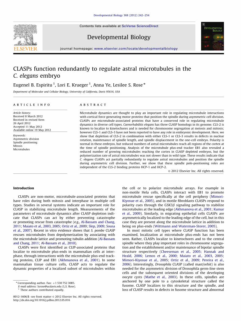
Developmental Biology 368 (2012) 242–254
Contents lists available at SciVerse ScienceDirect
Developmental Biology
0012-16
http://d
n Corr
E-m1 Th
journal homepage: www.elsevier.com/locate/developmentalbiology
CLASPs function redundantly to regulate astral microtubules in theC. elegans embryo
Eugenel B. Espiritu 1, Lori E. Krueger 1, Anna Ye, Lesilee S. Rose n
Department of Molecular and Cellular Biology, University of California, Davis 95616, USA
a r t i c l e i n f o
Article history:
Received 8 March 2012
Received in revised form
26 April 2012
Accepted 11 May 2012Available online 19 May 2012
Keywords:
Asymmetric division
Spindle positioning
Mitosis
Microtubules
06/$ - see front matter & 2012 Elsevier Inc. A
x.doi.org/10.1016/j.ydbio.2012.05.016
esponding author. Fax: þ1 530 752 3085.
ail address: [email protected] (L.S. Rose).
ese authors contributed equally.
a b s t r a c t
Microtubule dynamics are thought to play an important role in regulating microtubule interactions
with cortical force generating motor proteins that position the spindle during asymmetric cell division.
CLASPs are microtubule-associated proteins that have a conserved role in regulating microtubule
dynamics in diverse cell types. Caenorhabditis elegans has three CLASP homologs in its genome. CLS-2 is
known to localize to kinetochores and is needed for chromosome segregation at meiosis and mitosis;
however CLS-1 and CLS-3 have not been reported to have any role in embryonic development. Here, we
show that depletion of CLS-2 in combination with either CLS-1 or CLS-3 results in defects in nuclear
rotation, maintenance of spindle length, and spindle displacement in the one-cell embryo. Polarity is
normal in these embryos, but reduced numbers of astral microtubules reach all regions of the cortex at
the time of spindle positioning. Analysis of the microtubule plus-end tracker EB1 also revealed a
reduced number of growing microtubules reaching the cortex in CLASP depleted embryos, but the
polymerization rate of astral microtubules was not slower than in wild type. These results indicate that
C. elegans CLASPs act partially redundantly to regulate astral microtubules and position the spindle
during asymmetric cell division. Further, we show that these spindle pole-positioning roles are
independent of the CLS-2 binding proteins HCP-1 and HCP-2.
& 2012 Elsevier Inc. All rights reserved.
Introduction
CLASPs are non-motor, microtubule-associated proteins thathave roles during both mitosis and interphase in multiple celltypes. Studies in several systems indicate an important role forCLASP in stabilizing microtubule length. Measurements of theparameters of microtubule dynamics after CLASP depletion indi-cate that CLASPs can act by either preventing catastropheor promoting rescue from catastrophe (e.g., Al-Bassam and Chang,2011; Maiato et al., 2003, 2005; Ortiz et al., 2009; Slep, 2009; Sousaet al., 2007). Recent in vitro evidence shows that S. pombe CLASPrescues microtubules from depolymerization by associating withthe microtubule lattice and promoting tubulin addition (Al-Bassamand Chang, 2011; Al-Bassam et al., 2010).
CLASPs were first identified as CLIP-associated proteins thatlocalize to microtubule plus-ends in mammalian cells at inter-phase, through interactions with the microtubule plus-end track-ing proteins, CLIP and EB1 (Akhmanova et al., 2001). In somemammalian tissue culture cells, CLASP acts to regulate thedynamic properties of a localized subset of microtubules within
ll rights reserved.
the cell or to polarize microtubule arrays. For example innon-motile Hela cells, CLASPs interact with EB1 to promotemicrotubule rescue specifically at the cell periphery (Mimori-Kiyosue et al., 2005), and in motile fibroblasts CLASPs respond topolarity cues through the GSK3b signaling pathway to stabilizemicrotubules at the leading edge (Akhmanova et al., 2001; Kumaret al., 2009). Similarly, in migrating epithelial cells CLASPs areasymmetrically localized to the leading edge of the cell, but in thiscase they are present along the microtubule lattice in addition tobeing on plus-ends (Wittmann and Waterman-Storer, 2005).
In most mitotic cell types where CLASP function has beenexamined, localization at microtubule plus-ends has not beenseen. Rather, CLASPs localize to kinetochores and to the centralspindle where they play important roles in chromosome segrega-tion and the establishment and/or maintenance of bipolar spindlestructure respectively (Cheeseman et al., 2005; Hannak andHeald, 2006; Lemos et al., 2000; Maiato et al., 2003, 2005;Mimori-Kiyosue et al., 2005; Ortiz et al., 2009; Pereira et al.,2006). Interestingly, Drosophila CLASP (called mast/orbit) is alsoneeded for the asymmetric division of Drosophila germ-line stemcells and the subsequent oriented divisions of the developingoocyte cysts (Mathe et al., 2003). In these cells, spindles areanchored by one pole to a cytoskeletal structure called thefusome. CLASP localizes to this structure and the spindle, andloss of CLASP results in defects in fusome structure and abnormal

E.B. Espiritu et al. / Developmental Biology 368 (2012) 242–254 243
or monopolar spindles that fail to orient to the fusome. However,whether mast/orbit also plays a role in spindle orientation eventsthat are regulated by interactions of astral microtubules withpolarized cortical cues has not been addressed.
In the Caenorhabditis elegans one-cell embryo, as in many celltypes, PAR polarity proteins establish cortical domains requiredfor cell polarity. The PAR proteins also regulate the forces thatalign the spindle with the polarity axis to accomplish an asym-metric division (reviewed in Galli and van den Heuvel, 2008;Gonczy, 2008; Knoblich, 2010). Spindle positioning forces aregenerated when astral microtubule plus-ends interact with forcegenerators at the cell cortex, thereby translating cortical pullinginto forces acting on centrosomes or spindle poles. The PARproteins regulate cortical forces via a conserved pathway invol-ving Ga proteins and their partners GPR-1/2 and LIN-5 (known asLGN/PINS and NuMa/Mud in vertebrate cells and Drosophila
respectively). GPR-1/2 and LIN-5 are asymmetrically localized atthe cortex and interact with regulators of the microtubule motordynein, suggesting that the asymmetric activation of dynein leadsto spindle positioning events. In the C. elegans one-cell, asym-metric cortical pulling forces drive anteriorly directed nuclearcentration and rotation movements at prophase, as well asposteriorly-directed spindle displacement at metaphase andanaphase.
Microtubule-associated proteins that regulate microtubule plusend dynamics can potentially determine the frequency and dura-tion of growing microtubule plus ends and thus, interactions withdynein or other force-generators at the cortex (Kozlowski et al.,2007; Labbe et al., 2003; Srayko et al., 2005). Consistent with thisview, the XMAP215 ortholog, ZYG-9 and the doublecortin orhtologZYG-8 are required for spindle positioning in the C. elegans one-cellembryo. Mutations in the genes for either of these proteins resultin short astral microtubules and abnormal nuclear and spindlepositioning at all stages (zyg-9) or metaphase/anapahse (zyg-8),presumably due to a failure of microtubule-cortex interactions(Gonczy et al., 2001; Matthews et al., 1998; Srayko et al., 2003).However, the depletion of several other proteins shown to influ-ence microtubule dynamics in other systems, including an orthologof CLASP, CLS-2, the plus-end binding proteins EB1 and CLIP, andthe depolymerizing kinesin MCAK, has not been reported toproduce defects in asymmetric division (Srayko et al., 2005). Thefailure to identify a phenotype in these studies could be due toredundancy with gene family members or partially overlappingfunctions between different families.
Three genes in the C. elegans genome have homology to CLASP:cls-1 (C07H6.3), cls-2 (R107.6), and cls-3 (ZC84.3). One of theC. elegans CLASP homologs, CLS-2 localizes to the kinetochore-microtubule interface and is required for mitotic fidelity inembryos. Depletion of cls-2 by RNA interference leads to pre-mature separation of spindle poles before anaphase onset, andchromosome missegregation (Cheeseman et al., 2005). CLS-2 isalso required for meiosis where it plays roles in cytoplasmicstreaming and the structure of the anaphase spindle (Dumontet al., 2010; Yang et al., 2003). CLS-1 and 3 are similar to CLS-2throughout their lengths, except for an N-terminal extension ofCLS-1. This region of CLS-1 aligns with the first Tumor Over-expressed Gene domain-like domain (TOG-like) of mammalianCLASPs, suggesting that CLS-1 may have two TOG-like domains.This makes CLS-1 more similar to mammalian CLASPs than CLS-2and CLS-3, which contain only one predicted TOG-like domain.However, knockdown of CLS-1 or CLS-3 via RNA interference doesnot give lethality (Sonnichsen et al., 2005; Yang et al., 2003).
In this study, we use the C. elegans embryo as a system tostudy the roles of CLASP proteins in spindle movements that aregenerated by astral microtubules during asymmetric division.Using RNA interference of the three genes singly and in
combination, we found that the three C. elegans CLASP homologsfunction redundantly during nuclear rotation and spindle displa-cement. Together, these results provide evidence for a role forC. elegans CLASP in regulating astral microtubules during asym-metric cell division.
Materials and methods
Worm strains and growth
C. elegans were cultured on MYOB plates using standard methods,as previously described (Brenner, 1974; Church et al., 1995). Thefollowing strains were used: N2 (wild-type Bristol variant); AZ244,unc-119(ed3); ruIs57[pAZ147: pie-1::ß-tubulin::GFP; unc-119(þ)];FM102, lin-5(ev571ts) unc-119(ed3); ruIs57[pAZ147: pie-1::ß-tubu-
lin::GFP; unc-119(þ)]; FM125, (unc-119(ed3);ruIs57[pAZ147:pie-1::
b-tubulin::GFP;unc-119(þ);itIs37[unc-119(þ) pie-1::mCherry::H2B];JH1512, Isax1137 [pRF-4; pJH7.04 pie-1:GFP:Par-6; genomic DNA];
JH2648, [PAR-6::mCherry unc-119; axIS1928 [pEG66]; TH66,unc-119(ed3); Is[pie-1/EBP-2::GFP]. N2 worms were maintainedat 20 1C, while transgenic strains were maintained at 23–25 1C foroptimal transgene expression.
RNA interference
RNA interference (RNAi) was carried out by bacterial feeding(Timmons et al., 2001) using the following Ahringer librarybacterial clones where the numbers refer to nucleotide positionsin the unspliced CDS (Kamath et al., 2003): cls-2 618-1733 bp (III-4J10), cls-3 (III-4N20), cls-1 4214-5350 (III-4A13), hcp-1 (V-4H10),hcp-2 (V6P23). Bacteria were used undiluted for single RNAi, ormixed 1:1 for double RNAi respectively. Feeding was conducted at20 1C for the N2 background and 23–25 1C for GFP transgenicstrains; embryos were screened for depletion phenotypes from36–48 and 20–40 h post injection respectively, with the exceptionof hcp-1/2(RNAi) embryos for which 13/21 were examined at48–72 h. To generate dsRNA for injection, the inserts of plasmidsfrom the above clones were amplified with a T7 primer, and theproduct used as a template to synthesize dsRNA using theMEGAscripts Kit (Roche); dsRNA was resuspended at a finalconcentration of 0.5 or 1 mg/mL for each dsRNA. The entireinserts from the above constructs were also cloned in tandeminto the RNAi feeding vector L4440 to create pEE1 [cls-1;cls-2 ]and pEE2 [cls-2; cls-3]; dsRNA was generated from these newinserts and mixed with either cls-3 or cls-1 dsRNA to generate atriple RNAi mixture. L4 larvae were injected at room temperatureand subsequently stored and analyzed as above depending on thebackground strain. No difference in phenotype was seen fordouble RNAi carried out at 0.5 versus 1 mg/mL concentration.
Imaging and quantification
For live imaging experiments shown in Figs. 1–3, embryoswere cut from gravid hermaphrodites in egg buffer, mountedon 2% agarose pads and sealed under a coverslip. For thePAR-6::mCherry experiments, embryos mounted on agar exhib-ited strong autofluorescence of the egg shell. For that experimentand those involving hcp-1/2 and lin-5, embryos were mounted onpolylysine coated coverslips and inverted over spacers forimaging. DIC imaging of N2 and epifluorescence imaging ofworms expressing fusion proteins were carried out on an Olym-pus BX60 microscope, using an Olympus PlanFl 100X, 1.3 NAobjective or a PlanApo N 60X, 1.42 NA objective lens respectively.Images were acquired using a Hammatasu Orca 12-bit digitalcamera and OpenLab Software. For the quantification of spindle

cls-1; cls-3cls-2; cls-3wildtype cls-2 cls-2; cls-1
-70
-131
0
-60
-149
0
-64
-125
0
-75
-155
-69
-138
0
56
35
120
179
54
40
120
183
55
30
126
183
50
30
121
185
0
54
32
121
183
NE
BR
otat
ion
Ana
phas
e S
pind
le D
ispl
acem
ent
% Embryo Length0 25 50 75 100
0
250
150
-150
-50
50
100
200
-100
Tim
e to
NE
B (s
ec)
Fig. 1. Spindle positioning phenotypes in CLASP depleted embryos. (A) Still images from DIC time-lapse movies of representative embryos for wild type and the CLASP
RNAi treatments indicated. Anterior is to the left in these and all subsequent images. Time to NEB (0) is given for each frame; dots mark centrosomes. Scale bar is 10 mm.
(B) Spindle pole traces during the cell cycle generated from tracking each embryo in A are shown directly under each corresponding embryo; anterior spindle poles shown
in light blue, posterior in dark blue.
E.B. Espiritu et al. / Developmental Biology 368 (2012) 242–254244
movements shown in Figs. 1 and 2, DIC images were captured atone-second intervals, and tracked manually using the ManualTracking plugin for ImageJ (rsbweb.nih.gov/ij/plugins/track/track.html); data was transferred to Microsoft Excel for proces-sing. Scatter plots and statistical analysis were carried out usingPrismPlot (GraphPad Software, Inc). For DIC and epifluorescenceimaging experiments, brightfield timelapse images were capturedat 12 frames/min. Single fluorescent images were taken at 60, 120and 180 s post NEB to monitor the GFP::tubulin signal, or atpseudocleavage, NEB, and cytokinesis to examine PAR-6. Todetermine the extent of the PAR-6 domain, the free hand tool inImageJ was used to trace the cortex from the anterior to theposterior, and the intensity values were plotted. In wild type
embryos, the posterior cortex values were the same or lower thanthe adjacent cytoplasm. The absolute pixel intensities at theanterior cortex varied, but dropped in a gradient such that levelsreached 50% maximum at 50% egg length (EL) in all embryos,further dropping to the posterior minimum by 70% egg length. Inthe few CLASP embryos with extended domains, the gradient wasshifted such that the minima were reached at �80% EL, but theposterior-most cortex still exhibited an absence of PAR-6 signal asin wild type.
For confocal imaging of GFP::tubulin or EBP-2::GFP strains,embryos were mounted on agar as above and imaged on a 3iHybrid Spinning Disk Confocal- TIRF-Widefield Marianas micro-scope with a Plan-Apo 63x, 1.3 NA objective lens (Zeiss).

Rot
atio
n A
ngle
(deg
rees
)
0
90
30
60
cls-2; cls-1
WT cls-2 cls-2; cls-3
cls-1; cls-3
Cen
tratio
n P
oint
(%E
L)
25
50
cls-2; cls-1
WT cls-2 cls-2; cls-3
cls-1; cls-3
Spi
ndle
Dis
plac
emen
tTi
min
g (s
ec fr
om N
EB
)
cls-2; cls-1
WT cls-2 cls-2; cls-3
cls-1; cls-3
0
150
100
50
Max
Pos
ition
of P
oste
rior
Cen
troso
me
(%E
L)
cls-2; cls-1
WT cls-2 cls-2; cls-3
cls-1; cls-3
0
50
100
75
25
Max
Pos
ition
of A
nter
ior
Cen
troso
me
(%E
L)cls-2; cls-1
WT cls-2 cls-2; cls-3
cls-1; cls-3
0
50
100
75
25
Min
Spi
ndle
Len
gth
(μm
) 40
0
20
30
10
cls-2; cls-1
WT cls-2 cls-2; cls-3
cls-1; cls-3
75
Fig. 2. Quantification of defects exhibited by CLASP RNAi embryos. Scatter plots show specific measurements for all embryos in the data sets from Table 1. In each plot, the
means for each genotype are indicated by horizontal lines; the values for the means and statistical significance are summarized in Table 1. (A) Rotation angle of nuclear-
centrosome complex at NEB, where complete rotation onto the A/P axis¼01, complete failure¼901. (B) Centration point of the nuclear–centrosome complex, expressed as
percent egg length (%EL) where the anterior end of the embryo¼0%, posterior¼100%. (C) Minimum spindle length after NEB. (D) Maximum position (i.e. the farthest
posterior point obtained) of the anterior centrosome, expressed as percent egg length (%EL). (E) The time of onset of spindle displacement. (F) Maximum position of the
posterior centrosome.
E.B. Espiritu et al. / Developmental Biology 368 (2012) 242–254 245
Acquisition was controlled by Slidebook 5 software (3i Incorpo-rated). Embryos were excited with 488 and 561 wavelengthsfor 400 ms each sequentially incorporated into a 4 s intervalbetween acquisitions. In Slidebook, acquisitions were photo-bleach corrected following a single exponential fit. For higherresolution imaging of GFP::tubulin labeled microtubules, embryoswere filmed starting at NEB using the same microscope, acquisi-tion software, and a Plan-Apo 100� , 1.3 NA objective lens (Zeiss).Embryos were excited with 488 and 561 wavelengths for 800 msand 100 ms respectively, incorporated into a 1.52 s intervalbetween acquisitions. All raw images were exposed and scaledwith the same parameters, and were photobleach corrected asabove; however, because cls2; cls-1 and cls2; cls-3 embryosexhibited a lower signal to noise ratio, images for these wereadjusted using the auto-contrast tool in Image J in order to bettervisualize microtubule ends. Microtubule segments that camewithin 1 mm of the cortex were manually tracked with the ImageJpencil tool. Cortical contacts per time point were measured from
40 s before to 40 s after the onset of posterior spindle displace-ment (PD) and were then divided into eight 10-s windows. Nosignificant differences in the average number of cortical contactsin wild-type embryos were seen over this time period, and thusthe data were pooled into two groups (before PD and after PD) forpresentation in Fig. 4B. The ten time-points prior to posteriorspindle displacement (15.2 s total) were reanalyzed to track thenumber of contacts per four cortical domains defined by thelocation on the anterior-posterior axis, where 0% is most anteriorand 100% is most posterior. The length of the cortical regionmeasured in the anterior (0–25%) and posterior (75–100%)domains is longer than in the lateral domains due to the curvatureof the embryo, and so the number of microtubule contacts wasnormalized relative to length for graphing in Fig. 4C.
The plus-ends of growing microtubules were visualized inEBP-2::GFP expressing embryos excited with a 488 wavelengthfor 300 ms, incorporated into a 500 ms interval between acquisi-tions over 32 s. The 64 time points prior to posterior displacement

E.B. Espiritu et al. / Developmental Biology 368 (2012) 242–254246
(32 s total interval) were analyzed for the number of EBP-2::GFPcomets that grew past a radius of 10 mm from the centrosome orgrew to within 2 mm from the cortex; quantifications wererestricted to a half-circle radius on the outside of each centrosomeas shown in Fig. 6A (Srayko et al., 2005) to ensure scoring EBP-2::GFP comets on astral microtubules projected towards thecortex, rather than on interpolar microtubules. Five comets eachfor the anterior and posterior centrosomes were quantified foreach time interval per embryo, and are presented as an averagesum for five embryos per condition. For quantification of micro-tubule growth rates, EBP-2::GFP comets were tracked using theManual Tracking plugin in Image J. Growth rate was quantifiedper comet by dividing the distance traveled by the time intervalthe comet could be detected; 5 comets that persisted for at least5 time points were quantified per centrosome per embryo.
Antibodies and immunolocalization
The following regions were used as antigens for antibodyproduction: CLS-1 (aa42-333), CLS-2 (aa689-759), CLS-3 (aa660-736). The specified regions were amplified by PCR using cDNAtemplates (cls-3 and cls-1 full-length cDNA clones were obtainedfrom Open Biosystems, cls-2 cDNA from (Yang et al., 2003) andcloned into pGEX or pMAL protein purification vectors. MBPand GST fusion proteins were expressed in bacteria and purifiedusing amylose resin or Glutathione Sepharose 4B resin respec-tively according to the manufacturer’s instructions (AmershamBioscience, GE Healthcare). Purified GST-CLS-1, MBP-CLS-1 andMBP-CLS-3 were injected into rabbits and rats (Covance). Antiserawere purified on affinity columns made by cross linking GST-CLS-2 and GST-CLS-3 to Affigel-15, and GST-CLS-1 to Affigel-10(BioRad); antibodies were eluted with glycine-HCl, pH 2.5 andneutralized with 1M Tris pH 8.0. To test antibodies, purified GST-fusion proteins were separated on a 12% acrylamide gel, trans-ferred to nitrocellulose membrane (GE Healthcare) and incubatedwith primary antibodies (1:1000). Secondary antibodies (Licor)were diluted 1:20,000 in PBS, and images acquired on an OdysseyInfrared Imaging System.
For western blot analysis of embryo extracts, synchronized L4worms were placed onto control and RNAi plates. After 24 h, adultworms were collected and the embryos obtained by the alkalinehypochloride method (Lewis and Fleming, 1995). Embryo pelletswere resuspended in lysis buffer with protease inhibitor cocktail(Sigma-Aldrich), and stored in �80 1C until use. SDS loading dyewas added to embryo pellets and boiled at 95 1C for 1 min.Samples were loaded on 4–15% gradient gels (BioRad) andtransferred onto nitrocellulose membranes (GE Healthcare).Membranes were cut at approximate 65 kD and the lower portionincubated with mouse anti-tubulin antibody DM1a (1:1000 inPBS; Sigma-Aldrich), and the top portion with anti-CLS (1:100 inPBS) antibodies. Secondary antibodies (Licor) were diluted1:20,000 in PBS, and images acquired on an Odyssey InfraredImaging System.
In situ immunolocalization
Gravid hermaphrodites were rinsed in ddH20, mounted ontopolylysine-coated slides, and cut to release embryos. Embryoswere prepared for immunolocalization in liquid nitrogen,followed by fixation for 5 s in �20 oC methanol then 30 min in4% paraformaldehyde/.24M sorbitol/PEM (PEM is 100 mM PIPES/5 mM EDTA/ 5 mM MgCl2 pH6.9) as described in Toya et al.(2010)). CLS-2, CLS-3, and DM1a, (Sigma) antibodies were dilutedin PBS at 1:50, 1:10, and 1:500 respectively; fluorescently con-jugated secondaries, goat anti-rabbit FITC and goat anti-mouseRhodamine, were diluted in PBS at 1:100. CLS-2 and CLS-3 were
pre-absorbed with acetone powders of GST-expressing bacteriaand MBP-expressing bacteria respectively and secondaries werepre-absorbed with wild-type worm acetone powder prior to use.Embryos were stained with DAPI to visualize DNA and determinecell-cycle stage, and mounted in Vectashield mounting medium(Vector Laboratories, Inc.). Similar results were obtained withstandard MeOH fixation conditions (Miller and Shakes, 1995).Confocal sections were acquired on an Olympus Fv1000 FluoviewLaser Scanning Confocal Microscope, using a 60x Plan-Apo NA1.42 objective. For each embryo, Z-series of five confocal sectionswere taken at mid-embryo focal plane in 0.2 mm steps, withbelow-saturation acquisition settings. Images shown in panels aremaximum intensity projections made in Image J.
Results
CLASPs are required for spindle positioning in the one-cell embryo
To determine if C. elegans CLASPs act redundantly duringspindle positioning, we used RNA interference (RNAi) by bacterialfeeding in a wild-type background to deplete CLASP proteinssingly and in combination. Embryos were examined during thefirst division by DIC time-lapse microscopy. Wild-type embryosfollow a stereotypical sequence of nuclear and spindle move-ments during the first cell cycle (reviewed in Galli and Van DenHeuvel (2008) and Gonczy (2008)). Male and female pronucleimeet at the posterior of the embryo. The pronuclear-centrosomecomplex then moves to the center of the embryo (centration),simultaneously rotating to align the centrosomes along theanterior-posterior axis of cell polarity (nuclear rotation) beforenuclear envelope breakdown (NEB) (Fig.1, Movie 1). The spindleforms parallel to the anterior/posterior (A/P) axis and in manyembryos the entire spindle shifts slightly towards the posteriorduring metaphase. This movement and asymmetric elongation ofthe spindle at anaphase leads to unequal cleavage and aretogether referred to as posterior spindle displacement. Duringspindle elongation, the entire spindle also rocks as a unit, with theposterior spindle pole making larger oscillations perpendicular tothe A/P axis than the anterior pole.
Supplementary material related to this article can be foundonline at http://dx.doi.org/10.1016/j.ydbio.2012.05.016.
In cls-2(RNAi) embryos, nuclear centration and rotationappeared similar to wild type. However, at metaphase the spindlepoles separated abruptly and then oscillated independently ofeach other during anaphase (Fig. 1, Movie 2; Tables 1 and 2), asreported previously. These spindle pole movements indicate theabsence of a central spindle connecting the two poles and will bereferred to as the weak spindle phenotype (Cheeseman et al.,2005). In embryos depleted for cls-2 in combination with cls-1 orcls-3, we observed defects in nuclear centration and rotation,spindle length maintenance, and posterior spindle displacement.To characterize these defects, films were tracked to quantifycentrosome and spindle pole movements throughout the first cellcycle (Fig. 1 shows representative examples; data for all embryosis given in Fig. 2, Tables 1 and 2). In cls-2;cls-1 and cls-2;cls-3
(RNAi) embryos, pronuclei met at the posterior as in wild type,and the pronuclear complex moved towards the center of theembryo. For both cls-2;cls-1 and cls-2;cls-3 (RNAi), the mean angleof nuclear-centrosome alignment was different from wild type,and some embryos showed a complete failure of rotation wherethe final angle of the spindle relative to the A/P axis was between601 and 901 (Fig. 1A, Movies 3, 4; Fig. 2A; Tables 1 and 2).Although the final position of the nuclear-centrosome complex atthe end of centration in cls-2; cls-1 and cls-2; cls-3 (RNAi) embryoswas not different than wild type on average, centration was more

Table 1Analysis of spindle positioning in wild-type and clasp(RNAi) embryos.
Parameter measured wildtype cls-2 cls-2; cls-1 cls-2; cls-3 cls-1; cls-3
Rotation angle at NEB (deg.) 12 (14) 12 (14) 52 (20)a,b,c 47 (29)a,b,c 20 (21)
Centration point at NEB (% EL) 51.7 (3.2) 50.5 (4.3) 54.1 (5.0) 50.5 (4.9) 50.1 (2.7)
Maximum spindle length (mm) 25.0 (1.0) 26.1 (2.2) 23.2 (4.3) 25.8 (2.3) 23.2 (1.0)
Minimum spindle length (mm) 12.2 (0.6) 10.7 (2.0)a 7.6 (1.2)a,b,c 8.7 (1.9)a,b,c 11.3 (0.5)a
Spindle displacement timing (sec from NEB) 67 (14) 64 (13) 83 (18)a,b,c,d 64 (15) 54 (8)
Maximum position of anterior spindle pole (% EL) 46.8 (3.8) 45.5 (11.5) 65.7 (13.3)a,b,c 56.9 (14.1)a,b,c 50.6 (3.9)
Maximum position of posterior spindle pole (% EL) 82.5 (2.3) 84.9 (4.4) 84.8 (1.2) 84.6 (5.1) 83.6 (1.6)
All values are means for the given genotype with standard deviation in parentheses. Parameters were measured as indicated in the text. The number of embryos examined
is given in Table 2, N2.
All other comparisons between wild-type and clasp(RNAi) were tested and were not significantly different.a Data sets were tested and are significantly different from wildtype, po0.05 (students t-test).b Data sets were tested and are significantly different from cls-2, po0.05 (students t-test).c Data sets were tested and are significantly different from cls-1; cls-3, po0.05 (students t-test).d Data sets were tested and are significantly different from cls-2; cls-3, po0.05 (students t-test).
Table 2Frequency of phenotypes produced by depletion of clasps by RNAi.
Strain Backgrounda
(method)b
Gene Targeted Nc Rotationd
0–301 (% N)
Rotationd
31–901 (% N)
spindle
collapse e (% N)
Excessive spindle
displacementf (% N)
Weak
Spindleg (% N)
N2 (F) none 18 88 12 0 0 0
cls-2 17 88 12 24 29 93
cls-2;cls-1 6 17 83 100 88 100
cls-2; cls-3 17 35 65 59 83 100
cls-1;cls-3 8 63 37 0 0 0
hcp-1;hcp-2 21 86 14 0 0 100
GFP::tubulin (F) none 10 60 40 0 0 0
cls-2 7 43 57 86 57 86
cls-2;cls-1 4 25 75 100 50 75
cls-2; cls-3 8 25 75 75 37.5 88
cls-1;cls-3 12 75 25 0 0 0
GFP::tubulin (I) none 10 100 0 0 0 0
cls-2 10 90 10 60 0 80
cls-2;cls-1 10 70 30 60 10 100
cls-2; cls-3 11 73 27 18 0 100
PAR-6 (I) none 19 90 10 0 0 0
cls-2 16 94 6 75 33 50
cls-2;cls-1 9 67 33 22 22 70
cls-2; cls-3 8 75 25 38 50 100
cls-1;cls-2;cls-3 8 63 37 47 40 100
a Background strains used for the analysis were N2 (wild type), FM125 (expressing GFP::tubulin;mCHerry::H2B), or PAR-6 (GFP::PAR-6 or mCherry::PAR-6). GFP::
PAR-6 was used for RNAi of control, cls-2, and clas;cls-1, while mCherry::PAR-6 was used for control, cls-2 and cls-2;cls-3. The controls gave similar results and thus were pooled.b dsRNA was delivered by feeding (F) or injection (I) as outlined in the methods.c Number of embryos examined for each treatment; for N2(F), n¼5 for cls-1;cls-3 embryos examined at metaphase/anaphase.d Rotation angle of a line drawn through the centrosomes, relative to the A/P axis¼01, at NEB.e Spindle collapse was defined as any spindle shorter than the wild type range �2%, for that background and RNAi condition.f Excessive spindle displacement was defined as any spindle in which the anterior centrosome moved farther towards the posterior than the wild type range þ2%, for that
background and RNAi condition.g Weak spindle was the apparent absence of a central spindle as described in results.
E.B. Espiritu et al. / Developmental Biology 368 (2012) 242–254 247
variable (Table 1, Fig. 2B). Some embryos exhibited a partialfailure of centration (Fig. 1A, Movie 3: cls-2; cls-1 embryo showsan example) while others showed an over-centering phenotype(Fig. 1A, Movie 4: cls-2; cls-3).
After the centration/rotation phase, many cls-2; cls-1 and cls-2;
cls-3 (RNAi) embryos exhibited a spindle collapse phenotype inwhich the spindle poles did not maintain their normal separationafter NEB and instead moved closer together (Fig. 1 cls-2; cls-1 andcls-2; cls-3 show representative embryos). Quantification of spin-dle pole-to-pole distance showed that the average minimumspindle length after NEB for cls-2; cls-1 and cls-2; cls-3 (RNAi)embryos was significantly different from wild type (Table 1) andmany embryos exhibited a minimum spindle length smaller thanthe wild-type range (Fig. 2C). In the majority of embryos in thisdata set, spindle collapse was followed by an excessive spindle
displacement phenotype in which the entire spindle movedtowards the posterior (Fig. 1). The two poles of the collapsedspindle traveled as a unit, and thus the anterior spindle polemoved significantly farther to the posterior than in controls(Fig. 2D, Tables 1 and 2). The timing of this spindle displacementwas within the normal range for most embryos, but delayedin some (Fig. 2E, Table 1). In contrast, the posterior spindlepole did not travel farther towards the posterior on average thanin wild type (Fig. 2F, Table 1). After excessive spindle displace-ment, spindle poles moved apart at anaphase and revealed theweak spindle phenotype typical of cls-2 RNAi alone (Fig. 1,Table 2). The final position of both spindle poles was on theanterior-posterior axis, as in wild type (Fig. 1) and the embryoscompleted cytokinesis (n48 embryos for each type of RNAicondition).

Fig. 3. CLASP RNAi treatment affects chromosome segregation and spindle
structure. Spinning disk confocal images of GFP::tubulin; mCherry::histone from
control and RNAi treated embryos. Time is in relative to NEB (0 s); scale bar is
10 mm. Wild-type and cls-1;cls-3 embryos show normal chromosome congression
to a metaphase plate by approximately 100 s, followed by anaphase chromosome
segregation and spindle separation. The cls-2 embryo is shown at slightly earlier
timepoints because the spindle poles separate early. The cls-2;cls-3 example
illustrates spindle collapse at metaphase and excessive posterior spindle
displacement.
Pseudocleavage
3-slc ;2-slclortno
C
Bright Field PAR-6::mCherry
Fig. 4. Polarity is established normally in most CLASP RNAi embryos. Brightfield and
expressing PAR-6::mCherry. Psuedocleavage (just after polarity establishment) and NE
E.B. Espiritu et al. / Developmental Biology 368 (2012) 242–254248
Quantification of the same parameters in cls-2 single (RNAi)embryos revealed that some cls-2(RNAi) embryos exhibited shorterspindles and excessive spindle displacement (Tables 1 and 2;Fig. 2). For example, 6/17 cls-2(RNAi) embryos showed a minimumspindle length that was smaller than the wild type range (Fig. 2C,Table 1). The mean spindle length of these cls-2 embryos wassmaller than wild type, but still longer than in cls-2; cls-1 andcls-2;cls-3 (RNAi) embryos (Table 1). In contrast, the maximumlength of the spindle reached during anaphase was not signifi-cantly different in any of the CLASP depletion treatments comparedto wild type (Table 1).
Depletion of CLASPs by bacterial feeding in a strain expressingGFP::tubulin, mCherry::histone (McNally et al., 2006); Fig. 3, Table 2,Movies 6–10) was used for a more careful examination of chromo-some movements and spindle structure after single versus doubleCLASP depletion. As previously reported (Cheeseman et al., 2005), allcls-2 (RNAi) embryos showed a chromosome congression defect andeither a complete absence of microtubules (4/6) or greatly reducednumbers of microtubules (2/6) in the central spindle at anaphase(Movie 7). This phenotype was often accompanied by laggingchromosomes (2/6) or an extended mass of chromosome materialat anaphase that appeared stretched out on the remaining midzonemicrotubules (2/6). Similarly cls-2; cls-1 and cls-2; cls-3 double RNAiembryos showed chromosome congression defects (2/4 and 7/8respectively), missing or reduced central spindle microtubules (4/4,7/8), and lagging chromosomes or chromatin at anaphase (2/4, 2/8;Movie 8, 9). Thus, no additional spindle structure or chromosomephenotypes were seen after cls-2; cls-1 and cls-2; cls-3 doubledepletion compared to cls-2 RNAi alone. In some cls-2; cls-1 andcls-2; cls-3 embryos, although the chromosomes exhibited initialcongression defects, chromosomes appeared aligned in late meta-phase (Movie 9). Improved alignment correlated with spindle polecollapse. It was previously shown that attenuating cortical pullingforces suppressed the premature spindle pole separation defect andimproved chromosome segregation in cls-2(RNAi) embryos(Cheeseman et al., 2005). Thus, the spindle collapse phenotype ofcls-2; cls-1 and cls-2; cls-3 embryos, which prevents prematurespindle pole separation, likely allows more time for kinetochoreattachments to occur in these embryos.
Similar spindle positioning and spindle collapse phenotypeswere observed after single and double RNAi of CLASPs by bacterialfeeding in other backgrounds and by injection of dsRNA (Table 2).
NEB
Bright Field PAR-6::mCherry
epifluorescent images of representative control and cls-2;cls-3 (RNAi) embryos
B images are from the same embryo. Scale bar, 10 mm.
E.B. Espiritu et al. / Developmental Biology 368 (2012) 242–254 249
RNAi directed against all three CLASPs did not result in a strongerphenotype. Although the penetrance of each phenotype amongdata sets varied, the nuclear rotation and spindle displacementdefects were typically stronger in cls-2; cls-1 and cls-2; cls-3
double RNAi embryos compared to cls-2 single RNAi embryos(Table 2). In contrast, no obvious spindle positioning phenotypeswere observed in cls-1; cls-3 (RNAi) experiments, which werecarried out in parallel with the other double RNAi experiments(Figs.1 and 2 and Tables 1 and 2, Movie 5). The only differencebetween cls-1; cls-3 (RNAi) embryos and wild type was a slightlysmaller minimum spindle length (Table 1). Likewise, when cls-1
and cls-3 were depleted simultaneously in a strain expressingGFP::tubulin, mCherry::histone, chromosomes congressed on themetaphase plate and segregated normally, and the spindle-mid-zone microtubules appeared robust (11/11, Fig. 3; Movie 10).Together, these observations indicate that cls-1 and cls-3 actpartially redundantly with cls-2 during centration, rotation andspindle displacement in one-cell embryos, as well as in themaintenance of spindle length.
Defects in spindle positioning in CLASP depleted embryos are not due
to polarity defects
CLS-2 was previously shown to function prior to the first mitosisin C. elegans, during the microtubule-mediated cytoplasmic streamingthat occurs just after fertilization and in spindle formation andchromosome segregation in meiosis (Dumont et al., 2010; McNallyet al., 2010). After the completion of meiosis, the polarity axis forasymmetric division is established. The growth of the sperm’smicrotubule aster correlates with the timing of polarity establishment(Tsai and Ahringer, 2007; Wallenfang and Seydoux, 2000). Thus, thespindle positioning phenotypes of CLASP depleted embryos couldresult from pre-mitotic defects in microtubule function that affectpolarity. To test this, we analyzed embryos expressing GFP::PAR-6 ormCherry::PAR-6 during the first cell cycle (Zonies et al., 2010). Incls-2, cls-2; cls-1 or cls-2; cls-3 (RNAi) embryos, an anterior PAR-6domain of normal size formed by the time of pronuclear meeting inthe majority of embryos, just as in controls (14/16, 7/8, 7/8,respectively; Fig. 4). Similar results were observed in cls-1; cls-2;
cls-3 (RNAi) embryos (6/8 embryos exhibited a normal PAR-6domain). In the remaining embryos for all treatments, the PAR-6domain extended farther than 50% egg length, but PAR-6 remainedabsent at the very posterior cortex (see Methods for quantification).Of the 6 embryos among all treatments that showed an extendedPAR-6 domain, 3 of these had no spindle positioning defects. Further,of the normally polarized embryos, 3/14 cls-2 (RNAi) embryos, 4/ 7cls-2;cls-1, 3/7 cls-2;cls-3, and 5/7 cls-1;cls-2;cls-3 embryos showedeither a nuclear rotation defect, excessive spindle displacement, orboth. Thus, there was no correlation between the embryos with anextended PAR-6 domain and alterations in spindle positioning.
To test whether the posterior movements of the spindleobserved after CLASP depletion are due to the normal corticalforces, we examined the effects of a loss of lin-5 activity on thephenotype. LIN-5 is a component of the G protein pathway thatresults in cortical pulling forces and asymmetric spindle displace-ment in wild-type embryos. Strains expressing GFP::tubulin withor without the lin-5(ev571ts) mutation were used for this analysis(Park and Rose, 2008). For cls-2; cls-1 and cls-2; cls-3 (RNAi), 67%and 72% of the embryos exhibited spindle collapse and excessivespindle displacement respectively (n¼18). In contrast, none ofthe lin-5; cls-2; cls-1 (RNAi) and lin-5; cls-2; cls-3 (RNAi) embryosshowed excessive spindle displacement or any posterior move-ment of the spindle during metaphase (n¼19). Nonetheless,spindle collapse occurred in 61% of lin-5; clasp (RNAi) embryos.In addition, in the subset of embryos for which tubulin fluores-cence was examined, central spindle microtubules were absent or
greatly reduced in 8/9 lin-5; cls-2; cls-1 (RNAi) and 7/8 cls-2; cls-1
(RNAi) embryos, indicating that the weak spindle phenotype ispresent even when forces are attenuated. We conclude that thespindle positioning defects observed in CLASP depleted embryosare not caused by abnormal polarity but do depend on asym-metric cortical forces.
CLASPs are required for normal numbers of microtubule-to-cortex
contacts
CLASPs are known to affect microtubule dynamics and inparticular can promote long microtubule length by preventingcatastrophe (Al-Bassam et al., 2010; Maiato et al., 2003; Maiatoet al., 2005; Ortiz et al., 2009; Slep, 2009; Sousa et al., 2007). Thedefects in spindle positioning observed could therefore be caused byalterations in microtubule dynamics and length that reduce interac-tions of astral microtubules with the cortex. To assay for microtubuleinteractions with the cortex, we first examined astral microtubules inGFP::tubulin expressing embryos, using higher resolution imaging.Control and RNAi embryos were imaged from NEB throughanaphase, and the number of microtubules that contacted the cortexwere quantified (Fig. 5; see Methods for details). In this data set, thespindle positioning phenotypes were weaker, although manyembryos exhibited a collapsed spindle (Table 2). Nonetheless, cls-2
(RNAi) embryos exhibited significantly fewer microtubule-cortexcontacts when compared to control embryos (Fig. 5). In addition,cls-2; cls-1 and cls-2; cls-3 double RNAi embryos had fewer micro-tubules contacting the cortex than cls-2 single RNAi embryos orcontrols. These results demonstrate that the three CLASPs actredundantly in regulating astral microtubule contact with the cortex.
We next examined whether depletion of CLASPs affects micro-tubule-cortex contacts globally or only at certain regions of thecortex. The number of microtubule-cortex contacts in each of fourdomains across the anterior/posterior axis of the embryo werecalculated. Control embryos showed a similar number of corticalcontacts in all domains. In cls-2 and cls-2; cls-1 embryos, micro-tubule contacts were significantly reduced at all cortical regions(Fig. 5C). We conclude that depletion of CLASPs reduces astralmicrotubule contact with the cortex throughout the embryo.
To further characterize the effects of CLASP depletion onmicrotubules, we examined a strain expressing EBP-2::GFP afterdepletion of CLASPs by RNAi. EBP-2 is a C. elegans ortholog of EB1,which labels the plus ends of growing microtubules only and isthus visible as fluorescent ‘‘comets’’ (Srayko et al., 2005). Wefound no difference in the number of EBP-2::GFP cometsnucleated from the centrosome that grew to a distance of10 mm in cls-2(RNAi) embryos compared to wild type (Fig. 6),consistent with a previous study (Srayko et al., 2005). However,cls-2; cls-3 embryos had fewer growing microtubules extending tothis distance, and all CLASP depleted embryos showed signifi-cantly fewer EBP-2::GFP comets growing to within 2 mm of thecortex. As with the analysis of microtubules using GFP::tubulin,the double CLASP depletion embryos exhibited fewer growingmicrotubules near the cortex than embryos depleted for CLS-2alone. Measurements of the velocities of EBP-2::GFP comets, anassay for microtubule polymerization rate, showed a very smallincrease in cls-2 and cls-2;cls-1 embryos compared to controls;the difference in velocities between cls-2;cls-3 and controlembryos, and between cls-2;cls-3 and the other cls-2 depletions,was not significant (Fig. 6). Overall, these results indicate thatfewer growing microtubules reach the cortex in CLASP depletedembryos. In this data set, the excessive spindle displacementphenotype was observed in 0% of cls-2 (RNAi) embryos, 60% ofcls-2; cls-1 embryos and 37.5% cls-2; cls-3 embryos. Thus, thestrength of the spindle positioning phenotypes exhibited by

Aver
age
Num
ber M
icro
tubu
les
Con
tact
ing
Cor
tex
Per
Tim
epoi
nt
cls-2 cls-2; cls-3cls-2; cls-1control
12
8
4
Before PD After PD
0
1-slc;2-slc
cls
-2, c
ls-3
cls-
2co
ntro
l
control cls-2 cls-2; cls-1 25-50%0-25% 50-75% 75-100%
Aver
age
Num
ber M
icro
tubu
les
Con
tact
ing
Cor
tex
Per
Tim
epoi
ntP
er D
omai
n
0
1
2
3
Fig. 5. CLASP RNAi treated embryos display fewer microtubule cortical contacts.
(A) Still images from spinning disk time-lapse movies of representative embryos
for wild type and CLASP RNAi treatments as indicated. Zooms of anterior portions
of embryos are shown at right of each full embryo; arrowheads denote micro-
tubules contacting the cortex. Scale bar is 10 mm. (B) Quantification of the average
number of microtubules that came within 1 mm of the cortex per time-point
during 40 s intervals before and after posterior displacement (PD).
(C) Quantification of the average number of microtubules that contacted the
cortex per time-point in a specific region during the 15.2 s interval prior to
posterior displacement. Domains are illustrated in (A) separated by dash lines; 0%
is most anterior, 100% is most posterior of embryo. Error bars are standard error
of the mean. Brackets indicate statistical difference between RNAi conditions
(*po10�7; **po10�3).
E.B. Espiritu et al. / Developmental Biology 368 (2012) 242–254250
CLASP depleted embryos correlates with defects in the number ofmicrotubules that grow to and contact the cortex.
CLS-2 localize to the cytoplasm and mitotic spindle throughout early
development
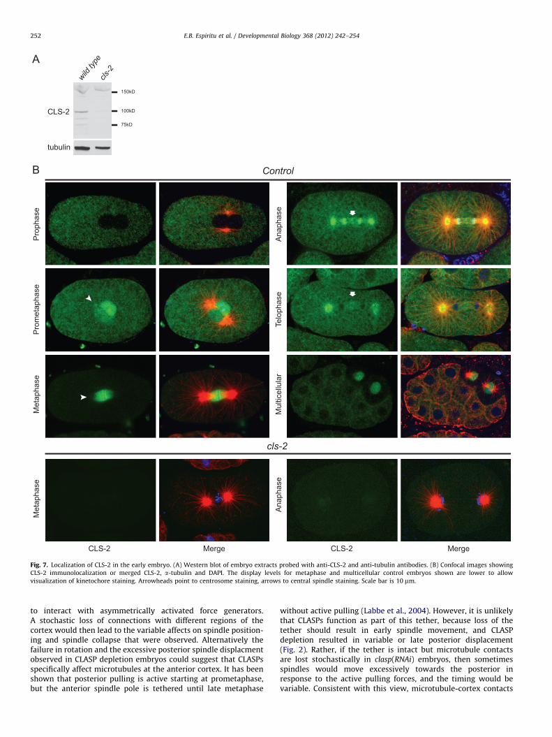
To further investigate the function of C. elegans CLASPs, we raisedantibodies to all three proteins. The region from amino acids 680–780 of CLS-2 showed the lowest homology among all three CLASPsand this was used as an antigen for raising antibodies against CLS-2and CLS-3. The 330 amino acid N-terminal region of CLS-1, which isnot present in CLS-2 or CLS-3, was used as antigen for CLS-1.Western blotting of embryo extracts with affinity-purified anti-CLS-2 antibodies revealed a band of the predicted size for CLS-2 in
embryo extracts made from wild type, which was greatly dimin-ished in extracts made from cls-2 RNAi treated worms; similarly,immunolocalization in embryos showed staining that was abolishedby RNAi (described below, Fig. 7). The anti-CLS-1 and anti-CLS-3antisera or purified antibodies recognized their respective fusionproteins on westerns. However, a specific signal that could bedetected in wild type but not clasp depletion embryos was notobserved with immunostaining or western blotting using thesereagents (unpublished results).
Previous studies in C. elegans showed that CLS-2 localizes weaklyat centrosomes and strongly to kinetochores in the one-cell mitoticembryo, but the localization pattern throughout early embryodivisions was not reported (Cheeseman et al., 2005). From analyzingstaining throughout the cell cycle (Fig. 7), we found that CLS-2 firstappeared on the centrosomes at prophase in some embryos (3/7). Byprometaphase, centrosome staining was evident in most embryosand the signal also entered the nucleus (8/9). CLS-2 localized to thekinetochore interfaces and on the spindle at metaphase in one-cellembryos, in addition to the centrosomes (n¼12). Spindle stainingpersisted into anaphase (17/19), especially in the mid spindle region(Fig. 7); anaphase spindle staining was not reported in C. elegans
mitotic embryos previously, but was shown for meiotic spindles(Cheeseman et al., 2005; Dumont et al., 2010). Centrosome stainingalso persisted through anaphase and into telophase (19/19 and 17/18 respectively).
All embryos also showed cytoplasmic staining which wasabsent after RNAi depletion. In multicellular embryos, interphasecells showed strong cytoplasmic staining that appeared enrichedat cell contacts but did not appear to colocalize with regions ofthe cells showing the densest tubulin staining (Fig. 7). Mitoticcells in multicellular embryos exhibited centrosome, kinetochoreand spindle staining through at least the 28-cell stage (n¼ 23,older embryos are not retained in these samples). The latterobservation suggests that CLS-2 continues to play a role in mitosisthroughout early embryogenesis.
CLS-2 has HCP independent roles in the one-cell embryo
HCP-1 and HCP-2 are CENP-F related proteins in C. elegans thatplay a critical role in both mitosis and meiosis. HCP-1 and HCP-2are redundant, but embryos depleted simultaneously for HCP-1 andHCP-2 (hereafter referred to as HCP-1/2) show the same chromo-some misorientation and weak spindle phenotypes during the firstdivision that were observed after CLS-2 depletion. CLS-2 associateswith HCP-1/2 in embryonic extracts, and analyses of multiplekinetochore proteins indicated that HCP-1/2 recruits CLS-2 tokinetochores (Cheeseman et al., 2005; Dumont et al., 2010). Todetermine if HCP-1/2 also play a role with CLASPs in spindlepositioning, we examined embryos after depletion of HCP-1/2 byRNAi. DIC imaging of the embryos during the first division showedthat the spindle poles separated abruptly after NEB and spindlepoles oscillated independently, as previously reported (Table 2).The spindles in these embryos did not collapse or exhibit position-ing defects, even after extended RNAi treatment (Table 2). Similarly,other kinetochore mutants show a phenotype of abrupt separationof spindle poles at metaphase without spindle pole collapse(Cheeseman et al., 2004; Grill et al., 2001; Oegema et al., 2001),while we observe spindle pole collapse in 25-75% of cls-2 singleRNAi embryos (Table 2). These results suggest that CLASPs haveHCP-1 and HCP-2 independent functions in the one-cell embryo.
Discussion
Our work demonstrates that CLS-1 and CLS-3 are partiallyredundant with CLS-2 during nuclear rotation, spindle length

cont
rol
2-slc ;1-slc c
ls-2
, cls
-3
cls-
2
Ave
rage
Num
berM
icro
tubu
les
Gro
win
g to
Rad
ius
Ave
rage
Mic
rotu
bule
Gro
wth
Rat
e (µ
m/s
ec)
1.2
.8
.4
0
30
20
10
010µm
controlcls-2cls-2; cls-1 cls-2; cls-3
1.4
Cortex
Fig. 6. Fewer microtubules polymerize towards the cortex in CLASP RNAi embryos. (A) Time lapse projection image of EBP-2::GFP for the 32 second interval prior to
posterior displacement showing the growth of actively polymerizing microtubules over the entire time interval. Scale bar is 10 mm. (B) Quantification of the average
number of EBP-2::GFP comets that passed a 10 mm radius or came within 2 mm of the cortex (as shown by the dashed lines in (A) during this interval. (C) Average
microtubule growth rate as measured by the distance traveled of EBP-2::GFP comets over time. N¼ 5 embryos for each measurement and RNAi condition (see Methods);
error bars are standard error of the mean. Brackets indicate statistical difference between RNAi conditions (po0.05), but for simplicity brackets are not shown for control
versus cls-2; cls-1 and cls-2; cls-3 in the graph for the cortex in (B).
E.B. Espiritu et al. / Developmental Biology 368 (2012) 242–254 251
maintenance and spindle displacement in C. elegans embryos. Thisredundancy explains why a role for CLS-1 and CLS-3 in earlyembryos was not uncovered in earlier studies that targeted thesegenes singly with RNAi. Depletion of CLS-1 and CLS-3 simulta-neously did not produce spindle positioning defects, nor did weobserve any clear difference in the phenotypes produced by cls-2;cls-1 RNAi versus cls-2; cls-3 RNAi. The simplest explanation forthese results is that all three CLASPs function interchangeably inregulating astral microtubules at this stage, but that there arehigher levels of CLS-2 than of CLS-1 and CLS-3 in the embryo.Alternatively, the CLS-2 protein could possess functional differ-ences that cause it to have a more dominant affect on microtubuledynamics in the one-cell embryo. Future analyses will be neededto resolve this issue.
In C. elegans, other kinetochore mutants show the weak spindlephenotype with premature spindle pole separation and absence ofcentral spindle microtubules during anaphase, similar to what wasreported for cls-2 in this and previous studies. However, thosekinetochore mutants do not appear to exhibit the spindle collapsethat we document here (Cheeseman et al., 2004, 2005; Grill et al.,2001; Oegema et al., 2001; Krueger and Rose, unpublished). Further,we showed that hcp-1;hcp-2(RNAi) embryos do not exhibit spindlepole collapse even after extended RNAi treatments. One explanationfor the spindle collapse phenotype is that CLASPs play an additionalrole in microtubule bundling in the central spindle, as described forS. pombe and Drosophila CLASPs in mitosis, and C. elegans andXenopus CLASPs in meiosis (Bratman and Chang, 2007; Dumontet al., 2010; Hannak and Heald, 2006; Maiato et al., 2002).Consistent with this view, we found that CLS-2 localizes to thecentral spindle during anaphase after the chromosomes havesegregated. Another explanation for the spindle collapse phenotypeis that it results from the loss of microtubule contacts with thecortex which reduces pulling forces on the spindle poles. Theabsence of spindle collapse and other spindle positioning pheno-types in hcp-1;hcp-2 embryos is also consistent with this model andindeed the two possibilities are not mutually exclusive.
Cortical pulling forces on astral microtubules are regulated bypolarity cues throughout the cell cycle in the one-cell embryo, andsuch pulling forces are necessary for rotation, spindle displacementand anaphase spindle pole separation (Galli and Van Den Heuvel,2008; Gonczy, 2008). We found that PAR-6 localization is normal inthe majority of CLASP RNAi embryos, and that the spindle movestowards the posterior as in wild type, albeit with more variabletiming. In contrast, there was a more severe change in microtubule-cortex contacts in the CLASP double RNAi treatments compared tocls-2 single RNAi, which does correlate with the stronger spindlepositioning defects. These results strongly suggest that the nuclearrotation and excessive spindle displacement phenotypes are causedby altered microtubule-cortex contacts.
To characterize the effects of CLASP depletion in more detail,we examined microtubule plus-end growth using an EBP-2::GFPreporter. The cls-2 and cls-2;cls-1(RNAi) embryos exhibitedslightly increased polymerization rates on average. A similareffect was observed after depletion of mammalian CLASPs andwas proposed to result from an increase in the soluble tubulinpool (due to the loss of long microtubules), which could enhancemicrotubule polymerization rate (Mimori-Kiyosue et al., 2005).We found no change in the number of EBP-2::GFP comets at adistance of 10 mm from the centrosome in cls-2(RNAi) embryoscompared to wild type, as previously reported (Srayko et al.,2005). Nonetheless, there were fewer EBP-2::GFP comets growingwithin 2 mm of the cortex in cls-2(RNAi) embryos and doubleCLASP depleted backgrounds, similar to what we observed forGFP::tubulin marked microtubules. Although we cannot directlymeasure the rate of microtubule catastrophe and rescue in oursystem, this decrease in the number of growing microtubulesreaching the cortex is consistent with a role for C. elegans CLASPsin suppressing catastrophe and promoting microtubule rescue, asreported for CLASPs in other organisms (Al-Bassam et al., 2010;Slep, 2009). We therefore propose that microtubules growtowards the cortex in CLASP depleted embryos, but thatfewer microtubules maintain this growth and reach the cortex
tl uM
ral ull eciesahpol eT
esahpat eM
esahpateM
esahpatemor P
esahpor P
CLS-2 Merge CLS-2 Merge
esahpanA
Control
cls-2
esahpanA
tubulin
CLS-2
wild ty
pecls
-2
150kD
100kD
75kD
Fig. 7. Localization of CLS-2 in the early embryo. (A) Western blot of embryo extracts probed with anti-CLS-2 and anti-tubulin antibodies. (B) Confocal images showing
CLS-2 immunolocalization or merged CLS-2, a-tubulin and DAPI. The display levels for metaphase and multicellular control embryos shown are lower to allow
visualization of kinetochore staining. Arrowheads point to centrosome staining, arrows to central spindle staining. Scale bar is 10 mm.
E.B. Espiritu et al. / Developmental Biology 368 (2012) 242–254252
to interact with asymmetrically activated force generators.A stochastic loss of connections with different regions of thecortex would then lead to the variable affects on spindle position-ing and spindle collapse that were observed. Alternatively thefailure in rotation and the excessive posterior spindle displacmentobserved in CLASP depletion embryos could suggest that CLASPsspecifically affect microtubules at the anterior cortex. It has beenshown that posterior pulling is active starting at prometaphase,but the anterior spindle pole is tethered until late metaphase
without active pulling (Labbe et al., 2004). However, it is unlikelythat CLASPs function as part of this tether, because loss of thetether should result in early spindle movement, and CLASPdepletion resulted in variable or late posterior displacement(Fig. 2). Rather, if the tether is intact but microtubule contactsare lost stochastically in clasp(RNAi) embryos, then sometimesspindles would move excessively towards the posterior inresponse to the active pulling forces, and the timing would bevariable. Consistent with this view, microtubule-cortex contacts

E.B. Espiritu et al. / Developmental Biology 368 (2012) 242–254 253
were reduced in all regions of clasp(RNAi) embryos, and we didnot observe any posterior movement of the spindle in clasp(RNAi)embryos in which asymmetric cortical forces were attenuated byloss of lin-5 activity. Excessive posterior displacement movementshave also been reported in zyg-8 mutants which have shortermicrotubules beginning at metaphase, and in embryos treatedwith nocodazole at metaphase (Gonczy et al., 2001). Interestinglyhowever, in those embryos the spindles stay in the posterior,whereas in clasp(RNAi) embryos, the anterior spindle pole movesback to a more normal position. Microtubules in zyg-8 andnocodazole treated embryos appear uniformly short. In contrast,some microtubules are long enough to contact the cortexin clasp(RNAi) embryos. Thus at anaphase, when centrosomesnormally nucleate more microtubules and anterior cortical pull-ing becomes active again, interactions of the microtubules withthe cortex in CLASP embryos may allow the anterior spindle poleto move anteriorly once again and the spindle poles to separate.
In summary, we have demonstrated a role for CLASPsin regulating astral microtubules during spindle positioningin mitotis. The three C. elegans CLASPS appear to functioninterchangeably to regulate microtubules in the one-cell embryo.This is similar to what has been seen in mammalian cells whereCLASPs were shown to function redundantly to regulate micro-tubule stability (Maiato et al., 2003; Mimori-Kiyosue et al., 2005;Pereira et al., 2006) during interphase and mitosis. However, theC. elegans CLASPS may have separable roles as well. The kineto-chore and meiosis defects caused by cls-2 depletion appear fullypenetrant (Cheeseman et al., 2005; Dumont et al., 2010), whilethe spindle positioning and spindle maintenance phenotypes weobserved were more severe after double depletion. These dataraise the possibility that CLS-2 has a unique function at thekinetochore compared to CLS-1 and CLS-3. Further, CLS-1 ispredicted to have two TOG-like domains, similar to mammalianCLASPs, whereas CLS-2 and CLS-3 are only predicted to have one.CLS-1 could therefore have unique functions. In addition, promo-ter fusions to CLS-1 and CLS-3 show differential expression inlarvae and adults (Hunt-Newbury et al., 2007; Lynch et al., 1995).Future work comparing each of these proteins’ biochemicalactivities and cellular roles will yield important insights into thedifferent functions of the CLASP family of proteins.
Acknowledgments
We thank Geraldine Seydoux for strains and Frank McNally forstrains and the cls-2 cDNA. Other strains were provided by theCaenorhabditis Genetics Center (funded by the NIH NationalCenter for Research Resources). Dae Hwi Park, Adam Hayashiand Hai Chi Pham provided valuable technical assistant. We thankFrank McNally and Jawdat Al-Bassam for comments on themanuscript, and members of the Rose and McNally labs forhelpful discussions. This research was supported by NIHR01GM68744 (partially funded via the ARRA) to L.R., an NSFPredoctoral Fellowship to L.K., and UC Davis Schwall and NIHPredoctoral Fellowships to E.E.
References
Akhmanova, A., Hoogenraad, C.C., Drabek, K., Stepanova, T., Dortland, B., Verkerk,T., Vermeulen, W., Burgering, B.M., De Zeeuw, C.I., Grosveld, F., Galjart, N.,2001. Clasps are CLIP-115 and -170 associating proteins involved in theregional regulation of microtubule dynamics in motile fibroblasts. Cell 104,923–935.
Al-Bassam, J., Chang, F., 2011. Regulation of microtubule dynamics by TOG-domainproteins XMAP215/Dis1 and CLAS. Trends Cell Biol. 21, 604–614.
Al-Bassam, J., Kim, H., Brouhard, G., van Oijen, A., Harrison, S.C., Chang, F., 2010.CLASP promotes microtubule rescue by recruiting tubulin dimers to themicrotubule. Dev Cell. 19, 245–258.
Bratman, S.V., Chang, F., 2007. Stabilization of overlapping microtubules by fissionyeast CLASP. Dev. Cell 13, 812–827.
Brenner, S., 1974. The genetics of Caenorhabditis elegans. Genetics 77, 71–94.Cheeseman, I.M., MacLeod, I., Yates 3rd, J.R., Oegema, K., Desai, A., 2005. The CENP-
F-like proteins HCP-1 and HCP-2 target CLASP to kinetochores to mediatechromosome segregation. Curr. Biol. 15, 771–777.
Cheeseman, I.M., Niessen, S., Anderson, S., Hyndman, F., Yates 3rd, J.R., Oegema, K.,Desai, A., 2004. A conserved protein network controls assembly of the outerkinetochore and its ability to sustain tension. Genes Dev. 18, 2255–2268.
Church, D.L., Guan, K.L., Lambie, E.J., 1995. Three genes of the MAP kinase cascade,mek-2, mpk-1/sur-1 and let-60 ras, are required for meiotic cell cycleprogression in Caenorhabditis elegans. Development 121, 2525–2535.
Dumont, J., Oegema, K., Desai, A., 2010. A kinetochore-independent mechanismdrives anaphase chromosome separation during acentrosomal meiosis. Nat.Cell Biol. 12, 894–901.
Galli, M., van den Heuvel, S., 2008. Determination of the cleavage plane in earlyC. elegans embryos. Annu. Rev. Genet. 42, 389–411.
Gonczy, P., 2008. Mechanisms of asymmetric cell division: flies and worms pavethe way. Nature reviews. Mol. Cell Biol. 9, 355–366.
Gonczy, P., Bellanger, J.M., Kirkham, M., Pozniakowski, A., Baumer, K., Phillips, J.B.,Hyman, A.A., 2001. zyg-8, a gene required for spindle positioning in C. elegans,encodes a doublecortin-related kinase that promotes microtubule assembly.Dev. Cell 1, 363–375.
Grill, S.W., Gonczy, P., Stelzer, E.H., Hyman, A.A., 2001. Polarity controls forcesgoverning asymmetric spindle positioning in the Caenorhabditis elegansembryo. Nature 409, 630–633.
Hannak, E., Heald, R., 2006. Xorbit/CLASP links dynamic microtubules to chromo-somes in the Xenopus meiotic spindle. J. Cell Biol. 172, 19–25.
Hunt-Newbury, R., Viveiros, R., Johnsen, R., Mah, A., Anastas, D., Fang, L., Halfnight,E., Lee, D., Lin, J., Lorch, A., McKay, S., Okada, H.M., Pan, J., Schulz, A.K., Tu, D.,Wong, K., Zhao, Z., Alexeyenko, A., Burglin, T., Sonnhammer, E., Schnabel, R.,Jones, S.J., Marra, M.A., Baillie, D.L., Moerman, D.G., 2007. High-throughputin vivo analysis of gene expression in Caenorhabditis elegans. PLoS Biol. 5, e237.
Kamath, R.S., Fraser, A.G., Dong, Y., Poulin, G., Durbin, R., Gotta, M., Kanapin, A.,Le Bot, N., Moreno, S., Sohrmann, M., Welchman, D.P., Zipperlen, P., Ahringer, J.,2003. Systematic functional analysis of the Caenorhabditis elegans genomeusing RNAi. Nature 421, 231–237.
Knoblich, J.A., 2010. Asymmetric cell division: recent developments and theirimplications for tumour biology. Nat. Rev. Mol. Cell Biol. 11, 849–860.
Kozlowski, C., Srayko, M., Nedelec, F., 2007. Cortical microtubule contacts positionthe spindle in C. elegans embryos. Cell 129, 499–510.
Kumar, P., Lyle, K.S., Gierke, S., Matov, A., Danuser, G., Wittmann, T., 2009.GSK3beta phosphorylation modulates CLASP-microtubule association andlamella microtubule attachment. J. Cell Biol. 184, 895–908.
Labbe, J.C., Maddox, P.S., Salmon, E.D., Goldstein, B., 2003. PAR proteins regulatemicrotubule dynamics at the cell cortex in C. elegans. Curr. Biol. 13, 707–714.
Labbe, J.C., McCarthy, E.K., Goldstein, B., 2004. The forces that position a mitoticspindle asymmetrically are tethered until after the time of spindle assembly. JCell Biol. 167, 245–256.
Lemos, C.L., Sampaio, P., Maiato, H., Costa, M., Omel’yanchuk, L.V., Liberal, V.,Sunkel, C.E, 2000. Mast, a conserved microtubule-associated protein requiredfor bipolar mitotic spindle organization. EMBO J. 19, 3668–3682.
Lewis, J.A., Fleming, J.T., 1995. Basic culture methods. Methods Cell Biol. 48, 3–29.Lynch, A.S., Briggs, D., Hope, I.A., 1995. Developmental expression pattern screen
for genes predicted in the C. elegans genome sequencing project. Nat. Genet.11, 309–313.
Maiato, H., Fairley, E.A., Rieder, C.L., Swedlow, J.R., Sunkel, C.E., Earnshaw, W.C.,2003. Human CLASP1 is an outer kinetochore component that regulatesspindle microtubule dynamics. Cell 113, 891–904.
Maiato, H., Khodjakov, A., Rieder, C.L., 2005. Drosophila CLASP is required for theincorporation of microtubule subunits into fluxing kinetochore fibres. Nat. CellBiol. 7, 42–47.
Maiato, H., Sampaio, P., Lemos, C.L., Findlay, J., Carmena, M., Earnshaw, W.C.,Sunkel, C.E., 2002. MAST/Orbit has a role in microtubule-kinetochore attach-ment and is essential for chromosome alignment and maintenance of spindlebipolarity. J. Cell Biol. 157, 749–760.
Mathe, E., Inoue, Y.H., Palframan, W., Brown, G., Glover, D.M., 2003. Orbit/Mast, theCLASP orthologue of Drosophila, is required for asymmetric stem cell and cystocytedivisions and development of the polarised microtubule network that intercon-nects oocyte and nurse cells during oogenesis. Development 130, 901–915.
Matthews, L.R., Carter, P., Thierry-Mieg, D., Kemphues, K., 1998. ZYG-9, a Caenor-habditis elegans protein required for microtubule organization and function, is acomponent of meiotic and mitotic spindle poles. J. Cell Biol. 141, 1159–1168.
McNally, K., Audhya, A., Oegema, K., McNally, F.J., 2006. Katanin controls mitoticand meiotic spindle length. J. Cell Biol. 175, 881–891.
McNally, K.L., Martin, J.L., Ellefson, M., McNally, F.J., 2010. Kinesin-dependenttransport results in polarized migration of the nucleus in oocytes and inwardmovement of yolk granules in meiotic embryos. Dev. Biol. 339, 126–140.
Miller, D.M., Shakes, D.C., 1995. Immunofluorescence microscopy. Methods CellBiol. 48, 365–394.
Mimori-Kiyosue, Y., Grigoriev, I., Lansbergen, G., Sasaki, H., Matsui, C., Severin, F.,Galjart, N., Grosveld, F., Vorobjev, I., Tsukita, S., Akhmanova, A., 2005. CLASP1and CLASP2 bind to EB1 and regulate microtubule plus-end dynamics at thecell cortex. J. Cell Biol. 168, 141–153.
Oegema, K., Desai, A., Rybina, S., Kirkham, M., Hyman, A.A., 2001. Functional analysisof kinetochore assembly in Caenorhabditis elegans. J Cell Biol. 153, 1209–1226.

E.B. Espiritu et al. / Developmental Biology 368 (2012) 242–254254
Ortiz, J., Funk, C., Schafer, A., Lechner, J., 2009. Stu1 inversely regulates kinetochorecapture and spindle stability. Gene Develop. 23, 2778–2791.
Park, D.H., Rose, L.S., 2008. Dynamic localization of LIN-5 and GPR-1/2 to
cortical force generation domains during spindle positioning. Dev. Biol. 315,42–54.
Pereira, A.L., Pereira, A.J., Maia, A.R., Drabek, K., Sayas, C.L., Hergert, P.J., Lince-Faria, M.,Matos, I., Duque, C., Stepanova, T., Rieder, C.L., Earnshaw, W.C., Galjart, N.,
Maiato, H., 2006. Mammalian CLASP1 and CLASP2 cooperate to ensure mitoticfidelity by regulating spindle and kinetochore function. Mol. Biol. Cell 17,4526–4542.
Slep, K.C., 2009. The role of TOG domains in microtubule plus end dynamics.Biochem. Soc. Trans. 37, 1002–1006.
Sonnichsen, B., Koski, L.B., Walsh, A., Marschall, P., Neumann, B., Brehm, M.,Alleaume, A.M., Artelt, J., Bettencourt, P., Cassin, E., Hewitson, M., Holz, C.,
Khan, M., Lazik, S., Martin, C., Nitzsche, B., Ruer, M., Stamford, J., Winzi, M.,Heinkel, R., Roder, M., Finell, J., Hantsch, H., Jones, S.J., Jones, M., Piano, F.,Gunsalus, K.C., Oegema, K., Gonczy, P., Coulson, A., Hyman, A.A., Echeverri, C.J.,
2005. Full-genome RNAi profiling of early embryogenesis in Caenorhabditis
elegans. Nature 434, 462–469.Sousa, A., Reis, R., Sampaio, P., Sunkel, C.E., 2007. The Drosophila CLASP homo-
logue, mast/orbit regulates the dynamic behaviour of interphase microtubulesby promoting the pause state. Cell Motil. Cytoskeleton 64, 605–620.
Srayko, M., Kaya, A., Stamford, J., Hyman, A.A., 2005. Identification and character-ization of factors required for microtubule growth and nucleation in the earlyC. elegans embryo. Dev. Cell 9, 223–236.
Srayko, M., Quintin, S., Schwager, A., Hyman, A.A., 2003. Caenorhabditis elegansTAC-1 and ZYG-9 form a complex that is essential for long astral and spindlemicrotubules. Curr. Biol. 13, 1506–1511.
Timmons, L., Court, D.L., Fire, A., 2001. Ingestion of bacterially expressed dsRNAscan produce specific and potent genetic interference in Caenorhabditis elegans.Gene 263, 103–112.
Toya, M., Iida, Y., Sugimoto, A., 2010. Imaging of mitotic spindle dynamics inCaenorhabditis elegans embryos. Methods Cell Biol. 97, 359–372.
Tsai, M.C., Ahringer, J., 2007. Microtubules are involved in anterior-posterior axisformation in C. elegans embryos. J. Cell Biol. 179, 397–402.
Wallenfang, M.R., Seydoux, G., 2000. Polarization of the anterior-posterior axis ofC. elegans is a microtubule-directed process. Nature 408, 89–92.
Wittmann, T., Waterman-Storer, C.M., 2005. Spatial regulation of CLASP affinity formicrotubules by Rac1 and GSK3beta in migrating epithelial cells. J. Cell Biol. 169,929–939.
Yang, H.Y., McNally, K., McNally, F.J., 2003. MEI-1/katanin is required for translocationof the meiosis I spindle to the oocyte cortex in C. elegans. Dev. Biol. 260, 245–259.
Zonies, S., Motegi, F., Hao, Y., Seydoux, G., 2010. Symmetry breaking andpolarization of the C. elegans zygote by the polarity protein PAR-2. Develop-ment 137, 1669–1677.