clinical evaluation of zero-echo-time mr imaging for the...
TRANSCRIPT

Clinical Evaluation of Zero-Echo-Time MR Imaging for theSegmentation of the Skull
Gaspar Delso*1, Florian Wiesinger*2, Laura I. Sacolick2, Sandeep S. Kaushik3, Dattesh D. Shanbhag3, Martin Hüllner†4,and Patrick Veit-Haibach†4
1MR Applications and Workflow, GE Healthcare, Waukesha, Wisconsin; 2GE Global Research, Munich, Germany; 3GE GlobalResearch, Bangalore, India; and 4Department of Medical Imaging, University Hospital, Zurich, Switzerland
MR-based attenuation correction is instrumental for integrated PET/
MR imaging. It is generally achieved by segmenting MR images into
a set of tissue classes with known attenuation properties (e.g., air,lung, bone, fat, soft tissue). Bone identification with MR imaging is,
however, quite challenging, because of the low proton density and
fast decay time of bone tissue. The clinical evaluation of a novel,
recently published method for zero-echo-time (ZTE)–based MR bonedepiction and segmentation in the head is presented here. Methods:A new paradigm for MR imaging bone segmentation, based on pro-
ton density–weighted ZTE imaging, was disclosed earlier in 2014. In
this study, we reviewed the bone maps obtained with this method on15 clinical datasets acquired with a PET/CT/MR trimodality setup.
The CT scans acquired for PET attenuation-correction purposes
were used as reference for the evaluation. Quantitative measurements
based on the Jaccard distance between ZTE and CT bone masks andqualitative scoring of anatomic accuracy by an experienced radiologist
and nuclear medicine physician were performed. Results: The aver-
age Jaccard distance between ZTE and CT bone masks evaluatedover the entire head was 52% ± 6% (range, 38%–63%). When only the
cranium was considered, the distance was 39% ± 4% (range, 32%–
49%). These results surpass previously reported attempts with dual-
echo ultrashort echo time, for which the Jaccard distance was in the47%–79% range (parietal and nasal regions, respectively). Anatomi-
cally, the calvaria is consistently well segmented, with frequent but
isolated voxel misclassifications. Air cavity walls and bone/fluid interfa-
ces with high anatomic detail, such as the inner ear, remain a challenge.Conclusion: This is the first, to our knowledge, clinical evaluation
of skull bone identification based on a ZTE sequence. The results
suggest that proton density–weighted ZTE imaging is an efficientmeans of obtaining high-resolution maps of bone tissue with sufficient
anatomic accuracy for, for example, PET attenuation correction.
Key Words: ZTE; bone; PET/MR; attenuation; skull
J Nucl Med 2015; 56:417–422DOI: 10.2967/jnumed.114.149997
Hybrid PET/MR imaging is an emerging modality with po-tential applications in oncology, cardiology, and neurology. Since
its commercial introduction in 2011, this new technology has beensteadily expanding its installed base. Currently, 4 models—2 fullyintegrated and 2 sequential—are commercially available for clin-ical applications.One of the main challenges of hybrid PET/MR imaging is
attenuation correction. Indeed, a critical step for quantitative PET isdetermining and compensating for the signal attenuation introducedby the patient. In PET/CT scanners, this attenuation correction canbe achieved adapting the x-ray attenuation information provided bythe CT in PET/CT scanners. In the case of PET/MR scanners, theattenuation is estimated by segmenting MR data to identifydifferent tissue classes (e.g., fat, soft tissue, lung) (1,2). It followsthat the accuracy of reconstructed PET data will be determined byhow many tissue classes can be correctly identified.Of particular relevance is the correct identification of bone
tissue, due to its distinctively high attenuation value. However, incontrast to CT, MR is intrinsically much less suited for thedepiction of cortical bone structures: the low proton density (PD)(;20% of water) and heterogeneous structure lead to weak andshort-lived signals (T2, ;390 ms at 3T) (3). Atlas methods havebeen proposed to overcome this obstacle (4–6), but they are notwell suited for patients with off-norm anatomy, a common occur-rence in PET indications such as oncology, for which postsurgicalexaminations are frequent.Conventional sequences with echo times in the millisecond
range are therefore too slow for direct bone signal detection. Incontrast, ultrashort-echo-time (UTE) sequences with center-outk-space acquisition enable sufficiently fast data acquisition tocapture the bone signals (7,8). Several studies have been publisheddiscussing the technical feasibility of UTE imaging for PET/MRattenuation correction. Generally, long T2 suppression methods (e.g.,echo subtraction, saturation prepulses, or multiple sequences) arenecessary to separate the bone signal from soft tissue (9). Argu-ably, the most widely accepted approach of bone segmentation forMR-based attenuation correction is based on the postprocessing ofdual-echo UTE images (10–17).A recent development has been the publication of a new bone
identification technique, based on 3-dimensional radial zero-echo-time(ZTE) imaging (18). This sequence (similar to pointwise encodingtime reduction with radial acquisition (19) and, to a certain extent,SWeep Imaging with Fourier Transformation (20)) has been provento provide high-resolution isotropic images, suitable for bone segmen-tation—1.4 mm3 in this study. Furthermore, bone segmentation isachieved without the need of preparation pulses or multiple echoes,making it a time-efficient acquisition. Rather than obtaining contrastfrom T2 relaxation time differences, this method explores the use ofPD differences.
Received Oct. 14, 2014; revision accepted Dec. 15, 2014.For correspondence or reprints contact: Gaspar Delso, PET/CT-MR Center,
University Hospital of Zurich, Wagistrasse 14, 8952 Schlieren, Switzerland.E-mail: [email protected]*Contributed equally to this work.†Contributed equally to this work.Published online Feb. 12, 2015.COPYRIGHT © 2015 by the Society of Nuclear Medicine and Molecular
Imaging, Inc.
CLINICAL EVALUATION OF SINGLE-ECHO ZTE • Delso et al. 417
by on July 12, 2018. For personal use only. jnm.snmjournals.org Downloaded from

The goal of the present study was to evaluate ZTE acquisitions forskull bone identification in PET/MR. For this purpose, a PET/CT/MRtrimodality setup has been used to acquire clinical patient datasets,providing both the MR sequences under study and the gold-standardCT reference.
MATERIALS AND METHODS
Data Acquisition
Data were acquired using a trimodality setup consisting of a Discovery750w 3T MR system (GE Healthcare) located in a room adjacent to
a Discovery 690 time-of-flight PET/CT system (GE Healthcare). Patientswere transported between the 2 systems using a dedicated transfer
device, enabling a consistent patient placement between the PET/CT andMR imaging systems (GE Healthcare) (21).
Fifteen patients referred for a clinical oncology PET/CT examina-tion were enrolled in this study. The average patient age was 596 17 y
(range, 32–88 y), the average weight was 75 6 9 kg (range, 66–100kg), and the average body mass index was 256 3 kg/m2 (range, 20–33
kg/m2). Sixty percent of the patients were men and 40% women.Eleven of the patients displayed artificial alterations of the skull (cra-
niotomies and craniectomies at various locations). The present study didnot involve any extra radiation dose delivered to the patients, since the
used CTwas part of the clinical routine PET/CT examination. This studywas approved by the institutional ethics committee, and written in-
formed consent was obtained from all patients before the examination.The voluntary MR examination was performed during the resting
time after the radiotracer injection, so the total time at the site for thepatient (PET/CT/MR, compared with standard PET/CT) was not altered,
allowing approximately 30 min of MR scan time. The MR protocol
included a ZTE acquisition, as well as further sequences used for theanatomic referencing of whole-body PET findings and not relevant for
the current study. Thirteen patients were scanned with a 32-channel headcoil (MR Instruments Inc.) and the remaining 2 with a head and neck
coil (geometry embracing method head and neck unit; GE Healthcare).The present study was based on the new bone imaging paradigm
recently proposed by Wiesinger et al., based on proton density (PD)–weighted ZTE (22,23). The acquisition lasted 172 s, covering a 26-cm
transaxial and axial field of view with a resolution of 1.4 · 1.4 · 1.4 mm.Four excitations were acquired, with a flip angle of 1� and 62.5-kHz
bandwidth. The k-space was parsed using a 3-dimensional radial tra-jectory. The missing region at the center of k-space was filled with
a second radial acquisition, with a reduced number of spokes andbandwidth, similar to the so-called water- and fat-suppressed proton
projection MRI method (24). Before reconstruction, the 2 acquisitionswere merged with a linear transition in the overlap region, to prevent the
formation of artifacts. The raw data were reconstructed offline usingstandard 3-dimensional gridding (25,26).
The PET/CT acquisition followed the standard protocol for a clinicaloncology study. The helical CT scan acquired for PET attenuation
correction (120 kV; 15–80 mAwith automatic dose modulation; rotation
time, 0.5 s; helical thickness, 3.75 mm; pitch, 39.37 mm/rotation;
matrix size, 512 · 512, 1.4 · 1.4 · 3.3 mm3) was used in this studyas ground truth for the evaluation of MR-based bone imaging. The
subsequent PET acquisition is not relevant for the present study.
Postprocessing
A detailed description of the segmentation procedure to generatebone and tissue masks from the reconstructed ZTE volumes can be
found in Wiesinger et al. (22). The procedure essentially consists ofa first intensity equalization step, followed by a set of histogram-based
thresholding operations: first, a logarithmic rescaling of the normal-ized image intensities is applied to enhance the bone tissue. Then,
a mask of the main body object is obtained to eliminate the back-ground and coil elements. Within this body region, the bone and air
regions are defined by thresholding operations based on a gaussianfitting of the main histogram peaks. Simple thresholding was also used
to identify internal air cavities. The precise threshold values weredefined based on the mean and full width at half maximum values
of the noise and signal peaks. The formulas to generate the thresholdsbased on these were defined empirically (62.25 · full width at half
maximum) but remained constant for all patients.
Data Analysis
The segmentation results were quantitatively evaluated using the
Jaccard distance between CT and ZTE bone masks. This overlapdistance is equal to 1 (or 100%) for maximal dissimilarity and to 0 (or
0%) for complete equality:
JdðI1; I2Þ5 jI1[ I2j 2 jI1\ I2jjI1[ I2j : Eq: 1
The CT datasets were automatically registered to the segmented ZTE
volumes using the rigid registration tool of the Integrated Registration
plugin in an Advantage Workstation (GE Healthcare).Notice that the results of the Jaccard distance are dependent on the
threshold used to generate the CT bone mask. We determined thisthreshold by plotting, for each patient, the Jaccard distance for a wide
range of possible threshold values. The optimal threshold for each
patient was recorded, and the average threshold over all patients wasused in the subsequent analysis.
This study of the quality of ZTE bone masks, compared with CT,was performed both considering the entire head image and consider-
ing only the cranium (to be precise, the axial span of the neuro-
cranium). Studying both areas separately is useful for 2 reasons: first,
it removes from the measure effects such as dental artifacts and jaw
and vertebral (nonrigid) misalignment. Second, it provides a better
idea of the different performance that can be expected when using
ZTE for the attenuation correction of brain PET studies, as opposed to
head and neck imaging.
In terms of clinical evaluation, each CT and ZTE image pair wasinspected by a dual board-certified radiologist and nuclear medicine
physician using the Advantage Workstation
Volume Viewer software. Axial, sagittal, and
coronal 2-dimensional views, as well as
volume renderings, were used to determine
the anatomic accuracy of ZTE bone maps in
comparison to the CT gold standard. Selected
areas of interest were assessed: the calvaria
was subdivided into frontal, parietal, tempo-
ral, and occipital areas. The inner ear (bone/
fluid interfaces), the mastoid (bone/air/fluid
interfaces), and the orbit (bone/muscle/fat
interfaces) were assessed. Calcifications
within the brain found consistently in adults
FIGURE 1. Axial (A and B) and sagittal views (C and D) of reconstructed ZTE dataset. Loga-
rithmic intensity rescaling (B and D) was applied to enhance bone tissue and facilitate its sepa-
ration from background and internal air cavities.
418 THE JOURNAL OF NUCLEAR MEDICINE • Vol. 56 • No. 3 • March 2015
by on July 12, 2018. For personal use only. jnm.snmjournals.org Downloaded from
were addressed as well (choroid plexus of lateral ventricles, pineal
gland). Midline brain/fluid interfaces (third ventricle, fourth ventricle)were also part of the assessment. Another defined area of interest was
artificial bone alterations, such as drilling holes or osteoplastic
craniectomy sites.
RESULTS
Figures 1A and 1C illustrate a typical single-echo ZTE dataset(in this case acquired with the geometry embracing method headand neck coil). Notice the flat contrast, uncommon in diagnosticimaging, intended to facilitate the segmentation. The use of very
low flip angles, an intrinsic property ofZTE imaging, has in this case the benefitof minimizing saturation effects in tissueswith long relaxation times. In conse-quence, the low-intensity range of the imageis occupied by little more than air and bonetissue, which is highlighted by the logarith-mic intensity rescaling of the images (Figs.1B and 1D).An example of the rescaled intensity
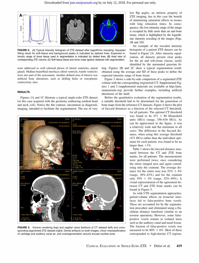
histogram of a patient ZTE dataset can befound in Figure 2A. After intensity equal-ization, all datasets present clear peaksfor the air and soft-tissue classes, easilyidentified by the automated gaussian fit-
ting. Figures 2B and 2C show a typical segmentation result,obtained using the average and SD of these peaks to define theexpected intensity range of bone tissue.Figure 3 shows a side-by-side comparison of a segmented ZTE
volume with the corresponding (registered) CT. Supplemental Fig-ures 1 and 2 (supplemental materials are available at http://jnm.snmjournals.org) provide further examples, including artificialalterations of the skull.Before the quantitative evaluation of the segmentation results,
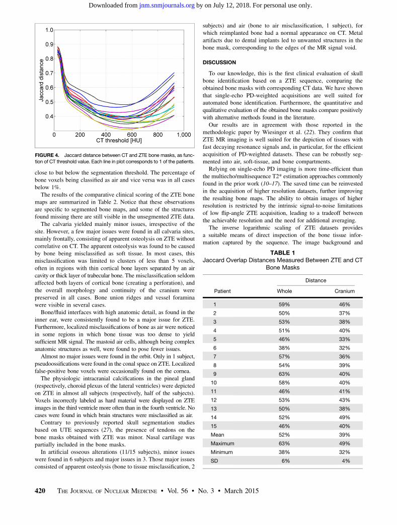
a suitable threshold had to be determined for the generation ofbone maps from the reference CT datasets. Figure 4 shows the plotof Jaccard distances as a function of the selected CT threshold,
for all patients. The optimal CT thresholdwas found to be 471 6 88 Hounsfieldunits (HUs) (range, 356–636 HUs). Ascan be appreciated in the figure, it wasa relatively wide and flat minimum in allcases. The difference in the Jaccard dis-tance, when using this average threshold(471 HUs) rather than the individual opti-mum for each patient, was found to be nolarger than 11%.Table 1 shows the Jaccard distance mea-
sured between the CT and ZTE bonemasks, for all patients. The measurementswere performed twice, once consideringthe entire imaged area and again consid-ering only the cranium. The average dis-tance for the entire area was 52% 6 6%(range, 38%–63%) and for the craniumonly 39% 6 4% (range, 32%–49%). Avisual representation of the agreement be-tween CT and ZTE bone masks can befound in Figure 5.As with UTE segmentation approaches,
partial-volume effects on tissue/air inter-faces led to false-positive bone voxels.These are accounted for by the segmenta-tion procedure and eliminated using a Eu-clidean distance transform (similar to anerosion operation). However, some false-positive voxels remain in isolated areassuch as the auditory canal and nasal fossae.The fraction of false-positive voxels wasmeasured to be 80% 6 6%. Most of thesecorresponded to high-density CT regions,
FIGURE 2. (A) Typical intensity histogram of ZTE dataset after logarithmic rescaling. Gaussian
fitting result for soft-tissue and background peaks is indicated by dashed lines. Expected in-
tensity range of bone tissue used in segmentation is indicated by dotted lines. (B) Axial view of
corresponding ZTE volume. (C) Soft tissue (blue) and bone mask (green) obtained with segmentation.
FIGURE 3. Volume rendering (top) and sagittal views (bottom) of CT dataset (left) and corre-
sponding segmented ZTE dataset (right). Dental artifacts on both images, minor misclassification
of cartilage and auditory canal air, and oversegmentation around sinusal cavities exist.
CLINICAL EVALUATION OF SINGLE-ECHO ZTE • Delso et al. 419
by on July 12, 2018. For personal use only. jnm.snmjournals.org Downloaded from
close to but below the segmentation threshold. The percentage ofbone voxels being classified as air and vice versa was in all casesbelow 1%.The results of the comparative clinical scoring of the ZTE bone
maps are summarized in Table 2. Notice that these observationsare specific to segmented bone maps, and some of the structuresfound missing there are still visible in the unsegmented ZTE data.The calvaria yielded mainly minor issues, irrespective of the
site. However, a few major issues were found in all calvaria sites,mainly frontally, consisting of apparent osteolysis on ZTE withoutcorrelative on CT. The apparent osteolysis was found to be causedby bone being misclassified as soft tissue. In most cases, thismisclassification was limited to clusters of less than 5 voxels,often in regions with thin cortical bone layers separated by an aircavity or thick layer of trabecular bone. The misclassification seldomaffected both layers of cortical bone (creating a perforation), andthe overall morphology and continuity of the cranium werepreserved in all cases. Bone union ridges and vessel foraminawere visible in several cases.Bone/fluid interfaces with high anatomic detail, as found in the
inner ear, were consistently found to be a major issue for ZTE.Furthermore, localized misclassifications of bone as air were noticedin some regions in which bone tissue was too dense to yieldsufficient MR signal. The mastoid air cells, although being complexanatomic structures as well, were found to pose fewer issues.Almost no major issues were found in the orbit. Only in 1 subject,
pseudoossifications were found in the conal space on ZTE. Localizedfalse-positive bone voxels were occasionally found on the cornea.The physiologic intracranial calcifications in the pineal gland
(respectively, choroid plexus of the lateral ventricles) were depictedon ZTE in almost all subjects (respectively, half of the subjects).Voxels incorrectly labeled as hard material were displayed on ZTEimages in the third ventricle more often than in the fourth ventricle. Nocases were found in which brain structures were misclassified as air.Contrary to previously reported skull segmentation studies
based on UTE sequences (27), the presence of tendons on thebone masks obtained with ZTE was minor. Nasal cartilage waspartially included in the bone masks.In artificial osseous alterations (11/15 subjects), minor issues
were found in 6 subjects and major issues in 3. Those major issuesconsisted of apparent osteolysis (bone to tissue misclassification, 2
subjects) and air (bone to air misclassification, 1 subject), forwhich reimplanted bone had a normal appearance on CT. Metalartifacts due to dental implants led to unwanted structures in thebone mask, corresponding to the edges of the MR signal void.
DISCUSSION
To our knowledge, this is the first clinical evaluation of skullbone identification based on a ZTE sequence, comparing theobtained bone masks with corresponding CT data. We have shownthat single-echo PD-weighted acquisitions are well suited forautomated bone identification. Furthermore, the quantitative andqualitative evaluation of the obtained bone masks compare positivelywith alternative methods found in the literature.Our results are in agreement with those reported in the
methodologic paper by Wiesinger et al. (22). They confirm thatZTE MR imaging is well suited for the depiction of tissues withfast decaying resonance signals and, in particular, for the efficientacquisition of PD-weighted datasets. These can be robustly seg-mented into air, soft-tissue, and bone compartments.Relying on single-echo PD imaging is more time-efficient than
the multiecho/multisequence T2* estimation approaches commonlyfound in the prior work (10–17). The saved time can be reinvestedin the acquisition of higher resolution datasets, further improvingthe resulting bone maps. The ability to obtain images of higherresolution is restricted by the intrinsic signal-to-noise limitationsof low flip-angle ZTE acquisition, leading to a tradeoff betweenthe achievable resolution and the need for additional averaging.The inverse logarithmic scaling of ZTE datasets provides
a suitable means of direct inspection of the bone tissue infor-mation captured by the sequence. The image background and
FIGURE 4. Jaccard distance between CT and ZTE bone masks, as func-
tion of CT threshold value. Each line in plot corresponds to 1 of the patients.
TABLE 1Jaccard Overlap Distances Measured Between ZTE and CT
Bone Masks
Distance
Patient Whole Cranium
1 59% 46%
2 50% 37%
3 53% 38%
4 51% 40%
5 46% 33%
6 38% 32%
7 57% 36%
8 54% 39%
9 63% 40%
10 58% 40%
11 46% 41%
12 53% 43%
13 50% 38%
14 52% 49%
15 46% 40%
Mean 52% 39%
Maximum 63% 49%
Minimum 38% 32%
SD 6% 4%
420 THE JOURNAL OF NUCLEAR MEDICINE • Vol. 56 • No. 3 • March 2015
by on July 12, 2018. For personal use only. jnm.snmjournals.org Downloaded from

partial-volume effects in the air–tissue interfaces must be removedbefore rendering tools can be used.The bone masks yielded by ZTE postprocessing were evaluated
relative to CT, the current gold standard for bone imaging. Quantitativeevaluation using the Jaccard distance shows better performance thanpreviously reported for dual-echo UTE (27), which ranged be-tween 47% and 79%, as opposed to the 38%–63% obtained inthis study.The wide minima found during the optimization of the threshold
for CT bone segmentation suggest that the bone structures detectablewith ZTE are relatively well defined in terms of CT intensity (i.e.,there are no undetected structures with similar attenuation properties,nor an excess of gradual intensity transitions). Hence, the ZTE bonemask is well suited for the purpose of attenuation modeling. Still,future work will aim at improving the processing of ZTE datasets toyield nonbinary estimations of tissue density.
Overall, the anatomic detail of ZTE bonemasks was insufficient for direct clinicalinspection but acceptable for attenuation-correction purposes. Despite air cavity differ-entiation working remarkably well becauseof the PD contrast of the sequence, theproblem of false-positive bone identificationin certain interfaces affected by partial-volumeeffects remains.The general morphology of the cranium
was correctly captured in the ZTE masks.Apparent osteolyses were found, especially
in the frontal calvaria. These were due to local (typically ,1 cm)misclassification of thin trabecular bone layers as soft tissue.Often, only 1 layer was affected, suggesting that the misclassifi-cation would have a limited impact on attenuation estimation.Block effects related to the implementation of the intensity in-homogeneity correction were also identified as an occasionalsource of local bone misclassification. This problem will needto be addressed in future studies.Structural detail in the inner ear was almost completely lost in
ZTE bone masks of all patients, compared with CT (severelylimiting their use for clinical diagnosis). Although structural detailwas also diminished in the mastoid, with loss of some thin air cellsepta, this did not affect the overall interpretation of the scans,because no relevant information was lost. More problematic, from thepoint of view of attenuation estimation, would be the misclassifica-tion of dense bone tissue as air. This is probably an inherent
FIGURE 5. Logarithmic rescaling of ZTE dataset (A and B) and corresponding map of agreement
between ZTE and CT bonemasks (C and D). Green pixels indicate true-positive bone identification, red
pixels indicate false-negative (missed bone), and yellow pixels indicate false-positive. Residual mis-
registration can be perceived in, for example, occipital region. Notice surgical bone alteration.
TABLE 2Clinical Scoring of ZTE Bone Masks in Comparison with CT Gold Standard
Patient
CalvariaInner
ear Mastoid Orbit
Cerebrospinal
fluid space
Choroid
plexusof lateral
ventricle
(calcification)
Pineal gland
(calcification)
Artificial
alterationFrontal Parietal Temporal Occipital
Third
ventricle
Fourth
ventricle
1 1 0 1 1 2 1 1 1 1 1 0 NA
2 1 1 1 0 2 1 0 0 0 1 0 1
3 0 1 1 1 2 1 1 2 0 1 1 1
4 0 1 1 1 1 1 0 2 0 1 0 1
5 1 1 0 1 1 1 2 2 0 1 0 0
6 1 1 1 1 1 1 1 1 0 0 0 2
7 2 0 1 1 2 1 1 2 1 0 0 0
8 2 1 1 1 2 1 0 0 0 0 1 1
9 1 1 1 1 2 1 1 0 1 0 0 NA
10 1 1 1 1 2 1 1 2 0 1 0 NA
11 1 2 2 2 2 1 1 0 0 1 0 2
12 1 1 1 1 1 1 0 0 0 0 0 NA
13 1 0 1 1 2 1 0 2 0 1 0 1
14 2 2 2 1 2 1 1 2 0 1 0 2
15 1 0 1 1 2 1 0 2 2 1 0 1
Mean 1.1 0.9 1.1 1.0 1.7 1.0 0.7 1.2 0.3 0.7 0.1 1.1
SD 0.6 0.6 0.5 0.4 0.5 0.0 0.6 0.9 0.6 0.5 0.4 0.7
1 5 minor issue; 0 5 no issue; 2 5 major issue; NA 5 not applicable.
CLINICAL EVALUATION OF SINGLE-ECHO ZTE • Delso et al. 421
by on July 12, 2018. For personal use only. jnm.snmjournals.org Downloaded from
limitation of the acquisition, due to the low density of signal-emittingtissue. Regardless of the highly localized nature of this issue, furtherstudy will be required to assess its impact in attenuation correction.Fat/muscle/bone interfaces such as in the retrobulbar region yielded
satisfactory results, except in 1 subject in whom apparent conalossification on ZTE was possibly inferred by eyeball movements.We could show that even small calcifications, such as in the
pineal gland, are reliably interpreted as hard structures by ZTE.The incorrect hard structures displayed by ZTE in the thirdventricle were likely induced by a combination of magnetizationsaturation and the thin, slitlike configuration of this cerebrospinalfluid space, with 2 brain/fluid interfaces being close, as opposed tothe fourth ventricle, for which pseudocalcifications were found lesscommonly. Classification issues in the ventricles have also beenreported for UTE-based methods (28). Anatomy-based postprocessingmay need to be included in the segmentation to mitigate this issue.Similarly, the throat and vertebrae area would benefit from dedicated
processing, which could account both for the lower density bone tissueand for the false-positives in the oropharyngeal cavity interfaces.Apparent lytic or even air-containing lesions in reimplanted
bone on ZTE, being observed in 3 of 11 subjects with post-operative changes, do have grave clinical influence. Extremecaution should therefore be taken when interpreting ZTE datasetsaround such areas. Signal voids around implanted metallic objectsare a serious limitation of the proposed method, and correctionapproaches are currently being investigated.Further work will be directed at extending the use of ZTE bone
segmentation to whole-body applications. Other potential appli-cations of the ZTE sequence for PET/MR imaging would be lungvisualization, to improve the estimation of parenchyma attenua-tion (29) and the detection of local coils.
CONCLUSION
This is the first, to our knowledge, clinical evaluation of skullbone identification based on a ZTE sequence. The results suggestthat single-echo, PD-weighted ZTE imaging is an efficient meansof obtaining anatomically accurate maps of bone tissue for thepurpose of PET attenuation correction. The segmentation resultsshow better bone depiction and separation from air cavities andcollagenous tissue than previously reported methods. Further workwill be aimed at obtaining nonbinary estimations of bone density.
DISCLOSURE
The costs of publication of this article were defrayed in part bythe payment of page charges. Therefore, and solely to indicate thisfact, this article is hereby marked “advertisement” in accordancewith 18 USC section 1734. No potential conflict of interest rele-vant to this article was reported.
REFERENCES
1. Martinez-Möller A, Souvatzoglou M, Delso G, et al. Tissue classification as
a potential approach for attenuation correction in whole-body PET/MRI: evalu-
ation with PET/CT data. J Nucl Med. 2009;50:520–526.
2. Hofmann M, Pichler B, Scholkopf B, Beyer T. Towards quantitative PET/MRI:
a review of MR-based attenuation correction techniques. Eur J Nucl Med Mol
Imaging. 2009;36(suppl 1):S93–S104.
3. Du J, Carl M, Bydder M, Takahashi A, Chung CB, Bydder GM. Qualitative and
quantitative ultrashort echo time (UTE) imaging of cortical bone. J Magn Reson.
2010;207:304–311.
4. Hofmann M, Steinke F, Scheel V, et al. MRI-based attenuation correction for
PET/MRI: a novel approach combining pattern recognition and atlas registration.
J Nucl Med. 2008;49:1875–1883.
5. Qian H, Shanbhag D, Kaushik S, et al. Whole-body PET/MR attenuation cor-
rection combining image segmentation, truncation completion and atlas-based
skull segmentation. Paper presented at: PET/MR and SPECT/MR: New Paradigms
for Combined Modalities in Molecular Imaging Conference; May 26–30, 2012;
Elba, Italy.
6. Malone IB, Ansorge RE, Williams GB, Nestor PJ, Carpenter TA, Fryer TD. At-
tenuation correction methods suitable for brain imaging with a PET/MRI scanner:
a comparison of tissue atlas and template attenuation map approaches. J Nucl Med.
2011;52:1142–1149.
7. Robson MD, Bydder GM. Clinical ultrashort echo time imaging of bone and
other connective tissues. NMR Biomed. 2006;19:765–780.
8. Robson MD, Gatehouse PD, Bydder M, Bydder GM. Magnetic resonance: an intro-
duction to ultrashort TE (UTE) imaging. J Comput Assist Tomogr. 2003;27:825–846.
9. Du J, Bydder M, Takahashi AM, Carl M, Chung CB, Bydder GM. Short T2
contrast with three-dimensional ultrashort echo time imaging. Magn Reson Im-
aging. 2011;29:470–482.
10. Keereman V, Fierens Y, Broux T, De Deene Y, Lonneux M, Vandenberghe S.
MRI-based attenuation correction for PET/MRI using ultrashort echo time se-
quences. J Nucl Med. 2010;51:812–818.
11. Wang L, Zhong X, Zang L, Tiwari D, Mao H. Ultra-short TE (UTE) imaging of
skull and a quantitative comparison of skull images obtained from MRI and CT.
Proc. Intl. Soc. Mag. Reson. Med. 2010;18:796.
12. Catana C, Van der Kouwe A, Benner T, et al. MR-Based PET attenuation cor-
rection for neurological studies using dual-echo UTE sequences. Proc. Intl. Soc.
Mag. Reson. Med. 2010;18:3953.
13. Catana C, van der Kouwe A, Benner T, et al. Toward implementing an MRI-
based PET attenuation-correction method for neurologic studies on the MR-PET
brain prototype. J Nucl Med. 2010;51:1431–1438.
14. Johansson A, Karlsson M, Nyholm T. CT substitute derived from MRI sequences
with ultrashort echo time. Med Phys. 2011;38:2708–2714.
15. Berker Y, Franke J, Salomon A, et al. MRI-based attenuation correction for
hybrid PET/MRI systems: a 4-class tissue segmentation technique using a com-
bined ultrashort-echo-time/Dixon MRI sequence. J Nucl Med. 2012;53:796–804.
16. Navalpakkam BK, Braun H, Kuwert T, Quick HH. Magnetic resonance-based
attenuation correction for PET/MR hybrid imaging using continuous valued
attenuation maps. Invest Radiol. 2013;48:323–332.
17. Delso G, Zeimpekis K, Carl M, Wiesinger F, Hüllner M, Veit-Haibach P. Cluster-
based segmentation of dual-echo ultra-short echo time images for PET/MR bone
localization. EJNMMI Physics. 2014;1:1–13.
18. Madio DP, Lowe IJ. Ultra-fast imaging using low flip angles and FIDs. Magn
Reson Med. 1995;34:525–529.
19. Grodzki DM, Jakob PM, Heismann B. Ultrashort echo time imaging using point-
wise encoding time reduction with radial acquisition (PETRA). Magn Reson
Med. 2012;67:510–518.
20. Idiyatullin D, Corum C, Park JY, Garwood M. Fast and quiet MRI using a swept
radiofrequency. J Magn Reson. 2006;181:342–349.
21. Veit-Haibach P, Kuhn FP, Wiesinger F, Delso G, von Schulthess G. PET-MR
imaging using a tri-modality PET/CT-MR system with a dedicated shuttle in
clinical routine. MAGMA. 2013;26:25–35.
22. Wiesinger F, Sacolick L, Kaushik S, Ahn S, Delso G, Shanbhag D. Zero TE bone
imaging. Paper presented at: ISMRM; May 10–16, 2014; Milan, Italy.
23. Wiesinger F, Sacolick LI, Menini A, et al. Zero TE MR bone imaging in the
head. Magn Reson Med. January 16, 2015 [Epub ahead of print].
24. Wu Y, Ackerman JL, Chesler DA, Graham L, Wang Y, Glimcher MJ. Density of
organic matrix of native mineralized bone measured by water- and fat-suppressed
proton projection MRI. Magn Reson Med. 2003;50:59–68.
25. Jackson JI, Meyer CH, Nishimura DG, Macovski A. Selection of a convolution
function for Fourier inversion using gridding. IEEE Trans Med Imaging.
1991;10:473–478.
26. Beatty PJ, Nishimura DG, Pauly JM. Rapid gridding reconstruction with
a minimal oversampling ratio. IEEE Trans Med Imaging. 2005;24:799–808.
27. Delso G, Carl M, Wiesinger F, et al. Anatomic evaluation of 3-dimensional
ultrashort-echo-time bone maps for PET/MR attenuation correction. J Nucl
Med. 2014;55:780–785.
28. Choi H, Cheon GJ, Kim H-J, et al. Segmentation-based MR attenuation correction
including bones also affects quantitation in brain studies: an initial result of 18F-FP-
CIT PET/MR for patients with parkinsonism. J Nucl Med. 2014;55:1617–1622.
29. Gibiino F, Sacolick L, Menini A, Landini L, Wiesinger F. Free-breathing, zero-
TE MR lung imaging. MAGMA. September 9, 2014 [Epub ahead of print].
422 THE JOURNAL OF NUCLEAR MEDICINE • Vol. 56 • No. 3 • March 2015
by on July 12, 2018. For personal use only. jnm.snmjournals.org Downloaded from
Doi: 10.2967/jnumed.114.149997Published online: February 12, 2015.
2015;56:417-422.J Nucl Med. Patrick Veit-HaibachGaspar Delso, Florian Wiesinger, Laura I. Sacolick, Sandeep S. Kaushik, Dattesh D. Shanbhag, Martin Hüllner and Clinical Evaluation of Zero-Echo-Time MR Imaging for the Segmentation of the Skull
http://jnm.snmjournals.org/content/56/3/417This article and updated information are available at:
http://jnm.snmjournals.org/site/subscriptions/online.xhtml
Information about subscriptions to JNM can be found at:
http://jnm.snmjournals.org/site/misc/permission.xhtmlInformation about reproducing figures, tables, or other portions of this article can be found online at:
(Print ISSN: 0161-5505, Online ISSN: 2159-662X)1850 Samuel Morse Drive, Reston, VA 20190.SNMMI | Society of Nuclear Medicine and Molecular Imaging
is published monthly.The Journal of Nuclear Medicine
© Copyright 2015 SNMMI; all rights reserved.
by on July 12, 2018. For personal use only. jnm.snmjournals.org Downloaded from