complement activation and regulation in...
TRANSCRIPT

1
COMPLEMENT ACTIVATION AND REGULATIONIN MUCOSAL INFECTIONS

2
© 2001 by Riina Rautemaa
Illustrations and book design by the author and Marja HämäläinenCover by Vilma Rautemaa
Printed at Saarijärven Offset Oy, Saarijärvi, FinlandE-book-ISBN 952-10-0188-7

3
COMPLEMENT ACTIVATION ANDREGULATION IN MUCOSALINFECTIONS
Riina Rautemaa
Department of Bacteriology and ImmunologyHaartman InstituteUniversity of HelsinkiHelsinki, Finland
Academic dissertation for the PhD degree
To be publicly discussed, with permission of the Medical Faculty ofthe University of Helsinki, in the Small Auditorium of the HaartmanInstitute, Helsinki on Wednesday 5th December, 2001,at 12 o’clock noon.

4
SUPERVISOR
Seppo Meri
MD, PhD, docentDepartment of Bacteriology and ImmunologyHaartman InstituteUniversity of Helsinki, Finland
REVIEWERS
Olli Vainio
MD, PhD, docentDepartment of Medical MicrobiologyUniversity of Turku, Finland
Timo Sorsa
DDS, PhD, DiplPerio, docentOral Pathology Research UnitBiomedicum Helsinki, Institute of DentistryUniversity of Helsinki, Finland
OPPONENT
Reinhard Würzner
MD, PhD, Assoc. ProfessorInstitute for HygieneUniversity of InnsbruckAustria

5
Be accurate when cooking toastNever try to guess
Cook it ‘til it smokes and thenTwenty seconds less1.

6
7
CONTENTS
1 PUBLICATIONS.......................................................................11
2 ABBREVIATIONS .....................................................................12
3 ABSTRACT............................................................................15
4 INTRODUCTION......................................................................18
5 REVIEW OF THE LITERATURE......................................................21
5.1 THE COMPLEMENT SYSTEM.....................................................21
5.1.1 Complement activation...................................................21
5.1.1.1 Classical pathway (CP) ............................................22
5.1.1.2 Alternative pathway (AP).........................................23
5.1.1.3 Lectin pathway (LP) ...............................................23
5.1.1.4 Terminal pathway .................................................24
5.1.1.5 Anaphylatoxins C3a and C5a .....................................24
5.1.2 Complement regulation ..................................................26
5.1.2.1 Fluid phase regulators ............................................26
5.1.2.2 Membrane regulators..............................................28
5.2 COMPLEMENT AND INFECTIONS................................................31
5.2.1 General......................................................................31
5.2.2 Complement deficiencies ................................................31
5.2.3 Adverse effects of C activation .........................................33
5.2.4 Adult periodontitis ........................................................34
5.2.5 H. pylori gastritis..........................................................37
5.2.6 Herpes simplex type 1 infection ........................................38
5.3 COMPLEMENT AND MICROBES..................................................40
5.3.1 Complement resistance mechanisms of bacteria.....................40
5.3.1.1 General ..............................................................40
5.3.1.2 LPS ...................................................................40
5.3.1.3 Capsule and peptidoglycan.......................................42
5.3.1.4 Membrane proteins ................................................45
5.3.1.5 Antibody blocking and antigenic variation.....................47
5.3.1.6 Utilization of membrane receptors and regulators...........48
8
5.3.2 Complement resistance mechanisms of viruses...................... 49
5.3.2.1 C component mediated adhesion ............................... 49
5.3.2.2 Misdirecting the C system........................................ 50
5.3.2.3 Mimicry of C regulators........................................... 50
5.3.2.4 Acquisition of host cell membrane regulators ................ 51
6 AIMS OF THE STUDY................................................................ 53
7 MATERIALS AND METHODS ........................................................ 54
7.1 MATERIALS ....................................................................... 54
7.1.1 Patients..................................................................... 54
7.1.1.1 Adult periodontitis (AdP, I) ...................................... 54
7.1.1.2 H. pylori-gastritis (III) ............................................ 55
7.1.1.3 Tissue preparation ................................................ 55
7.1.2 Bacteria .................................................................... 56
7.1.3 Eukaryotic cell culture ................................................... 56
7.1.4 HSV-1 infection............................................................ 57
7.1.5 Buffers ...................................................................... 57
7.1.6 Reagents and sera ........................................................ 58
7.1.7 Isolation of CD59 .......................................................... 60
7.2 METHODS ......................................................................... 61
7.2.1 Indirect immunofluorescence microscopy (IF) ....................... 61
7.2.2 CD59 binding tests ........................................................ 62
7.2.3 Flow cytometry............................................................ 62
7.2.4 Bactericidal assay......................................................... 63
7.2.5 Bioluminescence assay for bacteriolysis .............................. 63
7.2.6 Whole cell ELISA .......................................................... 64
7.2.7 SDS-PAGE................................................................... 65
7.2.8 Statistical methods ....................................................... 65
8 RESULTS AND DISCUSSION ........................................................ 66
8.1 ADULT PERIODONTITIS (AdP).................................................. 66
8.1.1 Complement activation in AdP ......................................... 66
8.1.2 Complement regulation in AdP ......................................... 67

9
8.2 BINDING OF CD59 TO BACTERIA IN VITRO ..................................................... 69
8.2.1 Binding of CD59 to E. coli........................................................................ 69
8.2.2 Binding of CD59 to H. pylori ................................................................... 70
8.3 INHIBITION OF C BY BINDING OF mCD59 TO E. COLI ANDH. PYLORI ............................................................................................................. 71
8.4 COMPLEMENT ACTIVATION AND REGULATION IN H.PYLORI GASTRITIS ...............................................................73
8.4.1 Activation of C in H. pylori gastritis....................................73
8.4.2 Regulation of C in H. pylori gastritis ...................................74
8.5 COMPLEMENT ACTIVATION AND REGULATION BY HSV-1-INFECTED EPITHELIAL AND NEURONAL CELLS ...............................75
9 CONCLUSIONS .......................................................................79
10 ACKNOWLEDGEMENTS..............................................................83
11 REFERENCES .........................................................................87
12 ORIGINAL PUBLICATIONS ........................................................ 108

10
11
1 PUBLICATIONS
This thesis is based on the following original publications:
I R. Rautemaa and S. Meri.
Protection of gingival epithelium against complement-
mediated damage by strong expression of the membrane
attack complex inhibitor protectin (CD59).
J. Dent. Res. 75:568-574, 1996.
II R. Rautemaa, G.A. Jarvis, P. Marnila and S. Meri.
Acquired resistance of Escherichia coli to complement lysis by
incorporation of GPI-anchored protectin (CD59).
Infect. Immun. 66:1928-1933, 1998.
III R. Rautemaa, H. Rautelin, P. Puolakkainen, A. Kokkola, P.
Kärkkäinen and S. Meri.
Survival of Helicobacter pylori from complement lysis by
binding of GPI-anchored protectin (CD59).
Gastroenterology 120:470-479, 2001.
IV R. Rautemaa, T. Helander and S. Meri
Herpes simplex -virus 1 infected epithelial and neuronal cells
differ in their susceptibility to complement attack.
Submitted, 2001.
The articles in this thesis have been reproduced with the permission
of the copyright holders.
12
2 ABBREVIATIONS
AP Alternative pathway of complement
AdP Adult periodontitis
BM Basement membrane
BSA Bovine serum albumin
C Complement
C1INH C1 inhibitor
CD59 Protectin
CFU Colony forming unit
CN Clusterin
CP Classical pathway of complement
DAF Decay-accelerating factor (CD55)
EDTA Ethylene-diamine tetra-acetic acid
ELISA Enzyme-linked immunosorbent assay
fH Factor H
fI Factor I
FITC Fluorescein isothiocyanate
GCF Gingival crevicular fluid
GPI Glycophosphoinositol
HSV-1 Herpes simplex -virus type 1
IC Immune complex
IF Immunofluorescence
Ig Immunoglobulin
LP Lectin pathway
LPS Lipopolysaccharide
mAb Monoclonal antibody
MAC Membrane attack complex of complement
MASP Mannose binding lectin associated serine protease
MBL Mannose binding lectin
mCD59 Membrane form of protectin
MCP Membrane cofactor protein (CD46)
13
NHS Normal human serum
NHShi Heat-inactivated normal human serum
NHSimm Immune normal human serum
NHSni Non-immune normal human serum
OD Optical density
pAb Polyclonal antibody
PBS Phosphate buffered saline
PFU Plaque forming unit
POX Peroxidase
sCD59 Soluble form of protectin
SD Standard deviation
SDS-PAGE Sodium-dodecyl-sulfate polyacrylamide gel electrophoresis
TP Terminal pathway of complement
TCC Terminal complement complex
VBS Veronal buffered saline
VN Vitronectin

14

15
3 ABS TRACT
The human complement (C) system i s an essenti al par t of the innate
immune system a nd has a central r ole in infla mmator y and immune
res ponses . It p artici pates in ops onization, chemota xis, leukocyte
activation and direct killi ng of bacter ia and infected cells. T he aim of
the present study was to analyze how ba cteria causi ng chr onic
infections and host cells a t the sites of inflammation pr otect
themselves agai nst C attack .
In the fi rst study C activa tion a nd reg ulation were inves tigated in a dult
per iodontitis (AdP). AdP is a chr onic i nflammatory diseas e of the tooth
sup porting appa ratus. Coars e granular d eposits of C components C3d
and C9 were seen in the sub epithelial tissues of the majority of AdP
(n= 18) pa tients . In the hea lthy controls (n=1 1) C d eposits were
detectable only in 1 case. C deposits w ere also obs erved on the
bas ement membra nes of both pocket and oral ep itheli um of both
hea lthy a nd AdP gingi va. In healthy gingiva C D59 wa s strongly
exp ressed on the memb ranes of epi thelia l cells and on the vascular
end otheli a of the und erlying connective tissue. In AdP CD59 was
str ongly expres sed by endothelial cells but i n the epithelia the
exp ression was granular and weaker than in the healthy gi ngiva. C9
dep osits were never s een at sites of CD59 exp ression. The gingi val
epi thelium and connective tissue endothelia a re thus well protected
aga inst d amage by the membr ane attack complex of C (MAC).
Protection of the und erlying connective tissue is, however, ins uffici ent
and may a llow d eposition of MAC a nd autologous tiss ue damage in AdP.
Some micr obes can eva de the host immune system and cause chroni c
infections. The second stud y examined w hether the g ram-negative
bacterium E. coli could acquire to its outer cell membra ne the
glycophos phoinositol- (GPI-) anchored C inhib itor C D59 released from
hos t cells. CD5 9 is a human cell membra ne reg ulator of C that i nhibits
the forma tion of MAC. Analysis by using radiolabeled CD59 ,

16
immunofluorescence mi croscopy, flow cytometry and w hole cell EL ISA
demonstra ted that CD5 9 bound to nonenca psulated E. coli strains
EH2 37 (Re) and EH234 (Ra) i n a ca lcium-depend ent ma nner. The
incorpora tion r equired the GPI-phospholipid moiety since no binding of
a p hospholipid-free s oluble form of CD5 9 was observed. Mg ++ di d not
enhance the binding a nd a p olysia lic acid cap sule p revented it (IH308 0
str ain, O 18:K1 :H8). Bound CD59 i nhibited the C5b-9 neoantigen
exp ression on C -treated bacteria. Protection agains t C lysis wa s
obs erved in both a colony counting assa y and a biolumines cence assay
where via ble EH 234 ba cteria expressing the lucifera se gene emitted
green lig ht in the pr esence of the luci ferine substrate.
The third study exami ned another gram-negative bacterium,
Helicobac ter pylori, w hich can cause a chronic infection that persists
for years . It i s sens itive to C lysis in vitro but it w as obs erved that H.
pylori acquires C-res istance by b inding glycolipid-tailed inhib itors from
the host. In noninfected mucosal gastri c biop sy samples (n=6) C D59 wa s
str ongly expres sed on the membranes of epithelial cells. In the H.
pylori-infected epithelia (n=10) the ex pressi on of CD59 w as gra nular
and more focused to the mucus. H. pylori in the g astric pits were often
pos itive for CD59 but negative for C5b-9. An opposi te pattern w as seen
on the surface mucosa . In vitro ana lyses using 125I-C D59 and
bacteriolysis a ssays showed that CD59 b ound to H. pylori and protected
the CagA virulence fa ctor express ing strains agains t C ki lling. In an
ELISA ass ay the bindi ng of CD59 correla ted inversely with the
app earance of the C5b -9 neoantigen. Binding of CD59 inhib its
membrane attack complex ass embly on H. pylori and may thereb y
contribute to their s urviva l on the gas tric mucosa. The r esults indicate
tha t CD59 can i ncorporate i nto the cell membr anes of nonencapsulated
bacteria in a functionally active form.
In the fourth s tudy the role of C activation and regulati on in a vira l
infection was examined. Herpes si mplex vi rus type 1 (HSV-1) infection
in neurons is lifelong and genera lly as ymptomatic. Reacti vation of the
latent infection results in skin blistering w hereas the r espective
17
per iphera l neur ons ar e rarely affected. Why the neuronal cells are
spa red while the epithelial cells are s acrifi ced is not w ell understood.
Dur ing the firs t hour s of H SV-1 i nfecti on in vitro the expr ession of the
end ogenous C regulators DAF and C D59 increased on b oth ep itheli al
HES cells and neurona l Paju cells . By 1 2 hour s the infected HES cells
had lost their ability to control C attack. T he exp ression of DAF and
CD5 9 decr eased and the cells beca me tar gets for MAC attack. In
contrast, C reg ulator expression on the neuronal Pa ju cells did not
decrease below the initial level and C5 b-9 depositi on was found only on
10% of the Paju cells at 12 hours . The results sugg est that HSV-infected
neuronal cells are better than ep itheli al cells in protecting themselves
aga inst C attack. Thi s may contri bute to the persis tence of a latent
HSV-1 infection in neuronal cells for p rolong ed per iods.
In conclusion, these studies show that the GP I-anchored p rotein CD59 is
a very dynamic complement i nhibitor. It can b ecome shed from host
cells dur ing infection and inflammation and b ecome incorp orated in a
functiona lly active form into new cell membra nes. W hen E. coli and H.
pylori bound CD59 they incr eased their resistance to C membrane
attack. T hus, s ome mi crobes are capable of recycling the releas ed GPI-
lip id anchored protei n and use it for their own protection agai nst host
C a ttack.

18
4 INTRODUCTION
The human body encounters foreign mater ial fr om the envir onment
constantly. The first barri ers sk in and the mucosal surfa ces need to
protect the bod y agai nst invasion by mi crobes , foreign antigens and
tox ic agents. T he immune system s hould recognize and remove all
for eign materia l but spare the body’s own via ble ti ssues. However, the
immune system s hould also b e able to recognize non-viable tissues as it
is responsible for the clea rance of tis sue debris g enerated dur ing
tra uma or during the normal cellular tur n-over .
The human immune system can be di vided into i nnate and acquired
immunity. In the acquired i mmunity B and T lymphocytes play a major
role. B lymphocytes g ive ri se to plasma cells that produce anti bodies
cap able of recognizing specific non-self anti gens. T lymp hocytes can
recognize infected cells ca rrying forei gn pep tides on their sur faces. The
acq uired immuni ty develops slowly but i t can very s pecifi cally
recognize its target. In contrast, the innate immune system can act
immediately when it g ets into contact w ith foreign invaders. The
innate immune s ystem includ es e. g. serum complement, pa ttern
recogniti on molecules , cytokines, natur al killer (NK) cells and
pha gocyti c cells.
The human complement (C) system has a central role in inflammatory
and immune resp onses. Complement was fi rst id entifi ed as a heat-
lab ile fa ctor i n serum that complemented anti bodies in ki lling bacter ia.
We now know tha t C is an important defense system a gainst microbial
infections as i t participates in opsoni zation, chemotaxis , leuk ocyte
activation and direct killi ng of bacter ia and of mi crobe-infected
cells2,3. C omplement serves a lso as an interface betw een the inna te
and acqui red immune s ystems as it augments antibody responses a nd
enhances immunologica l memory4. In addi tion, the C system is ca pable
of discri minati ng between d ifferent tar gets w ithout the help of
antibodies. It has an impor tant r ole in the clearance of immune

19
complexes and a poptotic cells, as well5. T he C s ystem is alw ays active
at a low rate a nd rea dily r ecogni zes and atta cks foreign materi al tha t is
invading human tissues. Therefore, the host needs over 10 inhib itors to
keep this power ful defence system under contr ol.
Some micr obes can eva de the host immune system and cause chroni c
infections. The abili ty of a micr obe to evade C activation is one of the
fea tures that mak es it a pathogen6. Moreover, an ability to r esist C is a
key virulence feature of ma ny bacteria7. Despite more than a century
of parallel res earch on bacteria and the C system r elatively li ttle is
known of the mechanis ms whereby p athogenic ba cteria can escape C-
related opsonop hagocytosis and di rect k illing . Comp ared w ith vi ruses
bacteria have had les s opportunities to acqui re genetic materia l from
mammalian cells . Instead, i t is more li kely that pa thogenicity in
bacteria has ar isen more accidentally a nd on the ba sis of selection
from natural mutants rather than by stealing or cop ying of genetic
cod es from the host. Factor s providing C resi stance in ba cteria include
external structures s uch as the capsule, peptidoglycan la yer,
lip opolys acchar ide si de cha ins, s pecifi c proteins on the outer
membrane, proteins encoded by vir ulence plasmids and C regulatory
proteins acquir ed from the host.
Ora l and gastri c mucosa ar e not sterile. In health, harmony ex ists
between the "normal" flora and the immune sys tem. T he number of
microbes is kep t under control and thei r inva sion to deep er tis sues i s
prevented by mechanis ms tha t provoke inflamma tion only mi nimally. If
a p athogen inva des the mucosa, an infla mmator y reaction d evelop s and
the immune system aims to d estroy the i nvader . In chronic condi tions
the infection p ersists desp ite the concerted activi ty of the immune
mechanism. An underlying k ey question in the present thesis work was
to examine how microb es can escap e from the i mmune defens e
sys tems. On the other hand, it wa s also asked why the infection
remains limited and w hat ha ppens to hos t cells at the site of i nfecti on.

20

21
5 REVIEW OF THE LITERATURE
5.1 THE COMPLEMENT SYSTEM
The complement (C) system is always active at a low rate and it
readily recognizes and surveys for foreign material entering human
tissues. Over 10 inhibitors are needed to keep the powerful defense
system under control. The biological importance of C is emphasized
by the severe symptoms that C deficiencies cause. These are usually
the result of an impaired activation or regulation of complement.
Most plasma C components are produced in the liver and/or
macrophages, and their concentrations in plasma are normally
relatively stable8. C6 and C7 are produced by polymorphonuclear
leukocytes which may release their constituents at local
inflammation sites9. The membrane components are produced
locally and act either as receptors for the C activation products or as
regulators of the C cascade8. The main C components are shown in
Table 1.
5.1.1 Complement activation
Complement can be activated via the classical, the lectin or the
alternative pathway (Fig. 1). Activation of the classical pathway
begins when C1q binds to specific antibodies on a target. C1q can
also bind to certain bacteria independently of antibodies10. The
lectin pathway is activated when the mannose binding lectin binds
to appropriate target sugar residues found, for example, on
mannose-rich serotypes of Salmonellae11. The alternative pathway is
continuously turning over at a slow rate in an antibody-independent
manner and will attack particles that are not specifically protected
against C.

22
Fi g u re 1. A c t i va t i o n o f t h e co m p l em e nt s ys t em . T he d as h ed l i ne s i nd i c at e en z y m a t i c a ct i vi t i es . A b - ag , ant i b o d y - a nt i g e n co m p l ex es ; c rp , c - r eac t i vep r o t ei n; m a n, m a nn o s e ; g l c na c, n- ac et y l g l uco s a m i n e; m b l , m ann o s eb i nd i n g l ec t i n ; m a s p , m b l - as s o ci a t e d s er i n e p ro t ea s e ; t cc , t er m i na l co m p l e m en t co m p l ex ; m ac , m em b r ane a t t a ck c o m p l e x . Fo r o t h er ab b r ev i at i o ns , s ee t e x t .
5.1.1.1 Classical pathway (CP)
In general, activation of the classical pathway of C begins when the
first component C1 binds to antibodies bound to their antigens. The
interaction takes place between the C1q portion of C1 and the Fc
parts of IgG or IgM12. CP can also become activated by complexes
containing chromatin or C-reactive protein (CRP)13 or by surface
blebs of apoptotic cells14. Binding to a target results in
conformational changes first in C1q and then in C1r. The activated
C1r-protease cleaves C1s to an active enzyme. Activated C1s is
responsible for the cleavage of C4 and C2. The cleavage of C4 leads
to the formation of soluble C4a and reactive C4b, which can form an
amide or hydroxyl ester bond with the target surface15. Bound C4b
forms a magnesium (Mg++) dependent reversible complex with C2,
which in turn becomes cleaved by C1s. The formed C4b2a complex is
the C3/C5 convertase of the CP. The C2a part of the convertase
cleaves C3 to C3a and C3b, and C5 to C5a and C5b. Before its
Classical pathway Lectin pathway Alternative pathway
Ab-Ag/CRP Man/GlcNAc Nonself particles
C1q+C1r+C1s MBL+MASP-1+MASP-2C2
C4
C4b2a C3
C5
C3b/C3(H2O)+B
C3bB D
C3bBb
C3b
C5b +C6+C7+C8+C9 TCC/MACTerminal pathway

23
cleavage C5 binds to C3b16. As one convertase cleaves multiple C3
and C5 molecules the initial signal becomes enhanced.
5.1.1.2 Alternative pathway (AP)
The alternative pathway is continuously activated at a low level in
plasma. C3 undergoes slow and spontaneous hydrolysis to C3(H2O)
and forms, together with Bb, the initial fluid phase C3-convertase of
AP (Fig. 1). The convertase cleaves C3 and the generated
metastable C3b can eventually attach covalently to amino or
hydroxyl groups on target surfaces. Surface bound C3b binds factor
B in a Mg++-dependent manner. The B part of the complex becomes
cleaved by fD and the actual C3 convertase of the AP, C3bBb, is
formed17. The complex is stabilized by properdin. The AP can also
be initiated by activation of the CP to yield the first C3b molecules.
Since C3bBb cleaves several C3 molecules into C3b subunits of new
C3 convertases an effective positive feedback amplification loop is
generated. Amplification leads to C3b opsonization for phagocytosis,
and the generation of C5b starts the activation of the terminal
pathway.
5.1.1.3 Lectin pathway (LP)
The lectin pathway closely resembles the classical pathway. Instead
of C1 the pathway is initiated by mannose binding lectin (MBL) that
binds to carbohydrates containing mannose or N-acetyl glucosamine
residues (Fig. 1). MBL belongs to a family of collectins and has
structural homology to C1q. MBL binds to MBL-associated serine
proteases MASP-1 and MASP-2 to form a C1-like complex. MBL binds
to the target structure calcium-dependently, and the binding results
in conformational changes and activation of the MASPs18,19. The cell-
bound complex cleaves C2 and C4 similarly as C1 of the CP. The rest
of the cascade is identical with that of the CP.

24
5.1.1.4 Terminal pathway
The terminal pathway begins when C5 becomes cleaved by either
the CP or the AP C5-convertase. The formed C5b binds to C6 in the
fluid phase. This induces a conformational change in C6 making it
capable of binding C7. The C5b-7 complex is hydrophobic and
capable of binding to lipid membranes. C8 in turn may bind either to
a cell-bound or a soluble C5b-7 complex. C5b-8 binds C9 and when
multiple C9 molecules polymerize and penetrate through the cell
membrane, a cytolytic membrane attack complex (MAC) is formed
and the membrane is perforated (Fig. 4)20. The terminal C complex
(TCC) is a joint name for both the soluble and the membrane
associated complexes (C5b-7, C5b-8, C5b-9, C5b-9n). Clusterin and
vitronectin are proteins that can keep the TCC in solution 21,22.
5.1.1.5 Anaphylatoxins C3a and C5a
Complement activation via all three pathways leads to generation of
small anaphylatoxins C3a and C5a. They are involved in the
generation of inflammation as they cause release of histamine,
leukotrienes and other phlogistic mediators from mast cells and
basophils. At low concentrations anaphylatoxins cause vascular
dilatation and at higher concentrations they (particularly C5a) can
cause uncontrolled contraction of smooth muscle and other
characteristics of anaphylaxis23. The C3a receptor is expressed on
eosinophils, neutrophils and monocytes and C3a has been shown to
have direct immunomodulatory effects on B cells. C3a and its
degradation product C3a-desArg have been demonstrated to
regulate the synthesis of tumor necrosis factor-α and interleukin-β24.
C3a and C3a-desArg also enhance cytokine synthesis in adherent
monocytes while at the site of inflammation and inhibit the
synthesis of proinflammatory cytokines in circulating cells. Both C3a
and C5a are chemotactic and induce leukocyte migration to the site
of inflammation.

25
Table 1. The main complement components.
Component Pathway Main function
C1q CP Binds Fc of IgG and IgM
C1r CP Activates C1s
C1s CP Cleaves C2 and C4
C4 CP, LP C4b is a part of the CP C3/C5 convertase, C4a is ananaphylatoxin
C2 CP, LP C2a is a part of the CP C3/C5 convertase
C3 CP, AP, LP C3b is an opsonin and a part of CP and AP C3/C5convertases, C3b binds fBC3a is an anaphylatoxin and chemotaxin
MBL LP Binds to certain carbohydrates, activates MASP-1
MASP-1 LP Activates MASP-2
MASP-2 LP Cleaves C2 and C4
Factor B AP Bb is a part of the AP C3/C5 convertase
Factor D AP Cleaves fB
Properdin AP Stabilizes the AP C3/C5 convertase
C5 TP C5b binds C6 and C7, C5a is an anaphylatoxin andchemotaxin
C6 TP Binds C5b and C7
C7 TP Binds C5b6 and C8
C8 TP Binds C5b-7 and C9
C9 TP Binds C5b-8 and C5b-9

26
5.1.2 Complement regulation
If uncontrolled, activation of the complement system would lead to
unnecessary consumption of C components and possible destruction
of autologous cells. Therefore C activation is strictly controlled both
on cell membranes and in the fluid phase by overlapping systems at
various stages (Fig. 2 and Table 2).
Figure 2. Regulators of the complement system. Dashed lines markenzymatic activities, curved dashed lines mark the inhibition sites of Cregulators. For abbreviations, see text.
5.1.2.1 Fluid phase regulators
Activated C1 in plasma and on immune-complexes is regulated by
C1-inhibitor (C1INH; Fig. 2). Irreversible covalent binding of C1INH
to activated C1r and C1s results in the dissociation of the proteins
from the complex and down-regulation of the CP. C1INH is a serine
protease inhibitor (serpin) and, in addition to C1r and C1s, it
inhibits plasmin, kallikrein and coagulation factors XIa and XIIa25.
DAF/CR1/C4bp/fI+ MCP/CR1/C4bp
fH/DAF/CR1
VitronectinClusterin
CD59
CP AP
C1C2
C4
C4b2a C3
C5
C3b/C3(H2O)+B
C3bB D
C3bBb
C3b
C5b +C6+C7+C8+C9 MACTP
C1INH
fH/CR1
fI+ fH/CR1/MCP
27
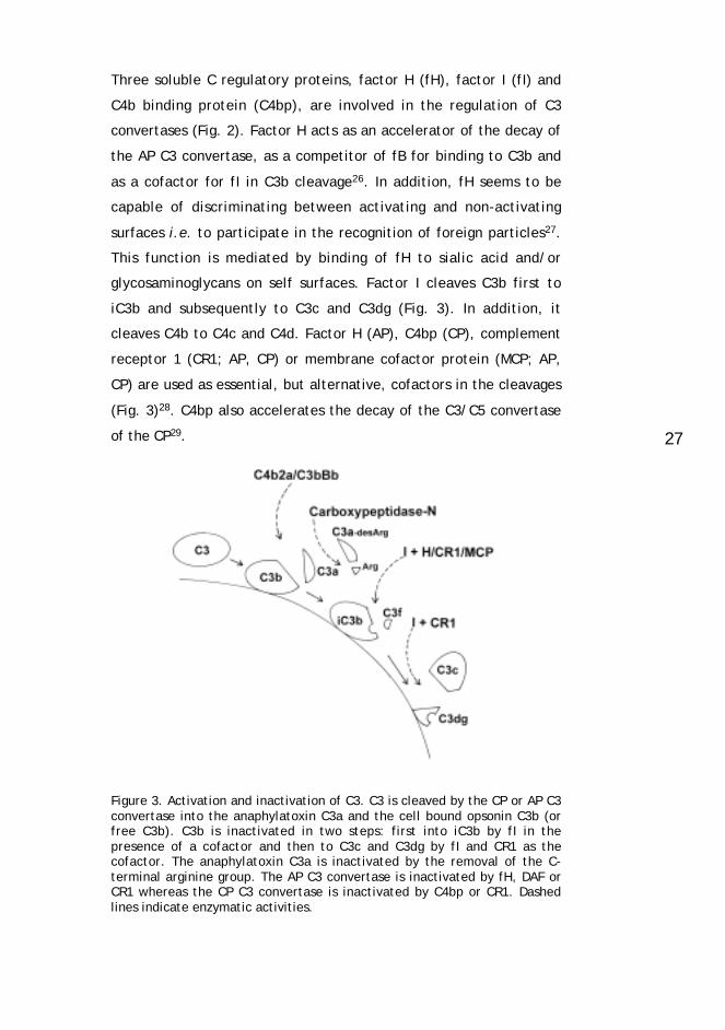
Three soluble C regulatory proteins, factor H (fH), factor I (fI) and
C4b binding protein (C4bp), are involved in the regulation of C3
convertases (Fig. 2). Factor H acts as an accelerator of the decay of
the AP C3 convertase, as a competitor of fB for binding to C3b and
as a cofactor for fI in C3b cleavage26. In addition, fH seems to be
capable of discriminating between activating and non-activating
surfaces i.e. to participate in the recognition of foreign particles27.
This function is mediated by binding of fH to sialic acid and/or
glycosaminoglycans on self surfaces. Factor I cleaves C3b first to
iC3b and subsequently to C3c and C3dg (Fig. 3). In addition, it
cleaves C4b to C4c and C4d. Factor H (AP), C4bp (CP), complement
receptor 1 (CR1; AP, CP) or membrane cofactor protein (MCP; AP,
CP) are used as essential, but alternative, cofactors in the cleavages
(Fig. 3)28. C4bp also accelerates the decay of the C3/C5 convertase
of the CP29.
Figure 3. Activation and inactivation of C3. C3 is cleaved by the CP or AP C3convertase into the anaphylatoxin C3a and the cell bound opsonin C3b (orfree C3b). C3b is inactivated in two steps: first into iC3b by fI in thepresence of a cofactor and then to C3c and C3dg by fI and CR1 as thecofactor. The anaphylatoxin C3a is inactivated by the removal of the C-terminal arginine group. The AP C3 convertase is inactivated by fH, DAF orCR1 whereas the CP C3 convertase is inactivated by C4bp or CR1. Dashedlines indicate enzymatic activities.

28
Vitronectin (VN) and clusterin function at the C5b-7 level. The
binding of these regulators to the C5b-7 complex prevents the
insertion of the complex into the membranes and thus inhibits the
formation of MAC21,22.
5.1.2.2 Membrane regulators
The membrane cofactor protein (MCP, CD46) is a glycoprotein with
a transmembrane domain and an intracellular cytoplasmic tail (cyt1
or cyt2). It acts as a cofactor for fI and promotes the cleavage of
C3b to iC3b or C4b to C4c and C4d (Fig. 2)30. It is expressed by
various cells including leukocytes and endothelial cells. As an
exception, red blood cells do not express MCP.
The complement receptor type 1 (CR1, C3b receptor, CD35) is a
transmembrane glycoprotein. It has decay accelerating activity and
cofactor activity for fI in cleaving cell-bound C3b first to iC3b and
subsequently to C3dg and fluid phase C3c (Fig. 3). CR1 also has a
central role in the clearance of immune complexes that have
activated C as it can act as a receptor for complex-bound C3b, C4b,
C1q and iC3b31,29.
Decay accelerating factor (DAF, CD55) is a glycosyl-phosphatidyl-
inositol- (GPI-) anchored membrane glycoprotein that inhibits C at
the C3 level. It accelerates the decay of both CP and AP C3/C5
convertases on the cells that express it. It is expressed on most cells
and the expression is upregulated by various pro-inflammatory
activators of cellular second-messenger stimuli31,29.
Protectin (CD59) is also a membrane glycoprotein linked to cell
membranes via a GPI-anchor. It inhibits C at the TCC level by
preventing the C5b-8 complex-catalyzed insertion and
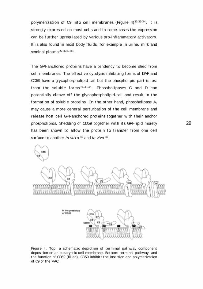
29
polymerization of C9 into cell membranes (Figure 4)32,33,34. It is
strongly expressed on most cells and in some cases the expression
can be further upregulated by various pro-inflammatory activators.
It is also found in most body fluids, for example in urine, milk and
seminal plasma35,36,37,38.
The GPI-anchored proteins have a tendency to become shed from
cell membranes. The effective cytolysis inhibiting forms of DAF and
CD59 have a glycophospholipid-tail but the phospholipid part is lost
from the soluble forms39,40,41. Phospholipases C and D can
potentially cleave off the glycophospholipid-tail and result in the
formation of soluble proteins. On the other hand, phospholipase A2
may cause a more general perturbation of the cell membrane and
release host cell GPI-anchored proteins together with their anchor
phospholipids. Shedding of CD59 together with its GPI-lipid moiety
has been shown to allow the protein to transfer from one cell
surface to another in vitro 42 and in vivo 43.
Figure 4. Top: a schematic depiction of terminal pathway componentdeposition on an eukaryotic cell membrane. Bottom: terminal pathway andthe function of CD59 (filled). CD59 inhibits the insertion and polymerizationof C9 of the MAC.

30
Table 2. The main complement regulators.
Protein Recognition Function
Soluble regulators
C1 inhibitor C1r, Cs Dissociation of C1s and C1r from the C1complex after covalent binding
Factor I C3b, C4b Degradation of C3b and C4b
Factor H C3b Decay accelerating activity for the C3/C5convertase of AP, cofactor for fI
C4b binding protein C4b Decay accelerating activity for the C3/C5convertase of CP, cofactor for fI
Clusterin C5b-7 Prevention of C5b-7 membrane insertion
Vitronectin C5b-7 Prevention of C5b-7 membrane insertion
Membrane regulators
CR1 (CD35) C3b, iC3b,C4b
Decay accelerating activity for the C3/C5convertases, cofactor for fI, immune complextransport
MCP (CD46) C3b, C4b Cofactor for fI
DAF (CD55) C3b, C4b Decay accelerating activity for the C3/C5convertases
Protectin (CD59) C8, C9 Inhibition of MAC

31
5.2 COMPLEMENT AND INFECTIONS
5.2.1 General
The clinical outcome of all infections is strongly modulated by the
complement system as it participates both in the neutralization of
invading microbes and in the generation of inflammation.
Complement-derived anaphylatoxins cause increased vascular
permeability, and chemotaxins attract leukocytes to the site of
microbial invasion. Both gram-negative and -positive bacteria can
become opsonized with C3b and iC3b, and the opsonized bacteria
become phagocytosed. MAC can be formed on gram-negative
bacteria exposing them to the lytic action of C. Although the
formation of MAC has been shown to be necessary for optimal killing
of some gram-negative bacteria44,45, opsonophagocytosis remains
the major antibacterial defense mechanism. Virus-infected cells and
virions may also become opsonized with C3b, iC3b or specific anti-
viral antibodies capable of activating the CP46.
5.2.2 Complement deficiencies
The complement system consists of more than 30 proteins, many of
which have multiple functions. Different deficiencies therefore
result in different clinical consequences. The major disorders
caused by C component deficiencies are recurrent infections and
immune complex (IC) diseases47. The deficiencies of C regulators
can also result in diseases with severe symptoms.
Three types of C component deficiency have been reported to
explain an increased susceptibility to pyogenic infections. Any
deficiency that affects opsonization causes a general susceptibility
to pyogenic microbes whereas defects in the lytic action of C
increase the susceptibility to neisserial infections48. Defense against

32
disseminated neisserial infection is dependent on the recognition of
the bacterial surface by antibody and complement. Individuals with
inherited deficiences in the terminal C components, which are
critical for neisserial killing, often experience disseminated systemic
or relapsing neisserial infections despite having an intact
opsonophagocytic system. The fact that these patients are not more
prone to other systemic infections, caused by e.g. Haemophilus
influenzae, suggests that there is a unique relationship between the
neisserial cell surface and the C system. Deficiencies of the lectin
pathway have been reported to result in an increased risk for
recurrent respiratory infections in early childhood11.
C1q deficiency causes a failure of the classical pathway and
accumulation of immune complexes (IC)49. C1q deficiency has been
related to the pathogenesis of systemic lupus erythematosus (SLE)
because it is a disease where ICs accumulate in different organs.
C1q is involved in the clearance of ICs and the balance between C1q
and IC is lost in SLE. C1q also participates in the clearance of
apoptotic cells. Impaired clearance of these cells and their
fragments has been found to predispose to autoimmune
conditions50. C1q deficiency has also been associated with a
susceptibility to infections caused by encapsulated bacteria and
fungi47.
C1INH deficiency results in a loss of regulation of C1 and a failure to
activate kallikrein. Hereditary angioedema (HAE) is caused by a
reduced C1INH level (type I) or by a reduced C1INH functional
activity (type II). Low C1INH leads to reduced levels of C2 and C4.
HAE is an autosomal dominantly inherited disease or occurs as a
sporadic “de novo” mutation in the C1INH gene. The patients have
characteristic episodes of painless swelling of the skin and mucosal
membranes4.
Dysfunction of factor H can cause two types of kidney diseases,
either membranoproliferative glomerulonephritis type II (MPGN II) or
hemolytic uremic syndrome (HUS). MPGN II is the consequence of a
33
total lack of factor H (or its function) whereas HUS develops when
mutations in the fH gene affect the function of the most C-terminal
domains of fH51,52,53.
Paroxysmal nocturnal hemoglobinuria (PNH) is a hemolytic disease
characterized by an increased sensitivity of a proportion of
erythrocytes to C-mediated lysis, granulocytopenia,
thrombocytopenia and a tendency for venous thrombosis54. The
increased susceptibility of the cells to C is due to their lack of DAF
and CD59. PNH is usually caused by a clonal somatic mutation in the
PIG-A gene that affects the GPI-anchor synthesis55. Isolated
deficiencies of the other membrane regulators are rare.
5.2.3 Adverse effects of C activation
The primary function of the C system is to protect the host against
microbial invasion. C also participates in the processing of non-self
material and in the removal of debris of self-origin. In most cases,
the C system is well controlled and inappropriate activation and
host cell destruction are prevented. However, C activation is not
always entirely beneficial to the host. During bacteremia a vast
amount of anaphylatoxins C3a and C5a are formed. They may lead
to a violent enhancement of inflammation and septic shock. The C
system becomes activated at a site of tissue necrosis and the
clearance is not totally restricted to the necrotic site itself. For
example, in myocardial infarction C activation is an inducer of the
reperfusion injury56,5 7 . The inflammatory changes during
cardiopulmonary bypass are also partially caused by activation of
the C system and the terminal pathway58,59. Autoantibodies present
in autoimmune diseases activate the CP, which can result in an
enhancement of inflammation and C attack against autologous cells.

34
5.2.4 Adult periodontitis
_Adult periodontitis (AdP) is a chronic inflammatory disease of the
tooth supporting apparatus. It is characterized by loss of clinical
tooth attachment due to destruction of the periodontal ligament and
loss of the adjacent supporting bone60. An important feature of AdP
is the accumulation of gingival crevicular fluid (GCF) in the gingival
crevice of periodontal lesions (Fig. 5). A healthy gingival crevice
produces small amounts of GCF, but production is markedly
increased at a site of inflammation. Inflammatory cells, neutrophil
granule constituents and a variety of plasma proteins including IgG,
IgM, IgA and interleukins have been identified in this fluid61,62,63.
Figure 5. A schematic structure of healthy (left) and chronically infectedAdP (right) periodontium.
AdP results from a complex interplay of a mixed bacterial infection
and host response. In addition, behavioral factors, like cigarette
smoking, modify the outcome of the disease. There is no direct
pathogen-disease link in human periodontitis but the presence of
some bacteria has a strong association with progressive disease64. It
has also been suggested that Herpes viruses have a role in the

35
development of destructive periodontal disease65. In AdP proteolytic
interstitial collagenases (matrix metalloproteinases) have a central
role in the periodontal degradation process66. The progression of the
disease can be stopped by inhibiting or down-regulating the activity
of the matrix metalloproteinases -8 and -1367. These enzymes have
been shown to originate predominantly from neutrophils68, which are
recruited to the site of inflammation e.g. by the chemotactic C
fragments (Fig 6).
_
Figure 6. An enlargement of the chronically infected area of AdPperiodontium. Mechanisms active in periodontal bone resorption: Mφ ,macrophage; PMN, polymorphonuclear neutrophil; T, T-lymphocyte; C,activated complement system; E, enzymes causing breakdown of the organicbone matrix; IL-1, interleukin-1; TNF, tumor necrosis factor; PGE2,prostaglandin E2; CK, cytokines; γ-IFN, γ−interferon; LPS, lipopolysaccharideof gram-negative bacteria. Activated osteoclasts are marked with multipledots, resting osteoclasts with single dots.

36
One of the strongest associations between a suspected pathogen
and destructive periodontal disease is provided by Actinobacillus
actinomycetemcomitans (A.a.). Although A.a. has especially been
implicated in juvenile forms of periodontitis, it has also been
associated with AdP69. A. actinomycetemcomitans is a small,
nonmotile, gram-negative, aerobic rod which causes a strong
antibody response especially in juvenile periodontitis.
Porphyromonas gingivalis is another probable periodontal pathogen
associated with the development of AdP 70. It is a gram-negative,
anaerobic, nonmotile, short rod of the black-pigmented Bacteroides
group, the members of which produce an exceptionally large array
of virulence factors like many proteases, endotoxin, NH3 and H2S71.
In response to an infection by Bacteroides-bacteria a local and
systemic immune response develops.
Activation products of the C components have been detected in the
GCF at the site of inflammation72,73. C3d, an activation product of
C3, is known to be present at high levels in the GCF of AdP patients,
but not in serum, suggesting a local activation of the C system74,75.
It has also been observed that the proportion of native C3 in GCF
significantly increases after periodontal treatment76. The rare
occurence of C4 cleavage in periodontal diseases suggests that C
activation in AdP occurs mainly via the alternative pathway74. As C
activation poses a threat to the nearby host cells, these should be
protected against C-mediated damage.
37
5.2.5 H. pylori gastritis
Helicobacter pylori is the causative agent of chronic gastritis and
the single most important factor in peptic ulcer disease77. There is
also a strong association between H. pylori infection and gastric
cancer78. However, it is not known why only a minority of H. pylori-
infected patients develop severe sequealae, nor are the
pathogenetic mechanisms underlying H. pylori infection well
understood. It has been suggested that the differences in the
outcome of the disease are due to various virulence factors present
in different H. pylori strains. The flagella and specific adhesins seem
to be important in H. pylori colonization whereas a vacuolating
cytotoxin and lipopolysaccharide mainly influence the degree of
inflammation in the gastric mucosa. H. pylori urease seems to be
necessary for controlling the acidity during initial colonization 79.
The presence of the cytotoxin-associated gene A (cagA) locus has
been shown to be associated with the peptic ulcer disease and
atrophic gastritis. It is the best known virulence factor of H. pylori
although the actual function of the CagA protein is still not known.
H. pylori induces a strong inflammatory response in the gastric
mucosa and expression of a wide spectrum of cytokines. Neutrophils
accumulate in the mucosa together with macrophages, lymphocytes
and plasma cells80. Both local and circulating antibodies can
regularly be demonstrated in infected patients81. All the machinery
needed for immune defense thus seems to be present, but still a
spontaneous recovery is rare and in the absence of treatment
chronic H. pylori infection persists for years. H. pylori bacteria are
mainly found in the mucus of the gastric lumen and only rarely
within tissues. How H. pylori survives in the gastric mucus is still
incompletely understood.
To colonize the human stomach H. pylori must survive the acidity of
the gastric juice, traverse through the gastric mucus layer and
evade the human C system, phagocytosis and other defense

38
mechanisms. H. pylori tolerates the acidity by producing large
amounts of urease that can hydrolyze gastric juice urea into
ammonium82. H. pylori urease also reduces opsonization by C and
thus interferes with the phagocytosis by granulocytes83. So far, no
answer has been offered to explain how the bacterium evades the
lytic action of complement present in the gastric mucus and
mucosa.
Helicobacter pylori bacteria do not survive in human serum because
of their susceptibility to the cytolytic activity of the plasma C
system84,85. Furthermore, it has been shown that unless opsonized
by the alternative pathway of C H. pylori may survive in
phagocytes86. The C system has been shown to become activated in
H. pylori-positive and negative gastritis87. C3b, as well as soluble
terminal C complexes (TCC), have been found in the gastric mucus.
5.2.6 Herpes simplex type 1 infection
Herpes simplex -virus type 1 (HSV-1) infects the majority of the
human population. Although the primary infection is usually
efficiently controlled by the immune system, the infection is lifelong
and complete clearance of the virus is seldom achieved. Latent virus
conceals itself in trigeminal, vagal or sacral ganglia and viral gene
expression is minimal during latency. The infection becomes
periodically reactivated resulting in virus shedding and recurrence of
symptoms. In addition to the classical symptoms of labial and
mucosal blistering, recurrent infections have also been linked to
post-herpetic neuralgia and facial paralyses. Severe manifestations,
like encephalitis, occur mainly in the newborn or in
immunocompromised individuals.
Both humoral and cellular defense mechanisms contribute to
resistance against HSV-1 infection and its disease manifestations88.
HSV-1 has developed many strategies to survive in the human host89.
One of the most important ones is its ability to establish a latent

39
infection in neurons. During the latent state viral genes are not
expressed and the exposure to the immune system is minimal. To be
able to infect another host the virus must, however, reactivate and
migrate from the neuronal cells. Reactivation and virus replication is
secured by the production of specific immune evasion molecules
capable of blocking antibody or C activity and preventing antigen
presentation by the HLA class I complex.
Herpes-viruses have been shown to encode proteins which interact
with human C proteins. Some of these are thought to prevent C
activation and others mediate virus entry into host cells (e.g. gp330
of the Epstein-Barr-virus). HSV-1 glycoprotein C (gC-1) is a relatively
well characterized viral immune evasion protein90,91. It binds to C3,
C3b, iC3b and C3c and inhibits alternative pathway C activation. In
addition to protecting the cell-free virus from C-mediated
neutralization the viral C regulators play a role in limiting host cell
destruction during the infection. In the absence of gC, HSV-1 is
readily neutralized by C by a C5-dependent mechanism92. Thus,
humoral defense against HSV is probably more dependent on
antibodies and the classical pathway of C than on the alternative
pathway. Actually, inherited partial C4 deficiencies predispose to
frequent and unusually severe intraoral HSV-1 infections93. While the
virus can effectively inhibit the alternative C pathway the host is
dependent on the classical pathway for defending itself against
severe infections.
Since HSV-1 has minimized its exposure to the immune system and
controls immune attacks it has been able to develop a long lasting
relationship with its host. It has been equally important for the host
to learn to prevent unnecessarily strong immune responses and
autologous cell destruction94. This is especially important when the
infected tissue is vital and poorly regenerating.

40
5.3 COMPLEMENT AND MICROBES
5.3.1 Complement resistance mechanisms of bacteria
5.3.1.1 General
As the complement system is a powerful defense system of the host,
any pathogenic microbe coming into contact with human blood or
plasma must have developed mechanisms to evade C attack. Most
gram-negative bacteria are sensitive to the lytic action of C in fresh
human serum, whereas some are resistant and therefore more
virulent. Some completely serum resistant strains of gram-negative
bacteria can actually grow in serum. The spectrum of the C
resistance mechanisms of microbes is far wider than our knowledge
of it. Interactions of each bacterial species with different
components of the large group of C components are involved in the
generation of distinct syndromes that each of the bacteria can
cause. We are still at a very early stage in understanding the C
evasion mechanisms of bacteria. Exploration of genomes of bacteria
will provide the basis of search for virulence factors that interfere
with the C system. This task is not as simple as in the case of
viruses, where readouts of the genomes have directly pointed out
proteins that are structurally homologous to human C inhibitors.
5.3.1.2 LPS
Lipopolysaccharides (LPS) are a major constituent of the outer
membranes of gram-negative bacteria. LPS becomes released from
bacterial surfaces at cell death and also from the surfaces of intact
bacteria in smaller amounts. LPS is also called endotoxin since it is
responsible for many noxious effects during an infection. LPS can
activate the classical and the alternative C pathways and in some

41
cases (e.g. Salmonella O:6,7 LPS) also the lectin pathway. LPS can
cause strong inflammatory reactions also independently of C. The O-
polysaccharide side chains of LPS can sterically hinder the access of
C components to the bacterial membrane. Long O-polysaccharide
side chains of LPS can prevent C1q from binding to the lipid A part
of LPS and activating the classical pathway. Most E. coli and
Salmonella strains readily bind and activate C1, but C1INH stops the
activation at an early stage on smooth strains containing long O-
polysaccharide side chains95. Long-chain LPS appears to be the
major determinant of serum resistance of Salmonellae96. In this case
C resistance depends both on the quantity and structure of the O-
antigen side chains97,98. Strains with smooth LPS show less bound
C3b than their rough, O-side chain deficient counterparts after
treatment with serum.
Activation of the alternative C pathway on target cell surfaces is
regulated by an interaction between surface-bound C3b and the key
regulator factor H. Factor H inhibits the alternative pathway C3
convertase and promotes inactivation of C3b into iC3b by factor I
(Fig. 2). Negatively charged sialic acid can enhance the binding of
factor H to C3b on the cell surface and prevent amplification of the
alternative pathway99,100,101. Since most microbes lack sialic acid on
their surfaces they are “activators” of the alternative pathway.
However, the presence of sialic acid on the bacterial surface can
provide resistance against C attack. This has been suggested to be
due to a direct interaction of the cell surface sialic acid with factor
H101. Meningococci can have a sialic acid capsule (group B) but sialic
acid also occurs as a terminal sugar on the lipo-oligosaccharide (LOS)
of both meningococci and gonococci102. Serum resistant strains of
gonococci are known to be more sialylated than the serum sensitive
ones. This leads to lower amounts of C3b and iC3b on the bacterial
surface103. Gonococci can also vary the sialylation of their LOS
structures during infection. Low level sialylation is important for
entering mucosal epithelial cells but high level sialylation gives
protection against C lysis104. Ram et al105 have recently located a
gonococcal LOS sialic acid binding site on factor H. The binding of
42
SCRs 16-20 of factor H to gonococcal sialic acid blocked C activation
at the level of C3. Neisseriae can sialylate their LOS glycolipids
either endogenously or exogenously but it has been suggested that
exogenously sialylated gonococcal LOS can interact with factor H
whereas endogenously sialylated meningococcal LOS and
meningococcal sialic acid capsule cannot103. A study by Estabrook et
al106 showed that the serum sensitivity of group C meningococci
correlated with the expression of free sialic acid acceptor sites on
their LOS. Both endogenous and exogenous LOS sialylation were
associated with an increased serum resistance by masking free
acceptor sites.
In addition to preventing C attack at the C3 level the LPS side chains
can restrict MAC assembly on the microbial surface. Opsonization
with C3b and iC3b is important for the intracellular pathogen
Burkholderia pseudomallei to gain entry to human phagocytic cells
and establish infection. Although C is activated on the surface of B.
pseudomallei the cells are not lysed by MAC. This has been
suggested to result from a weak ionic binding of C5b-9 to the
polysaccharide side chains of LPS instead of the bacterial
membrane107. It has also been shown that a long O-polysaccharide
side chain of Salmonellae can prevent insertion of the forming MAC
into the outer cell membrane and lead to its shedding96.
5.3.1.3 Capsule and peptidoglycan
The bacterial capsule has a very important role in protecting
microbes against the C attack they eventually encounter in the
human body. Polysaccharide capsules have been shown to inhibit
phagocytosis of both gram-positive and gram-negative bacteria. The
capsules and the thick peptidoglycan layer of gram-positive bacteria
sterically hinder the access of C molecules to the bacterial surface.
Even if the opsonins C3b or iC3b were formed they may become
embedded deep in the capsular network and be inaccessible to their
receptors on phagocytes. This has been most clearly demonstrated
43
with Staphylococcus aureus108, virulent pneumococci109 and
Escherichia coli110. However, opsonization with C is essential for the
phagocytic killing of many encapsulated bacteria.
The capsule is a major C resistance and virulence factor for a
number of pathogens. For example, capsule-deficient strains of
Staphylococcus aureus are opsonized for phagocytosis by C alone
whereas strains with a proper capsule need both specific antibodies
and C for their phagocytosis111. Also, nearly all invasive strains of
Haemophilus influenzae (Hib) are encapsulated. Patients with
invasive Hib disease have often multiple copies of the genes
responsible for the capsule expression112 and it has been shown that
amplification of these genes increases the resistance of the bacteria
to complement-dependent host defense113. Capsules of serogroup B
disease-causing meningococci are rich in sialic acid which is
relatively nonimmunogenic in human hosts and prevents C attack114.
Group B streptococci with type III capsular polysaccharides
containing sialic acid residues have been shown to inhibit C3
deposition by inhibiting the AP115. The exact mechanism for this has
not been defined yet, but as one possibility it has been thought that
sialylated structures bind factor H, the main fluid phase regulator of
the AP C3 convertase. On the other hand, colominic acid, an
essential component of the sialic-acid capsule of E. coli K1, which is
structurally identical to the serogroup B meningococcal capsule,
does not seem to interact directly with factor H101. The conflicting
results of the ability of sialic acid in capsules and lipo-
oligosaccharides to bind factor H probably results from the fact that
in the former case sialic acids are in a polymeric configuration and
in the latter case as terminal sugar moieties. Differences in the
organization of the capsular sialic acid polymers may also cause
differences in the amount of accessible terminal sugar moieties and
binding sites for factor H.
The steric barrier of the capsule and peptidoglycan also hinder very
efficiently the access of the lytic membrane attack complex (MAC)
to the membrane. Mainly due to their thick cell wall gram-positive
44
bacteria are not lysed by C. Accordingly, many of the encapsulated
gram-negative bacteria can evade the lytic action of C although
there are other more specific means for C resistance for these
bacteria. Ward and Inzana1 1 6 have reported that capsular
polysaccharides of Actinobacillus pleuropneumoniae can limit the
amount of bound C9 and the sensitivity of these bacteria to C lysis
although they do not limit the early C activation. The role of the
capsule of gram-negative bacteria in C resistance has been given
much less attention than that of gram-positive bacteria.
It has been thought that C-reactive protein (CRP), an acute phase
reactant, participates in defense against pneumococci by binding to
the pneumococcal cell wall C polysaccharide (CPS) and activating
the classical pathway. However, CPS is beneath the capsule and not
accessible to CRP except in aged bacteria. It has also been observed
that CRP can inhibit the alternative pathway of C117. There is also
the possibility that pneumococci may utilize CRP for the evasion of
the alternative pathway. Microbes that bind CRP could in principle
exploit it for their own benefit to avoid alternative pathway attack.
It has been observed that CRP binds the key alternative pathway
inhibitor, factor H, which could give the microbes protection against
alternative pathway activation118,119. Plasma levels of CRP rise up to
100-1000-fold during invasive bacterial infections and the
physiological purpose of this is still somewhat obscure. Since CRP
has been observed to activate the classical pathway and inhibit the
alternative pathway it is possible that it participates in the
clearance of cell remnants and other material via the activation of
the classical pathway and suppresses excessive alternative pathway-
mediated inflammatory responses at locations where it has become
bound. By increasing binding of factor H to surfaces coated with C3b
CRP may also promote inactivation of C3b into iC3b and generate
ligands for phagocytosis by macrophages119.
45
5.3.1.4 Membrane proteins
A classical example of a specific factor responsible for C resistance
of a microbe is the M protein family of Streptococcus pyogenes. M
proteins show great structural and antigenic variation between
strains and are considered to be the major virulence factor of S.
pyogenes. All molecules in the M protein family are fibrillar coiled-
coil dimers with variable N-terminal sequences. Most M proteins bind
human fibrinogen and sterically hinder the binding of C molecules to
the streptococcal cell wall120,121. Some M proteins inhibit
phagocytosis and allow the bacteria to grow in whole human blood
but the functions of many of them are still unknown122. M protein
can selectively bind factor H and thereby effectively block the
activity of the alternative pathway C3 convertase123. Recently, the
M protein binding site on factor H was mapped to the SCR7
domain124. This site binds also heparin but does not interact with
C3b. Therefore, binding of factor H by M protein does not
compromise the factor H-mediated C inhibitory activity. It has
recently been shown that the serum resistant strains of Lyme
disease pathogen Borrelia burgdoferi have surface proteins capable
of binding fH and fH-like protein 1125. This interaction promotes fI-
mediated degradation of C3b on the bacteria and their evasion from
C attack126,127.
It has also been shown that the highly variable region of many
members of the M protein family, e.g. Emm, can bind C4bp in a
functionally active form128,129. C4bp regulates the classical pathway
by decaying the classical pathway C3 convertase and by acting as a
cofactor for factor I in degrading C4b. The gram-negative bacterium
Bordetella pertussis expresses on its surface a filamentous
hemagglutinin which has for long been related to the virulence of B.
pertussis. Filamentous hemagglutinin has recently been shown to be
the C4bp binding structure on B. pertussis130.

46
Porins are the major outer membrane proteins of gonococci and
occur in two primary immunochemical classes Por1A and Por1B131.
Strains expressing Por1A have been associated with serum resistance
and disseminated disease. It was recently shown by Ram and
colleagues that Por1A can specifically bind factor H independently
of lipo-oligosaccharide (LOS) and thus downregulate the alternative
pathway of C132.
A factor on Y. enterocolitica associated with resistance to C is the
outer membrane protein Ail. When expressed in E. coli Ail has been
shown to inhibit C at the level of C5b-91 3 3. Rck is an outer
membrane protein of Salmonella typhimurium, structurally as well
as functionally homologous to Ail134. The C resistance mediated by
Rck is associated with a failure to form fully polymerized tubular
MAC, a mechanism analogous to the function of human CD59
(CD59)135. The TraT lipoprotein of Salmonellae and E. coli increases
the resistance of the bacteria to C killing probably by interfering
with the formation of C5b6 and correct assembly and membrane
insertion of the MAC136.
Many bacteria have proteases that can inactivate C proteins or
inhibit their accumulation on bacterial surfaces137,138. For example,
proteases expressed by Porphyromonas gingivalis have been shown
to be major virulence factors for the microbe. P. gingivalis
expresses a wide spectrum of proteases and some strains express an
arginine-specific cysteine protease capable of cleaving C3 and C5139.
C3 is cleaved into C3a-like and C3b-like molecules but C3b does not
become bound onto the bacterial surface140. The C5a-like
biologically active chemotactic factor cleaved from C5 can recruit
neutrophils to the site of infection and thus result in progression of
the disease141. Many strains of group A and B streptococci produce a
C5a inactivating C5a-ase and can therefore inhibit the inflammatory
response and opsonophagocytosis142. Streptococci and staphylococci
can specifically bind and activate plasminogen which provides the
bacteria with the ability to recruit proteolytic activity from the

47
host143. Such activity could also lead to the inactivation of C3b and
cessation of amplification of the C cascade144.
5.3.1.5 Antibody blocking and antigenic variation
A surface-bound antibody normally directs and facilitates C
activation to the target surface and will lead to cell lysis if C
activation is not inhibited. In some instances, however, an antibody
can block the lytic action of C or the binding of a specific C
activating antibody to the surface resulting in increased C resistance
of the microbe. For example, binding of human IgA1 may either
block or initiate C activation on meningococcal surfaces depending
on the molecular site to which IgA1 binds. If IgA1 binds to group C
polysaccharide capsule, it blocks lysis initiated by cocirculating IgG
and IgM145. Blocking IgG against the gonococcal outer membrane
protein Rmp prevents further activation of C in a different manner.
Once bound, blocking IgG refocuses C deposition to a new site that
does not lead to cell lysis146. Human natural anti-Gal antibody has
been proposed to block the alternative pathway and cell lysis when
bound to the LPS of S. marcescens. The mechanism for this is not
known147. An LPS-specific blocking antibody against A .
pleuropneumoniae has also been described116.
Most bacteria can vary their outer membrane protein composition to
some extent. Bacteria capable of varying their surface antigens can
evade complement-mediated killing and opsonophagocytosis as the
new antigens are not recognized by existing antibodies and the
classical pathway of C does not become activated. In bacteria,
however, the antigenic variation is not as wide-spread and extensive
as e.g. on certain protozoan parasites, like trypanosomes148.

48
5.3.1.6 Utilization of membrane receptors and regulators
Bacteria have various ways to exploit C proteins and C regulators for
adhesion, invasion and for their own protection. Mycobacteria are
intracellular pathogens that can invade host macrophages by
activating the alternative pathway and thus bind to the C receptors
CR1, CR3, and CR4149. It has also been shown that by binding C2a
from the host pathogenic mycobacteria can form a C3 convertase of
the classical pathway on their surfaces and utilize that during their
invasion150. Some organisms, particularly viruses, use membrane
regulators of C as their receptors when entering the human body. Of
bacteria, it has been shown that some pathogenic E. coli strains use
decay accelerating factor (DAF) on the urinary tract epithelial cells
as an attachment ligand151,152. The ligand on the bacterial side is the
Dr-adhesin.
Since the human C inhibitors DAF and CD59 have GPI-anchors they
can potentially insert themselves into membranes of other cells or
particles. It has been observed that DAF can be incorporated into the
membrane of red blood cells39 and CD59 can bind to high density
lipoprotein (HDL) particles42. Lipoteichoic acids (LTA) are membrane
teichoic acids of gram-positive bacteria. They become readily shed
from the bacterial surface and can spontaneously bind to
mammalian cell surfaces153. LTA bound to mammalian cells
sensitizes them to autologous C and redirects C activation away
from the bacterial surface at the same time154.
49
5.3.2 Complement resistance mechanisms of viruses
Viruses can exist in two forms: as extracellular virion particles and
as intracellular genomes. Virions are more resistant to physical
stress than plain genomes but more susceptible to immune control.
Virus genomes may be concealed in host cells with limited gene
expression and minimal exposure to the immune system. However,
to infect a new host the virus must replicate and migrate away from
the old host cell. Viruses have thus evolved different strategies to
evade host immune control mechanisms. These strategies include
the utilization and mimicry of the C system.
5.3.2.1 C component mediated adhesion
Virions need to adhere to the host cell in order to infect it. Many
viruses use C proteins for their adhesion. Epstein-Barr-virus gp350
binds to CR2 (C3d receptor; CD21) mediating virus adhesion and
entry into the host cells155. The measles virus uses MCP (CD46) as its
receptor156 and the West Nile virus uses CR3 (iC3b receptor;
CD11b/18)157 to enter the host cell. HIV-1 gp41 mimicks C3 and is
therefore capable of binding to C receptors CR 1 (C3b/C4b receptor;
CD35), 2 and 3158. It has also been suggested that HIV-1 gp41 also
binds to CR3-like molecules on Candida albicans159. This binding
causes enhancement of candidal proteinase release and suppression
of PMN phagocytosis. HIV-1 gp41 can thus indirectly contribute to
the virulence of HIV-1.

50
5.3.2.2 Misdirecting the C system
Virus replication results in viral antigen production on the host cell
surface and its exposure to the immune system. Some viruses can
cause shedding of strongly C activating surface structures into the
environment. This can lead to activation and consumption of C
components in the fluid phase instead of on the host cell membrane.
Upon budding several viruses coat themselves with a lipid bilayer
from the host cell membrane. In addition to viral proteins, this
membrane carries many “self” proteins including C regulators and
may thus be recognized as a non-activator of the AP.
5.3.2.3 Mimicry of C regulators
During their evolution many viruses have incorporated gene
segments of host C regulatory proteins in their genomes. The
infected cells and virions can thus express functional proteins that
control the C activation. For example, a vaccinia virus C control
protein (VCP) has been identified and the gene encoding the protein
has homology to a group of MCP-like eukaryotic C regulatory
proteins160. Herpesvirus saimiri expresses a MAC inhibiting protein
with sequence homology to human CD59161. It also has a structurally
and functionally DAF -like protein that accelerates the decay of the
AP C3 convertase. Epstein-Barr-virus expresses a protein that binds
to C3b, iC3b and C4b. HSV-1 and HSV-2 have a functionally CR1-like
protein (gC), whose structure, however, is distinct from CR1. The
HSV-1 glycoprotein C (gC-1) binds to C3, C3b, iC3b and C3c and
inhibits alternative C pathway activation92. HHV-8, the Kaposi's
sarcoma virus, expresses a functionally C4bp/DAF-like protein that
accelerates the decay of the AP C3 convertase162.

51
5.3.2.4 Acquisition of host cell membrane regulators
Several viruses coat themselves with a lipid bilayer from the host
cell membrane upon virion budding. The membrane carries many
host cell C regulators. DAF and CD59 have been shown to be
particularly important in protecting e.g. HIV virions from C
lysis163,164,165. Vaccinia virus acquires DAF, CD59 and MCP upon
budding. Cytomegalovirus even induces the expression of C
regulatory proteins on the host cell during viral replication to assure
the replication and a sufficient number of C regulators on the
budding virions166.

52
53
6 AIMS OF THE STUDY
The general aim of this study was to investigate the role of
complement (C) activation and regulation in chronic mucosal
infections.
The specific goals were to analyze:
I C activation and the expression of C regulators in
chronically infected mucosa in vivo (in adult
periodontitis and H. pylori-gastritis),
II whether gram-negative bacteria causing chronic
infections and persisting for long periods on mucosa can
acquire the host cell C inhibitor CD59 onto their
surfaces and thereby prevent MAC attack and,
III how a viral (HSV-1) infection of neuronal and epithelial
cells affects their susceptibility to C attack in vitro.

54
7 MATERIALS AND METHODS
7.1 MATERIALS
7.1.1 Patients
7.1.1.1 Adult periodontitis (AdP, I)
The patients who participated in study I received surgical treatment
at the Department of Periodontology or Oral Surgery at the Institute
of Dentistry, University of Helsinki. Two additional AdP samples
were obtained at autopsies at the Department of Pathology,
University of Helsinki, 1-2 days post mortem. The study was
approved by the ethical committee of the Institutes of Dentistry,
University of Helsinki, and the subjects were enrolled into the study
and treated in compliance with the Helsinki Agreement as revised in
1983.
Samples of healthy gingiva (controls) were obtained from 11
individuals undergoing operative extractions of clinically
noninflamed, partially erupted third molars. The controls had no
radiographic evidence for loss of tooth attachment and the
maximum pocket depth was 3.5 mm. Samples of diseased gingiva
(AdP) were collected from 18 patients with moderate to severe
generalized adult periodontitis as judged by clinical measurements
of pocket depths, loss of attachment, radiographic bone loss,
suppuration and gingival bleeding on probing. AdP patients chosen
had radiographic evidence for bone loss of 20 - 50% on many teeth
and a mean loss of attachment ranging from 4 to 6 mm. Samples
were obtained from premolar-molar regions during flap surgery of
the initial periodontal therapy. All sample sites were greater than 6
mm in probing depth and bled on probing. Results on the clinical
examination and status of patients are described in article I.

55
7.1.1.2 H. pylori-gastritis (III)
Gastric biopsies were obtained from a total of 60 (30 men, 30
women) patients undergoing routine diagnostic gastroscopic
examination at the Helsinki University Central Hospital in 1998 and
2000. Upper gastrointestinal endoscopies were performed with
Olympus GIFQ-100 video endoscopes. Three biopsy specimens were
obtained; two from the corpus and one from the antrum. Standard
histology was performed from antrum and corpus biopsies at our
University Central Hospital Laboratory Diagnostics Unit (Department
of Pathology). H. pylori infection was determined by standard
histology and by staining the corpus samples with a mAb against H.
pylori. The examination of the histological specimens is described in
article III.
Ten patients were found to have H. pylori infection (5 men and 5
women; age range 26-86 years, mean 61.8 years) and six
histologically H. pylori negative patients were chosen as controls (1
man and 5 women; age range 21-71 years, mean 59.4 years). The
subjects were enrolled into the study and treated in compliance
with the Helsinki Agreement as revised in 1983. The sampling from
the patients was approved by the Ethical Committee of the
Department of Surgery at the Helsinki University Central Hospital.
7.1.1.3 Tissue preparation
The biopsy samples for indirect immunofluorescence were cleaned
of macroscopic blood by rinsing with 0.9% NaCl and taken to the
laboratory on an ice bed. Samples were snap frozen using liquid
nitrogen within 30 minutes after biopsy and stored at -70°C during
the sample collection. For the immunohistochemical analyses the
samples were sectioned in a cryostat (4 µm) and stored at –20°C
until used.

56
7.1.2 Bacteria
For the experiments using E. coli (II) strains EH237 (LPS chemotype
Re)167, EH234 (LPS chemotype Ra)168 and an encapsulated strain
IH3080 (O18:K1:H8)169 were used (by the courtesy of Dr. M. Vaara at
our department). The luciferase gene expression vector pCSS962
(lucGR)170,171 and a helper plasmid pGB3172 were kindly provided and
cloned into the E. coli strains EH234 and JM103 (LPS chemotype Ra)
by Dr. M. Karp at the University of Turku, Finland. Bacteria were
grown at 37°C on Luria broth. Following overnight growth, the
bacteria were washed three times (1750g, 8 min), resuspended into
buffer and concentrated to approximately 2 x 10¹⁰ bacteria/ml for
binding analyses.
For the H. pylori experiments (III) strain NCTC 11637 (CagA+) and
two fresh clinical isolates 9:0 (CagA+) and 67:20 (CagA-, by the
courtesy of Professor L. Engstrand, Karolinska Institute, Stockholm,
Sweden) were cultured on Brucella agar plates (BBL, Cockeysville,
MD) supplemented with whole horse blood (7%). The plates were
incubated for 48 hours at 37°C in a microaerophilic atmosphere
generated by Anaerocult C (Merck, Darmstadt, Germany). The
bacteria were collected, washed three times (1750g, 8 min),
resuspended into buffer and adjusted to yield an OD value of 1.0 at
650 nm. Finally, the bacteria were concentrated to approximately 2
x 1010 bacteria/ml.
7.1.3 Eukaryotic cell culture
The Paju tumor cell line173 was established from a pleural metastasis
of a neural-crest-derived tumor (kindly provided by Prof. L. C.
Andersson at the Department of Pathology at our Institute). The Paju
cells were grown in RPMI 1640 medium supplemented with 10% fetal
calf serum, penicillin G (10 U/ml), streptomycin sulfate (50 mg/ml)

57
and 1 mmol/L glutamine. Human embryonal skin (HES) fibroblasts
were prepared using conventional methods from an embryonal skin
specimen (initiated by Prof. A. Vaheri at the Department of Virology
at our Institute). For subculturing, the cells were detached by
treatment with Versene/EDTA (Life Technologies Inc., Grand Island,
NY). For the experiments the cells were cultured in 24-well plates
on microscope glass cover slips for 48 hours.
7.1.4 HSV-1 infection
The HES and Paju cells were infected with Herpes simplex -virus
type 1 (15 pfu/cell). After incubating for 1 hour the cells were
washed twice with PBS and once with the RPMI 1640 medium to
remove the extracellular virus. To neutralize the viruses 0, 1, 2, 3,
5, 7 or 12 hours after infecting the cells they were treated (10 min
at -20°C) with acetone. Finally, the cells were washed three times
with phosphate buffered saline (PBS; pH 7.4) and stored at 4°C until
immunostained.
7.1.5 Buffers
Veronal buffered saline (VBS; 3.2 mM diethyl barbituric acid, 1.8 mM
diethyl barbituric acid sodium salt, 0.15 M NaCl, pH 7.3), VBS
containing MgCl2 (0.5 mM) and CaCl2 (0.15 mM) (VBS++) or phosphate
buffered saline (PBS; pH 7.4) were used as buffers. In some
instances, 0.05% BSA or 0.05% Tween 20 (T) was added to VBS (VBS-
BSA), VBS++ (VBS++-BSA) or PBS (PBS-T) to reduce nonspecific
reactions. In the whole cell ELISA, 0.5% BSA in VBS was used as a
blocking reagent. For the CD59 binding assay (articles II, III) stock
solutions of CaCl2 and MgCl2 were prepared into distilled H2O. The
cations were diluted into VBS to obtain eight final concentrations
ranging between 0-30 mM.

58
7.1.6 Reagents and sera
The antibodies and the sera were used as described in detail in the individual
articles. A summary is presented in tables 3-6.
Table 3. Antibodies against complement components
Ab Type Source Method* Used in
C1q pAb, rabbit Dakopatts, Glostrup, Denmark IF IV
C3c pAb, rabbit Dakopatts, Glostrup, Denmark IF III, IV
C3d pAb, rabbit Behringwerke AG, Marburg, Germany IF I
C4c pAb, rabbit Dakopatts, Glostrup, Denmark IF IV
C9 pAb, goat Quidel, San Diego, CA IF I
C5b-9 mAb IgG2a, mouse Quidel, San Diego, CA IF, WC-ELISA II, III, IV
Table 4. Antibodies against complement regulators
Ab Name Type Source Method* Used in
DAF (CD55) BRIC213 mAb IgG2b,mouse
Int. Blood Group Ref. Lab.,Elstree, UK
IF III, IV
MCP (CD46) GB24 mAb IgG1,mouse
Dr. K. Liszewski and Prof. J. P.Atkinson, Washington Univ.School of Med., St. Louis, MO,USA
IF IV
CD59 BRIC229 mAb IgG2b,mouse
Int. Blood Group Ref. Lab.,Elstree, UK
IF, FACS,WC-ELISA
I, II, III, IV
YTH53.1 biotinylatedmAb IgG2b,rat
Prof. H. Waldmann, SirWilliam Dunn School ofPathology, Oxford, England
Double IF III
Vitronectin pAb, rabbit Behringwerke AG, Marburg,Germany
IF I
* IF, immunofluorescence microscopy; FACS, Flow cytometric cell sorting; WC-ELISA,whole cell enzyme-linked immunosorbent assay

59
Table 5. Antibodies against microbes and control antibodies
Ab Type Source Method* Used in
H. pylori mAb IgG2a,mouse
Orion Diagnostica, Espoo, Finland IF, WC-ELISA III, IV
HSV-1 mAb IgG,mouse
Biodesign, ME, USA IF IV
rat podocalyxin mAb, mouse Dr. A. Miettinen at our department IF I
anti-idiotype AF1 mAb IgG,mouse
Dr. M. Kaartinen at our department IF II
anti-idiotype AF-16.1
mAb IgG1,mouse
Dr. M. Kaartinen at our department IF II, III
anti-idiotype 1.5.2 mAb IgG2b,mouse
Dr. M. Kaartinen at our department IF III
Table 6. Secondary antibodies
Ab Source Method* Used in
FITC anti-mouse IgG Dakopatts, Glostrup, Denmark IF I
Jackson ImmunoResearch Laboratories, WestGrove, PA
IF II, III
Alexa Molecular Probes, Eugene, OR, USA IF IV
FITC anti-rabbit IgG Dakopatts, Glostrup, Denmark IF I
Jackson ImmunoResearch Laboratories, WestGrove, PA
IF III
Alexa Molecular Probes, Eugene, OR, USA IF IV
FITC anti-goat IgG Dakopatts, Glostrup, Denmark IF I
Rhodamineneutravidin
Alexa Molecular Probes, Eugene, OR, USA Double IF III
POX anti-mouse IgG Jackson ImmunoResearch Laboratories, WestGrove, PA
WC-ELISA II, III
* IF, Immunofluorescence microscopy; WC-ELISA, whole cell enzyme-linkedimmunosorbent assay

60
Normal human serum (NHS) samples were obtained from healthy
laboratory personnel and kept frozen in aliquots at –70°C. In study IV
sera obtained from laboratory personnel with IgG-class antibodies
against HSV-1 were used as immune sera (NHSimm). Sera from
personnel without history of HSV infections and no antibodies against
HSV-1 or HSV-2 of IgG- or IgM-class were used as nonimmune sera
(NHSni). The HSV-1 antibodies were detected with a standard ELISA
method at the Helsinki University Central Hospital Laboratory
Diagnostics (Helsinki, Finland). Sera were heat-inactivated (NHShi)
by treatment at 56°C for 30 min. The sera were kept frozen in
aliquots at –70°C.
7.1.7 Isolation of CD59
A glycolipid-tailed membrane form of CD59 (mCD59) was purified
from human erythrocytes and a soluble form (sCD59) from human
urine. YTH53.1-Sepharose 4B affinity chromatography was used as
described earlier174. The used rat mAb YTH53.1 (IgG2b) against CD59
was obtained from Prof. H. Waldman, Dept. of Pathology, University
of Oxford, UK. Bound proteins were eluted with 100 mM glycine
buffer, pH 2.5, into tubes containing 50 µl of 1M Tris buffer, pH 9.0,
pooled and dialyzed against VBS. During purification mCD59
contained 0.1% NP40 detergent. The purity of the CD59 preparations
was confirmed by SDS-PAGE and was found to be over 80%. The
protein concentrations were analyzed by the BCA Protein Assay kit
(Pierce Chemical Co., Rockford, IL, USA).
Both mCD59 and sCD59 were radiolabeled with Na[125I] using the
Iodogen method (Pierce Chemical Co., Rockford, IL, USA). The 125I-
mCD59 (initial activity: 2x10⁷ cpm/µg) stock preparation contained
0.02% NP-40 detergent. 125I-sCD59 ( 1 0 7 cpm/µg) did not contain
detergent but for comparative binding assays it was reconstituted
with NP40 at concentrations equivalent to those used with 125I -
mCD59.

61
7.2.1 METHODS
7.2.1 Indirect immunofluorescence microscopy (IF)
Frozen sections or bacteria air-dried on microscope slides were fixed
in cold acetone (-20°C) for 10 minutes and prewashed with PBS-T or
PBS-BSA. The sections were incubated for 30 minutes at +22°C in a
humid chamber with the different primary antibodies (Tables 3-5) at
concentrations described in individual articles. Control incubations
were performed either by replacing the primary antibody with buffer
or with an irrelevant isotype-matching antibody at an equivalent
concentration (Table 5). After rinsing three times with buffer the
sections were incubated for 30 minutes at +22°C with fluorescein
isothiocyanate (FITC)-conjugated antibodies against mouse, rabbit or
goat immunoglobulins, respectively, or with the rhodamine
neutravidin conjugate for biotin detection (Table 6). After rinsing
again three times the samples were mounted in Mowiol (articles I-III)
or in the Immu-Mount™ mounting medium (Shandon Inc., Pittsburgh,
PA, USA), (article IV).
The samples for immunofluorescence-microscopy were examined
using a Zeiss (article I) or an Olympus BX50 (articles II-III) standard
microscope with a filter specific for FITC-fluorescence. Sections
were photographed with a Nikon automatic camera system using
Kodak Tri-X 400 film. In study IV a Hamamatsu ORCAIIIm digital color
camera (Hamamatsu Photonics, Hamamatsu City, Japan) together
with the Openlab 2.2.5 imaging application (Improvision, Coventry,
UK) was used for photography.

62
7.1.8 CD59 binding tests
Three different strains of E. coli (I) and three different strains of H.
pylori (III) were incubated with four different concentrations of 125I-
mCD59 and 125I-sCD59 (0.5 ng-0.5 µg/109 bacteria in a final volume
of 100 µl of VBS-BSA) at 37°C for 30 min with gentle shaking. To
study the effect of divalent cations on the CD59 binding, the
bacteria were incubated with 125I-mCD59 or 125I-sCD59 (0.4 µg/109
bacteria in a final volume of 100 µl) in the presence of eight
different concentrations (final concentrations 0-30 mM) of Ca++ or
Mg++. After washes, the cell-bound and free 125I-CD59 were separated
by centrifuging (5000g, 1 min) the mixtures through 20% sucrose (250
µl) in narrow (0.4 ml) test tubes. The bottom parts of the tubes
containing the cells were cut out, and radioactivities in both pellets
and supernatants were counted. To control for possible cation-
induced precipitation of CD59 the binding experiments were
repeated in the absence of bacteria.
7.1.9 Flow cytometry
For FACS analysis (II) the E. coli-bacteria were resuspended to
approximately 109 bacteria/ml. mCD59 was allowed to bind to the
EH237 bacteria as described above. After washing three times with
VBS-BSA, the bacteria were incubated for 30 min at 37°C with the
BRIC229 Ab (3.3 µg/ml) against CD59. After washing, the secondary
FITC-conjugated Ab was added and the bacteria were incubated at
37°C for 30 min, washed three times and examined immediately by
flow cytometry. Control stainings were performed by omitting CD59
or the primary Ab. All samples were examined by a Becton Dickinson
FACScan 440 (San Jose, CA, USA) flow cytometer with an argon laser
tuned to 488 nm at a power output of 15 mW. The data were
analyzed with the Lysys II software supplied by Becton Dickinson.
63
7.1.10 Bactericidal assay
The serum sensitivity of the E. coli and H. pylori -bacteria was
examined with the bactericidal assay. The bacteria were incubated
with or without mCD59 (0.01 µg/2x105 bacteria in 100 µl of VBS for
30 min at 37°C) as described above. After three washes with VBS++-
BSA the E. coli were incubated with 0, 1.7, 5 or 17% of NHS or NHShi
in a final volume of 500 µl. After washing, serial 10-fold dilutions of
bacterial suspensions were made into saline and 900 µl of each
dilution of was plated on Luria agar plates. The washed H. pylori
were incubated with 0, 1.7, 3.3 or 5.5% of NHS or NHShi and the 10-
fold dilutions were plated on Brucella-agar plates. After 15-hour
incubations at 37° C the colony forming units (CFUs) were counted.
The survival of bacteria in NHS was calculated relative to that in
NHShi.
7.1.11 Bioluminescence assay for bacteriolysis
The serum sensitivity of the EH234 and JM103 strains of E. coli was
also studied using a bioluminescence method. A structural gene for
luciferase from the Jamaican click beetle Pyrophorus
plagiophthalamus was cloned and expressed in the two strains of E.
coli. Incubations with mCD59 (0.4 µg/109 bacteria in 1 ml) and NHS
were performed as described above. In controls, mCD59 was omitted
or NHShi was used. The JM103 strain and the bioluminescence
method used have been described earlier175,176. Briefly, live bacteria
expressing the luciferase enzyme illuminate in the presence of the
luciferine substrate. Bacterial death leads to the loss of enzyme
production and activity resulting in a decrease in illumination. The
substrate, 0.25 mM D-luciferine in sodium citrate, pH 5.0, was added
90 minutes before measuring the luminescence. Luminoskan EL1
luminometer and Biolise software (Labsystems, Helsinki, Finland)
were used for data collection and analysis.

64
7.1.12 Whole cell ELISA
A modification of a whole cell ELISA method for C3 deposition onto
mycobacteria177 was used for analysis of CD59 binding and C5b-9
complex formation on E. coli and H. pylori. EH237 and IH3080
strains of E. coli and 11637, 9:0 and 67:20 strains of H. pylori were
incubated with or without mCD59 (2 µg/109 bacteria in a final
volume of 2 ml of VBS++) in the presence of 2.5 mM Ca++. The
bacteria were washed with VBS++-BSA and divided into aliquots. E.
coli were incubated with 0, 1.7, 5 and 17% and H. pylori with 0, 1.7,
5 and 17% of NHS or NHSi in a final volume of 500 µl. The bacteria
were then washed again and resuspended to 109 bacteria/ml. 50 µl
aliquots were dispensed into the wells of microtiter plates and
allowed to dry overnight at 37°C. Each well was washed three times
with PBS-T, incubated with 75 µl of 0.5% BSA in PBS for 1 h at 37°C
to block nonspecific protein binding sites and washed again. Six
replicate wells for each combination of experimental conditions
(EH237 or IH3080 of E. coli with or without mCD59, four
concentrations of NHS or NHShi and 11637, 9:0 and 67:20 of H.
pylori with or without mCD59, four concentrations of NHS or NHShi)
were incubated for 1 h at 37°C with or without 50 µl of the primary
antibody against CD59 or the C5b-9 neoepitope or H. pylori in
article III. The wells were washed and the secondary peroxidase-
conjugated Ab was added. The plates were incubated for 1 h at
37°C and washed thoroughly with PBS-T. Phenylenediamine
dihydrochloride substrate in ureoperoxidase (Dakopatts, Glostrup,
Denmark) was added and the reaction was stopped after 5 min with
50 µl of 20% sulfuric acid. The absorbances were read using a 492-
nm filter on an ELISA reader (Labsystems Multiskan MCC/340,
Helsinki, Finland). Absorbances in wells with NHShi-incubated
bacteria were subtracted as background in the C5b-9-ELISA and
absorbances in wells with CD59-uncoated bacteria were subtracted
as background in the CD59-ELISA. In study III the ODs were

65
calibrated with the amount of H. pylori in each row (OD of H. pylori
wells). The means and standard deviations of the absorbances for
the replicate wells of each type were calculated.
7.1.13 SDS-PAGE
The purity of the purified CD59 was examined by sodium-dodecyl-
sulfate polyacrylamide gel electrophoresis (SDS-PAGE). It was
performed with a 12% mini-gel (BioRad Laboratories, Richmond, CA,
USA) under reducing conditions. The reducing conditions were
achieved by using 5% β-mercaptoethanol. The gels were stained with
Coomassie brilliant blue which showed bands of 18 to 21 kDa for
CD59 under reducing conditions.
7.1.14 Statistical methods
The significance of the decrease in C5b-9 formation by binding of
CD59 was examined by the two-tailed Student’s t-test (II, III). The
correlation between bound CD59 and C5b-9 neoepitope expression
was analyzed by linear regression (II, III).
66
8 RESULTS AND DISCUSSION
8.1 ADULT PERIODONTITIS (AdP)
8.1.1 Complement activation in AdP
The C3 (C3d) and C9 components of complement were observed to
be deposited on the basement membranes (BM) of both control and
AdP gingival epithelium. The connective tissue of the control gingiva
(n=11) was mainly negative for C3 and C9 whereas evidence for
distinct C activation could be detected in the connective tissue of
the AdP gingiva (n=18). Coarse granular deposits of C3 were
detected below the BM in two thirds of the AdP cases and a similar
or more diffuse staining for C9 was seen in one third of the AdP
cases. The presence of C deposits in the connective tissue indicates
that, in addition to the previously described fluid phase cleavage of
C components in AdP73, C activation occurs also in the subepithelial
regions.
In addition to the subepithelial deposits of C components in AdP
patients linear deposits of both C3d and C9 were observed along the
BM of both pocket and oral epithelium in healthy and diseased
gingiva. This finding is analogous to the staining of C components in
other human tissues e.g. in the skin178 and in the kidney
glomerulus179. The C system, especially the alternative pathway,
readily recognizes and attacks foreign material entering human
tissues. To accomplish this the C system must be active at a low
level all the time. BMs are thin extracellular sheets that, in addition
to giving mechanical support for the overlying tissues, act as
semipermeable barriers. In gingiva BMs constantly filter plasma to
generate the gingival crevicular fluid (GCF). Thereby the BMs also
become exposed to C components. The presence of C3d on the BMs
of kidney glomeruli179, and in gingiva as observed here, could thus

67
be interpreted to be the result of a normal C turnover process.
Complement deposits in the connective tissue beneath the BM,
however, probably reflect disease associated C activation. This could
be a result of insufficient C regulating activity in this region.
8.1.2 Complement regulation in AdP
In gingival tissues of AdP patients the C components C3d and C9
seemed to be present at the same sites as vitronectin (VN), a plasma
inhibitor of the terminal C cascade. The presence of VN in these
deposits is in accordance with previous findings that vitronectin
colocalizes with components of MAC in glomerulonephritis21 and in
myocardial infarction 180,57. C activation products deposited on the
BM together with vitronectin. The inefficiency of vitronectin to
prevent the deposition of MAC has been considered to be due to its
other functions, especially in the clearance of cell debris after tissue
injury. Vitronectin could thus participate in the clearance of
nonviable cells from tissues by macrophages. The demonstration of
the αvβ3-integrin receptor for vitronectin on osteoclasts181 suggests
that vitronectin could also be involved in bone remodeling through
the activation of osteoclasts after binding of vitronectin-MAC
complexes to osteoclast vitronectin receptors.
The MAC inhibitor CD59 was found to be strongly expressed in the
epithelium of healthy human gingiva and present in the AdP gingiva.
No C components were seen in areas of CD59 expression. This
indicates that the gingival epithelium is well protected against MAC-
mediated cell damage. The expression of CD59 in AdP was different
from that in the healthy gingiva. The staining of the epithelium,
especially of the pocket epithelium, for CD59 was weaker in AdP and
often granular. The granular expression of CD59 in the AdP
epithelium could be due to a detachment or shedding of the
phospholipid-tailed form of CD59 from cell membranes. Somewhat
similar observations have been made e.g. in previous studies of
myocardial infarction182. Protectin was strongly expressed by

68
endothelial cells in both control and AdP gingival blood vessels. In a
previous study183 the expression of CD59 in cultured endothelial cells
was shown to be up-regulated up to threefold by proinflammatory
activators of cellular second messenger stimuli. In AdP a possible
increase in the vascular expression of CD59 may not have become
visualized because of an already strong expression in healthy
endothelial cells.
The active form of CD59 has a glycophospholipid-tail (GPI) but the
phospholipid part is lost from the soluble form of CD5935, 41, 184. The
GPI-anchor may not only provide increased lateral mobility for
membrane proteins184 but it may also allow the proteins to become
released from cell membranes. The granular expression pattern of
CD59 suggests that an inflammatory reaction and tissue damage may
induce instability of GPI-anchored molecules on the cell membranes.
The decrease in the expression of CD59 in AdP epithelium may also
result from an inability of the epithelial cells to upregulate CD59
expression or to maintain the synthesis of GPI-anchored proteins
during an enhanced turnover. The GPI-anchored proteins released
into the fluid phase may become incorporated in a functionally
active form into new cell membranes. Shedding could be a practical
and economical way of mobilizing useful proteins from nonviable
tissues for later use elsewhere.
The results of study I suggest that if periodontal inflammation
reaches connective tissue near the alveolar bone it is not protected
against MAC attack. The regulation of MAC formation and prevention
of tissue damage takes place in the epithelium of the periodontium
and in the endothelium of the blood vessels of the underlying
connective tissue. The epithelium is not as strongly affected as the
underlying connective tissue and the progression of the disease
apparently takes place in the deeper, more vulnerable, parts of the
gingiva.

69
8.2 BINDING OF CD59 TO BACTERIA IN VITRO
8.2.1 Binding of CD59 to E. coli
The fact that CD59 can be released from cell surfaces suggested that
the active form of CD59 could incorporate into outer cell membrane
of a microbe and increase its resistance to MAC-mediated lysis. The
ability of CD59 to bind to the surface of gram-negative bacteria was
examined directly using 125I-labeled CD59 in a binding assay. In the
absence of added divalent cations, phospholipid-tailed CD59 bound
to the non-encapsulated E. coli strain EH237 (Re chemotype) but not
to the non-encapsulated strain EH234 (Ra chemotype) or to the
encapsulated strain IH3080 (II). Protectin binding was dependent on
the glycolipid moiety since no binding of soluble CD59 (sCD59) to any
of the E. coli strains was observed. The addition of increasing
concentrations of Ca++, but not of Mg++, resulted in a significant
increase in the binding of phospholipid-tailed CD59 to both non-
encapsulated strains (EH234 and EH237). A detectable increase in
binding occurred at 0.5 mM Ca++ and at a near physiological
concentration of Ca++ (2.5 mM) an average of 50% of the lipid-tailed
CD59 bound to the non-encapsulated strains. Ca++ did not induce
binding of mCD59 to the encapsulated strain IH3080. Indirect
immunofluorescence microscopy showed that bacteria incubated
with mCD59 in the presence of Ca++ stained positive for CD59
whereas in the absence of Ca++ the staining was clearly less intense.
Flow cytometric analysis also showed that mCD59 bound to the
bacteria.
Results of study II showed that outer cell membranes of E. coli can
act as acceptors for GPI-anchored proteins. mCD59 bound to non-
encapsulated E. coli in the presence of Ca++-ions and protected the
bacteria against C lysis. The persistence of bound CD59 on the
bacteria and the inability of the soluble CD59 to bind to the
bacteria, it is likely that the association was due to a hydrophobic
interaction between the glycolipid-tail and the lipid A-rich bacterial

70
outer cell membrane. Since human cell membranes can analogously
bind only phospholipid-tailed CD59, it is probable that the
phospholipid moiety becomes incorporated into the bacterial outer
membrane in a manner similar to eukaryotic cell membranes. The
amount of CD59 offered to E. coli was estimated to result in a
density similar to that on cultured human endothelial cells183,185.
Increasing the concentration of Ca++ increased the density of mCD59
in the bacterial outer membrane. A subsequent decrease in the
proportion of lipid A on the membrane is, however, likely to cause
instability of the membrane.
A capsule prevented the binding of GPI-anchored CD59 to the
bacteria. It forms a steric barrier on the surface of the bacterium
and the probability of a lipid-rich micelle to pass the barrier and
enter the outer membrane is low. A long oligosaccharide side-chain
of LPS also seemed to hinder the binding of CD59 to the bacteria in
the absence of exogenously added Ca++-ions, suggesting partial steric
hindrance by the long oligosaccharide. This also indicates that the
bacterial outer membrane rather than the oligosaccharide side-chain
is the principal mCD59 binding structure. The role of CD59 as a
receptor for the bacteria examined was ruled out by the fact that no
binding of the soluble form of CD59 was observed.
8.2.2 Binding of CD59 to H. pylori
Since study II indicated that Escherichia coli was able to bind mCD59
it was next (III) considered that this could be a more common
property of gram-negative bacteria. Indirect immunofluorescence
staining analysis of biopsy samples from patients with H. pylori
gastritis showed that the H. pylori seen in the gastric pits often
stained positive for CD59. Since this suggested acquisition of CD59 by
H. pylori in vivo it was tested whether CD59 bound to the surface of
H. pylori in vitro. This was first examined using 125I-labeled CD59 in
a direct binding assay. On average, H. pylori strain 11637 bound 12-

71
18 % of the phospholipid-tailed CD59 offered. The CagA positive
clinical isolate 9:0 bound 16-17% and the CagA negative isolate 67:20
bound 10-12% of the mCD59 offered. The amount of mCD59 bound to
the 67:20 isolate was significantly smaller (p< 0.005) compared to
the two CagA positive strains. The difference between the two CagA
positive strains was not significant. As with E. coli CD59 binding was
dependent on the glycolipid-moiety since no binding of soluble
urinary CD59 (sCD59) to H. pylori was observed. In contrast with E.
coli, CD59 binding to H. pylori was only slightly enhanced by the
divalent cation Ca++. Mg++ had no effect on the binding.
The H. pylori strains studied bound significantly less mCD59 than
Escherichia coli under similar conditions. This could be explained by
differences in the structures of the outer cell membranes and in the
outer surface area per bacterium. The 10-18% binding still results in
approximately 40,000 - 75,000 molecules per bacterium. Unlike with
E. coli, the binding was only weakly enhanced by Ca++ which also
may be due to a different organization of LPS and other outer
membrane structures. As with E. coli, the binding of CD59 to H.
pylori was dependent on the phospholipid-tail since no binding of
the soluble urinary form of CD59 was observed.
8.3 INHIBITION OF C BY BINDING OF mCD59 TO E.COLI AND H. PYLORI
The ability of bacteria-bound mCD59 to prevent C-mediated
bacteriolysis was studied by counting viable bacteria after exposure
to increasing concentrations of NHS. The binding of mCD59 increased
the resistance of both E. coli strains (II) and all three H. pylori
strains (III) against active human serum. 50% of the E. coli EH237
with bound mCD59 survived an exposure to 1.0% NHS compared with
only 12% survival of the native bacteria. The E. coli with bound
mCD59 needed at least three times higher serum concentrations
than native bacteria for 50% killing. The increase in C resistance of
H. pylori was statistically significant for the CagA positive strains
72
11637 and 9:0 when serum concentrations up to 1% (p< 0.01) or 3%
(p<0.05) were used, respectively. The difference was not significant
for the CagA negative 67:20 isolate.
The ability of bacteria-bound mCD59 to prevent C-mediated
bacteriolysis was also studied by measuring changes in the
luminescence of luciferase-transfected bacteria (II). In the
bioluminescence assay more than 90% of the E. coli EH234 with
bound mCD59 survived the exposure to 1.0% NHS compared with 50%
survival of the native bacteria. The bacteria with bound mCD59
needed at least twice the amount of serum as native bacteria for
50% lysis.
The ability of membrane-bound mCD59 to prevent deposition of the
C membrane attack complex was studied with a whole cell ELISA.
After treatment with serum at concentrations up to 3%, the mCD59-
coated E. coli expressed significantly less (p<0.05) C5b-9 neoepitope
than the uncoated bacteria. C5b-9 deposition correlated negatively
with the amount of mCD59 binding to the EH237 strain (r=-0.973,
p<0.05). The mCD59-coated CagA positive H. pylori expressed
significantly less (p< 0.05) C5b-9 neoepitope than the uncoated
bacteria when 5% serum was used. The bacteria-bound mCD59 did
not have a similar effect on the CagA negative strain 67:20. C5b-9
deposition correlated negatively with the amount of mCD59 bound to
the H. pylori. The correlation was statistically significant (p< 0.01)
only for the CagA positive strains 11637 and 9:0.
Bacteria-bound mCD59 was found to protect the bacteria against
complement-mediated lysis in vitro. In accordance with inhibiting
bacteriolysis, the bacteria-bound CD59 inhibited the expression of
the C5b-9 neoepitope on the bacteria. This further suggests that
CD59 inhibited the MAC similarly as on eukaryotic cells. A difference
was seen between the CagA positive and negative H. pylori strains.
The CagA negative strain bound less CD59 and appeared to benefit
less from CD59 binding to it. This finding could be related to the

73
observations that CagA positive strains of H. pylori are often found
deep in the gastric pits and in closer contact with the gastric
mucosa whereas CagA negative strains colonize more superficial
parts186.
8.4 COMPLEMENT ACTIVATION AND REGULATION IN H.PYLORI GASTRITIS
8.4.1 Activation of C in H. pylori gastritis
Activation products of complement C3 were observed to be
deposited on the surface and below the gastric epithelia of both H.
pylori-infected and noninfected patients (III). The staining was often
granular and more intense in the H. pylori-infected mucosa than in
the controls. Positive staining for C3 could also be seen around
connective tissue blood vessels. The H. pylori bacteria seen in the
superficial mucus were often opsonized with C3 and coccoid in
shape. The mucus of the gastric pits and the bacteria there were
often negative for C3.
In general, the staining for the C5b-9 neoepitope was positive in the
same areas as the staining for C3. C5b-9 was observed to be
deposited around blood vessels and below the epithelia of both H.
pylori-infected and noninfected gastric mucosa. In the H. pylori-
infected mucosa the staining for C5b-9 was often granular and more
intense than in the controls. The H. pylori seen in the superficial
mucus were often positive for C5b-9 and coccoid in shape. The
mucus of the gastric pits and the H. pylori there were in most cases
negative for C5b-9.
Evidence for local mucosal C activation was seen in patients with H.
pylori gastritis but also to some extent in patients without H. pylori
infection. The presence of C activation products could thus in part
be due to a normal C turnover process. It is also possible that other
activators of C are present in the gastric mucosa.
74
8.4.2 Regulation of C in H. pylori gastritis
The glycolipid-tailed C inhibitors DAF and CD59 were found to be
expressed on the epithelial and endothelial cell membranes of the
gastric mucosa (III). In general, the staining for CD59 was more
intense than for DAF and the vascular endothelia stained most
prominently. The staining of the epithelial cells for CD59 was often
apically granular or less intense in the infected mucosa, especially in
the gastric pits. The staining for DAF, however, was intensified in
infected mucosa. The gastric mucus of the patients, but not of the
controls, was in most cases positive for CD59 and DAF suggesting
shedding or release of the proteins.
The granular staining pattern and shedding of CD59 to the mucus
was found to be typical for H. pylori infection. H. pylori produces a
phospholipase PldA187, which could be responsible for sensitizing
local host tissue to complement activation during H. pylori infection.
On the other hand, the endogenous phospholipase A2 from
neutrophils may induce mucosal tissue to lose its phospholipid-
anchored regulators or the ability to bind factor H. Unlike
phopholipases C and D, phospholipase A2 and the H. pylori
phospholipase PldA may cause a more general perturbation of the
cell membrane and release of host cell GPI-anchored proteins
together with their anchor phospholipids188. This could make the
mucosa susceptible to complement-mediated damage. It is likely
that the closer a bacterium is to the host tissue the more instability
it causes and the more it is exposed to the C system. Shedding of
CD59 together with its GPI-lipid-moiety may allow its incorporation
into neighbouring cells, including bacteria.
The H. pylori seen in the gastric pits stained often positive for CD59
whereas bacteria outside the pits were negative. In areas where the
H. pylori bacteria stained positive for CD59 they stained negative for
C5b-9. This suggests that the transferred CD59 had been functional.
No positive staining of H. pylori for DAF could be detected although

75
the staining for DAF on the mucosal cells and mucus was intensified
in the infected mucosa. The simultaneous incorporation of the two
GPI-anchored C regulators, CD59 and DAF could result in a further
increase in the C resistance of the bacteria.
8.5 COMPLEMENT ACTIVATION AND REGULATION BYHSV-1-INFECTED EPITHELIAL AND NEURONALCELLS
To analyze the effects of a viral infection on complement regulator
expression human epithelial HES and neuronal Paju cells were
infected with the Herpes simplex virus type 1 (HSV-1) (IV). When the
noninfected cells were exposed to C in fresh human serum the cells
did not show any specific, positive staining for the C components
studied on either cell type. After infecting HES cells with HSV-1 they
soon began to activate C. Exposure to non-immune serum resulted in
weak, granular C3, C4 and C5b-9 deposits three to five hours after
infecting the cells and C1q deposition could be detected at the 12-
hour time point. When the infected HES cells were exposed to
immune serum, C1q, C3, C4 and C5b-9 deposits were first detected
at the 1 and 2 -hour time points. By 5 hours nearly half of the HES
cells had deposited C components. Twelve hours after infecting the
cells nearly all of them stained positively for early C components
and three fourths of cells were positive for C5b-9. Initially, the
positive staining of individual HES cells for C components was seen
mainly on the contracted cells and cytoplasm adjacent to the
nucleus but by 12 hours all cells stained positively throughout. In
addition, the exposure to immune serum resulted in a spindle-
shaped morphology and a further reduction in cell size.
When the HSV-1-infected Paju cells were exposed to nonimmune
serum, weak, granular C3 and C4 deposits could be detected three
to five hours after infecting the cells but no C1q or C5b-9 deposits
were seen at any time point. This suggested weak activation of the
AP. When the Paju cells were treated with immune serum weak C
component deposition was detectable within one hour after

76
infecting the cells. By two hours one tenth of the cells had bound
C1q and C4 and one fourth of the cells were positive for C3. Less
than one tenth of the Paju cells were positive for C5b-9 at the same
time point. The staining intensity and the percentage of Paju cells
positive for C1q, C3, C4 and C5b-9 decreased by three hours. By 12
hours one third of the Paju cells stained positive for C1q and C4,
nearly half for C3 and only one tenth for C5b-9.
Patients with recurrent HSV-1 infections normally develop antibodies
against HSV-antigens capable of activating the classical pathway. In
the fourth study HSV-1 infection rapidly resulted in HSV protein
expression on the surfaces of both HES and Paju cells. In contrast to
HES cells, the expression of viral antigens on Paju cells started to
decrease after 3 hours. At the 12-hour time point all HES cells, but
only 40 % of the Paju cells, expressed viral antigens. This suggested
that the infection became latent in the neuronal cells. Despite their
C-activating potential only 10 % of Paju cells deposited terminal C
components from immune serum during the 12-hour follow-up. This
indicated that C activation was relatively well controlled on this cell
type.
Intact HES cells stained weakly positive for MCP (CD46). The staining
was evenly distributed over the epithelial cells. The expression was
down-regulated three hours after infecting the cells. Intact Paju
cells did not express MCP but HSV-1 infection induced expression
which lasted for approximately 3 hours. The glycolipid-tailed
regulator DAF (CD55) was expressed on intact HES and Paju cells.
The staining for DAF was markedly intensified during the first 3 to 5
hours of the HSV-1 infection. Similar increase in intensity was
observed in the inflamed epithelia of the periodontium (I) and
gastric mucosa (III). DAF staining was located to cell membranes and
the contracted HES cells, and the shrunk extensions of Paju cells
stained strongly positive. During the next few hours the staining
intensity for DAF decreased in both cell types and was totally lost in
HES cells by 12 hours. In contrast, the Paju cells remained positive
for DAF at all the time points studied.

77
CD59 was strongly expressed on intact HES and Paju cells and the
staining was evenly distributed on the cell membranes. No change in
the overall staining intensity for CD59 was detected during the first
5 hours of infection in either cell type. The contracted extensions of
the Paju cells stained strongly positive for CD59. Seven hours after
infecting the cells a decrease in staining for CD59 started to occur in
both cell types. By 12 hours there was a homogeneous loss of CD59
staining in the HES cells. In Paju cells, on the other hand, CD59 was
lost only from a proportion of the cells while most cells retained
strong CD59 expression. The loss of CD59 from the HES cells
resembled that seen in the inflamed epithelial cells in studies I and
III.
With time the ability of the HES cells to control the C system
weakened resulting in MAC deposition on the majority of the
epithelial cells after exposure to serum. In contrast, the neuronal
Paju cells mostly retained their C regulator expression and
prevented MAC formation on the majority of the cells.
Human epithelial HES cells lost their ability to control C activation
after they become infected with HSV-1. These cells could lose the
GPI-anchored C regulators e.g. by membrane vesiculation, shedding
or through the activity of the phospholipases that cleave the GPI-
anchor182,189. Activation of the C system by the infected epithelial
cells may be involved in the clearance of the infected cells.
Compared to the epithelial cells, the neuronal cells are spared from
C attack during the HSV-1 infection. A persistence of membrane C
regulators and possible additional protective mechanisms could
explain the limited susceptibility of neuronal cells to C attack. It has
recently been suggested that HSV-1 infection can reactivate in oral
mucosa in the absence of clinical symptoms. Furthermore, HSV-1-
shedding could be detected also in recrudescence190. The results of
this study support the possibility of limited viral replication without
cellular destruction especially in neuronal cells.
78
The HSV-1 virus encodes the gC protein which has a well defined C
regulatory activity92. This protein protects the maturing virions by
inhibiting C lysis of the infected cells. The viral C control proteins
inhibit lysis of most cells. As gC is mainly an alternative pathway
inhibitor191 the final extent of C regulation depends on the ability of
the host to control the classical pathway. Cellular resistance thus
depends on the combined activities of the viral and host cell C
regulators. Noninfected cells of both types expressed the major
cell-bound C regulators DAF, MCP and CD59. During the first hours of
infection the expression of DAF and MCP intensified in both cell
types whereas no up-regulation of the already strong expression of
CD59 could be detected. However, we found that the contracted
extensions of the Paju cells stained strongly positive for both CD59
and DAF. This probably is a result of up-regulation, re-arrangement
and concentration of the proteins on the extensions. It is known
that activated sprouting Paju cells up-regulate DAF expression on
their surfaces173. DAF is thus the most dynamic membrane C
regulator in its expression. Changes in the cellular environment can
include signaling that leads to upregulation of DAF.
A recent study showed that an inherited C4 deficiency (together
with HLA homozygosity) may predispose to frequent and unusually
severe intraoral HSV-1 infections93. This suggests that a functional
classical pathway of the C system is important in controlling HSV-1
infections. Future studies should explore whether deficiencies in the
CP could predispose to more severe HSV-1 infections, like
encephalitis. On the other hand, the present and earlier studies
show that HSV-1-infected neuronal cells can control activation of
the C system on their surfaces during the infection. However, the C
system becomes activated and lytic MAC complexes are formed on
one tenth of the infected neuronal cells. The activated C system
could thus also be responsible for neuronal damage and
malfunctions.

79
9 CONCLUSIONS
In general, an infection triggers an inflammatory response. The
blood vessels in the infected area dilate and increased blood flow
enables the anti-microbial mechanisms to rapidly reach the site and
eliminate the invader. However, the inflammatory response also has
many adverse effects which often cause more problems than the
infection itself. The complement system has a central role in
mediating inflammation and is thus responsible for a part of the
adverse effects. The activation products C5a and C5b-9 activate
white blood cells which in turn evoke a number of other
inflammatory byproducts, including injurious cytokines,
inflammatory enzymes, and cell adhesion molecules. Together,
these byproducts can lead to the destruction of host tissue, as seen
in many inflammatory diseases.
The present study has shown that the C system is active also in
healthy gingival and gastric mucosa. It was found that the healthy
mucosa is well protected against C activation and deposition of the
terminal complexes is rare. However, the semipermeable barrier of
the epithelial basement membrane (BM) is poorly protected against
C activation and C deposits are systematically present. An infection
enhances the activation and coarse deposits of C3 and C9 in the
connective tissue can often be seen. The BM becomes disrupted and
potentially leaky in many areas. It was also observed that the
infection upregulated the production of the GPI-anchored inhibitors,
DAF and CD59, while it also increased their instability. Granular
staining for CD59 was typical for infected gingival and gastric
mucosa and shedding of this protective protein into the gastric
mucus was detected. Despite the shedding, MAC deposits were rare
in the areas of CD59 expression. The BM and the underlying
connective tissue were areas most susceptible to C damage.

80
Microbes have many mechanisms to evade complement-mediated
killing. Identification of factors involved in complement resistance
will be of much help in designing specific vaccines and new anti-
microbial therapies. This study demonstrated a new mechanism
whereby microbes can resist C-mediated lysis. It was found that E.
coli and H. pylori bacteria may bind the GPI-anchored CD59 protein
(protectin) released from host cell surfaces to their outer cell
membranes. Binding of CD59 inhibits membrane attack complex
assembly on the bacteria and may thereby contribute to their
survival. The persistence of bound CD59 on the bacteria and the
inability of the soluble CD59 to bind to the bacteria suggest that the
GPI -lipid tail incorporates into the outer membrane analogously as
to eukaryotic cell membranes (Fig. 4 and 7). This mechanism may
partly explain the resistance of H. pylori against an active C system
and the persistence of the chronic infection in the stomach. So far,
it is the only suggested mechanism explaining how the normally C
sensitive H. pylori could escape direct killing by C in vivo. It is
probable that this mechanism functions mainly locally and under
moderate C exposure and not e.g. in blood, which is a hostile
environment for H. pylori.
Figure 7. A schematic structure of the cell membrane of gram-negativebacteria (left) and a suggested binding site of human CD59 (right). Thelarge, globular structures mark membrane proteins
81
As an example of the effects of a viral infection we studied the
resistance of HSV-1-infected neuronal and epithelial cells to C
activation in vitro. It was observed that both types of cells began to
activate C after infection with HSV-1. Intact cells were well
protected against C activation but an HSV-1 infection caused the
epithelial cells to lose their protection. In the neuronal cells,
however, the infection became latent and the expression of the
membrane regulators of C persisted. HSV-1-infected neuronal cells
were better than the epithelial cells in protecting themselves
against C attack. Nevertheless, a small proportion of the infected
neuronal cells deposited MAC on their surfaces suggesting their
vulnerability to C attack, as well. It has recently been suggested
that HSV-1 infection can reactivate in oral mucosa in the absence of
clinical symptoms, and that HSV-1-shedding could be detected also
in recrudescence190. The results of this study support the possibility
of limited viral replication without cellular destruction, especially in
neuronal cells.
In developing new therapies for mucosal infections it is important to
understand their effects on the C system. Vaccinations and antibody
treatments aim to strengthen the immune responses. However,
additional C activation and excessive chemotaxin and anaphylatoxin
production may excessively amplify inflammatory reactions both
locally and systemically. On the other hand, treatments that enable
specific control of certain C components could inhibit unnecessary
tissue destruction. For example, the first medication of this type
that will probably soon be in use is an anti-inflammatory antibody
that selectively and potently binds to C5 and blocks the production
of the inflammatory byproducts192. It inhibits inflammation
dramatically but leaves the early steps of infection-preventing
functions of C intact. A better understanding of the C system will
enable the development of more sophisticated treatments for many
infectious and inflammatory diseases.

82

83
10 ACKNOWLEDGEMENTS
This study was carried out at the Department of Bacteriology and
Immunology, Haartman Institute, University of Helsinki and at the
Helsinki University Central Hospital during the years 1995-2001. I am
grateful to all those who have been working with me during these
years and made my work possible. I am grateful to Acting professors
Risto Renkonen and Seppo Meri for providing such excellent working
facilities.
I want to express my gratitude to my supervisor Docent Seppo Meri.
He has shared his vast knowledge of immunology and guided my
work with enthusiasm, optimism and care. He has given me much
freedom in the choice of scientific topics and encouraged me to
develop my responsibility for scientific endeavours. I am most
grateful to Docent Hilpi Rautelin for her scientific expertise as well
as for her enormous patience and willingness to help in all kinds of
areas. I am grateful to Eva Grönblad-Saksela, DDS, PhD, for guiding
me into immunology and to Seppo’s group.
I warmly thank all collaborators and coauthors. I am particularly
grateful to Gary Jarvis, the Associate Professor of the Department
of Laboratory Medicine of University of California in San Francisco
for his continuos support and help. Gary’s vast knowledge of
microbiology, his positive and friendly attitude towards my work
and our fruitful discussions have truly guided me through the years.
I am indebted to Docent Päivi Kärkkäinen at the Department of
Pathology of our institute who patiently taught me gastric
morphology and pathology and took me for many long sightseeings
with her microscope. I also thank Docent Pauli Puolakkainen and
Arto Kokkola, MD, PhD, of the Department of Surgery of the Helsinki
University Central Hospital. It was such a pleasure to work with
Pauli and Arto who shared their expertise with pleasure and
obtained the samples of gastric mucosa for me almost in no time. I

84
also want to thank Pertti Marnila, MSc, from the Institute of Food
research, Agricultural Research Centre of Finland. It was such an
exotic experience for me to run the long experiments in the
countryside lab and to check the cows while incubating. I also want
to thank Pertti for the stimulating discussions we had during our
project. I warmly thank Tuula Helander, MSc, at the Department of
Pathology of our institute for the pleasant collaboration. I am
grateful for the thought and time Tuula put into our project despite
all other responsibilities she had.
I am indebted to Docent Malcolm Richardson for guiding me with the
English language and for reviewing the language of this book. I
gratefully acknowledge Docent Martti Vaara at our department for
his valuable comments and for providing the E. coli strains used. I
thank Dr. Matti Karp, University of Turku, Finland, for providing the
bioluminescent bacteria. I gratefully acknowledge Professor L. C.
Andersson at the Department of Pathology at our Institute for
providing the neuronal Paju cell line used. I thank Professor A.
Vaheri at the Department of Virology at our Institute for providing
the epithelial HES cells. I am indebted to Juha Paatsama, DDS, MD
of the Department of Oral Surgery, Institutes of Dentistry,
University of Helsinki for obtaining the samples of healthy gingiva. I
thank Monica Schoultz, MSc., for technical assistance with the flow
cytometric analyses.
I warmly thank all collaborators in our research group. My sincere
thanks to Sakari Jokiranta, MD, PhD for the constructive discussions
and his positive attitude. I acknowledge Jens Hellwage, PhD, for his
endless support and constructive criticism. I am grateful to Juha
Hakulinen, MSc, and Jorma Tissari, MSc, for their help and for
sharing their expertise. I also thank Mervi Närkiö-Mäkelä, MD, PhD
for brain-storming and for making work in the lab fun. I want to
thank Taru Meri, MSc, for her help and for creating a pleasant
atmosphere in the lab. I want to thank Hanna Jarva, MD, for helping
to make work in the lab more organized and easier. I am indebted
to Timo Lehto, MSc, PhD, for setting up the protectin purification

85
system in the lab, for some purified protectin and for guiding me in
my first protein purifications. I also want thank our technician
Marjatta Ahonen for her technical assistance and precise work. In
addition, I thank everybody in our group for the nice moments
together.
I am deeply grateful to all my good friends for the non-scientific life
outside the lab and for taking me out to the real world. I also
acknowledge my dear colleagues at our practice for interest in my
work and for understanding my little peculiarities including the
sample collections. I also warmly thank our nurses for co-operation
and for keeping things running during these years.
Finally, I am most grateful to my family for making life outside the
lab pleasant. I am especially grateful to my dear husband Arto for
keeping up the spirit and for sharing his fuzzy sense of humor with
me. I also want to thank him for being a good father to our two
children and for being an innovative cook for us all. Arto made my
work possible and together with our children Joose and Vilma, life
delightful and worth living. Special thanks belong to my dearest
parents Marja and Paavo Hämäläinen for their never-ending love,
guidance and support. I also thank them for helping me to design
and edit this book.
I acknowledge the financial and material support from the Finnish
Dental Society, the Sigrid Jusélius Foundation, the Academy of
Finland, the Haartman Institute and the University of Helsinki.
Riina Rautemaa
Helsinki, November, 2001.

86
87
11 REFERENCES
1. McGough R, Brandreth G, friends. The big book of littlepoems. London: André Deutsch Classics; 1999.
2. Brown EJ, Joiner KA, Frank MM. The role of complement inhost resistance to bacteria. Springer Semin. Immunopathol.1983;6:349-360.
3. Fearon DT. The complement system and adaptive Immunity.Semin. Immunol. 1998;10:355-361.
4. Walport MJ. Advances in immunology: Complement (First oftwo parts). New Engl. J. Med. 2001;344:1058-1066.
5. Taylor PR, Carugati A, Fadok VA, Cook HT, Andrews M, CarrollMC, Savill JS, Henson PM, Botto M, Walport MJ. A hierarchicalrole for classical pathway complement proteins in theclearance of apoptotic cells in vivo. J. Exp. Med.2000;192:359-366.
6. Würzner R. Evasion of pathogens by avoiding recognition oreradication by complement, in part via molecular mimicry.Mol. Immunol. 1999;36:249-260.
7. Rautemaa R, Meri S. Complement-resistance mechanisms ofbacteria. Microbes Inf. 1999;1:785-794.
8. Rother K, Till GO, Hänsch GM, eds. The complement system. 2ed. Berlin: Springer-Verlag; 1997.
9. Høgåsen AKM, Würzner R, Abrahamsen TG, Dierich MP. Humanpolymorphonuclear leukocytes store large amounts of terminalcomplement components C7 and C6, which may be releasedon stimulation. J. Immunol. 1995;154:4734-4740.
10. Betz SJ, Isliker H. Antibody-independent interactions betweenEscherichia coli J5 and human complement components. J.Immunol. 1981;127:1748-1754.
11. Turner MW. Mannose-binding lectin: the pluripotent moleculeof the innate immune system. Immunol. Today. 1996;17:532-540.
88
12. Lucisano Valim YM, Lachmann PJ. The effect of antibodyisotype and antigenic epitope density on the complement-fixing activity of immune complexes: a systematic study usingchimaeric anti-NIP antibodies with human Fc regions. Exp.Immunol. 1991;84:1-8.
13. Claus DR, Siegel J, Petras K, Osmand AP, Gewurz H.Interactions of C-reactive protein with the first component ofhuman complemnt. J. Immunol. 1977;119:187-192.
14. Navratil JS, Watkins SC, Wisnieski JJ, Ahern JM. The globularheads of C1q specifically recognize surface blebs of apoptoticvascular endothelial cells. J. Immunol. 2001;166:3231-3239.
15. Dodds AW, Ren XD, Willis AC, Law SK. The reaction mechanismof the internal thioester in the human complement componentC4. Nature. 1996;379:177-179.
16. Vogt W, Schmidt G, Von Buttlar B, Dieminger L. A newfunction of activated third component of complement: bindingto C5, an essential step for C5 activation. Immunology.1978;34:29-40.
17. Müller-Eberhard HJ, Götze O. C3 proactivator convertase andits mode of action. J. Exp. Med. 1972;135:1003-1008.
18. Sato T, Endo Y, Matsushita M, Fujita T. Molecularcharacterization of a novel serine protease involved inactivation of the complement system by mannose bindingprotein. Int. Immunol. 1994;6:665-669.
19. Thiel S, Vorup-Jensen T, Stover CM, Schwaeble W, Laursen SB,Poulsen K, Willis AC, Eggleton P, Hansen S, Holmskov U, ReidKB, Jensenius JC. A second serine protease associated withmannan-binding lectin that activates complement. Nature.1997;386:506-510.
20. Bhakdi S, Tranum-Jensen J. C5b-9 assembly: average bindingof one C9 molecule to C5b-8 without poly-C9 formationgenerates a stable transmembrane pore. J. Immunol.1986;136:2999-3005.
21. Bhakdi S, Kaflein R, Halstensen TS, Hugo F, Preissner KT,Mollnes TE. Complement S-protein (vitronectin) is associatedwith cytolytic membrane bound C5b-9 complexes. Clin. Exp.Immunol. 1988;74:459-464.
22. Milis L, Morris CA, Sheehan MC, Charlesworth JA, Pussell BA.Vitronectin-mediated inhibition of complement: evidence fordifferent binding sites for C5b-7 and C9. Clin. Exp. Immunol.1993;92:114-119.

89
23. Ember JA, Jagels MA, Hügli TE. Characterization ofcomplement anaphylatoxins and their biological responses. In:Volanakis JE, Frank MM, eds. The human complement systemin health and disease. New York: Marcel Dekker, Inc.;1998:241-284.
24. Takabayashi T, Vannier E, Clark BD, Margolis NH, Dinarello CA,Burke JF. A new biologic role for C3a and C3a desArg:Regulation of TNF-a and IL-1b synthesis. J. Immunol.1996;156:3455-3460.
25. Ratnoff OD, Pensky J, Ogston D, Naff GB. The inhibition ofplasmin, plasma kallikrein, plasma permeability factor, andthe C'1r subcomponent of the first component of complementby serum C'1 esterase inhibitor. J. Exp. Med. 1969;129:315-331.
26. Whaley K, Ruddy S. Modulation of C3b haemolytic activity byplasma protein distinct from the C3b inactivator. Science.1976;193:1011-1013.
27. Kazatchkine MD, Fearon DT, Austen KF. Human alternativecomplement pathway: membrane-associated sialic acidregulates the competition between B and ß1H for cell-boundC3b. J. Immunol. 1979;122:75-81.
28. Ross GD, Lambris JD, Cain JA, Newman SL. Generation ofthree different fragments of bound C3 with purified factor I orserum. I. Requirements for factor H vs CR1 cofactor activity.J. Immunol. 1982;129:2051-2060.
29. Liszewski MK, Atkinson JP. Regulatory proteins ofcomplement. In: Volanakis JE, Frank MM, eds. The humancomplement system in health and disease. New York: MarcelDekker, Inc.; 1998:149-166.
30. Seya T, Ballard L, Bora N, McNearney T, Atkinson JP.Distribution of membrane cofactor protein (MCP) ofcomplement on human peripheral blood cells. Eur. J.Immunol. 1988;18:1289-1294.
31. Morgan BP, Meri S. Membrane proteins that protect againstcomplement lysis. Springer. Semin. Immunopathol.1994;15:369-396.
32. Davies A, Simmons DL, Hale G, Harrison RA, Tighe H,Lachmann PJ, Waldmann H. CD59, an LY-6-like proteinexpressed in human lymphoid cells, regulates the action of thecomplement membrane attack complex on homologous cells.J. Exp. Med. 1989;170:637-654.

90
33. Meri S, Morgan BP, Wing M, Jones J, Davies A, Podack E,Lachmann PJ. Human protectin (CD59), an 18-20-kDhomologous complement restriction factor, does not restrictperforin-mediated lysis. J. Exp. Med. 1990;172:367-370.
34. Rollins SA, Sims PJ. The complement-inhibitory activity ofCD59 resides in its capacity to block incorporation of C9 intomembrane C5b-9. J. Immunol. 1990;144:3478-3483.
35. Nakano Y, Noda K, Endo T, Kobata A, Tomita M. Structuralstudy on the glycosyl-phosphatidylinositol anchor and theasparagine-linked sugar chain of a soluble form of CD59 inhuman urine. Arch. Biochem. Biophys. 1994;311:117-126.
36. Hakulinen J, Meri S. Shedding and enrichment of theglycolipid-anchored complement lysis inhibitor protectin(CD59) into milk fat globules. Immunology. 1995;85:495-501.
37. Rooney IA, Heuser JE, Atkinson JP. GPI-anchored complementregulatory proteins in seminal plasma. An analysis of theirphysical condition and the mechanisms of their binding toexogenous cells. J. Clin. Invest. 1996;97:1675-1686.
38. Ogundele MO. Role and significance of the complement systemin mucosal immunity: Particular reference to the humanbreast milk complement [Review]. Immunol. Cell Biol.2001;79:1-10.
39. Medof ME, Walter EI, Rutgers JL, Knowles DM, Nussenzweig V.Identification of the complement decay-accelerating factor(DAF) on epithelium and glandular cells and in body fluids. J.Exp. Med. 1987;165:848-864.
40. Lehto T, Meri S. Interactions of soluble CD59 with terminalcomplement complexes. J. Immunol. 1993;151:4941-4949.
41. Meri S, Lehto T, Sutton CW, Tyynelä J, Baumann M. Structuralcomposition and functional characterization of soluble CD59:heterogeneity of the oligosaccharide and glycophosphoinositol(GPI) anchor revealed by laser-desorption mass spectrometricanalysis. Biochemical J. 1996;316:923-935.
42. Väkevä A, Jauhiainen M, Ehnholm C, Lehto T, Meri S. High-density lipoproteins can act as carriers of glycophosphoinositollipid-anchored CD59 in human plasma. Immuno logy.1994;82:28-33.
43. Kooyman DL, Byrne GW, McClellan S, Nielsen D, Tone M,Waldmann H, Coffman TM, McCurry KR, Platt JL, Logan JS. Invivo transfer of GPI-linked complement restriction factorsfrom erythrocytes to the endothelium. Science. 1995;269:89-92.

91
44. Joiner KA, Schmetz MA, Sanders ME, Murray TG, Hammer CH,Dourmashkin R, Frank MM. Multimeric complement componentC9 is necessary for killing of Escherichia coli J5 by terminalattack complex C5b-9. Proc. Natl. Acad. Sci. USA.1985;82:4808-4812.
45. Tomlinson S, Taylor PW, Morgan BP, Luzio JP. Killing of Gram-negative bacteria by complement. Biochem. J. 1989;263:505-511.
46. Tortorella D. Viral subversion of the immune system. Annu.Rev. Immunol. 2000;18:861-926.
47. Rother K, Späth PJ, Wüthrich B, Colten HR, Kirschfink M,Binder M, Tedesco F, Pausa M, Hänsch GM, Daha MR.Complement deficiencies in humans. In: Rother K, Till GO,Hänsch GM, eds. The complement system. 2 ed. Berlin:Springer-Verlag; 1997:351-461.
48. Würzner R, Orren A, Lachmann PJ. Inherited deficiencies ofthe terminal components of human complement.Immunodefic. Rev. 1992;3:123-147.
49. Nash JT, Taylor PR, Botto M, Norsworthy PJ, Davies KA,Walport MJ. Immune complex processing in C1q-deficientmice. Clin. Exp. Immunol. 2001;123:196-202.
50. Walport MJ, Davies KA, Morley BJ, Botto M. Complementdeficiency and autoimmunity. Ann. NY Acad. Sci.1997;815:267-281.
51. Warwicker P, Goodship TH, Goodship JA. Three newpolymorphisms in the human complement factor H gene andpromoter region. Immunogenetics. 1997;46:437-438.
52. Jansen JH, Hogasen K, Harboe M, Hovig T. In situ complementact ivat ion in porc ine membranopro l i ferat iveglomerulonephritis type II. Kidney Int. 1998;53:331-349.
53. Richards A, Buddles MR, Donne RL, Kaplan BS, Kirk E, VenningMC, Tielemans CL, Goodship JA, Goodship TH. Factor Hmutations in hemolytic uremic syndrome cluster in exons 18-20, a domain important for host cell recognition. Am. J.Human Gen. 2001;68:485-490.
54. Rosse WF. Phosphatidylinositol-linked proteins and paroxysmalnocturnal hemoglobinuria. Blood. 1990;75:1595-1601.
55. Takeda J, Miyata T, Kawagoe K, Iida Y, Endo Y, Fujita T,Takahashi M, Kitana T, Kinoshita T. Deficiency of the GPIanchor caused by a somatic mutation of the PIG-A gene inparoxysmal nocturnal hemoglobinuria. Cell. 1993;73:703-711.

92
56. Weisman HF, Bartow T, Leppo MK, Marsh HC, Carson GR,Concino MF, Boyle MP, Roux KH, Weisfeldt ML, Fearon DT.Soluble human complement receptor type 1: in vivo inhibitorof complement suppressing post-ischemic myocardialinflammation and necrosis. Science. 1990;249:146-151.
57. Väkevä A, Laurila P, Meri S. Regulation of complementmembrane attack complex formation in myocardial infarction.Am. J. Pathol. 1993;143:65-75.
58. Aronen M, Leijala M, Meri S. Value of C-reactive protein inreflecting the magnitude of complement activation in childrenundergoing open hart surgery. Intensive Care Med.1990;16:128-132.
59. Lazar HL, Bao Y, Gaudiani J, Rivers S, Marsh H. Totalcomplement inhibition: an effective strategy to limit ischemicinjury during coronary revascularization on cardiopulmonarybypass. Circulation. 1999;100:1438-1442.
60. Hamada S, Holt SC, McGhee JR, eds. Periodontal diseasepathogens & host immune responses. Tokio: Quintessencebooks; 1991.
61. Landi L, Amar S, Polins AS, Van Dyke TE. Host mechanisms inthe pathogenesis of periodontal disease. Current Opinion inPeriodontology. 1997;4:3-10.
62. Giannobile WV. Crevicular fluid biomarkers of oral bone loss.Current Opinion in Periodontology. 1997;4:11-20.
63. Kinane DF. Regulators of tissue destruction and homeostasis asdiagnostic aids in periodontology. Per iodonto logy .2000;24:215-225.
64. Haffajee AD, Socransky SS. Microbial etiological agents ofdestructive periodontal diseases. Periodontology. 2000;5:78-111.
65. Slots J, Contreras A. Herpesviruses: a unifying causative factorin periodontitis? Oral Microbiol. Immunol. 2000;15:277-280.
66. Chen HY, Cox SW, Eley BM, Mantyla P, Ronka H, Sorsa T.Matrix metalloproteinase-8 levels and elastase activities ingingival crevicular fluid from chronic adult periodontitispatients. J. Clin. Periodontol. 2000;27:366-369.
67. Golub LM, Lee HM, Greenwald RA, Ryan ME, Sorsa T, Salo T,Giannobile WV. A matrix metalloproteinase inhibitor reducesbone-type collagen degradation fragments and specificcollagenases in gingival crevicular fluid during adultperiodontitis. Inflamm. res. 1997;46:310-319.

93
68. Sorsa T, Suomalainen K, Uitto V-J. The role of gingivalcrevicular fluid and salivary interstitial collagenases in humanperiodontal diseases. Archs. Oral Biol. (Suppl.). 1990;35:193-196.
69. S lo t s J , Fe ik D , Rams TE. Actinobacillusactinomycetemcomitans and Bacteroides intermedius inhuman periodontits: age relationship and mutual association.J. Clin. Periodontol. 1990;17:659-662.
70. Lamont RJ, Jenkinson HF. Subgingival colonization byPorphyromonas gingivalis. Oral Microbiol. Immunol.2000;15:341-349.
71. Kadowaki T, Nakayama K, Okamoto K, Abe N, Baba A, Shi Y,Ratnayake DB, Yamamoto K. Porphyromonas gingivalisproteinases as virulence determinants in progression ofperiodontal diseases. J. Biochem. 2000;128:153-159.
72. Patters MR, Niekrash CE, Lang NP. Assessment of complementcleavage in gingival fluid during exprimental gingivitis in man.J. Clin. Periodontol. 1989;16:33-37.
73. Schenkein HA. The complement system in periodontal disease.In: Genco RJ, Mergenhagen SE, eds. Host-parasite interactionsin periodontal disease. Washington D. C.: American Societyfor Microbiology; 1982:299-308.
74. Schenkein HA, Genco RJ. Gingival fluid and serum inperiodontal diseases: II. Evidence for cleavage of complementcomponennts C3, C3 proactivator (factor B), and C4 in gingivalfluid. J. Periodontol. 1977;48:778-784.
75. Schifferle RE, Wilson ME, Levine MJ, Genco RJ. Activation ofserum complement by polysaccharide-containing antigens ofPorphyromonas gingivalis. J. Periodont. Res. 1993;28:248-254.
76. Niekrash CE, Patters MR. Simultaneous assessment ofcomplement components C3, C4, and B and their cleavageproducts in human gingival fluid. II. Longitudinal changesduring periodontal therapy. J. Periodont. Res. 1985;20:268-275.
77. Marshall BJ. Helicobacter pylori. Am J Gastroenterol.1994;89:116-128.
78. International Agency for Research on Cancer. Schistosomes,liver flukes and Helicobacter pylori. In: IARC Monogr. Eval.Carcinog. Risks Hum.; 1994:177-220.
79. Marshall BJ, Barrett LJ, Prakash C, McCallum RW, GuerrantRL. Urea protects Helicobacter (Campylobacter) pylori fromthe bactericidal effect of acid. G a s t r o e n t e r o l o g y.1990;99:697-702.

94
80. Warren JR, Marshall B. Unidentified curved bacilli on gastricepithelium in active chronic gastritis. Lancet. 1983;i:1273-1275.
81. Rathbone BJ, Wyatt JI, Worsley BW, Shires SE, TrejdosiewiczLK, Heatley RV, Losowsky MS. Systemic and local antibodyresponses to gastric Campylobacter pyloridis in non-ulcerdyspepsia. Gut. 1986;27:642-647.
82. Eaton KA, Brooks CL, Morgan DR, Krakowka S. Essential role ofurease in pathogenesis of gastritis induced by Helicobacterpylori in gnotobiotic piglets. Infect. Immun. 1991;59:2470-2475.
83. Rokita E, Makristathis A, Presterl E, Rotter ML, Hirschl AM.Helicobacter pylori urease significantly reduces opsonizationby human complement. J. Inf. Dis. 1998;178:1521-1525.
84. Gonzales-Valencia G, Perez-Perez GI, Washburn RG, Blaser MJ.Susceptibility of Helicobacter pylori to the bactericidalactivity of human serum. Helicobacter. 1996;1:28-33.
85. Clyne M, Thomas J, Weaver L, Drumm B. In vitro evaluation ofthe role of antibodies against Helicobacter pylori in inhibitingadherence of the organism to gastric cells. Gut. 1997;40:731-738.
86. Rautelin H, Blomberg B, Fredlund H, Järnerot G, Danielsson D.Incidence of Helicobacter pylori strains activating neutrophilsin patients with peptic ulcer disease. Gut. 1993;34:599-603.
87. Berstad AE, Brandtzaeg P, Stave R, Halstensen TS. Epitheliumrelated deposition of activated complement in Helicobacterpylori associated gastritis. Gut. 1997;40:196-203.
88. Rouse BT, Gierynska M. Immunity to Herpes simplex virus: ahypothesis. Herpes. 2001;8:2-5.
89. Cunningham AL, Mikloska Z. The holy grail: immune control ofhuman herpes simplex virus infection and disese. Herpes.2001;8:6-10.
90. Friedman HM, Cohen GH, Eisenberg RJ, Seidel CA, Cines DB.Glycoprotein C of Herpes simplex virus 1 acts as a receptor forthe C3b complement component on infected cells. Nature.1984;309:633-635.
91. Friedman HM, Wang L, Fishman NO, Lambris JD, Eisenberg RJ,Cohen GH, Lubinski J. Immune evasion properties of Herpessimplex virus type 1 glycoprotein gC. J. Virol. 1996;70:4253-4260.
95
92. Friedman HM, Wang L, Pangburn MK, Lambris JD, Lubinski J.Novel mechanism of antibody-independent complementneutralization of Herpes simplex virus type 1. J. Immunol.2000;165:4528-4536.
93. Seppänen M, Lokki M-L, Timonen T, Lappalainen M, Jarva H,Järvinen A, Valtonen V, Meri S. Complement C4 deficiency andHLA homozygosity in patients with frequent intraoral Herpessimplex type 1 infections. Clin. Inf. Dis. 2001:33;1604-1607.
94. Brier AM, Wohlenberg C, Rosenthal J, Mage M, Notkins AL.Inhibition or enhancement of immunological injury of virus-infected cells. Proc. Natl. Acad. Sci. USA. 1971;68:3073-3077.
95. Tenner AJ, Ziccardi RJ, Cooper NR. Antibody-independent C1activation by E. coli. J. Immunol. 1984;133:886-891.
96. Joiner KA, Hammer CH, Brown EJ, Frank MM. Studies on themechanism of bacterial resistance to complement-mediatedkilling. II. C8 and C9 release C5b67 from the surface ofSalmonella minnesota S218 because the terminal complexdoes not insert into the bacterial outer membrane. J. Exp.Med. 1982;155:809-819.
97. Joiner KA, Grossman N, Schmetz M, Leive L. C3 bindspreferentially to long-chain lipopolysaccharide duringalternative pathway activation by Salmonella montevideo. J.Immunol. 1986;136:710-715.
98. Grossman N, Joiner KA, Frank MM, Leive L. C3b binding, butnot its breakdown, is affected by the structure of the O-antigen polysaccharide in lipopolysaccharide fromSalmonellae. J. Immunol. 1986;136:2208-2215.
99. Fearon DT. Regulation by membrane sialic acid of beta1H-dependent decay-dissociation of amplification C3 convertaseof the alternative complement pathway. Proc. Natl. Acad. Sci.USA. 1978;751971-1975.
100. Pangburn MK, Müller-Eberhard HJ. Complement C3convertase: cell surface restriction of ß1H control andgeneration of restriction on neuraminidase-treated cells. Proc.Natl. Acad. Sci. USA. 1978;75:2416-2420.
101. Meri S, Pangburn MK. Discrimination between activators andnonactivators of the alternative pathway of complement:regulation via a sialic acid/polyanion binding site on factor H.Proc. Natl. Acad. Sci. USA. 1990;87:3982-3986.

96
102. Mandrell RE, Lesse AJ, Sugai JV, Shero M, Griffiss JM, Cole JA,Parsons NJ, Smith H, Morse SA, Apicella MA. In vitro and invivo modification of Neisseria gonorrhoeae lipooligosaccharideepitope structure by sialylation. J. Exp. Med. 1990;171:1649-1664.
103. Jarvis GA. Analysis of C3 deposition and degradation onNeisseria meningitidis and Neisseria gonorrhoeae. Infect.Immun. 1994;62:1755-1760.
104. van Putten JP. Phase variation of lipopolysaccharide directsinterconversion of invasive and immuno-resistant phenotypesof Neisseria gonorrhoeae. EMBO J. 1993;12:4043-4051.
105. Ram S, Sharma AK, Simpson SD, Gulati S, McQuillen DP,Pangburn MK, Rice PA. A novel sialic acid binding site onfactor H mediates serum resistance of sialylated Neisseriagonorrhoeae. J. Exp. Med. 1998;187:743-752.
106. Estabrook MM, Griffiss JM, Jarvis GA. Sialylation of Neisseriameningitidis lipooligosaccharide inhibits serum bactericidalactivity by masking lacto-N-neotetraose. Infect. Immun.1997;65:4436-4444.
107. Egan AM, Gordon DL. Burkholderia pseudomallei activatescomplement and is ingested but not killed bypolymorphonuclear leukocytes. Infect. Immun. 1996;64:4952-4959.
108. Wilkinson BJ, Sisson SP, Kim Y, Peterson PK. Localization ofthe third component of complement on the cell wall ofencapsulated Staphylococcus aureus M: implications for themechanism of resistance to phagocytosis. Infect. Immun.1979;26:1159-1163.
109. Winkelstein JA, Abramovitz AS, Tomasz A. Activation of C3 viathe alternative complement pathway results in fixation of C3bto the pneumococcal cell wall. J. Immunol. 1980;124:2502-2506.
110. Taylor PW, Robinson MK. Determinants that increase theserum resistance of Escherichia coli. Infect. Immun.1980;29:278-280.
111. Thakker M, Park J-S, Carey V, Lee JC. Staphylococcus aureusserotype 5 capsular polysaccharide is antiphagocytic andenhances bacterial virulence in a murine bacteremia model.Infect. Immun. 1998;66:5183-5189.
112. Corn PG, Anders J, Takala AK, Kayhty H, Hoiseth SK. Genesinvolved in Haemophilus influenzae type b capsule expressionare frequently amplified. J. Inf. Dis. 1993;167:356-364.

97
113. Noel GJ, Brittingham A, Granato AA, Mosser DM. Effect ofamplification of the Cap b locus on complement-mediatedbacteriolysis and opsonization of type b Haemophilusinfluenzae. Infect. Immun. 1996;64:4769-4775.
114. Craven DE, Peppler MS, Frasch CE, Mocca LF, McGrath PP,Washington G. Adherence of isolates of Neisseria meningitidisfrom patients and carriers to human buccal epithelial cells. J.Inf. Dis. 1980;142:556-568.
115. Marques MB, Kasper DL, Pangburn MK, Wessels MR. Preventionof C3 deposition by capsular polysaccharide is a virulencemechanism of type III group B streptococci. Infect. Immun.1992;60:3986-3993.
116. Ward CK, Inzana TJ. Resistance of Actinobacilluspleuropneumoniae to bactericidal antibody and complement ismediated by capsular polysaccharide and blocking antibodyspecific for lipopolysaccharide. J. Immunol. 1994;153:2110-2121.
117. Mold C, Gewurz H. Inhibitory effect of C-reactive protein onalternative C pathway activation by liposomes andStreptococcus pneumoniae. J. Immunol. 1981;127:2089-2092.
118. Aronen M, Lehto T, Meri S. Regulation of alternative pathwaycomplement activation by an interaction of C-reactive proteinwith factor H. Immunol. Inf. Dis. 1993;3:83-87.
119. Jarva H, Jokiranta TS, Hellwage J, Zipfel PF, Meri S.Regulation of complement activation by C-reactive protein:targeting the complement inhibitory activity of factor H by aninteraction with short consensus repeat domains 7 and 8-11. J.Immunol. 1999;163:3957-3962.
120. Weis JJ, Law SK, Levine RP, Cleary PP. Resistance tophagocytosis by group A streptococci: failure of depositedcomplement opsonins to interact with cellular receptors. J.Immunol. 1985;134:500-505.
121. Whitnack E, Dale JB, Beachey EH. Common protectiveantigens of group A streptococcal M proteins masked byfibrinogen. J. Exp. Med. 1984;159:1201-1212.
122. Fischetti VA. Streptococcal M protein: molecular design andbiological behavior. Clin. Microbiol. Rev. 1989;2:285-314.
123. Horstmann RD, Sievertsen HJ, Knobloch J, Fischetti VA.Antiphagocytic activity of streptococcal M protein: selectivebinding of complement control protein factor H. Proc. Natl.Acad. Sci. USA. 1988;85:1657-1661.

98
124. Kotarsky H, Hellwage J, Johnsson E, Skerka C, Svensson HG,Lindahl G, Sjobring U, Zipfel PF. Identification of a domain inhuman factor H and factor H-like protein-1 required for theinteraction with streptococcal M proteins. J. Immunol.1998;160:3349-3354.
125. Hellwage J, Meri T, Heikkila T, Alitalo A, Panelius J, LahdenneP, Seppala IJ, Meri S. The complement regulator factor Hbinds to the surface protein OspE of Borrelia burgdorferi. J.Biol. Chem. 2001;276:8427-8435.
126. Alitalo A, Meri T, Ramo L, Jokiranta TS, Heikkila T, Seppala IJ,Oksi J, Viljanen M, Meri S. Complement evasion by Borreliaburgdorferi: serum-resistant strains promote C3b inactivation.Infect. Immun. 2001;69:3685-3691.
127. Kraiczy P, Hunfeld KP, Peters S, Wurzner R, Ackert G, WilskeB, Brade V. Borreliacidal activity of early Lyme disease seraagainst complement-resistant Borrelia afzelii FEM1 wild-typeand an OspC-lacking FEM1 variant. J. Med. Microbiol.2000;49:917-928.
128. Thern A, Stenberg L, Dahlback B, Lindahl G. Ig-binding surfaceproteins of Streptococcus pyogenes also bind human C4b-binding protein (C4BP), a regulatory component of thecomplement system. J. Immunol. 1995;154:375-386.
129. Johnsson E, Thern A, Dahlbäck B, Heden LO, Wikström M,Lindahl G. A highly variable region in members of thestreptococcal M protein family binds the human complementregulator C4BP. J. Immunol. 1996;157:3021-3029.
130. Berggård K, Johnsson E, Mooi FR, Lindahl G. Bordetellapertussis binds the human complement regulator C4BP: role offilamentous hemagglutinin. Infect. Immun. 1997;65:3638-3643.
131. Knapp JS, Tam MR, Nowinski RC, Holmes KK, Sandstrom EG.Serological classification of Neisseria gonorrhoeae with use ofmonoclonal antibodies to gonococcal outer membrane proteinI. J. Inf. Dis. 1984;150:44-48.
132. Ram S, McQuillen DP, Gulati S, Elkins C, Pangburn MK, RicePA. Binding of complement factor H to loop 5 of porin protein1A: a molecular mechanism of serum resistance ofnonsialylated Neisseria gonorrhoeae. J. Exp. Med.1998;188:671-680.
133. Rossi V, Tatar LD, Beer KB, Miller VL, Esser AF. Structuralstudies of Ail-mediated resistance of E. coli to killing bycomplement. Mol. Immunol. 1998;35:397.
99
134. Bliska JB, Falkow S. Bacterial resistance to complement killingmediated by the Ail protein of Yersinia enterocolitica. Proc.Natl. Acad. Sci. USA. 1992;89:3561-3565.
135. Heffernan EJ, Wu L, Louie J, Okamoto S, Fierer J, Guiney DG.Specificity of the complement resistance and cell associationphenotypes encoded by the outer membrane protein genes rckfrom Salmonella typhimurium and ail from Yersiniaenterocolitica. Infect. Immun. 1994;62:5183-5186.
136. Pramoonjago P, Kaneko M, Kinoshita T, Ohtsubo E, Takeda J,Hong KS, Inagi R, Inoue K. Role of TraT protein, ananticomplementary protein produced in Escherichia coli byR100 factor, in serum resistance. J. Immunol. 1992;148:827-836.
137. Schultz DR, Miller KD. Elastase of Pseudomonas aeruginosa:inactivation of complement components and complement-derived chemotactic and phagocytic factors. Infect. Immun.1974;10:128-135.
138. Schenkein HA. The effect of periodontal proteolyticBacteroides species on proteins of the human complementsystem. J. Periodont. Res. 1988;23:187-192.
139. Chen Z, Potempa J, Polanowski A, Wikstrom M, Travis J.Purification and characterization of a 50-kDa cysteineproteinase (gingipain) from Porphyromonas gingivalis. J. Biol.Chem. 1992;267:18896-18901.
140. Schenkein HA. Failure of Bacteroides gingivalis W83 toaccumulate bound C3 following opsonization with serum. J.Periodont. Res. 1989;24:20-27.
141. Wingrove JA, DiScipio RG, Chen Z, Potempa J, Travis J, HugliTE. Activation of complement components C3 and C5 by acysteine proteinase (gingipain-1) from Porphyromonas(Bacteroides) gingivalis. J. Biol. Chem. 1992;267:18902-18907.
142. Takahashi S, Nagano Y, Nagano N, Hayashi O, Taguchi F,Okuwaki Y. Role of C5a-ase in group B streptococcal resistanceto opsonophagocytic killing. Infect. Immun. 1995;63:4764-4769.
143. Kuusela P, Ullberg M, Saksela O, Kronvall G. Tissue-typeplasminogen activator-mediated activation of plasminogen onthe surface of group A, C, and G streptococci. Infect. Immun.1992;60:196-201.
144. Thomas ML, Janatova J, Gray WR, Tack BF. Third componentof human complement: localization of the internal thiolesterbond. Proc. Natl. Acad. Sci. USA. 1982;79:1054-1058.
100
145. Jarvis GA, Griffiss JM. Human IgA1 blockade of IgG-initiatedlysis of Neisseria meningitidis is a function of antigen-bindingfragment binding to the polysaccharide capsule. J. Immunol.1991;147:1962-1967.
146. Joiner KA, Scales R, Warren KA, Frank MM, Rice PA.Mechanism of action of blocking immunoglobulin G forNeisseria gonorrhoeae. J. Clin. Invest. 1985;76:1765-1772.
147. Hamadeh RM, Jarvis GA, Galili U, Mandrell RE, Zhou P, GriffissJM. Human natural anti-Gal IgG regulates alternativecomplement pathway activation on bacterial surfaces. J. Clin.Invest. 1992;89:1223-1235.
148. Jokiranta TS, Jokipii L, Meri S. Complement resistance ofparasites. Scand. J. Immunol. 1995;42:9-20.
149. Schlesinger LS, Horwitz MA. Phagocytosis of Mycobacteriumleprae by human monocyte-derived macrophages is mediatedby complement receptors CR1 (CD35), CR3 (CD11b/CD18), andCR4 (CD11c/CD18) and IFN-gamma activation inhibitscomplement receptor function and phagocytosis of thisbacterium. J. Immunol. 1991;147:1983-1994.
150. Schorey JS, Carroll MC, Brown EJ. A macrophage invasionmechanism of pathogenic mycobacteria. S c i ence .1997;277:1091-1093.
151. Kaul A, Nowicki BJ, Martens MG, Goluszko P, Hart A, NagamaniM, Kumar D, Pham TQ, Nowicki S. Decay-accelerating factor isexpressed in the human endometrium and may serve as theattachment ligand for Dr pili of Escherichia coli. Am. J.Reprod. Immunol. 1994;32:194-199.
152. Nowicki B, Hart A, Coyne KE, Lublin DM, Nowicki S. Shortconsensus repeat-3 domain of recombinant decay-acceleratingfactor is recognized by Escherichia coli recombinant Dradhesin in a model of a cell-cell interaction. J. Exp. Med.1993;178:2115-2121.
153. Beachey EH, Dale JB, Simpson WA, Evans JD, Knox KW, Ofek I,Wicken AJ. Erythrocyte binding properties of streptococcallipoteichoic acids. Infect. Immun. 1979;23:618-625.
154. Hummell DS, Winkelstein JA. Bacterial lipoteichoic acidsensitizes host cells for destruction by autologouscomplement. J. Clin. Invest. 1986;77:1533-1538.
155. Nemerow GR, Mold C, Schwend VK, Tollefson V, Cooper NR.Identification of gp350 as the viral glycoprotein mediatingattachment of Epstein-Barr Virus (EBV) to the EBV/C3dreceptor of B cells: sequence homology of gp350 and C3complement fragment C3d. J. Virol. 1987;61:1416-1420.

101
156. Dorig RE, Marcil A, Chopra A, Richardson CD. The human CD46molecule is a receptor for measles virus (Edmonston strain).Cell. 1993;75:295-305.
157. Cooper NR. Complement evasion strategies of micro-organisms. Immunol. Today. 1991;12:327-331.
158. Sölder BM, Reisinger EC, Köfler D, Bitterlich G, Lehr HA,Wachter H, Dierich MP. Complement receptors: another portof entry for HIV. Lancet. 1989;ii:271-272.
159. Würzner R, Gruber A, Stoiber H, Spruth M, Chen YH, Lukasser-Vogl E, Schwendinger MG, Dierich MP. Humanimmunodeficiency virus type 1 gp41 binds to Candida albicansvia complement C3-like regions. J. Inf. Dis. 1997;176:492-498.
160. Isaacs SN, Kotwal GJ, Moss B. Vaccinia virus complement-control protein prevents antibody-dependent complement-enhanced neutralization of infectivity and contributes tovirulence. Proc. Natl. Acad. Sci. 1992;89:628-632.
161. Albrecht J-C, Nicholas J, Cameron KR, Newman C,Fleckenstein B, Honess RW. Herpesvirus saimiri has a genespecifying a homologue of the cellular membrane glycoproteinCD59. Virology. 1992;190:527-530.
162. Russo JJ, Bohenzky RA, Chien MC, Chen J, Yan M, MaddalenaD, Parry JP, Peruzzi D, Edelman IS, Chang Y, Moore PS.Nucleotide sequence of the Kaposi sarcoma-associatedherpesvirus (HHV8). Proc. Natl. Acad. Sci. USA.1996;93:14862-14867.
163. Stoiber H, Pinter C, Siccardi AG, Clivio A, Dierich MP. Efficientdestruction of human immunodeficiency virus in human serumby inhibiting the protective action of complement factor Hand decay accelerating factor (DAF, CD55). J. Exp. Med.1996;183:307-310.
164. Nakamura M, Okada H, Sasaki H, Yoshida K, Kamada M, OkadaN, Terada M, Ohno T. Quantification of the CD55 and CD59,membrane inhibitors of complement on HIV-1 particles as afunction of complement-mediated virolysis. Microbiol.Immunol. 1996;40:561-567.
165. Saifuddin M, Hedayati T, Atkinson JP, Holguin MH, Parker CJ,Spear GT. Human immunodeficiency virus type 1 incorporatesboth glycosyl phosphatidylinositol-anchored CD55 and CD59and integral membrane CD46 at levels that protect fromcomplement-mediated destruction. J. Gen. Virol.1997;78:1907-1911.

102
166. Hengel H, Brune W, Koszinowski UH. Immune evasion bycytomegalovirus - survival strategies of a highly adaptedopportunist. Trends Microbiol. 1998;6:190-197.
167. Vuorio R, Vaara M. The lipid A biosynthesis mutation IpxA2 ofEscherichia coli results in drast ic antibiot icsupersusceptibility. Antimicrob. Agents Chemother.1992;36:826-829.
168. Boman HG, Monner DA. Characterization oflipopolysaccharides from Eschericia coli K-12 mutants. J .Bacteriol. 1975;121:455-464.
169. Vaara M, Viljanen P, Vaara T, Mäkelä PH. An outer membrane-disorganizing peptide PMBN sensitizes E. coli strains to serumbactericidal action. J. Immunol. 1984;132:2582-2589.
170. Lampinen J, Koivisto L, Wahlsten M, Mäntsälä P, Karp M.Expression of luciferase genes from different origins in Bacillussubtilis. Mol. Gen. Genet. 1992;232:498-504.
171. Wood KV, Lam YA, Seliger HH, McElroy WD. ComplementaryDNA coding click beetle luciferases can elicit bioluminescenceof different colors. Science. 1989;244:700-702.
172. Le Grice SFJ, Beuck V, Mous J. Expression of biologicallyactive human T-cell lymphotropic virus type III reversetransciptase in Bacillus subtilis. Gene. 1987;55:95-103.
173. Zhang K-Z, Junnikkala S, Erlander MG, Guo H, Westberg JA,Meri S, Andersson LC. Up-regulated expression of decay-accelerating factor (CD55) confers increased complementresistance to sprouting neural cells. Eur. J. Immunol.1998;28:1189-1196.
174. Meri S, Morgan BP, Davies A, Daniels RH, Olavesen MG,Waldmann H, Lachmann PJ. Human protectin (CD59), an18,000-20,000 MW complement lysis restricting factor, inhibitsC5b-8 catalysed insertion of C9 into lipid bilayers.Immunology. 1990;71:1-9.
175. Virta M, Åkerman KEO, Saviranta P, Oker-Blom C, Karp MT.Real-time measurement of cell permeabilization with low-molecular-weight membranolytic agents. J. Antimicrob.Chemother. 1995;36:303-315.
176. Vir ta M, Karp M, Rönnemaa S , Lili us E-M. Kinetic measurement ofthe membr anolytic activity of ser um complement by usingbioluminescent bacter ia. J. Immunol. Methods. 1 997:20 1;21 5-21.

103
177. Schlesinger LS, Bellinger-Kawahara NR, Payne NR, Horwitz MA.Phagocytosis of Mycobacterium tuberculosis is mediated byhuman monocyte complement receptors and complementcomponent C3. J. Immunol. 1990;144:2771-2780.
178. Yancey KB, Basset-Seguin N. Studies of C3d,g in normal humanepidermal basement membrane. J Invest Dermatol.1990;94:122S-127S.
179. Leivo I, Engvall E. C3d fragment of complement interacts withlaminin and binds to basement membranes of glomerulus andtrophoblast. J. Cell Biol. 1986;103:1091-1100.
180. Rus HG, Niculescu F, Vlaicu R. Presence of C5b-9 complementcomplex and S-protein in human myocardial areas withnecrosis and sclerosis. Immunol Lett. 1987;16:15-20.
181. Steffensen B, Duong AH, Milam SB, Potempa CL, Winborn WB,Magnuson VL, Chen D, Zardeneta G, Klebe RJ.Immunohistochemical localization of cell adhesion proteinsand integrins in the periodontium. J. Periodontol.1992;63:584-592.
182. Väkevä A, Laurila P, Meri S. Loss of expression of protectin(CD59) is associated with complement membrane attackcomplex deposition in myocardial infarction. Lab. Invest.1992;67:608-616.
183. Meri S, Mattila P, Renkonen R. Regulation of CD59 expressionon the human endothelial cell line EA.hy 926. Eur. J.Immunol. 1993;23:2511-2516.
184. Sugita Y, Ito K, Shiozuka K, Suzuki H, Gushima H, Tomita M,Masuho Y. Recombinant soluble CD59 inhibits reactivehaemolysis with complement. Immunology. 1994;82:34-41.
185. Smit J, Kamio Y, Nikaido H. Outer membrane of Salmonellatyphimurium: chemical analysis and freeze-fracture studieswith lipopolysaccharide mutants. J. Bact. 1975;124:942-958.
186. Blaser MJ. Science, medicine, and the future: Helicobacterpylori and gastric diseases. Br. Med. J. 1998;316:1507-1510.
187. Dorrell N, Martino MC, Stabler RA, Ward SJ, Zhang ZW,McColm AA, Farthing MJG, Wren BW. Characterization ofHelicobacter pylori PldA, a phospholipase with a role incolonization of the gastric mucosa. Gastroenterology.1999;117:1098-1104.

104
188. Hack CE, Wolbink G-J, Schalkwijk C, Speijer H, Hermens WT,van den Bosch H. A role for secretory phospholipase A2 and C-
reactive protein in the removal of injured cells. Immunol.today. 1997;18:111-115.
189. Morgan BP. Complement membrane attack on nucleated cells:resistance, recovery and non-lethal effects. Biochem. J.1989;264:1.
190. Knaup B, Schünemann S, Wolff MH. Subclinical reactivation ofherpes simplex virus type 1 in oral cavity. Oral MicrobiolImmunol. 2000;15:281-283.
191. Harris SL, Frank I, Yee A, Cohen GH, Eisenberg RJ, FriedmanHM. Glycoprotein C of Herpes simplex virus type 1 preventscomplement-mediated cell lysis and virus neutralization. J.Inf. Dis. 1990;162:331-337.
192. Fitch JC, Rollins S, Matis L, Alford B, Aranki S, Collard CD,Dewar M, Elefteriades J, Hines R, Kopf G, Kraker P, Li L,O'Hara R, Rinder C, Rinder H, Shaw R, Smith B, Stahl G,Shernan SK. Pharmacology and biological efficacy of arecombinant, humanized, single-chain antibody C5complement inhibitor in patients undergoing coronary arterybypass graft surgery with cardiopulmonary bypass.Circulation. 1999;100:2499-2506.