complex visual hallucinations in the hemianopic field
TRANSCRIPT
Journal ofNeurology, Neurosurgery, and Psychiatry 1985;48: 29-38
Complex visual hallucinations in the hemianopic fieldHANS W KOLMEL
From the Department ofNeurology, Free University ofBerlin, Berlin
SUMMARY From 120 patients with an homonymous hemianopia 16 experienced complex visualhallucinations in the hemianopic field. The brain lesion was located in the occipital lobe, thoughdamage was not limited to this area. Complex hallucinations appeared after a latent period. Theywere weak in colour and stereotypical in appearance, which allowed differentiation from visualhallucinations of other causes. Different behaviour after saccadic eye movement differentiatedbetween complex visual hallucinations in the hemianopic field and visual auras of an epilepticorigin.
Visual hallucinations which appear in thehemianopic field are not rare in neurological prac-tice. A common problem is differentiation betweenhallucinations as precursors or equivalents of epilep-tic seizures and hallucinations as isolatedphenomena in the hemianopic field, the sole concernof the current study. A detailed case report pub-lished by Seguin in 1886' is often cited as one of thefirst descriptions of visual hallucinations in thehemianopic field. However, this case report illus-trates the difficulty involved in a purelyphenomenological differentiation between visualhallucinations as epileptic auras and as isolated vis-ual phenomena.Most of the reported phenomena are visual auras
in epileptic seizures. Large series were first present-ed by Uhthoff2 3 and by Eskuchen.4 A large numberof the patients included here certainly suffered fromseizure disorders. Henschen5-7 did extensive workon visual hallucinations in hemianopic fields. He wasless concerned with differentiation between epilep-tic and non-epileptic phenomena than with thesignificance of these sensations as localising signs.Descriptions of unilateral visual hallucinationsassociated with occipital lobe tumours were pro-vided by Allen8 and by Horrax and Putnam.9 How-ever, only eight of 40 patients in Allen' s seriesreported visual hallucinations, while six describedphosphenes. Two patients with complex visual hal-lucinations experienced auditory hallucinations aswell. Thirty eight patients suffered homonymous
Address for reprint requests: Priv Doz Dr Hans W Kolmel,Department of Neurology, Klinikum Charlottenburg, FreieUniversitat Berlin, Spandauer Damm 130 D 1000 Berlin 19.
Received 21 March 1984 and in revised form 16 May 1984.Accepted 21 May 1984.
hemianopia in the course of their illness. It is notclear whether there was a temporal correlation bet-ween the onset of the hallucination and thedevelopment of a visual defect. The study includedpatients with tumours extending to the parietal ortemporal regions, so that it is not possible to ascer-tain whether the visual hallucinations were symp-toms of a circumscribed brain lesion, limited to theoccipital lobe, for example. The study published byHorrax and Putnam9 was limited to occipital lobetumours. According to their findings the authorsconcluded that tumours limited to the occipital lobecaused "unformed hallucinations". Cushing'0 andHorrax" studied the influence of temporal lobetumours on vision. They presumed that complexvisual hallucinations suggest a temporal lesion.Gloning et al'2 studied 106 patients with visual hal-lucinations of various aetiologies and concluded thatcomplex hallucinations in the hemianopic field haveno value in localising intracranial lesion, while thosewhich develop without visual defects suggest a tem-poral lobe process. Anastasopoulos'3 1' suggestedthat destruction of visual centers explains the factthat patients with tumours and homonymoushemianopia do not report visual hallucinations.The significance of visual hallucinations in the
hemianopic field as localising signs has been a mat-ter of controversy. Weinberger and Grant'5 studieda group of patients with chiasmal lesions and con-cluded that visual hallucinations have little or nolocalising value. Lance'6 commented that the resultsof these were not conclusive in that on the contraryvisual hallucinations may have value as localisingsigns, especially when they appear unilaterally, as inhemianopic fields.
Experimental studies with stimulation of the vis-ual cortex resulted in phosphenes. However, Lo-
29
guest. Protected by copyright.
on Novem
ber 25, 2021 byhttp://jnnp.bm
j.com/
J Neurol N
eurosurg Psychiatry: first published as 10.1136/jnnp.48.1.29 on 1 January 1985. D
ownloaded from
30
wenstein and Borchardt'" observed that increasinglystrong stimulation may result in transition fromphosphenes to complex visual hallucinations. Thelatter have been reproduced experimentally at theborder of the occipital and temporal regions and inthe temporal lobe. Foerster'8 and Penfield andPerot'9 have provided extensive reports on thesefindings. The following study attempts initially todescribe complex visual hallucinations in thehemianopic field and to define the pathogenesis ofthese hallucinations as well as to locate their origins.Visual hallucinations which were interpreted asauras in epileptic seizures were excluded from thestudy, as were all forms of illusion such as dysmor-phopsias, micropsias or macropsias, visual persever-ation, monocular diplopia or polyopia of centralorigin. It should be emphasised that all patients inthe study recognised a causal relationship betweenthe visual hallucinations and the visual field defect,and considered the visual phenomena unreal. As aresult, the phenomena would be termed pseudohal-lucinations as defined by Jaspers.20
Patients
One hundred and twenty patients with homonymoushemianopia or quadrantanopia were studied at regularintervals over a period of 6 years. The shortest observationperiod was not less than one year for an individual patient.Ninety five patients had experienced their loss of vision onthe day of their first neurological examination or a few dayspreviously. The remaining 25 patients had experiencedtheir loss of vision one or more years before inclusion inthe study. Of these patients 88 had suffered from cere-brovascular disease, 29 space-occupying lesion, and threeof the patients had either trauma or infectious disease.A thorough neurological examination was performed,
and ophthalmologic studies included determinations ofvisual fields with conventional dynamic perimetry (Gold-mann perimeter), tests of visual acuity and foveal colourrecognition using Ishihara charts, as well as measurementof macular vision (tangent screen) in selected cases.
Cranial computed tomography was performed in almostall cases in order to determine cause and location of thebrain lesion. Electroencephalograms were recorded insome patients, though never while the patient experiencedcomplex visual hallucinations.
Methods
With the exception of the study by Lance'6 there are nosystematic investigations of complex hallucinations in thehemianopic field in the literature. Our personal interviewswere adapted to the personalities of the individual patients,their power of observation, their anxiety and the type ofcomplex hallucinations which they experienced. Allpatients were initially informed of the possibility of suchsensations before they reported on their own experiences.We conducted an unstructured interview based on the fol-
Kolmel
lowing questions:1 When did the visual field defect or disorder of visionoccur and when were hallucinations first observed?2 During what period of time were hallucinationsobserved? How long did the individual sensations last?3 How often did the hallucinations occur, and did theyoccur regularly?4 In which part of the visual field were the hallucinationsobserved?5 Were the hallucinations coloured or not?6 Were the hallucinations moving or static? Whenmotion was observed, was it always in the same direction?7 Was there a variety of hallucinations, or were theyalways identical?8 Were the objects in the hallucination of normal size, ordid they appear smaller or larger than normal? Were theydeformed?9 Was the content of the hallucinations in any way famil-iar? What associations were recognised?10 Was it possible to influence the hallucinations in anyway? How did opening or closing the eyes influence them?What happened to the hallucinations when the eyes weremoved, or when a near or distant point was fixed?11 Did the patient suspect the cause of his visual halluci-nations?12 When were the hallucinations last experienced?During the interviews the patients attempted to convince
the interviewer of the insignificance of the visual hallucina-tions. Each question on the part of the interviewer wascountered with another question from the patient. Themajority of the latter became increasingly mistrustful asthe interviewer's questions became more precise.
Case reports
Static hallucinationsCase 4 This 68-year-old patient was righthanded and hadundergone surgery for recurrence of an oligodendrogliomain the left parieto-occipital region. Homonymoushemianopia was an unavoidable sequela of the operation,since growth of the tumour had been extensive. The visualfield defect remained unchanged, while right hemiparesisand amnesic aphasia disappeared completely within a fewdays of surgery. The patient experienced a variety of col-oured and non-coloured phosphenes in the hemianopicfield between the second and thirtieth postoperative days.These sensations did not disturb him. In contrast, on the4th postoperative day he related the strange sensation to afellow patient and remarked that he thought he was losinghis mind. Several small men had suddenly appeared on theright side where he was in fact blind. Approximatelytwenty figures were standing quite still in a row. Sometimesthey appeared in several rows and slightly displaced againstone another to form a regular pattern. The figures resem-bled one another closely, were well dressed and wore tophats like English gentlemen. Their clothing was grey. Thefaces were not clearly recognizable, and when he tried tofix a single figure all disappeared at once. Any othermotion of the eyes or closing the lids caused the figures todisappear. The patient was able to observe these imagesfor up to 30 seconds when he did not look at them directly.The gentlemen appeared five times until the 8th postopera-
guest. Protected by copyright.
on Novem
ber 25, 2021 byhttp://jnnp.bm
j.com/
J Neurol N
eurosurg Psychiatry: first published as 10.1136/jnnp.48.1.29 on 1 January 1985. D
ownloaded from

Complex visual hallucinations in the hemianopic field
tive day and were never seen again. However, during thenext days the patient saw a "fat old lady" who appearedsuddenly and at irregular intervals to the right of the mid-line. She wore a coat and a large hat, was motionless andturned her head slightly away from the patient. After fourappearances between the 8th and 10th postoperative daysthe hallucination disappeared for good.Summary: The patient initially observed a variety of col-oured and uncoloured phosphenes. Complex hallucina-tions were observed independent of the phosphenes bet-ween the 4th and 10th postoperative days. Two differentstatic colourless figures were observed. The appearance ofmultiple identical objects resulted in a pattern. Ocularmovement caused the sensation to disappear.
Moving hallucinationsCase 3 This patient was a 66-year-old right-handedfemale. She appeared well-groomed, self-confident andassured, although a number of chronic illnesses includingdiabetes mellitus, hyperlipidaemia and hyperuricaemia hadcaused her problems for a number of years. A cardiacpacemaker had been implanted three years previously. Onarising one morning she experienced a sudden violentattack of dizziness accompanied by stabbling pain in theforehead and nausea. Her physician noted elevated bloodpressure and prepared an injection of an antihypertensiveagent. At the moment of injection the patient saw brilliantflashes of light in the left visual field. The patient describeda regular system of bright coloured circles and stars whichcreated a fireworks display. After recovering from her ini-tial shock the patient noticed that the visual sensations haddisappeared, but she was blind on the left side.
Determination of the visual fields on the same dayrevealed complete left homonymous hemianopia with pre-servation of macular vision. Neurological and n,euro-psychological findings were normal, while cranial com-puted tomography demonstrated slight cortical atrophyand a large zone of infarction in the right cortical atrophyoccipital region. The EEG 10 days later demonstrated a
delta focus in the right temporal region. The visual disor-der did not improve. One day after homonymoushemianopia developed, the patient noticed strange sensa-tions in the blind field, but she did not report these experi-ences for fear that she might be considered mad. She ulti-mately confided the following: "I often saw hands, whichsuddenly appeared and approached me from the left. Atfirst they resembled a man's hand, but then they trans-formed themselves into claws which seemed to want tograsp me. These hands or paws threatened me almost allday long except when I rolled my eyes, which caused thehands to disappear for a short period. On several occasionsI also saw approximately 50 peasant women who marchedup to me in goose-step. They reminded me of a painting byHieronymus Bosch. The women were simple in appearanceand wore earth-coloured clothing and white caps. For sev-eral weeks images resembling birds fluttered past me ateye-level from left to right, but only when I became excitedor moved very quickly. Several times three or four animalsresembling wolves or foxes appeared on the left. They hadreddish bushy tails and stalked silently about my feet.Sometimes they transformed themselves suddenly intosnakes and I was so paralysed by fear that I remained
31
seated until they disappeared. I was always convinced thatall these apparitions had something to do with my illnessand that they were unreal." The events described remainedessentially unchanged in appearance over a period ofapproximately two months. They apeared less often, usu-ally only when the patient became excited, moved quicklyor was visited by a man whom she did not like.Summary: The patient first experienced phosphenes in theleft visual field for short periods. On the second day ofillness moving hallucinations developed in the blind leftfield. Four different scenes appeared at irregular intervals.The apparitions moved from the periphery to the center.Colours were seen only once. In one scene multiple imagesof a single object (the peasant women) appeared. Ocularmovement caused the sensations to disappear. Thephenomena were observed less often and were limited tospecific situations after two months of the illness.
Special types of hallucinations
HeautoscopiaCase 6 The patient was an alert and self-possessed 74-year-old female with a history of diabetes mellitus for sev-eral years. Moderate congestive heart failure had beenadequately treated with digitalis. One morning she realisedthat her vision had deteriorated quite suddenly. Objectsappeared blurred and she had the impression of wearing athin grey veil before both eyes. Left homonymoushemianopia and left hemineglect were diagnosed on thesame day. On the second or third day of illness the patientsuddenly noticed a seated figure on the left. "It wasn' t hardto realise that it was I myself who was sitting there. Ilooked younger and fresher than I do now. My doublesmiled at me in a friendly way, as though she wanted to tellme something. It was a rather pleasant experience. I hadthe opportunity of looking at myself for 10 to 20 secondsand then the image disappeared. The experience was repe-ated several times over the next seven days. The imageschanged later, and I saw moving shadows which I could notrecognise in place of my own seated image. These appari-tions also disappeared on the 12th day of illness." Thepatient added that one might consider her mad on the basisof this tale. However, she knew that she was not mad andthat these visual experiences were merely "marginalappearances". She had the feeling that she had beenmarked by death.CT scan demonstrated a large space-occupying
hypodense zone in the right parieto-occipital region. Lossof vision remained unchanged for the next few months.The patient was found dead in bed one morning fourmonths after the initial event.Summary: The patient experienced heautoscopia in theblind field two days after development of homonymoushemianopia. This experience was repeated for approxi-mately nine days and was replaced by moving shadowsperhaps phosphenes- for five more days.
Hallucinatory palinopiaCase 10 The patient was an exceptionally active and heal-thy 75-year-old man. There was no history of serious ill-ness. He had suffered occipital lobe infarction with righthomonymous hemianopia and slight aphasia four years
guest. Protected by copyright.
on Novem
ber 25, 2021 byhttp://jnnp.bm
j.com/
J Neurol N
eurosurg Psychiatry: first published as 10.1136/jnnp.48.1.29 on 1 January 1985. D
ownloaded from
32
earlier. He was referred to the neurology service by anophthalmologist who had requested an opinion on thepatient's homonymous hemianopia.The patient reported the following: "About four years
ago I experienced severe temporal headache on awaken-ing. At the same time I noticed that my vision wasimpaired, so I took a bus to visit my ophthalmologist. As Ilooked out the window of the bus, I could not believe myeyes. A gigantic wall of blue tiles stretched from theground up to the sky. I remembered that these were thesame tiles that I had used on the walls of my bathroom fourmonths previously. I wiped my eyes in order to look at thetiled wall more closely, but it disappeared immediately,only to reappear, disappear and reappear once moresomewhat later, disturbing both my vision and my state ofmind. The same day objects and people appeared suddenlyon the right. They were standing or sitting quite still, wererather shadowy and pale in colour. The tiled wall appearedless frequently during the subsequent few days, but I saw avast landscape in its place. Although I was quite awake, Ifelt as though I were in a dream and thought that a FataMorgana in the desert must be similar in appearance. Thefigures appeared more frequently, and I regretted that theyalways kept their faces turned away from me and that Icould not recognise them. I had the feeling, though, that Ihad seen many of them before. When I tried to regard aperson more closely, he disappeared. I was certain thatthese apparitions had something to do with my loss of vis-ion and were not real. The figures appeared up to 40 timesa day during the following weeks, and irritated me to suchan extent that I began to defend myself against them. Ieither fixed them directly or made gesture in their directionwith my hand in order to maintain their irreality. Thiscaused them to disappear. The figures appear rarely now,usually in stress situation.Summary: Hallucinations first developed in thehemianopic field after a latency period of several hours.They were introduced by hallucinatory palinopia involvingobjects which the patient had seen several months before.Other static hallucinations began to appear in addition tothe palinopia. Ocular movement caused all the hallucina-tions to disappear. The latter continued to appear in stresssituations months after brain infarction.
Results
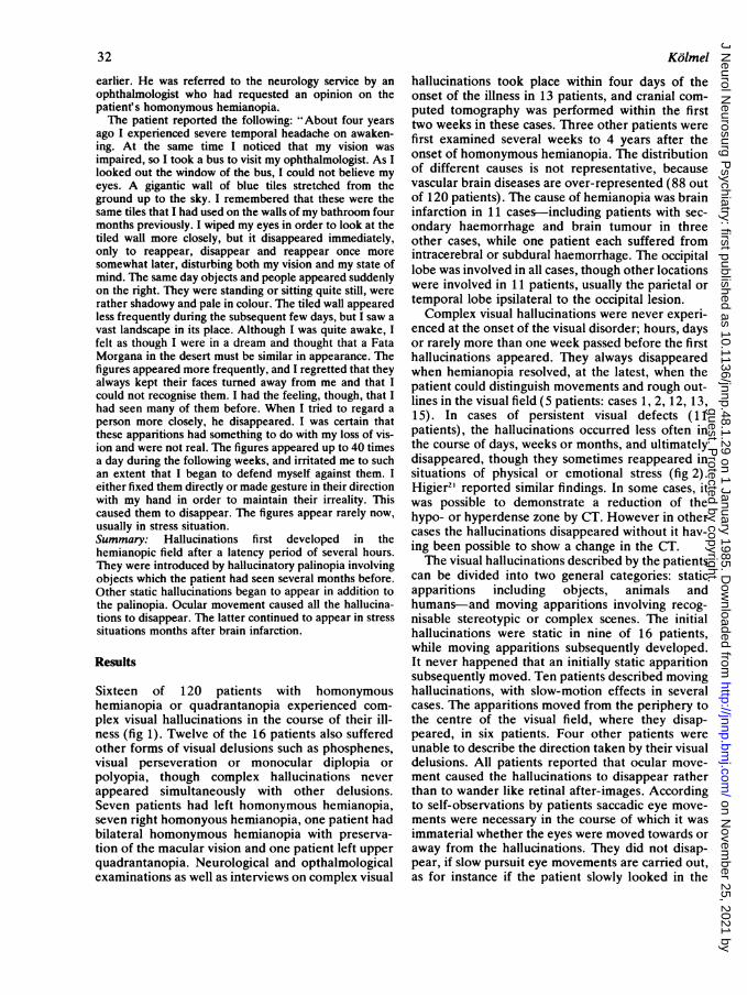
Sixteen of 120 patients with homonymoushemianopia or quadrantanopia experienced com-plex visual hallucinations in the course of their ill-ness (fig 1). Twelve of the 16 patients also sufferedother forms of visual delusions such as phosphenes,visual perseveration or monocular diplopia orpolyopia, though complex hallucinations neverappeared simultaneously with other delusions.Seven patients had left homonymous hemianopia,seven right homonyous hemianopia, one patient hadbilateral homonymous hemianopia with preserva-tion of the macular vision and one patient left upperquadrantanopia. Neurological and opthalmologicalexaminations as well as interviews on complex visual
Kolmel
hallucinations took place within four days of theonset of the illness in 13 patients, and cranial com-puted tomography was performed within the firsttwo weeks in these cases. Three other patients werefirst examined several weeks to 4 years after theonset of homonymous hemianopia. The distributionof different causes is not representative, becausevascular brain diseases are over-represented (88 outof 120 patients). The cause of hemianopia was braininfarction in 1 1 cases-including patients with sec-ondary haemorrhage and brain tumour in threeother cases, while one patient each suffered fromintracerebral or subdural haemorrhage. The occipitallobe was involved in all cases, though other locationswere involved in 11 patients, usually the parietal ortemporal lobe ipsilateral to the occipital lesion.Complex visual hallucinations were never experi-
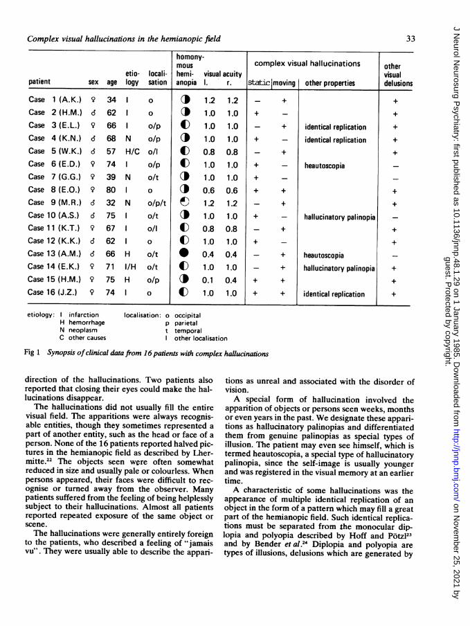
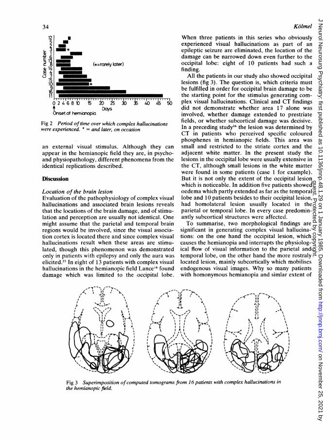
enced at the onset of the visual disorder; hours, daysor rarely more than one week passed before the firsthallucinations appeared. They always disappearedwhen hemianopia resolved, at the latest, when thepatient could distinguish movements and rough out-lines in the visual field (5 patients: cases 1, 2, 12, 13,15). In cases of persistent visual defects (11patients), the hallucinations occurred less often inthe course of days, weeks or months, and ultimatelydisappeared, though they sometimes reappeared insituations of physical or emotional stress (fig 2).Higier2' reported similar findings. In some cases, itwas possible to demonstrate a reduction of thehypo- or hyperdense zone by CT. However in othercases the hallucinations disappeared without it hav-ing been possible to show a change in the CT.The visual hallucinations described by the patients
can be divided into two general categories: staticapparitions including objects, animals andhumans-and moving apparitions involving recog-nisable stereotypic or complex scenes. The initialhallucinations were static in nine of 16 patients,while moving apparitions subsequently developed.It never happened that an initially static apparitionsubsequently moved. Ten patients described movinghallucinations, with slow-motion effects in severalcases. The apparitions moved from the periphery tothe centre of the visual field, where they disap-peared, in six patients. Four other patients wereunable to describe the direction taken by their visualdelusions. All patients reported that ocular move-ment caused the hallucinations to disappear ratherthan to wander like retinal after-images. Accordingto self-observations by patients saccadic eye move-ments were necessary in the course of which it wasimmaterial whether the eyes were moved towards oraway from the hallucinations. They did not disap-pear, if slow pursuit eye movements are carried out,as for instance if the patient slowly looked in the
guest. Protected by copyright.
on Novem
ber 25, 2021 byhttp://jnnp.bm
j.com/
J Neurol N
eurosurg Psychiatry: first published as 10.1136/jnnp.48.1.29 on 1 January 1985. D
ownloaded from
Complex visual hallucinations in the hemianopic field
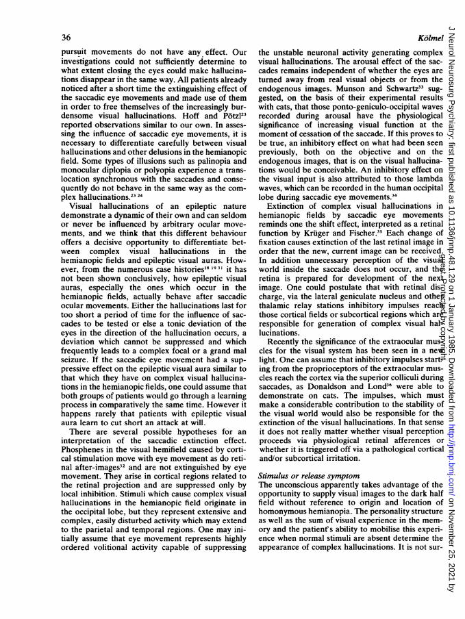
homony-mous complex visual hallucinations other
etio- locali- hemi- visual acuity visualpatient sex age logy sation anopia 1. r. static moving other properties delusions
Case 1 (A.K.) 9 34 1 o ci 1.2 1.2 - + +Case 2 (H.M.) d 62 1 o @ 1.0 1.0 + - +Case 3 (E.L.) 9 66 1 o/p E 1.0 1.0 - + identical replication +Case 4 (K.N.) d 68 N o/p I 1.0 1.0 + - identical replication +Case 5 (W.K.) d 57 H/C o/l C 0.8 0.8 - + +Case 6 (E.D.) 9 74 1 o/p E 1.0 1.0 + - heautoscopiaCase 7 (G.G.) 9 39 N o/t I 1.0 1.0 + -Case 8 (E.O.) 9 80 1 o 0.6 0.6 + + +Case 9 (M.R.) d 32 N o/p/t 1.2 1.2 - + +Case 10 (A.S.) d 75 1 o/t ) 1.0 1.0 + - hallucinatory palinopia -
Case 11 (K.T.) 9 67 1 o/l C 0.8 0.8 - + +Case 12 (K.K.) d 62 1 o C 1.0 1.0 + - +Case 13 (A.M.) d 66 H o/t * 0.4 0.4 - + heautoscopiaCase 14 (E.K.) 9 71 I/H o/t C 1.0 1.0 - + hallucinatory palinopia +Case 15 (H.M.) 9 75 H o/p (P 0.1 0.4 + + +Case 16 (J.Z.) 9 74 1 o C 1.0 1.0 + + identical replication +
etiology: infarctionH hemorrhageN neoplasmC other causes
localisation: o occipitalp parietalt temporal
other localisation
Fig 1 Synopsis ofclinical data from 16 patients with complex hallucinations
direction of the hallucinations. Two patients alsoreported that closing their eyes could make the hal-lucinations disappear.
The hallucinations did not usually fill the entirevisual field. The apparitions were always recognis-able entities, though they sometimes represented apart of another entity, such as the head or face of aperson. None of the 16 patients reported halved pic-tures in the hemianopic field as described by Lher-mitte.22 The objects seen were often somewhatreduced in size and usually pale or colourless. Whenpersons appeared, their faces were difficult to rec-ognise or turned away from the observer. Manypatients suffered from the feeling of being helplesslysubject to their hallucinations. Almost all patientsreported repeated exposure of the same object orscene.The hallucinations were generally entirely foreign
to the patients, who described a feeling of "jamaisvu". They were usually able to describe the appari-
tions as unreal and associated with the disorder ofvision.A special form of hallucination involved the
apparition of objects or persons seen weeks, monthsor even years in the past. We designate these appari-tions as hallucinatory palinopias and differentiatedthem from genuine palinopias as special types ofillusion. The patient may even see himself, which istermed heautoscopia, a special type of hallucinatorypalinopia, since the self-image is usually youngerand was registered in the visual memory at an earliertime.A characteristic of some hallucinations was the
appearance of multiple identical replication of anobject in the form of a pattern which may fill a greatpart of the hemianopic field. Such identical replica-tions must be separated from the monocular dip-lopia and polyopia described by Hoff and P6tzl23and by Bender et al.24 Diplopia and polyopia aretypes of illusions, delusions which are generated by
33
guest. Protected by copyright.
on Novem
ber 25, 2021 byhttp://jnnp.bm
j.com/
J Neurol N
eurosurg Psychiatry: first published as 10.1136/jnnp.48.1.29 on 1 January 1985. D
ownloaded from
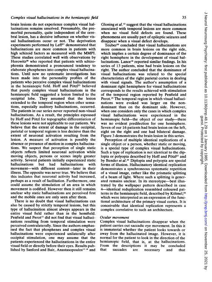
When three patients in this series who obviouslyexperienced visual hallucinations as part of anepileptic seizure are eliminated, the location of thedamage can be narrowed down even further to theoccipital lobe: eight of 10 patients had such afinding.
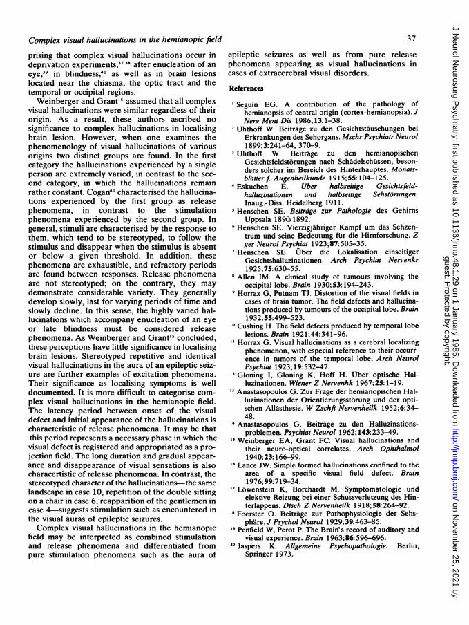
All the patients in our study also showed occipitallesions (fig 3). The question is, which criteria mustbe fulfilled in order for occipital brain damage to be
___________ the starting point for the stimulus generating com-3' '40,45 50 plex visual hallucinations. Clinical and CT findings
did not demonstrate whether area 17 alone wasinvolved, whether damage extended to prestriate
ex hallucinations fields, or whether subcortical damage was decisive.sionhalsatos In a preceding study26 the lesion was determined by
CT in patients who perceived specific colouredphosphenes in hemianopic fields. This area was
hough they can small and restricted to the striate cortex and they are, in psycho- adjacent white matter. In the present study thenomena from the lesions in the occipital lobe were usually extensive in
the CT, although small lesions in the white matterwere found in some patients (case 1 for example).But it is not only the extent of the occipital lesionwhich is noticeable. In addition five patients showedoedema which partly extended as far as the temporal
of complex visual lobe and 10 patients besides to their occipital lesion,n lesions reveals had homolateral lesion usually located in thege, and of stimu- parietal or temporal lobe. In every case predomin-ot identical. One antly subcortical structures were affected.d temporal brain To summarise, two morphological findings areie visual associa- significant in generating complex visual hallucina-:e complex visual tions: on the one hand the occipital lesion, whichareas are stimu- causes the hemianopia and interrupts the physiolog-as demonstrated ical flow of visual information to the parietal and)nly the aura was temporal lobe, on the other hand the more rostralyth complex visual located lesion, mainly subcortically which mobilisesId Lance'6 found endogenous visual images. Why so many patientse ,occipital lobe. with homonymous hemianopia and simlar extent of
125
i 14
16 _________11
024681
Onset of herrncnopta
=rarely later)
au 25Days
Fig 2 Period oftime over which complkwere experienced. * = and later, on occa
an external visual stimulus. Altiappear in the hemianopic field the:and physiopathology, different pheiidentical replications described.
Discussion
Location of the brain lesionEvaluation of the pathophysiologyhallucinations and associated braiithat the locations of the brain damalation and perception are usually nomight assume that the parietal anmregions would be involved, since tition cortex is located there and sinchallucinations result when these .lated, though this phenomenon wonly in patients with epilepsy and celicited.25 In eight of 13 patients withallucinations in the hemianopic fiedamage which was limited to th
,~_I , ,
'1'f',.r'
Fig 3 Superimposition ofcomputed tomograms from 16 patients with complex hallucinations inthe hemianopic field.
Kolmel34
guest. Protected by copyright.
on Novem
ber 25, 2021 byhttp://jnnp.bm
j.com/
J Neurol N
eurosurg Psychiatry: first published as 10.1136/jnnp.48.1.29 on 1 January 1985. D
ownloaded from
Complex visual hallucinations in the hemianopic fieldbrain lesions do not experience complex visual hal-lucinations remains unsolved. Presumably, the pre-morbid personality, quite independent of the cere-bral lesion, has a decisive influence on whether vis-ual hallucinations occur, or not. The deprivationexperiments performed by LeffZ' demonstrated thathallucinations are more common in patients withhigh schizoid factors as measured with the MMPI.These studies correlated well with observations byHorowitz28 who reported that patients with schizo-phrenia demonstrated a pronounced tendency toelaborate phosphenes into complex visual hallucina-tions. Until now no systematic investigations hasbeen made into the personality profiles of thepatients who perceive complex visual hallucinationsin the hemianopic field. Hoff and Potzl23 believedthat purely complex visual hallucinations in thehemianopic fi'eld suggested a lesion limited to theoccipital lobe. They assumed that the lesionextended to the temporal region when other sensa-tions, especially auditory hallucinations, occurred.The patients in our series never reported non-visual-hallucinations. As a result, the principles espousedby Hoff and Potzl for topographic differentiation ofthese lesions were not applicable to our patients. Webelieve that the extent pf damage in the occipital,parietal or temporal regions is less decisive than theextent of neuronal activation resulting from thelesion. A measure of activation is possibly theabsence or presence of motion in complex hallucina-tions. We suspect that perception of single staticobjects reflects limited neuronal activation whilemoving objects, persons or scenes imply greateractivity. Several patients initially experienced statichallucinations but had hallucinations withmovement-with different content-later in theirillness. The opposite was never true. We believe thatthis indicates that neuronal activity had increased,perhaps as a result of facilitation. Furthermore, onecould assume the stimulation of an area in whichmovement is codified. However then it still remainsunclear why static hallucinations are perceived firstand the mobile ones are only seen after them.There is no doubt that visual hallucinations can
also be caused by strictly temporal lesions, but thistype of hallucination almost always appears in theentire visual field rather than in the hemifield.Penfield and Perot'9 did not find that visual halluci-nations resulting from temporal stimulation wereperceived contralaterally. Since the authors emphas-ised the fact that phosphenes and complex visualhallucinations were experienced unilaterally afteroccipital stimulation, one may assume that thepatients experienced the hallucinations in the entirevisual field or directly before their eyes. Results pub-lished by Sanford and Bair, Anastasopoulos'3 14 and
35
Gloning et al.'2 suggest that the visual hallucinationsassociated with temporal lesions are more commonwhen no visual field defects are found. Thesephenomena are usually part of epileptic seizures anddisappear when a visual defect develops.
Teuber30 concluded that visual hallucinations aremore common in brain lesions on the right side,which implies a certain degree of dominance of theright hemisphere in the development of visual hal-lucinations. Lance'6 reported similar findings. In hisseries of 13 patients, nine had brain lesions on theright. The author concluded that the dominance ofvisual hallucinations was related to the specialcharacteristics of the right parietal cortex in dealingwith visual signals. The author's assumption of adominant right hemisphere for visual hallucinationscorresponds to the results achieved with stimulationof the temporal region reported by Penfield andPerot.'9 The temporal region in which visual halluci-nations were evoked was larger on the non-dominant than on the dominant side. However,when one considers only the cases in which complexvisual hallucinations were experienced in thehemianopic field-the object of our study-therewas no evident predilection for one hemisphere.Seven of our 16 patients had brain lesion on the left,eight on the right and one had bilateral damage.Figure 3 demonstrates the brain lesion in this series.
Perception of multiple identical replications of asingle object or a person, whether static or moving,is a special type of complex visual hallucinations.Such a type of replication is not identical to the dip-lopia or polyopia described by Hoff and Potzl23 andby Bender et al.24 Diplopia and polyopia are specialforms of illusion. Hallucinatory identical replicationdemonstrates a synchroneous systematic repetitionof a visual image, rather like the prismatic splittingof a beam of light. Where such a splitting is gener-ated remains unclear. In its stereotype-best illus-trated by the wallpaper pattern described in case4-identical reduplication- resembled coloured pat-terns in the hemianopic field, described by Kolmel,26which were interpreted as an expression of the func-tional architecture of the primary visual cortex. It isconceivable that identical replication represents acomplex correlative to such an architecture.
Ocular movementComplex visual hallucinations disappear when thepatient carries out saccadic eye movement. In this itis immaterial whether the patient looks towards oraway from the hallucinated image. However, it isnormal for the patient to look in the direction of thehemianopic field, that is, at the hallucinations.From the descriptions it may be concludedthat saccades are necessary and that
guest. Protected by copyright.
on Novem
ber 25, 2021 byhttp://jnnp.bm
j.com/
J Neurol N
eurosurg Psychiatry: first published as 10.1136/jnnp.48.1.29 on 1 January 1985. D
ownloaded from
36
pursuit movements do not have any effect. Ourinvestigations could not sufficiently determine towhat extent closing the eyes could make hallucina-tions disappear in the same way. All patients alreadynoticed after a short time the extinguishing effect ofthe saccadic eye movements and made use of themin order to free themselves of the increasingly bur-densome visual hallucinations. Hoff and Potzl23reported observations similar to our own. In asses-sing the influence of saccadic eye movements, it isnecessary to differentiate carefully between visualhallucinations and other delusions in the hemianopicfield. Some types of illusions such as palinopia andmonocular diplopia or polyopia experience a trans-location synchronous with the saccades and conse-quently do not behave in the same way as the com-plex hallucinations.23 24
Visual hallucinations of an epileptic naturedemonstrate a dynamic of their own and can seldomor never be influenced by arbitrary ocular move-ments, and we think that this different behaviouroffers a decisive opportunity to differentiate bet-ween complex visual hallucinations in thehemianopic fields and epileptic visual auras. How-ever, from the numerous case histories'8 1931 it hasnot been shown conclusively, how epileptic visualauras, especially the ones which occur in thehemianopic fields, actually behave after saccadicocular movements. Either the hallucinations last fortoo short a period of time for the influence of sac-cades to be tested or else a tonic deviation of theeyes in the direction of the hallucination occurs, adeviation which cannot be suppressed and whichfrequently leads to a complex focal or a grand malseizure. If the saccadic eye movement had a sup-pressive effect on the epileptic visual aura similar tothat which they have on complex visual hallucina-tions in the hemianopic fields, one could assume thatboth groups of patients would go through a learningprocess in comparatively the same time. However ithappens rarely that patients with epileptic visualaura learn to cut short an attack at will.There are several possible hypotheses for an
interpretation of the saccadic extinction effect.Phosphenes in the visual hemifield caused by corti-cal stimulation move with eye movement as do reti-nal after-images32 and are not extinguished by eyemovement. They arise in cortical regions related tothe retinal projection and are suppressed only bylocal inhibition. Stimuli which cause complex visualhallucinations in the hemianopic field originate inthe occipital lobe, but they represent extensive andcomplex, easily disturbed activity which may extendto the parietal and temporal regions. One may ini-tially assume that eye movement represents highlyordered volitional activity capable of suppressing
Kolmel
the unstable neuronal activity generating complexvisual hallucinations. The arousal effect of the sac-cades remains independent of whether the eyes areturned away from real visual objects or from theendogenous images. Munson and Schwartz33 sug-gested, on the basis of their experimental resultswith cats, that those ponto-geniculo-occipital wavesrecorded during arousal have the physiologicalsignificance of increasing visual function at themoment of cessation of the saccade. If this proves tobe true, an inhibitory effect on what had been seenpreviously, both on the objective and on theendogenous images, that is on the visual hallucina-tions would be conceivable. An inhibitory effect onthe visual input is also attributed to those lambdawaves, which can be recorded in the human occipitallobe during saccadic eye movements.34
Extinction of complex visual hallucinations inhemianopic fields by saccadic eye movementsreminds one the shift effect, interpreted as a retinalfunction by Kruger and Fischer.35 Each change offixation causes extinction of the last retinal image inorder that the new, current image can be received.In addition unnecessary perception of the visualworld inside the saccade does not occur, and theretina is prepared for development of the nextimage. One could postulate that with retinal dis-charge, via the lateral geniculate nucleus and otherthalamic relay stations inhibitory impulses reachthose cortical fields or subcortical regions which areresponsible for generation of complex visual hal-lucinations.
Recently the significance of the extraocular mus-cles for the visual system has been seen in a newlight. One can assume that inhibitory impulses start-ing from the proprioceptors of the extraocular mus-cles reach the cortex via the superior colliculi duringsaccades, as Donaldson and Lond36 were able todemonstrate on cats. The impulses, which mustmake a considerable contribution to the stability ofthe visual world would also be responsible for theextinction of the visual hallucinations. In that senseit does not really matter whether visual perceptionproceeds via physiological retinal afferences orwhether it is triggered off via a pathological corticaland/or subcortical irritation.
Stimulus or release symptomThe unconscious apparently takes advantage of theopportunity to supply visual images to the dark halffield without reference to origin and location ofhomonymous hemianopia. The personality structureas well as the sum of visual experience in the mem-ory and the patient's ability to mobilise this experi-ence when normal stimuli are absent determine theappearance of complex hallucinations. It is not sur-
guest. Protected by copyright.
on Novem
ber 25, 2021 byhttp://jnnp.bm
j.com/
J Neurol N
eurosurg Psychiatry: first published as 10.1136/jnnp.48.1.29 on 1 January 1985. D
ownloaded from
Complex visual hallucinations in the hemianopic fieldprising that complex visual hallucinations occur indeprivation experiments,37 38 after enucleation of aneye,39 in blindness,40 as well as in brain lesionslocated near the chiasma, the optic tract and thetemporal or occipital regions.Weinberger and Grant'5 assumed that all complex
visual hallucinations were similar regardless of theirorigin. As a result, these authors ascribed nosignificance to complex hallucinations in localisingbrain lesion. However, when one examines thephenomenology of visual hallucinations of variousorigins two distinct groups are found. In the firstcategory the hallucinations experienced by a singleperson are extremely varied, in contrast to the sec-ond category, in which the hallucinations remainrather constant. Cogan4I characterised the hallucina-tions experienced by the first group as releasephenomena, in contrast to the stimulationphenomena experienced by the second group. Ingeneral, stimuli are characterised by the response tothem, which tend to be stereotyped, to follow thestimulus and disappear when the stimulus is absentor below a given threshold. In addition, thesephenomena are exhaustible, and refractory periodsare found between responses. Release phenomenaare not stereotyped; on the contrary, they maydemonstrate considerable variety. They generallydevelop slowly, last for varying periods of time andslowly decline. In this sense, the highly varied hal-lucinations which accompany enucleation of an eyeor late blindness must be considered releasephenomena. As Weinberger and Grant'5 concluded,these perceptions have little significance in localisingbrain lesions. Stereotyped repetitive and identicalvisual hallucinations in the aura of an epileptic seiz-ure are further examples of excitation phenomena.Their significance as localising symptoms is welldocumented. It is more difficult to categorise com-plex visual hallucinations in the hemianopic field.The latency period between onset of the visualdefect and initial appearance of the hallucinations ischaracteristic of release phenomena. It may be thatthis period represents a necessary phase in which thevisual defect is registered and appropriated as a pro-jection field. The long duration and gradual appear-ance and disappearance of visual sensations is alsocharacertistic of release phenomena. In contrast, thestereotyped character of the hallucinations-the samelandscape in case 10, repetition of the double sittingon a chair in case 6, reapparition of the gentlemen incase 4-suggests stimulation such as encountered inthe visual auras of epileptic seizures.Complex visual hallucinations in the hemianopic
field may be interpreted as combined stimulationand release phenomena and differentiated frompure stimulation phenomena such as the aura of
37
epileptic seizures as well as from pure releasephenomena appearing as visual hallucinations incases of extracerebral visual disorders.
References
'Seguin EG. A contribution of the pathology ofhemianopsis of central origin (cortex-hemianopsia). JNerv Ment Dis 1986;13:1-38.
2 Uhthoff W. Beitrage zu den Gesichtstauschungen beiErkrankungen des Sehorgans. Mschr Psychiatr Neurol1899;3:241-64, 370-9.
3Uhthoff W. Beitrage zu den hemianopischenGesichtsfeldstorungen nach Schadelschussen, beson-ders solcher im Bereich des Hinterhauptes. Monats-blatter f Augenheilkunde 1915;55: 104-125.
Eskuchen E. Uber halbseitige Gesichtsfeld-halluzinationen und halbseitige Sehstorungen.Inaug.-Diss. Heidelberg 1911.
5 Henschen SE. Beitrage zur Pathologie des GehirnsUppsala 1890/1892.
6 Henschen SE. Vierzigjahriger Kampf um das Sehzen-trum und seine Bedeutung fur die Hirnforschung. Zges Neurol Psychiat 1923;87:505-35.
7Henschen SE. Uber die Lokalisation einseitigerGesichtshalluzinationen. Arch Psychiat Nervenkr1925;75:630-55.
8 Allen IM. A clinical study of tumours involving theoccipital lobe. Brain 1930;53: 194-243.
Horrax G, Putnam TJ. Distortion of the visual fields incases of brain tumor. The field defects and hallucina-tions produced by tumours of the occipital lobe. Brain1932;55:499-523.
'0 Cushing H. The field defects produced by temporal lobelesions. Brain 1921;44:341-96.
"Horrax G. Visual hallucinations as a cerebral localizingphenomenon, with especial reference to their occurr-ence in tumors of the temporal lobe. Arch NeurolPsychiat 1923; 19:532-47.
12 Gloning I, Gloning K, Hoff H. Uber optische Hal-luzinationen. Wiener Z Nervenhk 1967;25: 1-19.
13 Anastasopoulos G. Zur Frage der hemianopischen Hal-luzinationen der Orientierungsstorung und der opti-schen AllWsthesie. W Zschft Nervenheilk 1952;6:34-48.
Anastasopoulos G. Beitrage zu den Halluzinations-problemen. Psychiat Neurol 1962; 143:233-49.
'5 Weinberger EA, Grant FC. Visual hallucinations andtheir neuro-optical correlates. Arch Ophthalmol1940; 23:166-99.
16 Lance JW. Simple formed hallucinations confined to thearea of a specific visual field defect. Brain1976;99:719-34.
L6wenstein K, Borchardt M. Symptomatologie undelektive Reizung bei einer Schussverletzung des Hin-terlappens. Dtsch Z Nervenheilk 1918;58: 264-92.
8 Foerster 0. Beitrage zur Pathophysiologie der Sehs-phare. J Psychol Neurol 1929;39:463-85.
'9 Penfield W, Perot P. The Brain's record of auditory andvisual experience. Brain 1963;86: 596-696.
20 Jaspers K. Ailgemeine Psychopathologie. Berlin,Springer 1973.
guest. Protected by copyright.
on Novem
ber 25, 2021 byhttp://jnnp.bm
j.com/
J Neurol N
eurosurg Psychiatry: first published as 10.1136/jnnp.48.1.29 on 1 January 1985. D
ownloaded from

38
21 Higier H. Uber unilaterale Halluzinationen. Wien Klinik1894;20: 139-70.
22 Lhermitte J. Les Hallucinations Clinique et Physio-pathologie. Paris, Doin 1951.
23 Hoff H, Plotzl 0. Uber Polyopie und gerichtetehemianopische Halluzinationen. Jahrbucher fiirPsychiatrie 1937;54:55-88.
24 Bender MB, Feldman M, Sobin AJ. Palinopsia. Brain1968;91:321-38.
25 Penfield W, Rasmussen T. The Cerebral Cortex ofMan.New York Macmillan 1952:135-47, 165-6.
26 Kolmel HW. Coloured patterns in hemianopic fields.Brain 1984; 107:155-67.
27 Leff JP. Perceptual phenomenia and personality in sen-sory deprivation. Br J Psychiat 1968; 114: 1499-508.
28 Horowitz MJ. The imagery of visual hallucinations. JNerv Ment Dis 1964; 138:513-23.
29 Sanford HS, Bair HL. Visual disturbances associatedwith tumors of the temporal lobe. Arch NeurolPsychiat 1939;42: 21-43.
30 Teuber HL. Effect of brain wounds implications right orleft hemisphere in Man. In: Mountcastle VB, ed.Interhemispheric Relations and Cerebral Dominance.Baltimore, John Hopkins Press, 1961.
31 Russel WR, Whitty CWM. Studies in traumatic epilepsy,III. Visual fits. J Neurol Neurosurg Psychiat1955; 18:79-96.
32 Brindley GS, Lewin W. The sensations produced byelectrical stimulation of the visual cortex. J Physiol(Lond) 1968; 196: 479-83.
33 Munson JB, Schwartz KS. Lateral geniculate and occipi-tal cortex spikes with eye movements in awake and
Kolmel
sleeping cats: temporal and functional correlations.Exp Neurol 1972;35:300-4.
34 Kurtzberg D, Vaughan HG. Electrophysilogical obser-vations on the visumotor system and visual neurosen-sorium. In: Desmedt JE, ed, Visual Evoked Potentialsin Man: New Developments. Oxford, Clarendon Press,1977:314-31
Kruger J, Fischer B. Strong periphery effect in cat retinalganglion cells. Exitatory responses in on- and off-center- neurones to single grid displacements. ExpBrain Res 1973; 18:316-8
36 Donaldson IML, Long AC. Interactions between extra-ocular proprioceptive and visual signals in the superiorcolliculus of the cat. J Physiol (Lond) 1980;298: 85-110.
37 Kempe P, Reimer CH. Halluzinatorische Phanomenebei Reizentzug. Nervenarzt 1976;47: 701-7.
38 Zuckermann M. Hallucinations, reported sensations andimages. In: Zubeck JP, ed. Sensory Deptvation: Fif-teen Years of Research. New York, Appleton-Centry-Crofts. 1969:85-125.
39 Lauber HL, Lewin B. A clinical and psychological studyof optic hallucinations in the elimination of vision.Arch Psychiat Nervenkr 1958; 197:15-31.
40 Jacob H. Der Erlebniswandel bei Spaterblindeten. ZurPsychopathologie der optischen Wahrnehmung.Abhandlung zur Psychiatrie, Psychologie,Psychopathologie und Grenzgebieten Bd. 1. Hamburg,Nolke Verlag. 1949.
4' Cogan DG. Visual hallucinations as release phenomena.Graefes Arch Klin Opthalmol 1973; 188:139-50.
guest. Protected by copyright.
on Novem
ber 25, 2021 byhttp://jnnp.bm
j.com/
J Neurol N
eurosurg Psychiatry: first published as 10.1136/jnnp.48.1.29 on 1 January 1985. D
ownloaded from