condroitina review acido solfato dispositivo medico per il ... acta_4_16.pdf · m. pedrazzoli, l....
TRANSCRIPT

w
Official Journal of the Italian Society of Otorhinolaryngology Head and Neck Surgery
Organo Ufficiale della Società Italiana di Otorinolaringologia e Chirurgia Cervico-Facciale 4
Otorhinolaryngologica Italica
August 2016
Volume 36
ReviewHuman papillomavirus and head and neck carcinomas:
focus on evidence in the babel of published data
Head and neckParapharyngeal space tumours: video-assisted minimally invasive
transcervical approach
Surgical complications in orbital decompression for Graves’ orbitopathy
OtologyThe audiological and take results of perichondrium attached cartilage island
graft in tympanoplasty: PACIT
Role of Kabat rehabilitation in facial nerve palsy: a randomised study on severe cases of Bell’s palsy
Laryngology Malignant salivary gland tumours of the larynx: a single institution review
Evaluation of pharyngeal muscle activity through nasopharyngeal surface electromyography in a cohort of dysphagic patients with acute ischaemic stroke
RhinologyMonolateral sinonasal complications of dental disease or treatment:
when does endoscopic endonasal surgery require an intraoral approach?
AudiologyPossibility of differentiation of cochlear electrodes in radiological measurements
of the intracochlear and chorda-facial angle position
Clinical techniques and technologyPrevention of bisphosphonate-related mandibular fractures
Vastus lateralis myofascial free flap in tongue reconstruction
Letter to the Editor The relevance of counseling in patients with nasal polyps
Case series and reportsEffects of simultaneous palatal expansion and mandibular
advancement in a child suffering from OSA
Arterio-venous malformation of the mandible. Case report and review of literaturePO
STE
ITALI
ANE
SPA
- Spe
dizio
ne in
Abb
onam
ento
Pos
tale
- D.
L. 3
53/2
003
conv
. in
L. 2
7/02
/200
4 n°
46
art.
1, c
omm
a 1,
DCB
PIS
A - I
scriz
ione
al t
ribun
ale
di P
isa a
l n. 1
0 de
l 30-
07-9
3 - F
inito
di s
tam
pare
pre
sso
IGP,
Pisa
- lu
glio
201
6 - -
Con
tiene
Sup
plem
ento
- Co
ntin
e I.E
.. IS
SN: 0
392-
100X
(Prin
t) - I
SSN:
182
7-67
5X (O
nlin
e)
Acta O
torhinolaryngologica Italica, XX
XV
I/4, 249-343, 2016Dispositivo medico per il trattamentoDispositivo medico per il trattamento Acido ialuronico
Condroitina solfato
Poloxamer 407
una difesa originale e innovativa
Dispositivo medico per il trattamentodel re�usso gastro-esofageo
Dispositivo medico per il trattamento del re�usso gastro-esofageo
Alfa Wassermann fa parte del Gruppo Alfasigma

Official Journal of the Italian Society of Otorhinolaryngology - Head and Neck SurgeryOrgano Ufficiale della Società Italiana di Otorinolaringologia e Chirurgia Cervico-Facciale
Otorhinolaryngologica Italica
Editorial BoardEditor-in-Chief: G. Paludetti President of S.I.O.: C.A. LeoneFormer Presidents of S.I.O. and Editors-in-Chief: I. De Vincentiis, D. Felisati, L. Coppo, G. Zaoli, P. Miani, G. Motta, L. Marcucci, A. Ottaviani, P. Puxeddu, M. Maurizi, G. Sperati, D. Passali, E. de Campora, A. Sartoris, P. Laudadio, M. De Benedetto, S. Conticello, D. Casolino, A. Rinaldi Ceroni, M. Piemonte, A. Staffieri, F. Chiesa, R. Fiorella, A. Camaioni, A. Serra, G. Spriano, R. Filipo
Editorial StaffEditor-in-Chief: G. Paludetti Deputy Editor: J. Galli Associate Editors: G. Almadori, F. OttavianiEditorial Coordinator: E. De CorsoEditorial Assistant: P. Moore Treasurer:L. de Campora
Italian Scientific BoardL. Bellussi, G. Danesi, C. Grandi, A. Martini, L. Pignataro, F. Raso, R. Speciale, I. Tasca
International Scientific BoardJ. Betka, P. Clement, M. Halmagyi, L.P. Kowalski, M. Pais Clemente, J. Shah, H. Stammberger, R. Laszig, G. O’Donoghue, R.J. Salvi, R. Leemans, M. Remacle, F. Marshal, H.P. Zenner, B. Scola Yurrita, R.W. Gilbert
Editorial OfficeEditor-in-Chief: G. PaludettiDepartment of Head and Neck Surgery - OtorhinolaryngologyCatholic University of the Sacred Heart “A. Gemelli” Hospital L.go F. Vito, 1 - 00168 Rome, Italy Tel. +39 06 30154439 Fax + 39 06 3051194 [email protected]
Editorial Coordinator:E. De [email protected]
Argomenti di Acta Otorhinolaryngologica ItalicaEditor-in-Chief: G. Paludetti Editorial Coordinator: M.R. [email protected]
© Copyright 2016 bySocietà Italiana di Otorinolaringologia e Chirurgia Cervico-FaccialeVia Luigi Pigorini, 6/300162 Rome, Italy
PublisherPacini Editore SrlVia Gherardesca, 156121 Pisa, ItalyTel. +39 050 313011Fax +39 050 [email protected]
Acta Otorhinolaryngologica Italica is cited in Index Medicus, MEDLINE, PubMed Central, Science Citation Index Expanded, Scopus, DOAJ, Open-J Gate, Free Medical Journals, Index Copernicus, Socolar
Journal Citation Reports: Impact Factor 1.531Acta Otorhinolaryngologica Italica is available on Google Scholar
Volume 36 – Number 4 – August 2016
Former Editors-in-Chief: C. Calearo†, E. de Campora, A. Staffieri, M. Piemonte, F. Chiesa

ReviewHuman papillomavirus and head and neck carcinomas: focus on evidence in the babel of published dataPapillomavirus umano e carcinomi del tratto aerodigestivo: il punto sulle evidenze nella babele dei dati scientificiP. Morbini, M. Benazzo . . . . . . . . . . . . . . . . . . . . . . . . . . . . . . . . . . . . . . . . . . . . . . . . . . . . . . . . . . . . . . . . . . . . . . . . . pag. 249Head and neckParapharyngeal space tumours: video-assisted minimally invasive transcervical approachTumori dello spazio parafaringeo: approccio transcervicale video-assistito mini invasivoF. Pilolli, L. Giordano, A. Galli, M. Bussi . . . . . . . . . . . . . . . . . . . . . . . . . . . . . . . . . . . . . . . . . . . . . . . . . . . . . . . . . . . » 259Surgical complications in orbital decompression for Graves’ orbitopathyComplicanze chirurgiche in pazienti sottoposti a decompressione orbitaria per oftalmopatia di GravesS. Sellari-Franceschini, I. Dallan, A. Bajraktari, G. Fiacchini, M. Nardi, R. Rocchi, C. Marcocci, M. Marinò, A.P. Casani . . . . . . . . . . . . . . . . . . . . . . . . . . . . . . . . . . . . . . . . . . . . . . . . . . . . . . . . . . . . . . . . . . . . . . . . . . . . . . . . . . . » 265OtologyThe audiological and take results of perichondrium attached cartilage island graft in tympanoplasty: PACIT Risultati audiologici e rate di attecchimento dell’innesto di cartilagine con pericondrio nella timpanoplastica: PACITF. Solmaz, D. Akduman, M. Haksever, E. Gündoğdu, M. Yanılmaz, A. Mescioğlu . . . . . . . . . . . . . . . . . . . . . . . . . . . . » 275Role of Kabat rehabilitation in facial nerve palsy: a randomised study on severe cases of Bell’s palsy Ruolo della riabilitazione Kabat nella paralisi del nervo facciale: studio randomizzato su casi severi di paralisi di BellS. Monini, C.M. Iacolucci, M. Di Traglia, A.I. Lazzarino, M. Barbara . . . . . . . . . . . . . . . . . . . . . . . . . . . . . . . . . . . . . » 282LaryngologyMalignant salivary gland tumours of the larynx: a single institution review Tumori maligni delle ghiandole salivari della laringe: un’unica review istituzionaleS. Karatayli-Ozgursoy, J.A. Bishop, A.T. Hillel, L.M. Akst, S.R. Best . . . . . . . . . . . . . . . . . . . . . . . . . . . . . . . . . . . . . » 289Evaluation of pharyngeal muscle activity through nasopharyngeal surface electromyography in a cohort of dysphagic patients with acute ischaemic stroke Valutazione dell’attività muscolare faringea attraverso elettromiografia di superficie nasofaringea in pazienti disfagici affetti da ictus ischemico acutoN.M. Giannantoni, M. Minisci, V. Brunetti, E. Scarano, E. Testani, C. Vollono, E. De Corso, G. Bastanza, L. D’Alatri, G. Della Marca . . . . . . . . . . . . . . . . . . . . . . . . . . . . . . . . . . . . . . . . . . . . . . . . . . . . . . . . . . . . . . . . . . . . . . . . . . . . . . . . » 295RhinologyMonolateral sinonasal complications of dental disease or treatment: when does endoscopic endonasal surgery require an intraoral approach?Complicanze sinusali monolaterali da patologia o trattamenti dentali: quando la chirurgia endoscopica endonasale necessita un approccio intraorale?G.L. Fadda, M. Berrone, E. Crosetti, G. Succo . . . . . . . . . . . . . . . . . . . . . . . . . . . . . . . . . . . . . . . . . . . . . . . . . . . . . . . » 300AudiologyPossibility of differentiation of cochlear electrodes in radiological measurements of the intracochlear and chorda-facial angle positionPossibilità di differenziazione degli elettrodi cocleari nelle misurazioni radiologiche della posizione intracocleare e dell’angolo cordo-faccialeI. Diogo, U. Walliczeck, J. Taube, N. Franke, A. Teymoortash, J. Werner, C. Güldner . . . . . . . . . . . . . . . . . . . . . . . . . » 310Clinical techniques and technologyPrevention of bisphosphonate-related mandibular fracturesPrevenzione delle fratture mandibolari conseguenti alla necrosi ossea da difosfonatiM. Pedrazzoli, L. Autelitano, F. Biglioli . . . . . . . . . . . . . . . . . . . . . . . . . . . . . . . . . . . . . . . . . . . . . . . . . . . . . . . . . . . . » 317Vastus lateralis myofascial free flap in tongue reconstruction Lembo miofasciale di vasto laterale nella ricostruzione della linguaR. Pellini, A. De Virgilio, G. Mercante, B. Pichi, V. Manciocco, P. Marchesi, F. Ferreli, G. Spriano . . . . . . . . . . . . . . » 321Letter to the Editor The relevance of counseling in patients with nasal polyps L’importanza del counseling nei pazienti affetti da poliposi nasaleM. Gelardi, N. De Candia, N. Quaranta, C. Russo, P. Pecoraro, M. Mancini, P. Luperto, G. Lombardo, A. Macchi, C. Bocciolini, A. Ciofalo, E. De Corso, G. Ciprandi . . . . . . . . . . . . . . . . . . . . . . . . . . . . . . . . . . . . . . . . . . . . . . . . . . . » 326Case series and reportsEffects of simultaneous palatal expansion and mandibular advancement in a child suffering from OSAEffetti di simultanei espansione palatale e avanzamento mandibolare in un paziente pediatrico con apnee ostruttive notturneA. Galeotti, P. Festa, M. Pavone, G.C. De Vincentiis . . . . . . . . . . . . . . . . . . . . . . . . . . . . . . . . . . . . . . . . . . . . . . . . . . . » 328Arterio-venous malformation of the mandible. Case report and review of literatureMalformazione arterovenosa della mandibola. Caso clinico e revisione della letteraturaR. Spreafico, L. Sordo, R. Bellotto, M. Schipano, A. Rescaldani, F. Parmigiani . . . . . . . . . . . . . . . . . . . . . . . . . . . . . . » 333
In Memoriam of Prof. Pietro Ferrara . . . . . . . . . . . . . . . . . . . . . . . . . . . . . . . . . . . . . . . . . . . . . . . . . . . . . . . . . . » 337Notiziario SIO . . . . . . . . . . . . . . . . . . . . . . . . . . . . . . . . . . . . . . . . . . . . . . . . . . . . . . . . . . . . . . . . . . . . . . . . . . . . . . . . » 338
Calendar of events - Italian and International Meetings and Courses . . . . . . . . . . . . . . . . . . . . . . . . . . . . . » 341
Information for authors including editorial standards for the preparation of manuscripts available on-line: www.actaitalica.it

249
ACTA oTorhinolAryngologiCA iTAliCA 2016;36:249-258; doi: 10.14639/0392-100X-853
Review
Human papillomavirus and head and neck carcinomas: focus on evidence in the babel of published dataPapillomavirus umano e carcinomi del tratto aerodigestivo: il punto sulle evidenze nella babele dei dati scientifici
P. Morbini1, M. benazzo2
1 Department of Molecular Medicine, Unit of Pathology, and 2 Department of otolaryngology, University of Pavia and Foundation irCCS Policlinico S. Matteo, Pavia, italy
SUMMARY
Human papillomavirus (HPV)-associated squamous cell carcinoma of the oropharynx is a well-defined entity mostly affecting young to middle-aged male non-smokers. It is generally associated with a favourable outcome, and for this reason a less intensive therapeutic ap-proach has been proposed for this subset of patients. The incidence of HPV-associated oropharyngeal cancers is rapidly increasing in most Western countries, but detailed epidemiological data are not available for the Italian population. Furthermore, among other head and neck regions, a smaller proportion of oral high-grade dysplasia and cancers seems to depend on HPV infection, whereas its role in laryngeal cancer is recognised as less relevant. HPV-dependent neoplastic transformation depends on the expression of viral oncogenes in the infected host cell that can only be directly documented through viral oncogene mRNA identification. The consensus on how to classify these patients from clinical and laboratory diagnostic points of view is still limited, with different approaches based on one or more diagnostic techniques including p16 immunostaining, in situ hybridisation and polymerase chain reation (PCR) amplification of viral DNA. The possibility of early diagnosis relying on the identification of HPV infection in oral and oropharyngeal exfoliated cells has so far provided unsatisfactory results, although viral persistence after treatment has been associated with risk of recurrence. Presently, sufficient data are not available to document the natural history and progression from tonsillar HPV infection to oropharyngeal cancer development, and to clearly define the modality of transmission and risk exposure, among which sexual behaviours appear to play a relevant role. The diffusion of HPV vaccina-tion and its administration to both genders will undoubtedly dramatically modify the epidemiology of HPV-related head and neck cancers in the coming years.
KEY WORDS: HPV • Oropharyngeal cancer • Oral cancer • Diagnosis
RIASSUNTO
I carcinomi squamosi dell’orofaringe associati all’infezione da papillomavirus umano (HPV) costituiscono ormai una entità ben carat-terizzata, che interessa prevalentemente maschi, giovani adulti o di mezza età, non fumatori. Essi hanno generalmente una prognosi più favorevole rispetto alla controparte non associate ad infezione, e per questo è stato proposto di dedicare a questo gruppo di pazienti un approccio terapeutico meno aggressivo. L’incidenza dei carcinomi dell’orofaringe associati a HPV è in rapido aumento nella maggior parte dei paesi occidentali, ma per quanto riguarda la popolazione italiana non sono disponibili dati epidemiologici in merito. Per quanto riguarda le altre regioni del distretto testa-collo, una più modesta porzione di lesioni displastiche di alto grado e di neoplasie appare essere correlata all’infezione da HPV, mentre il ruolo del virus nei tumori della laringe è stato parzialmente ridimensionato. HPV determina la trasformazione neoplastica delle cellule infettate tramite l’espressione dei suoi due oncogeni, E6 ed E7, che interagiscono con i meccani-smi di apoptosi e regolazione del ciclo cellulare della cellula ospite. L’unica metodica in grado di documentare con certezza l’espressione degli oncogeni virali è attualmente l’amplificazione dell’RNA messaggero trascritto dai due oncogeni. Il consenso riguardo la strategia per l’identificazione dei pazienti affetti da carcinoma dell’orofaringe associato a HPV dal punto di vista clinico e diagnostico è tuttora limitato. Le metodiche diagnostiche più utilizzate, singolarmente o in combinazione, comprendono l’immunocolorazione con anticorpi diretti contro p16, l’ibridazione in situ per genotipi virali ad alto rischio e l’amplificazione del DNA virale mediate PCR. La possibilità di ottenere una diagnosi precoce grazie all’identificazione dell’infezione virale nelle cellule epiteliali esfoliate dal cavo orale o dall’orofaringe non ha finora fornito risultati soddisfacenti, tuttavia la persistenza del virus nel cavo orale in pazienti trattati per carcinoma dell’orofaringe ha dimostrato una significativa associazione con il rischio di recidiva del tumore. Non sono ancora disponibili sufficienti dati che documen-tino in maniera dettagliata la storia naturale dell’infezione a la sua progressione verso lo sviluppo di una neoplasia, e che definiscano con chiarezza le modalità di trasmissione e i fattori di rischio, comunque è chiaro che i comportamenti sessuali hanno un peso rilevante nel determinare il rischio di sviluppo di neoplasia dell’orofaringe HPV-correlata. La progressive diffusione nelle giovani generazioni del vaccino contro HPV, e soprattutto la sua estensione agli adolescenti di entrambi i generi è sicuramente destinata a modificare in maniera rilevante anche l’epidemiologica dei tumori HPV-correlati nel distretto testa-collo nel prossimo futuro.
PAROLE CHIAVE: HPV • Carcinoma dell’orofaringe • Carcinoma orale • Diagnosi
Acta Otorhinolaryngol Ital 2016;36:249-258

P. Morbini, M. Benazzo
250
IntroductionHead and neck cancers rank as the sixth most common cancer worldwide and represent a serious challenge for the health community. The typical tumour is a squamous cell carcinoma (SCC) with variable grade of differentia-tion (from well to undifferentiated); it predominantly af-fects males in their fifth to sixth decade of life and, in Western countries, it is strongly related to tobacco smoke and alcohol abuse. The estimated annual burden of head and neck squamous cell carcinomas (HNSCC) is about 650,000 cases and the rate of death is approximately 50% 1. The incidence of head and neck (HN) SCC has remained stable or even declined since the late 1980s because of a gradual decrease in typical risk behaviours. Despite this, SCCs occurring in the oropharyngeal (OP) region (partic-ularly in the tonsils and base of the tongue) have increased from 2-3% to 5.5% of all HNSCCs in the USA and other countries 2 3. Consistently, this increase has been shown to affect young men (30-40 years of age) with limited or no exposure to the typical risk factors. This epidemiologi-cal shift first suggested the involvement of a new driver cause for this type of cancer, which has been supported by epidemiologic and molecular evidence showing a causal role of human papillomavirus (HPV) in the subset of HNSCC originating from the oropharynx. The involve-ment of HPV in oral and oropharyngeal carcinogenesis was first proposed by Syrjanen in 1983 4 and subsequently confirmed by several studies and recognised by the inter-national scientific community 5. At present, there is gen-eral agreement that clinical and prognostic implications of HPV-related OPSCC differ from those of conventional, tobacco-related SCC, and for this reason its treatment needs to be adapted accordingly 6-8.Although HPV is unequivocally recognised as a causative agent for a subset of OPSCC, the abundance of published data regarding the biology and natural history of HPV infection, identification methods and best clinical man-agement of patients with HPV-related cancer can be over-whelming for the general medical professional. In this review, the authors will focus on the main implications of HPV biological, diagnostic and prognostic implications in the wealth of published data on HPV investigation and detection in HN cancer.
Definition of HPV-associated carcinomaGeneral consensus has been reached on the definition of HPV-associated tumours, which requires the expression of viral oncogenic proteins E6 and E7, responsible for the neoplastic transformation of infected cells. Less solid evidence, however, supports the belief that HPV-DNA in-tegration into the host cell genome is an essential step for virus oncogene expression in oropharyngeal cancer, as in
the case in cervical carcinomas 9. Since the first observa-tion by Snijders et al. in 1992 10, several studies have indeed documented the presence of HPV oncogene transcripts in tumours with prevalent episomal viral genomes 9. Inde-pendently of the process leading to oncogene expression, HPV E6/E7 mRNA identification is considered the gold standard for classification of HPV-related tumours in the head and neck, although for patient stratification and epi-demiological purposes more accessible strategies based on DNA or protein expression are generally accepted.
Epidemiological burden of HPV-associated head and neck carcinomasThe prevalence of HPV-associated head and neck carci-nomas shows great variation among geographical areas, and is strongly associated with the anatomical site of the tumour. A recent, comprehensive, meta-analysis that con-sidered the methods used to assess HPV tumour status(11) provided definite evidence that, among HN compartments, the attributable fraction of E6/E7 mRNA and HPV-DNA positive cases is highest in the oropharynx, close to 40%, whereas it is 16.3% in the oral cavity and less than 10% in the larynx. This result is of great relevance because it provides further confirmation for the current practice of reserving HPV testing to oropharyngeal cancer. Moreo-ver, despite very different prevalences of HPV infection in tumours occurring at subsites such as the larynx 12, no clear prognostic implications have been documented for HPV-positive SCC occurring at these subsites 13. Despite being so far limited to OPSCC, HPV oncogen-esis in the head and neck region appears to be responsi-ble worldwide for an increasingly relevant burden of new cases that are rapidly changing the traditional landscape of HN oncology. In countries where this phenomenon is more relevant, such as the USA and Northern European countries, the incidence of OPSCC among men younger than 60 years of age has been steadily increasing over the last 3 decades, despite the stability or even reduction of oral cancer incidence 14. Several studies provided evidence for a role of HPV in the shift of HNSCC epidemiology: in Sweden, Ramqvist et al. reported a substantial increase in tonsillar and tongue base SCC during the period 1960-2006, among which the proportion of HPV-related tu-mours increased from 23% to more than 90% in the most recent years 15. Studies in the USA have shown a similar trend and projected that, by 2020, HPV-related OPSCC will become the most common HPV-associated tumour, exceeding cervical cancer 2. The epidemiology of HPV-associated OPSCC in Italy is less well documented. The Cancer Incidence in Five Continents survey 14 reported a moderate parallel reduction of both oral and oropharyn-geal cancer incidence over the last 3 decades, but nation-wide data on HPV-related cancer incidence have not been collected. By reviewing the scientific literature produced

HPV infection in head and neck cancer
251
in Italy, partial data from the north suggests a lower but nonetheless increasing prevalence: two studies published by the Istituto Tumori of Milan with a 6-year interval from each other reported a prevalence increasing from 17% 16 to 50% 17. Two recent studies reported, respectively, a 32% and 39.8% prevalence of HPV-associated OPSCC in consecutive series of oropharyngeal cancers collected in two different Roman Institutes between 2009-2011 and 2010-2014 18 19. Our group could document a similar rais-ing trend: since the beginning of HPV analysis in 1997 until 2010 less than 30% of OPSCC were HPV-positive 20, while the proportion increased to 48% in the years 2011-2013 21 and has recently reached 50%.The clinical relevance of the increasing prevalence of HPV-related SCC is not limited to the number of cases, but needs to take into account the specific characteristics of the affected population, which generally differs from the typical HN tumour patient population in younger age, lack of tobacco and alcohol exposure and higher socioec-onomic status 22. This implies a shift towards a therapeutic approach that takes into account the longer expected life span and lower risk of second exposure-related tumours is needed, as well as the need for increased risk aware-ness for young non-smoker males who have so far been considered at ‘low risk’ for their lack of exposure to con-ventional HNSCC carcinogens.
Diagnostic classification of HPV-associated carcinomaCorrectly diagnosing HPV-associated HNSCC now rep-resents one of the major challenges faced by otolaryn-gologists and HN pathologists. When HPV-associated OPSCC was recognised as an independent subgroup of HNSCC 22, more than of a decade of experience had al-ready been accumulated on the diagnosis of HPV-related cervical cancers, and several diagnostic platforms had been implemented and patented for HPV identification, mostly based on DNA identification and/or amplification. Customary cooperative patterns in pathology units led to the widespread translation of the diagnostic protocols for gynaecological cancer into the HN field. This resulted in a wealth of data that can hardly be comparatively analysed, in part because of the heterogeneity of tests employed, and in part because of the inherent difficulty in precise-ly defining the subsite of tumour origin. Much evidence supports a correlation between HPV oncogenesis and tumours developing from the epithelium of the tonsillar crypts 23. However, defining the specific subsite of tumour origin in advanced lesions diffusely extending beyond the limits of the original anatomical structures can be diffi-cult, both with clinical evaluation and imaging, and in-deed most studies do not specify the topographic criteria of classification. Although histological evidence of non-keratinising, so-called ‘basaloid’ morphology is sugges-
tive of a deep tonsillar origin of SCC 24, HPV expression in conventional keratinising SCC is also observed 25. For this reason, studies aimed at correlating HPV status with precise topographical data and histomorphology are re-quired to further clarify the issue of the specific anatomic site of infection and tumour transformation. With the above-mentioned limitations, the proportion of HPV-DNA-positive cases in different oropharyngeal subsites ranges from 53.9% (CI 46.4-61.3) to 47.8 (CI 43.1-61.8), respectively, in the tonsil and tongue base 11, However, at variance with cervical tumours, the presence of HPV-DNA in OPSCC cells does not equal HPV-driven oncogenesis 26, being possibly explained by passenger infections facilitated by lowered immune resistance (tu-mour, therapy, previous smoking), although other expla-nations, including false-negative mRNA amplification and contaminations, should be taken into account. As previously mentioned, HPV-mediated oncogenesis de-pends on the expression of the two viral oncogene pro-teins E6 and E7 that interact with cellular pathways of apoptosis and cell cycle control, and which are also the unequivocal markers of HPV-associated SCC 27. Although the availability of standardised laboratory methods for mRNA extraction and amplification is increasing, it is not yet easily available for routine diagnosis in general pathol-ogy labs, mostly because fresh samples are still required for reliable results. Therefore, we are relying on different diagnostic strategies, whose diagnostic accuracy has to be assessed against the gold standard of HPV-mRNA ampli-fication. Currently used tests that can be applied on routine formalin-fixed, paraffin-embedded samples include DNA amplification, DNA in situ hybridisation (ISH) and immu-nohistochemical identification of the cell cycle regulator p16ink4a. As far as PCR amplification is concerned, the vast range of primer sets, most of them patented, targeting either DNA sequences shared by more high-risk (HR) and low-risk (LR) HPV genotypes 28 or specific sequences for a sin-gle genotype (esp. HPV16), and the parallel heterogeneity of methods used to analyse and interpret PCR results, make it very difficult to assess overall PCR accuracy with respect to the gold standard of HPV oncogene mRNA expression. The reported sensitivity of various PCR tests is generally high (60-99%), whereas the specificity is low (33-76%) 29-36. Recently, real-time quantitative (q-) PCR has been applied to the study of HPV-associated OPSCC. Although the use of different experimental approaches (primer sets, target gene, technologies) also impairs comparisons in this set-ting, this approach can increase test accuracy given that HPV oncogene mRNA expression is strictly correlated with high viral load 32 35. Commercial qPCR platforms for HPV screening and typing of cervical samples have been released on the market by several companies, but their use in OPSCC is still limited. A recent study analysed the per-formance of the Roche CobasTM HPV test on cytological samples of HNSCC, documenting 100% sensitivity and

P. Morbini, M. Benazzo
252
86% specificity compared with p16 ICH and ISH, but fur-ther studies are required to establish the test accuracy with respect to mRNA expression 37.ISH has acquired a relevant role in HPV identification in OPSCC because of the development of standardised auto-mated protocols using either genotype-specific (HPV16) probes or probes that target several HR genotypes, and the inherent morphological correlations on tissue slides (Fig. 1a). Although the reported sensitivity is ideally 1-2 viral copies per cell 23, ISH sensitivity with respect to mRNA gold standard is generally lower than that of PCR, although its specificity is good (88-100%) 29 30 32 38-40.p16 immunostaining is the only method showing, albeit indirectly, evidence of HPV transcriptional activity, and contemporarily allowing morphological correlations (Fig. 1b). p16 is a cyclin-dependent kinase inhibitor whose ex-pression is linked via a negative feed-back loop with pRB expression; pRB inactivation by HPV E7 leads to p16 over-expression that can be demonstrated immunohistochemi-cally with specific monoclonal antibodies, while normal expression levels rest below the detection threshold. The prognostic role of p16 expression is sufficiently well docu-mented to support its use in oropharyngeal cancer patient classification even independently of HPV status 41 42, to the point that it was the only selection criterion in multicen-tre trials aimed at assessing the efficacy of deintensified therapy protocols in HPV-positive patients 43 44. It is neces-sary to remark however, that while p16 sensitivity is very high compared to the gold standard of mRNA expression, consistent evidence shows that its specificity is lower (72-80%) 29 30 32 45. Poor specificity is due to the presence of other regulatory pathways of p16 expression apart from HPV on-cogenes 46. Recent studies have suggested that p16 overex-pression in HPV-negative tumour cells may be associated with mechanisms of cell senescence 47. A further issue that challenges the use of p16 as a surrogate marker of HPV oncogenic infection is its common expression in normal tonsil reticulated cells from which HPV-positive SCC are believed to originate 47. p16-negative, HPV-mRNA-positive cases have also been reported by some authors 48. Finally, the optimal cut-off of expression for positivity has not yet been univocally established, although 70% nuclear and cy-toplasmic positivity appears to better predict the presence of HPV 49. Despite these limitations, p16 immunostaining is still considered an acceptable and accessible surrogate marker for the classification of HPV-related OPSCC 31 50 51, provided that the interpretation of staining results follows the reported guidelines and is not translated to non-oro-pharyngeal sites 52 53.
Diagnostic algorithmsThe suboptimal diagnostic accuracy of the above-summa-rised diagnostic methods can be partially overcome by the use of diagnostic algorithms that pair, either in parallel or sequentially, more than one test. Given a sensitivity ap-
proaching 100% and lower cost, there is general agree-ment on the use of p16 immunostaining as the first diag-nostic step, followed by either HPV-DNA amplification or ISH 29 54. More recently, RNAscopeTM (Advanced Cell Diagnostics, Hayward, CA) has been validated as a new ISH method to directly document the presence of HPV mRNA in histological tissue sections 39 55 (Fig. 1c, d). De-
Fig. 1. Light micrographs showing a positive reaction in iSH (a), p16 positive immunostaining (b), and mrna iSH positive reaction (c) in HPV-positive oPSCC.
B
C
A

HPV infection in head and neck cancer
253
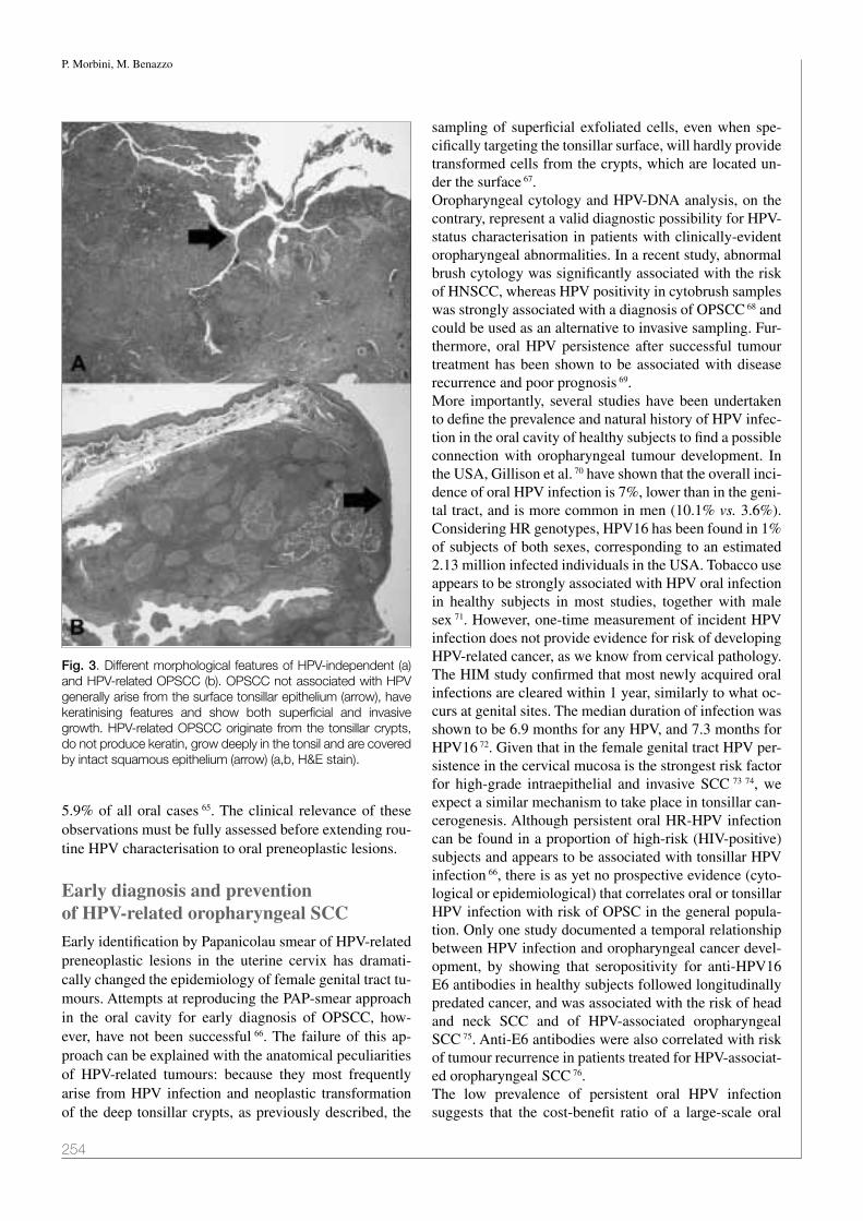
spite its excellent diagnostic accuracy 39 56, application of RNAscope in routine diagnosis is probably limited by its high cost and complex protocols. We have demonstrated that a stepwise diagnostic algorithm that includes both ISH and HR-HPV-DNA amplification (Fig. 2) can cor-rectly classify all mRNA ISH-positive cases 21; however, RNAscope is the only tool for the direct recognition of HPV transcriptional activity in paraffin-embedded sam-ples. A further issue is whether only HPV16 or all HR geno-types should be investigated in OPSCC, and which viral gene should be amplified. In our experience, we adopted a very sensitive broad-spectrum primer set (SPF10) paired with reverse-line blot genotyping [LiPA, Genotyping As-say Version Extra (Fujirebio Europe, Ghent, Belgium)] that is widely used in genital and head and neck pathology in paraffin samples 2 57 and found non-HPV16 HR infec-tions in 5-10% of cases. When amplifying viral sequences comprised in the L1-L2 region in oropharyngeal SCC, false negative and discordant results can be observed be-cause of possible L1 deletion upon viral integration 20 58 59. The amplification of the HPV16 E6 gene in a separate re-action can help in resolving ambiguous results (i.e. p16+/ISH+/PCR-). Topographic correlations. Evidence is accumulating that supports a specific correlation between HPV infection and neoplastic transformation of the reticulated epithe-lium lining the tonsillar crypts 23, which can in turn ex-plain the typical non-keratinising or ‘basaloid’ morphol-ogy of HPV-associated OPSCC (Fig. 3) 24. The biological peculiarities of tonsillar structures have been claimed to
be responsible for this selective tropism. The deep mu-cosal crypts may trap HPV viral particles and prolong the contact time between the virus and the mucosa; fur-thermore, tonsillar reticulated epithelia have intercellular ‘gaps’ that mimic the microlesions known to allow viral access to the basal cell layers in the cervical epithelium 60. Another possibility is that the local immune environment of the tonsil is directly involved in malignant transforma-tion. Although HPV-related tumours can by no means arise from superficial tonsillar keratinising epithelium 25, within the oropharyngeal region HPV-related oncogen-esis appears to be strongly related to tonsillar structures, whereas other oropharyngeal subsites are rarely involved. Importantly, a clear distinction of the site of origin of the tumour is often not straightforward from a clinical point of view, especially with larger tumours. Therefore, new cases of OPSCC should not be excluded from HPV test-ing on a purely topographical basis.
Non-oropharyngeal HPV-associated SCCWe have previously mentioned the low impact of HPV infection in non-oropharyngeal oncogenesis. One possi-ble exception can be the oral cavity, where HPV infection has been demonstrated in the periodontal pockets 61, and HPV replication appears to be favoured by the epithelial cell proliferation induced by chronic periodontal inflam-mation 62. Recent studies documented HPV association in a morphologically characteristic subset of oral high-grade dysplasia and SCC, mostly originating from the floor of the mouth and mobile tongue 63 64, which accounts for
Fig. 2.The diagnostic algorithm currently in use at our centre for the assessment of HPV-status in oPSCC.
P. Morbini, M. Benazzo
254
5.9% of all oral cases 65. The clinical relevance of these observations must be fully assessed before extending rou-tine HPV characterisation to oral preneoplastic lesions.
Early diagnosis and prevention of HPV-related oropharyngeal SCC
Early identification by Papanicolau smear of HPV-related preneoplastic lesions in the uterine cervix has dramati-cally changed the epidemiology of female genital tract tu-mours. Attempts at reproducing the PAP-smear approach in the oral cavity for early diagnosis of OPSCC, how-ever, have not been successful 66. The failure of this ap-proach can be explained with the anatomical peculiarities of HPV-related tumours: because they most frequently arise from HPV infection and neoplastic transformation of the deep tonsillar crypts, as previously described, the
sampling of superficial exfoliated cells, even when spe-cifically targeting the tonsillar surface, will hardly provide transformed cells from the crypts, which are located un-der the surface 67.Oropharyngeal cytology and HPV-DNA analysis, on the contrary, represent a valid diagnostic possibility for HPV-status characterisation in patients with clinically-evident oropharyngeal abnormalities. In a recent study, abnormal brush cytology was significantly associated with the risk of HNSCC, whereas HPV positivity in cytobrush samples was strongly associated with a diagnosis of OPSCC 68 and could be used as an alternative to invasive sampling. Fur-thermore, oral HPV persistence after successful tumour treatment has been shown to be associated with disease recurrence and poor prognosis 69.More importantly, several studies have been undertaken to define the prevalence and natural history of HPV infec-tion in the oral cavity of healthy subjects to find a possible connection with oropharyngeal tumour development. In the USA, Gillison et al. 70 have shown that the overall inci-dence of oral HPV infection is 7%, lower than in the geni-tal tract, and is more common in men (10.1% vs. 3.6%). Considering HR genotypes, HPV16 has been found in 1% of subjects of both sexes, corresponding to an estimated 2.13 million infected individuals in the USA. Tobacco use appears to be strongly associated with HPV oral infection in healthy subjects in most studies, together with male sex 71. However, one-time measurement of incident HPV infection does not provide evidence for risk of developing HPV-related cancer, as we know from cervical pathology. The HIM study confirmed that most newly acquired oral infections are cleared within 1 year, similarly to what oc-curs at genital sites. The median duration of infection was shown to be 6.9 months for any HPV, and 7.3 months for HPV16 72. Given that in the female genital tract HPV per-sistence in the cervical mucosa is the strongest risk factor for high-grade intraepithelial and invasive SCC 73 74, we expect a similar mechanism to take place in tonsillar can-cerogenesis. Although persistent oral HR-HPV infection can be found in a proportion of high-risk (HIV-positive) subjects and appears to be associated with tonsillar HPV infection 66, there is as yet no prospective evidence (cyto-logical or epidemiological) that correlates oral or tonsillar HPV infection with risk of OPSC in the general popula-tion. Only one study documented a temporal relationship between HPV infection and oropharyngeal cancer devel-opment, by showing that seropositivity for anti-HPV16 E6 antibodies in healthy subjects followed longitudinally predated cancer, and was associated with the risk of head and neck SCC and of HPV-associated oropharyngeal SCC 75. Anti-E6 antibodies were also correlated with risk of tumour recurrence in patients treated for HPV-associat-ed oropharyngeal SCC 76.The low prevalence of persistent oral HPV infection suggests that the cost-benefit ratio of a large-scale oral
Fig. 3. Different morphological features of HPV-independent (a) and HPV-related oPSCC (b). oPSCC not associated with HPV generally arise from the surface tonsillar epithelium (arrow), have keratinising features and show both superficial and invasive growth. HPV-related oPSCC originate from the tonsillar crypts, do not produce keratin, grow deeply in the tonsil and are covered by intact squamous epithelium (arrow) (a,b, H&e stain).

HPV infection in head and neck cancer
255
screening programme to identify subjects at risk of oro-pharyngeal SCC would be extremely low, especially in our population where HPV-associated tumours account for a relatively limited proportion of cases. An alternative strategy could be that of targeting subjects at increased risk of oral HPV infection for screening. In addition to HIV-positive immunosuppressed patients 66, it has been demonstrated that the risk of acquiring oral HPV infec-tion is related to sexual behaviour, both hetero- and ho-mosexual 77 78. Preliminary evidence suggests that, besides individual sexual habits, the partners of patients with HPV-related squamous epithelial lesions of the genital area are at increased risk for oral HPV infection and oro-pharyngeal SCC 79-81, so they could represent a potential target for oral HPV infection and oropharyngeal cancer screening protocols.
Future perspectivesQuadrivalent (HPV 6/11/16/18) and bivalent (HPV 16/18) anti-HPV vaccines have been available since 2006 and 2007, respectively. Vaccination policies vary worldwide concerning age of administration, population coverage and gender. Only a few countries have so far introduced gender-neutral vaccination for pre-adolescents, and its in-troduction was generally delayed by a few years with re-spect to female vaccinations 82. Despite this variability, in countries where coverage has been high a dramatic reduc-tion in cervical high-grade squamous intraepithelial le-sions as well as warts 83 84 has been observed. It is relevant to note that a significant reduction in cervical but also oral HPV prevalence was demonstrated in the Swedish female population a few years after the introduction of the vac-cination 85. Although the interval between tonsillar HPV infection and SCC diagnosis is unknown, we expect that vaccination benefits in HN cancer epidemiology would be delayed by several years. Notably, a small retrospective study has suggested that previous resection of the palatine tonsils significantly reduces the risk of tonsillar carcino-ma, even though it does not affect the risk of HPV-related cancers arising in other oropharyngeal subsites, and opens new interesting opportunities for primary prevention 86.
Conclusions Five-year survival rates are significantly different for patients with HPV-related OPSCC compared with HPV-negative patients (75-80% vs. 45-50%) 6 16 87 88. Based on the higher survival rates registered among patients with HPV-positive OPSCC, the application of de-intensified protocols has been proposed in this patient group, re-gardless of the specific treatment strategy (surgery, radia-tion therapy, concurrent chemoradiotherapy or induction chemotherapy plus concurrent chemoradiation). In addi-tion, the reduced risk of second malignancies in patients
with HPV-related OPSCC 89 is also expected to modify the natural history of OPSCC patients, and further sup-ports the need for treatments that do not persistently affect patients’ quality of life. Future clinical research will pro-vide further insights, but the combination of tumour HPV status, pack/year amount of tobacco exposure, and cancer stage should already be used routinely to classify patients as having low, intermediate or high risk as proposed by reputed authors 6.
AcknowledgementsThe work was partially supported by grant Ricerca Fi-nalizzata Ministero della Salute project code RF-2011-02351315.
References1 Ferlay J, Shin HR, Bray F, et al. Estimates of worldwide
burden of cancer in 2008: globoscan 2008. Int J Cancer 2010;127:2893-917.
2 Chaturvedi AK, Engels EA, Pfeiffer RM, et al. Human papil-lomavirus and rising oropharyngeal cancer incidence in the United States. J Clin Oncol 2011;29:4294-301.
3 Hammarstedt L, Dahlstrand H, Lindquist D, et al. The inci-dence of tonsillar cancer in Sweden is increasing. Acta Oto-laryngol 2007;127:988-92.
4 Syrjanen K, Syrjanen S, Lamberg M, et al. Morphological and immunohistochemical evidence suggesting human pap-illoma virus (HPV) involvement in oral squamous cell car-cinogenesis. Int J Oral Surg 1983;12:418-24.
5 Adelstein DJ, Ridge JA, Gillison ML, et al. Head and neck squamous cell cancer and the human papillomavirus: sum-mary of a National Cancer Institute State of the Science Meeting, November 9-10, 2008, Washington, DC. Head Neck 2009;31:1393-422.
6 Ang KK, Harris J, Wheeler R, et al. Human papilloma virus and survival of patients with oropharyngeal cancer. N Engl J Med 2010;363:24-35.
7 Ragin CC, Taioli E. Survival of squamous cell carcinoma of the head and neck in relation to human papilloma vi-rus infection: review and meta-analysis. Int J Cancer 2007;121:1813-20.
8 Boscolo-Rizzo P, Del Mistro A, Bussu F, et al. New insights in-to human papillomavirus-associated head and neck squamous cell carcinoma. Acta Otorhinolaryngol Ital 2013;33:77-87.
9 Gao G, Johnson SH, Kasperbauer JL, et al. Mate pair se-quencing of oropharyngeal squamous cell carcinomas re-veals that HPV integration occurs much less frequently than in cervical cancer. J Clin Virol 2014;59:195-200.
10 Snijders PJ, Meijer CJ, van den Brule AJ, et al. Human papil-lomavirus (HPV) type 16 and 33 E6/E7 region transcripts in tonsillar carcinomas can originate from integrated and epi-somal HPV DNA. J Gen Virol 1992;73:2059-66.
11 Ndiaye C, Mena M, Alemany L, et al. HPV DNA, E6/E7 mRNA, and p16INK4a detection in head and neck can-cers: a systematic review and meta-analysis. Lancet Oncol 2014;15:1319-31.

P. Morbini, M. Benazzo
256
12 Torrente MC, Rodrigo JP, Haigentz M Jr, et al. Human papillomavirus infections in laryngeal cancer. Head Neck 2011;33:581-6.
13 Young RJ, Urban D, Angel C, et al. Frequency and prog-nostic significance of p16(INK4A) protein overexpression and transcriptionally active human papillomavirus infec-tion in laryngeal squamous cell carcinoma. Br J Cancer 2015;112:1098-104.
14 Chaturvedi AK, Anderson WF, Lortet-Tieulent J, et al. Worldwide trends in incidence rates for oral cavity and oro-pharyngeal cancers. J Clin Oncol 2013;31:4550-9.
15 Ramqvist T, Dalianis T. Oropharyngeal cancer epidemic and human papillomavirus. Emerg Infect Dis 2010;16:1671-7.
16 Licitra L, Perrone F, Bossi P, et al. High-risk human papil-lomavirus affects prognosis in patients with surgically treat-ed oropharyngeal squamous cell carcinoma. J Clin Oncol 2006;24:5630-6.
17 Granata R, Miceli R, Orlandi E, et al. Tumor stage, human papillomavirus and smoking status affect the survival of patients with oropharyngeal cancer: an Italian validation study. Ann Oncol 2012;23:1832-7.
18 Bussu F, Sali M, Gallus R, et al. Human papillomavirus (HPV) infection in squamous cell carcinomas arising from the oropharynx: detection of HPV DNA and p16 immunohis-tochemistry as diagnostic and prognostic indicators - a pilot study. Int J Radiat Oncol Biol Phys 2014;89:1115-20.
19 Donà MG, Spriano G, Pichi B, et al. Human papillomavirus infection and p16 overexpression in oropharyngeal squa-mous cell carcinoma: a case series from 2010 to 2014. Futu-re Microbiol 2015;10:1283-91.
20 Morbini P, Dal Bello B, Alberizzi P, et al. Oral HPV infection and persistence in patients with head and neck cancer. Oral Surg Oral Med Oral Pathol Oral Radiol 2013;116:474-8.
21 Morbini P, Alberizzi P, Tinelli C, et al. Identification of tran-scriptionally active HPV infection in formalin-fixed, paraf-fin-embedded biopsies of oropharyngeal carcinoma. Hum Pathol 2015;46:681-9.
22 Gillison ML. Human papillomavirus-associated head and neck cancer is a distinct epidemiologic, clinical, and molecu-lar entity. Semin Oncol 2004;31:744-54.
23 Kim SH, Koo BS, Kang S, et al. HPV integration begins in the tonsillar crypt and leads to the alteration of p16, EGFR and c-myc during tumor formation. Int J Cancer 2007;120:1418-25.
24 Chernock RD. Morphologic features of conventional squa-mous cell carcinoma of the oropharynx: ‘keratinizing’ and ‘nonkeratinizing’ histologic types as the basis for a consist-ent classification system. Head Neck Pathol 2012;6:S41-7.
25 Cai C, Chernock RD, Pittman ME, et al. Keratinizing-type squamous cell carcinoma of the oropharynx: p16 overexpres-sion is associated with positive high-risk HPV status and im-proved survival. Am J Surg Pathol 2014;38:809-15.
26 Mirghani H, Amen F, Moreau F, et al. Human papilloma vi-rus testing in oropharyngeal squamous cell carcinoma: what the clinician should know. Oral Oncol 2014;50:1-9.
27 Pim D, Banks L. Interaction of viral oncoproteins with cel-lular target molecules: infection with high-risk vs low-risk human papillomaviruses. APMIS 2010;116:471-93.
28 Iftner T, Villa LL. Chapter 12: Human papillomavirus tech-nologies. J Natl Cancer Inst Monogr 2003;31:80-8.
29 Smeets SJ, Hesselink AT, Speel EJ, et al. A novel algorithm for reliable detection of human papillomavirus in paraffin embedded head and neck cancer specimen. Int J Cancer 2007;121:2465-72.
30 Schache AG, Liloglou T, Risk JM, et al. Evaluation of human papilloma virus diagnostic testing in oropharyngeal squa-mous cell carcinoma: sensitivity, specificity, and prognostic discrimination. Clin Cancer Res 2011;17:6262-71.
31 Hoffmann M, Ihloff AS, Görögh T, et al. p16(INK4a) overexpression predicts translational active human pap-illomavirus infection in tonsillar cancer. Int J Cancer 2010;127:1595-602.
32 Jordan RC, Lingen MW, Perez-Ordonez B, et al. Validation of methods for oropharyngeal cancer HPV status determi-nation in US cooperative group trials. Am J Surg Pathol 2012;36:945-54.
33 Weinberger PM, Yu Z, Haffty BG, et al. Molecular classifi-cation identifies a subset of human papillomavirus--associ-ated oropharyngeal cancers with favorable prognosis. J Clin Oncol 2006;24:736-47.
34 Holzinger D, Schmitt M, Dyckhoff G, et al. Viral RNA patterns and high viral load reliably define oropharynx carcinomas with active HPV16 involvement. Cancer Res 2012;72:4993-5003.
35 Jung AC, Briolat J, Millon R, et al. Biological and clinical relevance of transcriptionally active human papillomavirus (HPV) infection in oropharynx squamous cell carcinoma. Int J Cancer 2010;126:1882-94.
36 O’Regan EM, Toner ME, Finn SP, et al. p16(INK4A) genet-ic and epigenetic profiles differ in relation to age and site in head and neck squamous cell carcinomas. Hum Pathol 2008;39:452-8.
37 Kerr DA, Pitman MB, Sweeney B, et al. Performance of the Roche cobas 4800 high-risk human papillomavirus test in cytologic preparations of squamous cell carcinoma of the head and neck. Cancer Cytopathol 2014;122:167-74.
38 Lizard G, Démares-Poulet MJ, Roignot P, et al. In situ hy-bridization detection of single-copy human papillomavirus on isolated cells, using a catalyzed signal amplification sys-tem: GenPoint. Diagn Cytopathol 2001;24:112-6.
39 Schache AG, Liloglou T, Risk JM, et al. Validation of a novel diagnostic standard in HPV-positive oropharyngeal squa-mous cell carcinoma. Br J Cancer 2013;108:1332-9.
40 Thavaraj S, Stokes A, Guerra E, et al. Evaluation of human papillomavirus testing for squamous cell carcinoma of the tonsil in clinical practice. J Clin Pathol 2011;64:308-12.
41 Oguejiofor KK, Hall JS, Mani N, et al. The prognostic sig-nificance of the biomarker p16 in oropharyngeal squamous cell carcinoma. Clin Oncol (R Coll Radiol) 2013;25:630-8.
42 Lewis JS Jr, Thorstad WL, Chernock RD, et al. p16 posi-tive oropharyngeal squamous cell carcinoma:an entity with a favorable prognosis regardless of tumor HPV status. Am J Surg Pathol 2010;34:1088-96.
43 Mehra R, Ang KK, Burtness B. Management of human papillomavirus-positive and human papillomavirus-negative head and neck cancer. Semin Radiat Oncol 2012;22:194-7.
44 Kimple RJ, Harari PM. Is radiation dose reduction the right answer for HPV-positive head and neck cancer? Oral Oncol 2014;50:560-4.

HPV infection in head and neck cancer
257
45 Bussu F, Sali M, Gallus R, et al. HPV infection in squamous cell carcinomas arising from different mucosal sites of the head and neck region. Is p16 immunohistochemistry a reli-able surrogate marker? Br J Cancer 2013;108:1157-62.
46 Sartor M, Steingrimsdottir H, Elamin F, et al. Role of p16/MTS1, cyclin D1 and RB in primary oral cancer and oral cancer cell lines. Br J Cancer 1999;80:79-86.
47 Klingenberg B, Hafkamp HC, Haesevoets A, et al. p16 IN-K4A overexpression is frequently detected in tumour-free tonsil tissue without association with HPV. Histopathology 2010;56:957-67.
48 Hoffmann M, Tribius S, Quabius ES, et al. HPV DNA, E6*I-mRNA expression and p16INK4A immunohistochemistry in head and neck cancer - how valid is p16INK4A as surrogate marker? Cancer Lett 2012;323:88-96.
49 Grønhøj Larsen C, Gyldenløve M, Jensen DH, et al. Correla-tion between human papillomavirus and p16 overexpression in oropharyngeal tumours: a systematic review. Br J Cancer 2014;110:1587-94.
50 Klussmann JP, Gültekin E, Weissenborn SJ, et al. Expression of p16 protein identifies a distinct entity of tonsillar carci-nomas associated with human papillomavirus. Am J Pathol 2003;162:747-53.
51 Thomas J, Primeaux T. Is p16 immunohistochemistry a more cost-effective method for identification of human papilloma virus-associated head and neck squamous cell carcinoma? Ann Diagn Pathol 2012;16:91-9.
52 El-Naggar AK, Westra WH. p16 expression as a surrogate marker for HPV-related oropharyngeal carcinoma: a guide for interpretative relevance and consistency. Head Neck 2012;34:459-61.
53 Westra WH. Detection of human papillomavirus (HPV) in clinical samples: evolving methods and strategies for the ac-curate determination of HPV status of head and neck carci-nomas. Oral Oncol 2014;50:771-9.
54 Westra WH. The changing face of head and neck cancer in the 21st century: the impact of HPV on the epidemiology and pathology of oral cancer. Head Neck Pathol 2009;3:78-81.
55 Ukpo OC, Flanagan JJ, Ma X, et al. High-risk human papil-lomavirus E6/E7 mRNA detection by a novel in situ hybridi-zation assay strongly correlates with p16 expression and pa-tient outcomes in oropharyngeal squamous cell carcinoma. Am J Surg Pathol 2011;35:1343-50.
56 Mirghani H, Casiraghi O, Amen F, et al. Diagnosis of HPV-driven head and neck cancer with a single test in routine clinical practice. Mod Pathol 2015;28:1518-2.
57 Alberizzi P, Spinillo A, Gardella B, et al. Evaluation of the HPV typing INNO-LiPA EXTRA assay on formalin-fixed paraffin-embedded cervical biopsy samples. J Clin Virol 2014;61:535-9.
58 Duray A, Descamps G, Arafa M, et al. High incidence of high-risk HPV in benign and malignant lesions of the larynx. Int J Oncol 2011;39:51-9.
59 Tate JE, Yang YC, Shen J, et al. A comparison of early (E7) and late (L1) primer-mediated amplification of pap-illomaviral DNA in cervical neoplasia. Mol Cell Probes 1996;10:347-51.
60 Blitzer GC, Smith MA, Harris SL, et al. Review of the clini-cal and biologic aspects of human papillomavirus-positive
squamous cell carcinomas of the head and neck. Int J Radiat Oncol Biol Phys 2014;88:761-70.
61 Hormia M, Willberg J, Ruokonen H, et al. Marginal peri-odontium as a potential reservoir of human papillomavirus in oral mucosa. J Periodontol 2005;76:358-63.
62 Tezal M. Interaction between chronic inflammation and oral HPV infection in the etiology of head and neck cancers. Int J Otolaryngol 2012;2012:575242.
63 Woo SB, Cashman EC, Lerman MA. Human papillomavi-rus-associated oral intraepithelial neoplasia. Mod Pathol 2013;26:1288-97.
64 McCord C, Xu J, Xu W, et al. Association of high risk human papillomavirus infection with oral epithelial dysplasia. Oral Surg Oral Med Oral Pathol Oral Radiol 2013;115:541-9.
65 Lingen MW, Xiao W, Schmitt A, et al. Low etiologic fraction for high-risk human papillomavirus in oral cavity squamous cell carcinomas. Oral Oncol 2013;49:1-8.
66 Fakhry C, Rosenthal BT, Clark DP, et al. Associations be-tween oral HPV16 infection and cytopathology: evaluation of an oropharyngeal “pap-test equivalent” in high-risk pop-ulations. Cancer Prev Res 2011;4:1378-84.
67 Lingen MW. Brush-based cytology screening in the ton-sils and cervix: there is a difference! Cancer Prev Res 2011;4:1350-2.
68 Donà MG, Giuliani M, Vocaturo A, et al. Cytology and hu-man papillomavirus testing on cytobrushing samples from patients with head and neck squamous cell carcinoma. Can-cer 2014;120:3477-84.
69 Rettig EM, Wentz A, Posner MR, et al. Prognostic implica-tion of persistent human papillomavirus type 16 DNA detec-tion in oral rinses for human papillomavirus-related oro-pharyngeal carcinoma. JAMA Oncol 2015;1:907-15.
70 Gillison ML, Broutian T, Pickard RK, et al. Prevalence of oral HPV infection in the United States, 2009-2010. JAMA 2012;307:693-703.
71 Fakhry C, Gillison ML, D’Souza G. Tobacco use and oral HPV-16 infection. JAMA 2014;312:1465-7.
72 Kreimer AR, Pierce Campbell CM, et al. Incidence and clearance of oral human papillomavirus infection in men: the HIM cohort study. Lancet 2013;382:877-87.
73 Bodily J, Laimins LA. Persistence of human papillomavirus infection: keys to malignant progression. Trends Microbiol 2011;19:33-9.
74 Wheeler CM. The natural history of cervical human papil-lomavirus infections and cervical cancer: gaps in knowl-edge and future horizons. Obstet Gynecol Clin North Am 2013;40:165-76.
75 Mork J, Lie AK, Glattre E, et al. Human papillomavirus in-fection as a risk factor for squamous-cell carcinoma of the head and neck. N Engl J Med 2001;344:1125-31.
76 Koslabova E, Hamsikova E, Salakova M, et al. Markers of HPV infection and survival in patients with head and neck tumors. Int J Cancer 2013;133:1832-9.
77 D’Souza G, Kreimer AR, Viscidi R, et al. Case-control study of human papillomavirus and oropharyngeal cancer. N Engl J Med 2007;356:1944-56.
78 D’Souza G, Cullen K, Bowie J, et al. Differences in oral sexual behaviors by gender, age, and race explain observed

P. Morbini, M. Benazzo
258
differences in prevalence of oral human papillomavirus in-fection. PLoS One 2014;9:e86023.
79 Hemminki K, Dong C. Cancer in husbands of cervical can-cer patients. Epidemiology 2000;11:347-9.
80 Hemminki K, Dong C, Frisch M. Tonsillar and other upper aerodigestive tract cancers among cervical cancer patients and their husbands. Eur J Cancer Prev 2000;9:433-7.
81 D’Souza G, Gross ND, Pai SI, et al. Oral human papillomavi-rus (HPV) infection in HPV-positive patients with oropharyn-geal cancer and their partners. J Clin Oncol;32:2408-15.
82 Garland SM, Molesworth EG, Machalek DA, et al. How to best measure the effectiveness of male human papillomavi-rus vaccine programs? Clin Microbiol Infect 2015;S1198-743X(15)00570-4.
83 Pollock KG, Kavanagh K, Potts A, et al. Reduction of low- and high-grade cervical abnormalities associated with high uptake of the HPV bivalent vaccine in Scotland. Br J Cancer 2014:111:1824-30.
84 Dominiak-Felden G, Gobbo C, Simondon F. Evaluating the early benefit of quadrivalent HPV vaccine on genital warts in Belgium: a cohort study. PLoS One 2015;10:e0132404.
85 Grün N, Ährlund-Richter A, Franzén J, et al. Oral human papillomavirus (HPV) prevalence in youth and cervical HPV prevalence in women attending a youth clinic in Sweden, a follow up-study 2013-2014 after gradual introduction of public HPV vaccination. Infect Dis 2015;47:57-61.
86 Fakhry C, Andersen KK, Christensen J, et al. The impact of tonsillectomy upon the risk of oropharyngeal carcinoma di-agnosis and prognosis in the Danish cancer registry. Cancer Prev Res 2015:8:583-9.
87 Lindquist D, Romanitan M, Hammarstedt L, et al. Human papilloma virus is a favourable prognostic in tonsillar can-cer and its oncogenic role is supported by the expression of E6 and E7. Mol Oncol 2007;1:350-5.
88 Lassen P, Eriksen JG, Hamilton-Dutoit S, et al. Effect of HPV-associated p16INK4A expression on response to radio-therapy and survival in squamous cell carcinoma of the head and neck. J Clin Oncol 2009; 27:1992-8.
89 Diaz DA, Reis IM, Weed DT, et al. Head and neck second primary cancer rates in the human papillomavirus era: A population-based analysis. Head Neck 2016;38(Suppl 1):E873-83.
Received: September 24, 2015 - Accepted: March 19, 2016
Address for correspondence: Marco Benazzo, Department of Otorhinolaryngology, Fondazione IRCCS Policlinico San Matteo, 27100 Pavia, Italy. E-mail: [email protected]

259
ACTA oTorhinolAryngologiCA iTAliCA 2016;36:259-264; doi: 10.14639/0392-100X-709
Head and neck
Parapharyngeal space tumours: video-assisted minimally invasive transcervical approachTumori dello spazio parafaringeo: approccio transcervicale video-assistito mini invasivo
F. PiLoLLi, L. GiorDano, a. GaLLi, M. bUSSiDepartment of otorhinolaryngology, San raffaele Scientific institute, Milan, italy
SUMMARY
The purpose of the present study was to evaluate the advantages of a video-assisted, minimally invasive transcervical approach to benign and malignant parapharyngeal space (PPS) tumours. Ten patients affected by benign and malignant PPS neoplasms underwent a combined transcervical and video-assisted minimally invasive approach, using Hopkins telescopes. We describe the operative technique and perform a review of the literature. Definitive histology revealed 3 pleomorphic adenomas, 2 schwannomas, 2 metastatic papillary thyroid carcinomas, one carcinoma ex pleomorphic adenoma, one cavernous haemangioma and one basal cell adenoma. Mean tumour size was 37.2 mm (range: 19-60). Operation time ranged from 75 min to 185 min (mean: 146.7). One case was converted to transcervical-transparotid approach. Pa-tients were discharged on postoperative day 2-5. One patients presented hypoglossal nerve paresis. The minimally invasive video-assisted transcervical approach is safe and feasible for selected benign and malignant PPS tumours. Furthermore, it offers harmless dissection in a deep and narrow space, accurate haemostasis and continuous control of critical anatomic structures.
KEY WORDS: Parapharyngeal space tumour • Video-assisted • Endoscopic • Pleomorphic adenoma • Papillary thyroid cancer
RIASSUNTO
L’obiettivo dello studio è stato valutare i vantaggi di un approccio transcervicale mini-invasivo video-assistito per l’exeresi di neofor-mazioni maligne e benigne dello spazio parafaringeo. Sono stati trattati 10 pazienti con approccio trans-cervicale mini-invasivo video-assistito con l’utilizzo di telescopi di Hopkins. Viene descritta la tecnica chirurgica e una revisione della letteratura. L’esame istologico definitivo è stato in 3 casi di adenoma pleomorfo, in 2 casi di schwannoma, 2 metastasi linfonodali da carcinoma tiroideo, un carcinoma ex adenoma pleomorfo, un emangioma cavernoso ed un adenoma a cellule basali. La dimensione massima delle neoformazioni è stata in media di 37,2 mm (da 19 a 60 mm). Il tempo chirurgico è stato dai 75 ai 185 minuti (media 146,7). In un caso è stata necessaria la conversione ad approccio transcervicale-transparotideo. I pazienti sono stati dimessi dalla seconda alla quinta giornata postoperatoria. In un caso è stata osservata paresi definitiva del nervo ipoglosso. L’approccio trans-cervicale mini-invasivo video-assistito è sicuro e offre la possibilità di seguire esattamente il piano di clivaggio, permettendo un’emostasi accurata e avendo sempre il controllo delle strutture anatomiche più critiche.
PAROLE CHIAVE: Tumori spazio parafaringeo • Video-assistito • Endoscopico • Adenoma pleomorfo • Carcinoma tiroideo
Acta Otorhinolaryngol Ital 2016;36:259-264
IntroductionParapharyngeal space (PPS) is classically described as an inverted pyramid-like area with the floor at the skull base and apex at the greater horn of the hyoid bone. The tensor-vascular-styloid fascia divides PPS into prestyloid and retrostyloid compartments. Neoplasms arising in the PPS are rare tumours account-ing for 0.5 to 1% of all head and neck masses 1; 82% are benign and 18% are malignant: pleomorphic adenoma is the most common histotype (29%).Most PPS lesions need first-line surgical treatment per-formed with a transoral, transcervical, transparotid, or transmandibular approach, alone or in combination.
Recently, endoscopic and robotic approaches have been widely applied in head and neck surgery to minimise tis-sue trauma and wound-related complications and improve cosmetic outcomes. Reports on their use in PPS surgery are extremely limited.
Materials and methods
PatientsTen patients with PPS tumours were treated with transcer-vical video-assisted surgery at the Department of Otorhi-nolaryngology – Head and Neck Surgery of the Univer-sity Vita-Salute San Raffaele, Milan, Italy from July 2012
F. Pilolli et al.
260
to March 2015. Mean age was 58.2 years (range: 42-72). The opportunity to opt for a video-assisted approach was mainly evaluated with magnetic resonance imaging (MRI): we enrolled only patients affected by tumours smaller than 6-7 cm in their largest diameter and with a definite cleavage plane from nearby structures.Only 4 patients were symptomatic (Table I). Diagnostic workup included contrast-enhanced MRI. Computed to-mography (CT) was required in 4 cases for better radio-logical assessment. Preoperative ultrasound-guided fine needle aspiration cytology (FNAC) was performed in 3 patients: directly on the PPS mass in 2 cases and on a cervical node in the other.
Operative techniqueAll procedures were performed under general anaesthe-sia by the same surgical team. As previously described 2 3, video-assisted dissection is performed through a minimal cervical incision (depending on the tumour size) made in a natural skin crease, approximately 3 cm below the mandibular angle at the level of the digastric muscle. The aim of this approach is to reach the whole PPS through a small anatomical corridor, wide enough to allow use of an endoscope and some endoscopic instruments. A skin flap is elevated in the subplatysmal plane. The submandibu-lar gland is retracted anteriorly and the tail of the parotid gland posterosuperiorly. The posterior belly of digastric muscle could be divided or cranially retracted. The hypo-glossal nerve is then identified and preserved.The next steps are performed under assistance of 0° and 30° Hopkins telescopes using a high definition camera. During video-assisted surgical steps, the second surgeon keeps the telescope: this allows the first surgeon to use
both hands. The third surgeon provides a wider surgi-cal field using retractors. Operative room setup is shown in Figure. 1.Thereafter, the internal carotid artery is identified. Tumour dissection is performed upwards and circumferentially in an extracapsular plane: nearby vessels and nerves are care-fully retracted from the mass. At this point, the suction-dissector becomes a useful tool to maintain a bloodless surgical field. The tumour is then released and removed en bloc. Endoscopic inspection confirms the completeness of the resection. The posterior belly of the digastric muscle is reapproximated if previously divided. A suction drain is placed inside the wound, which is closed in layers. The drain is removed as soon as daily drainage falls below 20 ml: the patient can be discharged the day after.
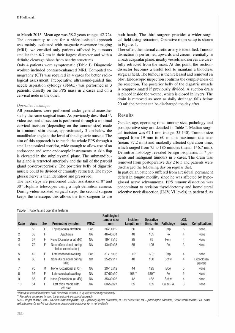
ResultsGender, age, operating time, tumour size, pathology and postoperative stay are detailed in Table I. Median surgi-cal incision was 67.1 mm (range: 35-140). Tumour size ranged from 19 mm to 60 mm in maximum diameter (mean: 37.2 mm) and markedly affected operation time, which ranged from 75 to 185 minutes (mean: 146.7 min). Definitive histology revealed benign neoplasms in 7 pa-tients and malignant tumours in 3 cases. The drain was removed from postoperative day 2 to 5 and patients were discharged the following day on regular diet. In particular, patient 6 suffered from a residual, permanent deficit in tongue motility since he was affected by hypo-glossal nerve schwannoma. PPS tumour dissection was concomitant to revision thyroidectomy and homolateral selective neck dissection (II-IV, VI levels) in patient 5, as
Table I. Patients and operative features.
Case Ages Sex Presenting symptom FNAC
Radiological tumour size,
mmIncision
Length, mmOperative time, min Pathology
LOS, days Complications
1 53 F Thyroglobulin elevation Pap 36x14x19 56 170 Pap 6 None
2 53 F Dysphagia NA 46x45x31 48 165 PA 4 None
3 57 F None (Occasional at MRI) NA 19x17x15 35 75 Hem 4 None
4 72 F None (Occasional during clinical examination)
NA 43x40x35 85 105 PA 3 None
5 42 F Laterocervical swelling Pap 31x15x16 140* 170* Pap 4 None
6 60 F None (Occasional during MRI)
NC 25x22x17 48 130 Schw 4 Hypoglossal paresis
7 70 M None (Occasional at CT) NA 20x13x12 44 125 BCA 5 None
8 56 F Laterocervical swelling NA 57x50x30 108** 180** PA 5 None
9 65 F None (Occasional at MRI) NA 35x30x25 42 162 Schw 4 None
10 54 F Left otitis media with effusion
NA 60x59x27 65 185 Ca ex-PA 3 None
*Procedure included selective neck dissection (levels II-IV, VI) and revision thyroidectomy.** Procedure converted to open transcervical-transparotid approachLOS = length of stay; Hem = cavernous haemangioma; Pap = papillary thyroid carcinoma; NC: not conclusive; PA = pleomorphic adenoma; Schw: schwannoma; BCA: basal cell adenoma; Ca ex-PA: carcinoma ex pleomorphic adenoma; NA = not available

Parapharyngeal space tumours: video-assisted minimally invasive transcervical approach
261
he was affected by metastatic papillary thyroid cancer: in-cision length and operative time were longer. Skull base adhesion of the mass made conversion to transcervical-transparotid approach unavoidable in patient 8. There-fore, excluding cases 5 and 8, median incision length was
52.9 mm (range: 35-85) and mean operative time was 139.6 min (range: 75-185).After a mean follow-up period of 22 months (from 2 to 37 months), neither radiologically nor clinically relapse was detected into the PPS.
DiscussionSurgery is the mainstay of treatment of most PPS tumours. The anatomic complexity (Fig. 2) of the PPS had led to the development of several surgical approaches. Tumour size, proximity to cervical neurovascular structures and histotype should guide the surgeon in tailoring the strat-egy for treatment. The transcervical approach is commonly used for most PPS neoplasms 4: it provides good local disease control with minimal risk of facial nerve injury and good cosmet-ic results. However, it is not considered safe for masses with significant vertical extension or radiological suspi-cion of invasion of cranial foramina 5.The transparotid approach is used for tumours of the deep lobe of parotid gland 6. It offers a wide access to PPS, but the risk of facial nerve injury is higher due to its unavoidable extensive dissection and retraction during the procedure 2 7.Many authors 8-10 have addressed the need for additional approaches to obtain oncologically safe results, such as mandibulotomy. In particular, Malone et al. 10 described 40% of combined techniques. The transmandibular ap-
Fig. 1. Operative room setup during video-assisted steps.
Fig. 2. Anatomy of the PPS.

F. Pilolli et al.
262
proach ensures very wide exposure of the PPS and should be considered for highly vascularised neoplasms, recur-rent tumours, malignant masses and lesions invading the skull base. However, it should be kept in mind that this technique often results in important nerve injuries, maloc-clusion and malunion 2. The orbitozygomatic-middle fossa approach is another technique reported in literature, although it has been de-scribed for a restricted number of extremely large tumours involving the skull base 11.The transoral approach is the most controversial. It pro-vides limited, direct access to the PPS and makes identifi-cation of neurovascular structures more difficult. Moreo-ver, it is linked to a higher risk of intra-surgical tumour rupture, incomplete removal of the mass, uncontrollable haemorrhage and facial nerve injury 4.Some authors 12 suggested robotic transoral resection for large benign masses that are accessible from the oropharynx and involving the poststyloid space. This approach offers a high rate of disease control and a low risk of post-operative complications, such as lockjaw or cranial nerve injuries.Endoscopic visualisation has been introduced relatively recently in order to obtain better neoplasm control and
improve wound cosmetic outcomes. Dallan et al. 13 identi-fied some critical surgical landmarks in endoscopic tran-soral PPS dissection of six fresh human cadaver heads. Another anatomic study was conducted by Taniguchi et al. to assess the feasibility of an endoscopic transnasal route 14. The first endoscopic PPS approach on a living person was published in 2010 for paediatric transnasal abscess drainage 15. Subsequent reports were published with transvestibular 16, transoral 17 and transcervical 2 ap-proaches for benign PPS tumours.The traditional transcervical approach provides very lim-ited surgical exposure to the PPS: in fact, it is 5-6 cm deep from the cutaneous surface (Fig. 3d). Surgeons are forced to work in a long, dark and narrow tunnel. Digital explora-tion and digitoclasia are certainly helpful, but direct visual control is not possible during these operations.The goal of this early experience was to appreciate the advantages of an endoscopic approach, especially from the surgeon’s perspective. Using 0° and angled telescopes it is possible to constantly check relationships between the mass and nearby vessels or cranial nerves. The close visual control and magnification of the image allow the surgeon to follow the tumour surface, easing the recogni-
Fig. 3. Case 10: A, pre-operative RMN; B, medialisation of the left pharyngeal wall; C, video-assisted dissection of PPS tumour using suction-dissector; D, demonstration of the deep and narrow surgical tunnel for PPS tumours dissection; E, surgical specimen; F, aesthetic result on postoperative day 11.

Parapharyngeal space tumours: video-assisted minimally invasive transcervical approach
263
tion of cleavage planes, even in lobulated neoplasms. This latter feature reduces the risk of tissue spillage that could have dramatic consequences even for benign lesions.The video-assisted technique simplifies the identification of small vessels, allowing accurate haemostasis. Further-more, using the suction-dissector (Fig. 4) it is possible to work in a near-bloodless surgical field due to one-hand simultaneous or alternate dissection and aspiration.In our series, the nature and the dimensions of the masses markedly influenced operation time. Average surgical time (146.7 min) was similar to that reported by Beswick et al. 2 (133 min) in the only video-assisted PPS tumour dissection previously reported in the literature.Hospitalisation time was similar to our non-video-assisted approaches, but was higher than that reported by Beswick et al. 2, perhaps due to our prudential attitude in removal of drains.In summary, a minimally invasive video-assisted tran-scervical approach should be considered for PPS tumours smaller than 7 cm in their largest diameter. In our opinion, histotype is not an indication itself: even selected malig-nant neoplasms could be excised with this technique if a definite cleavage plane is recognisable and if the histo-type does not require removal of marginal healthy tissues around the mass. We effectively treated three malignant tumours: 2 expected nodal metastases of thyroid papillary carcinomas and an occasional carcinoma ex pleomor-phic adenoma. In all these cases surgery was definitive. Nonetheless, after our preliminary experience we would not recommended this technique for malignant masses in-vading adjacent tissues: a video-assisted minimally inva-
sive transcervical approach cannot offer sufficient access to PPS. Furthermore, we consider it dangerous to use a video-assisted approach for hypervascular tumours (e.g., paragangliomas).
Conclusions A minimally invasive video-assisted transcervical ap-proach is a new technique for excision of sizable benign and selected malignant PPS tumours. It allows clear iden-tification of critical surgical landmarks and guides the dissection through the right cleavage plane, offering the chance for accurate hemostasis while decreasing surgical complications.
References1 Riffat F, Dwivedi RC, Palme C, et al. A systematic review of
1143 parapharyngeal space tumors reported over 20 years. Oral Oncol 2014;50:421-30.
2 Beswick DM, Vaezi A, Caicedo-Granados E, et al. Minimally invasive surgery for parapharyngeal space tumors. Laryngo-scope 2012;122:1072-8.
3 Giordano L, Pilolli F, Toma S, et al. Parapharyngeal me-tastases from thyroid cancer: surgical management of two cases with minimally-invasive video-assisted technique. Acta Otorhinolaryngol Ital 2015;35:289-92.
4 Kuet ML, Kasbekar AV, Masterson L, et al. Management of tumors arising from the parapharyngeal space: a systematic review of 1,293 cases reported over 25 years. Laryngoscope 2015;125:1372-81.
5 Basaran B, Polat B, Unsaler S, et al. Parapharyngeal space tumours: the efficiency of a transcervical approach without mandibulotomy through review of 44 cases. Acta Otorhi-nolaryngol Ital 2014;34:310-6.
6 Casani AP, Cerchiai N, Dallan I, et al. Benign tumours affect-ing the deep lobe of the parotid gland: how to select the op-timal surgical approach. Acta Otorhinolaryngol Ital organo 2015;35:80-7.
7 Khafif A, Segev Y, Kaplan DM, et al. Surgical management of parapharyngeal space tumors: a 10-year review. Otolar-yngol Head Neck Surg 2005;132:401-6.
8 Hamza A, Fagan JJ, Weissman JL, et al. Neurilemomas of the parapharyngeal space. Arch Otolaryngol Head Neck Surg 1997;123:622-6.
9 Cohen S, Burkey B, Netterville J. Surgical management of parapharyngeal space masses. Head Neck 2005:669-75.
10 Malone JP, Agrawal A, Schuller DE. Safety and efficacy of transcervical resection of parapharyngeal space neoplasms. Ann Otol Rhinol Laryngol 2001;110:1093-8.
11 O’Malley BW, Quon H, Leonhardt FD, et al. Transoral robot-ic surgery for parapharyngeal space tumors. ORL J Otorhi-nolaryngol Relat Spec 2010 [cited 2012 Sep 5];72:332-6.
12 Lee HS, Kim J, Lee HJ, et al. Transoral robotic surgery for neurogenic tumors of the prestyloid parapharyngeal space. Auris Nasus Larynx 2012;39:434-7.
13 Dallan I, Seccia V, Muscatello L, et al. Transoral endo-scopic anatomy of the parapharyngeal space: a step-by-step
Fig. 4. Video-assisted dissection of PPS tumour (patient 2) from pre-verte-bral fascia using suction-dissector.

F. Pilolli et al.
264
logical approach with surgical considerations. Head Neck 2011;33:557-61.
14 Taniguchi M, Kohmura E. Endoscopic transnasal transmaxil-lary transpterygoid approach to the parapharyngeal space: an anatomic study. Minim Invasive Neurosurg 2010;53:255-60.
15 Lee C-H, Lee T-J, Chen C-W. Transnasal endoscopic ap-proach for drainage of pediatric parapharyngeal space ab-scess. Otolaryngol Head Neck Surg 2010;143:467-8.
16 Chan JYK, Li RJ, Lim M, et al. Endoscopic transvestibular paramandibular exploration of the infratemporal fossa and parapharyngeal space: a minimally invasive approach to the middle cranial base. Laryngoscope 2011;121:2075-80.
17 Chiang TY, Chen MK. Endoscope-assisted transoral exci-sion of a huge parapharyngeal pleomorphic adenoma. B-ENT 2011;7:143-6.
Address for correspondence: Pilolli Francesco, San Raffaele Scientific Institute, Department of Otorhinolaryngology, “San Raffaele” Hospital, via Olgettina 58, 20132 Milan, Italy. Tel. +39 02 26432172, +39 02 26433530. Fax +39 02 26433508. E-mail: [email protected]
Received: May 4, 2015 - Accepted: January 10, 2016

265
ACTA oTorhinolAryngologiCA iTAliCA 2016;36:265-274; doi: 10.14639/0392-100X-1082
Head and neck
Surgical complications in orbital decompression for Graves’ orbitopathyComplicanze chirurgiche in pazienti sottoposti a decompressione orbitaria per oftalmopatia di Graves
S. SeLLari-FranCeSCHini1, i. DaLLan1, a. bajrakTari1, G. FiaCCHini1, M. narDi2, r. roCCHi3, C. MarCoCCi3, M. Marinò3, a.P. CaSani11 First enT Unit, azienda ospedaliero-Universitaria Pisana, University of Pisa, italy; 2 Unit of ophthalmology, azienda ospedaliero-Universitaria Pisana, University of Pisa, italy; 3 Unit of endocrinology, azienda ospedaliero-Universitaria Pisana, University of Pisa, italy
SUMMARY
The objective of this study is to analyse the complications of orbital decompression in Graves’ orbitopathy. The clinical records of 946 patients who had been operated on with orbital decompression for Graves’ orbitopathy were reviewed and the intra- and post-operative complications with minimum follow-up of six months were analysed. An extensive review of the literature was carried out to compare results. In the case-series reported here the most frequent complications were: wasting of the temporal region (100%) in patients operated on using a coronal approach; permanent hypoesthesia of V2 (13%) and V1 (8%) in patients operated on with an upper eyelid incision. In only one patient was a total monolateral lesion of V2 reported. The most severe complications consisted in reduction of visual acuity in 5 patients, and CSF leak with cerebral complications in 2 patients, who were operated on with a non-endoscopic endonasal approach. Three patients had intra-operative haemorrhages and 3 patients had post-operative haemorrhages requiring further surgical intervention. The incidence of symptomatic sinusitis/mucoceles was 0.75%. In conclusion, orbital decompression carried out with endoscopic endonasal technique and via transpalpebral accesses appears to be associated with a low incidence of complications. Knowledge of the causes of the possible complications in the different surgical approaches can definitely help to reduce their incidence.
KEY WORDS: Graves’ orbitopathy • Orbital decompression • Different approaches • Complications
RIASSUNTO
L’obiettivo di questo studio è analizzare le complicanze della decompressione orbitaria in pazienti affetti da oftalmopatia Basedowiana. Abbiamo analizzato 946 pazienti sottoposti a decompressione orbitaria per orbitopatia di Graves e le complicanze intra- e post-operatorie con un follow-up minimo di 6 mesi. Abbiamo eseguito inoltre un’estesa revisione della letteratura per comparare i risultati. Nel nostro stu-dio le più frequenti complicanze sono state: atrofia della regione temporale (100%) nei pazienti sottoposti a decompressione con approccio coronale; ipoestesia permanente di V2 (13%) e V1 (8%) in pazienti sottoposti a decompressione con approccio transpalpebrale superiore. Un solo paziente ha avuto una lesione totale monolaterale di V2. Le complicanze più gravi sono state la riduzione dell’acuità visiva, che si è verificata in 5 pazienti, e la perdita di liquido cerebrospinale con complicanze cerebrali, verificatesi in 2 pazienti, entrambi operati con approccio endonasale non endoscopico. 3 pazienti hanno avuto un’emorragia intraoperatoria mentre 3 pazienti un’emorragia postopera-toria che ha richiesto un secondo intervento chirurgico. L’incidenza delle sinusiti/mucoceli sintomatici è stata dello 0,75%. In conclusione abbiamo evidenziato come la decompressione orbitaria eseguita con tecnica endoscopica endonasale e con accessi transpalpebrali sia una procedura chirurgica con una bassa incidenza di complicanze. La conoscenza delle cause delle possibili complicanze nei differenti approcci chirurgici può sicuramente aiutare a ridurre la loro incidenza.
PAROLE CHIAVE: Orbitopatia di Graves • Decompressione orbitaria • Approcci differenti • Complicanze
Acta Otorhinolaryngol Ital 2016;36:265-274
Introduction
Graves’ Orbitopathy (GO) is an auto-immune inflamma-tory disease that causes an increase in the volume of or-bital adipose tissue and the extrinsic muscles of the eye. Proportionally to the prevalence of oedema or fibrosis, clinical manifestations of GO are extremely variable. The surgical indication for orbital decompression can thus
be attributed to mild aesthetic problems, but also to se-vere proptosis, subluxation of the ocular globe, corneal exposure, in the presence or absence of diplopia and/or strabismus. Most of the publications available on orbital decompression of GO describe case-studies focusing on patients with differing pathological situations, operated on with different techniques 1. To further complicate the

S. Sellari-Franceschini et al.
266
situation, surgical techniques are generally not described in detail, and chosen by the surgeon on the basis of his specialisation 2. In fact, this makes evaluation of surgical complications very difficult. To our knowledge, few publications have reported the rate of complications adequately 3-12. Leong et al. 12 reported that the global incidence of complications was 9.3%, while the rate of severe complications with long-term se-quelae was estimated to be 0.12%. The authors underline that many studies do not mention complications, although this does not necessarily mean that no complications arose. Moreover, Boboridis and Bunce 1 mentioned that it is difficult to reach any safe conclusions regarding the efficacy and possible complications of various techniques. However, it is of fundamental importance to investigate more in depth possible complications, and above all their causes, to further improve surgical techniques and achieve better results. In this work, the complications that occurred in the case-histories of 946 patients with the use of different surgical techniques are collected and analysed. Furthermore, the causes are critically evaluated and the techniques imple-mented to reduce the incidence. A careful comparison with data from literature is presented.
Materials and methodsClinical records of 946 (1762 orbits) of 957 consecutive patients (11 patients lost to follow-up) operated on by a single surgeon from December 1992 to December 2014 were collected. Post-operative data refer to a minimum follow-up period of 6 months. The local research ethics committee approved the study.The first surgical technique used was the trans-antral infero-medial decompression (TA) according to Walsh and Ogura 13, (21 patients, 39 orbits). Next, the three-wall decompression with coronal approach (C) according to Mourits et al. 14 (21 patients, 38 orbits) was used. Our vari-ation on this technique was the infero-medial approach performed with non-endoscopic trans-nasal access. The combination of the two techniques (TA + C) was carried out in 30 patients (54 orbits). Since 2000 orbital decompression has been performed on the three walls (lateral, medial and inferior) with or without removal of adipose tissue, adapting the tech-nique to the severity of the proptosis and/or the pres-ence of optic neuropathy and eyes imbalance. Lateral (L) and medial (M) orbital wall surgery was performed as described previously 15 16. The orbital floor was ap-proached with trans-inferior eyelid access or with the “swinging eyelid” technique 17. Due to the fact that eve-ry complication is correlated to the surgical approach, it is important to know the exact number of each surgical procedures to understand the incidence rate. The lat-eral orbital wall approach was used 1489 times, medial
orbital wall approach 1200 times and infero orbital ap-proach 278 times. Adipose tissue was removed, but only in combination with decompression of one or more bony walls and almost always with external approaches. In the bilateral forms, both eyes were decompressed during the same operation to better balance the level of decompression between the two sides.The following were all considered as major compli-cations: death, loss or reduction of visual acuity, ce-rebrospinal fluid leaks (CSF), central nervous system pathologies, significant bleeding that significantly pro-longed the duration of the operation or which rendered a second surgical intervention necessary, permanent impairment of the supraorbital nerve (V1) or the in-fraorbital nerve (V2) and total palpebral ptosis. Minor complications included injury of the dura mater of the medial and/or anterior cranial fossae, easily controlled bleeding, transient hypo-aesthesia of the forehead (V1) and of the cheek and upper lip (V2), sinusitis, muco-cele, corneal lesions, cutaneous palpebral lesions, mild ptosis, wasting of the temporal region, oscillopsia and subcutaneous emphysema.Possible causes and technical tips are analysed in order to reduce the incidence of single complications.The problem of post-operative diplopia, considered by some authors to be a common sequela of the decompres-sion operation 18 19, and certainly closely linked to the dis-ease of the extrinsic muscles of the eye 20, was not taken into account.A systematic review of the literature was performed us-ing large biomedical databases (PubMed and Cochrane) and combining the key words “orbital decompression”, “complications”, “Graves’ disease”, “Graves’ orbi-topathy”, “ophthalmopathy” and “thyroid eye disease”. We critically analysed and selected 40 of 189 articles that reported the following complications: loss of vi-sual acuity, cerebral complications, haemorrhages and haematomas, hypesthesia and dysesthesia in areas of innervation of the first (V1) and second (V2) branches of the trigeminal nerves, symptomatic sinusitis and mu-coceles.
ResultsThe rates of complications in our patients with the surgi-cal techniques used are shown in Table I.
DeathNo death was correlated with the intervention of orbital decompression during the entire period examined.
Loss of visual acuityWe had five patients with mono-lateral loss of visual acu-ity, three following an endonasal approach, though not

Surgical complications in orbital decompression for Graves’ orbitopathy
267
endoscopic, during the first years of our experience. The other two patients had a balanced decompression. One de-veloped ocular keratitis and the other remained without a certain cause. Only partial recovery of visual acuity was seen during follow-up.
Cerebral complications and CSF leaksWe observed one case of meningitis after a CSF leakage not identified and not treated intra-operatively and one case of pneumocephalus. Another eight CSF leaks were identified and managed intra-operatively, without compli-cations. One patient presented a cavernous sinus bleeding and haematoma that was managed post-operatively in a conservative fashion. We observed 38 cases of mono-lateral lesion of the me-ninges of the middle or anterior cranial fossa, during lat-eral approaches with removal of the greater wing of the sphenoid bone. All cases were managed conservatively in 2-3 days, with compressive packing of the eye.
Haemorrhages/haematomasWe had 2 intra-operative major bleedings, during a lateral approach, which complicated the intervention, but both patients recovered without sequelae.In the postoperative period we had a haematoma of the temporal muscle, after a coronal approach, which required
intervention with general anaesthesia. Similarly, three pa-tients were taken back into surgery within 24 hours after first surgery for intraorbital lateral bleeding.
Hypo/anaesthesia of V1, V2 innervation areasWe had 260 cases of mono-lateral hypesthesia of V1 (17.5%) one month after surgery; 118 (8%) continued to report permanent hypesthesia 6 months after surgery.419 cases operated on externally had mono-lateral hypes-thesia of V2 (28%) at one month after surgery; 196 (13%) continued to report permanent hypesthesia 6 months after surgery. We had 9 cases (9.7%) of hypesthesia of V2 in patients operated on with the TA technique. One patient (0.36%) reported the section of V2, with total deficit, dur-ing a revision procedure performed on the orbital floor with an inferior eyelid approach.
Symptomatic sinusitis/mucoceleWe had 5 patients with persistent post-operative sinusi-tis (two frontal and three maxillary sinusitis), that were managed successfully with medical therapy. Furthermore, other four patients were operated on for mucocele (two posterior-ethmoidal and two frontal-ethmoidal) 5-8 years after orbital decompression. All these patients were oper-ated on with endoscopic transnasal access.
Table I. Surgical complications.
Complications Number of patients (Surgical technique)
Rate of complications (Surgical technique)
Death 0 0%
Loss of visual acuity 3 (TA); 2 (L+M) 3.23% (TA); 0.13% (L+M)
CSF leaks 8 (M); 38 (L) 0.67% (M); 2.55% (L)
Pneumocephalus 1 (M) 0.08%(M)
Meningitis 1 (M) 0.08% (M)
Cavernous sinus bleeding 1 (M) 0.08% (M)
Major bleeding 2 (L) 0.13% (L)
Haematoma of the temporal muscle 1 (C) 1.09% (C)
Intraorbital bleeding 3 (M) 0.25% (M)
Epistaxis 1 (M) 0.08% (M)
Hypoesthesia of V1 118 (L) 8% (L)
Hypoesthesia of V2 196 (S); 9 (TA) 13% (S); 9.68% (TA)
Anaesthesia of V2 1 (I) 0.36% (I)
Symptomatic chronic sinusitis 5 (M) 0.42% (M)
Mucocele 4 (M) 0.33% (M)
Corneal ulcers 8 (L) 0.54% (L)
Section of the levator muscle 7 (L) 0.47% (L)
Small areas of burn on the upper eyelid 30 (L) 2.01% (L)
Wasting of the temporal region 51 (C) 100% (C)
Oscillopsia 2 (L) 0.13% (L)
Hyposmia 4 (M) 0.33% (M)
Subcutaneous emphysema 6 (M) 0.5% (M)TA: transantral. C: coronal. L: lateral wall. M: medial wall. I: inferior wall.

S. Sellari-Franceschini et al.
268
Palpebral lesionsIn 7 patients (0.47%) section of the levator muscle of the upper eyelid occurred with access through the upper eyelid. Six patients had no sequelae because the levator muscle was sutured with absorbable stitches at the end of the surgery. In 1 patient the lesion was not recognised during the intervention, but the patient refused corrective surgery. In 30 patients (2.01%) operated on with a lateral approach, small areas of burn on the skin of the upper eyelid, caused by the drill, occurred.
Corneal ulcersWe had some cases of corneal de-epithelisation and 8 cases (0.54%) of corneal ulcer caused by intra-operative manoeuvres, all during an external approach; all these re-solved successfully with medical treatment.
Wasting of the temporal region21 patients (100%) operated on with a coronal approach manifested hollowing, of various degrees, of the temporal region. These patients complained of mild disorders with mastication, but no aesthetic damage.
Oscillopsia 2 patients (0.13%) operated on with lateral approach, in-cluding complete removal of the greater wing of the sphe-noid bone, manifested oscillopsia during mastication. The disorder was well tolerated and the patients refused cor-rective surgery.
Hyposmia 4 patients (0.33%) operated on with an endonasal ap-proach complained of hyposmia.
Subcutaneous emphysema6 patients (0.5%) operated on with endo-nasal approach presented inferior palpebral emphysema in the post-oper-ative period, after having violently blown their noses. All these cases resolved spontaneously.
DiscussionMany works reporting the results of decompression inter-vention are available in the literature, although in general these describe small groups of patients often presenting varying clinical situations. Moreover, orbital decompres-sion is performed with different techniques, depending more on the specialisation and experience of the surgeon than on ocular muscle impairment. Since the typology of complications is closely associated with the technique uti-lised, we wished to carry out an accurate analysis of the complications arising in our patients, in view of the fact that we used all the approaches described with the excep-tion of Olivari’s technique 21, despite the removal of adi-pose tissue being an integral part of many of our interven-
tions. Leong et al. 12 reported that of 4176 decompressed orbits, the incidence of complications was 9.3%. We feel that, rather than making a simple evaluation, it is impor-tant to distinguish between major and minor complica-tions and to try to understand the causes. Leong refers that no death occurred as a consequence of orbital decompres-sion, but in the medical literature 2 deaths have been de-scribed following post-operative complications with the TA and infero-medial transpalpebral technique 8 22.The worst ocular complication is total or partial loss of visual acuity. In our case-series, there were had 5 cases. Three were at the beginning of our experience with a non-endoscopic endonasal approaches. In the first case (TA technique), it was most likely that traction maneuvers on adipose tissue, which was very fibrotic, were performed to increase decompression, causing severe damage, more probably on a vascular basis, of the optic nerve. In the other 2 patients, bone fragments (anterior section of the lateral wall of the sphenoid sinus in one case and a small fragment of lamina papyracea in the other) directly or indi-rectly compressed the nerve: with endoscopic techniques, this complication may easily be avoided. Another patient developed ocular keratitis after balanced decompression. The last patient had total loss of visual acuity after bal-anced decompression partially recovered after steroid therapy. The cause of the visual loss was not identified because postoperative MRI and CT scan were negative. In the literature, 2 cases of monolateral blindness 23 and 2 cases of visual loss 3 have been reported, all of whom were operated on with infero-medial decompression. Warren et al. 24 reported 5 patients who developed postoperative pan-ophthalmitis and progressed to blindness. Jernfors et al.10 reported one bilateral and one monolateral postoperative blindness in patients operated on due to progressive optic neuropathy.We report 1 case of meningitis and 1 of pneumocephalus due to incorrect closure of CSF leaks in patients oper-ated on with a non-endoscopic endonasal approach. Sub-sequently, 8 other CSF leaks were closed with a middle turbinate mucoperiosteal flap. There are 28 other cases reported in the literature of CSF leaks and other major cerebral complications (Table II). Although in some cases spontaneous closure of the fistulas is described, we strong-ly recommend performing skull base reconstruction. On the opposite side, small lesions of the meninges of the anterior and middle cranial fossa with CSF tears (through lateral access) usually represent a minor concern which does not require any skull base reconstruction. We found 5 similar cases in the literature 7 25 26. In our experience, in the case of lesions of less than 2-3 mm, maintenance of drainage without suction and moderate compressive eye-packing resolve the leaks in a few days.We had 2 major bleedings, during lateral approaches, which were managed intra-operatively. One patient who was operated on with a coronal approach presented bleed-

Surgical complications in orbital decompression for Graves’ orbitopathy
269
Table II. Cerebral complications and treatment after orbital decompression reported in literature.
Authors Surgical technique Number of patients
Number of orbits
Cerebral complications Treatment
Bailey et al. 28 3-walls 55 97 2 CSF leaks 1 nasal packing; 1 neurosurg op
De Santo 23 TA 200 399 4 CSF leaks No treatment for the leak
Garrity et al. 3 TA 428 851 15 CSF leaks (4 meningitis, 1 patient died);
1 frontal lobe haematoma
4 second operations
Kashkouli et al. 9 Transconjunctival approach 1 (case report) 1 subarachnoid haemorrhage and frontal
lobe ischemia
No treatment for the leak
Kasperbauer et al. 33 Medial wall and floor(endoscopic approach)
59 88 2 CSF leaks 2 closed intraop
McCormick et al. 8 Medial wall and floor (inferomedial approach)
2 (case report) 2 CSF leaks (1 died) No treatment for the leak
Murchison et al. 34 Medial wall and floor (transconjunctival approach)
1 (case report) 1 Meningoencephalocele Resection of the herniated brain tissue and skull-base reconstruction
Nadeau et al. 35 1 orbit: medial wall16 orbits: medial and lateral
walls6 orbits: medial wall and
floor50 orbits: medial, lateral
and floor
40 73 1 CSF leak No treatment for the leak
Schaefer et al. 36 Inferomedial approach 41 72 1 CSF leak No treatment for the leak
Warren et al. 24 TA 305 610 1 CSF leak with meningitis No treatment for the leakTA: transantral. CSF: cerebrospinal fluid.
Table III. Haemorrhages and haematomas after orbital decompression reported in the literature.
Authors Surgical technique Number of patients Number of orbits Bleeding
Antisdel et al. 37 3-walls 50 86 1 epistaxis (second operation)
Eloy et al. 38 Endoscopic approach 16 27 1 epistaxis
Garrity et al. 3 TA 428 851 1 frontal lobe haematoma 11 blood transfusions
Jernfors et al. 10 TA / Transnasal endoscopic approach 78 144 2 epistaxis
Kashkouli et al. 9 Transconjunctival approach 1 (case report) 1 subarachnoid haemorrhage and frontal
lobe ischaemia
Kikkawa et al. 39 3-walls 9 1 haemorrhage (second operation)
Lund et al. 40 59 orbits: inferomedial approach 33 59 3 moderate haemorrhages
McCormick et al. 8 Medial wall and floor (transconjunctival approach)
2 (case report) 1 epistaxis
Metson et al. 19 8 orbits: lateral wall 33 orbits: medial and lateral walls
26 41 1 epistaxis
Nadeau et al. 35 1 orbit: medial wall 16 orbits: medial and lateral wall
6 orbits: medial wall and floor 50 orbits: medial, lateral and floor
40 73 1 epistaxis
Olivari 21 Fat removal 57 108 1 haematoma
Pezato et al. 41 3-walls 15 17 1 haematoma
Ulualp et al. 30 Medial wall (endo) and floor (transconjunctival approach)
15 28 1 haematoma
White et al. 42 Medial (endo) and lateral walls 34 64 2 epistaxisTA: transantral.

S. Sellari-Franceschini et al.
270
ing in the area of the temporalis muscle (which was sec-tioned and sutured with this technique). This patient re-quired a revision procedure. Two other patients presented bleeding in the lateral orbit compartment, starting from the adipose tissue. Currently, our strategy is to carefully check bleeding of the adipose tissue that has been re-ar-ranged laterally at the end of the intervention, and we use drainage with suction for 24 hours. More or less severe bleedings have been reported in the literature, described in all surgical approaches, although they are more preva-lent in the endonasal approaches (Table III).In 9 patients operated on with the TA technique, hypo-esthesia of V2 occurred. Subsequently, with both endo-scopic and non-endoscopic endonasal approaches, we had no more sensitivity disorders of V2 while hypoesthesia of limited areas of the forehead (8%), of the cheek, lip and wing of the nose (13%) were frequent with external access. The cause of impairment of the nerve is associ-ated with drilling of the lower tract of the greater wing of the sphenoid bone, near the inferior orbital fissure, prior to the entrance of V2 in the bony canal of the maxillary
bone. Only 1 patient had a monolateral section of V2: this happened during a revision procedure of the orbital floor. In the literature, this complication has been reported to ranges from 0.7% to 32% for V2 and around 5% for V1 (Table IV).The incidence of symptomatic sinusitis is quite low, considering that some patients already present signs of chronic sinusitis at the moment of the orbital decompres-sion. We have had only 4 mucoceles treated surgically. This incidence, however, is likely to be underestimated insomuch as small mucoceles can remain asymptomatic for years and some patients may not refer to our hospital for postoperative sinusitis. Our data is in agreement with those in the medical literature. The incidence of symp-tomatic sinusitis and mucoceles was low (Table V).We had 1 case of complete unilateral lesion of the levator palpebrae muscle, not identified during surgery. We ad-vise performing the superior eyelid approach carefully in order to reduce the risk of damaging the levator palpebrae muscle: in the event of partial or complete cut, immediate suturing of the muscle should resolve the problem 15.
Table IV. Hypesthesia and dysesthesia in areas of innervation of V1/V2 after orbital decompression reported in the literature.
Authors Surgical technique Number of
patients
Number of orbits
Number of transient V1
hypoesthesia
Number of permanent V1 hypoesthesia
Number of transient V2
hypoesthesia
Number of permanent V2 hypoesthesia
Bailey et al. 28 3-walls 55 97 5 (5%)
Barkhuysen et al. 43
3-walls (transconjunctival approach) 7 14 2 (14.3%) 1 (7.1%)
Carrasco et al. 44 63 orbits: TA65 orbits: transconjunctival approach
75 128 29 (46%) WO 4 (6.1%) transconj
De Santo 23 TA 200 399 200 (50%) 10 (2.5%)
Garrity et al. 3 TA 428 851 Frequent 23 (2.7%)
Goh et al. 45 10 orbits: lateral wall 65 orbits: medial and lateral wall5 orbits: medial wall and floor2 orbits: lateral wall and floor69 orbits: medial, lateral and floor
88 151 15 (9.9%) 1 (0.7%)
Jernfors et al. 10 TA / Transnasal endoscopic approach 78 144 25 (32%)
Kalmann et al. 4 3-walls (coronal approach) 125 250 Almost all 1 (0.4%) 6 (2.4%) 1 (0.4%)
Kingdom et al. 46 3-walls 77 114 1 (0.9%)
Liao et al. 47 Transforniceal 35 62 2 (3.2%) 1 (1.6%)
Lund et al. 40 35 orbits: Patterson’s approach24 orbits: endoscopic approach
33 59 4 (6.8%)
Maroon et al. 26 4-walls (ext) 4 7 1 (14.3%)
Michel et al. 48 Endoscopic approach 78 145 4 (2.8%)
Olivari 21 Fat removal 57 108 2 (1.9%) 5 (4.6%)
Sasim et al. 27 92 orbits: coronal approach47 orbits: swinging eyelid approach
74 139 5% of the coronal
approach
25% of the swinging eyelid
approach
Tjon et al. 49 TA 75 3 (4%)
Warren et al. 24 TA 305 610 20% 5%TA: transantral.

Surgical complications in orbital decompression for Graves’ orbitopathy
271
All patients we operated with coronal approach presented hollowing of various degrees in the temporal regions, and temporary disorders of mastication. These complications are also described in coronal approaches and lateral orbi-totomies 25 27-29. With lateral approaches we had some cases of lesions of the cornea. Currently, our strategy is to temporarily close the eyelids during intervention with two 5-0 nylon stitches 15.
With the combined technique of swinging and endona-sal endoscopy, a case of entrapment of the medial rectus muscle has also been described. This problem required a secondary procedure 30.Olivari 31 described several possible complications of transpalpebral lipectomy, but in his vast experience (697 patients) he reported only 3 retro-orbital haematomas which were operated on immediately, 16 cases of paresis
Table V. Symptomatic sinusitis and mucoceles after orbital decompression reported in literature.
Authors Surgical technique Number of patients
Number of orbits
Number of frontal sinusitis
Number of maxillary sinusitis
Number of sinus surgery
Antisdel et al. 37 3-walls 50 86 1 2 3
Bough et al. 50 TA 1 (case report)
2 2 2
Cansiz et al. 51 18 orbits: TA + medial wall (endoscopic approach)8 orbits: TA + medial wall (endoscopic approach) + lateral wall
19 26 1 1
Carrasco et al. 44
63 orbits: TA65 orbits: transconjunctival approach
75 128 10 (6 TA, 4 transconjunctival)
Eloy et al. 38 Endoscopic approach 16 27 1 1
Garrity et al. 3 TA 428 851 18 (not specified what kind of
sinusitis)
0
Goh et al. 45 10 orbits: lateral wall65 orbits: medial and lateral wall5 orbits: medial wall and floor2 orbits: lateral wall and floor69 orbits: medial, lateral and floor
88 151 3 1
Hanabury et al. 52
TA 28 4 1
Jernfors et al. 10
TA / Transnasal endoscopic approach 78 144 14 3
Kasperbauer et al. 33
Medial wall and floor (endo) 59 88 3 (2 maxillary sinusitis, 1 ethmoid
mucocele)
3
Lee 53 External ethmoidectomy 1 (case report)
1 1 1
Leung et al. 11 Endoscopic approach 20 29 4 1 3
Lund et al. 40 35 orbits: Patterson’s approach 24 orbits: endoscopic approach
33 59 1 2 2
Mensink et al. 54
3-walls 1 (case report)
1 1
Michel et al. 48 Endoscopic approach 78 145 1 1
Nadeau et al. 35 1 orbit: medial wall16 orbits: medial and lateral wall6 orbits: medial wall and floor50 orbits: medial, lateral and floor
40 73 8 (20%, not specified what kind
of sinusitis)
4
Remulla et al. 5 Endoscopic approach 3 (case report)
3
Rizk et al. 55 18 orbits: TA + medial wall (endoscopic approach)2 orbits: medial wall (endoscopic approach)
10 20 1 ethmoid sinusitis 0
Rose et al. 56 3-walls 6 6 6 Silent Sinus Syndrome
6
TA: transantral

S. Sellari-Franceschini et al.
272
of V1, in some other cases temporary paresis of the supra-trochlear nerve, and 2 intra-orbital infections. With the same technique, Kazim et al. 32 described only 2 cases of myogenic impairment of inferior oblique muscle function.
Conclusions The incidence of 9.3% of complications reported by Leong et al. 12 is likely to be underestimated since many publications do not report any complications. Further-more, many surgeons utilise the same surgical technique in all patients and often report data relating to small case-series: this makes it difficult to understand which method is associated with minor complications 1.From the analysis of our data and of those present in literature, we conclude that the most severe complica-tions, such as diminishing/loss of visual acuity and CSF leaks, are more frequent during trans-maxillary and non-endoscopic endonasal approaches. Certainly, cor-rect closure of accidental CSF leaks has significantly reduced the incidence of severe complications. We do not consider small meningeal lesions caused during ex-ternal approaches as major complications as the use of drainage without suction and moderate eye-packing for a few days are usually sufficient to resolve this prob-lem. Lesions of V1 and V2 were much more frequent with external access, and therefore maximum attention is required to not traumatise these nerves during sur-gery. The incidence is likely to be underestimated also for this complication because patients tend not to refer minor complaints unless specifically asked. Generally speaking, bleedings have a low incidence. For lateral approaches, the use of drainage with suction for 24 hours seems to be very useful in managing mild to moder-ate bleeding. Symptomatic sinusitis was not frequently reported, but in the endoscopic approach it is important to perform exten-sive antrostomy involving the natural ostium, remove the lower two thirds of the middle turbinate and carry out a complete anterior sphenoidotomy to allow adequate ven-tilation of the sinuses. We advise keeping the anterior one third of the lamina papyracea in place to avoid the risk of frontal sinus dysventilation. It is also advisable to protect the cornea and conjunctiva during surgery, especially during external approaches, by closing the eyelids with stitches. The introduction of endoscopic endonasal techniques through the medial wall and with superior and inferior eyelid incisions, as used in blepharoplasty, have made this intervention less invasive and have reduced the incidence of complica-tions 15.It should be noted that most complications represent a mi-nor problem for the patient compared to a pathology with important functional and psychological impairments.
References1 Boboridis KG, Bunce C. Surgical orbital decompression for
thyroid eye disease. Cochrane Database of Syst Rev 2011;7.2 Dharmasena A, Keenan TD, Goldacre MJ. Orbital de-
compression for thyroid-associated orbitopathy in Eng-land: trends over time and geographical variation. Orbit 2014;33:109-14.
3 Garrity JA, Fatourechi V, Bergstralh EJ, et al. Results of transantral orbital decompression in 428 patients with severe Graves’ ophthalmopathy. Am J Ophthalmol 1993;116:533-47.
4 Kalmann R, Mourits MP, Van Der Pol JP, et al. Coronal ap-proach for rehabilitative orbital decompression in Graves’ ophthalmopathy. Br J Ophthalmol 1997;81:41-5.
5 Remulla HD, Gliklich RE, Metson R, et al. Delayed orbital infection after endoscopic orbital decompression for dysthy-roid orbitopathy. Ophthalmology 2000;107:947-50.
6 Korinth MC, Ince A, Banghard W, et al. Follow-up of ex-tended pterional orbital decompression in severe Graves’ ophthalmopathy. Acta Neurochir (Wien) 2002;144:113-20.
7 Graham SM, Brown CL, Carter KD, et al. Medial and lateral orbital wall surgery for balanced decompression in thyroid eye disease. Laryngoscope 2003;113:1206-9.
8 McCormick CD, Bearden WH, Hunts JH, et al. Cerebral vasospasm and ischemia after orbital decompression for graves ophthalmopathy. Ophthal Plast Reconstr Surg 2004;20:347-51.
9 Kashkouli MB, Khalatbari MR, Falavarjani KG et al. Suba-rachnoid hemorrhage following uneventful thyroid orbital decompression. Ophthal Plast Reconstr Surg 2007;23:316-8.
10 Jernfors M, Välimäki MJ, Setälä K, et al. Efficacy and safety of orbital decompression in treatment of thyroid-associated ophthalmopathy: long-term follow-up of 78 patients. Clin Endocrinol (Oxf) 2007;67:101-7.
11 Leung MK, Platt MP, Metson R. Revision endoscopic orbital decompression in the management of Graves’ orbitopathy. Otolaryngol Head Neck Surg 2009;141:46-51.
12 Leong SC, Karkos PD, Macewen CJ, et al. A systematic re-view of outcomes following surgical decompression for dys-thyroid orbitopathy. Laryngoscope 2009;119:1106-15.
13 Walsh TE, Ogura JH. Transantral orbital decompression for malignant exophthalmos. Laryngoscope 1957;67:544-68.
14 Mourits MP, Koornneef L, Wiersinga WM, et al. Orbital de-compression for Graves’ ophthalmopathy by inferomedial, by inferomedial plus lateral, and by coronal approach. Oph-thalmology 1990;97:636-41.
15 Sellari-Franceschini S. Balanced orbital decompression in Graves’ orbitopathy. Oper Tech Otolaryngol Head Neck Surg 2012;23:219-26.
16 Sellari-Franceschini S, Lenzi R, Santoro A, et al. Lateral wall orbital decompression in Graves’ orbitopathy. Int J Oral Maxillofac Surg 2010;39:16-20.
17 Rootman J. Orbital Surgery. A Conceptual Approach. 2nd ed. Philadelphia: Lippincott & Williams 2014.
18 Shorr N, Neuhaus RW, Baylis HI. Ocular motility problems after orbital decompression for dysthyroid ophthalmopathy. Ophthalmology 1982;89:323-8.

Surgical complications in orbital decompression for Graves’ orbitopathy
273
19 Metson R, Shore JW, Gliklich RE, et al. Endoscopic orbital decompression under local anesthesia. Otolaryngol Head Neck Surg 1995;113:661-7.
20 European Group on Graves’ Orbitopathy (EUGOGO), Mourits MP, Bijl H, Altea MA, et al. Outcome of orbital decompression for disfiguring proptosis in patients with Graves’ orbitopathy using various surgical procedures. Br J Ophthalmol 2009;93:1518-23.
21 Olivari N. Transpalpebral decompression of endocrine oph-thalmopathy (Graves’ disease) by removal of intraorbital fat: experience with 147 operations over 5 years. Plast Reconstr Surg 1991;87:627-41.
22 Garrity JA. The surgical management of Graves’ ophthal-mopathy. Curr Opin Ophthalmol 1994;5:39-44.
23 De Santo LW. The total rehabilitation of Graves’ ophthal-mopathy. Laryngoscope 1980;90:1652-78.
24 Warren JD, Spector JG, Burde R. Long-term follow-up and recent observations on 305 cases of orbital decompression for dysthyroid orbitopathy. Laryngoscope 1989;99:35-40.
25 Goldberg RA, Weinberg DA, Shorr N, et al. Maximal, three-wall, orbital decompression through a coronal approach. Ophthalmic Surg Lasers 1997;28:832-43.
26 Maroon JC, Kennerdell JS. Radical orbital decompres-sion for severe dysthyroid exophthalmos. J Neurosurg 1982;56:260-6.
27 Sasim IV, de Graaf ME, Berendschot TT, et al. Coronal or swinging eyelid decompression for patients with disfiguring proptosis in Graves’ orbitopathy? Comparison of results in one center. Ophthalmology 2005;112:1310-5.
28 Bailey KL, Tower RN, Dailey RA. Customized, single-in-cision, three-wall orbital decompression. Ophthal Plast Re-constr Surg 2005;21:1-9.
29 Gilliland GD, Clayton E. Beveled osteotomy with lateral wall advancement and interpositional bone grafting for severe thyroid orbitopathy. Ophthal Plast Reconstr Surg 2007;23:192-6.
30 Ulualp SO, Massaro BM, Toohill RJ. Course of proptosis in patients with Graves’ disease after endoscopic orbital de-compression. Laryngoscope 1999;109:1217-22.
31 Olivari N. Endocrine Ophthalmopathy. Heidelberg, DE: Kaden Verlag 2001.
32 Kazim M, Kennerdell JS, Rothfus W, et al. Orbital lym-phangioma. Correlation of magnetic resonance images and intraoperative findings. Ophthalmology 1992;99:1588-94.
33 Kasperbauer JL, Hinkley L. Endoscopic orbital de-compression for Graves’ ophthalmopathy. Am J Rhinol 2005;19:603-6.
34 Murchison AP, Schaberg M, Rosen MR, et al. Large menin-goencephalocele after orbital decompression. Ophthal Plast Reconstr Surg 2012;28:e64-5.
35 Nadeau S, Pouliot D, Molgat Y. Orbital decompression in Graves’ orbitopathy: a combined endoscopic and external lateral approach. J Otolaryngol 2005;34:109-15.
36 Schaefer SD, Soliemanzadeh P, Della Rocca DA, et al. En-doscopic and transconjunctival orbital decompression for thyroid-related orbital apex compression. Laryngoscope 2003;113:508-13.
37 Antisdel JL, Gumber D, Holmes J, et al. Management
of sinonasal complications after endoscopic orbital de-compression for Graves’ orbitopathy. Laryngoscope 2013;123:2094-8.
38 Eloy P, Trussart C, Jouzdani E, et al. Transnasal endoscopic orbital decompression and Graves’ ophtalmopathy. Acta Otorhinolaryngol Belg 2000;54:165-74.
39 Kikkawa DO, Pornpanich K, Cruz RC Jr, et al. Graded or-bital decompression based on severity of proptosis. Ophthal-mology 2002;109:1219-24.
40 Lund VJ, Larkin G, Fells P, et al. Orbital decompression for thyroid eye disease: a comparison of external and endoscop-ic techniques. J Laryngol Otol 1997;111:1051-5.
41 Pezato R, Pereira MD, Manso PG, et al. Three-wall decom-pression technique using transpalpebral and endonasal ap-proach in patients with Graves’ ophthalmopathy. Rhinology 2003;41:231-4.
42 White WA, White WL, Shapiro PE. Combined endoscopic medial and inferior orbital decompression with transcuta-neous lateral orbital decompression in Graves’ orbitopathy. Ophthalmology 2003;110:1827-32.
43 Barkhuysen R, Nielsen CC, Klevering BJ, et al. The transconjunctival approach with lateral canthal extension for three-wall orbital decompression in thyroid orbitopathy. J Craniomaxillofac Surg 2009;37:127-31.
44 Carrasco JR, Castillo I, Bilyk JR, et al. Incidence of infraor-bital hypesthesia and sinusitis after orbital decompression for thyroid-related orbitopathy: a comparison of surgical techniques. Ophthal Plast Reconstr Surg 2005;21:188-91.
45 Goh MS, McNab AA. Orbital decompression in Graves’ or-bitopathy: efficacy and safety. Intern Med J 2005;35:586-91.
46 Kingdom TT, Davies BW, Durairaj VD. Orbital decompression for the management of thyroid eye disease: an analysis of out-comes and complications. Laryngoscope 2015;125:2034-40.
47 Liao SL, Shih MJ, Chang TC, et al. Transforniceal lateral deep bone decompression--a modified technique to prevent postoperative diplopia in patients with disfiguring exoph-thalmos due to dysthyroid orbitopathy. J Formos Med Assoc 2006;105:611-6.
48 Michel O, Oberländer N, Neugebauer P, et al. Follow-up of transnasal orbital decompression in severe Graves’ ophthal-mopathy. Ophthalmology 2001;108:400-4.
49 Tjon F, Sang M, Knegt P, et al. Transantral orbital decom-pression for Graves’ disease. Clin Otolaryngol Allied Sci 1994;19:290-4.
50 Bough ID Jr, Huang JJ, Pribitkin EA. Orbital decompression for Graves’ disease complicated by sinusitis. Ann Otol Rhi-nol Laryngol 1994;103:988-90.
51 Cansiz H, Yilmaz S, Karaman E, et al. Three-wall orbital de-compression superiority to 2-wall orbital decompression in thyroid-associated ophthalmopathy. J Oral Maxillofac Surg 2006;64:763-9.
52 Hanabury MR, Cole TB, Clark CE, et al. Surgical treatment for malignant exophthalmos of endocrine origin. Laryngo-scope 1984;94:1193-5.
53 Lee WC. Recurrent frontal sinusitis complicating orbital decom-pression in Graves’ disease. J Laryngol Otol 1996;110:670-2.
54 Mensink HW, Paridaens D. Fronto-ethmoidal mucocele after coronal orbital decompression. Orbit 2006;25:129-31.

S. Sellari-Franceschini et al.
274
55 Rizk SS, Papageorge A, Liberatore LA, et al. Bilateral simul-taneous orbital decompression for Graves’ orbitopathy with a combined endoscopic and Caldwell-Luc approach. Otolar-yngol Head Neck Surg 2000;122:216-21.
56 Rose GE, Lund VJ. Clinical features and treatment of late enophthalmos after orbital decompression: a condition sug-
gesting cause for idiopathic “imploding antrum” (silent si-nus) syndrome. Ophthalmology 2003;110:819-26.
57 Vaseghi M, Tarin TT, Levin PS, et al. Minimally invasive or-bital decompression for Graves’ ophthalmopathy. Ann Otol Rhinol Laryngol 2003;112:57-62.
Address for correspondence: Giacomo Fiacchini, Azienda Ospe-daliero-Universitaria Pisana, University of Pisa, First ENT Unit; via Paradisa 2, 56124, Pisa, Italy. Tel. +39 050 997546. E-mail: [email protected]
Received: January 31, 2016- Accepted: March 2, 2016

275
ACTA oTorhinolAryngologiCA iTAliCA 2016;36:275-281; doi: 10.14639/0392-100X-660
Otology
The audiological and take results of perichondrium attached cartilage island graft in tympanoplasty: PACITRisultati audiologici e rate di attecchimento dell’innesto di cartilagine con pericondrio nella timpanoplastica: PACIT
F. SoLMaz1, D. akDUMan2, M. HakSeVer1, e. GünDoğDU1, M. YaniLMaz3, a. MeSCioğLU4
1 bursa High education Training and research Hospital, Department of otorhinolaryngology, bursa, Turkey; 2 Duzce University, Faculty of Medicine, Department of otorhinolaryngology, Duzce, Turkey; 3 Medient Hospital, Department of otorhinolaryngology, istanbul, Turkey; 4 bursa Çekirge State Hospital, Department of otorhinolaryngology, bursa, Turkey
SUMMARY
Cartilage is one of the most preferable grafts for tympanoplasty (TPL). The anatomical and audiological results and take rates of perichon-drium attached cartilage island graft in tympanoplasty (PACIT) are presented herein. One hundred ninety four ears of 191 patients (108 male, 83 female) were evaluated retrospectively in terms of the type of surgery, graft take rate and hearing results. Type I, II, and III TPL were performed in 127 (65.46%), 45 (23.20%), and 22 (11.34%) ears, respectively. The overall mean preoperative pure tone average-air bone gaps (PTA-ABGs) for TPL types were 33.74 ± 9.60, 52.58 ± 9.07, and 56.58 ± 10.27 dB HL, respectively; postoperative mean values for TPL groups were 18.55 ± 9.25, 31.21 ± 4.36, and 44.84 ± 12.45 dB HL. Postoperative hearing results showed an improvement (≥ 10 dB) in 76.81% of ears with a mean gain of 20 dB HL (range 10-40 dB). However, 19.07% of ears showed no change (< 10, ≥ 0 dB) in hearing, and hearing worsened in 4.12% of ears (< 0 dB) postoperatively. Overall, graft take was 91.24% at least 13 months (mean 68.64) after surgery with a graft failure rate of 8.76%. Graft take was successful in TPL groups. Postoperative PTA-ABG results demonstrated significant improvement. The long-term eligibility of perichondrium attached cartilage island graft in TPL is emphasised with this study.
KEY WORDS: Chronic otitis media • Cartilage tympanoplasty • Cholesteatoma • Mastoidectomy • Graft take rate • Pure tone audiogram • Air bone gap • PACIT
RIASSUNTO
La cartilagine rappresenta una delle opzioni più interessanti per il confezionamento dell’innesto nella timpanoplastica (TPL). Col presente studio presentiamo i nostri risultati audiologici e il rate di attecchimento nei casi di TPL trattati con innesto di cartilagine con pericondrio (PACIT). Sono stati analizzati, in termini di tipo di chirurgia effettuata, attecchimento dell’innesto e risultati audiologici, 194 orecchi di 191 pazienti (108 maschi, 83 donne). Sono state effettuate 127 (65,46%) TPL tipo I, 45 (23,20%) tipo II e 22 (11,34%) tipo III. Il gap medio fra via aerea e via ossea all’audiometria tonale preoperatoria è stato rispettivamente 33,74 ± 9,60, 52,58 ± 9,07, e 56,58 ± 10,27 dB HL; i valori nel postoperatorio sono stai invece 18,55 ± 9,25, 31,21 ± 4,36, and 44,84 ± 12,45 dB HL. Nel postoperatorio di è registrato un miglioramento della soglia (≥ 10dB) nel 76,81% degli orecchi valutati, con un recupero medio di 20 dB HL (range 10-40 dB). Tuttavia il 19,07% degli orecchi valutati non ha mostrato un miglioramento della soglia uditiva, e il 4,12% ha manifestato un peggioramento della soglia. L’innesto ha attecchito correttamente nel 91,24% dei casi con follow-up di almeno 13 mesi con una media di 68,64 mesi, mentre si è registrato un fallimento nel 8,76% dei casi. In considerazione dei livelli postoperatori della soglia uditiva e dell’elevato rate di attecchi-menti registrato, il presente studio ha evidenziato l’efficacia a lungo termine dell’innesto di cartilagine con pericondrio.
PAROLE CHIAVE: Otite media cronica • Timpanoplastica con innesto di cartilagine • Colesteatoma • Mastoidectomia • Attecchimento dell’innesto • Audiometria tonale • Gap ossea-aerea • PACIT
Acta Otorhinolaryngol Ital 2016;36:275-281
Introduction
Tympanoplasty (TPL) and tympano-mastoidectomy op-erations have been performed in the treatment of chronic otitis media (COM) since 1774 1. Temporalis fascia and perichondrium can be used as a graft material to repair
tympanic membrane with high success rates, up to 95%. These materials may not be appropriate to use in cases with severe tubal dysfunction, total tympanic membrane perfo-ration, fibro-adhesive processes and revision operations. In such situations, a cartilage graft is a good choice because of its stability, and there is no need for extra instruments dur-

F. Solmaz et al.
276
ing the operation 2. Cartilage grafts are also used in revision surgery, in which the tympanic membrane was previously reconstructed with fascia, and in recurrent cholesteatoma cases 3. Cartilage grafts may be used in different ways, such as perichondrium cartilage island, palisade or shield graft and cartilage reinforcement technique 4 5.The aim of this study was to establish the long term re-sults of type I-II-III cartilage TPL achieved with a carti-lage island graft with its perichondrium attached to one side, defined as perichondrium attached cartilage island tympanoplasty (PACIT). Patients were evaluated for ex-tent of disease, surgical procedures, take rates and hearing results. An overview of the literature on the cartilage TPL is also presented.
Materials and methodsThe study was approved by the Ethics Committee of Bursa Sevket Yilmaz Training and Research Hospital. The study included 194 ears in 191 patients (83 females, 108 males) with a mean age of 42.64 years who underwent PACIT. The operations were classified as originally described by Wullstein in 1956 6. The patients who underwent type I, II and III TPL were included in the study. Operations which were performed along with partial or total ossicular re-placement prosthesis were excluded from the study.All operations were performed by two senior surgeons (FS, DA) between 2002 and 2012 at Department of Otolaryngology. In the cases of bilateral TPL, the sec-ond operation was performed with an interval of at least 6 months. Surgical records and patient charts were re-viewed retrospectively. Data on background variables (gender, age, side, TM perforation and middle ear pathology), surgical ap-proach, complications and anatomical and functional outcome were collected from the patient charts. Each patient had a pure tone audiogram (PTA) at 0.5, 1, 2 and 4 KHz frequencies preoperatively and at least 6 weeks postoperatively. The failure of middle ear surgery was accepted when there was a re-perforation, lateralisation or retraction of the tympanic membrane, graft failure, sensorineural hearing loss, facial nerve injury and re-sidual cholesteatoma. All patients included in the study were regularly monitored for at least one year following surgery.All operations were performed under general anaesthesia. Four quadrants of the external ear canal and both sides of tragus were infiltrated using 1% lidocaine with epi-nephrine 1:100,000 before the operation. The surgery was performed in a post auricular or endaural approach. After the edges of the tympanic membrane perforation were de-nuded and tympanic membrane remnants with tympano-sclerosis were removed, a tympanomeatal flap (vascular strip) was elevated from 12 to 6 o’clock. Then the mid-dle ear was explored, the status of the ossicular chain was
checked, and any pathology in this area was removed. If there was a fixation or defect on the ossicular chain, mo-bilisation and reconstruction of the chain was performed. Continuity of the ossicular chain was restored with a strut (reshaped incus or cortical bone) in type II tympanoplasty (Fig. 1b,c,d). In the cases of type III tympanoplasty, the graft was placed directly on the capitulum of stapes as myringo-stapediopexy (Fig. 1e,f). Primary type II and III tympanoplasty operations were performed with intact ca-nal wall (ICW) and canal wall down (CWD) procedures. We did not use partial or total ossicular replacement pros-thesis in any of cases included in this study. The cartilage graft with bilateral perichondrium was har-vested from the tragus without thinning out. To prepare a perichondrium attached cartilage graft (PACG), the peri-chondrium on one side of cartilage was elevated and left attached to spread on the external ear canal and adhere to the tympanomeatal flap subsequently (Fig. 1a). Next, the cartilage was sized to the tympanic annulus and a V-shaped piece of cartilage was excised to accommo-date the graft to the manubrium of malleus in type I TPL (Fig. 2).The PACG was placed with the underlay technique. The attached perichondrium part of graft was spread on the pos-terior quadrant of external auditory canal or mastoid cav-ity. The middle ear was packed with gelfoam, and gelfoam pledgets impregnated with antibiotic ointment (Furacin®, Zentiva, Istanbul, Turkey) were placed lateral to the graft for stabilisation and to secure proper positioning of the tympanomeatal flap. Skin and subcutaneous incisions were closed in two layers. A rolled gauze pack impregnated with antibiotic ointment was placed in the external ear canal, and a mastoid dressing was applied for 3 days. Skin sutures were removed 1 week after surgery. Ab-sorbable gelatine sponges were partially suctioned from the external ear canal in CWD and ICW cases at 1 and 2 weeks after the surgery, respectively. Ear drops with steroid (Onadron®, IE Ulagay-Menarini Group, Istanbul, Turkey) and antibiotic (Siprogut®, Bilim Pharmaceuticals, Istanbul, Turkey) were prescribed for 2 weeks. The take of tympanic membrane was examined and audiometry was carried out at least 6 weeks after surgery. The patients were followed up postoperatively by serial audiometries at 1.5, 6, and 12 months and yearly thereafter. Ears were evaluated for the closure of tympanic mem-brane (take rates) at least 6 weeks after the operation. Long term take rates with respect to tympanoplasty types were evaluated on the basis of last visit after surgery. Pre-operative and postoperative pure tone average-air bone gaps (PTA-ABG) regarding to TPL types were compared with Student’s t test. A p value less than 0.05 was consid-ered significant. The improvements in air conduction levels were also eval-uated. Ears were rated with respect to the difference be-tween preoperative and postoperative air conduction lev-

Perichondrium attached cartilage tympanoplasty (PACIT)
277
els (dB) as improved (≥ 10 dB), no change (< 10, 0 ≥ dB) and worsened (< 0 dB).
ResultsThe study included 191 patients (194 ears) who underwent PACIT. Eighty-three (85 ears) (43.81%) were female and 108 (109 ears) (56.19%) were male. The age of patients ranged from 18 to 59, with a mean age of 42.64 ± 8.8 years. The mean follow-up time was 68.64 ± 8.0 months (range 13 to 110). Three patients (3 female, 1 male) had underwent bilateral TPL. Of the remaining 188 ears, 86 had right (45.87%) TPL and 102 had left (54.12%) TPL.All patients required total tympanic membrane reconstruc-
tion for the following causes: 120 ears (61.85%) had central perforation, 33 (17.01%) ears had large -total/subtotal- per-foration and 41 (21.14%) had perforation with cholestea-toma. The distribution of preoperative middle ear disease and tympanic membrane perforations is shown in Table I.Type I TPL without mastoidectomy was performed in 127 (65.46%) ears, type II tympanoplasty in 45 (23.20%) (ICW = 33, CWD = 13) and 22 (11.34%) ears underwent type III TPL (ICW = 5, CWD = 16). Of the 41 ears with cholesteatoma, 29 had CWD and 12 had ICW procedures.There were no complications such as haematoma, facial nerve injury, wound infection, fistula, or sensorineural hear-ing loss. During the follow-up period, recurrent cholestea-toma occurred in 2 patients (4.88%) at 11 and 18 months
Fig. 1. Schematic drawings of PACIT types. Type I (a), type II; partial ossicular reconstruction, cartilage graft plus interposition with reshaped incus or cortical bone on stapedial capitulum in intact canal wall and canal wall down tympanoplasty (b,c,d), type III; intact canal wall with myringostapediopexy (e) and canal wall down tympanoplasty with myringostapediopexy (f).
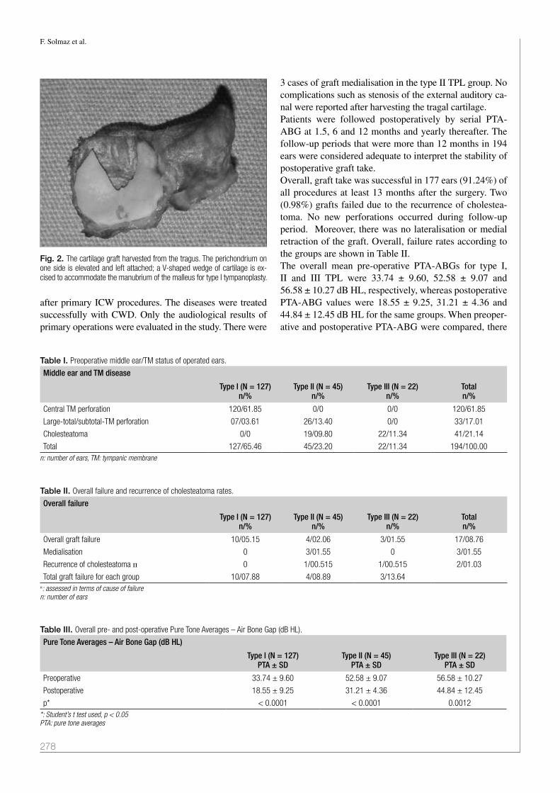
F. Solmaz et al.
278
after primary ICW procedures. The diseases were treated successfully with CWD. Only the audiological results of primary operations were evaluated in the study. There were
3 cases of graft medialisation in the type II TPL group. No complications such as stenosis of the external auditory ca-nal were reported after harvesting the tragal cartilage. Patients were followed postoperatively by serial PTA-ABG at 1.5, 6 and 12 months and yearly thereafter. The follow-up periods that were more than 12 months in 194 ears were considered adequate to interpret the stability of postoperative graft take. Overall, graft take was successful in 177 ears (91.24%) of all procedures at least 13 months after the surgery. Two (0.98%) grafts failed due to the recurrence of cholestea-toma. No new perforations occurred during follow-up period. Moreover, there was no lateralisation or medial retraction of the graft. Overall, failure rates according to the groups are shown in Table II.The overall mean pre-operative PTA-ABGs for type I, II and III TPL were 33.74 ± 9.60, 52.58 ± 9.07 and 56.58 ± 10.27 dB HL, respectively, whereas postoperative PTA-ABG values were 18.55 ± 9.25, 31.21 ± 4.36 and 44.84 ± 12.45 dB HL for the same groups. When preoper-ative and postoperative PTA-ABG were compared, there
Fig. 2. The cartilage graft harvested from the tragus. The perichondrium on one side is elevated and left attached; a V-shaped wedge of cartilage is ex-cised to accommodate the manubrium of the malleus for type I tympanoplasty.
Table I. Preoperative middle ear/TM status of operated ears.
Middle ear and TM disease
Type I (N = 127)n/%
Type II (N = 45)n/%
Type III (N = 22)n/%
Totaln/%
Central TM perforation 120/61.85 0/0 0/0 120/61.85
Large-total/subtotal-TM perforation 07/03.61 26/13.40 0/0 33/17.01
Cholesteatoma 0/0 19/09.80 22/11.34 41/21.14
Total 127/65.46 45/23.20 22/11.34 194/100.00n: number of ears, TM: tympanic membrane
Table II. Overall failure and recurrence of cholesteatoma rates.
Overall failure
Type I (N = 127)n/%
Type II (N = 45)n/%
Type III (N = 22)n/%
Totaln/%
Overall graft failure 10/05.15 4/02.06 3/01.55 17/08.76
Medialisation 0 3/01.55 0 3/01.55
Recurrence of cholesteatoma п 0 1/00.515 1/00.515 2/01.03
Total graft failure for each group 10/07.88 4/08.89 3/13.64п: assessed in terms of cause of failuren: number of ears
Table III. Overall pre- and post-operative Pure Tone Averages – Air Bone Gap (dB HL).
Pure Tone Averages – Air Bone Gap (dB HL)
Type I (N = 127)PTA ± SD
Type II (N = 45)PTA ± SD
Type III (N = 22)PTA ± SD
Preoperative 33.74 ± 9.60 52.58 ± 9.07 56.58 ± 10.27
Postoperative 18.55 ± 9.25 31.21 ± 4.36 44.84 ± 12.45
p* < 0.0001 < 0.0001 0.0012*: Student’s t test used, p < 0.05 PTA: pure tone averages

Perichondrium attached cartilage tympanoplasty (PACIT)
279
was a statistically significant improvement in PTA-ABG for all three TPL types (p < 0.05) (Table III).Evaluation of the difference between pre- and post-op-erative air conduction levels revealed that the latter de-creased in 8 (4.12%) ears; the early postoperative hearing results (at 1.5 and 3 months after surgery) remained stable in all patients. Postoperative hearing results in our study showed an improvement (≥ 10 dB) in 149 ears (76.81%) with a mean gain of 20 dB (range 10-40 dB). The dif-ference between pre- and post-operative air conduction levels was unchanged (< 10, 0 ≥ dB) in 37 (19.07%) ears. The improvements in postoperative hearing levels regard-ing tympanoplasty types are summarized in Table IV.
DiscussionTympanoplasty is performed to repair the tympanic mem-brane. With this surgical procedure there are two main targets. The first aim is to close the tympanic membrane and to have a closed middle ear, which will prevent the ear from subsequent infection and drainage. The second is to provide free movement of ossicles, which will result in better sound transmission and hearing. Skin, fascia la-ta, vein, temporalis fascia, perichondrium and dura mater have been used in the literature 7. Today, temporalis fascia is the most commonly employed material for tympanic membrane reconstruction 8.The use of cartilage is proposed in middle ear surgery,
since it offers a reliable technique in cases of advanced middle ear pathology and eustachian tube dysfunction 9.
In 1963, Salen 10 and Jansen 11 first reported the use of cartilage composite grafts for tympanic membrane re-construction. The interest for this material has risen in last decade. Clinical and experimental studies show that the cartilage is well tolerated in tympanoplastic sur-gery 12 13.
Despite the thickness of cartilage, various authors pointed out equal or better hearing results for the cartilage com-paring to temporalis fascia in tympanoplasty 9 14. Shield cartilage TPL using sliced tragal cartilage-perichondrium composite graft in terms of functional and anatomic re-sults has been studied. The perforation closure and air bone gap closure was reported to be successful 15. In a systematic review on 1,475 patients comparing cartilage and fascia graft in TPL, no statistically significant differ-ence between cartilage and temporalis fascia regarding function and hearing outcome was reported. In addition, TPL using cartilage with or without perichondrium was reported to have better morphological outcomes than TPL using temporalis fascia 16.Cartilage grafts may be used in different ways, such as perichondrium cartilage island graft, palisade or shield graft and cartilage reinforcement techniques 4 5. The use of cartilage as shield graft in tympanic membrane recon-struction is an easily applied technique, and is recom-mend by many authors 17. The reconstructed membrane
Table IV. Hearing improvement after the operations in tympanoplasty groups.
Audiological change**(dB HL)
Type I (N = 127)n/%
Type II (N = 45)n/%
Type III (N = 22)n/%
Overalln/%
Improved (≥ 10 dB 106/83.46 29/64.45 13/59.09 148/76.29
No Change (< 10, 0 ≥ dB) 21/16.54 10/22.22 04/18.18 35/18.04
Worsened (< 0 dB) 00/00.00 06/13.33 05/22.73 11/05.67
Total 127/100.00 45/100.00 22/100.00 194/100.00**: The difference = preoperative air conductive level – postoperative air conduction level (dB HL)n: number of ears
Fig. 3. Postoperative appearance of cartilage graft in type I, II and III tympanoplasty. Type I in a right ear at postoperative 15th month (a); type II-intact canal wall in a right ear at postoperative 13th month (b); type III-canal wall down at 18th month postoperatively. Facial ridge (single asterisk), cartilage graft (double asterisk), mastoid cavity (three asterisks) (c).

F. Solmaz et al.
280
will have resistance against the negative middle ear pres-sure, and long term integrity will be provided. Cartilage graft is mostly preferred in the posterosuperior retraction pockets of TM, since recurrent retractions are observed with the use of fascia graft over time 18.The cartilage graft may be harvested from tragus or con-cha. We prefer the tragal cartilage, because of its flat sur-face, ideal thickness and sufficient size. It was used as car-tilage island graft with the perichondrium attached to one side, since its thickness is less than 1 mm.17
Guneri et al. 3 reported a mean gain of 20 dB (64%) in a retrospective clinical study presenting their experience with cartilage grafts in ear surgery; postoperative hearing levels were improved in all type III tympanoplasties with ossicular reconstruction without mastoidectomies (100%), followed by CWD tympanomastoidectomy with ossicular reconstruc-tion (66%), type I TPL (62%), and ICW tympanomastoidec-tomy with ossicular reconstruction (25%).Cavaliere et al. 7 reported their personal experience with “tragal cartilage shield” graft in TPL with 306 adult pa-tients. Graft take was achieved in 304 patients (99.35%) and there was no immediate post-operative complication. The overall preoperative PTA-ABG was 43.79 ± 7.07 dB, whereas it was 10.43 ± 5.25 dB at one year after the sur-gery. Conchal cartilage, shaped as a shield, was used to replace the entire tympanic membrane and reconstruct the ossicular chain in patients with an absent incus as type III cartilage “shield” TPL. It was reported as an effective technique for hearing improvement in selected patients with chronic otitis media 19.Ozbek et al. 20 studied the long-term efficacy of cartilage palisades in TPL for atelectatic ears. The mean follow-up time was 44.5 months. The closure of tympanic mem-brane perforation was achieved in 91% of ears. Postop-erative PTA-ABG was less than 20 dB in 71% of ears. The average preoperative and postoperative ABG values, including all types of tympanoplasty operations (type I, II and III), were 28.4 ± 5.8 and 16.9 ± 6.7 dB, respectively (p < 0.001). They concluded that palisade cartilage tym-panoplasty was an effective technique for tympanic mem-brane closure and hearing improvement in atelectatic ears. They also indicated that the mastoidectomy did not affect anatomic or audiological outcomes. The reported follow-up rates in the current literature are between 19.9 months and 12 years according to our search 3 7 20-22. Mean follow-up time was 68.64 months in the present study. It is a relatively long time period, com-pared with many studies in the literature. Of the 41 ears with cholesteatoma, recurrent disease emerged in 2 patients after ICW procedures, which were managed successfully with CWD. The overall take rate was 91.24% for all patients; in type I, II, III TPL take rates were 92.12%, 91.11% and 86.36% respectively. No new re-perforations occurred during follow-up period. The post-operative hearing results in our series showed an improve-
ment in 76.29%, no change in 18.04% and a worsening in 5.67% of ears. Of the 11 ears whose hearing levels wors-ened postoperatively, 6 had primary CWD + Type II TPL and 5 had primary CWD + Type III TPL. Decreasing hearing levels may be due to the structural changes of middle ear during surgery.
ConclusionsOur study represents the results of PACG in type I, II and III primary TPL for treatment of COM with or without cholesteatoma. A limitation of study is the heterogeneity of the middle ear pathologies: we did not evaluate the ef-fect of atelectasis, cholesteatoma and revision on the suc-cess rate of TPL. The inherent disadvantages of a retro-spective study prevent formation of a true control group, and there may also be bias in selection of techniques. The overall graft take rate for PACIT was 91.24% at least 13 months after surgery. Postoperative PTA-ABG results demonstrated a significant improvement in our study for all types of TPL. The worsening hearing results were ob-served in the ears that underwent CWD due to cholestea-toma. This situation may depend on the structural changes of middle ear during surgery, rather than the type of graft used for tympanic membrane reconstruction. A cartilage island graft with the perichondrium attached to one side harvested from tragus may be useful in type I, II and III TPL due to high stability and good hearing results.
References1 Wolferman A, Farrior JB. Classification of tympanoplasty.
Arch Otolaryngol 1971;94:380.2 Kerr AG, Byrene JET, Smyth GDL. Cartilage homografts in
the middle ear: longterm histologic study. J Laryngol Otol 1973;87:1193-9.
3 Güneri EA, Ikiz AO, Erdag TK, et al. Cartilage tympa-noplasty: Indications, techniques and results. J Otol-Head Neck Surg 2009;38:362-8.
4 Dornhoffer J. Cartilage tympanoplasty: indications, tech-niques and outcomes in a 1,000-patient series. Laryngo-scope 2003;113:1844-56.
5 Neumann A, Schultz-Coulon HJ, Jahnke K. Type III tympan-oplasty applying the palisade technique: a study of 61 cases. Otol Neurotol 2003;24:33-7.
6 Wullstein H. Theory and practice of tympanoplasty. Laryn-goscope 1956;66:1076-93.
7 Cavaliere M, Mottola G, Rondinelli M, et al. Tragal carti-lage in tympanoplasty: anatomic and functional results in 306 cases. Acta Otorhinolaryngol Ital 2009;29:27-32.
8 Sheehy JL, Anderson RG. Myringoplasty. A review of 472 cases. Ann Otol Rhinol Laryngol 1980;89:331-4.
9 Amedee RG, Mann WJ, Riechelmann H. Cartilage palisade tympanoplasty. Am J Otol 1989;10:447-50.
10 Salen B. Myringoplasty using septum cartilage. Acta Otolar-yngol 1963;188:82-91.

Perichondrium attached cartilage tympanoplasty (PACIT)
281
11 Jansen C. Cartilage-tympanoplasty. Laryngoscope 1963;73:1288-302
12 Yamamoto E, Iwanaga M, Fukumoto M. Histologic study of homograft cartilage implanted in the middle ear. Otolaryngol Head Neck Surg 1988;98:546-51.
13 Hamed M, Samir M, El Bigermy M. Fate of cartilage mate-rial used in middle ear surgery light and electron microscopy study. Auris Nasus Larynx 1999;26:257-62.
14 Heermann J. Autograft tragal and conchal palisade cartilage and perichondrium in tympanomastoid reconstruction. Ear Nose Throat J 1992;71:344-9.
15 Khan MM, Parab SR. Primary cartilage tympanoplasty: our technique and results. Am J Otolaryngol 2011;32:381-7.
16 Mohamad SH, Khan I, Hussain SS. Is cartilage tympano-plasty more effective than fascia tympanoplasty? A system-atic review. Otol Neurotol 2012;33:699-705.
17 Gerber MJ, Mason JC, Lambert PR. Hearing results af-
ter primary cartilage tympanoplasty. Laryngoscope 2000;110:1994-9.
18 Couloigner V, Molony N, Viala P, et al. Cartilage tympa-noplasty for posterosuperior retraction pockets of the pars tensa in children. Otol Neurotol 2003;24:264-9.
19 Kyrodimos E, Sismanis A, Santos D. Type III cartilage “shield” tympanoplasty: an effective procedure for hearing improvement. Otolaryngol Head Neck Surg 2007;136:982-5.
20 Ozbek C, Ciftçi O, Ozdem C. Long-term anatomic and func-tional results of cartilage tympanoplasty in atelectatic ears. Eur Arch Otorhinolaryngol 2010;267:507-13.
21 Onal K, Arslanoglu S, Oncel S, et al. Perichondrium/carti-lage island flap and temporalis muscle fascia in type I tym-panoplasty. J Otolaryngol Head Neck Surg 2011;40:295-9.
22 Velepic MM, Manestar D, Starcević R et al. Full thick-ness cartilage palisade tympanoplasty with malleus inter-position: a study of the long term results. Coll Antropol 2014;38:283-7.
Address for correspondence: Davut Akduman, Duzce Universite-si, Tip Fakültesi, Kulak Burun Boğaz Hastalıkları Anabilim Dalı, Duzce, Turkey. Tel. +90 380 542 14 16. Fax +90 380 542 13 02. E-mail: [email protected]
Received: March 27, 2015 - Accepted: January 10, 2016
282
ACTA OTORHINOLARYNGOLOGICA ITALICA 2016;36:282-288; doi: 10.14639/0392-100X-783
Otology
Role of Kabat rehabilitation in facial nerve palsy: a randomised study on severe cases of Bell’s palsy Ruolo della riabilitazione Kabat nella paralisi del nervo facciale: studio randomizzato su casi severi di paralisi di Bell
S. Monini1, C.M. iaCoLUCCi1, M. Di TraGLia2, a.i. Lazzarino3, M. barbara1
1 otorhinolaryngology Clinic, neSMoS Department, Medicine and Psychology, Sapienza University, rome, italy; 2 Department of Public Health and infectious Diseases, Sapienza University, rome, italy; 3 epidemiology and Statistics, imperial College, London, Uk
SUMMARY
The treatment of Bell’s palsy (BP), based on steroids and/or antiviral drugs, may still leave a certain percentage of affected subjects with disfiguring sequelae due to incomplete recovery. The different procedures of physical rehabilitation have not been demonstrated to play a favourable role in this disorder. The aim of the present study was to compare functional outcomes in severe cases of Bell’s palsy when treated by steroids alone or by steroids accompanied by Kabat physical rehabilitation. This prospective study included 94 subjects who showed sudden facial nerve (FN) palsy with House-Brackmann grade IV or V and who were divided into two groups on the basis of the therapeutic approach: one group (a) was treated by steroids, and the other (b) received steroids in combination with physical rehabilita-tion. Medical treatment consisted in administration of steroids at a dosage of 60 mg per day for 15 days; physical rehabilitative treatment consisted in proprioceptive neuromuscular facilitation according to Kabat, and was administered to one of the two groups of subjects. Recovery rate, degree of recovery and time for recovery were compared between the two groups using the Mann-Whitney and univariate logistic regression statistical tests (Ward test). Kabat patients (group b) had about 20 times the odds of improving by three HB grades or more (OR = 17.73, 95% CI = 5.72 to 54.98, p < 0.001) than patients who did not receive physical treatment (group a). The mean speed of recovery in group b was the half of that recorded for group a (non-Kabat subjects). No difference was observed in the incidence of synk-ineses between the two groups. Steroid treatment appears to provide better and faster recovery in severe cases (HB IV and V) of BP when complemented with Kabat physical rehabilitation.
KEY WORDS: Bell’s palsy • Physical rehabilitation • Kabat • House-Brackmann grading system • Steroid treatment
RIASSUNTO
La terapia della paralisi di Bell, incentrata su farmaci steroidei e/o antivirali, può ancora far esitare nei soggetti affetti sequele disfigu-ranti per un recupero incompleto. Le diverse procedure riabilitative non si sono dimostrate al giorno d’oggi in grado di giocare un ruolo favorevole in questo senso. Scopo di questo lavoro è stato quello di mettere a confronto i risultati funzionali di pazienti affetti da forme severe di paralisi di Bell, quando trattati con solo cortisone con quelli nei quali al cortisone è stata affiancata una terapia riabilitativa secondo Kabat. Lo studio prospettico ha incluso 94 soggetti con paralisi di Bell di grado IV e V secondo House-Brackmann (HB) a loro volta suddivisi in due gruppi: (a) trattato con terapia steroidea; (b) trattato con terapia steroidea e riabilitazione Kabat. Il trattamento medico è consistito di 60 mg di prednisolone al giorno per 15 giorni; la terapia riabilitativa è consistita nel trattamento di facilitazione neuromuscolare propiocettiva secondo Kabat. Percentuale, grado e tempi di recupero sono stati comparati utilizzando l’analisi statistica Mann-Whitney e il test di regressione logistica multivariata (Ward test). I pazienti Kabat (gruppo b) hanno avuto 20 volte di più la possibi-lità di migliorare di 3 o più gradi HB (OR = 17,73, 95% IC = 5,72 a 54,98, p < 0,001) rispetto a quelli di gruppo a. La velocità media di recupero nel gruppo b è risultata la metà di quella registrata nel gruppo a. Nessuna differenza è stata invece riscontrata sull’incidenza di sincinesie. Si può dunque concludere che la terapia steroidea permette un migliore e più rapido recupero dei casi severi di paralisi di Bell, quando associata a terapia riabilitativa Kabat.
PAROLE CHIAVE: Paralisi di Bell • Terapia riabilitativa • Kabat • Classificazione House-Brackmann • Terapia steroidea
Acta Otorhinolaryngol Ital 2016;36:282-288
IntroductionBell’s palsy (BP) is an acute, sudden unilateral periph-eral paralysis of the facial nerve (FN) that accounts for almost 75% of all acute facial palsies (FP). The annual
incidence varies between 11 and 40 cases per 100,000 subjects 1 2 or even higher, as indicated by a recent sur-vey 3.The aetiology of BP is still unknown, but reactivation of a latent Herpes simplex virus would seem to play a major

Kabat rehabilitation in severe Bell’s palsy
283
role 4. Inflammation and entrapment of the nerve in the narrow bony labyrinthine segment of the Fallopian canal have specifically been proposed as possible mechanisms for inducing nerve sufferance 4 5.The therapeutic options are not universally accepted in terms of priority. Medical treatment is based on adminis-tration of corticosteroids, and several studies have shown that they may be able to reduce the time of recovery and occurrence of sequelae 6 7, while no evidence has been gathered on the adjunctive role of antiviral drugs 7. Surgical decompression of the FN has also been proposed in the past, with the rationale to release the edematous nerve from its bony canal, but its efficacy has been re-cently debated 8 and, in recent years, the number of these procedures has drastically reduced. Several procedures of FN rehabilitation have also been applied in combination with medical treatment, mostly in the post-surgical FP setting, although a recent Cochrane review was unable to identify any significant benefit or harm by any type of rehabilitative procedure 9. When analysing physical rehabilitative procedures, the recent scientific contributions have examined the application of manual therapies 10-12, electrical stimulation 13, exercises and, ago-puncture 14 15 and laser 16. Barbara et al. compared clinical outcomes and ENoG values of patients affected by BP and rehabilitated with the Kabat method 17, show-ing that the better and faster recovery of an early rehabili-tative protocol was independent of the nerve condition as assessed by ENoG findings 18.The purpose of the present study was to shed light on the favourable role played by Kabat physical rehabilitation in the outcomes of subjects affected by severe BP. Clinical outcomes were compared between two different proto-cols: pharmacological treatment associated with physical rehabilitation and pharmacological treatment alone.
Materials and methodsA series of consecutive subjects admitted to the Emergency Department (ED) of a tertiary University Hospital with diagnosis of BP during the years 2005-2012 were consid-ered. In all, FN palsy was graded according to the House-Brackmann (HB) system 19. From a total of 390 patients, only those staged HB IV or higher were taken into consid-eration. Pregnant women and individuals with involvement of other cranial nerves were also excluded from the study. Ninety-six patients, hence composing the study group, were enrolled: HB IV (66 subjects) and HB V (28 subjects).Randomisation of these patients was performed by the personnel working at the ED that, on the basis of indi-vidual judgment on the FN severity, sent 66 BP patients home and 28 to our FN Center for consultation. In this way, two separate groups were identified: • study group a, formed by 66 patients, aged 16-90 years
(mean = 57.1), 30 females and 36 males, 58 with HB
IV and 8 with HB V. All these subjects were sent home by the ED with medical steroid protocol treatment, eye drops for eye protection and paracetamol in case of pain;
• study group b, formed by 28 patients, aged 32-74 years (mean = 55.7), 13 males, 15 females, 10 with HB IV and 18 with HB V. All these patients were sent for consultations to our FN Centre by ED staff. At our Centre, standard facial movements (brow lift, gen-tle eye closure, forced eye closure, snarling, smiling and lip pucking) were documented in each patient by video-recording with a mobile phone, and reviewed by a senior ENT to assign the appropriate HB grade. The Kabat physical rehabilitation was started within 7 days from BP onset, and was associated for two weeks with medical treatment, consisting of daily prednisolone (2 mg/kg), for 10 days and tapered off within 2 weeks.
Group b subjects were evaluated at our Centre twice a week, during the rehabilitation sessions and at the end of the cycle, defined with the maximum improvement of FN function that remained stable for a least 1 month of follow-up. Once a week from FP onset, group b pa-tients were re-evaluated, so as to define the progression or eventual recovery in days via direct clinical observa-tion.At day 7 from FP onset, group a subjects received a tel-ephone contact and were classified according to the ca-pability of eye closure, so as to distinguish grade III or better from grade IV or worse. Next, they were asked to self-assess and report in a diary, from that day on, facial features such as:1. symmetry of the face at rest: if the right and left sides
of the face appeared similar at rest (without any move-ment);
2. facial motion through different facial movements (brow lift, gentle eye closure, forced eye closure, snarl-ing, smiling and lip picking).
Kabat rehabilitation consists in the facilitation of the vol-untary response of an impaired muscle through the global pattern of an entire muscular section that undergoes re-sistance. This method appears to be extremely rational for facial muscles, since most of the face muscular fibers run diagonally, with easy irradiation to the upper facial region due to the cross-FN innervations. Three regional fulcra were taken into consideration: upper, intermediate and lower fulcrum. The first (forehead and eyes) is connected via a vertical axis to the intermediate one (nose), while the lower mimic-chewing-articulatory fulcrum lies along the horizontal axis. Hence, action on the upper fulcrum also involves the other two fulcra. The manipulation of these three fulcra is carried out by utilising both contralateral contractions and basic propioceptive stimulation, includ-ing stretching, maximal resistance, manual contact and verbal input. In the upper fulcrum, the activation of the

S. Monini et al.
284
frontal, corrugators and orbicularis muscles is carried out by their upwards or downwards traction, which is always in a vertical plane depending on the specific function that needs to be activated. In the intermediate fulcrum, activa-tion of the common elevator muscle of the ala nasi and the upper lip is also carried out using traction movements, in this case contrary to the normal direction, following a vertical line. For the lower fulcrum, the maneuvers are carried out on the risorium and orbicularis oris muscles in a horizontal plane, and on the mental muscle in a vertical plane. For statistical purposes, the time of recovery has been ex-pressed in days. The time of recovery and the final HB degree of palsy were compared between groups.
Statistical analysisThe dataset was cleaned before analysis with respect to inconsistent and missing values, duplicates, outliers, digit preference and normality of continuous variables. The non-parametric Mann-Whitney-Wilcoxon (MWW) U test was used to verify the null hypothesis that the two study groups are the same against an alternative hypothesis, namely that one group would tend to have better recovery values than the other. Univariate logistic regression (Ward test) was used to compare all rehabilitated (group b) and non-rehabilitated (group a) patients for the final grade of improvement, degree of improvement (1-grade, 2 grades, 3 grades and 4-grades) and timing of recovery. The effect of treatment was evaluated by three different approaches, adjusted for age and gender: i) multiple linear regression to model the paired change of HB grade between before and after treatment (at the end of follow-up); ii) multi-ple logistic regression to model the chances of having a paired decrease in disease status by three HB grades or
more, before and after treatment (at the end of follow-up); iii) multiple Cox proportional hazards regression for time of improvement (speed of recovery), with follow-up time in days (information about patient status was retrieved daily). Statistical assumptions underlying the tests were systematically checked.
ResultsGrade IV at baseline was recorded in 87.9% of group a and in 35.7% of group b. Grade V at baseline was pre-sent in 12.1% of group a and 64.3% of group b. Group b subjects with grade V at baseline underwent Kabat treatment and showed better improvement after treat-ment with a faster recovery time in comparison with group a (64.6 days in respect to 117.1 days in group a). Moreover, at the end of follow-up, in group b there was no case of one-grade improvement, while an improve-ment by four grades was found in 25.0% of subjects; in group a, one-grade improvement was observed in 16.7% of patients, and none showed a four-grade im-provement (Table I).When comparing (MWW) the rehabilitated (group b) versus non-rehabilitated (group a) groups, independently of the initial FP grade, significant differences were found (p = 0.009546) and shown in Figure 1. With respect to the entire study group, a greater significance between the two modalities of treatment was identified when taking into account individually grade IV (p = 0.0001349) and grade V subjects (p = 0.009025) (Fig. 2 and 3). The multiple linear regression model for the change in HB grade after treatment is reported in Table II. With respect to group a, in the rehabilitated group (b) the HB grade decreased by one further point (coefficient = -0.99%, 95% CI = -1.27/1.71, p < 0.001), in HB IV (coefficient = 0.86,
Table I. Sample description by categories of treatment strategy.
Variable
Non-Kabat Kabat
p*
(n = 66) (n = 28)
Mean or proportion SD Min Max
Mean or proportion SD Min Max
Age 56.8 17.9 16 90 55.8 12.7 32 74 0.778
Male 54.5% 57.1% 0.817
HB V at onset 12.1% 64.3% < 0.001
Synkineses after treatment 12.1% 7.1% 0.479
Time to first improvement (days) 117.1 90.3 34 365 64.6 50.7 12 194
Grade improvement † at the end of follow-up 2.0 0.6 1 3 3.0 0.7 2 4 <0.001
1-grade improvement 16.7 0.0
2-grades improvement 69.7 28.6
3-grades improvement 13.6 46.4
4-grades improvement 0.0 25.0* P values were computed using univariate logistic regression (Wald test) with Kabat as outcome.* Negative values indicate improvement, i.e. decrease in grade.† Improvement means decrease in grade after treatment.

Kabat rehabilitation in severe Bell’s palsy
285
95% CI = -1.23/0.49, p < 0.001) and in HB V ((coefficient = -1.10, 95% CI = -1.82/0.38, p < 0.005).Group b patients showed about 20 times the odds of improv-ing by three HB grades or more (OR = 17.73, 95% CI = 5.72 to 54.98, p < 0.001) than group a (Table III). Group b HB IV patients had 65 times the odds of a three-grade improvement
than those in group a (non rehabilitated), while a three-grade improvement was only observed in HB V patients in group b. The mean speed of recovery in group b was half of that recorded in group a (HR = 2.19, 95% CI = 1.37 to 3.51, p = 0.001). Group b HB IV patients had seven times shorter times of recovery than similar grades of group a
Fig. 1. Comparison between rehabilitated and non-rehabilitated groups (Mann-Whitney (U-test), HB IV and V together, independently of the severity of facial palsy at onset. A significant difference in the outcome was found between groups (p = 0.009546).
Fig. 2. Comparison between rehabilitated and non-rehabilitated groups (Mann-Whitney (U-test) in HB IV subjects, independently of the severity of facial palsy at onset. A significant difference in the outcome was found between groups (p = 0.0001349).
Fig. 3. Comparison between rehabilitated and non-rehabilitated groups (Mann-Whitney (U-test), in HB V subjects, independently of the severity of facial palsy at onset. A significant difference in the outcome was found between groups (p = 0.009025).
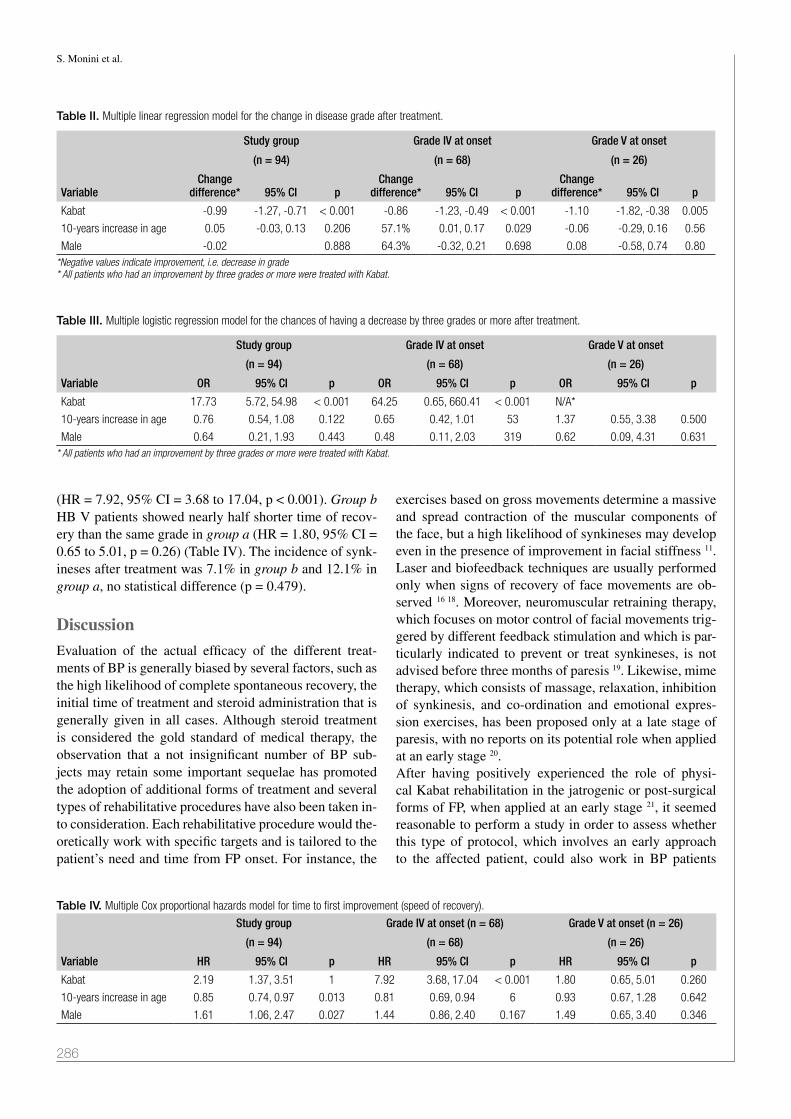
S. Monini et al.
286
(HR = 7.92, 95% CI = 3.68 to 17.04, p < 0.001). Group b HB V patients showed nearly half shorter time of recov-ery than the same grade in group a (HR = 1.80, 95% CI = 0.65 to 5.01, p = 0.26) (Table IV). The incidence of synk-ineses after treatment was 7.1% in group b and 12.1% in group a, no statistical difference (p = 0.479).
DiscussionEvaluation of the actual efficacy of the different treat-ments of BP is generally biased by several factors, such as the high likelihood of complete spontaneous recovery, the initial time of treatment and steroid administration that is generally given in all cases. Although steroid treatment is considered the gold standard of medical therapy, the observation that a not insignificant number of BP sub-jects may retain some important sequelae has promoted the adoption of additional forms of treatment and several types of rehabilitative procedures have also been taken in-to consideration. Each rehabilitative procedure would the-oretically work with specific targets and is tailored to the patient’s need and time from FP onset. For instance, the
exercises based on gross movements determine a massive and spread contraction of the muscular components of the face, but a high likelihood of synkineses may develop even in the presence of improvement in facial stiffness 11. Laser and biofeedback techniques are usually performed only when signs of recovery of face movements are ob-served 16 18. Moreover, neuromuscular retraining therapy, which focuses on motor control of facial movements trig-gered by different feedback stimulation and which is par-ticularly indicated to prevent or treat synkineses, is not advised before three months of paresis 19. Likewise, mime therapy, which consists of massage, relaxation, inhibition of synkinesis, and co-ordination and emotional expres-sion exercises, has been proposed only at a late stage of paresis, with no reports on its potential role when applied at an early stage 20. After having positively experienced the role of physi-cal Kabat rehabilitation in the jatrogenic or post-surgical forms of FP, when applied at an early stage 21, it seemed reasonable to perform a study in order to assess whether this type of protocol, which involves an early approach to the affected patient, could also work in BP patients
Table II. Multiple linear regression model for the change in disease grade after treatment.
Variable
Study group Grade IV at onset Grade V at onset
(n = 94) (n = 68) (n = 26)
Change difference* 95% CI p
Change difference* 95% CI p
Change difference* 95% CI p
Kabat -0.99 -1.27, -0.71 < 0.001 -0.86 -1.23, -0.49 < 0.001 -1.10 -1.82, -0.38 0.005
10-years increase in age 0.05 -0.03, 0.13 0.206 57.1% 0.01, 0.17 0.029 -0.06 -0.29, 0.16 0.56
Male -0.02 0.888 64.3% -0.32, 0.21 0.698 0.08 -0.58, 0.74 0.80*Negative values indicate improvement, i.e. decrease in grade* All patients who had an improvement by three grades or more were treated with Kabat.
Table III. Multiple logistic regression model for the chances of having a decrease by three grades or more after treatment.
Variable
Study group Grade IV at onset Grade V at onset
(n = 94) (n = 68) (n = 26)
OR 95% CI p OR 95% CI p OR 95% CI p
Kabat 17.73 5.72, 54.98 < 0.001 64.25 0.65, 660.41 < 0.001 N/A*
10-years increase in age 0.76 0.54, 1.08 0.122 0.65 0.42, 1.01 53 1.37 0.55, 3.38 0.500
Male 0.64 0.21, 1.93 0.443 0.48 0.11, 2.03 319 0.62 0.09, 4.31 0.631* All patients who had an improvement by three grades or more were treated with Kabat.
Table IV. Multiple Cox proportional hazards model for time to first improvement (speed of recovery).
Variable
Study group Grade IV at onset (n = 68) Grade V at onset (n = 26)
(n = 94) (n = 68) (n = 26)
HR 95% CI p HR 95% CI p HR 95% CI p
Kabat 2.19 1.37, 3.51 1 7.92 3.68, 17.04 < 0.001 1.80 0.65, 5.01 0.260
10-years increase in age 0.85 0.74, 0.97 0.013 0.81 0.69, 0.94 6 0.93 0.67, 1.28 0.642
Male 1.61 1.06, 2.47 0.027 1.44 0.86, 2.40 0.167 1.49 0.65, 3.40 0.346

Kabat rehabilitation in severe Bell’s palsy
287
presenting with a HB grade IV or worse. In fact, to our knowledge, studies comparing the effect of physical treatment to that of exclusive steroid treatment in BP are scanty. In the present study, the composition of the study groups was not achieved by randomisation, but by the in-dividual attitude of the ED staff to either send patients home or to our attention for consultation, although the former protocol mostly included less severe cases. In both instances, all patients received steroid treatment, while the Kabat rehabilitation was only performed on subjects that were sent and followed directly at our Clinic and that formed study group b. In the present study, only the most severe cases, i.e. HB IV and V subjects, were taken into account for the final evaluation. Through this methodology, it was possible to have comparable patients in the study groups, as well as in HB IV or V subjects separately, for assessment of the primary outcomes of the study, i.e. recovery time, final HB grade and probability to achieve a 3-grade HB im-provement. Although group a (steroids only) patients were numeri-cally greater than those of group b, this latter included only the most severe HB grades and therefore all were ad-dressed to rehabilitative treatment. The use of such physi-cal rehabilitation led to a faster recovery time in group b, nearly half of that recorded in group a subjects, while for HB IV subjects it was reduced by seven times less than in group a. After physical rehabilitation, the degree of improvement (grade reduction at the HB) at the time of definitive clinical outcome was significant in comparison to the final condition of patients treated only with steroids, since patients treated simultaneously with pharmacologi-cal and rehabilitative therapy recovered in a significant number to normality (HB I), while in most of the patients who received only steroids, the maximum recovery was HB II. When considering HB IV only, the significance value of difference between rehabilitated and non-rehabilitated cases was ten times less than that observed in the entire group (HB IV and V); the same finding was observed in HB V, with less significance than HB IV, due to a marked intersection of values at the HB III level. This is why in the most severe subjects (HBV), there was a prevalence of recovery to HB III in non-rehabilitated cases and to HBI in rehabilitated subjects.Moreover, group b patients recovered by three grades two times more frequently than those in group a, and in group b the degree decreased, i.e. improved, by one further point, in total and single grades, compared to group a. In group b, moreover, a 20 fold higher prob-ability of improvement by three HB grades was seen, and the most severe cases had similar improvement only when rehabilitated.Most of the severe (HB V), non-rehabilitated patients reached as maximum result of HB III, while similar cases
when undergoing Kabat rehabilitation could even obtain a normal facial function (HB I).
Conclusions It is possible to conclude that, when Kabat rehabilitation is associated with standard steroid treatment in the case of severe BP, affected subjects may be likely to have a fast-er and better recovery than those in whom only medical treatment is applied. It would therefore be worthwhile to always include this type of physical rehabilitation in pa-tients with BP, especially in the most severe cases which may carry the risk of disfiguring facial sequelae.
References1 De Diego-Sastre JI, Prim-Espada MP, Fernàndez-
Garcìa F. The epidemiology of Bell’s palsy. Rev Neurol 2005;41:287-90.
2 Finsterer J. Management of peripheral facial nerve palsy. Eur Arch Otorhinolaryngol 2008;265:743-52.
3 Monini S, Lazzarino AI, Iacolucci C, et al. Epidemiology of Bell’s palsy in an Italian Health District: incidence and case-control study. Acta Otolaryngol It 2010;30:198-206.
4 Murakami S, Mizobuchi M, Nakashiro Y, et al. Bell’s palsy and herpes simplex virus: identification of viral DNA in endoneurial fluid and muscle. Ann Intern Med 1996;124:27-33.
5 McCormick DP. Herpes-simplex virus as a cause of Bell’s palsy. Lancet 1997;1:937-39.
6 Engström M, Berg T, Stjernguist-Desatnik A, et al. Predniso-lone and valaciclovir in Bell’s palsy: a randomised, double-blind, placebo-controlled, multicentre trial. Lancet Neurol 1998;7:993-1000.
7 Sullivan FM, Swan IRC, Donnan PT, et al. Early treatment with prednisolone or acyclovir in Bell’s palsy. New Eng J Med 2007;357:1598-607.
8 McAllister K, Walker D, Donnan PT, et al. Surgical inter-ventions for the early management of Bell’s palsy. Cochrane Database Syst Rev 2011;2:CD007468.
9 Mosforth J, Taverner D. Physiotherapy for Bell’s palsy. Br Med J 1958;2:675-77.
10 Flores PF, Medina RZ, Haro LG. Idiopathic peripheral fa-cial paralysis treatment: physic therapy versus prednisone [Tratamiento de la parálisis facial periférica idiopática: ter-apia física versus prednisona]. Revista médica del Instituto Mexicano del Seguro Social 1998;36:217-21.
11 Manikandan N. Effect of facial neuromuscular re-education on facial symmetry in patients with Bell’s palsy: a rand-omized controlled trial. Clin Rehabilitation 2007;21:338-43.
12 Alakram P, Puckree T. Effects of electrical stimulation on House-Brackmann scores in early Bell’s palsy. Physiothera-py Theory Pract 2010;26:160-6.
13 Wang XH, Zhang LM, Han M, et al. Treatment of Bell’s palsy with combination of traditional Chinese medicine and western medicine. Hua Xi Kou Qiang Yi Xue Za Zhi 2004;22:211-13.
14 Zhou ZL, Zuo C, Cheng SL, et al. Application of grading

S. Monini et al.
288
evaluation on facial nerve function of Bell’s palsy treated with electroacupuncture. Zhongguo Zhen Jiu 2013;33:692-6.
15 Alayat MS, Elsodany AM, El Fiky AA. Efficacy of high and low level laser therapy in the treatment of Bell’s palsy: a ran-domized double blind placebo-controlled trial. Lasers Med Sci 2014;29:335-42.
16 Kabat H, Knott M. Proprioceptive facilitation therapy for paralysis. Physiotherapy 1954;40:171-6.
17 Barbara M, Antonini G, Vestri A, et al. Role of Kabat physi-cal rehabilitation in Bell’s palsy: a randomized trial. Acta Otolaryngol 2010;130:167-72.
18 House JW, Brackmann DE. Facial nerve grading system. Otolaryngol Head Neck Surg 1985;93:146-7.
19 Diels HJ, Combs D. Neuromuscular retraining for facial pa-ralysis. Otolaryngol Clin North Am 1997;30:727-43.
20 Barbara M, Monini S, Buffoni A, et al. Early rehabilitation of facial nerve deficit after acoustic neuroma surgery. Acta Otolaryngol 2003;123:932-5.
21 Beurskens CH, Heymans PG. Mime therapy improves facial symmetry in people with long-term facial nerve paresis: A randomized controlled trial. Aus J Physioth 2006;52:177-83.
Address for correspondence: Maurizio Barbara, Sapienza Univer-sity Medicine and Psychology, NESMOS Department, via di Grot-tarossa 1035, 00189 Rome, Italy. Tel. +39 06 33775628. E-mail: [email protected]
Received: December 15, 2015 - Accepted: March 4, 2016

289
ACTA oTorhinolAryngologiCA iTAliCA 2016;36:289-294; doi: 10.14639/0392-100X-807
Laryngology
Malignant salivary gland tumours of the larynx: a single institution review Tumori maligni delle ghiandole salivari della laringe: un’unica review istituzionale
S. karaTaYLi-ozGUrSoY1 2, j.a. biSHoP1 3, a.T. HiLLeL1, L.M. akST1, S.r. beST1
1 Department of otolaryngology, Head and neck Surgery, johns Hopkins School of Medicine, baltimore, USa; 2 Department of otolaryngology, Ufuk University, ankara, Turkey; 3 Departments of Pathology, johns Hopkins School of Medicine, baltimore, USa
SUMMARY
Malignant salivary gland tumours of the larynx are very rare, with limited reports of clinical outcomes. We present the decade-long experi-ence of a single institution. A 10-year retrospective chart review of a tertiary head and neck cancer centre was performed. Index patients were identified from a review of a pathology database, and reviewed by a head and neck pathologist. Patient demographics, presenting signs and symptoms, treatment modalities and clinical outcomes were extracted from electronic medical records. Six patients were included, with an age range of 44 to 69. All six had malignant laryngeal salivary gland tumours. Pathologies included: three adenoid cystic carcinoma (2 supraglottic, 1 subglottic), one mucoepidermoid carcinoma (supraglottic), one epithelial-myoepithelial carcinoma (supraglottic) and one adenocarcinoma (transglottic). All were treated with surgery (2 endolaryngeal, 4 open) and five of six with the addition of adjuvant therapy (4 radiotherapy, 1 concurrent chemoradiation). One patient had smoking history; no patients had significant alcohol history. With 4.5 years of median follow-up, none of the patients has had recurrence or local/distant metastasis. Salivary gland tumours of the larynx present in mid to late-age, and can be successfully managed with a multi-modality approach, resulting in excellent local and regional control rates.
KEY WORDS: Larynx • Cancer • Salivary • Chemotherapy • Surgery • Partial laryngectomy
RIASSUNTO
I tumori a istotipo salivare della laringe sono molto rari, con pochi report in letteratura in merito al loro andamento clinico. Nel presente manoscritto discutiamo un’esperienza di 10 anni presso una singola struttura. Abbiamo condotto una review retrospettiva della casistica di un centro di oncologia della testa e del collo di terzo livello. I pazienti sono stati individuati mediante analisi di un database e sono stati revisionati da un Anatomo Patologo testa collo. I dati inerenti la clinica, le modalità di trattamento e gli esiti sono stati prelevati da archivi elettronici. Sono stati inclusi sei pazienti nello studio, con un range di età dai 44 ai 69 anni. Tutti e sei erano affetti da neoplasie maligne a istotipo salivare della laringe. Gli istotipi includevano: tre carcinomi adenoido-cistici (2 sopraglottico, 1 sottoglottico), un carcino-ma mucoepidermoidale (sopraglottico), un carcinoma epiteliale-mioepiteliale (sopraglottico), e un adenocarcinoma (transglottico). Tutti sono stati sottoposti a trattamento chirurgico (2 chirurgie laser, 4 open) e 5 dei 6 pazienti sono stati successivamente sottoposti a terapia adjuvante (4 a radioterapia, 1 a radio-chemioterapia concomitante). Un paziente era fumatore; nessun paziente aveva storia di abuso di alcolici. A un follow-up con mediana di 4,5 anni nessuno dei pazienti ha presentato recidiva o metastasi locali o a distanza. I tumori a istotipo salivare della laringe si presentano solitamente in pazienti della seconda/terza età, e possono essere trattati con successo mediante approcci multimodali, con un ottimo controllo locoregionale di malattia.
PAROLE CHIAVE: Laringe • Cancro • Salivare • Chemioterapia • Chirurgia • Laringectomia parziale
Acta Otorhinolaryngol Ital 2016;36:289-294
IntroductionMalignant salivary gland tumours of the larynx are very rare neoplasms, which account for < 1% of all laryngeal malignancies 1. The most common malignant minor sali-vary gland tumours are adenoid cystic (32-69%) and mu-coepidermoid carcinomas (15-35%) 2; adenocarcinomas are less frequent, and epithelial-myoepithelial carcinomas are even more rare 3. Unlike squamous cell carcinoma, which is strongly associated with inhaled tobacco use, malignant salivary tumours in the larynx have no strong
association with smoking and appear to occur equally in both sexes. These tumours arise from subepithelial mu-cous glands in the larynx, which are found most common-ly in the subglottis and supraglottis 4. In the true glottis, the possible areas of origin are floor of the ventricle and below the anterior commissure in the subglottis. Because these tumours do not arise on the free edge of the true vo-cal folds which are covered with thin squamous epitheli-um, there is often no voice change to detect these tumours at an early stage. They typically present in a subepithelial

S. Karatayli-Ozgursoy et al.
290
fashion, and can grow to a large size before they present with dysphonia or airway symptoms.The aim of this study is to present our experience over the last decade with these rare tumours to provide insight into the multimodality treatment that is typically required for long-term oncologic success.
Materials and methodsIn an IRB-approval protocol, the pathology archives of the Johns Hopkins Hospital and clinical records of pa-tients with laryngeal cancer were reviewed to identify patients with malignant salivary gland laryngeal lesions between January 2004 and December 2013. Age, gender, presenting symptoms, location of the tumour, pathology, TNM classifications, treatment modality (surgery and/or chemo radiotherapy) and disease status was extracted from patient charts.
ResultsSix patients (3M, 3F) were included, with an age at diag-nosis range of 44 to 69 (mean: 56 years). Pathologic clas-sifications are summarised in Table I and included: three adenoid cystic carcinomas, one mucoepidermoid carci-noma, one epithelial-myoepithelial carcinoma and one adenocarcinoma, not otherwise specified. The most com-mon presenting symptom was dysphonia. Half of patients presented with advanced stage disease, but all except the adenocarcinoma presented without regional or distant metastases. Only one patient was a smoker (T4 subglottic adenoid cystic carcinoma). Treatment modalities and oncologic results are summa-rised in Table I. All patients were treated primarily with
surgery, and negative surgical margins were obtained in all cases. The type of surgical approach varied with each case, but two were successfully managed with endola-ryngeal surgery, while four required an open surgical ap-proach. All but one patient underwent additional adjuvant therapy regardless of pathologic type or surgical margins (4 radiotherapy, 1 concurrent chemoradiation). After 4.5 years of median follow-up, none of the patients has had recurrence or local/distant metastasis. To highlight the unique presentation and management of these cases, three case studies with pathologic images are presented below.
Representative casesCase two: A 44-year-old male professor presented with dysphonia lasting two years. He denied any difficulty swallowing; however, he had a significant amount of coughing while eating. On fiberoptic laryngoscopy a la-ryngeal mass on the epiglottis was identified with normal vocal fold mobility (Fig. 1), and a staging CT scan dem-onstrated a large bulky supraglottic lesion on the laryn-geal surface of the epiglottis from the tip to the petiole, extending into the preepiglottic space, with fat planes preserved along the hyoid bone. Direct laryngoscopy and biopsy was performed. The lesion was consistent with ad-enoid cystic carcinoma with both tubular and cribriform histology (Fig. 2).With these findings, he underwent an open supraglottic laryngectomy. The final pathology demonstrated adenoid cystic carcinoma with perineural invasion and no defini-tive venous/lymphatic invasion and extensive involve-ment of the paraglottic space, staging the tumour as T3. Surgery was followed by post-operative adjuvant radia-tion therapy. At latest follow-up, 1.5 years after surgery, he was free of disease.
Table I. Patient demographics and tumour characteristics, treatments and uutcomes.
Case Age Gender Histology Primary Site Stage Tobacco Clinical presentation Surgical management Approach Surgical margins
Adjuvant therapy Laryngeal preservation
Follow-up period Outcome
1 61 F Adenoid cystic carcinoma
Supraglottisİnfrahyoidepiglottis
T1N0M0 N Dysphagia SupraglotticLaryngectomy
Open - RT Y 3 y ANED
2 44 M Adenoid cystic carcinoma
Supraglottisİnfrahyoidepiglottis
T3N0M0 N Dysphonia SupraglotticLaryngectomy
Open - RT Y 13 m ANED
3 57 F Adenoid cystic carcinoma
Subglottis T4N0M0 Y Hemoptysis TL +İpsilateral ND
Open - RT N 14 y ANED
4 69 M Mucoepidermoid carcinoma
Supraglottisİntra-
arytenoid
T1N0M0 N Dysphonia PartialLaryngectomy
Laser Endoscopic - RT Y 4.5 y ANED
5 58 F Epithelial-myoepitheialcarcinoma
Supraglottissuprahyoid-
epiglottis
T1N0M0 N Cough SupraglotticLaryngectomy
Laser Endoscopic - None Y 6 y ANED
6 47 M Adenocarcinoma Transglottic T4N2cM0 N Dysphonia TL +Bilateral Selective ND,
Levels 1-5
Open - RT / CT N 11.5 y ANED
TL-Total laryngectomy; ND- Neck dissection; RT- Radiation therapy; CT- Chemotherapy; ANED- Alive with no evidence of disease

Malignant salivary gland tumours of the larynx: a single institution review
291
Case four: A 69-year-old man presented with hoarseness and a history of slow and progressive voice change in the last 2 months. He denied pain, bleeding, cough, dysphagia, or otalgia. He was a non-smoker but had persistent gastro-oe-sophageal reflux. A suspicious mass was noted in the poste-rior commissure of the larynx on flexible laryngoscopy, and biopsy revealed a mucoepidermoid carcinoma in the larynx. He was then referred to our centre for management.The lesion was centred in the posterior commissure, di-rectly between the arytenoids, approximately 1 cm in maximum diameter. A preoperative MRI of the neck re-vealed a fullness of the posterior commissure with no
well-defined mass, and no pathological cervical lymphad-enopathy. The lesion was therefore completely excised endoscopically, using sharp instrumentation and a tissue shaver. Final pathology demonstrated ‘intermediate grade mucoepidermoid carcinoma’ (Fig. 3). All surgical mar-gins were negative and post-operative radiation was used as adjuvant treatment. On last follow-up, 8 years after treatment, there was no evidence of disease.Case five: A 55-year-old woman complained of persistent cough of one year duration. She had presented one year prior for the same complaint and was noted at that time to have a small, benign appearing lesion of the epiglot-tis, which was diagnosed as presumptive epiglottic cyst. A chest x- ray performed as part of her work-up revealed
Table I. Patient demographics and tumour characteristics, treatments and uutcomes.
Case Age Gender Histology Primary Site Stage Tobacco Clinical presentation Surgical management Approach Surgical margins
Adjuvant therapy Laryngeal preservation
Follow-up period Outcome
1 61 F Adenoid cystic carcinoma
Supraglottisİnfrahyoidepiglottis
T1N0M0 N Dysphagia SupraglotticLaryngectomy
Open - RT Y 3 y ANED
2 44 M Adenoid cystic carcinoma
Supraglottisİnfrahyoidepiglottis
T3N0M0 N Dysphonia SupraglotticLaryngectomy
Open - RT Y 13 m ANED
3 57 F Adenoid cystic carcinoma
Subglottis T4N0M0 Y Hemoptysis TL +İpsilateral ND
Open - RT N 14 y ANED
4 69 M Mucoepidermoid carcinoma
Supraglottisİntra-
arytenoid
T1N0M0 N Dysphonia PartialLaryngectomy
Laser Endoscopic - RT Y 4.5 y ANED
5 58 F Epithelial-myoepitheialcarcinoma
Supraglottissuprahyoid-
epiglottis
T1N0M0 N Cough SupraglotticLaryngectomy
Laser Endoscopic - None Y 6 y ANED
6 47 M Adenocarcinoma Transglottic T4N2cM0 N Dysphonia TL +Bilateral Selective ND,
Levels 1-5
Open - RT / CT N 11.5 y ANED
TL-Total laryngectomy; ND- Neck dissection; RT- Radiation therapy; CT- Chemotherapy; ANED- Alive with no evidence of disease
Fig. 1. Flexible laryngoscopy showing a large mass of the epiglottis that did not impair vocal fold mobility, but did cause dysphonia through a mass effect on the supraglottic.
Fig. 2. This case of adenoid cystic carcinoma consists of tubules and cri-briform collections of cells with minimal cytoplasm and hyperchromatic, an-gulated nuclei. Prominent false ducts (asterisk) and subtle true ducts (arrow) are present, as is classic for this tumour type.

S. Karatayli-Ozgursoy et al.
292
non-small cell lung carcinoma, for which she received chemotherapy with etoposide and cisplatin, radiation, and left lobectomy with mediastinal and hilar lymph node dis-section. Despite treatment for lung cancer, her cough and throat irritation persisted and she therefore represented to clinic. She was a life-long non-smoker. On flexible examination, the mass on the epiglottis was demonstrated again and was larger in size (Fig. 4). There-fore, endoscopic biopsy and excision of the mass was performed with CO2 laser. The lesion was centred on the laryngeal surface of the epiglottis and completely excised to normal-appearing margins. The pathology of the le-sion returned as epithelial-myoepithelial carcinoma of the epiglottis (Fig. 5), with tumour extending to multiple cauterised specimen edges. Therefore, two months later, she underwent a repeat suspension microlaryngoscopy and CO2 laser excision of epiglottis with all final margins negative for tumour. She has been regularly followed up for 6 years, and has no evidence of disease.
DiscussionIn the larynx, salivary glands have a distinct anatomic dis-tribution 4, and occur mainly in the subglottis and supra-glottis. Relative rates of salivary malignancies for these subsites differ by series 2 5, but true vocal cord involvement is rarely reported. This is in contradistinction to squa-mous cell carcinoma, which most commonly originates in the squamous epithelium covering the true vocal folds, and explains the difference in presenting symptoms and stage 6. Most of our cases (67%) had supraglottic tumours and therefore the most common presenting symptom was hoarseness, dysphagia and cough. It is interesting to note that the supraglottic tumours in our series mimic the pat-
terns of spread seen in traditional squamous cell carcino-ma – invasion in the preepiglottic but not to the true vocal cord, allowing for laryngeal preservation surgery. This sug-gests that the barriers to tumour spread within the larynx, such as the quadrangular membrane and conus elasticus, are resistant to tumours of all histologies. As in most other series, we report a male to female ratio of 1:1 1 2 7 8, with a minority of patients having a history of smoking 2. The predominant pathologic diagnosis in the literature is adenoid cystic carcinoma, consistent with the 50% rate reported in our series 2 5. In the early stages, salivary gland tumours may not present with any symptoms due to slow submucosal spread. Two of three adenoid cystic carcino-ma cases were diagnosed as T3 and T4 tumours. One of
Fig. 3. Mucoepidermoid carcinoma consists of variable numbers of squa-moid cells, mucous cells and intermediate cells. This laryngeal intermediate-grade mucoepidermoid carcinoma consists primarily of uniform intermediate cells with abundant eosinophilic cytoplasm, with focal mucous cells (asterisk) and squamoid cells (arrow).
Fig. 4. Flexible laryngoscopy demonstrating an ulcerative mass on the la-ryngeal surface of the epiglottis with normal vocal fold mobility.
Fig. 5. This case of laryngeal epithelial-myoepithelial carcinoma was bipha-sic, with an inner layer of ductal cells (asterisk) tightly coupled with an outer layer of myoepithelial cells with clear cytoplasm (arrow).

Malignant salivary gland tumours of the larynx: a single institution review
293
these patients had perineural invasion. There are no stand-ardised therapeutic decisions with prospective studies due to the rarity and different histopathological types. Tumour type, grade, location and symptoms should be taken into consideration while choosing treatment options. Postop-erative radiotherapy is widely accepted as adjuvant mo-dality, especially in cases with a high-grade tumour, un-clear or positive margins, and perineural spread 8-12. All three patients in our series with adenoid cystic carcinomas had adjuvant radiotherapy and none had recurrence. Nod-al disease was not present in any patient in our study, and a low rate of nodal disease is consistent with previously published reports 1.One of our patients had supraglottic mucoepidermoid carcinoma. About 60% of patients with mucoepidermoid carcinomas of the larynx are localised in the supraglot-tic area 13. In patients with high-grade mucoepidermoid carcinomas, neck dissections have been suggested regard-less of clinical nodal status 13; others conclude that neck dissections are not indicated in salivary gland tumours of the larynx unless nodal metastasis is clinically appar-ent 1 2 7 8. Radiotherapy is also recommended in high-grade cases because of the high incidence of local recurrence 14. Our patient underwent endoscopic excision, and given the patient’s intermediate grade pathology he also underwent adjuvant radiation therapy.The most common site of epithelial-myoepithelial carci-noma (EMC) is the parotid gland or submandibular gland. EMC of the larynx is very rare. Only four cases have been previously reported in the literature to date 3 15-17; one of them being the present case 15. Information about demo-graphics, treatment and prognosis of EMC in the larynx is scarce because of its rarity and there is no consensus for management. In our case, long-term disease control was achieved with surgical excision, but as highlighted in the case presentation, submucosal spread and an unusual pa-thology has the potential to create diagnostic uncertainty and inadequate initial resection if the surgeon is anticipat-ing a benign pathology. Therefore, our case required re-resection to achieve negative margins after final pathology from the surgical specimen revealed EMC and positive margins. Finally, one patient in the current series presented with advanced adenocarcinoma, not otherwise specified (NOS) with significant local and nodal disease. Only 0.35% to 0.5% of all laryngeal malignancies are adenocarcinomas NOS, and are mostly seen in males 18, as in our case. Ad-enocarcinomas NOS of the larynx tend to metastasise to both regional and distant site 19-21 and therefore require ag-gressive surgical management and adjuvant therapy. Our patient did not have any recurrence or metastasis after to-tal laryngectomy and adjuvant chemoradiotherapy after a follow-up of 11.5 years.Careful long-term follow-up for laryngeal salivary tu-mours is critical. Distant metastases to the lung is a hall-
mark of adenoid cystic carcinoma and long-term follow up is mandatory, since recurrences may occur after more than 10 years after primary treatment 2 8 12 22. Nevertheless, adjuvant therapy may have a role in achieving long-term disease control, as the majority of our patients received adjuvant treatment and none had recurrences and/or local or distal metastasis after a median follow-up of 4.5 years.
ConclusionsSalivary gland malignancies are rare tumours of the lar-ynx that present in mid- to late-age, and can be success-fully managed with a multi-modality approach, resulting in excellent local and regional control rates.
This work was presented at the 10th Congress of the Eu-ropean Laryngology Society, April 9-12, 2014, Antalya, Turkey.
References1 Batsakis JG, Luna MA, El-Naggar AK. Nonsquamous
carcinomas of the larynx. Ann Otol Rhinol Laryngol 1992;101:1024-6.
2 Ganly I, Patel SG, Coleman M, et al. Malignant minor sali-vary gland tumors of the larynx. Arch Otolaryngol Head Neck Surg 2006;132:767-70.
3 Moukarbel RV, Kwan K, Fung K. Laryngeal epithelial-my-oepithelial carcinoma treated with partial laryngectomy. J Otolaryngol Head Neck Surg 2010;39:E39-41.
4 Bak-Pedersen K, Nielsen KO. Subepithelial mucous glands in the adult human larynx. Studies on number, distribution and density. Acta Otolaryngol 1986;102:341-52.
5 Nielsen TK, Bjørndal K, Krogdahl A, et al. Salivary gland carcinomas of the larynx: a national study in Denmark. Au-ris Nasus Larynx 2012;39:611-4.
6 Levine HL, Tubbs R. Nonsquamous neoplasms of the larynx. Otolaryngol Clin North Am 1986;19:475-88.
7 Veivers D, de Vito A, Luna-Ortiz K, et al. Supracricoid par-tial laryngectomy for non-squamous cell carcinoma of the larynx. J Laryngol Otol 2001;115:388-92.
8 Del Negro A, Ichihara E, Tincani AJ, et al. Laryngeal ad-enoid cystic carcinoma: case report. Sao Paulo Med J 2007;125:295-6.
9 Wang MC, Liu CY, Li WY, et al. Salivary gland carcinoma of the larynx. J Chin Med Assoc 2006;69:322-5.
10 Zvrko E, Golubović M. Laryngeal adenoid cystic carcinoma. ActaOtorhinolaryngol Ital 2009;29:279-82.
11 Calis AB, Coskun BU, Seven H, et al. Laryngeal mucoepi-dermoid carcinoma: report of two cases. Auris Nasus Larynx 2006;33:211-4.
12 Tincani AJ, Del Negro A, Araújo PP, et al. Management of salivary gland adenoid cystic carcinoma: institutional expe-rience of a case series. Sao Paulo Med J 2006;124:26-30.
13 Prgomet D, Bilić M, Bumber Z, et al. Mucoepidermoid car-cinoma of the larynx: report of three cases. J Laryngol Otol 2003;117:998-1000.

S. Karatayli-Ozgursoy et al.
294
14 Gomes V, Costarelli L, Cimino G, et al. Mucoepider-moid carcinoma of the larynx. Eur Arch Otorhinolaryngol 1990;248:31-4.
15 Visaya JM, Chu EA, Schmieg J, et al. Pathology quiz case 1. Epithelial-myoepithelial carcinoma (EMC). Arch Otolaryn-gol Head Neck Surg 2009;135:832.
16 Mikaelian DO, Contrucci RB, Batsakis JG. Epithelial-my-oepithelial carcinoma of the subglottic region: a case pres-entation and review of the literature. Otolaryngol Head Neck Surg 1986;95:104-6.
17 Yan Y, Wang L, Ke J, Sun S, et al. Diagnosis and treatment of nonsquamous cell neoplasms located in subglottis. Lin Chung Er Bi Yan Hou Tou Jing Wai Ke Za Zhi 2014;28:182-5.
18 Ebru T, Omer Y, Fulya OP, et al. Primary mucinous adeno-
carcinoma of the larynx in female patient: a rare entity. Ann Diagn Pathol 2012;16:402-6.
19 Haberman PJ, Haberman RS. Laryngeal adenocarcinoma, not otherwise specified, treated with carbon dioxide laser excision and postoperative radiotherapy. Ann Otol Rhinol Laryngol 1992;101:920-4.
20 Fechner RE. Adenocarcinoma of the larynx. Can J Otolaryn-gol 1975;4:284-9.
21 Hilger AW, Prichard AJ, Jones T. Adenocarcinoma of the lar-ynx--a distant metastasis from a rectal primary. J Laryngol Otol 1998;112:199-201.
22 Monin DL, Sparano A, Litzky LA, et al. Low-grade mucoep-idermoid carcinoma of the subglottis treated with organ-preservation surgery. Ear Nose Throat J 2006;85:332-6.
Address for correspondence: Simon R.A. Best, Department of Oto-laryngology, Head and Neck Surgery, Johns Hopkins School of Me-dicine, 601 N. Caroline Street, Baltimore, MD 21287, USA. Tel. 410 955 1654. Fax 410 955 0035
Received: August 13, 2015 - Accepted: January 10, 2016

295
ACTA oTorhinolAryngologiCA iTAliCA 2016;36:295-299; doi: 10.14639/0392-100X-1124
Laryngology
Evaluation of pharyngeal muscle activity through nasopharyngeal surface electromyography in a cohort of dysphagic patients with acute ischaemic stroke Valutazione dell’attività muscolare faringea attraverso elettromiografia di superficie nasofaringea in pazienti disfagici affetti da ictus ischemico acuto
n.M. GiannanToni1, M. MiniSCi1, V. brUneTTi1, e. SCarano2, e. TeSTani1, C. VoLLono1, e. De CorSo2, G. baSTanza2, L. D’aLaTri2, G. DeLLa MarCa1
1 institute of neurology, Catholic University, rome, italy; 2 institute of otolaryngology, Catholic University, rome, italy
SUMMARY
Oro-pharyngeal dysphagia is frequently present during the acute phase of stroke. The aim of the present study was to evaluate whether the recording of surface EMG using a nasopharyngeal (NP) electrode could be applied to evaluation of pharyngeal muscle activity in acute stroke patients and if this neurophysiological measure is related with clinical assessment of swallowing. Patients were examined and clinical severity was assessed with the National Institute of Health Stroke Scale (NIHSS) score; dysphagia was evaluated through bedside screening test using the Gugging Swallowing Scale (GUSS). Extension of the ischaemic lesion was measured by quantitative score, based on CT scan [Alberta Stroke Programme Early CT Score (ASPECTS)]. We analysed 70 patients; 50 were classified as dysphagic (Dys+), and 20 as non-dysphagic (Dys–). Each participant underwent a surface NP EMG recording performed with a NP electrode, made of a Teflon isolated steel catheter, with a length of 16 cm and a tip diameter of 1.5 mm. The electrode was inserted through the nasal cavity, rotated and positioned approximately 3 mm antero-inferior to the salpingo-palatine fold. At least four consecutive swallowing-induced EMG bursts were recorded and analysed for each participant. Swallowing always induced a repetitive, polyphasic burst of activation of the EMG, lasting around 0.25 to 1 sec, with an amplitude of around 100-600mV. Two parameters of the EMG potentials recorded with the NP electrode were analyzed: duration and amplitude. The duration of the EMG burst was increased in Dys+ patients with a statistically significant difference compared to Dys- patients (p < 0.001). The amplitude was slightly reduced in the Dys+ group, but statistically significant differences were not observed (p = 0,775). Nevertheless, the burst amplitude showed a significant inverse correlation with NIHSS [r(48) = –0.31; p < 0.05] and ASPECTS scores [r(48) = –0.27; p < 0.05], meaning that the burst amplitude progressively reduced with an increase of clinical severity (NIHSS) and topographic extension of brain lesions in CT (AS-PECTS). These results suggest that NP recordings can give a semi-quantitative measure of swallowing difficulties originating from pharyngeal dysfunction, in fact, electromyographic findings suggest reduced pharyngeal motility.
KEY WORDS: Dysphagia • Acute stroke • Nasopharyngeal electrode • Surface EMG • GUSS
RIASSUNTO
La disfagia orofaringea è spesso presente durante la fase acuta di un ictus. Lo scopo di questo lavoro è stato quello di valutare se la registrazione elettromiografica di superficie tramite un elettrodo nasofaringeo può essere impiegata per testare l’attività muscolare del faringe nei pazienti con ictus acuto e se queste misurazioni elettrofisiologiche possono essere correlate con la valutazione clinica della deglutizione. Dal punto di vista clinico la severità del quadro è stata valutata mediante l’utilizzo della scala del National Institute of Health Stroke (NIHSS); la disfagia è stata valutata mediante il test di screening Gugging Swallowing Scale (GUSS); l’estensione della lesione ischemica alla TAC è stata misurata attraverso l’Alberta Stroke Programme Early CT Score (ASPECTS). Abbiamo valutato 70 pazienti di cui 50 disfagici (Dys+), e 20 non disfagici (Dys–). Ciascun partecipante è stato sottoposto a un’elettromiografia di superficie registrata mediante un elettrodo NP costituito da un catetere di Teflon isolato in acciaio (lungo 16 cm e con un diametro in punta di 1,5 mm). L’elettrodo è stato inserito attraverso la cavità nasale, ruotato e posizionato approssimativamente 3 mm antero-inferiormente rispetto alla volta salpingo-palatina. Per ogni partecipante sono state registrate ed analizzate le risposte elettromiografiche di almeno quattro deglutizioni volontarie ripetute. La deglutizione induce sempre all’elettromiografia burst ripetitivi e polifasici di durata compresa fra 0,25 e 1 secondo, con un’ampiezza intorno ai 100-600mV. I disfagici hanno mostrano una maggiore durata del burst rilevato all’elettromiografia rispetto ai non disfagici, con una differenza statisticamente significativa (p < 0,001), ma non hanno mostrano differenze in termini di ampiezza del burst stesso (p = 0,775); quest’ultima invece era inversamente correlata con lo NIHSS score [r(48) = –0,31; p < 0,05)] e con lo ASPECTS score [r(48) = –0,27; p < 0,05]. Questi risultati suggeriscono che le registrazioni nasofaringee possono rappresentare un indice semi-quantitativo delle difficoltà deglutitorie secondarie a disfunzione faringea ed in particolare, i risultati dell’elettromiografia sarebbero indicativi di una ridotta motilità faringea durante la fase acuta di un ictus.
PAROLE CHIAVE: Disfagia • Ictus acuto • Elettrodo nasofaringeo • EMG di superficie • GUSS
Acta Otorhinolaryngol Ital 2016;36:295-299

N.M. Giannantoni et al.
296
IntroductionDysphagia is a common consequence of ischaemic stroke. Oropharyngeal dysphagia is frequently present during the acute phase of stroke: clinically evident dysphagia, evalu-ated through bedside screening techniques, is reported in 37-45% of patients in the first three days after stroke on-set, largely varying depending on the diagnostic method applied and the dimension and site of stroke lesion 1. The presence of dysphagia increases the risk of aspiration pneumonia, malnutrition and dehydration, and can there-fore be potentially life-threatening deserving rapid iden-tification and treatment 2. Dysphagia has generally a fa-vourable course, and its prevalence declines significantly within the first week after stroke 3-5. At three months, most patients recover completely 4 6. Methods for detecting dysphagia include non-instru-mental bedside screening and instrumental methods such as videofluoroscopic study of swallowing or flexible endoscopic evaluation with sensory testing, which are re-garded as the gold standard for evaluation of swallowing, although not always feasible in the acute setting. Even though bedside screenings are not as sensitive and stand-ardised as instrumental methods, they are commonly used in daily clinical practice for their ease and repeatability 7. In a previous paper 8 we used a nasopharyngeal (NP), sur-face electrode, applied trans-nasally, to record the electro-myographic (EMG) activity of pharyngeal muscles during swallowing (Fig. 1). We observed that the NP electrode was able to record polyphasic reproducible EMG poten-tials during swallowing, with duration of approximately 0.5-1.5 sec and average amplitude of 100-600 mV. The polyphasic response recorded by the NP electrode is char-acterised by an initial downward deflection (when the po-tential wave travels towards the recording electrode) fol-
lowed by an upward deflection (when the potential moves away from the electrode) (Fig. 2); thus, it reflects a wave of muscular activation travelling in the pharynx. In the present study, our aim was to evaluate whether this recording technique may be applied to the evaluation of pharyngeal muscle activity in acute stroke patients and if this neurophysiological measure is related with clinical assessment of swallowing.
Materials and methodsA cohort of acute ischaemic stroke patients was prospec-tively enrolled in this study. All patients were evaluated within 48 hours of stroke onset. The diagnosis of stroke was always confirmed by both computed tomography (CT) and magnetic resonance imaging (MRI). Extension of the ischaemic lesion was measured by quantitative score based on CT scan [Alberta Stroke Programme Early CT Score (ASPECTS) 9]. Clinical evaluation included neurological examination and bedside clinical assessment for dysphagia. Clinical severity was assessed with the Na-tional Institute of Health Stroke Scale (NIHSS) 10. As the neurophysiological method used in this study was based on the EMG measure of voluntary swallowing, patients unable to produce a voluntary pharyngeal contraction (i.e. patients comatose, or patients with severe aphasia, unable to understand the request) were excluded from the study. Patients with a history of head and neck damage, neuro-logic disease other than cerebrovascular disorders or cur-rent dysphagia were also excluded from the study. The ethical committee of the hospital approved the study, and patients gave their consent to participate.
Clinical and neurophysiological measure of pharyngeal muscle activityClinical evaluation of dysphagia and neurophysiologic re-cording were performed in the same session, in the first 48 hours following stroke presentation. For the evaluation of dysphagia, the Gugging Swallowing Score (GUSS) 11 was applied. The GUSS is a validated clinical bedside test for dysphagia, which includes a preliminary indirect four step assessment of swallowing followed by a direct swal-lowing test that consists of three sequentially performed subtests, starting with semisolid, liquid and finally solid textures. The GUSS score ranges from 0 to 20; patients with GUSS = 20 were classified as non dysphagic (Dys–), patients with a GUSS equal or below 19 were considered dysphagic (Dys+) 11. Each participant underwent a surface NP EMG recording performed with a NP electrode, made of a Teflon isolated steel catheter, with a length of 16 cm, tip diameter 1.5 mm (Fig. 1). The electrode was inserted through the nasal cav-ity, rotated, and positioned approximately 3 mm antero-inferior to the salpingo-palatine fold, as described by Su et al. 12 In this way, it was placed in a site that allowed record-Fig. 1. The naso-pharyngeal electrode.

Surface electromyography in dysphagic patients with acute ischaemic stroke
297
ing the activity of the posterior oropharyngeal muscles. The outer part of the electrode was fixed with a tape to the nos-tril to reduce movement artifacts. A reference electrode was placed on the mastoid bone. The EMG signal was acquired witha Micromed® SystemPlus digital polygraph; sampling rate was 1024 Hz, high pass filter was 10 Hz, low pass filter was 500 Hz and gain was 5 mV/mm. Patients were lying in a supine position during the recording. After electrode placement, subjects were invited to swallow repeatedly, and the position of the electrode was slightly moved, until the maximum amplitude of the swallowing-related potentials was obtained. Then, the patients were asked to swallow re-peatedly, at least four times per recording. The parameters measured were the duration of the recorded potential (Fig. 2, from the initial deflection from baseline to the return to baseline) and the peak-to-peak amplitude. For each patient, we measured the average duration and amplitude of the four best responses.
Statistical analysisThe following variables were analysed: age, gender, NIHSS, ASPECTS and GUSS score, amplitude (Amp) and duration of the EMG potential (T
dur). Statistical analy-
sis was performed in multiple steps. In a first step, we eval-uated the normality of the distribution of the Amp and T
dur
of the EMG potentials, using the Shapiro-Wilk test, with a significance level of p < 0.05. When the distribution was normal in both the samples (Dys+ groupand Dys– group) comparison was made using a Student’s t-test. When the distribution was not normal, a non-parametric test was applied (Mann-Whitney U-test). Significance level was set at p < 0.05. Categorical variables were compared with Fisher’s exact test. Within the Dys+ group, correlations were tested between Amp and T
dur of the EMG potentials
versus age, NIHSS, ASPECTS and GUSS score, using Pearson’s correlation coefficient. The critical value of Pearson’s correlation coefficient was set to r(48) = 0.28, corresponding to a significance level of p < 0.05.
Results
Seventy patients were enrolled, 46 men and 24 women. Mean age was 67.30 ± 13.1 years; mean scores of NIHSS, ASPECTS and GUSS was 7.44 ± 6.86, 2.90 ± 2.26 and 12.51 ± 7.14, respectively; 50 patients were classified as Dys+ and 20 as Dys–. Mean and SD of NIHSS, ASPECTS and GUSS in the two groups are reported in Table I. Each recording session lasted 5 to 15 min. At least four consecutive swallowing-induced EMG bursts were re-corded and analysed for each participant. Swallowing al-ways induced a repetitive polyphasic burst of activation of the EMG, lasting around 0.25 to 1 sec, with amplitude around 100-600mV (Fig. 2). The Shapiro-Wilk test showed a normal distribution for Amp and T
dur of the EMG potentials in the Dys– group,
whereas the distribution was not normal in the Dys+ group either for the duration (p < 0.001) or for the amplitude (p = 0.040). Consequently, comparison between Dys+ and Dys– group was performed with a non-parametric test (Mann-Whitney U-test). Compared to Dys– patients, Dys+ patients showed older age (Dys+ = 70.1 ± 11.8 years; Dys– = 60.3 ± 14.0 years; U-test = 293.0; p = 0.007), while no difference was observed in gender composition (Fisher’s exact test p = 0.782). Dys+ patients showed a greater NIH score (Dys+ = 9.2 ± 7.3; Dys– = 3.1 ± 2.3; U-test = 182.0; p < 0.001), while no difference was observed in the ASPECTS score (Dys+ = 3.2 ± 2.5; Dys– = 2.1 ± 1.1; U-test = 423.5; p = 0.307). Regarding EMG data, the Dys+ group showed longer EMG burst du-ration (Dys+ = 809 ± 505 msec; Dys– = 338 ± 107 msec; U-test = 127.0; p < 0.001), but no differences in ampli-tude (Dys+ = 424 ± 190 mV; Dys– = 405 ± 75 msec; U-test = 522.0; p = 0.775). Finally, in the correlation analysis the EMG burst amplitude was inversely correlated with NIHSS [r(48) = –0.31; p < 0.05] and the ASPECTS score [r(48) = –0.27; p < 0.05].
Fig. 2. naso-pharyngeal eMG recording. The arrows indicate swallowing-induced eMG bursts. Time scale and amplitude calibra-tion are in the upper left corner.

N.M. Giannantoni et al.
298
DiscussionWe studied a population of acute ischaemic stroke pa-tients in which surface EMG evaluation of pharyngeal muscles was performed using a surface nasopharyngeal electrode. In our sample, stroke-related dysphagia was present in 71% of patients, and was associated with older age, greater clinical severity (higher NIHSS score) and larger brain lesions (higher ASPECT score). These find-
ings are in agreement with data from the literature, which suggest that swallowing impairment in stroke is more fre-quent and severe in patients with extensive brain damage and severe clinical deficits 4 13 14.The NP recording results suggest that the EMG naso-pharyngeal burst recorded in dysphagic patients are dif-ferent from those observed in non-dysphagic ones. Two parameters of the EMG potentials recorded with the NP electrode were analysed: duration and amplitude. The du-ration of the EMG burst was increased in Dys+ patients with a statistically significant difference compared to Dys- patients. The amplitude was slightly reduced in the Dys+ group, but statistically significant differences were not observed. When we analysed correlations between clinical parameters and EMG data, the burst amplitude showed a significant inverse correlation with NIHSS and ASPECTS scores, meaning that the burst amplitude progressively reduced with the increase of clinical sever-ity (NIHSS) and topographic extension of brain lesions by CT scan (ASPECTS). The increased duration of the EMG burst with a slightly reduced amplitude suggests a temporal dispersion of neuromuscular inputs and a loss of synchrony between muscle cells, which results in less efficient muscular activation. Swallowing difficulties in stroke patients may be due to several mechanisms. Physiologically, the swallowing se-quence is triggered by sensory inputs and completed by a complex bilateral reflex muscular response, organised by a brain stem central pattern generator and modulated by various peripheral and supranuclear controls 14-18. Epi-demiological studies in stroke populations have outlined three major mechanism at the basis of neurogenic dyspha-gia: 1) impaired pharyngeal motility due to hypoactivation of the tongue, incomplete closure of the laryngeal sphinc-ter and/or delayed opening of upper oesophagus sphinc-ter; 2) sensory pharyngeal and laryngeal impairment; 3) altered tongue coordination 19 20. The surface EMG re-cordings described in the present study, which estimate oropharyngeal muscle activity, can only give information concerning pharyngeal motility. With these limitations,
Table I. Clinical features of the study population, Dys+ and Dys- groups, and results of statistical comparison.
Condition Age Gender NIHSS ASPECTS GUSS Amp Tdur
years n n n n mV msec
All Mean 67.30 46M 24W 7.44 2.89 12.51 418.80 674.40
SD 13.14 6..87 2.27 7.14 165.20 479.90
Dys+ (n = 50) Mean 70.10 32M 18W 9.20 3.20 9.52 424.2 809.0
SD 11.79 7.31 2.54 6.31 190.1 505.1
Dys- (n = 20) Mean 60.30 14M 6W 3.05 2.10 20.00 405.3 337.9
SD 14.00 2.28 1.07 0.00 75.4 107.3
Mann Whitney U-test 293.0 182.0 423.5 522.0 127.0
p 0.007 < 0.001 0.307 0.775 < 0.001
Fisher 0.78
Fig. 3. Correlation plots between eMG mean burst amplitude and niHSS (upper panel) and aSPeCT score (lower panel).

Surface electromyography in dysphagic patients with acute ischaemic stroke
299
we suggest that NP recordings can give a semi-quantita-tive measure of swallowing difficulties originating from pharyngeal dysfunction. In particular, increased duration of the EMG burst suggests reduced pharyngeal motility, and burst amplitude is related with the extension and clin-ical severity of the stroke. If confirmed, these data suggest that surface NP-EMG recording can be helpful in bedside, non-invasive assessment of stroke patients with swallow-ing impairment.
References1 Martino R, Foley N, Bhogal S, et al. Dysphagia after stroke:
incidence, diagnosis, and pulmonary complications. Stroke 2005;36:2756-63.
2 Yeh SJ, Huang KY, Wang TG, et al. Dysphagia screening decreases pneumonia in acute stroke patients admitted to the stroke intensive care unit. J Neurol Sci 2011;306:38-41.
3 Perry L, Love CP. Screening for dysphagia and aspiration in acute stroke: a systematic review. Dysphagia 2001;16:7-18.
4 Barer DH. The natural history and functional consequences of dysphagia after hemispheric stroke. J Neurol Neurosurg Psychiatry 1989;52:236-41.
5 Meng NH, Wang TG, Lien IN. Dysphagia in patients with brainstem stroke: incidence and outcome. Am J Phys Med Rehabil 2000;79:170-5.
6 Broadley S, Croser D, Cottrell J, et al. Predictors of pro-longed dysphagia following acute stroke. J Clin Neurosci 2003;10:300-5.
7 Umay EK, Unlu E, Saylam GK, et al. Evaluation of dyspha-gia in early stroke patients by bedside, endoscopic, and elec-trophysiological methods. Dysphagia 2013;28:395-403.
8 Picciotti PM, Della Marca G, Restuccia D, et al. Tensor veli palatini electromyography with surface electrode applied transnasally. Acta Otorhinolaryngol Ital 2005;25:120-4.
9 Finlayson O, John V, Yeung R, et al. Interobserver agree-
ment of ASPECT score distribution for noncontrast CT, CT angiography, and CT perfusion in acute stroke. Stroke 2013;44:234-6.
10 Goldstein LB, Bertels C, Davis JN. Interrater reliability of the NIH stroke scale. Arch Neurol 1989;46:660-2.
11 Trapl M, Enderle P, Nowotny M, et al. Dysphagia bedside screening for acute-stroke patients: the Gugging Swallowing Screen. Stroke 2007;38:2948-52.
12 Su CY, Hsu SP, Chee CY. Electromyographic study of ten-sor and levator veli palatini muscles in patients with naso-pharyngeal carcinoma. Implications for eustachian tube dys-function. Cancer 1993;71:1193-200.
13 Carrion S, Cabre M, Monteis R, et al. Oropharyngeal dys-phagia is a prevalent risk factor for malnutrition in a cohort of older patients admitted with an acute disease to a general hospital. Clin Nutr 2015;34:436-42.
14 Celifarco A, Gerard G, Faegenburg D, et al. Dysphagia as the sole manifestation of bilateral strokes. Am J Gastroenterol 1990;85:610-3.
15 Cola MG, Daniels SK, Corey DM, et al. Relevance of sub-cortical stroke in dysphagia. Stroke 2010;41:482-6.
16 Daniels SK, Corey DM, Barnes CL, et al. Cortical represen-tation of swallowing: a modified dual task paradigm. Percept Mot Skills 2002;94:1029-40.
17 Daniels SK, Corey DM, Fraychinaud A, et al. Swallowing lateralization: the effects of modified dual-task interference. Dysphagia 2006;21:21-7.
18 Daniels SK, Corey DM, Hadskey LD, et al. Mechanism of se-quential swallowing during straw drinking in healthy young and older adults. J Speech Lang Hear Res 2004;47:33-45.
19 Aviv JE, Martin JH, Sacco RL, et al. Supraglottic and phar-yngeal sensory abnormalities in stroke patients with dyspha-gia. Ann Otol Rhinol Laryngol 1996;105:92-7.
20 Daniels SK, Brailey K, Foundas AL. Lingual discoordina-tion and dysphagia following acute stroke: analyses of lesion localization. Dysphagia 1999;14:85-92.
Address for correspondence: Giacomo Della Marca, Institute of Neurology, Catholic University, 00168 Rome, Italy. Tel. +39 06 30156321. Fax +39 06 35501909. E-mail: [email protected]
Received: March 5, 2016 - Accepted: April 23, 2016

300
ACTA OTORHINOLARYNGOLOGICA ITALICA 2016;36:300-309; doi: 10.14639/0392-100X-904
Rhinology
Monolateral sinonasal complications of dental disease or treatment: when does endoscopic endonasal surgery require an intraoral approach?Complicanze sinusali monolaterali da patologia o trattamenti dentali: quando la chirurgia endoscopica endonasale necessita un approccio intraorale?
G.L. FaDDa1, M. berrone2, e. CroSeTTi1, G. SUCCo1
1 enT Dept., San Luigi Gonzaga Hospital, University of Turin, italy; 2 Dept. of oncology, resident, PhD program in experimental Medicine and Therapy, University of Turin, italy
SUMMARY
The widespread use of dental implants and reconstructive procedures for their positioning has led to an increase in sinonasal complications of dental disease and treatment (SCDDT). Diagnosis requires accurate dental and rhinological evaluation, including computed tomography (CT). The aim of this study is to investigate a multidisciplinary approach for the treatment of SCDDT by combining endoscopic endonasal surgery (EES) and an intraoral approach on the basis of a preliminary classification system already proposed by other authors. Moreover, we analysed the percentage of odontogenic maxillary sinusitis extending to the anterior ethmoidal sinuses and bacteria involved in the pathogenesis of SCDDT. Between January 2012 and August 2015, in our series of 31 patients, 16/31 patients (51.6%) were treated with EES, 3/31 patients (9.7%) with an intraoral approach and 12/31 patients (38.7%) with a combined approach. All patients reported improve-ment in sinusitis symptoms confirmed by clinical examinations and CT scan. No significant complications were recorded and revision surgery was not required. Finally, the results of this preliminary study suggest that a multidisciplinary approach to SCDDT from diagnosis to therapy allows more precise diagnosis and comprehensive therapy to achieve a rapid recovery and minimise the risk of recurrence.
KEY WORDS: Odontogenic maxillary sinusitis • Chronic rhinosinusitis • FESS • Sinus floor elevation • Oral implant
RIASSUNTO
L’utilizzo diffuso degli impianti dentali e delle procedure ricostruttive per il loro posizionamento ha portato un aumento delle complicanze sinusali da patologia o trattamenti dentali (SCDDT). La diagnosi richiede una valutazione dentale e rinologica accurata, compresa la tomografia computerizzata (TC). Lo scopo di questo studio è stato quello di considerare un approccio multidisciplinare per il trattamento delle SCDDT, combinando la chirurgia endoscopica endonasale (EES) e l’approccio intraorale sulla base di un sistema di classificazione preliminare già proposto da altri autori. Inoltre, gli autori hanno analizzato la percentuale di sinusite mascellare a eziologica odontogena che si estende a interessare i seni etmoidali anteriori come anche i batteri coinvolti nella patogenesi delle SCDDT. Tra il gennaio 2012 e agosto 2015, nella nostra casistica di 31 pazienti, 16/31 pazienti (51,6%) sono stati trattati con approccio EES, 3/31 pazienti (9,7%) con approccio intraorale, e 12/31 pazienti (38,7%) con approccio combinato. Tutti i pazienti hanno riferito un miglioramento dei sintomi della rinosinusite, confermato attraverso i risultati degli esami clinici e della TC di controllo. Non è stata osservata nessuna complicanza significativa, né si è ricorsi a una revisione chirurgica. Infine, i risultati di questo studio preliminare suggeriscono che un approccio mul-tidisciplinare delle SCDDT dalla diagnosi alla terapia permette una diagnosi più precisa e una terapia più esauriente, così da ottenere un rapido recupero, riducendo al minimo il rischio di recidiva.
PAROLE CHIAVE: Sinusite mascellare odontogena • Rinosinusite cronica • Chirurgia endoscopica funzionale sinusale • Rialzo del pavimento del seno mascellare • Impianti orali
Acta Otorhinolaryngol Ital 2016;36:300-309
Introduction
Sinonasal complications of dental disease and treatment (SCDDT) are a significant disorder of the paranasal sinus-es 1-4 accounting for 10-12% of all cases of chronic maxil-lary sinusitis (CMS) 5-12. In recent publications, it has been reported that 30-40% of CMS cases are of a dental origin 12-
14 and 8% of all EES are due to odontogenic aetiologies 14.
SCDDT occurs when the Schneiderian membrane is violated by conditions such as: a) oroantral fistulae (OAF) 8 9 15 16; b) chronic periapical odontogenic infections of the maxillary posterior teeth; c) odontogenic cystitis; d) iatrogenic factors including intraoral foreign bodies (den-tal fillings, tooth roots in traumatic extraction and parts of broken instruments); e) penetration of dental implants

Surgical approaches to sinonasal complications of odontogenic disease
301
into the maxillary sinus; f) elevation of the sinus floor fol-lowed by dislocation of the grafting material and/or dental implant into the sinusal cavity 6 10 111-21; g) maxillary osteo-myelitis or maxillary medication related osteonecrosis of the jaw (MRONJ) 15 22.SCDDT deserves special consideration because it dif-fers from other forms of rhinogenous sinusitis in terms of pathophysiology, microbiology, diagnosis and manage-ment 6 10 17. A close collaboration between ENT, oral and maxillofacial specialists is essential for accurate diagno-sis and optimal treatment of both sinusitis and the odon-togenic source.Our study included 31 patients with SCDDT who under-went surgery between January 2012 and August 2015. Despite its limited nature, the aims of this preliminary study were: a) to apply a classification system proposed by Felisati et al. 1, partially modified by us (Table I), for surgical treatment of SCDDT combining endoscopic en-donasal surgery (EES) and an intraoral approach where necessary; b) to analyse the percentage of odontogenic maxillary sinusitis extending to the anterior ethmoid si-nuses and investigate the presence of anatomical varia-tions from maxillofacial computed tomography (CT) scans; and c) to investigate the bacteria and fungi involved in the pathogenesis of SCDDT (Table II).
Materials and methodsBetween January 2012 and August 2015, 31 patients underwent surgery for SCDDT at the ENT Department in San Luigi Gonzaga Hospital, Turin, Italy (Table I). Eighteen patients were women (58.1%) and 13 were men (41.9%), aged from 30 to 75 years, with an average age of 51.3 (SD: 13.28), which is similar to that found by other authors 7 8 23. The male to female ratio was 1:1.4, similar to that reported in previous studies 7 10 12. Three patients underwent urgent surgery (# 18, 19, 22). The guidelines from the Helsinki Declaration were followed in this study, and informed consent was obtained from all patients.All patients had a history of dental treatment and did not respond to medical therapy. Patients with a history of bi-lateral chronic rhinosinusitis, allergic rhinitis, or asthma, or OAF after tooth extraction without sinusitis, or those treated with only antibiotics after dental treatment were not included. We decided to limit fungal forms to only fungus balls by excluding invasive forms because these conditions are usually not of odontogenic origin. The in-terval from the dental procedure to first visit for symp-toms was less 1 month in 3 patients (# 18,19,22) and 8-12 months for the remaining patients.The suspected diagnosis of SCDDT was based on the presence of unilateral signs and symptoms of sinusitis, nasal endoscopy and oral examination. The definitive di-agnosis was made by axial and coronal contiguous 1 mm CT scans, with post-processing sagittal CT scans 24 show-
ing inflammation of the maxillary sinus, OAF, periapical lesions, dental roots, dental materials, implants, materi-als for sinus augmentation, or parts of broken instruments in the maxillary sinus. The diagnosis of anterior ethmoid sinusitis and/or the presence of anatomical variations was obtained from coronal CT scans. Recently, we have started using cone beam computed tomography (CBCT) to evaluate the dental cause of sinusitis, particularly in pa-tients with peri-implant disease.In the presence of peri-implant osteitis with sinusitis or other kinds of maxillary dentoalveolar infection associ-ated with sinusitis or in presence of OAF, endoscopic endonasal surgery (EES) has been used along with an in-traoral approach for the removal of infected bone or den-tal implants and the closure of oroantral communication. Therefore, by analysing the results of the literature, we have done is in accordance with what proposed by Feli-sati’s classification.Since August 2013, we implemented a standardised ex-pert-team composed of ENT specialists and oral surgeons to resolve these pathologies with either EES alone, or a combination of EES and an intraoral approach.
Endoscopic endonasal surgery (EES)With regards to these pathologies, EES not only has the objective of eliminating infection in the involved parana-sal sinuses and removing infected grafting material from the maxillary sinus, but also removing any obstacle to correct sinus drainage and ventilation.EES was performed under general anaesthesia with a local anaesthetic solution containing epinephrine to minimise bleeding. A rigid 0°, 4.0-mm endoscope (Karl Storz, Germany) was used to perform turbinoplasty of the middle turbinate, concha bullosa, or paradoxical middle turbinate. Then, an inferior uncinectomy was made and the natural ostium was identified. The os-tium was enlarged in a postero-inferior direction to a size sufficient for clear visualisation of the maxillary sinus and for effective drainage after healing. SCDDT treatment generally requires a type II or III maxillary sinusotomy 25. Rigid 45° and 70°, 4.0-mm endoscopes (Karl Storz) were used for inspection and treatment of the inferior maxillary recess of the maxillary sinus and to allow the use of angled and curved instruments inside the sinus. In a type II sinusotomy, it is opened further posteriorly and inferiorly (< 2 cm diameter). In type III sinusotomy, the antrostomy is extended close to the level of the posterior wall of the maxillary antrum, anterior to the lacrimal sac, and inferior to the base of the infe-rior turbinate 25. In patients whose maxillary sinus infec-tions were associated with ethmoidal cells, an anterior ethmoidectomy was performed with total removal of the uncinate process. Moreover, concomitant anatomical variations (significant septal deviation, concha bullosa, paradoxical middle turbinate, Haller’s cell, hypertrophy

G.L. Fadda et al.
302
of the uncinate process) were corrected to eliminate eve-ry possible obstacle to the recovery of sinus functional-ity, if present.
Intraoral surgeryThe objective of the intraoral approach was to remove graft-ing material in the inferior portion and anterior recess of the maxillary sinus not reachable with endoscopy, remove necrotic bone, perform periapical endodontic surgery, and where necessary, close the oroantral communication by re-moving fistulae and performing a closure with local flaps.When the EES had been completed, a full thickness buccal mucoperiosteal flap was prepared in the lateral-posterior maxilla according to the position of the OAF or infected implant allowing easy access to the alveolar process. In the case of maxillary osteitis or peri-implantitis, a meticu-lous revision of the alveolar process was performed using a diamond bur. In all cases, bone revision or implant re-moval led to an oroantral communication, and a local flap was used to close the communication with the maxillary sinus. Before suturing the flaps, an additional endoscopic control was performed and the maxillary sinus mucosa was washed with antibiotic solution (rifamycin). When the communication was very narrow (0.5-0.8 mm), a sim-ple mucoperiosteal flap was used to close the communica-tion and sutures were applied after careful flap mobilisa-tion by periosteal incisions.In the case of larger oroantral communication, a pedi-cled buccal fat pad flap (PBFPF) was used 22. The ap-proach to the buccal fat pad was made by periosteal incision in the posterolateral region of the maxilla, and the fat pad was transferred onto the defect; after that, two or three holes were made in the lateral maxillary wall to secure the flap in the correct position without tension using a resorbable 3/0 suture. A trans-mucosal suture was then made to fix the flap on the palatal as-pect. Finally, a mucosal flap was prepared to form a vestibular flap to cover the fat pad flap. A horizontal counter incision was made through the periosteum to release the mucosal flap and it was sutured over the PB-FPF without tension. From the fifth postoperative day onward, physical therapy consisting of active mouth-opening exercises is strongly recommended.The hospitalisation period after surgery was 24 hours and nasal packaging was removed 48 h after surgery. All patients were instructed to: (i) follow antibiotic therapy (amoxicillin-clavulanic acid 2 g/day or levofloxacin 500 mg/day) before and after surgery, for 14 days total; (ii) administer saline nasal sprays and nasal wash with saline; (iii) apply nasal unguent for 60-90 days after surgery; (iv) optimise oral hygiene with chlorhexidine for 10-12 days until oral suture removal; (v) carry out physical therapy consisting of active mouth-opening exercises from the fifth postoperative day onward.
ResultsThe results of the study are shown in Tables I and II, and some clinical cases are presented in Figures 1-7.Conventional dental treatment (class 3b) was the most common cause of SCDDT, found in nine patients (35.5%). Sinus floor elevation and grafting procedures with OAF (class 1) were present in five patients (16.1%), and with-out OAF (class 1a) in two patients (6.5%); peri-implant osteitis with sinusitis (class 2a) in six patients (19.4%); a odontogenic cyst (DCY) in five patients (16.2%) of which 3/5 with OAF (class 3a) while 2/5 without OAF (class 3b); implant dislocation with sinusitis and without OAF (class 2c) in three patients (9.7%); and a supernu-merary tooth (ST) was present in one patient (3.1%). Unilateral purulent rhinorrhoea was the most common presenting sign and symptom in 20 patients (64.5%), fol-lowed by nasal obstruction in 19 patients (61.3%), post-nasal drip in 18 patients (58.1%), bad smell in 17 patients (54.9%), facial pain in 14 patients (45.2%) and swollen cheek in 12 patients (38.7%).A paranasal sinus CT scan was carried out in all cases. In 16 of 31patients (51.6%), maxillary sinusitis was associ-ated with anterior ethmoid sinusitis, in 4 of 31 patients (12.9%) maxillary sinusitis was associated with anterior ethmoid and frontal sinusitis, and in 16 of 31 patients (51.6%) concomitant anatomic variations were observed. Twenty-six of 31 patients (83.9%) presented obstruction of the ostiomeatal complex (OMC); in nine of 26 patients (35%), concomitant anatomic variations were observed. Saibene Am et al. 26 retrospectively evaluated 315 surgi-cally treated SCDDT patients and in 18.7% have found bilateral involvement.Sixteen of 31 patients (51.6%) were treated with EES; in 12 of 31 patients (38.7%), EES was combined with an intraoral approach, while the remaining 3 of 31 patients (9.7%) were only treated with an intraoral approach for re-moval of a large dentigerous cyst in two patients and a su-pernumerary tooth in the third patient. Anterior ethmoid-ectomy was performed in 16/31 (51.6%) patients whose maxillary sinus infection was associated with anterior ethmoidal cells. Cases affected by concomitant anatomi-cal variations (16/31 patients) were treated to eliminate every possible obstacle to recovery of sinus functionality.Follow-up was performed with nasal endoscopy (rigid optic 0-45°, 3 mm) and scheduled at 1-3-5-8-12-16-24 weeks, and then after 1 year. No major intraoperative or immediate postoperative complications were observed in any patient, and no recurrences were observed during the follow-up period. A minor complication, nasal synechia, was seen in one case (# 14). Twenty-nine of 31 patients had CT scans about 4-5 months after surgery, which showed a significant improvement in line with clinical and radiological findings.Preoperative bacterial culture, endoscopically obtained
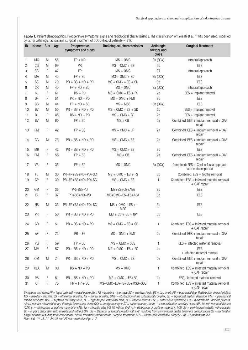
Surgical approaches to sinonasal complications of odontogenic disease
303
Table I. Patient demographics. Preoperative symptoms, signs and radiological characteristics. The classification of Felisati et al. 12 has been used, modified by us for aetiologic factors and surgical treatment of SCOD (No. of patients = 31).
ID Name Sex Age Preoperative symptoms and signs
Radiological characteristics Aetiologic factors and
class
Surgical Treatment
1 MG M 55 FP + NO MS + OMC 3a (DCY) Intraoral approach
2 CG M 69 PR MS + OMC + ES 3b EES
3 SG F 47 FP MS + OMC ST Intraoral approach
4 MA M 45 FP + SC MS + OMC + SD 3b (DCY) EES
5 SS M 70 PR + BS + NO + PD MS + OMC + ES + SD 3b EES
6 CR M 40 FP + NO + SC MS + OMC 3a (DCY) Intraoral approach
7 CL F 61 BS + PD MS + OMC + ES + FS 2c EES + implant removal
8 DF F 51 PR + NO + PD MS + OMC + PMT 3b EES
9 CC M 44 FP + NO + SC MS + MSS 3b (DCY) EES
10 BV M 50 PR + BS + NO + PD MS + OMC + ES + SD 2c EES + implant removal
11 BL F 45 BS + NO + PD MS + OMC + BE 2c EES + implant removal
12 BV M 60 FP + SC MS + CB 2a Combined: EES + implant removal + OAF repair
13 PM F 42 FP + SC MS + OMC + UP 2a Combined: EES + implant removal + OAF repair
14 CC M 73 PR + BS + NO + PD MS + OMC + ES 2a Combined: EES + implant removal + OAF repair
15 MR F 42 PR + BS + NO + PD MS + OMC + ES 3b EES
16 PM F 56 FP + SC MS + CB 2a Combined: EES + implant removal + OAF repair
17 VR F 35 FP + SC MS + OMC 3a (DCY) Combined: EES + Canine fossa approach with endoscopic aid
18 FL M 36 PR+FP+BS+NO+PD+SC MS + OMC + ES + FS 3b Combined: EES + tooths removal
19 CP F 39 PR+FP+BS+NO+PD+SC MS + OMC + ES 1 Combined: EES + infected material removal + OAF repair
20 GM F 36 PR+BS+PD MS+ES+CB+AEA 3b EES
21 FA F 37 PR+BS+NO+PD MS+OMC+ES+FS+AEA 3b EES
22 NS M 33 PR+FP+BS+NO+PD+SC MS + OMC + ES +MSS
3b EES
23 PR F 56 PR + BS + NO + PD MS + CB + BE + UP 3b EES
24 GR F 51 PR + BS + NO + PD MS + OMC + ES + CB 1 Combined: EES + infected material removal + OAF repair
25 AF F 72 PR + FP MS + OMC + PMT 2a Combined: EES + implant removal + OAF repair
26 PG F 59 FP + SC MS + OMC + SSS 1 EES + infected material removal
27 MM F 57 PR + BS + NO + PD MS + OMC + ES + FS 1a EES+ infected material removal
28 OM M 74 PR + BS + NO + PD MS + OMC + ES 2a Combined: EES + implant removal + OAF repair
29 CLA M 30 BS + NO + PD MS + OMC 1 Combined: EES + infected material removal + OAF repair
30 PS F 51 PR + BS + NO + PD MS + OMC + ES+FS 1a EES+ infected material removal
31 OI F 75 PR + FP + SC MS+OMC+ES+FS+CB+MSS+SSS 1 Combined: EES + infected material removal + OAF repair
Symptoms and signs: FP = facial pain; NO = nasal obstruction; PR = purulent rhinorrhea; SC = swollen cheek; BS = bad smell; PD = post-nasal drip. Radiological characteristics: MS = maxillary sinusitis; ES = ethmoidal sinusitis; FS = frontal sinusitis; OMC = obstruction of the ostiomeatal complex; SD = significant septum deviation; PMT = paradoxical middle turbinate; MSS = septated maxillary sinus; BE = hypertrophic ethmoidal bulla; CB= concha bullosa; SSS = silent sinus syndrome; PU = hypertrophic uncinate process; AEA = anterior ethmoidal artery. Etiologic factors and class: DCY = dentigerous cyst; ST = supernumerary tooth; 1 = sinusitis after maxillary sinus (MS) lift with oroantral fistulae (OAF) (+/– dislocation of grafting material in MS); 1a = sinusitis after MS lift without OAF (+/– dislocation of grafting material in MS); 2a = peri-implant osteitis with sinusitis; 2c = implant dislocation with sinusitis and without OAF; 3a = Bacterial or fungal sinusitis with OAF resulting from conventional dental treatment complications 3b = bacterial or fungal sinusitis resulting from conventional dental treatment complications. Surgical treatment: EES = endoscopic endonasal surgery; OAF = oroantral fistulae.Note: # 6, 10, 18, 21, 24, 26 and 27 are reported in Figs 1–7.

G.L. Fadda et al.
304
from the middle meatus with nasal swab, was performed on 20/31 patients with unilateral purulent rhinorrhoea. Bacteriological examination was positive in 8/20 patients (Table II), while it was negative on the remaining 12/20 patients. Positive intraoperative cultures were obtained in eight pa-tients, and new specific antibiotic therapies guided by an-tibiograms were prescribed. The predominant organisms identified using classic biochemical methods were Gram-positive aerobes (Table II). No anaerobes were observed in our cases, while Saibene AM et al. reported the pres-ence of anaerobes in 14% of cases 27. Drago L et al. 28 recently described the identification of Dialister pneumosintes in a case of chronic maxillary si-nusitis of odontogenic origin. D. pneumosintes is a known endodontic and periodontal pathogen found in necrotic pulp, subgingival plaque and deep periodontal pockets. In agreement with other authors 23 29, the removed extra-mucosal material of fungal aspect was sent for histopatho-logical analysis as well as a biopsy of the mucosal sinus wall (Table II). The biopsy of the mucosal sinus was done when a fungal infection was suspected in advance. On the basis of histopathological results, the presence of extramucosal non-invasive fungal hyphae forms were found in five cases (# 2,3,5,21,23). In 3/5 cases, the causa-tive agent was Aspergillus (Table II). Furthermore, other
dental-related microbes caused by Actinomyces spp. can be found in histopathological examination 30. In no case was the biopsy of the mucosal sinus positive for fungal hyphae, but they were all non-invasive extramucosal fun-gal forms. Therefore, no local or general antifungal treat-ment was administered, as reported by other authors 27 31.
DiscussionThe incidence of SCDDT is very low despite the high frequency of dental pathologies 8. In a meta-analysis by Arias-Irimia et al. 7, the most common cause of SCDDT was iatrogenic effects (55.97%) – extrusion of endodon-tic obturation materials in the maxillary sinus, amalgama remaining after apicoectomy, elevation of the sinus floor with poorly positioned dental implants or those which had migrated to the maxillary sinus with OAF – followed by periodontitis (40.38%) and dentigerous cysts (6.66%). In a retrospective study of 27 patients with SCDDT, Lee and Lee reported that implant related causes were the most common (37%), followed by dental extraction-related
Fig. 1. Patient 6. SCDDT resulting from odontogenic cyst (Class 3a). A) Coronal maxillofacial CT, and B) clinical examination showing a bulging of the upper gingiva caused by the cyst.
Fig. 2. Patient 10. SCDDT resulting from implant dislocation (Class 2c). A) Coronal CT scan showing the implant inside the maxillary sinus, near the natural ostium but unable to be dragged in the ostiomeatal complex due to the small size of the natural ostium itself; B) Endoscopic endonasal surgery and implant removal.
Fig. 3. Patient 18. SCDDT resulting from dental treatment complicated by bacterial sinusitis (Class 3b). A) Swelling on the left cheek and B) relevant purulent secretion in the middle meatal and olfactory cleft of the left side with a fetid purulent discharge, in a patient with C) dental caries and presence of dental pathology; D) Coronal CT scan showing complete obliteration of the left maxillary sinus and the anterior ethmoid cells as well as the obstruc-tion of the frontal recess. Note the asymmetrical skull base; E) Nine-month postoperative coronal CT scan showing complete aeration of all paranasal sinuses; F) as well as the complete absence of swelling on the cheek. G, H) The postoperative endoscopic view 9 months after endoscopic endonasal surgery showing the appearance of the anterior ethmoidectomy, turbinoplas-ty of the middle turbinate and maxillary sinusotomy. I) Intraoral appearance of healing after teeth extraction.

Surgical approaches to sinonasal complications of odontogenic disease
305
complications (29.6%) and dentigerous cysts (11.1%); radicular cyst, dental caries and a supernumerary tooth accounted for 7.4% of cases 8. In our study, the most com-mon cause of SCDDT was conventional dental treatment in 29% of patients.Classic symptoms of SCDDT can include unilateral pu-rulent rhinorrhoea and nasal obstruction, bad smell and taste, hyposmia, headache, post-nasal drip and ipsilateral cheek pain 6 8 12. In a series of 21 patients with SCDDT, Longhini and Ferguson reported dental pain in only 29% of patients 13. In our study, 70.1% of patients complained of unilateral purulent rhinorrhoea as the main symptom.The diagnostic work-up requires evaluation of symptoms, history of dental treatment, dental examination and na-sal endoscopy. In addition, in the presence of unilateral purulent rhinorrhoea, we recommend preoperative and/or intraoperative bacterial culture from the middle meatus under endoscopic guidance, so as to prescribe antibiotics guided by an antibiogram.CT is the gold standard in the diagnosis of SCDDT due to its high resolution and ability to discern bone and soft tissue 17. It can show the relationship between the odon-togenic origin and the maxillary sinus, foreign bodies within the maxillary sinus, opacification of the maxil-lary sinus and OMC, OAF, periapical lesions defined as a rounded lucency adjacent to the roots of a tooth, dental roots, dental materials, dental implants, material for sinus elevation, or parts of broken instruments in the maxillary
sinus as well as the spread of inflammation into the other paranasal sinuses and anatomical variations 5 32 33. In the presence of suspected fungal balls on CT scans, magnetic resonance imaging (MRI) with gadolinium can also be useful (Fig. 4).Previous studies have identified anterior ethmoid involve-ment during SCDDT without determining its preva-lence 34 35. In our series, although limited, 16/31 patients (51.6%) also presented unilateral anterior ethmoid sinusi-tis, while 16/31 patients (51.6%) had anatomical varia-tions so as to cause obstruction of the OMC.However, Saibene AM et al. found in a retrospective study of 315 patients surgically treated for SCDDT that 18.7% of cases were affected by bilateral sinonasal involve-ment 26. Di Pasquale D et al. reported a case of bilateral odontogenic sinusitis after a bilateral maxillary sinus aug-mentation 36 similar to other authors 37.Recently, cone beam computed tomography (CBCT) has been introduced in dental and maxillofacial imaging. It has several advantages over traditional CT including uti-lising approximately 10% of the radiation dose of con-ventional CT, higher resolution and is a chairside pro-cess 38. The technique is gaining popularity in the field of implant dentistry as there is frequently a need to assess the thickness of the floor of the maxillary sinus and to eliminate the presence of concurrent sinus disease be-fore implantation. However, at the moment, because of its high costs, CBCT,
Table II. Comparison between preoperative and intraoperative positive bacterial cultures and histological examination. Note: # 6, 18, 21, 24, 26 and 27 are reported in Figs 1-7.
ID Name Preoperative bacterial culture
Intraoperative bacterial culture Histological examination
1 MG Odontogenic cyst
2 CG Fungal Hyphae
3 SG Aspergillus
5 SS Staphylococcus aureus (g+) Sinonasal aspergillosis
6 CR Odontogenic cyst
8 DF Streptococcus constellatus (g+) Chronic rhinosinusitis
9 CC Mucocele
14 CC Streptococcus intermedius (g+) Chronic rhinosinusitis
18 FL Staphylococcus aureus (g+) Streptococcus constellatus (g+) Chronic rhinosinusitis
21 FA Aspergillus Fungal hyphae
23 PR Aspergillus Aspergillus
24 GR Streptococcus parasanguinis (g+) Heterologous bone
2 6 PG Strepto coccus intermedius (g+)
Chronic rhinosinusitis
27 MM Staphylococcus epidermidis (g+)
Streptococcus constellatus (g+) and Staphylococcus epidermidis (g+)
Heterologous bone
28 OM Staphylococcus aureus (g+) Streptococcus intermedius (g+) Chronic rhinosinusitis with nasal polyps
29 CLA Osteoma
30 PS Stenotrophomonas maltophilia (g-)
Streptococcus anginousus (g+) Chronic rhinosinusitis with nasal polyps
31 OI Staphylococcus epidermidis (g+) Chronic rhinosinusitis with nasal polyps + heterologous bone

G.L. Fadda et al.
306
even if it allows examination of all paranasal sinuses and alveolar processes, is only complementary to traditional CT.A combination of medical and surgical approaches is gen-erally required for the treatment of SCOD. The Caldwell-Luc technique was the first to be described and used for SCDDT, while EES is a recent addition 9 21 39. Moreover, the Caldwell-Luc technique can induce sinusal sclerosis, atelectasis and hypoplasia of the maxillary sinus 40 as well as silent sinus syndrome (Fig. 6), infraorbital nerve dam-age, facial swelling, facial and teeth paraesthesia, OAF and recurrent sinusitis 40 41. In addition, the Caldwell-Luc operation is an absolute contraindication to performing sinus elevation 42.Furthermore, the maxillary sinus has an effective muco-ciliary clearance to the natural ostium; this remains after EES through middle meatal antrostomy, but not after the Caldwell-Luc technique, because the artificial antrostomy is made in the inferior meatus 43. EES can be considered to be a relevant improvement for several reasons: (i) it is less invasive with low morbidity 34 40; (ii) it allows recov-ery of normal sinus function through spontaneous drain-age from the natural ostium; (iii) it eliminates the need for total sinus mucosa removal, as originally proposed by Caldwell and Luc; (iv) it is possible to surgically manage the other paranasal cavities involved in the infection as well as that of widening the ostium and to treat anatomic variations that might contribute to normal ventilation of the OMC 44-46. Lopatin et al. 9 were the first to report 70 cases of SCDDT treated with EES, and since then, EES has been the surgical technique indicated in the treatment of this disease. In our series, 28/31 (90.3%) of patients were treated with EES or EES combined with oral surgery and none experienced any complications.In accordance with the literature we wish to stress the importance of close collaboration between the implan-tologist, maxillofacial/oral and ENT specialists to treat complex cases of SCDDT, to distinguish osteitis or os-teomyelitis of the alveolar process and to prevent recur-rence and complications 47 48. In this regard, the surgical treatment for SCDDT as already reported in Felisati et al. classification 1 is represented in most cases by a mul-
Fig. 4. Patient 21. SCDDT resulting from dental treatment complicated by fungal ball sinusitis (Class 3b). A) Preoperative endoscopic view of purulent secretions in the right middle meatus; B, C) Coronal and axial maxillofa-cial CT scans showing radiodense material and erosion of bone in the right maxillary sinus indicative of aspergillosis with obstruction of the ostiomeatal complex; D, E) Coronal and axial T1 magnetic resonance image in the same patient showing iso- or hypointensity on T1-weighted images in the right maxillary sinus.
Fig. 5. Patient 24. SCDDT resulting from maxillary sinus lift with OAF (Class 1). Preoperative coronal CT scans showing A) unilateral left maxillary sinusi-tis and obstruction of the ostiomeatal complex, and B) maxillary osteitis af-ter bone graft in the posterior maxillary alveolar process; C) Preoperative intraoral view: no clinical evidence of infection; D) Intraoral approach with trapezoidal flap, and E) after bone graft removal and bone debridement; F) In-traoperative view showing a probe, inserted through the oroantral communi-cation, visible from maxillary sinusotomy (45° endoscope); G) Buccal fat pad flap preparation, then Rehrmann buccal flap was used for pedicled buccal fat pad flap (PBFPF) covering; H, I) Control coronal CT scans, 6 months af-ter surgery showing complete aeration of the maxillary and ethmoid sinuses on the left side as well as removal of the bone graft and its repair; L) Nasal endoscopy showing the maintenance of the maxillary sinusotomy and the absence of mucosal degeneration (45° endoscope).
Fig. 6. Patient 26. SCDDT resulting from possible complications in the Caldwell-Luc technique. A) Coronal CT scan after Caldwell-Luc technique and inferior meatotomy for left odontogenic sinusitis; B) Coronal CT scan 7 years after Caldwell-Luc technique showing atelectasis and sclerosis of the left maxillary sinus, hypoplasia and progressive enophthalmos in silent sinus syndrome.

Surgical approaches to sinonasal complications of odontogenic disease
307
tidisciplinary approach combining EES with an intraoral approach (Classes 1, 2, 3a). We have added subclass “1a” to include patients suffering from sinusitis after maxillary sinus lift without OAF, who request only EES. In the combined approach, the intraoral approach, allows treatment of pathologies that are impossible to treat with EES only 1, such as: (i) removal of infected dental im-plants 49 with apical portions penetrating into the maxil-lary sinus or any other migrated material 50; (ii) foreign bodies or odontogenic cyst 51; (iii) removal of infected grafting material which can be more difficult to eliminate with endoscopy; (iv) maxillary osteitis or osteomyelitis; (v) periapical odontogenic infections of the teeth; (vi) dentigerous cysts; (vii) dental extraction-related compli-cations; and (viii) the closure of OAF. In addition, OAF must be quickly closed as its persistence intensifies the possibility of inflammation of the sinus by infection from the oral cavity. Concerning the EES approach: (i) it is possible to surgically manage the paranasal cavities even-tually involved in the infection, which are not reachable via an intraoral approach while preserving physiological sinonasal cavity function; (ii) it is possible to eliminate the anatomical variations that might contribute as co-factors to infection or obstruction of OMC; (iii) it is also possible to treat a foreign body, implant or grafting material dis-located into the sinusal cavity, fungus ball, or persistent maxillary sinus problems dependent on dental pathology under the control of rigid 4 mm, 45° and 70° angled endo-
scopes and a microdebrider with 40° curved blade, which are useful to treat the deepest regions of the maxillary si-nus such as the alveolar recess. Furthermore, a one-step surgical procedure including simultaneously EES and a sinus floor elevation procedure through an intra-oral ap-proach can be performed in selected cases. Thus, treat-ment of local contraindications to sinus augmentation can help prevent a second surgical procedure and a reduce the waiting period before final prosthetic rehabilitation 52-55.Finally, a review of the current literature indicates that the results of treatment of SCDDT, where the most common causes are iatrogenic effects and periodontitis, have a high success rate in terms of healing (80-100%) with low com-plication rates (trigeminal neuralgia, orbital haematoma, visual disturbance, cerebrospinal fluid leak, nasal syne-chiae), recurrence (OAF, sinusitis) and revision surgery (< 10%) as well as a reduction in the time required for rehabilitation 9 18 21 34 39.
ConclusionsThe possibility of SCDDT should always be considered when a patient has unilateral nasal symptoms that do not respond to medical treatment. Despite the limited number of cases treated on the basis of these preliminary satisfac-tory results, implantologist, maxillofacial/oral specialists and rhinological consultations are mandatory. The same is needed for an accurate diagnosis of infections of den-tal origin and associated sinusitis in SCDDT to resolve the odontogenic source and sinus infection in the shortest possible time without risking relapse.The results of our study seem to be in accordance with the classification system proposed by Felisati, to which we referred. This could be useful in the standardisation of surgical treatment protocols, according to pathological conditions, to better harmonise cases reported by different authors. Nonetheless, we think that the number of the sub-groups could be increased, especially for the pathological conditions included in Class 3.Lastly, in our experience we can assert that EES must be associated with an intraoral approach whenever the max-illary alveolar process is affected by an infectious disease such as osteitis, osteomyelitis, peri-implant pathology, teeth periapical lesions, maxillary sinus lift complica-tions, oroantral fistulae, or oroantral communication.
References1 Felisati G, Chiapasco M, Lozza P, et al. Sinonasal complica-
tions resulting from dental treatment: outcome-oriented pro-posal of classification and surgical protocol. Am J Rhinol Allergy 2013;27:101-6.
2 Saibene AM, Lozza P. Endoscopic sinus surgery and in-traoral approaches in sinus oral pathology. J Craniofac Surg 2015;26:322-3.
3 Wang KL, Nichols BG, Poetker DM, et al. Odontogenic si-
Fig. 7. Patient 27. SCDDT as a result of elevation of the maxillary sinus floor with dislocation of grafting material (Class 1a). The patient was treated in 1990 for the elevation of the maxillary sinus floor. In 2007, three dental implants were added to the patient, which later were removed because of in-fection. The patient referred to us for a swollen cheek and unilateral purulent rhinorrhoea, without symptoms of OAF. She refused any oral procedures, and underwent EES. A, B) Coronal and sagittal CT scans showing filling material within the right maxillary sinus (MS), erosion of the inferior wall of the MS de-termined by the sinus floor lifting procedure and complete obliteration of both MS and the ostiomeatal complex; C) Purulent secretions in the middle meatal of the right nasal cavity; D) Removal of the grafting material used for sinus floor elevation and ethmoidectomy; E, F) Postoperative nasal endoscopy 5 months after surgery showing the restitutio ad integrum of the MS and the anterior ethmoid. Note the turbinoplasty of the middle turbinate.

G.L. Fadda et al.
308
nusitis: a case series studying diagnosis and management. Int Forum Allergy Rhinol 2015;5:597-601.
4 Giovannetti F, Priore P, Raponi I, et al. Endoscopic si-nus surgery in sinus-oral pathology. J Craniofac Surg 2014;25:991-4.
5 Mehra P, Jeong D. Maxillary sinusitis of odontogenic origin. Curr Allergy Asthma Rep 2009;9:238-43.
6 Brook I. Sinusitis of odontogenic origin. Otolaryngol Head Neck Surg 2006;135:349-55
7 Arias-Irimia O, Barona-Doraldo C, Santos-Marino JA, et al. Meta-analysis of the etiology of odontogenic maxillary si-nusitis. Med Oral Patol Oral Cir Bucal 2010;15:e70-3.
8 Lee KC, Lee SJ. Clinical features and treatments of odonto-genic sinusitis. Yonsei Med J 2010;51:932-7.
9 Lopatin AS, Sysolyatin SP, Sysolyatin PG, et al. Chronic maxillary sinusitis of dental origin: is external surgical ap-proach mandatory? Laryngoscope 2002;112:1056-9.
10 Mehra P, Murad H. Maxillary sinus disease of odontogenic origin. Otolaryngol Clin North Am 2004;37:347-64.
11 Kretzschmar DP, Kretzschmar JL. Rhinosinusitis: review from a dental perspective. Oral Surg Oral Med Oral Pathol Oral Radiol Endod 2003;96:128-35.
12 Patel NA, Ferguson BJ. Odontogenic sinusitis: an ancient but under-appreciated cause of maxillary sinusitis. Curr Opin Otolaryngol Head Neck Surg 2012;20:24-8.
13 Longhini AB, Ferguson BJ. Clinical aspects of odontogenic maxillary sinusitis: a case series. Int Forum Allergy Rhinol 2011;1:409-15.
14 Hoskison E, Daniel M, Rowson JE, et al. Evidence of an in-crease in the incidence of odontogenic sinusitis over the last decade in the UK. J Laryngol Otol 2012;126:43-6.
15 Racić A, Dotlić J, Janosević L. Oral surgery as risk fac-tor of odontogenic maxillary sinusitis. Srp Arh Celok Lek 2006;134:191-4.
16 Boyne PJ, James RA. Grafting of the maxillary sinus floor with autogenous marrow and bone. J Oral Surg 1980;38:613-6.
17 Simuntis R, Kubilius R, Vaitkus S. Odontogenic maxillary sinusitis: a review. Stomatologija 2014;16:39-43.
18 Chiapasco M, Felisati G, Maccari A, et al. The management of complications following displacement of oral implants in the paranasal sinuses: a multicenter clinical report and proposed treatment protocols. Int J Oral Maxillofac Surg 2009;38:1273-8.
19 Fusetti S, Emanuelli E, Ghirotto C, et al. Chronic oroantral fistula: combined endoscopic and intraoral approach under local anesthesia. Am J Otolaryngol 2013;34:323-6.
20 Tomomatsu N, Uzawa N, Aragaki K, et al. Aperture width of the osteomeatal complex as a predictor of successful treat-ment of odontogenic maxillary sinusitis. Int J Oral Maxil-lofac Surg 2014;43:1386-90.
21 Costa F, Emanuelli E, Robiony M, et al. Endoscopic surgical treatment of chronic maxillary sinusitis of dental origin. J Oral Maxillofac Surg 2007;65:223-8.
22 Berrone M, Florindi FU, Carbone V, et al. Medication-relat-ed osteonecrosis of the posterior maxilla: surgical treatment using a pedicled buccal fat pad flap: case reports. J Oral Maxillofac Surg 2015;15:900-3.
23 Naghibzadeh B, Razmpa E, Alavi Sh, et al. Prevalence of fungal infection among Iranian patients with chronic sinusi-tis. Acta Otorhinolaryngol Ital 2011;31:35-8.
24 Manfrè L, Ferlito S, Conticello S, et al. A comparative study of the pterygopalatine fossa by computed tomography and magnetic resonance. Radiol Med 1994;3:183-9.
25 Simmen D, Jones N. Indication for infundibulotomy and maxillary sinusotomy. In: Simmen D, Jones N (Eds.). Man-ual of endoscopic sinus and skull base surgery. Second Edi-tion. Stuttgart, New York: Thieme;2014, pp. 92-4.
26 Saibene AM, Pipolo GC, Lozza P, et al. Redefining bounda-ries in odontogenic sinusitis: a retrospective evaluation of extramaxillary involvement in 315 patients. Int Forum Al-lergy Rhinol 2014;4:1020-3.
27 Saibene AM, Vassena C, Pipolo C, et al. Odontogenic and rhinogenic chronic sinusitis: a modern microbiological com-parison. Int Forum Allergy Rhinol 2015 Sep 8.
28 Drago L, Vassena C, Saibene AM, et al. A case of coinfection in a chronic maxillary sinusitis of odontogenic origin: identi-fication of Dialister pneumosintes. J Endod 2013;39:1084-7.
29 Nicolai P, Mensi M, Marsili F, et al. Maxillary fungus ball: zinc-oxide endodontic materials as a risk factor. Acta Otorhi-nolaryngol Ital 2015;35:93-6.
30 Saibene AM, Di Pasquale D, Pipolo C, et al. Actinomycosis mimicking sinonasal malignant disease. BMJ Case Rep 2013.
31 Dufour X, Kauffmann-Lacroix C, Ferrie JC, et al. Para-nasal sinus fungus ball: epidemiology, clinical features and diagnosis. A retrospective analysis of 173 cases from a single medical center in France, 1989-2002. Med Mycol 2006;44:61-7.
32 Crovetto-Martinez C, Martin-Arregui FJ, Lopez-de-Maturana AZ, et al. Frequency of the odontogenic maxillary sinusitis ex-tended to the anterior ethmoid sinus and response to surgical treatment. Med Oral Patol Oral Cir Buccal 2014;1:409-13.
33 Konen E, Faibel M, Kleinbaum Y, et al. The value of the occipitomental (Waters’) view in diagnosis of sinusitis: a comparative study with computed tomography. Clin Radiol 2000;55:856-60.
34 Andric M, Saranovic V, Drazic R, et al. Functional endo-scopic sinus surgery as an adjunctive treatment for closure of oroantral fistulae: a retrospective analysis. Oral Surg Oral Med Oral Pathol Oral Radiol Endod 2010;109:510-6.
35 Stammberger H, Zinreich SJ, Koop W, et al. Surgical treat-ment of chronic recurrent sinusitis – the Caldwell-Luc versus a functional endoscopic technique. HNO 1987;35:93-105.
36 Di Pasquale D, Saibene AM, Bebi V, et al. Calcifications afloat: bad omens in maxillary sinus augmentation. BMJ Case Rep 2013 Dec 17.
37 Felisati G, Saibene AM, Lenzi R, et al. Late recovery from foreign body sinusitis after maxillary sinus floor augmenta-tion. BMJ Case Rep 2012 Dec 12.
38 Nair UP, Nair MK. Maxillary sinusitis of odontogenic origin: cone-beam volumetric computerized tomography-aided di-agnosis. Oral Surg Oral Med Oral Pathol Oral Radiol Endod 2010;110:e53-7.
39 Albu S, Baciut M. Failures in endoscopic surgery of the maxillary sinus. Otolaryngol Head Neck Surg 2010;142:196-201.

Surgical approaches to sinonasal complications of odontogenic disease
309
40 Nemec SF, Peloschek P, Koelblinger C, et al. Sinonasal imaging after Caldwell-Luc surgery: MDCT findings of an abandoned procedure in times of functional endoscopic sinus surgery. Eur J Radiol 2009;70:31-4.
41 DeFreitas J, Lucent FE. The Caldwell-Luc procedure: in-stitutional review of 670 cases: 1975-1985. Laryngoscope 1988;98:1297-300.
42 van den Bergh JP, ten Bruggenkate CM, Disch FJ, et al. Ana-tomical aspects of sinus floor elevations. Clin Oral Implants Res 2000;11:256-65.
43 Kennedy DW, Shaalan H. Reevaluation of maxillary sinus surgery: experimental study in rabbits. Ann Otol Rhinol Lar-yngol 1989;98:901-6.
44 Pignataro L, Mantovani M, Torretta S, et al. ENT assessment in the integrated management of candidate for maxillary si-nus lift. Acta Otorhinolaryngol Ital 2008;28:110-9.
45 Testori T, Capelli M, Galli F, et al. Complications associated with maxillary sinus lift: diagnostic/therapeutic interdisci-plinary consensus and clinical recommendations in reducing the incidence. Ital Oral Surg 2011;10:139-40.
46 Fadda GL, Rosso S, Succo G, et al. Multiparametric statis-tical correlations between paranasal sinus anatomic varia-tions and chronic rhinosinusitis. Acta Otorhinolaryngol Ital 2012;32:244-51.
47 Felisati G, Saibene AM, Pipolo C, et al. Implantology and otorhinolaryngology team-up to solve a complicated case. Implant Dent. 2014;23:617-21.
48 Mantovani M. The ENT’s role in sinus lift management doesn’t need misleading messages. Acta Otorhinolaryngol Ital 2012;32:404.
49 Albu S. The ‘double-barrel’ approach to the removal of den-tal implants from the maxillary sinus. Int J Oral Maxillofac Surg 2013;42:1529-32.
50 Mantovani M, Pipolo C, Messina F, et al. Antral retriever and displaced dental implants in the maxillary sinus. J Crani-ofac Surg 2011;22:2275-7.
51 Biglioli F, Goisis M. Access to the maxillary sinus using a bone flap on a mucosal pedicle: Preliminary report. J Cra-niomaxillofac Surg 2002;30:255-9.
52 Felisati G, Borloni R, Chiapasco M, et al. Maxillary sinus elevation in conjunction with transnasal endoscopic treat-ment of rhino-sinusal pathoses: preliminary results on 10 consecutively treated patients. Acta Otorhinolaryngol Ital 2010;30:289-93.
53 Felisati G, Borloni R, Chiapasco M, et al. Reply to: “the ENT’s role in sinus lift management doesn’t need misleading messages”. Acta Otorhinolaryngol Ital 2013;33:47-8.
54 Testori T, Drago L, Wallace SS, et al. Prevention and treat-ment of postoperative infections after sinus elevation sur-gery: clinical consensus and recommendations. Int J Dent 2012;2012:365809.
55 Colletti G, Felisati G, Biglioli F, et al. Maxillary reconstruc-tion and placement of dental implants after treatment of a maxillary sinus fungus ball. Int J Oral Maxillofac Implants 2010;25:1041-4.
Address for correspondence: GianLuca Fadda, via Juvarra 9/E, 10072 Caselle Torinese (Turin), Italy. Tel. +39 119026463, Fax +39 11 9026583. E-mail: [email protected]
Received: November 2, 2015 - Accepted: January 10, 2016

310
ACTA OTORHINOLARYNGOLOGICA ITALICA 2016;36:310-316; doi: 10.14639/0392-100X-878
Audiology
Possibility of differentiation of cochlear electrodes in radiological measurements of the intracochlear and chorda-facial angle positionPossibilità di differenziazione degli elettrodi cocleari nelle misurazioni radiologiche della posizione intracocleare e dell’angolo cordo-facciale
i. DioGo, U. WaLLiCzeCk, j. TaUbe, n. Franke, a. TeYMoorTaSH, j. Werner, C. GüLDnerUniversity Department of orL, Head and neck Surgery; UkGM, Marburg, Germany
SUMMARY
Due to an increasing number of cochlear implantations, quality control has become more important. In addition to intraoperative biophysi-cal measurements, radiological imaging is another possibility. An upcoming technique regarding this is Cone Beam CT (CBCT). Sixty-five data sets (35 Nucleus Contour Advance–Cochlear; 30 Flex Soft–MedEl) of postoperative imaging by CBCT (Accu-I-tomo F17, Morita, Kyoto, Japan) underwent further evaluation. Insertion angle, height of the cochlea, distance of the electrode to the medial or lateral wall, angle between chorda tympani and facial nerve and the precise position of the electrode cable in the facial-chordal angle were determined. The typical difference between the perimodiolar and lateral course of the electrodes could also be shown in radiological measurements. This demonstrates the accuracy and advantage of CBCT in visualisation of small structures with fewer metal artifacts. Furthermore, in 75% of patients, the angle of the chorda and facial nerve could be visualised. Significant differences in dependence of the electrode type for the relation of them to the facial nerve could be seen. In conclusion, CBCT achieves reliable visualisation and detailed imaging-based measurements of the intracochlear position of different cochlea electrodes. Additionally, clinically known differences can be reproduced. Even visualisation of the position of the electrode in the chorda-facial angle is possible. Therefore, CBCT is a useful tool in intra- and postoperative control of cochlear implants.
KEY WORDS: Cochlear implant • Cone beam computed tomography • Visualisation of cochlea • Measurement of cochlea • Facial nerve
RIASSUNTO
Con l’incremento del numero di impianti cocleari effettuati, il controllo di qualità è divenuto sempre più importante. Oltre alle misurazioni biofisiche intraoperatorie ci si può avvalere dell’imaging radiologico. Una nuova tecnica utilizzata in questo campo è il Cone Beam CT (CBCT). Nel presente studio sono stati valutati 65 casi (35 Nucleus Contour Advance–Cochlear; 30 Flex Soft–MedEl) studiati mediante CBCT (Accu-I-tomo F17, Morita, Kyoto, Japan). Nello specifico sono stati rilevati: l’angolo di inserzione, l’altezza dell’impianto, la distanza dell’elettrodo dalla parete mediale o laterale, l’angolo tra la corda del timpano e il nervo facciale e la posizione precisa del filo dell’elettrodo nell’angolo cordo-facciale. È stato inoltre possibile valutare la differenza tra il decorso peri-modiolare e laterale degli elettrodi. I dati presentati dimostrano l’accuratezza e il vantaggio della CBCT nella visualizzazione di piccole strutture grazie al ridotto numero di artefatti da indurimento del fascio. Inoltre nel 75% dei pazienti è stato possibile visualizzare l’angolo tra la corda del timpano e il nervo facciale. È stato possibile notare differenze significative fra i vari tipi di elettrodo in funzione del tipo di rapporto con il nervo facciale. In conclusione mediante la CBCT è possibile ottenere una visualizzazione precisa e dettagliate misurazioni della posizione intra-cocleare dei diversi elettrodi. È persino possibile la corretta valutazione della posizione dell’elettrodo rispetto all’angolo cordo- facciale. La CBCT è quindi, dal nostro punto di vista, un utile strumento per il controllo intra e post-operatorio degli impianti cocleari.
PAROLE CHIAVE: Impianto cocleare • Cone beam computed tomography • Visualizzazione della coclea • Misurazioni della coclea • Nervo facciale
Acta Otorhinolaryngol Ital 2016;36:310-316
Introduction
Cochlear implantation is currently the standard in reha-bilitation of hereditary or acquired high grade inner ear hearing loss. This results in increasing standardisation and improvement of surgical techniques and technical development of newly dedicated implants. To achieve
this and to prevent complications, intra- or postoperative evaluation of the implanted situation is necessary. In ad-dition to biophysical measurements, radiological imaging is the only way to get information about the intracochlear position and the relation of the electrode to anatomic im-portant structures. In the beginning, conventional radiog-raphy was used. Today, more information is needed, and

Differentiation of cochlear electrodes by CBCT
311
thus computed tomography (CT) and cone beam comput-ed tomography (CBCT) are used and recommended by current guidelines 1 2. Regarding the irradiation, naturally conventional plain radiography has the lowest dose, but in almost the same manner the lowest grade of information. Considering CBCT and CT, the former has about the half to a third of the radiation dose of CT, which is a main advantage particularly in children 3-5.Imaging methods methods with fewer artifacts are need-ed, not only to visualise that the electrode is inside the cochlea, but also to present an impression of the detailed intracochlear position and distances to the modiolus or lat-eral wall. Therefore, CBCT has been introduced into ENT imaging with a special focus on visualisation of middle and inner ear implants 6-8. Despite some studies that have focused on possibilities 9-12 and limitations 13 14 of this method in comparison to CT, some questions still remain. Can different types of electrodes (modiolar vs. lateral po-sition) also be differentiated not only by impression of the observer, but also reproduced by measurements based on CBCT images? Is it possible to visualise the angle be-tween the chorda tympani and facial nerve and the rela-tion of the electrode/cable to these structures 15? The cur-rent paper aims to answer some of these questions.
MethodsAll of our data on patients with a cochlear implant and imaging of their implant by CBCT two typical groups (N = 82) were analysed. Inclusion criteria were: one of the following implant types, existing row data for de-tailed radiological measurement, low enough artifacts for precise intracochlear measurements and full insertion of the electrode. The first group consisted of patients with a perimodiolar positioned electrode of the cochlea (Contour Advance 512; N = 35). The second group consisted of
patients with the lateral wall electrode of MedEl (Flex soft standard electrode; N = 30). All electrodes were inserted through the regular or enlarged round window.Of the 65 patients, 52% (N = 33) were female and 48% (N = 32%) were male. The mean age at the time of im-aging was 51.6 years (range 5 to 88 years). Forty-five percent (N = 29) of the data sets concerned the left ear, whereas 55% (N = 36) were right ears. In three patients, both ears were analysed. All images were performed on the day of operation or the day after using a CBCT device from Morita (Accu-I-to-mo, F17, Kyoto, Japan). The tube current ranged between 84.0 and 90.0 kV. The tube voltage was between 3.0 mA and 8.0 mA. The primary size of the acquired voxels was at 0.08 mm. Imaging analyses and measurements were performed using One Volume Viewer (I-Dixel 2.0, Mori-ta, Kyoto, Japan).According to a consensus paper, the insertion angle of each single electrode was measured in relation to the en-trance of the cochlea (Fig. 1) 16. In the background of the known difficulties of evaluation in the middle and apical turn and to compare both electrode types, only the first 360° of the cochlea was analyzed. Based on this reconstructed image, for each single elec-trode the following parameters were measured. First, the diameter of the cochlea was determined on a line orthogo-nal to the lateral and medial wall through the electrode. Second, along the same line, the diameter of the electrode, and third the distance of the electrode to the lateral wall (in the case of group 1) or the distance of the electrode to the medial wall (in the case of group 2) were measured (Fig. 2). Due to the relationship of the electrode and its cable and the facial nerve with the chorda tympani, all 82 patients were reviewed for visibility. Four patients were excluded due to a field of view that was too small. Fourteen pa-
Fig. 1. Visualisation of the way of measurement the insertion angle (in accordance to 16). The baseline is defined by round window edge and turning point of cochlea to vestibule (a). The angle is measured with a standard tool between the baseline and a line through the single electrode orthogonal to the lateral and medial wall of the cochlea (b). angle measurements were performed for each single electrode (C).
A B C

I. Diogo et al.
312
tients had to be excluded due to missing visibility of the chorda tympani, and two more due to too many artifacts. In all, 62 of the 82 (75%) underwent further analysis. A standardised multiplanar reconstruction resulted in a view showing the chorda tympani, facial nerve, hori-zontal semicircular canal and the cable of the electrode. The angle between the chorda tympani and facial nerve was determined by a standard angle measurement option (Fig. 3). The following measurements were performed on a parallel horizontal line to the semicircular canal at the point of the electrode in the chorda-facial-angle. The di-ameter of the facial nerve, thickness of the bony coverage
in direction to the electrode and distance of the electrode to the bony canal of facial nerve were determined at this point (Fig. 4).
ResultsRegarding the group of the Contour Advance electrode, in 9 of 35 patients (26%) all 22 electrodes were in the first 360° of the cochlea. In all patients, the first 18 electrodes could be detected in the first 360°. In the Flex-soft group, the first seven electrodes were within the first 360° in all patients. In all patients of this group, electrodes 10 to 12 were deeper than 360°.
Fig. 2. The diameter of the cochlea (a) and the electrode (b) were measured. in case of perimodiolar electrodes, the distance from lateral wall to electrode (C) and in case of lateral wall electrodes, the distance from medial wall to electrode (D) were determined.
Fig. 3. Typical image of the chorda tympani (a) and the angle to the facial nerve (b) is presented (a). The angle between both struc-tures was measured in each case (b).
A
A B
B C D
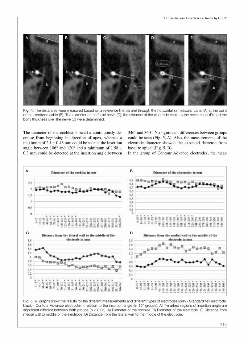
Differentiation of cochlear electrodes by CBCT
313
The diameter of the cochlea showed a continuously de-crease from beginning in direction of apex, whereas a maximum of 2.1 ± 0.43 mm could be seen at the insertion angle between 106° and 120° and a minimum of 1.58 ± 0.3 mm could be detected at the insertion angle between
346° and 360°. No significant differences between groups could be seen (Fig. 5, A). Also, the measurements of the electrode diameter showed the expected decrease from basal to apical (Fig. 5, B).In the group of Contour Advance electrodes, the mean
Fig. 4. The distances were measured based on a reference line parallel through the horizontal semicircular canal (a) at the point of the electrode cable (b). The diameter of the facial nerve (C), the distance of the electrode cable to the nerve canal (D) and the bony thickness over the nerve (D) were determined.
Fig. 5. all graphs show the results for the different measurements and different types of electrodes (grey - Standard flex electrode; black - Contour advance electrode) in relation to the insertion angle (in 15° groups). all *-marked regions of insertion angle are significant different between both groups (p < 0.05). a) Diameter of the cochlea. b) Diameter of the electrode. C) Distance from medial wall to middle of the electrode. D) Distance from the lateral wall to the middle of the electrode.
A
A B
DC
B C D E

I. Diogo et al.
314
distance from the electrode to the medial cochlea wall was 0.78 ± 0.12 mm whereas in the Flex-soft group, the mean was at 1.4 ± 0.15 mm. The opposite could be seen in the mean distance of the electrode to the lateral wall (Contour advance group: 1.0 ± 0.18 mm vs. Flex soft group: 0.63 ± 0.16 mm). All differences were significant for the mean and each value of the separate 15°-angle-group (p < 0.01). In both measurement curves, a decrease from basal to api-cal could again be detected (Fig. 5, C and D).Measurements at the point of chorda-facial-angle are sum-marised in Table I. The mean angle for all patients between the chorda tympani and facial nerve was 22.6 ± 9.5°. No significant difference could be seen between the groups (group 1: 23.7 ± 9° vs. group 2: 21.2 ±10°; p = 0.61). The diameter of the bony facial nerve canal was 1.8 ± 0.4 mm. Interestingly, a significant difference was found between groups (group 1: 1.7 ± 0.4 mm vs. group 2: 2.0 ± 0.4 mm; p = 0.001). The mean distance from the electrode to the facial nerve was 1.8 ± 0.7 mm. No difference between groups was detected (group 1: 1.7 ± 0.6 mm vs. groups 2: 1.8 ± 0.7 mm; p = 0.41). The mean bony coverage of the facial nerve was 0.8 ± 0.4 mm. Again, no significant dif-ference was seen (group 1: 0.78 ± 0.36 mm vs. group 2: 0.84 ± 0.37 mm; p = 0.35).
DiscussionThe number of cochlear implantations has been increas-ing for many years. This is because of consequent new-born hearing screening programmes and implantation of patients with single sided deafness and residual hearing 17. For further development of electrodes and improvement of surgical techniques, sufficient postoperative visualisa-tion of the implanted electrode is necessary. Conventional radiography only shows implantation into the cochlea or not, and gives no information about the detailed anatomy and specific intracochlear position. CT is much better, but due to the high range of metal artifacts, the potential of specific analyses is still low 18-20. CBCT is upcoming technique with high potential for visualisation of bony anatomy and implants due to fewer artifacts in compari-son to CT3. Several publications have shown its potential in visualisation of cochlear electrodes and its accuracy in comparison to histological examination 11 21-23. Even in CBCT, metal artifacts exist and result in reduced power of determination in medial and apical turn of the cochle-ar 13 14. The next steps of development will be automatic
analyses of the images and imaging fusion of pre- and postoperative data 6. Therefore, precise measurements of the electrode and its intracochlear position are needed. Additionally, measurement-based differentiation of dif-ferent electrode types is necessary. The current study fo-cused on this topic. As expected, a decreasing diameter of the cochlea could be detected. This is in accordance with the knowledge based on anatomic studies and shows the accuracy of the performed measurements 24 25. A relevant inter-individual range of the size of the cochlea exists and leaves open the question of the sense of standardised length of elec-trodes 24 26. Variability could be seen in the number of implanted electrodes into the individual cochlea. In the group of Contour Advance electrodes, only 26% (9/35) of all 22 electrodes were within in the basal turn (first 360°) of the cochlea. Based on the different design, all patients with the Flex Soft electrode were inserted deeper than the basal turn. The impact of implantation depth on speech understanding and hearing quality of music is still con-troversial 27 28. Particularly with this background, and the focus of inner ear trauma, visualisation of the inner struc-ture of the cochlea before and after implantation remains a focus of research. The current study demonstrates that it is possible to measure the visible and well known dif-ferences of the different implant types. Thus, a significant difference in the distances of the electrode to the medial and lateral cochlea wall was found for both groups. A sec-ond indicator of accuracy was the determined diameter of the electrode itself, which was in concordance with the information from the manufacturers. Another frequent problem in cochlear implantation sur-gery is the relationship to the facial nerve, the chorda tym-pani and the risk of unexpected postoperative facial nerve stimulation 29. This might be caused, for example, by ex-tracochlear electrodes, thin bony coverage of facial nerve or direct electric stimulation of the middle ear- or vestibu-lar part of the facial nerve 29. Because of this problem, the second part of this study analysed the potential of CBCT in visualisation of the chorda-facial angle and relationship to the electrode. Preclinical examinations of temporal bones showed the principal possibility of visualisation of the chorda tympani in CT and CBCT 15 30. No clinical data based on daily routine imaging data were found in the lit-erature. Therefore, astonishingly, in a fairly high number of patients – 75% (64/82) – the chorda-facial angle could
Table I. Results of measurements at the point of the chorda-facial angle.
All implants together Cochlear MedEl p-Wert
Chorda-facial angle (°) 22.6 ± 9.5 23.7 ± 9 21.2 ± 10 0.61
Diameter of bony facial nerve canal (mm) 1.8 ± 0.4 1.7 ± 0.4 2.0 ± 0.4 0.001
Distance from electrode to facial nerve canal (mm) 1.8 ± 0.7 1.7 ± 0.7 1.8 ± 0.7 0.41
Thickness of bone above facial nerve (mm) 0.8 ± 0.4 0.78 ± 0.36 0.84 ± 0.37 0.35

Differentiation of cochlear electrodes by CBCT
315
be visualised and analysed. The mean angle was 22.6 ± 10° and the distance of the electrode to the bony canal of the facial nerve was about 1.8 mm with a thickness of the bony coverage of about 0.8 mm. The diameter of the facial nerve canal was determined at 1.8 mm and is in principal accordance with other studies 31. Interestingly, a significant difference in the diameter of facial nerve canal could be detected between groups. We interpret this as a result of different insertion angles of the different types of implants. This might lead to a slightly different position regarding the height of the electrode in the chorda-facial angle, and in conclusion, to a different diameter of the facial nerve canal at the corresponding position. However, this problem should be addressed for detailed analyses in further studies.
ConclusionsCBCT has high potential for visualisation of different types of cochlear implants and achieves a reliable mea-surement-based analysis of the detailed intracochlear position of the electrode. Furthermore, visualisation and analyses of the chorda-facial angle and its relation to the electrode cable are frequently possible. Therefore, CBCT should be regarded as a useful tool for further radiologi-cal/audiological studies.
References1 Dammann F, Grees H, Kösling S, et al. Algorithmen für die
Durchführung radiologischer Untersuchungen der Kopf-Hals-Region. AWMF 2015;039/093.
2 German Society of Otorhinolaryngology H.a.N.S. Cochlea-Implantat Versorgung einschließlich zentral-auditorischer Implantate. AWMF 2012;017-071
3 Theunisse HJ, Joemai RM, Maal TJ, et al. Cone-beam CT versus multi-slice CT systems for postoperative imaging of cochlear implantation - a phantom study on image quality and radiation exposure using human temporal bones. Otol Neurotol 2015;36:592-9.
4 Stratemann SA, Huang JC, Maki K, et al. Comparison of cone beam computed tomography imaging with physical measures. Dentomaxillofac Radiol 2008;37:80-93.
5 Eggers G, Klein J, Welzel T, et al. Geometric accuracy of digital volume tomography and conventional computed to-mography. Br J Oral Maxillofac Surg 2008;46:639-44.
6 Aschendorff A. Imaging in cochlear implant patients. Laryn-gorhinootologie 2011;90(Suppl 1):S16-21.
7 Güldner C, Heinrichs J, Weiss R, et al. Prospective evalua-tion of reliability of cone beam computed tomography in de-tecting different position of vibroplasty middle ear implants. Clin Otolaryngol 2013;38:217-24.
8 Güldner C, Heinrichs J, Weiss R, et al. Visualisation of the bonebridge by means of CT and CBCT. Eur J Med Res 2013;18:30.
9 Zou J, Hannula M, Lehto K, et al. X-ray microtomographic confirmation of the reliability of CBCT in identifying the sca-
lar location of cochlear implant electrode after round win-dow insertion. Hear Res 2015;326:59-65.
10 Zou J, Lahelma J, Koivisto J, et al. Imaging cochlear implan-tation with round window insertion in human temporal bones and cochlear morphological variation using high-resolution cone beam CT. Acta Otolaryngol 2015;135:466-72.
11 Kurzweg T, Dalchow CV, Bremke M, et al. The value of digital volume tomography in assessing the position of coch-lear implant arrays in temporal bone specimens. Ear Hear 2011;31:413-9.
12 Aschendorff A, Kubalek R, Turowski B, et al. Quality con-trol after cochlear implant surgery by means of rotational tomography. Otol Neurotol 2005;26:34-7.
13 Diogo I, Franke N, Steinbach-Hundt S, et al. Differences of radiological artefacts in cochlear implantation in tem-poral bone and complete head. Cochlear Implants Int 2013;15:112-7.
14 Güldner C, Wiegand S, Weiss R, et al. Artifacts of the elec-trode in cochlea implantation and limits in analysis of deep insertion in cone beam tomography (CBT). Eur Arch Otorhi-nolaryngol 2012;269:767-72.
15 Hiraumi H, Suzuki R, Yamamoto N, et al. The sensitivity and accuracy of a cone beam CT in detecting the chorda tympani. Eur Arch Otorhinolaryngol 2016;273:873-7.
16 Verbist BM, Skinner MW, Cohen LT, et al. Consensus panel on a cochlear coordinate system applicable in histologic, physiologic, and radiologic studies of the human cochlea. Otol Neurotol 2010;31:722-30.
17 Stark T, Helbig S. Cochleaimplantatversorgung: Indikation im Wandel. HNO 2011;59:605-14.
18 Swartz JD, Russell KB, Wolfson RJ, et al. High resolution computed tomography in evaluation of the temporal bone. Head Neck Surg 1984;6:921-31.
19 Lecerf P, Bakhos D, Cottier JP, et al. Midmodiolar recon-struction as a valuable tool to determine the exact posi-tion of the cochlear implant electrode array. Otol Neurotol 2011;32:1075-81.
20 Todt I, Rademacher G, Wagner J, et al. Evaluation of cochle-ar implant electrode position after a modified round window insertion by means of a 64-multislice CT. Acta Otolaryngol 2009;129:966-70.
21 Boyer E, Karkas A, Attye A, et al. Scalar localization by cone-beam computed tomography of cochlear implant carriers: a comparative study between straight and periomodiolar pre-curved electrode arrays. Otol Neurotol 2015;36:422-9.
22 Xu J, Reh DD, Carey JP, et al. Technical assessment of a cone-beam CT scanner for otolaryngology imaging: image quality, dose, and technique protocols. Med Phys 2012;39:4932-42.
23 Bremke M, Luers JC, Stenner M, et al. Radiologic examina-tions in human temporal bone specimens using digital vol-ume tomography and high-resolution computed tomography after implantation of middle ear prosthesis and cochlear im-plant electrode array. Otol Neurotol 2013;34:1321-1328
24 Erixon E, Hogstorp H, Wadin K, et al. Variational anatomy of the human cochlea: implications for cochlear implanta-tion. Otol Neurotol 2009;30:14-22.
25 Dimopoulos P, Muren C. Anatomic variations of the cochlea and relations to other temporal bone structures. Acta Radiol 1990;31:439-44.

I. Diogo et al.
316
26 Avci E, Nauwelaers T, Lenarz T, et al. Variations in microanat-omy of the human cochlea. J Comp Neurol 2014;522:3245-61.
27 Hochmair I, Arnold W, Nopp P, et al. Deep electrode in-sertion in cochlear implants: apical morphology, elec-trodes and speech perception results. Acta Otolaryngol 2003;123:612-7.
28 Gani M, Valentini G, Sigrist A, et al. Implications of deep electrode insertion on cochlear implant fitting. J Assoc Res Otolaryngol 2007;8:69-83.
29 Ahn JH, Oh SH, Chung JW, et al. Facial nerve stimula-
tion after cochlear implantation according to types of Nucleus 24-channel electrode arrays. Acta Otolaryngol 2009;129:588-91.
30 Erovic BM, Chan HH, Daly MJ, et al. Intraoperative cone-beam computed tomography and multi-slice computed to-mography in temporal bone imaging for surgical treatment. Otolaryngol Head Neck Surg;150:107-14.
31 Komori M, Yamada K, Hinohira Y, et al. Width of the normal facial canal measured by high-resolution cone-beam com-puted tomography. Acta Otolaryngol 2013;133:1227-32.
Address for correspondence: Christian Güldner, University Ho-spital of Marburg, Department of ENT, Head- and Neck Surgery, Baldingerstraße, 35043 Marburg, Germany. Tel. +49 64215869052. Fax +49 64215866367. E-mail: [email protected]
Received: October 10, 2014 - Accepted: February 13, 2016

317
ACTA oTorhinolAryngologiCA iTAliCA 2016;36:317-320; doi: 10.14639/0392-100X-823
Clinical techniques and technology
Prevention of bisphosphonate-related mandibular fracturesPrevenzione delle fratture mandibolari conseguenti alla necrosi ossea da difosfonati
M. PeDrazzoLi, L. aUTeLiTano, F. biGLioLi Department of Maxillo-facial Surgery, San Paolo Hospital, University of Milan, italy
SUMMARY
Mandibular fracture is usually the clinical end of bisphosphonate-related osteonecrosis of the jaw. This is a painful complication and pa-tients cannot feed as usual, with a worsening of their quality of life. The goal of treatment in bisphosphonate related osteonecrosis of jaw (BRONJ) patients is to slow progression of bone necrosis. We present a novel technique for treatment of severe mandibular BRONJ in stage 3 patients that present with a high risk to develop fracture, since they have a residual unaffected mandibular bone height less than 6 mm. We treated 10 patients in this clinical situation with an extra-oral application of a reconstructive plate superficial to the platysma, to keep the plate separated from the infected site to avoid contamination and consequent need of removal, followed by an intraoral approach for ac-tive curettage of mandibular necrosis. The preservation of blood supply to the mandible and avoidance of direct contact of the infected site with the reconstructive plate are some advantages of this technique. This plate allows enhancement of mandibular strength, allowing proper treatment of the BRONJ site on the oral side without fear of causing a mandibular fracture when the residual mandible is thin. This technical solution guarantees these patients an extended disease-free period since it is effective in preventing mandibular fractures in patients with low mandibular residual height left after the BRONJ onset.
KEY WORDS: Bisphosphonate jaw fracture • Pathologic mandibular fracture • Extra-platysma stabilization • BRONJ Treatment
RIASSUNTO
La frattura della mandibola rappresenta solitamente l’evento finale nei pazienti che presentano una progressione della necrosi ossea deri-vante dall’impiego dei difosfonati. Si tratta di una grave complicanza molto dolorosa che impedisce ai pazienti di alimentarsi correttamen-te, essendo pertanto un fattore che peggiora notevolmente la loro qualità di vita. L’obiettivo del trattamento dei pazienti che presentano la necrosi ossea legata ai difosfonati (BRONJ) dovrebbe essere rallentare la progressione della malattia. Presentiamo una soluzione tecnica per il trattamento dei pazienti che presentano necrosi mandibolare in stadio 3 ad alto rischio di sviluppare una frattura, avendo un’altezza mandibolare residua di osso sano inferiore a 6 mm. Il trattamento consiste nel posizionamento di una placca ricostruttiva mandibolare per via extra-orale in un piano superficiale al muscolo platisma per tenere i mezzi di sintesi separati dal sito infettivo e non farli contami-nare con conseguente necessità di doverli rimuovere, seguito dal courettage per via endorale della necrosi mandibolare. Il rispetto della vascolarizzazione mandibolare e l’assenza di contatto diretto tra il sito di osteonecrosi e la placca ricostruttiva rappresentano alcuni dei vantaggi di questa metodica. La placca ricostruttiva rinforza la mandibola e consente di aggredire energicamente l’area di necrosi man-dibolare, senza esporre il paziente a rischio di frattura iatrogena. Questo garantisce al paziente un rallentamento della progressione della malattia e impedisce la frattura patologica della mandibola, inevitabile epilogo delle necrosi ossee mandibolari.
PAROLE CHIAVE: Frattura mandibolare da difosfonati • Frattura patologica della mandibola • Stabilizzazione extra-platismatica • Trattamento delle necrosi ossee da difosfonati
Acta Otorhinolaryngol Ital 2016;36:317-320
Introduction
Mandibular fracture is usually the clinical end of bis-phosphonate-related osteonecrosis of the jaw (BRONJ). This situation is associated with a sudden worsening of patients’ quality of life, since they start experiencing pain and cannot eat in a normal fashion. Since at present there is no evidence of an effective medical or surgical therapy to produce complete healing of the necrotic bone, the goal of treatment in BRONJ patients is to slow the progression of bone necrosis. Worsening of the pathology includes
further fistulae formation and mandibular pathologic frac-ture due to osteolysis extending to the inferior border.We decided to apply a reconstructive mandibular plate generally used to stabilise BRONJ-related mandibular fractures in patients who have not developed a mandibu-lar fracture, but who are at a high risk of its development.Since there is no literature on this topic, we arbitrarily selected a minimal residual thickness of unaffected man-dibular bone of 6 mm (Fig. 1) to apply the plate on an extra-platysmatic dissection plane 1. When the residual height of normal mandibular bone is more than 6 mm, we

M. Pedrazzoli et al.
318
treat patients by simple curettage of the bone necrosis and possible removal of the sequestrum.The proposed technique consists of the application of a reconstructive plate in a plane superficial to the platysma with an extraoral surgical access followed by an intraoral approach for active curettage of mandibular necrosis and rinsing of the surgical site. The main advantages of this technique are related to the maintenance of the blood sup-ply to the mandible and avoidance of direct contact of the infected site with the reconstructive plate.The position of this stable plate on a plane superficial to the platysma also allows the proper treatment of the BRONJ site on the oral side, without fear of causing a mandibular fracture due to the surgical curettage, and can allow mandibular resection when clinically needed with-out loss of occlusion.
Description of clinical techniqueA sterile drape is placed over the mouth and face to avoid any direct contamination of the cervical field by the infected bone. After inducing vasoconstriction with 1:200,000 epinephrine into the submandibular region, a 10-cm skin incision is made ~2 cm inferior to the man-dibular border, following its profile. The dissection plane is immediately over the platysma muscle, 5 cm in front of and 5 cm behind the site of BRONJ. A 2.5-mm recon-structive locking plate is modelled with the help of a tem-plate and then placed superficial to the platysma. Stabili-sation is obtained through mono-cortical locking screws placed a few cm from the BRONJ site, under plentiful rinsing solution (Fig. 2). Meticulous haemostasis and the application of an aesthetic suture completes the extra-oral surgical procedure.The cervical suture is covered by a sterile-drape dressing, and the BRONJ site is accessed through an intraoral ap-proach with minimal to no dissection of the periosteum. Surgery is limited to curettage and rinsing of the residual
bone. Rotating burs are avoided, while piezosurgical in-strumentation is preferred. Bony sequestrum is removed when present.Between 2013 and 2015 we operated on 10 patients af-fected by BRONJ of the mandible who were at risk to develop a mandibular fracture. Clinical data are shown in Table I.All patients were discharged after 2 days, with prompt relief of pain and allowance of feeding. We observed no facial nerve lesions. After a mean follow up time of 22.2 months, we saw no infection of plates and all means of fixation are stable at the time of writing. In one patient, after 12 months we observed local progression of disease that was treated by intra-oral courettage without need for plate removal.
DiscussionBRONJ is a clinical situation first reported in the modern era by Marx in 2003 2. Other authors have observed that a similar disease was described 100 years earlier, in 1899 by Dearden in the British Medical Journal in workers in the white phosphorus mines and in match factories; this was known as “Phossy Jaw”. It is possible that long-term exposure to phosphorus in these workers induced the pre-cipitation of bisphosphonates in bone leading to a clinical condition similar to the well-known BRONJ.The aetiopathological hypothesis of this disease is that bisphosphonates are powerful osteoclast inhibitors, and long-term use can result in suppression of bone turnover with increased trabecular bone density, inducing vascular insufficiency and causing bone necrosis. Moreover, the impaired bone turnover leads to failed removal of old os-teocytes. Osteocytes are not immortal cells; thus, when these cells die bone necrosis occurs 3.BRONJ is a clinical situation that is observed increasingly frequently. The effectiveness of bisphosphonates in prevent-ing bone pain and reabsorption in osteoporosis and control-
Fig. 2. Intraoperative view: the position of the 2.5 mm plate in the extra-platysmatic plane.
Fig. 1. Panoramic radiograph reveals wide bone destruction in the left mandibular angle, with residual height less than 6 mm.

Prevention of BRONJ related mandibular fractures
319
ling bone metastasis in the oncologic population leads to the increased use of this therapy. BRONJ is related to both IV and oral administration, the latter being less risky 1.The clinical presentation of BRONJ patients can differ. Attempting to standardise the management of these pa-tients, the American Association of Oral and Maxillo-facial Surgeons guidelines proposed a modified staging system in 2009 4:• At-risk category. Includes asymptomatic patients pre-
viously treated with either oral or IV bisphosphonates;• Stage 0. No clinical evidence of necrotic bone, but non-
specific clinical findings and symptoms;• Stage 1. Exposed necrotic bone, asymptomatic, and
with no evidence of infection;• Stage 2. Exposed and necrotic bone associated with in-
fection as evidenced by pain and erythema in the region of exposed bone with or without purulent drainage;
• Stage 3. Exposed and necrotic bone in patients with pain, infection, and one or more of the following: exposed and necrotic bone extending beyond the region of alveolar bone, resulting in pathologic fracture, extraoral fistula, oro-antral/oro-nasal communication, or osteolysis extending to the inferior border of the mandible or the sinus floor.
In the early stages conservative treatment is usually ad-vised, while in approaching more complex clinical situ-ations, as with stage 2-3 patients, the correct choice is challenging since management is not yet standardised. Surgical debridement/resection, in combination with anti-biotic therapy, may offer a long-term palliative and valid solution, but progression to a more complex situation is more likely to occur with time.In fact, usually the last stage is clinically characterised by pain, swelling, fistulae and finally mandibular fracture. In this situation patients cannot properly eat and, even if the fracture is reduced and stabilised with surgery, the frac-ture is not likely to heal. On the contrary, the presence of osteosynthesis material over the fracture may sustain a chronic infection.
Kuijpers et al. in 2011 5 described a unique case of sponta-neous healing of a BRONJ-related mandibular fracture in a 74-year-old woman, thus showing the persistent healing potential of bisphosphonate-treated bone, but of course this is not a rule.Deciding not to treat patients often means sentencing them to a quick decline of their quality of life because of the high degree of pain. In selected cases they can even be treated with free flap reconstruction after mandible re-section 6, but most of these patients are old and in poor general condition, thus their clinical situation cannot be addressed with complex surgical procedures.Therefore, we decided to stabilise the mandibular bone af-fected by BRONJ before fracture in those cases in which this clinical evolution is highly predictable, leading to a bet-ter clinical situation and preventing painful complications. We arbitrarily decided to apply a mandibular reconstruc-tive plate using the previously described extra-platysma technique 1 when the residual height of the unaffected mandible is no more than 6 mm (Fig. 3). The skin incision in the neck is set 2 cm inferior to the lower mandibular border to improve the aesthetic result. The presence of os-teosynthesis material over the platysma and placement of the screws distant from the infected surgical site reduce the risks of maintenance of chronic infection and worsen-ing of osteonecrosis.This plate is fixed in a plane superficial to the platysma muscle so that surgical dissection is not dangerous for the facial nerve since the marginalis mandibulae branch lies deeper inside. However, placing the screws without direct vision of the nerve branch could lead to nerve injuries. This is rare, and has never occurred in our patients.This technique is an application of well-known concepts of stabilisation of atrophic jaws, without removal of the periosteal support to the residual stumps; consequently, blood supply to the affected mandible is maintained 7. That is likely important for spontaneous healing of the necrotic bone and preventing expansion of the osteitis.
Table I. Clinical data of patients.
Patient name/gender/age Residual unaffected mandibular height
(mm)
Bisphosphonate means of administration
Disease Follow-up time(months)
M.E./F/ 65 2 IV Multiple myeloma 36
B.M./F/56 1 IV Breast cancer metastasis 26
M.P./M/70 4 IM Paget 24
L.S./F/72 6 Oral Osteoporosis 18
G.V./F/78 6 Oral Osteoporosis 30
A.F./M/72 1 IM Melanoma metastasis 20
T.A./F/74 3 IM Breast cancer metastasis 20
L.V./M/77 5 IM Multiple Myeloma 26
A.P./F/82 4 Oral Osteoporosis 14
S.S./F/75 3 IM Osteoporosis 8

M. Pedrazzoli et al.
320
Use of copious rinsing solution when drilling the screws is also advised to reduce the risk of BRONJ progression. The use of a locking instead of a compressive plate is important to avoid forcing of the weakened mandibular bone, which could lead to a mandibular fracture. The use of monocortical screws is intended to avoid exces-sive mechanical stress on the bone; the use of bicorti-cal screws could lead to overloading. The oral approach stage of the operation is limited to curettage and rinsing of the surgical site. This is usually performed trying to avoid round burs and using ultrasound surgical devices such as a piezoelectric drill.The presence of cutaneous fistulae does not represent a barrier to the application of this technique. In fact, in three cases we removed the fistula at the time of neck skin inci-sion, and then closed it with a lower platysma pedicled flap to separate the necrotic site from the dissection plane where the reconstructive plate was going to be positioned.Rapid relief and preservation of normal masticatory func-tion are achieved, and patients usually recover promptly.
The technique is painless, and patients can easily eat and be discharged after a few days, and thus this surgical so-lution can also be used in patients in poor general clini-cal conditions.The suggested technique is easy to apply and in our pa-tients was effective in preventing mandibular fractures in patients with low mandibular residual height after BRONJ onset.
References1 Biglioli F, Pedrazzoli M. Extra-platysma fixation of bisphos-
phonate-related mandibular fractures: a suggested technical solution. Int J Oral Maxillofac Surg 2013;42:611-4.
2 Marx RE. Pamidronate (Aredia) and Zoledronate (Zometa) induced avascular necrosis of the jaws: a growing epidemic. J Oral Maxillofac Surg 2003:61:1115-8.
3 Ruggiero SL. Emerging concepts in the management and treatment of osteonecrosis of the jaw. Oral Maxillofac Surg Clin North Am 2013 25:1:11-20.
4 Ruggiero SL, Dodson TB, Assael LA, et al. American As-sociation of Oral and Maxillofacial Surgeons position paper on bisphosphonate-related osteonecrosis of the jaws--2009 update. J Oral Maxillofac Surg 2009:67(Suppl 5):2-12.
5 Kuijpers SC, van Roessel EW, van Merkesteyn JP. Unusual case of a conservatively treated pathological fracture after sequestrectomy in a patient with long-term oral bisphospho-nate use. J Craniomaxillofac Surg 2011:39:69-72.
6 Colletti G, Autelitano L, Rabbiosi D, et al. Technical refine-ments in mandibular reconstruction with free fibula flaps: outcome-oriented retrospective review of 99 cases. Acta Otorhinolaryngol Ital. 2014 Oct;34(5):342-8.
7 Ellis E III, Price C. Treatment protocol for fractures of the atrophic mandible. J Oral Maxillofac Surg 2008:66:421-35.
Address for correspondence: Marco Pedrazzoli, via di Rudinì 8, 20142 Milan, Italy. Fax. +39 02 81844704. E-mail: [email protected]
Received: November 8, 2015 - Accepted: February 2, 2016
Fig. 3. Postoperative panoramic radiograph (at 24 months) showing the stable position of the reconstructive plate and extraordinary regrowth of mandibular bone.

321
ACTA oTorhinolAryngologiCA iTAliCA 2016;36:321-325; doi: 10.14639/0392-100X-1031
Clinical techniques and technology
Vastus lateralis myofascial free flap in tongue reconstruction Lembo miofasciale di vasto laterale nella ricostruzione della lingua
r. PeLLini1, a. De VirGiLio1 2, G. MerCanTe1, b. PiCHi1, V. ManCioCCo1, P. MarCHeSi1, F. FerreLi1, G. SPriano1
1 Department of otolaryngology-Head and neck Surgery, regina elena national Cancer institute, rome, italy; 2 Department organs of Sense, enT Section, University of rome “La Sapienza”, rome, italy
SUMMARY
In the last decade, the antero-lateral thigh free flap (ALT) has become the most popular free flap for tongue reconstruction because of less donor site morbidity and better cosmetic outcomes. However, fascio-cutaneous ALT may be insufficient to reconstruct major tongue de-fects, while its muscular-cutaneous variant (using the vastus lateralis muscle) may be too bulky. The present study describes our preliminary experience of tongue reconstruction with vastus lateralis myofascial flap, which could potentially offer unique advantages in head and neck reconstruction including adequate bulk when needed, optimal functional results and obliteration of dead space thus preventing fistulas and infections with minimal morbidity.
KEY WORDS: Free flap • Glossectomy • Rectus femoris • Tongue reconstruction • Vastus lateralis
RIASSUNTO
Nell’ultimo decennio il lembo antero-laterale di coscia (ALT) è diventato il lembo libero più utilizzato nella ricostruzione della lingua, dal momento che esso è caratterizzato da bassa morbidità a livello del sito donatore e da migliori risultati estetici. Tuttavia, l’ALT fascio-cuta-neo può essere insufficiente nella ricostruzione nei difetti maggiori (es. glossectomia totale) mentre la sua variante muscolo-cutanea (che include il muscolo vasto laterale)’ può essere troppo voluminosa. Scopo dello studio è quello di descrivere la nostra esperienza preliminare nella ricostruzione della lingua utilizzando il lembo libero mio-fasciale di vasto laterale che potrebbe a nostro parere offrire notevoli van-taggi nella ricostruzione testa-collo come: possibilità di confezionare un lembo voluminoso quando necessario, ottimi risultati funzionali, obliterazione di spazi morti con prevenzione dello sviluppo di fistola e infezione con minima morbidità a livello del sito donatore.
PAROLE CHIAVE: Lembo libero • Glossectomia • Retto femorale • Ricostruzione della lingua • Vasto laterale
Acta Otorhinolaryngol Ital 2016;36:321-325
IntroductionNowadays many free flaps have been described and suc-cessfully used in oral cavity reconstruction 1 2. In the last decade, the antero-lateral thigh free flap (ALT) become the most popular because of less donor site morbidity and better cosmetic outcomes 2. However, fascio-cutaneous ALT may be insufficient to reconstruct major tongue defects, while its muscular-cutaneous variant (using the vastus lateralis muscle) may be too bulky, especially in patients with a high body mass index (BMI).The aim of our study was to describe surgical technique and analyze the feasibility of vastus lateralis myofascial free flap (VLMFF) in tongue reconstruction, which to our knowledge has not been reported in the current literature.
Clinical techniqueThe National Cancer Institute ‘Regina Elena’ of Rome
Ethics Committee approved the pilot study. Three pa-tients affected by tongue squamous cell carcinoma (SCC) were treated from July to October 2015. All patients were staged with biopsy of the tumor, head and neck contrast MRI/CT and PET-CT. All patients underwent a temporary tracheostomy and nasogastric feeding tube placement. In Table I we summarised the patients’ clinical data. Recon-structions were performed by VLMFF.
VLMFF harvesting and insettingA line was drawn from the anterior superior iliac spine to the lateral border of the patella (Fig. 1a). The incision was made along the aforementioned line between the inferior and superior third. A suprafascial dissection was extended at least 5 cm medially and laterally ligating the skin per-forators (Fig. 1b). The intermuscular septum between the vastus lateralis and rectus femoris was visualised and the curved line with lateral concavity, medial to the intermus-

R. Pellini et al.
322
cular septum, was marked with a surgical pen and incised (Fig. 1c). Fascia was elevated medial-to-lateral to identify the intermuscular septum between the vastus lateralis and rectus femoris (Fig. 1d). The intermuscular septum was then gently dissected and the descending branch of the lateral circumflex femoral artery (DLCF) was identified along with the vastus lateralis motor nerve (Fig. 2a). A fascia paddle was then included over the vastus lateralis exceeding 20% of the desired muscle flap size in all direc-tions (Fig. 2b). VLMFF was raised in a caudal-cephalic
direction using a harmonic ultrasonic scalpel (Harmonic Focus, Ethicon Endo-Surgery, Cincinnati, USA) accord-ing to the desired shape and respecting the neurovascular pedicle (Fig. 2c). Pedicle dissection continued until ade-quate neuro-vascular pedicle length was reached (Fig. 2d). Harvesting times were 40, 42 and 47 min, respectively.In cases 1 and 3, the DLCF artery was anastomised with the superior thyroid artery, while in case 2 with the facial artery. Venous anastomoses were performed in all cases end to side to the internal jugular vein. Nerve anastomo-
Fig. 1. VLMFF harvesting: a) incision line; b) suprafascial dissection; c) a curved line with lateral concavity is marked with a surgical pen; and d) elevated medial-to-lateral.
Table I. Clinical data.
Case Gender/age
pTNM Surgery Complications Decannulation/ NG tube removal
Adjuvant therapy
1 F/70 yT2N0M0 Subtotal glossectomy + bilateral ND (I-IV)
None 5/13 days No
2 M/59 T4N2cM0 Subtotal glossectomy + bilateral ND (I-V;I-IV)
None 8/16 days Chemoradiation
3 F/69 pT2N0M0 Hemiglossectomy + ipsilateral ND (I-III)
None 4/9 days No
ND: neck dissection

Vastus lateralis myofascial free flap
323
ses between the hypoglossal nerve and vastus lateralis motor nerve was performed in all cases. In Figure 3 we show VLMFF insetting and 30-day post-operative re-sults.
DiscussionThe present study describes our preliminary experience of tongue reconstruction with VLMFF. VLMFF seems feasible, not technically demanding and safe. In our first three cases, we did not experience major complications such as necrosis, local infection or donor site impairment. VLMFF it is not time or budget consuming compared to the fascio-cutaneous and myocutaneous ALT variants since it does not imply further surgical steps and does not require adjunctive preoperative studies 3. Harvesting of VLMFF required on average of 43 minutes.VLMFF does not require harvesting of a skin island and consequently does not require preoperative study of vas-cular anatomy. This results in less closure tension and possibly in less donor site complications and better scar
healing. Due to its rich vascular supply, VLMFF does not require any perforator branch dissection either supra/sub fascial or intramuscular. This represents an important ad-vantage since the aforementioned surgical steps imply a higher incidence of flap vascular supply impairment. VLMFF has also some recipient site advantages. Thanks to the high number of muscular perforators, when the main vascular pedicle is preserved VLMFF could theoretically be shaped freely basing on the reconstruction necessity. However, we should point out that a 20-30% flap mass reduction due to muscular shrinkage should be expected.Another advantage is represented by the satisfactory cos-metic results. ALT flap classically includes a skin island that is responsible for a certain degree of color mismatch. In VLMFF, the use of muscle implies a better cosmetic result in tongue reconstruction. Moreover, thigh fascia protection adequately prevented fistula development in our 3 patients. A criticism to our technique could be in terms of donor site postoperative morbidity since the vastus lateralis muscle is sacrificed. However, as reported by Hanasono et al. 4 in a series of 220 ALT free flap and later by Bi-
Fig. 2. VLMFF harvesting: a) the descending branch of the lateral circumflex femoral artery is identified; b) a fascia paddle is included over the vastus later-alis; c) VLMFF is raised including an adequate neuro-vascular pedicle; d) VLMFF completed harvesting.

R. Pellini et al.
324
anchi et al. 5 in a series of 98 musculo-cutaneous ALT free flap, the harvesting of the muscular component does not seem to significantly increase donor-site morbidity. Hanasono et al. 5 reported only an 8% of thigh weakness after muscle harvesting that was not associated with the amount of muscle harvested or motor nerve transection. Our 3 patients did not report thigh weakness and did not complain of any limitations in daily physical activity at 30 days after surgery.
Conclusions
The free VLMF flap could potentially offer unique advan-
tages in head and neck reconstruction, including adequate bulk when needed, optimal functional results and oblit-eration of dead space, thus preventing fistulas and infec-tions with minimal morbidity.
AcknowledgementsArmando De Virgilio (corresponding author) has full ac-cess to all data in the study and takes responsibility for the integrity of the data and accuracy of data analysis.
References1 Cigna E, Rizzo MI, Greco A, et al. Retromolar trigone recon-
Fig. 3. Post-operative results: a) VLMFF insetting in patient 2. Tongue reconstruction with VLMFF in patient 2 (b) and 1 (c), 30 days after surgery; d) tongue reconstruction with VLMFF in patient 3, 30 days after surgery.

Vastus lateralis myofascial free flap
325
structive surgery: prospective comparative analysis between free flaps. Ann Surg Oncol 2015;22:272-8.
2 Pellini R, Mercante G, Spriano G. Step-by-step mandibular reconstruction with free fibula flap modelling. Acta Otorhi-nolaryngol Ital 2012;32:405-9.
3 Wolff KD, Grundmann A. The free vastus lateralis flap: An anatomic study with case reports. Plast Reconstr Surg 1992;89:469.
4 Hanasono MM, Skoracki RJ, Yu P. A prospective study of donor-site morbidity after anterolateral thigh fasciocutane-ous and myocutaneous free flap harvest in 220 patients. Plast Reconstr Surg 2010;125:209-14.
5 Bianchi B, Ferri A, Ferrari S, et al. The free anterolateral thigh musculocutaneous flap for head and neck reconstruc-tion: one surgeon’s experience in 92 cases. Microsurgery 2012;32:87-95.
Address for correspondence: Armando De Virgilio, Department of Otolaryngology-Head and Neck Surgery, Regina Elena National Cancer Institute, via Elio Chianesi 53, 00144 Rome, Italy. Tel. +39 06 52665065. Fax +39 06 52665066. E-mail: [email protected]
Received: January 10, 2016 - Accepted: March 4, 2016

326
ACTA OTORHINOLARYNGOLOGICA ITALICA 2016;36:326-327; doi: 10.14639/0392-100X-1315
Letter to the Editor
The relevance of counseling in patients with nasal polyps L’importanza del counseling nei pazienti affetti da poliposi nasale
M. GeLarDi1, n. De CanDia1, n. QUaranTa1, C. rUSSo2, P. PeCoraro3, M. ManCini4, P. LUPerTo5, G. LoMbarDo6, a. MaCCHi7, C. boCCioLini8, a. CioFaLo9, e. De CorSo10, G. CiPranDi11
1 Section of otolaryngology, Department of basic Medical Science, neuroscience and Sensory organs, University of bari, italy; 2 U.o.C. of otolaryngology, ospedale Di Venere, Carbonara di bari (ba), italy; 3 orL-asp 6- Palermo, italy; 4 Section of otolaryngology, azienda area Vasta romagna-riccione, italy; 5 Section of otolaryngology, aSL br1 brindisi, italy; 6 Section of otolaryngology, aSP of agrigento, italy; 7 orL Clinic University of insubriae, Varese, italy; 8 U.o.C. of otolaryngology Hospital Maggiore of bologna, italy; 9 Department of Sensory organs, Sapienza University of roma, italy; 10 otorhinolaryngology, Fondazione Policlinico a. Gemelli, Università Cattolica del Sacro Cuore, rome, italy; 11 Department of Medicine, istituto di ricovero e Cura a Carattere Scientifico (i.r.C.C.S.), azienda ospedaliera Universitaria San Martino, Genoa, italy
Acta Otorhinolaryngol Ital 2016;36:326-327
To the Editor,Management of nasal polyps (NP) is based on medical and surgical strategies 1-3. At present, medical therapy might be preferable, but it may not be long-lasting. In fact, NP patients have frequent relapses after medical and/or surgical treatment, and thus there is the need for adequate follow-up. In this regard, we previously proposed a clinical-cytological grading to calculate a prognostic index of relapse 4. Consequently, NP patients must be carefully assessed and followed over time, us-ing appropriate exams. Therefore, it is fundamental to approach NP patients by giving appropriate counseling, such as adequate information and communication, to manage their expectations 5. In this regard, Oscarsson and colleagues performed a long-term prospective study enrolling 33 patients with untreated NP to investigate the natural history 6. These authors concluded that oc-casional NP are frequently part of a chronic disorder that
do not necessarily evolve into a more relevant condition over time. Thus, treatment should be decided considering both the presence of NP and severity of symptoms. This issue highlights the relevance of paying careful attention to NP patients and performing appropriate follow-up. Accordingly, we performed a multicentre survey with the aim to evaluate the grade of medical communication in 375 NP patients (206 males, mean age 46.8 years). Table I reports the questions and answers. This investi-gation underlines that patients were often misinformed, and consequently overall patient satisfaction grade is rather low. Accordingly, surgeons should do a better job in explaining the natural history of NP. This issue is par-ticularly relevant as active and shared participation of the patient is fundamental to achieve optimal manage-ment. We firmly believe that medical communication is very relevant in NP management. Thus, a precise and thorough counseling is mandatory in all NP patients.
Table I. Questions and answers about NP management.
Questions Yes No
Did the doctor inform you that NP is a chronic disorder and that long-term follow-up is needed? 68% 32%
Did you know that NP have a high probability of relapse? 64% 36%
Did the doctor inform you that another operation might be needed because of relapse? 71% 29%
Did the doctor point out that operation is not curative and will not improve smell or taste? 70% 30%
Did you have periodical check-ups after surgery? 83% 17%
Did the doctor perform anterior rhinoscopy after surgery? 67% 33%
Did the doctor perform nasal fiber-endoscopy after surgery? 33% 67%
Did the doctor prescribe medications to prevent relapse? 83% 17%
Did you take medications to prevent relapse? 75% 25%
Is your overall satisfaction good? 26% 74%

The relevance of counseling in patients with nasal polyps
327
References1 Bhattacharyya N, Harvey RJ, Rosenfeld RM. Cochrane Cor-
ner: Extracts from The Cochrane Library: Interventions for Chronic Rhinosinusitis with Polyps. Otolaryngol Head Neck Surg 2015;153:315-9.
2 Martinez-Devesa P, Patiar S. WITHDRAWN: Oral ste-roids for nasal polyps. Cochrane Database Syst Rev 2016;4:CD005232
3 Kalish L, Snidvongs K, Sivasubramaniam R, et al. WITH-DRAWN: Topical steroids for nasal polyps. Cochrane Data-base Syst Rev 2016;4:CD006549.
4 Gelardi M, Fiorella R, Fiorella ML, et al. Nasal-sinus poly-posis: clinical-cytological grading and prognostic index of relapse. J Biol Reg 2009;23:181-8S.
5 Contrera KJ, Ishii LE, Setzen G, et al. Accountable Care Organizations and Otolaryngology. Otolaryngol Head Neck Surg 2015;153:170-4.
6 Oscarsson M, Johansson L, Bende M. What happens with untreated nasal polyps over time? A 13-year pro-spective study. Ann Otol Rhin Laryng 2016 May 17. pii: 0003489416649971. [Epub ahead of print].
Address for correspondence: Giorgio Ciprandi, largo R. Benzi 10, 16132 Genoa, Italy. Tel. +39 10 35331820. Fax +39 10 3538664. E-mail: [email protected]
Received: June 14, 2016 - Accepted: June 23, 2016

328
ACTA OTORHINOLARYNGOLOGICA ITALICA 2016;36:328-332; doi: 10.14639/0392-100X-548
Case series and reports
Effects of simultaneous palatal expansion and mandibular advancement in a child suffering from OSAEffetti di simultanei espansione palatale e avanzamento mandibolare in un paziente pediatrico con apnee ostruttive notturne
a. GaLeoTTi1, P. FeSTa1, M. PaVone2, G.C. De VinCenTiiS3
1 Dentistry Unit, Department of Pediatric Surgery, 2 respiratory Unit, Department of Pediatric Surgery, 3 otolaryngology Unit, Department of Pediatric Surgery, “bambino Gesù” Children’s research Hospital, rome, italy
SUMMARY
This clinical report describes a child suffering from obstructive sleep apnoea (OSA) and class II skeletal malocclusion with maxillary contraction and anterior open bite. He presented moderate obstructive sleep apnoea with large impact on quality of life of patient and par-ents. He was treated using an innovative orthodontic device (Sleep Apnea Twin Expander) to simultaneously carry out palatal expansion and mandibular advancement. After orthodontic therapy, the OSA-18 questionnaire demonstrated an improvement of the main respiratory symptoms, while cardiorespiratory sleep study revealed a reduction in obstructive sleep apnoea events. Post-treatment, clinical assessment and cephalometric analysis showed a reduction of sagittal maxillary discrepancy and an extension of upper airway space. In conclusion, this case report suggests that orthodontic treatment might be a valuable alternative treatment in children with obstructive sleep apnoea related to craniofacial anomalies.
KEY WORDS: Obstructive sleep apnea • Palatal expansion • Mandibular advancement appliances
RIASSUNTO
Questo caso clinico illustra il trattamento di un bambino affetto da apnee ostruttive nel sonno (OSA) che presenta una malocclusione di classe II scheletrica da retrusione mandibolare con contrazione del mascellare superiore e morso aperto anteriore. Il paziente presenta apnee ostruttive del sonno di grado moderato con un alto impatto sulla qualità della vita del paziente e dei genitori. Il paziente è stato trattato utilizzando un dispositivo ortodontico innovativo (Sleep Apnea Twin Expander), al fine di realizzare l’espansione del palato e l’avanzamento mandibolare contemporaneamente. Dopo la terapia ortodontica, il questionario sulla qualità della vita ha evidenziato un miglioramento dei principali sintomi respiratori e lo studio cardiorespiratorio del sonno ha rivelato una riduzione degli eventi di apnee ostruttive. Al termine della terapia, la valutazione clinica e l’analisi cefalometrica hanno evidenziato una riduzione della discrepanza sagittale e verticale tra il mascellare superiore e la mandibola e un ampliamento dello spazio delle vie aeree superiori. In conclusione, questo case report suggerisce che il trattamento ortodontico può essere una valida terapia alternativa nei bambini con apnea ostruttiva del sonno associata ad anomalie cranio-facciali.
PAROLE CHIAVE: Apnee ostruttive notturne • Espansione palatale • Apparecchi di avanzamento mandibolare
Acta Otorhinolaryngol Ital 2016;36:328-332
Introduction
Obstructive sleep apnoea (OSA) in children is a “sleep breathing disorder characterised by prolonged partial airway obstruction and/or intermittent complete obstruction that interrupts normal ventilation and normal sleep patterns” 1. OSA in children is associated with a series of daytime and night time symptoms, such as daytime sleepiness, morning headache, snoring, laboured breathing, restless sleep and nocturnal enuresis 2. Untreated OSA can result in serious cardiovascular, neurocognitive, behavioural and metabolic problems 2. The “gold standard” for the diagnosis of OSA is
polysomnography even if nocturnal cardiorespiratory sleep study (CRSS) represents a good alternative as an abbreviated diagnostic tool 3. The main risk factors of OSA in children include adenotonsillar hypertrophy, obesity, neuromuscular disorders and craniofacial anomalies 3. The first line therapy for children with OSA is adenotonsillectomy, but it is not universally effective due to multifactorial aetiology of the disease 4. Some authors have suggested the presence of a strong association between sleep breathing disorders and mouth breathing, abnormal placement of the tongue and anomalies in orofacial anatomy 5. The most common cranio-facial anomalies reported are retrognathism, maxillary con-

Effects of simultaneous palatal expansion and mandibular advancement in a child suffering from OSA
329
traction usually associated with unilateral or bilateral cross bite and open bite 6. The potential benefit of an orthodontic approach in patients with OSA and malocclusion was previ-ously described by several authors 7-9. We report our experi-ence on treatment of paediatric OSA with an innovative oral device that we called the “Sleep Apnea Twin Expander”.
Clinical casePatient: A 5-year-old boy D.S. referred to “Bambino Gesù” Children’s Research Hospital for sleep apnoeas since the age of 30 months. The patient underwent a baseline home noc-turnal cardiorespiratory sleep study (CRSS) which revealed moderate OSA with apnoea/hypopnoea index (AHI) of 6 events per hour of sleep and no alteration in oxygen saturation (mean of 98%, minimal 92%). Clinical history revealed poor appetite, no allergies, diurnal and nocturnal mouth breathing, loud snoring and repeated apnoeas during sleep. On physical and ear nose and throat (ENT) examination the patient showed good general conditions, pectus excavatum and patological tonsillar hypertrophy grade III) with no signs of inflamma-tion. The initial extraoral and intraoral photographs are shown in Figure 1. The lateral cephalogram (Fig. 1) showed adenoi-dal hypertrophy (75% of obstruction) confirmed by fiberoptic evaluation. The cephalometric analysis (Table I) showed a class II skeletal malocclusion, increased maxillary-mandib-
ular divergence, reduced rhinopharyngeal and oropharyngeal airway dimension. The quality of life (QOL) of the patient was measured with a questionnaire (OSA-18) 10. The score reported in our patient was 81 which suggested a large impact on QOL for both the patient and parents.Treatment: after multidisciplinary assessment, the child was prescribed a conservative treatment including ortho-dontic treatment and medical therapy. In order to correct malocclusion, we developed an innovative custom-made device, that we called “Sleep Apnea Twin Expander”. For development of the device, anatomical impressions and a wax bite registration in maximal intercuspation position
Table I. Cephalometric analysis. Maxillary and mandibular skeletal assess-ment: maxillary position in relation to cranial base (SNA angle), mandibular position in relation to cranial base (SNB angle), maxillomandibular sagittal differential (ANB angle). Vertical skeletal assessment: maxillo-mandibular divergence (ANS-PNS/GO-GN angle). Airway dimensions: rhinopharyngeal airway dimension (AD1-PNS), oropharyngeal airway dimension (PAS-PPAS).
Pre-treatment Post-treatment Normal value
SNA 73.5 73.4 81.4 ± 1.7
SNB 66.1 67.1 76.4 ± 1.4
ANB 7.4 6.3 5.0 ± 1.5
PNS-ANS/GO-GN 39.8 34.4 29 ± 5.0
AD1-PNS 7.3 7.9 18.9 ± 5.2
PAS-PPAS 3.1 4.8 12.0 ± 3.2
Fig. 1. Pre-treatment extraoral, intraoral photographs, lateral cephalogram.

A. Galeotti et al.
330
and in maximum mandibular protrusion were taken. The device (Fig. 2) is a double plate consisting of two sepa-rate elements, an upper fixed and a lower removable. It is a combination between the bite block expander by Mc-Namara 11 and twin block appliance by Clark 12. The upper part is a modified bite block palatal expander, consisting of a Hyrax-type screw 10 mm (Philoshopy 1, Lancer) se-cured to the maxillary arch with a 0.045-inch wire frame-work imbedded in acrylic covering the buccal, lingual and occlusal surfaces of the deciduous canine and first and sec-ond deciduous molars. The acrylic was 2 mm thick at the canines and 7 mm thick at the first and second deciduous molars creating steep inclined planes interlocked at about 70° to the occlusal plane. The lower appliance consisted of removable bite plane of acrylic retained with 0.9-mm ball clasps placed in the mandibular interproximal areas of first and second deciduous molars. The mandible was guided to protrude forward by the inclined planes in acrylic. Re-activation of the advancement was carried out with an ad-justable screw mechanism on the lower splint to achieve movement of inclined planes for gradual advancement of the mandible. This appliance allows to carry out palatal expansion and mandibular advancement simultaneously in order to obtain an extension of upper airway area and a possibile improvement of OSA symptoms in a short period. The upper bite block expander was bonded to the maxillary arch with resin reinforced glass ionomer cement (Fuji Or-tho, GC America). The mother of the patient was instructed to advance the screw once a day for 30 days. In addition, after one week the patient was instructed to wear the lower device during the night. The otorhinolaringologist pre-scribed intranasal corticosteroids. The advancement screw on the lower appliance was initially activated at 50% of the
maximum mandibular protrusion. Thirty days after the oral device placement, the patient’s mother referred a reduction of snoring and disappearance of apnoea events. Intraoral examination revealed an expansion of the transverse diam-eter of the upper jaw and an upper interincisal diastema. The screw of the lower plate was reactivated to increase mandibular advancement until 60% of maximal protrusion, the patient was instructed to wear it 14 hours per day, to stop activations in upper expander and was instructed about nasal breathing exercises. The patient was seen at 4-week intervals for an average of 10 additional months. After four months of orthodontic therapy the patient performed a sec-ond home nocturnal CRSS with device in situ in order to evaluate the effectiveness of the occlusal device in reducing
Fig. 2. Sleep apnea Twin expander in situ.
Fig. 3a-b. Lateral cephalogram pre- and post-treatment. Cephalometric points used in the analysis of the child. Points: a, a-point; anS: anterior nasal spine; b: b-point; Gn: gnathion; Go: gonion; n: nasion; PnS: posterior nasal spine; aD1: ade-noid inferior; S: sella; PPaS: posterior pharyngeal wall; PaS: anterior pharyngeal wall around gonial angle

Effects of simultaneous palatal expansion and mandibular advancement in a child suffering from OSA
331
apnoea events at night. The CRSS showed the absence of significant apnoeas with oxygen saturation within normal limits (AHI = 0.6; oxygen saturation mean of 98.2%, mini-mal 94%). After eight months of retention, the upper appli-ance was removed, and lateral cephalogram was performed. The patient was prescribed to use the device as removable during the night. The comparison between cephalometric analysis pre- and post-orthodontic treatment (Fig. 3, Ta-ble I) showed an increase in the rhinopharyngeal and oro-pharyngeal space and a reduction in maxillo-mandibular sagittal and vertical discrepancy. The examination of the patient after orthodontic treatment showed an improve-ment in face appearance, lip competence and dry lips, and the intraoral view showed a class I malocclusion and reduction of the overjet and open bite (Fig. 4). The OSA-18 QOL questionnaire administrated to the parents at the end of orthodontic treatment showed that the score was reduced to 20, suggesting a large improvement. ENT as-sessment at the end of the treatment showed reduction in hypertrophy of palatal tonsils and adenoids to grade II, and with no indication for surgical treatment.
DiscussionIn this case report we describe the successful treatment of moderate OSA with a new oral device in a 5-year-old boy.
Although adenotonsillectomy represents the first choice for treatment of OSA in children, we decided to treat him with an orthodontic approach based on the moderate degree of OSA with a normal oxygen saturation index and the pos-sibility of a conservative therapeutic strategy. The Sleep Apnea Twin Expander device was suitable for the patient described because it combined maxillary expansion and mandibular advancement. Orthodontic treatment resulted in an improvement of OSA symptoms through modifica-tion of the facial skeletal components. The positive effects of maxillary expansion and mandibular anterior reposition-ing have been previously described in children 7-9. The rapid maxillary expansion can produce significant increment of the total nasal volume 13 and mandibular advancement en-larges the retrolingual space and at the same time promotes lingual advancement during the night 7. These aspects, however, have not been confirmed by literature review be-cause of the limited number of studies and further studies are necessary to examine the effect of orthodontic device in a large sample of paediatric patients 14 15.In our patient, breathing pattern and skeletal improve-ments were documented: the post-treatment lateral cepha-logram showed an increase of rhinopharyngeal and oro-pharyngeal space, a reduction of maxillo-mandibular sagittal and vertical discrepancy, AHI passed from 6 to 0.6 events/night and the OSA-18 score decreased from 81
Fig. 4. Post-treatment extraoral, intraoral photographs, lateral cephalogram.

A. Galeotti et al.
332
to 20. Moreover, a reduction of the adenoidal and tonsillar hypertrophy degree was demonstrated, similar to data re-ported in a previous study 7. We hypothese that this effect may be related to the enlargement of the upper airway and retropharyngeal spaces that produce a relative reduction of the hypertrophy. The successful therapy of this case confirms the importance of a multidisciplinary approach in diagnosis and therapy of paediatric OSA. The early or-thodontic approach, aimed to obtain simultameus palatal expansion of the upper maxillary and the mandibular ad-vancement at the first phase of the treatment, associated with medical and breathing exercises, may modify the skeletal maxillo-mandibular anatomy by preventing ob-struction of the upper airway. Considering the results of this case, further studies are warranted to examine the ef-fects of orthodontic treatment with the Sleep Apnea Twin Expander in a large number of paediatric OSA patients with similar craniofacial anomalies.
AcknowledgementsThe authors thank to Dr. Vincenzo D’Antò for ideation of “Sleep Apnea Twin Expander”. The authors acknowledge Professor Ambrosina Michelotti, Dr. Renato Cutrera, Dr. Emanuela Sitzia and Dr. Valeria Viarani for their contribu-tion to this case report.
References1 American Thoracic Society. Standards and indications for
cardiopulmonary sleep studies in children. Am J Respir Crit Care Med 1996;153:866-78.
2 American Academy of Pediatrics. Clinical practice guide-line: diagnosis and management of childhood obstructive sleep apnea syndrome. Pediatrics 2002;109:704-12.
3 Marcus CL, Brooks LJ, Draper KA, et al. Diagnosis and management of childhood obstructive sleep apnea syndrome. Pediatrics 2012;130:e714-55.
4 Friedman M, Wilson M, Lin HC, et al. Updated systematic review of tonsillectomy and adenoidectomy for treatment of pediatric obstructive sleep apnea/hypopnea syndrome. Oto-laryngol Head Neck Surg 2009;140:800-8.
5 Huang YS, Guilleminault C. Pediatric obstructive sleep ap-nea and the critical role of oral-facial growth: evidences. Front Neurol 2013;3:184.
6 Zucconi M, Caprioglio A, Calori G, et al. Craniofacial modifications in children with habitual snoring and ob-structive sleep apnoea: a case-control study. Eur Respir J 1999;13:411-7.
7 Villa MP, Bernkopf E, Pagani J, et al. Randomized controlled study of an oral jaw-positioning appliance for the treatment of obstructive sleep apnea in children with malocclusion. Am J Respir Crit Care Med 2002;165:123-27.
8 Pirelli P, Saponara M, Guilleminault C. Rapid maxillary ex-pansion in children with obstructive sleep apnea syndrome. Sleep 2004;27:761-6.
9 Marino A, Ranieri R, Chiarotti F, et al. Rapid maxillary ex-pansion in children with Obstructive Sleep Apnoea Syndrome (OSAS). Eur J Paediatr Dent 2012;13:57-63.
10 Franco RA Jr, Rosenfeld RM, Rao M. Quality of life for chil-dren with obstructive sleep apnea. Otolaryngol Head Neck Surg 2000;123:9-16.
11 McNamara JA Jr, Brudon WL, Kokich VG. Orthodontics and Dentofacial Orthopedics. Needham Press; 2001.
12 Clark WJ. Twin-block functional therapy. London: Mosby-Wolfe; 1995.
13 Cordasco G, Nucera R, Fastuca R, et al. Effects of orthopedic maxillary expansion on nasal cavity size in growing subjects: a low dose computer tomography clinical trial. Int J Pediatr Otorhinolaryngol 2012;76:1547-51.
14 Kiliç N, Oktay H. Effects of rapid maxillary expansion on nasal breathing and some naso-respiratory and breathing problems in growing children: a literature review. Int J Pedi-atr Otorhinolaryngol 2008;72:1595-601.
15 Carvalho FR, Lentini-Oliveira D, Machado MA, et al. Oral appliances and functional orthopaedic appliances for ob-structive sleep apnoea in children. Cochrane Database Syst Rev 2007;18:CD005520.
Address for correspondence: Angela Galeotti, Dentistry Unit, De-partment of Pediatric Surgery, “Bambino Gesù” Children’s Hospi-tal, piazza S. Onofrio 4, 00165 Rome, Italy. Tel. +39 06 68594632. E-mail: [email protected]
Received: November 10, 2014 - Accepted: February 4, 2015

333
ACTA OTORHINOLARYNGOLOGICA ITALICA 2016;36:333-336; doi: 10.14639/0392-100X-1351
Case series and reports
Arterio-venous malformation of the mandible. Case report and review of literatureMalformazione arterovenosa della mandibola. Caso clinico e revisione della letteratura
r. SPreaFiCo, L. SorDo, r. beLLoTTo, M. SCHiPano, a. reSCaLDani, F. ParMiGianiDivision of otorhinolaryngology, ospedale Civile di Vimercate, azienda ospedaliera di Desio e Vimercate, Vimercate (Mb)
SUMMARY
Arteriovenous malformation (AVM) of the head and neck is a rare and potentially life threatening entity due to massive haemorrhage. There are several indications for treatment, including age of the patient and location, extent and type of vascular malformation. Endovas-cular therapy can effectively cure most lesions with limited tissue involvement. Surgery can be used in selected cases in combination with embolization. Here we report the case of a young woman affected by a massive AVM on the left side of the mandible and submandibular region, and also review the literature on AVM with special attention to treatment strategies.
KEY WORDS: Arterio-venous malformation • Mandible • Submandibular region • Treatment
RIASSUNTO
Le malformazioni arterovenose (MAV) del distretto cervico-facciale sono patologie rare e potenzialmente letali a causa delle imponenti emorragie che possono determinare. Il trattamento dipende dall’età del paziente, dalla sede, dall’estensione e dalla tipologia della mal-formazione. La terapia endovascolare è efficace nella maggior parte dei casi che presentano un’estensione limitata. In casi selezionati, e sempre in associazione con l’embolizzazione, si può ricorrere alla chirurgia. Nel presente articolo riportiamo il caso di una giovane donna affetta da un’estesa MAV dell’emimandibola e della regione sottomandibolare sinistra. Viene inoltre effettuata una revisione della letteratura prodotta su questo argomento con particolare attenzione alla strategia di trattamento.
PAROLE CHIAVE: Malformazione artero-venosa • Mandibola • Regione sottomandibolare • Trattamento
Acta Otorhinolaryngol Ital 2016;36:333-336
IntroductionAccording to Mulliken and Young, two types of vascular lesions can be recognized, which depend on the intrinsic properties of endovascular cells, namely haemangiomas and vascular malformations 1. Haemangiomas demon-strate endothelial hyperplasia and enlarge by cellular proliferation. Clinically, haemangiomas usually appear in early infancy, grow rapidly during the first months of life, then slowly involuting over 5 or 6 years 2 3.Arteriovenous malformations (AVMs) in contrast to the abnormal proliferation of endothelial cells of a haemangi-oma, display progressive ectasia of abnormal vessels, lined by flat endothelium. AVMs occur as a result of errors in embryogenesis, are always present at birth, may manifest at any time during life and grow proportionately with the child 3 4. AVMs can be subdivided based on the rate of blood flow: “slow flow” (capillary, venous, lymphatic or mixed) and “fast flow” (arterial, arteriovenous, fistulae or shunt) subtypes 5. Vascular malformations are frequently seen in the skin, but rarely affect the visceral organs or bones; ap-
proximately 51% occur in the head and neck, with a male-female ratio of 1 to 1.5. About 50% of all bone involvement occur in the skull and the maxillo-facial area 6 7.Lesions of the mandible are rare and potentially life-threatening entities that can present as innocuous episodes of gingival bleeding, slow-growing expansile masses, or severe haemorrhage. A biopsy or even a simple tooth ex-traction can cause a catastrophic bleeding that may even lead to death 8-12. However, the clinical course of malfor-mations involving the mandible and the maxilla remains unclear and unpredictable. Treatment may be surgical or non-surgical. The latter includes intravascular emboliza-tion with coil and/or sclerosing solutions. Surgical resec-tion is reserved for lesions that are extensive and/or re-fractory to endovascular therapy. Cure is defined as the complete eradication of disease or permanent resolution of symptoms with complete devascularization 8-16.Herein, we report the case of a 23-year-old woman with an AVM of the mandible and submandibular region, and review the relevant literature, focusing on the best options for treatment.

R. Spreafico et al.
334
Case reportIn December 2009, a 23-year-old woman, after tooth ex-traction, presented at our ENT division with a massive haemorrhage from the left side of the mandible.The bleeding was initially controlled by packing the man-dible, and blood transfusions were required. The patient reported no malocclusion and no history of infection, and family history was also unremarkable. She was affected by insulin dependent diabetes mellitus. The only extraoral finding was a slight left facial swelling.Retrospective analysis of the radiograms used for dental cure revealed the following. By panoramic radiograph, an extensive radiolucency in the left mandible involving the molar area (Fig. 1) was evident, while by dental scan, an abnormal enlargement of the left alveolar canal with inte-gral cortical bone (Fig. 2) was present.In the subsequent, urgent surgical intervention, a tempo-rary ligation of the external carotid artery was ineffective-ly attempted because of a large anastomotic blood supply. The haemorrhage was then controlled by filling the man-dibular cavity with sheets of oxidized cellulose, taking advantage of the natural pathway created by the previous tooth extraction. A haemostatic suture of the alveolar mu-cosa was finally done.In the strong suspicion of a vascular malformation, the patient underwent head and neck magnetic resonance im-aging (MRI) and magnetic resonance with angiography (MRA). The examination showed an extensive highly vascularized lesion involving the left side of the mandible and left submandibular region (Fig. 3).Ten days later, after the correction of severe diabetic de-compensation, an angiography was performed. This study
confirmed the diagnosis of arteriovenous malformation in the sites mentioned before, and associated with feeding vessels from the external carotid artery. Superselective coil embolization was performed of the facial artery, lin-gual artery and internal maxillary artery.The day after, follow-up MRI and MRA revealed a subto-tal closure of the AVM (Fig. 4). Two days later, the patient underwent surgical interven-tion. Passing through the incision of the previous carotid ligation, we removed a large highly vascularized lesion occupying the left submandibular region and substituting the submandibular gland itself. From the empty alveolar ridge, the intraosseous component of the AVM was in-jected with sodium tetradecyl sulphate (3%). There was no damage to the mandibular branch of the facial nerve. There were no intraoperative complications, and the es-
Fig. 1. Extensive radiolucency in the left mandible by panoramic radio-graph.
Fig. 2. Abnormal enlargement of the left alveolar canal with integral cortical bone by dental scan.
Fig. 3. MRI and MRA scans showing an extensive highly vascularized lesion involving the left side of the mandible and left submandibular region.
Fig. 4. Subtotal closure of the AVM on MRI and MRA performed the day after embolization.
Fig. 5. Two months after intervention, post-surgical MRI and MRA showed an empty submandibular triangle and almost complete reabsorption of the AVM in the mandible.

Arterio-venous malformation of the mandible
335
timated blood loss was 500 ml. The postoperative period was uneventful, and the patient was discharged on the fifth postoperative day. The final histopatological diagnosis was arterio-venous malformation.At the time of writing, after six months of follow-up, the patient has not experienced symptoms of trigeminal neu-ralgia. A minor bleeding episode was controlled with in-traosseous injection of methylprednisolone acetate. Sub-sequently, no further episodes of bleeding were observed. Two months after the operation, post-surgical MRI and MRA showed an empty submandibular triangle and al-most complete reabsorption of the AVM in the mandible (Fig. 5). Bone fractures and tissue necrosis or infection have not occurred.
DiscussionAVMs are extremely rare entities that can be life threat-ening if left untreated due to massive blood loss during tooth extraction or biopsy. Although rare, 50% of all intra-osseous AVMs occur in the maxillo-facial region and are extremely infrequent in the mandible 17. Very few cases of extraosseous submandibular AVMs have been reported, with 6 cases cited in the literature (5 females and 1 male) 18-
20. Our study confirms this trend. To our knowledge, this is the first documented case of a combined AVM involving both the mandible and the submandibular region. AVMs usually present with non-specific symptoms including bruit, dental loosening, swelling of soft tissues, change in skin and mucosal colour and dysesthesia of the lower lip or chin. Although CT, MRI and MRA may localize the arterio-venous shunt lesion, superselective arteriography remains an essential tool for diagnosis and planning of treatment 8-10 13 14. Management of AVMs is usually com-plex and requires a multidisciplinary team for successful outcome. Observation may be used as a temporary meas-ure in special situations, such as extreme age, pregnancy or refusal of therapy. No spontaneous regressions have been documented. On the other hand, it has been reported that the volume of the lesion may gradually increase 9.Arterial ligature was used in the past as a purely symp-tomatic treatment or before surgery. At present, it is well known that ligation of external carotid artery should not be performed, firstly as many anastomoses promote the rapid appearance of a collateral circulation, and secondly because future embolization would be impossible 9 10 13. In addition, in our experience, emergency ligation of the external carotid artery is insufficient to stop the haemor-rhage because of a large and already developed network of anastomotic blood vessels.At present, superselective angiographic embolization is considered first line treatment, alone or in combination with surgical approach to reduce intraoperative bleed-ing. Occlusion of the lesion is obtained using movable balloon, coils or liquid glue. Endovascular therapy as de-
finitive treatment for the AVMs of the mandible has been reported to have a success rate of 70%. This avoids mu-tilating surgery and related sequelae, especially if AVMs occur before adolescence 8-10 13-16.However, serious complications after embolization (e.g., occlusion of pulmonary or cerebral vessels) or recurrence of AVMs should be considered. In some cases, the pro-cedure has to be carried out in several sessions such as previous history of arterial ligation or high flow charac-teristics of the lesion or microshunts, which are invisible during hyperselective catheterization and therefore inac-cessible to treatment 9 11 13.Therefore, intraosseous injection of sclerosing agents should be used to achieve further obliteration of the AVMs 16. In this case, we used sodium tetradecyl sulphate 3% which in our division is regularly employed to treat many different vascular lesions of the head and neck with a high cure rate and very low incidence of side effects.Surgery is not the treatment of choice for AVMs of the mandible. Block resection of the affected area has been suggested, and temporary reconstruction with alloplastic bone plate or with the patient’s own free, previously curet-ted mandibular segment has been reported. More definite bony reconstruction is recommended as soon as possible with a free fibular graft or iliac crest to avoid facial de-formities and allow dental implantation 8-10 11 12 21.In the past years, some authors have commented that radi-cal surgical resection as in threating cancer 22 23. This has been tempered by acknowledgement that radical resec-tion and reconstruction of an extensive benign vascular lesion of the maxillo-facial and mandibular area typically causes severe disfigurement with considerable morbidity. Furthermore, recurrences may occur even after radical re-section, and relapses are reported to be even more difficult to treat 8.On the other hand, surgical excision of AVMs involv-ing soft tissues such as the submandibular space can be achieved without severe sequelae 18. No data on relapse have been reported for this very rare condition. Accord-ing to the literature, we tailored treatment of the present case by performing superselective arterial embolization, leaving the intraosseous part of the AVM in situ and re-secting only the part located in submandibular region. Surgery should always be performed after embolization, with a short interval between the procedures, based on the general recommendation that surgery should take place within 48 hours to 2 weeks to avoid revascularization of the lesion. In this period, we recommend MRI and MRA angiography studies to evaluate the effective devasculari-zation of AVMs.In conclusion, considering the present case and literature data, we can define the following general principles of treatment.Arterial ligation should not be performed, even in emer-gency settings. Superselective arterial embolization is the

R. Spreafico et al.
336
Address for correspondence: Roberto Spreafico, via Pertini 1, 20059 Vimercate (MB), Italy. Tel. +39 039 6654495. E-mail: [email protected]
Received: December 1, 2014 - Accepted: April 14, 2015
treatment of choice and can be repeated in the case of re-lapse. Intraosseous injection of sclerosing agents can be used in the attempt to reduce the number of arterial em-bolizations. In AVMs of the mandible, surgery should be reserved only for cases that are refractory to endovascular therapy and/or for therapeutic complications that are not otherwise treatable (bone fractures and/or necrosis). In AVMs of the soft tissue, surgery could be used if there are no significant side effects. Lastly, the interval between embolization and surgery should be as short as possible, and MRI and MRA should be repeated before surgery.
References1 Mulliken JB, Young AE. Vascular birthmarks: hemangiomas
and malformations. WB Saunders;1988.2 Mulliken JB, Glowacki J. Hemangiomas and vascular
malformations in infants and children: a classification based on endothelial characteristics. Plast Reconstr Surg 1982;69:412-22.
3 Waner M, Suen JY. Management of congenital vascular le-sions of the head and neck. Oncology 1995;9:989-94.
4 Higuera S, Gorldey K, Metry DW, et al. Management of hea-mangiomas and pediatric vascular malformations. J Cranio-fac Surg 2006;17:783-9.
5 Hurwitz DJ, Kerber CW. Hemodinamic considerations in the treatment of arteriovenous malformations of the face and scalp. Plast Reconstr Surg 1981;67:421-32.
6 Persky MS. Congenital vascular lesions of the head and neck. Laryngoscope 1986;96:1002-15.
7 Unni KK, Ivins JC, Beabout JW, et al. Hemangioma, heman-giopericytoma and hemangioendothelioma of bone. Cancer 1971;27:1403-14.
8 Yoshiga K, Tanimoto K, Okui T, et al. High-flow arteriov-enous malformation of the mandible: treatment and 7-year follow-up. Brit J Oral Maxillofac Surg 2003;41:348-50.
9 Giauoi L, Princ G, Chiras J. Treatment of vascular malfor-mations of the mandible: a description of 12 cases. Int J Oral Maxillofac Surg 2003;32:132-6.
10 Sakkas N, Schramm A, Metzeger MC, et al. Arteriovenous malformation of the mandible: a life-threatening situation. Ann Hematol 2007;86:409-13.
11 Hohlweg-Majert B, Metzeger MC, Schramm A, et al. Com-
plication of intraoral procedures in patients with vascular tumors. J Craniofac Surg 2008;19:816-9.
12 Shum JW, Clayman L. Resection and immediate reconstruc-tion of a pediatric vascular malformations in the mandible: case report. Oral Surg Oral Med Oral Pathol Radiol Endod 2010;109:517-24.
13 Persky MS, Yoo HJ, Berenstein A. Management of vascular malformations of the mandible and maxilla. Laryngoscope 2003;113:1885-92.
14 Benndorf G, Kim DM, Menneking H, et al. Endovascular management of a mandibular aeìrteriovenous malformation in a patient with severe hemophilia A. AJNR Am J Neurora-diol 2004;25:614-7.
15 Fan XD, Su LX, Zheng JW, et al. Ethanol embolization of arteriovenous malformation of the mandible. AJNR Am J Neuroradiol 2009;30:1178-83.
16 Chen W, Ye T, Xu L, et al. A multidisciplinary approach to treating maxillofacial arteriovenous malformations in children. Oral Surg Oral Med Oral Pathol Radiol Endod 2009;108;41-7.
17 Mohammadi H, Said-al-Naief NA, Hetfez LB. Arteriovenous malformation of the mandible: report of a case with a note on the differential diagnosis. Oral Surg Oral Med Oral Pathol Radiol Endod 1997;84;286-9.
18 Burns JC, Julian J, Alexander J. Arteriovenous malforma-tion of the submandibular triangle. J Oral Maxillofac Surg 1989;43:294-6.
19 Fine MJ, Holliday RA, Roland JT. Clinically unsuspected venous malformation limited to the submandibular triangle: CT findings. AJNR Am J Neuroradiol 1995;16:491-4.
20 Stocks HM, Sacksman A, Thompson P, et al. First docu-mented case of a submandibular arteriovenous malformation in a male patient. Am J Otolaryngol Head Neck Med Surg 2007;28:275-6.
21 Behnia H, Motamedi MHK. Treatment of central arteriov-enous malformation of the mandible via resection and im-mediate replantation of the segment: a case report. J Oral Maxillofac Surg 1997;55:79-84.
22 Azzolini A, Bertani A, Riberti C. Superselective emboliza-tion and immediate surgical treatment: our present approach to treatment of large vascular hemangiomas of the face. Ann Plast Urg 1982;9:42-60.
23 Withers EH, Franklin JD, Lynch JB. Case report: resection of a massive arteriovenous malformation of the head and neck. Plast Reconstr Surg 1979;63:566-9.

337
ACTA oTorhinolAryngologiCA iTAliCA 2016;36:337
In Memoriam of Pietro Ferrara (1934-2015)
We were all deeply saddened by the loss of Professor Pie-ro Ferrara, Director of the Clinic of Otorhinolaryngology of the University of Palermo, from 1980 to 2004.Prof. Pietro Ferrara was born at San Giuseppe Jato (Paler-mo) January 4th 1934.Graduated in Medicine and Surgery on 19 July 1958. Otorhinolaryngology professional qualification on 12 De-cember 1961.Anesthesiology professional qualification in 1963.Academic career in University of Palermo: he was Vol-untary Assistant from 1960 to 1962. He had entrusted as-sistant from 1962 to 1964 and Ordinary Assistant in 1964.He was Lecturer in Otorhinolaryngology from 1969. From 1974 to 1980 he had entrusted of the teaching of Otorhinolaryngology.From the 1980 Ordinary Professor professional qualifica-tion in Otorhinolaryngology.He was Chief Medical Executive of the Complex Unity of Otorhinolaryngology and Manager of the Department of the Special Surgeries in Azienda Ospedaliera Universi-taria Policlinico of Palermo.His teacher was Professor Ettore Borghesan, who was in-ternationally known for his research on the internal ear. Professor Ferrara inherited a passion for research and a deep-felt attachment to the Institution from his teacher and dedicated himself to the study of Histopathology and Histochemistry in the field of Otorhinolaryngology After brilliantly completing his specialization in Otorhi-nolaryngology in 1961, he progressively and successfully scaled all the steps of his academic career . Director of Otorhinolaryngology clinic, Director of the School of Specialization, and finally, Director of the De-partment of Otorhinolaryngology.His scientific research in various fields of Otology, Vesti-
bology and Otosurgery, and clinical and experimental On-cology, were published in many highly regarded journals. Professor Ferrara was also a Speaker and Chairman at many national and international congresses, an Organizer of courses and meetings.He was Member of numerous Italian and international Scientific Societies: Italian Society of Otorhinolaryngol-ogy, Italian Society of Audiology, Italian Society of Pho-niatry , Italian Society of Pediatric Otorhinolaryngology, Sicilian Society of Otorhinolaryngology, International Association of Physicians in Audiology (IAPA), Prosper Ménière Society, Fondation Portmann, Politzer Society, European Academy of Otology & Neuro-Otology, Euro-pean Skull Base Society. He was also President of the Sicilian Board of Otorhi-nolaryngology, Audiology, and Phoniatry.He was a brilliant otosurgeon and a passionate researcher who was always open to innovation. He published more than 200 articles (indexed/Peer reviewed Journals) 1 book chapters and more than 100 abstracts presented as oral presentation or posters at national and international sci-entific congress).Main topics of his activity were: otology, neurotology, middle ear surgery, neuroradiology, audiology.My memories go back to those first years in which I was student and attended the Department of Otorhinolaryn-gology of the University of Palermo. In those years, I as-sisted Professor Ferrara both in clinical research projects and in the wards and surgery rooms.His pupils will remember always his great sense of duty, dedication to the work, and his teaching.
Riccardo Speciale

338
ACTA OTORHINOLARYNGOLOGICA ITALICA 2016;36:338-340
Notiziario SIO
CONSIGLIO DIRETTIVO SIO (2016/2017)Leone C.A. Presidente SIO (Scad. Maggio 2017)Cassandro E. Vice-Presidente (Scad. Maggio 2017) Vicini C. Vice-Presidente (Scad. Maggio 2018)Scasso F. Segretario (Scad. Maggio 2017)Gallo A. Tesoriere (Scad. Maggio 2017)Ralli G. Bibliotecario (Scad. Maggio 2018)Capasso P. Segretario del Presidente (Scad. Maggio 2017)Barillari U. Consigliere (Scad. Maggio 2018)Benazzo M. Consigliere (Scad. Maggio 2018)Croce A. Consigliere (Scad. Maggio 2018)Cuda D. Consigliere (Scad. Maggio 2018)Gianformaggio C. Consigliere (Scad. Maggio 2018)Greco A. Consigliere (Scad. Maggio 2018)Pisani P. Consigliere (Scad. Maggio 2018)Santandrea L. Consigliere (Scad. Maggio 2018)Tortoriello G. Consigliere (Scad. Maggio 2018)Tirelli G. Consigliere (Scad. Maggio 2018)Filipo R. Past president (Scad. Maggio 2017)Paiano M.T. Consigliere agg. LP (Scad. Maggio 2018)
Comitato permanente per l’aggiornamento dello statuto e del regolamento Leone C.A. Presidente SIO (Scad. Maggio 2017)Lauriello M. Segretario dell’AUORL (Scad. Maggio 2018)Rugiu M.G. Segretario dell’AOOI (Scad. Maggio 2018)Scasso F. Segretario SIO (Scad. Maggio 2017)Maiolino L., Motta S., Passali GC. Colleghi nominati dall’AUORL (Scad. Maggio 2017)Della Vecchia L., Iemma M.,Piemonte M.. Colleghi nominati dall’AOOI (Scad. Maggio 2018)
Revisori dei conti SIOFiorella M.L. (Scad. Maggio 2020)Sessa M. (Scad. Maggio 2020)
Collegio dei Proibiviri SIOChiesa F. (Scad. Maggio 2018)Conticello S. (Scad. Maggio 2018)De Benedetto M. (Scad. Maggio 2018)Fiorella R. (Scad. Maggio 2018)Passali D. (Scad. Maggio 2018)
Coordinatore del Controllo di QualitàDella Vecchia L. (Scad. Maggio 2019)
Organo di consulenza SIOBussi M., Camaioni A., Casolino D., Conticello S., Coppo L., Danesi G., De Benedetto M., De Campora E., De Campora L., De Vincentiis I., Felisati D., Fiorella R., Laudadio P., Marcucci L., Miani P., Motta G., Ottaviani A., Passali D., Paludetti G., Piemonte M., Puxeddu P., Rinaldi Ceroni A., Sartoris A., Serra A., Sperati G., Spriano G., Zaoli G.
Notiziario SIO
339
DelegatiEUFOS Cuda D. (Scad. Maggio 2018) Fiorella M.L. (Scad. Maggio 2017)IFOS Piemonte M. (Scad. Maggio 2018) Galli J. (Scad. Maggio 2017)UEMS Cuda D. (Scad. Maggio 2018) Motta G. (Scad. Maggio 2017)EHNS Spriano G. (Scad. Maggio 2018) Motta G. (Scad. Maggio 2017)
CONSIGLIO DIRETTIVO AOOIDanesi G. Presidente (Scad. Maggio 2018)Barbara M. Vice-Presidente (Scad. Maggio 2018)Della Vecchia L. Vice-Presidente (Scad. Maggio 2018)Radici M. Vice-Presidente (Scad. Maggio 2018)Rugiu M.G. Segretario-tesoriere (Scad. Maggio 2018)Achilli V. Consigliere (Scad. Maggio 2018)Boccuzzi S. Consigliere (Scad. Maggio 2018)Campanini A. Consigliere (Scad. Maggio 2018)De Campora L. Consigliere (Scad. Maggio 2018)Miani C. Consigliere (Scad. Maggio 2018)Napolitano A. Consigliere (Scad. Maggio 2018)Panetti G. Consigliere (Scad. Maggio 2018)Panu F. Consigliere (Scad. Maggio 2018)Raso F. Consigliere (Scad. Maggio 2018)
Collegio dei probiviri AOOIBarbieri F. (Scad. Maggio 2020)De Benedetto M. (Scad. Maggio 2020)Laudadio P. (Scad. Maggio 2020)Richichi M. (Scad. Maggio 2020)Villari G. (Scad. Maggio 2020)
DelegatiIFOS Piemonte M. (Scad. Maggio 2018)UEMS EUFOS Cuda D. (Scad. Maggio 2018)EHNS Spriano G. (Scad. Maggio 2018)
Notiziario SIO
340
CONSIGLIO DIRETTIVO AUORLBussi M. Presidente (Scad. Maggio 2017)Paludetti G. Vice-Presidente (Scad. Maggio 2017)Nicolai P. Vice-Presidente (Scad. Maggio 2017)Lauriello M. Segretario-tesoriere (Scad. Maggio 2018)Aluffi Valletti P. Consigliere (Scad. Maggio 2017)Maiolino L. Consigliere (Scad. Maggio 2017)Martini A. Consigliere (Scad. Maggio 2017)Motta G. Consigliere (Scad. Maggio 2017)Passali F.M. Consigliere (Scad. Maggio 2017)Quaranta N. Consigliere (Scad. Maggio 2017)Sellari Franceschini S. Consigliere (Scad. Maggio 2017)Cassandro E. Past-President (Scad. Maggio 2017)
Collegio dei probiviriBarillari U. (Scad. Maggio 2017)Fusetti M. (Scad. Maggio 2017)Marchiori C. (Scad. Maggio 2017)Rinaldi Ceroni A. (Scad. Maggio 2017)Staffieri A. (Scad. Maggio 2017)
Revisori dei contiBussi M. (Scad. Maggio 2017)Mancini P. (Scad. Maggio 2017)
Comitato permanente per l’aggiornamento dello statuto e del regolamentoBellussi L. (Scad. Maggio 2017)Chiarella G. (Scad. Maggio 2017)Ralli G. (Scad. Maggio 2017)
DelegatiUEMS Motta G. (Scad. Maggio 2017)EUFOS Fiorella M.L. (Scad. Maggio 2017)IFOS Galli J. (Scad. Maggio 2017)EHNS Motta G. (Scad. Maggio 2017 )
Congresso Nazionale S.I.O. Sorrento 2017Relazione UfficialeCianfrone G.: Controversie diagnostico-terapeutiche in otorinolaringoiatria
Temi di ComunicazioneTemi liberi
Tavola RotondaRicci Maccarini A.: Insidie e strategie nella gestione della patologia della voce
Relazione Ufficiale 2018Scasso F., Ferrari G., De Vincentiis G.C.: Patologia infettiva emergente e riemergente in otorinolaringoiatria.

341
ACTA OTORHINOLARYNGOLOGICA ITALICA 2016;36:341-343
Calendar of events – Italian and International Meetings and Courses
Acta Otorhinolaryngol Ital 2016;36:341-343
information, following the style of the present list, should be submitted to the editorial Secretariat of acta otorhinolaryngologica italica ([email protected]). In accordance with the Regulations of S.I.O. and Ch.C.-F. (Art. 8) Members of the Society organis-ing Courses, Congresses or other scientific events should inform the Secretary of the Association (A.U.O.R.L., A.O.O.I.) within the deadlines set down in the respective Statutes and Regulations.
August-december 201612th OTOLOGICAL MICROSURGERy COURSE • August 29-31, 2016 • Bern – Switzerland
e-mail: [email protected] – Website: http://otology.swiss-meeting.org
4th INTERNATIONAL SyMPOSIUM ON OTOSCLEROSIS, DIAGNOSIS AND TREATMENTAugust 31- September 2, 2016 • Utrecht – The Netherlands
e-mail: [email protected] – Website: http://www.otosclerosis2016.com/
ENDOSCOPIC COURSE FOR PARANASAL SINUS AND SKULL BASE SURGERy September 1-3, 2016 • Bern – Switzerland
e-mail: [email protected] – Website: http://paranasal.swiss-meeting.org
INTERNATIONAL MEETING OF RHINOPLASTy SOCIETIES September 8-10, 2016 • Versailles (Paris) – France
Chairmen: Wolfgang Gubisch, bahman Guyuron – e-mail: [email protected] – Website: www.im-rhis2016.com/en/
EUROSAS SURGERY • September 15-17, 2016 • Rimini – Italy
organising Secretariat: Filippo Montevecchi, andrea De Vito – e-mail: [email protected]; [email protected] – Website: www.eurosas2016.com/
I WORKSHOP NAzIONALE – DAy SURGERy IN OTORINOLARINGOIATRIASeptember 16-17, 2016 • Rome – Italy
Presidenti: Fabrizio ottaviani, Gianluca bellocchi. Website: www.daysurgeryitalia.it
OTOLOGy 3.0: L’OTOLOGIA NEL TERzO MILLENNIO • September 22-24, 2016 • Padova – Italy
Director: alessandro Martini – e-mail: [email protected]
LA CHIRURGIA ENDSCOPICA RINOSINUSALE DALLA TEORIA ALLA PRATICASeptember 26-28, 2016 • Pavia – Italy
responsabile Scientifico: F. Pagella, U.o.C di otorinolaringoiatria, Fondazione irCCS, Policlinico San Mat-teo, Pavia, italy – Website: www.nadirex.com. organizing Secretariat: nadirex internetional Srl – e-mail: [email protected]
INSTRUCTIONAL WORKSHOP EUROPEAN ACADEMy OF OTOLOGy AND NEURO-OTOLOGy September 28 - October 1, 2016 • İzmir – Turkey
President: o. nuri ozgirgin – Website: www.eaono.org
28th CONGRESS OF UNION OF EUROPEAN PHONIATERS: PHONIATRICS AND COMMUNICATION September 29 - October 1, 2016 • Bilbao – Spain
Website: http://phoniatrics-bilbaocongress.com
SIMPOSIO ITALO FRANCESE DI VESTIBOLOGIA • September 30 - October 1, 2016 • Matera – Italy
responsabili Scientifici: G.a. Libonati, b.Cohen, Hotel Del Campo, Matera, italy. organizing Secretariat: Prisco Provider Srl – e-mail: [email protected] – Website: www.priscoprovider.it

Cal
end
ar o
f ev
ents
342
CORSO PRATICO DI ANATOMIA CHIRURGICA E DISSEzIONE SPERIMENTALE OTOLOGICA 2° LIVELLO - XX EDIzIONE • October 3-7, 2016 • Sanremo (IM) – Italy
President: S. nosengo – e-mail: [email protected] – a cura di: a. Tombolini, F. baricalla – Co-ordinato da: a. Tombolini – Website: www.studiumorl.com
IFHNOS 2016 • October 5-7, 2016 • Praha – Czech Republic
e-mail: [email protected] – Website: www.ifhnosprague2016.org/
RHINOLOGy & RHyNOALLERGOLOGy INTERNATIONAL CONFERENCE 5th BULGARIAN ITALIAN RHINOLOGy MEETING • October 13-15, 2016 • Senigallia – Italy
Directors: Dilyana Vitcheva, alessandro Varini, Giuseppe Frau, alessandro bucci. Scientific Secretariat: a. bucci, Finis africae Country House S.P. Sant’ angelo 155 Senigallia (an), italy – Website: www.rhinology.eu. organizing Secretariat: events Congress & Communication – Website: wwww.rhinology.eu
SWISS ENDOSCOPIC EAR SURGERy COURSE (SEES) • October 14-15, 2016 • Bern – Switzerland
e-mail: [email protected] – Website: http://sees.swiss-meeting.org
6th INTERNATIONAL COURSE ON FUNCTIONAL AND AESTHETIC SURGERy OF THE NOSE – LIVE SURGERy • October 16-19, 2016 • Imola (BO) – Italy
Course Director: ignazio Tasca – Scientific Secretariat, e.n.T. Department, imola Hospital, italy – Tel. +39 0542 662101/293 – Fax +39 0542 662284 – e-mail: [email protected] – executive Secretariat: a & r eventi sas di Verlicchi Clara e C., via r. benassi 28, 40068 San Lazzaro di Savena (bo), italy – Tel. +39 051 474238 – Fax +39 051 4839525 – e-mail: [email protected] – Website: www.imolarhinoplasty2016.com
4° MASTER DI LARINGOLOGIA ONCOLOGICA • October 17-20, 2016 • Vittorio Veneto – Italy
Scientific Secretriat: a. bertolin, ospedale Civile di Vittorio Veneto, Vittorio Veneto, italy. organizing Secretariat: nord est Congressi – e-mail: [email protected] – Website: www.nordestcongressi.it
ANzHNCS ANNUAL SCIENTIFIC MEETING AND THE IFHNOS 2016 WORLD TOUROctober 25-27, 2016 • The Langham Auckland – New Zealand
e-mail: [email protected] – Website: www.ifhnosauckland2016.org
15th INTERNATIONAL CONGRESS OF IRANIAN SOCIETy OF OTOLARyNGOLOGy, HEAD AND NECK SURGERy • November 8-11, 2016 • Tehran – Iran
e-mail: [email protected] – Website: www.iranent.org/congress/
APPROCCIO PRATICO ALLA VPPB LEzIONI TEORICHE E PRATICHENovember 10-12, 2016 • Matera – Italy
Scientific Secretariat: G.a. Liberati, Hotel Del Campo, Matera, italy. organizing Secretariat: Prisco Provider Srl – e-mail: [email protected] – Website: www.priscoprovider.it
4° CONGRESSO AGGIORNAMENTI IN ORL “ENDORL” • November 12, 2016 • Montegranaro (Fermo) – Italy
President: Luigi Fasanella – Scientific Secretariat: Cesare Carlucci – Tel. +39 0733 823030 – e-mail: [email protected]
HANDS-ON COURSE ON NEW ENDOSCOPIC APPROACHES TO LATERAL SKUL BASE, INNER EAR AND CEREBELLO-PONTINE ANGLENovember 12-13, 2016 • Verona – Italy and November 14, 2016 • Modena – Italy
Scientific Secretariat: L. Presutti, D. Marchionni, aoU di Modena Policlinico. organizing Secretariat: iCLo Srl – e-mail: [email protected] – Website: www.iclo.eu
CORSO DI ANATOMIA CHIRURGICA ENDOSCOPICA DEI SENI PARANASALI E DELLA BASE CRANICA November 13-15, 2016 • Verona – Italy
Scientific Secretariat: F. Pagella, e. emanuelli, iCLo San Francesco di Sales Teaching and research Center di arezzo (Via a. einstein, 12). organizing Secretariat: iCLo Srl – e-mail: [email protected] – Website: www.iclo.eu
MASTER DI DISSEzIONE E CHIRURGIA ENDOSCOPICA DEI SENI PARANASALI E DEL BASICRANIO November 14-18, 2016 • Milan – Italy
Director: alberto Dragonetti – e-mail: [email protected]
9th INTERNATIONAL SyMPOSIUM ON RECENT ADVANCES IN RHINOSINUSITIS AND NASAL POLyPOSIS (ISRNP 2016) • November 21-23, 2016 • Kuala Lumpur – Malaysia
e-mail: [email protected] – Website: http://www.isrnp2016.net/
Cal
end
ar o
f ev
ents
343
CORSO MICROSCOPICO E VIDEOENDOSCOPICO DI DISSEzIONE SU CADAVERE PROPEDEUTICO ALLA CHIRURGIA OTOLOGICA • November 24-25, 2016 • Milan – Italy
Presidents: raffaele Pugliese, alberto Dragonetti – e-mail: [email protected]
RHINOFORUM 2016 • December 1-3, 2016 • Warsaw – Poland
e-mail: [email protected] – Website: http://rhinoforum.pl/en
jAnuAry-december 2017CORSO DI DISSEzIONE OTOLOGICA, OTONEUROLOGICA E IMPLANTOLOGIA UDITIVA, DISSEzIONE ENDOSCOPICA DELL’ORECCHIO MEDIO E INTERNO • January 10-12, 2017 • Paris – France
Directors: olivier Sterkers and Daniele bernardeschi
12th SURGIcAl ANAtOmY IN HEAd&NEck cANcERS pROcEdURES • March 1-3, 2017 • Arezzo – Italy
Scientific Secretariat: M. benazzo, F.G. Chiesa, iCLo San Francesco di Sales Teaching and research Center di arezzo (Via a. einstein, 12). organizing Secretariat: iCLo Srl – e-mail: [email protected] – Website: www.iclo.eu
9th MILANO MASTERCLASS • March 24-28, 2017 • Milan – Italy
Charimen: Paolo Castelnuovo and Pietro Palma – Website: www.milanomasterclass.it
2nd WORLD CONGRESS ON ENDOSCOPIC EAR SURGERy • April 27-29, 2017 • Bologna – Italy
Chairmen: Livio Presutti, Muaaz Tarabichi, Daniele Marchioni
104° CONGRESSO NAzIONALE SIO – SOCIETA ITALIANA DI OTORINOLARINGOLOGIA E CHIRURGIA CERVICO-FACCIALE • May 24-27, 2017 • Sorrento – Italy
President: Carlo antonio Leone – Website: www.sioechcf.it
9th INTERNATIONAL BIENNALE MILANO MASTERCLASS – THE NOSE INSIDE OUTMarch 24-28, 2017 • Milan – Italy
Scientific Secretariat: P. Castelnuovo, P. Palma – Website: www.milanomasterclass.it. auditorium “iL SoLe 24 ore” Via Monte rosa, 91 - 20149 Milano. organizing Secretariat: Mz Congressi Srl – e-mail: [email protected]
IFOS PARIS 2017 - ENT WORLD CONGRESS • June 24-28, 2017 • Paris – France
Tel. +33 (0)1 44 64 15 15 – e-mail: [email protected] – Website: www.ifosparis2017.org
4th cONGRESS OF tHE EUROpEAN ORl-HNS • October 19-22, 2017 • Antalya – Turkey
e-mail: [email protected] – Website: www.ceorlhns2017.com
CORSI PRATICI DI VIDEOCHIRURGIA ENDOSCOPICA NASO-SINUALE E DEL BASICRANIO November 13-17, 2017 • Milan – Italy
Scientific Secretariat: a. Dragonetti, aSST Grande ospedale Metropolitano niguarda, Milan, italy. organizing Secretariat: eurocompany Srl – e-mail: [email protected]
17th ASEAN ORl HNS cONGRESS • November 16-18, 2017 • Myanmar
e-mail: [email protected]
jAnuAry-december 201815th INTERNATIONAL CONFERENCE ON COCHLEAR IMPLANTS AND OTHER IMPLANTABLE AUDITORy TECHNOLOGIES • June 13-16, 2018 • Antwerp – Belgium
Chairman: Paul Van de Heyning – e-mail: [email protected] – Website: www.ci2018.org
6th WORLD CONGRESS OF THE INTERNATIONAL FEDERATION OF HEAD AND NECK ONCOLOGIC SOCIETIES • September 1-5, 2018 • Buenos Aires – Argentina
Website: http://ifhnos2018.org/
jAnuAry-december 201914th ASIA-OCEANIA ORL-HNS CONGRESS 2019 • January 9-13, 2019 • Hyderabad – India
e-mail: [email protected] – Website: http://14asiaoceania.com/