condroitina review acido solfato dispositivo medico per il ... · pseudoaneurisma di origine...
TRANSCRIPT

w
Official Journal of the Italian Society of Otorhinolaryngology Head and Neck Surgery
Organo Ufficiale della Società Italiana di Otorinolaringologia e Chirurgia Cervico-Facciale 2
Otorhinolaryngologica Italica
April 2016
Volume 36
ISSN 0392-100X
ReviewAspects of cerebral plasticity related to clinical features in acute vestibular
neuritis: a “starting point” review from neuroimaging studies
Head and neckIntraoperative radiation therapy as adjuvant treatment in locally advanced stage tumours involving the middle ear: a hypothesis-generating retrospective study
Post-operative pain management in head and neck cancer patients: predictive factors and efficacy of therapy
RhinologyThe effects of inferior turbinoplasty on nasal airflow during cosmetic rhinoplasty
In vivo tissue response and durability of five novel synthetic polymers in a rabbit model
Skull base Endoscopic endonasal approach to the craniocervical junction:
the importance of anterior C1 arch preservation or its reconstruction
AudiologyIdiopathic sensorineural hearing loss in the only hearing ear
OtologyEarly and late surgical site infections in ear surgery
Can the learning curve in stapes surgery predict future functional outcome?
Clinical techniques and technologyOral cavity reconstruction with the masseter flap
Case series and reportsEndonasal endoscopic resection of ossifying fibroma involving the ethmoid
sinus, orbit and anterior skull base: case report and literature review
Traumatic intra-sphenoidal pseudoaneurysm lodged inside the fractured sphenoidal sinus
POST
E ITA
LIAN
E SP
A - S
pedi
zione
in A
bbon
amen
to P
osta
le -
D.L.
353
/200
3 co
nv. i
n L.
27/
02/2
004
n° 4
6 ar
t. 1,
com
ma
1, D
CB P
ISA
- Isc
rizio
ne a
l trib
unal
e di
Pisa
al n
. 10
del 3
0-07
-93
- Fin
ito d
i sta
mpa
re p
ress
o IG
P, Pi
sa -
april
e 20
16 -
Cont
iene
I.E.
Acta O
torhinolaryngologica Italica, XX
XV
I/2, 75-153, 2016Dispositivo medico per il trattamentoDispositivo medico per il trattamento Acido ialuronico
Condroitina solfato
Poloxamer 407
una difesa originale e innovativa
Dispositivo medico per il trattamentodel re�usso gastro-esofageo
Dispositivo medico per il trattamento del re�usso gastro-esofageo
Alfa Wassermann fa parte del Gruppo Alfasigma

Official Journal of the Italian Society of Otorhinolaryngology - Head and Neck SurgeryOrgano Ufficiale della Società Italiana di Otorinolaringologia e Chirurgia Cervico-Facciale
Otorhinolaryngologica Italica
Editorial BoardEditor-in-Chief: G. Paludetti President of S.I.O.: R. FilipoFormer Presidents of S.I.O. and Editors-in-Chief: I. De Vincentiis, D. Felisati, L. Coppo, G. Zaoli, P. Miani, G. Motta, L. Marcucci, A. Ottaviani, P. Puxeddu, M. Maurizi, G. Sperati, D. Passali, E. de Campora, A. Sartoris, P. Laudadio, M. De Benedetto, S. Conticello, D. Casolino, A. Rinaldi Ceroni, M. Piemonte, A. Staffieri, F. Chiesa, R. Fiorella, A. Camaioni, A. Serra, G. Spriano
Editorial StaffEditor-in-Chief: G. Paludetti Deputy Editor: J. Galli Associate Editors: G. Almadori, F. OttavianiEditorial Coordinator: E. De CorsoEditorial Assistant: P. Moore Treasurer:L. de Campora
Italian Scientific BoardL. Bellussi, G. Danesi, C. Grandi, A. Martini, L. Pignataro, F. Raso, R. Speciale, I. Tasca
International Scientific BoardJ. Betka, P. Clement, M. Halmagyi, L.P. Kowalski, M. Pais Clemente, J. Shah, H. Stammberger, R. Laszig, G. O’Donoghue, R.J. Salvi, R. Leemans, M. Remacle, F. Marshal, H.P. Zenner, B. Scola Yurrita, R.W. Gilbert
Editorial OfficeEditor-in-Chief: G. PaludettiDepartment of Head and Neck Surgery - OtorhinolaryngologyCatholic University of the Sacred Heart “A. Gemelli” Hospital L.go F. Vito, 1 - 00168 Rome, Italy Tel. +39 06 30154439 Fax + 39 06 3051194 [email protected]
Editorial Coordinator:E. De [email protected]
Editorial Secretary:R. [email protected]
Argomenti di Acta Otorhinolaryngologica ItalicaEditor-in-Chief: G. Paludetti Editorial Coordinator: M.R. [email protected]
© Copyright 2016 bySocietà Italiana di Otorinolaringologia e Chirurgia Cervico-FaccialeVia Luigi Pigorini, 6/300162 Rome, Italy
PublisherPacini Editore SrlVia Gherardesca, 156121 Pisa, ItalyTel. +39 050 313011Fax +39 050 [email protected]
Acta Otorhinolaryngologica Italica is cited in Index Medicus, MEDLINE, PubMed Central, Science Citation Index Expanded, Scopus, DOAJ, Open-J Gate, Free Medical Journals, Index Copernicus, Socolar
Journal Citation Reports: Impact Factor 1.640Acta Otorhinolaryngologica Italica is available on Google Scholar
Volume 36 – Number 2 – April 2016
Former Editors-in-Chief: C. Calearo†, E. de Campora, A. Staffieri, M. Piemonte, F. Chiesa

ReviewAspects of cerebral plasticity related to clinical features in acute vestibular neuritis: a “starting point” review from neuroimaging studiesLa plasticità cerebrale correlata alle caratteristiche cliniche nella neuronite vestibolare acuta: una revisione della letteratura di neuroimagingA. Micarelli, A. Chiaravalloti, O. Schillaci, F. Ottaviani, M. Alessandrini . . . . . . . . . . . . . . . . . . . . . . . . . . . . . . . . . . pag. 75
Head and neckIntraoperative radiation therapy as adjuvant treatment in locally advanced stage tumours involving the middle ear: a hypothesis-generating retrospective studyRadioterapia intraoperatoria nei tumori maligni avanzati estesi all’orecchio medio: valutazione da uno studio retrospettivoG. Cristalli, G. Mercante, L. Marucci, A. Soriani, S. Telera, G. Spriano . . . . . . . . . . . . . . . . . . . . . . . . . . . . . . . . . . . . » 85
Post-operative pain management in head and neck cancer patients: predictive factors and efficacy of therapy Dolore post-operatorio nei pazienti affetti da neoplasia testa-collo: fattori predittivi ed efficacia della terapiaC. Bianchini, M. Malagò, L. Crema, C. Aimoni, T. Matarazzo, S. Bortolazzi, A. Ciorba, S. Pelucchi, A. Pastore . . . . » 91
RhinologyThe effects of inferior turbinoplasty on nasal airflow during cosmetic rhinoplastyGli effetti sul flusso aereo nasale della turbinoplastica inferiore in corso di rinosettoplasticaR. Zojaji, M. Keshavarzmanesh, M. Bakhshaee, R. Behdani, S. Esmaeelzadeh, M. Mazloum Farsi Baf . . . . . . . . . . . » 97
In vivo tissue response and durability of five novel synthetic polymers in a rabbit model Biocompatibilità e durata in vivo di cinque nuovi polimeri sintetici testati su coniglioE. Sahin, C. Cingi, G. Eskiizmir, N. Altintoprak, A. Calli, C. Calli, I. Yilgör, E. Yilgör . . . . . . . . . . . . . . . . . . . . . . . . » 101
Skull base Endoscopic endonasal approach to the craniocervical junction: the importance of anterior C1 arch preservation or its reconstructionApproccio endoscopico endonasale alla giunzione craniocervicale: l’importanza di preservare o ricostruire l’arco anteriore dell’atlanteM. Re, M. Iacoangeli, L. Di Somma, L. Alvaro, D. Nasi, G. Magliulo, F.M. Gioacchini, D. Fradeani, M. Scerrati . . . » 107
AudiologyIdiopathic sensorineural hearing loss in the only hearing ear Ipoacusia neurosensoriale idiopatica nell’unico orecchio udenteS. Berrettini, A. De Vito, L. Bruschini, S. Fortunato, F. Forli . . . . . . . . . . . . . . . . . . . . . . . . . . . . . . . . . . . . . . . . . . . . . » 119
OtologyEarly and late surgical site infections in ear surgeryComplicanze infettive locali precoci e tardive nella chirurgia otologicaP.L. Bastier, C. Leroyer, A. Lashéras, A.-M. Rogues, V. Darrouzet, V. Franco-Vidal . . . . . . . . . . . . . . . . . . . . . . . . . . » 127
Can the learning curve in stapes surgery predict future functional outcome? L’analisi della curva di apprendimento della chirurgia dell’otosclerosi può aiutare a predirre i risultati funzionali?B. Sergi, G. Paludetti . . . . . . . . . . . . . . . . . . . . . . . . . . . . . . . . . . . . . . . . . . . . . . . . . . . . . . . . . . . . . . . . . . . . . . . . . . . » 135
Clinical techniques and technologyOral cavity reconstruction with the masseter flapRicostruzione del cavo orale con lembo massetereR. Mahieu, S. Russo, T. Gualtieri, G. Colletti, A. Deganello . . . . . . . . . . . . . . . . . . . . . . . . . . . . . . . . . . . . . . . . . . . . . » 139
Case series and reportsEndonasal endoscopic resection of ossifying fibroma involving the ethmoid sinus, orbit and anterior skull base: case report and literature reviewResezione endoscopica di un fibroma ossificante interessante il seno etmoidale, l’orbita e il basicranio anteriore: case report e revisione della letteraturaM. Jurlina, N. Skitarelić, D. Passali, F.M. Passali, R. Mladina . . . . . . . . . . . . . . . . . . . . . . . . . . . . . . . . . . . . . . . . . . . . » 144
Traumatic intra-sphenoidal pseudoaneurysm lodged inside the fractured sphenoidal sinus Pseudoaneurisma di origine traumatica localizzato in un seno sfenoidale fratturatoP. Pelliccia, M. Bartolomeo, G. Iannetti, A. Bonafé, M. Makeieff . . . . . . . . . . . . . . . . . . . . . . . . . . . . . . . . . . . . . . . . . » 149
Calendar of events - Italian and International Meetings and Courses . . . . . . . . . . . . . . . . . . . . . . . . . . . . . » 153
Information for authors including editorial standards for the preparation of manuscripts available on-line: www.actaitalica.it

75
ACTA oTorhinolAryngologiCA iTAliCA 2016;36:75-84; doi: 10.14639/0392-100X-642
review
Aspects of cerebral plasticity related to clinical features in acute vestibular neuritis: a “starting point” review from neuroimaging studiesLa plasticità cerebrale correlata alle caratteristiche cliniche nella neuronite vestibolare acuta: una revisione della letteratura di neuroimaging
A. MICARELLI1, 2, A. CHIARAVALLOTI3, O. SCHILLACI3, 4, F. OTTAVIANI1, M. ALESSANDRINI11 Ear-Nose-Throat Unit, “Tor Vergata” University, Rome, Italy; 2 Systems Medicine Department, Neuroscience Unit, “Tor Vergata” University, Rome, Italy; 3 Department of Biomedicine and Prevention, “Tor Vergata” University, Rome, Italy; 4 IRCCS Neuromed, Pozzilli, Italy
SUMMAry
Vestibular neuritis (Vn) is one of the most common causes of vertigo and is characterised by a sudden unilateral vestibular failure (UVF). Many neuroimaging studies in the last 10 years have focused on brain changes related to sudden vestibular deafferentation as in Vn. how-ever, most of these studies, also due to different possibilities across diverse centres, were based on different times of first acquisition from the onset of Vn symptoms, neuroimaging techniques, statistical analysis and correlation with otoneurological and psychological findings. in the present review, the authors aim to merge together the similarities and discrepancies across various investigations that have employed neuroimaging techniques and group analysis with the purpose of better understanding about how the brain changes and what characteristic clinical features may relate to each other in the acute phase of Vn. Six studies that strictly met inclusion criteria were analysed to assess cortical-subcortical correlates of acute clinical features related to Vn. The present review clearly reveals that sudden UVF may induce a wide variety of cortical and subcortical responses – with changes in different sensory modules – as a result of acute plasticity in the central nervous system.
KEy WorDS: Vestibular neuritis • Neuroimaging • Cerebral • Group analysis • Vertigo
riASSUnTo
La neuronite vestibolare (NV) rappresenta una delle cause più frequenti di vertigine ed è definita come caratterizzata da una perdita vestibolare monolaterale (UVF) improvvisa. Negli ultimi dieci anni molti studi sono stati condotti al fine di valutare il coinvolgimento ce-rebrale in corso di deafferentazioni vestibolari improvvise, come quelle in corso di NV. Tuttavia, la maggior parte di essi, anche per le non omogenee possibilità nei vari centri di studio, sono stati eseguiti con diverse tempistiche di acquisizione dall’insorgenza dei sintomi, mol-teplici tecniche di neuroimmagini, disparate analisi statistiche e correlazioni con i reperti otoneurologici e neuropsicologici. Pertanto nella presente revisione gli autori hanno avuto l’obiettivo di far emergere somiglianze e discrepanze nei lavori che hanno impiegato tecniche di neuroimmagini ed analisi statistica di gruppaggio, con l’intento di approfondire le modalità con cui i cambiamenti cerebrali correlassero con i reperti clinici durante la fase acuta di NV. A tal scopo, sei lavori – selezionati secondo i criteri di inclusione – sono stati analizzati al fine di rivelare quegli aspetti corticali e sottocorticali correlati ai corrispettivi clinici delle fasi acute nella NV. In conclusione la presente revisione mostra chiaramente come una UVF improvvisa sia in grado di generare un’ampia varietà di risposte corticali e sottocorticali – con cambiamenti in differenti moduli sensoriali – come risultato di una plasticità critica del sistema nervoso centrale.
PArolE ChiAVE: Neuronite vestibolare • Neuroimmagini • Cerebrale • Analisi di gruppo • Vertigine
Acta Otorhinolaryngol Ital 2016;36:75-84
IntroductionVestibular neuritis (Vn) is one of the most common causes of vertigo 1, and is defined as sudden unilateral labyrinthine failure, which is probably due to reactivation of latent herpes simplex virus 1 in the geniculate gangli-on 2 or to other infectious diseases of the inner ear. As is known, the characteristic signs and symptoms of Vn
include sudden onset of severe rotational vertigo associ-ated with horizontal rotatory peripheral vestibular spon-taneous nystagmus toward the unaffected ear, postural imbalance, nausea, vomiting, emotional disturbances and no other neurologic or cochlear symptoms and findings 3. This symptomatology can be very severe in the first few days (acute Vn) due to a sudden loss of environmental landmarks determining cerebral changes 4.

A. Micarelli et al.
76
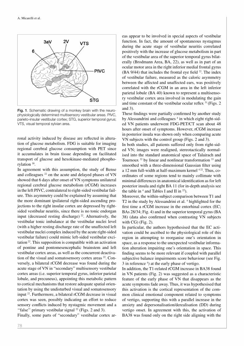
Functional neuronatomy of vestibular networksThe vestibular system is based on the principle of fusion of bilateral sensors, the input of which is distributed in a bilaterally organised neuronal network 5. The core circuit-ry of this network includes ocular motor function that me-diates the vestibular-ocular reflex (Vor) and is imbedded in a complex multisensory system containing numerous ascending and descending pathways subserving percep-tual, postural and vegetative functions as well as naviga-tion and spatial memory 5. Thus, vestibular input, which is fed into the Vor structures, is also fed into adjacent but separate fibres for perception and balance control 5. As ocular motor function consists of a rapid three-neuron arc (for a more comprehensive review: see reference 5) as-cending from both labyrinths – via the vestibular nuclei – to their corresponding pair of extraocular eye muscles (for review see reference 6), perceptual functions operate via pathways that run through the lateral and ventroposterior lateral thalamus to the multisensory cortical neural net-work. The latter includes, in the brain of monkey, a num-ber of temporo-parietal cortex areas such as the strongly interconnected area 2v, area 3aV and the parieto-insular vestibular cortex (PiVC), as well as retroinsular areas, su-perior temporal gyrus, inferior parietal lobule 5 7-11 (Fig. 1). At this level, metabolic studies during irrigation in right- and left-handed human volunteers have shown that the handedness of subjects and the side of the stimula-tion affect bilateral cortical activation pattern of vestibular areas. in fact, vestibular dominance in the non-dominant hemisphere and stronger activation occurring in the hemi-sphere ipsilateral to the stimulated ear were found 12. interestingly, navigation function seems to be mediated by ‘head direction cells’ in the thalamus (for review see reference 13) and ‘place cells’ in the hippocampus 14. Vari-ous anatomical connections have been proposed to join the vestibular nuclei to the hippocampus 15 16. Using func-tional Mri, Vitte and co-workers 17 demonstrated that ves-tibular caloric stimulation even activates the hippocampal formation in humans. The postural control of head and body is mediated via the descending tracts such as the medial vestibulo-spinal tract for head position and the lateral vestibulo-spinal tract for head and body position in space 18. Finally, vegetative functions are conveyed by pathways from the vestibular nuclei to the locus coeruleus, nucleus of the solitary tract, area postrema and the central nucleus of the amygdale 19
as well as the parabrachial nucleus, infralimbic cortex and hypothalamus 20 21. Since neuroimaging protocols are available, many stud-ies in the last 10 years have focused on the cortical and subcortical changes related to sudden vestibular deaffer-entation as in Vn. however, most of these, also due to different possibilities across diverse centers, were based on different times of first acquisition from the onset of Vn symptoms, neuroimaging techniques, statistical analysis
and correlation with otoneurological and psychological findings.The aim of the present review is to merge the similarities and discrepancies across studies that employed neuroim-aging techniques with the purpose of better understanding brain changes and characteristic clinical features during the acute phase of Vn.
Materials and methodsStudy selection and inclusion/exclusion criteriaA thorough analysis on PubMed was searched using the following key words: vestibular neuritis, unilateral vestibu-lar failure (UVF), neuroimaging, magnetic resonance im-aging (Mri), positron emission tomography (PET), near infra-red spectroscopy (nirS), single positron emission computer tomography (SPECT), voxel-based morphome-try (VBM), cerebral, cortical, sub-cortical and cerebellum.only studies written in English were selected. Studies fo-cusing on simultaneous cochleo-vestibular, vestibular sur-gical de-afferentation and/or bilateral vestibular impair-ment were excluded as well as those not enrolling acute Vn patients and not employing voxel-based analysis.herein, T1 will be used only to indicate acute phase of Vn and T2 only for the delayed phase, even if Vn sub-jects were only studied during the latter.
ResultsA preliminary examination of the existing literature high-lighted that four major neuroimaging techniques have been employed in the study of acute phase of Vn: [18F] fluorode-oxyglucose (FDg) - PET/computer tomography (CT) 4 22 23, SPECT 24, VBM 25-27 and functional Mri (fMri) 28 29. According to the above-mentioned criteria, the studies by Alessandrini et al. 24, helmchen et al. 26 and Zu Eulenburg et al. 25 were excluded, and the present review included 6 studies.Moreover, all included studies 4 22 23 27-29 investigated cer-ebral correlates of acute and delayed phase of Vn and in three 4 28 29 acute phase images were compared to both de-layed and control groups (Cg).Differences in subjects, time of acquisitions from the Vn symptoms onset, neuroimaging technique, statistical anal-ysis and contingent correlations with neuropsychological and otoneurological tests are shown in Table i.
DiscussionCortical correlates of acute and delayed VN phase
FDG-PET/CT imaging studiesUnder physiological conditions, as well as in several dis-eases affecting the brain, glucose metabolism is tightly connected to neuronal activity. Therefore, changes in neu-

Neuroimaging studies in acute vestibular neuritis
77
Tab
le I.
Sys
tem
atic
ana
lysis
of n
ine
stud
ies.
Stud
ySu
bjec
ts/
Sam
ple
Side
of
VN
Neur
oim
agin
g te
chni
que
T1T2
Pres
ence
of
cont
rol g
roup
Mai
n Ot
o-ne
urol
ogic
al
test
Neur
opsy
chol
ogic
al
and
clin
ical
test
Stat
istic
al
anal
ysis
/imag
es
hand
ling
Incl
usio
n
Bens
e et
al.
2004
5 rig
ht-h
ande
d pa
tient
s (4
mal
e, 1
fe
mal
e; m
ean
age
: 64
year
s ±
10)
5 rig
ht
VNFD
G-PE
T/CT
6.6
days
af
ter s
ymp-
tom
ons
et
3 m
onth
s af
-te
r sym
ptom
s on
set
None
DC-E
OG, S
VV, c
alor
ic
test
ing
None
SPM
99Ye
s
Ales
sand
rini
et a
l. 20
099
right
-han
ded
patie
nts
(4 m
ale,
5
fem
ale;
mea
n ag
e: 5
1.6
year
s ±
13.8
)
7 le
ft VN
; 2
right
VN
SPEC
T72
hou
rs
afte
r sym
p-to
m o
nset
1 m
onth
afte
r sy
mpt
oms
onse
t
None
ENG
None
visua
l eva
luat
ion
by a
wel
l exp
e-rie
nced
nuc
lear
ph
ysic
ian
No
Helm
chen
et
al. 2
009
15 ri
ght-
hand
ed p
atie
nts
(8 m
ales
, 7
fem
ales
; mea
n ag
e: 4
9 ±
13.
9)
4 le
ft VN
; 11
rig
ht
VN
VBM
None
3 m
onth
s af
ter
sym
ptom
ons
et15
(8 m
ales
, 7
fem
ales
; mea
n ag
e: 4
9 ±
13.
7 ye
ars)
CVS,
SVDS
,SV
V,Ca
loric
test
ing,
DC-
EOG
SVDS
VBM
tool
box
for
SPM
2No
Zu E
ulen
-bu
rg e
t al.
2010
22 ri
ght-
hand
ed p
atie
nts
(9 fe
-m
ales
, 13
mal
es; m
ean
age:
56
.7±
10.4
)
10
right
VN
, 12
left
VN
VBM
None
2.5±
1.6
year
s af
ter s
ympt
o-m
onse
t
Not s
peci
fied
in
the
text
Calo
ric te
stin
g,DC
-EOG
, HIT,
Und
erbe
rger
te
st, V
EMPs
, rot
ator
y ch
ai
test
, SVV
VSS,
VHQ
VBM
tool
box
for
SPM
5No
Ales
sand
rini
et a
l. 20
138
right
han
ded
patie
nts
(five
fe-
mal
es, 3
mal
es; m
ean
age:
48±
7 ye
ars)
8 rig
ht
VNFD
G-PE
T/CT
48±
6 ho
urs
sym
ptom
on
set
1 m
onth
afte
r sy
mpt
om o
nset
30 (1
6 fe
mal
e,
14 m
ales
; mea
n ag
e 49
.5±
12
year
s)
DC-E
OGZu
ng In
stru
men
t, de
pers
onal
izatio
n/de
-re
aliza
tion
inve
ntor
y, Go
mez
test
SPM
2Ye
s
Ales
sand
rini
et a
l. 20
148
right
han
ded
patie
nts
(5 fe
mal
es,
3 m
ales
; mea
n ag
e 48
±7
year
s)8
right
VN
FDG-
PET/
CT48
±6
hour
s sy
mpt
om
onse
t
1 m
onth
afte
r sy
mpt
om o
nset
None
DC-E
OG, B
ucke
t tes
tZu
ng In
stru
men
t, de
pers
onal
izatio
n/de
-re
aliza
tion
inve
ntor
y, Go
mez
test
AAL
Yes
Hong
et a
l. 20
149
right
-han
ded
patie
nts
(6 m
ales
, 3
fem
ales
; mea
n ag
e: 4
9.2
± 1
8.1
year
s)
5 rig
ht
VN;
4 le
ft VN
VBM
72 h
ours
af
ter s
ymp-
tom
ons
et
3 m
onth
s af
ter
sym
ptom
ons
etNo
neVN
G, C
alor
ic te
stin
g, ro
ta-
tory
cha
i tes
tK-
DHI
VBM
tool
box
for
SPM
8Ye
s
Helm
chen
et
al. 2
013
20 ri
ght-
hand
ed p
atie
nts
(11
mal
es,
9 fe
mal
es; m
ean
age:
55.
1 ±
13.
9)10
rig
ht
VN;
10 le
ft VN
fMRI
72 h
ours
af
ter s
ymp-
tom
ons
et
96.6
± 2
4 da
ys a
fter
sym
ptom
ons
et
20 (1
1 m
ales
, 9
fem
ales
; mea
n ag
e: 5
0.2
± 1
1.7
year
s)
DC-E
OG, C
alor
ic te
stin
g,
SVV,
HIT,
sta
tic p
ostu
rog-
raph
y
CVS,
SVD
S, V
ADL
SPM
8, IC
AYe
s
Klin
gner
et
al. 2
014
14 ri
ght-
hand
ed p
atie
nts
(6 fe
-m
ales
, 8 m
ales
; mea
n ag
e: 5
1.1
±
10.4
yea
rs)
7 rig
ht
VN,
7 le
ft VN
fMRI
4.9
± 1
.9
days
afte
r sy
mpt
om
onse
t
12 ±
4.6
m
onth
s af
ter
sym
ptom
ons
et
28 a
ge a
nd g
en-
der m
atch
ed
cont
rols
(12
fe-
mal
es, 1
6 m
ales
)
VNG,
Cal
oric
test
ing,
Sac
-ca
dic
eye
mov
emen
ts,
smoo
th p
ursu
it, o
ptok
inet
-ic
nys
tagm
us, g
aze
test
None
SPM
8, IC
AYe
s
VN, v
estib
ular
neu
ritis;
T1,
acu
te p
hase
of V
N; T
2, d
elay
ed p
hase
of V
N; F
DG-P
ET/C
T, [1
8F] fl
uoro
deox
yglu
cose
– p
ositr
on e
miss
ion
tom
ogra
phy/
com
pute
r tom
ogra
phy;
SPE
CT, s
ingl
e po
sitro
n em
issio
n co
mpu
ter t
omog
raph
y; V
BM, v
oxel
-bas
ed
mor
phom
etry
; fM
RI, f
unct
iona
l mag
netic
reso
nanc
e im
agin
g; D
C-EO
G, b
inoc
ular
ele
ctro
ocul
ogra
phy;
SVV
, sub
ject
ive v
isual
ver
tical
; ENG
, ele
ctro
nyst
agm
ogra
phy;
CVS
, clin
ical v
estib
ular
sco
re; S
VDS,
sub
ject
ive v
estib
ular
disa
bilit
y sc
ore;
HIT,
he
ad im
pulse
test
; VEM
Ps, v
estib
ular
evo
ked
myo
geni
c po
tent
ials;
VSS
, ver
tigo
seve
rity
scor
e; V
HQ, v
ertig
o ha
ndica
p qu
estio
nnai
re; K
-DHI
, Kor
ean
vers
ion
of th
e di
zzin
ess
hand
icap
inve
ntor
y; V
NG, v
ideo
nyst
agm
ogra
phy;
SVD
S, s
elf-a
sses
smen
t of
ves
tibul
ar d
isabi
lity;
VAD
L, s
elf-a
sses
smen
t of v
estib
ular
disa
bilit
y in
dai
ly lif
e; S
PM, s
tatis
tical
par
amet
ric m
appi
ng, A
AL, a
utom
ated
ana
tom
ical l
abel
ling,
ICA,
inde
pend
ent c
ompo
nent
ana
lysis.
A. Micarelli et al.
78
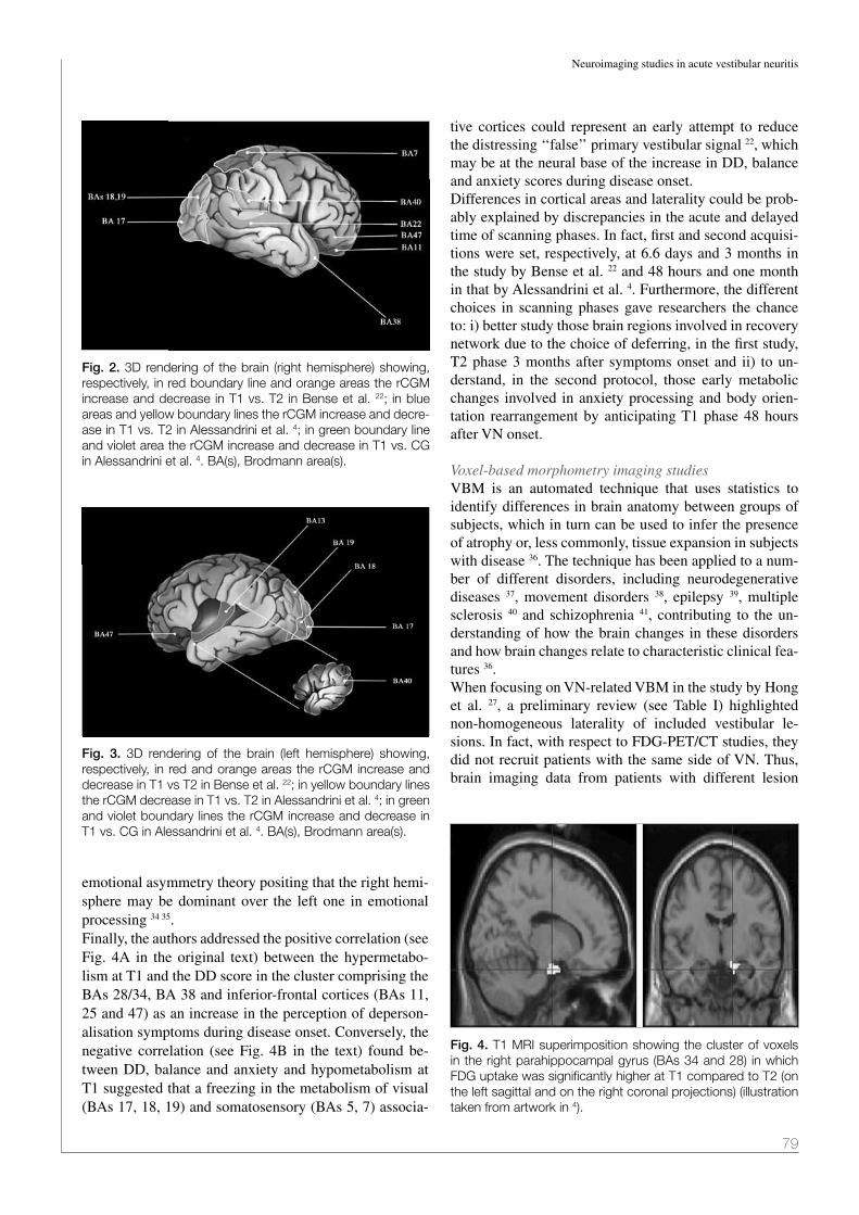
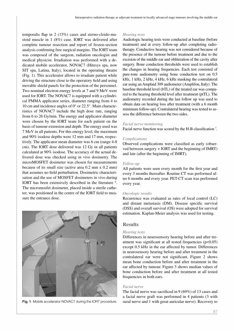
ronal activity induced by disease are reflected in altera-tion of glucose metabolism. FDg is suitable for imaging regional cerebral glucose consumption with PET since it accumulates in brain tissue depending on facilitated transport of glucose and hexokinase-mediated phospho-rylation 30. in agreement with this assumption, the study of Bense and colleagues 22 on the acute and delayed phases of Vn showed that 6 days after onset of Vn symptoms unilateral regional cerebral glucose metabolism (rCgM) increases in the left PiVC, contralateral to right-sided vestibular fail-ure. This asymmetry could be explained by assuming that the more dominant ipsilateral right-sided ascending pro-jections to the right insular cortex are depressed by right-sided vestibular neuritis, since there is no tonic endorgan input (decreased resting discharge) 22. Alternatively, the vestibular tonic imbalance at the vestibular nuclei level (with a higher resting discharge rate of the unaffected left vestibular nuclei complex induced by the acute right-sided vestibular failure) could mimic left-sided vestibular exci-tation 22. This supposition is compatible with an activation of pontine and pontomesencephalic brainstem and left vestibular cortex areas, as well as the concurrent deactiva-tion of the visual and somatosensory cortex areas 22. Con-versely, a bilateral rCgM decrease was found during the acute stage of Vn in “secondary” multisensory vestibular cortex areas (i.e. superior temporal gyrus, inferior parietal lobule, and precuneus), appointing this metabolic pattern to cortical mechanisms that restore adequate spatial orien-tation by using the undisturbed visual and somatosensory input 22. Furthermore, a bilateral rCgM decrease in visual cortex was seen, possibly indicating an effort to reduce sensory conflicts induced by nystagmic movement and a ‘‘false’’ primary vestibular signal 22 (Figs. 2 and 3). Finally, some parts of “secondary” vestibular cortex ar-
eas appear to be involved in special aspects of vestibular function. in fact, the amount of spontaneous nystagmus during the acute stage of vestibular neuritis correlated positively with the increase of glucose metabolism in part of the vestibular area of the superior temporal gyrus bilat-erally (Brodmann Area, BA, 22), as well as in part of an ocular motor area in the right inferior medial frontal gyrus (BA 9/44) that includes the frontal eye field 22. The index of vestibular failure, measured as the caloric asymmetry between the affected and unaffected ears, was positively correlated with the rCgM in an area in the left inferior parietal lobule (BA 40) known to represent a multisenso-ry vestibular cortex area involved in modulating the gain and time constant of the vestibular ocular reflex 31 (Figs. 2 and 3).These findings were partially confirmed by another study by Alessandrini and colleagues 4 in which eight right-sid-ed Vn patients underwent FDg-PET/CT scan about 48 hours after onset of symptoms. however, rCgM increase in posterior insula was shown only when comparing acute Vn subjects with the control group (Figs. 2 and 3).in both studies, all patients suffered only from right-sid-ed Vn; images were realigned, stereotactically normal-ised into the standard anatomical space of Talairach and Tournoux 32 by linear and nonlinear transformation 33 and smoothed with a three-dimensional gaussian filter using a 12 mm full-width at half-maximum kernel 4 22. Thus, co-ordinates of some regions tend to mainly collimate with minimal differences in anatomical identification as for left posterior insula and right BA 11 (for in-depth analysis see the table in 4 and Tables i and ii in 22). Moreover, the within-subject comparison between T1 and T2 in the study by Alessandrini et al. 4 highlighted for the first time a rCgM increase in the entorhinal cortex (EC; BAs 28/34; Fig. 4) and in the superior temporal gyrus (BA 38) (data also confirmed when contrasting Vn subjects with Cg) (Fig. 2). in particular, the authors hypothesised that the EC acti-vation could be ascribed to the physiological role of this region in attempting to reorganise one’s orientation in space, as a response to the unexpected vestibular informa-tion alteration impairing one’s orientation in space. This finding seems to be more relevant if coupled with parallel subjective balance impairments score behaviour (see Fig. 3 in reference 4) at the early phase of vertigo. in addition, the T1-related rCgM increase in BA38 found in Vn patients (Fig. 2) was suggested as a characteristic feature of the early phase of Vn that disappears as the acute symptoms fade away. Thus, it was hypothesised that this activation is the cortical representation of the com-mon clinical emotional component related to symptoms of vertigo, supporting this with a parallel increase in the anxiety and depersonalisation/derealisation (DD) during vertigo onset. in agreement with this, the activation of BA38 was found only on the right side aligning with the
Fig. 1. Schematic drawing of a monkey brain with the neuro-physiologically determined multisensory vestibular areas. PIVC, parieto-insular vestibular cortex; STG, superior temporal gyrus; VTS, visual temporal sylvian area.
Neuroimaging studies in acute vestibular neuritis
79
emotional asymmetry theory positing that the right hemi-sphere may be dominant over the left one in emotional processing 34 35. Finally, the authors addressed the positive correlation (see Fig. 4A in the original text) between the hypermetabo-lism at T1 and the DD score in the cluster comprising the BAs 28/34, BA 38 and inferior-frontal cortices (BAs 11, 25 and 47) as an increase in the perception of deperson-alisation symptoms during disease onset. Conversely, the negative correlation (see Fig. 4B in the text) found be-tween DD, balance and anxiety and hypometabolism at T1 suggested that a freezing in the metabolism of visual (BAs 17, 18, 19) and somatosensory (BAs 5, 7) associa-
tive cortices could represent an early attempt to reduce the distressing ‘‘false’’ primary vestibular signal 22, which may be at the neural base of the increase in DD, balance and anxiety scores during disease onset.Differences in cortical areas and laterality could be prob-ably explained by discrepancies in the acute and delayed time of scanning phases. in fact, first and second acquisi-tions were set, respectively, at 6.6 days and 3 months in the study by Bense et al. 22 and 48 hours and one month in that by Alessandrini et al. 4. Furthermore, the different choices in scanning phases gave researchers the chance to: i) better study those brain regions involved in recovery network due to the choice of deferring, in the first study, T2 phase 3 months after symptoms onset and ii) to un-derstand, in the second protocol, those early metabolic changes involved in anxiety processing and body orien-tation rearrangement by anticipating T1 phase 48 hours after Vn onset.
Voxel-based morphometry imaging studiesVBM is an automated technique that uses statistics to identify differences in brain anatomy between groups of subjects, which in turn can be used to infer the presence of atrophy or, less commonly, tissue expansion in subjects with disease 36. The technique has been applied to a num-ber of different disorders, including neurodegenerative diseases 37, movement disorders 38, epilepsy 39, multiple sclerosis 40 and schizophrenia 41, contributing to the un-derstanding of how the brain changes in these disorders and how brain changes relate to characteristic clinical fea-tures 36.When focusing on Vn-related VBM in the study by hong et al. 27, a preliminary review (see Table i) highlighted non-homogeneous laterality of included vestibular le-sions. in fact, with respect to FDg-PET/CT studies, they did not recruit patients with the same side of Vn. Thus, brain imaging data from patients with different lesion
Fig. 2. 3D rendering of the brain (right hemisphere) showing, respectively, in red boundary line and orange areas the rCGM increase and decrease in T1 vs. T2 in Bense et al. 22; in blue areas and yellow boundary lines the rCGM increase and decre-ase in T1 vs. T2 in Alessandrini et al. 4; in green boundary line and violet area the rCGM increase and decrease in T1 vs. CG in Alessandrini et al. 4. BA(s), Brodmann area(s).
Fig. 3. 3D rendering of the brain (left hemisphere) showing, respectively, in red and orange areas the rCGM increase and decrease in T1 vs T2 in Bense et al. 22; in yellow boundary lines the rCGM decrease in T1 vs. T2 in Alessandrini et al. 4; in green and violet boundary lines the rCGM increase and decrease in T1 vs. CG in Alessandrini et al. 4. BA(s), Brodmann area(s).
Fig. 4. T1 MRI superimposition showing the cluster of voxels in the right parahippocampal gyrus (BAs 34 and 28) in which FDG uptake was significantly higher at T1 compared to T2 (on the left sagittal and on the right coronal projections) (illustration taken from artwork in 4).

A. Micarelli et al.
80
sides were collapsed, in contrast with most previous VBM studies that artificially equalised the lesion side by flip-ping brain images of patients with lesions on the opposite side (e.g., flipping images of left Vn patients to simulate right Vn) 26 42 43. Moreover, nine right-handed Vn subjects were studied in T1 and T2 phase and the within-subject model found a significant decrease in grey matter volume (gMV) in the right superior medial gyrus, right middle orbital gyrus, cerebellar vermis and right cerebellar hemi-sphere in T1 compared to T2 (see Table ii in the origi-nal text). however, authors focused their discussion on gMV increase related to processes involved in vestibular compensation (T2) rather than in cortical atrophy found in these regions during the acute stage. This aspect may be in line with the employed technique, which is more useful in visualising brain volume changes over the time rather than in the acute stage of disease when a paucity of corti-cal hypertrophy/atrophy is found.
Sub-cortical structure involvement related to the early phase of VNAlessandrini et al. 23 used the same Vn patients of a previ-ous study 4 to provide an exclusive cerebellar analysis of FDg uptake changes comparing T1 and T2, by using ana-tomical automatic labelling (AAl) structural volumes of interest (Vois). The authors attempted to correlate these findings with those at the cortical level due to cerebellar involvement in different cerebro-cerebellar loops previ-ously highlighted 44.in particular, a relative hypometabolism in the early phase of Vn in anterior cerebellar lobe was found, includ-ing vermis 1-2, 3 and 6, and bilateral lobule iii and Vi (Fig. 5). These findings were postulated to be consistent with a cortical rCgM decrease in bilateral sensory-motor and parietal cortices, suggesting the relative rCgM de-crease in the anterior cerebellar lobe is associated with such hypometabolism 4. in addition, the hypometabolism seen in the anterior cerebellar lobe was hypothesised to support a bottom-up regulation of sensory conflict during controversial inflow between optical and vestibular input, as it occurs during the early phases of Vn 23. Thus, such a behaviour was interpreted by the authors as a realign-ment of the relationship between sensory inputs 45 such as those coming from optical 46, proprioceptive 47 48 and vestibular 49 organs and cerebellum 50 via olivary climb-ing fibers 51 that could convey an erroneous signal arising from mismatch between mentioned sensory inflow. Moreover, the study by Alessandrini et al. 23 highlighted a relative hypometabolism in the nodulus and flocculus at T1 compared to T2, suggesting a primary adaptive be-haviour of these regions in response to abrupt conflict-ing inputs conveyed by retinal slip and vestibular loss in the early phase of Vn. Curiously, the negative correlation found between metabolism in the right lobule 10 and slow phase velocity (SPV) scores highlighted a specific pivotal
role of the flocculus in modulating and controlling nys-tagmus parameters and in adapting Vor by mediating the functional interaction between vestibular inputs and the eye movement network 52. Finally, the study found a significant rCgM increase when comparing T1 to T2 in right crus i and a significant positive correlation between the metabolism found in this structure at Vn onset and anxiety score (see Table i and Figs. 2 and 3 in the text of 23). These data, along with anxiety/rCgM correlation findings in crus i, were inter-preted to add information regarding the function of this region during the acute phase of Vn and in subserving cortical emotional processing affecting the early stages of disease 23.hong et al. found a T1-related gMV increase (a relative T2-related gMV decrease) in cerebellar vermis and right lobule Viiia 27. in line with the discussion topic of their longitudial work (see chapter 1.2 in the text above), the authors explained the former finding as the consequence of a dominance of afferent input in the cerebellar vermis (gM atrophy following loss of peripheral sensory input), which could be underpinned by those cortico-cerebellar loop previously mentioned. The latter finding was found to be related over time with a decrease in nystagmus after impulse acceleration and deceleration; gM atrophy in this area was also associated with better recovery of periph-eral vestibular sense 27. however, although speculative, authors highlighted this area functions to subserve the earliest stages of Vn, and hypothesised that as peripheral vestibular function recovers, the functional contribution from this area may decrease and, subsequently, decrease gMV.Finally, these data could be not completely conflicting as it was chosen to investigate patients in different T2 mo-ments with a more strengthened rehabilitation protocol in
Fig. 5. 3D rendering of cerebellum showing in orange and gre-en colours the VOIs in which FDG uptake was significantly lo-wer and higher, respectively, at T1 compared to T2. On the top the ventral; on the bottom the dorsal cerebellar surface (illustra-tion modified from artwork in 23).

Neuroimaging studies in acute vestibular neuritis
81
hong et al. and by using different methods of scanning and data and imaging handling.
Functional connectivity in VNit has been shown that brain networks known to support visual, motor, attentional, or cognitive functions show spontaneous 53 and anticorrelated fluctuations 54 even without a specific task. in fact, large-amplitude spontane-ous low-frequency (0.1 hz) fluctuations in blood-oxygen-level dependent (BolD) signal, investigated by fMri, are temporally correlated across functionally related areas 55. By using these methods, many authors have attempted to identify changes in functional connectivity within neural networks 56 57 with the basic idea that spontaneous fluc-tuations in brain activity during rest reflect the intercon-nectivity of brain areas necessary to accommodate highly diverse processing demands. indeed, using resting state analysis it has been shown that the brain is organised into ‘‘dynamic, anticorrelated functional networks’’ 54. According to these aspects, helmchen and colleagues 28 evaluated the BolD signal changes in 20 right-handed Vn patients with acute Vn at T1 (within 3 days from symptom onset) and 3 months later (T2). in addition, they compared T1 brain images both with T2 phase and an age- and sex-matched Cg. For patients with right pe-ripheral vestibular failure, the smoothed images were mirrored along the y axis so that the left side was the le-sion side for all patients. They performed two statistical analyses, an independent component analysis (iCA) and a region-of-interest analysis based on the local-to-global ratio. A similar fMri approach was recently adopted by Klingner et al. 28 in order to discover the inter-network functional connectivity changes in 14 patients during the early (mean: 4.9 ± 1.9 days after onset of symptoms) and delayed (mean: 12 ± 4.6 months) phase of Vn.in the former study, one component (‘‘component 50’’) showed significant between-group changes in resting-state activity at T1. This component revealed a functional network of the parietal lobe, medial aspect of the superior parietal lobule, posterior cingulate cortex, middle frontal gyrus, middle temporal gyrus, parahippocampal gyrus, anterior cingulate cortex, insular cortex, caudate nucleus, thalamus and midbrain. Vn patients at T1 showed de-creased resting-state activity in the contralateral intrapa-rietal sulcus (iPS) in close vicinity to the rostro-dorsal aspect of the iPl, i.e. the supramarginal gyrus (SMg), compared with Cg. When the two measurements of the patients were compared, a change in resting-state activity in the same region became apparent indicating normalisa-tion of resting-state activity in patients over time.While the network revealed by component 50 in that study likely supports multiple functions, it is interesting to note that several of the implied areas have been shown to process vestibular signals (see review in 5). Within this neural network, significant differences between patients
and controls were found in iPS contralateral to the ves-tibular lesion. The iPS 58 extends ventrally to the parietal operculum, including the parietal opercular area oP2, which has re-cently been identified as the core region for vestibular processing in humans 59, and therefore suggested to be the human equivalent to the PiVC previously described in monkey 60. This findings – and subsequent seed-based functional connectivity analysis based on resting-state os-cillations 59 – led some authors to suggest that the iPl is a cortical multisensory integration area 59.Moreover, in Vn patients of this study the iPS coordinates of reduced resting-state activity overlapped with the ven-tral intraparietal area (ViP) contained in its fundus, the neuronal responses of which are more strongly influenced by vestibular than visual inputs 61 and are strongly linked to heading, which make them well equipped to play a role in multisensory integration for heading perception 62. in addition, effective connectivity analysis has shown a role of the iPS for memory retrieval 63. The SMg along the iPS, which also showed changes of resting-state activ-ity in patients in that study, is critical for mediating spatial working memory and shifts in spatial attention 64. This is of interest as the iPS is linked to the hippocampal forma-tion 65, and reduced resting-state activity in iPS might also influence hippocampal and parahippocampal function. Thus, it was speculated that reduced vestibular input 66 leads to changes of resting-state activity in iPS, which in turn may trigger adaptive hippocampal reorganisation and impairment in navigational and spatial orientation tasks.however, Vn patients might suffer from impaired spatial navigation which has been shown for bilateral 67, but not unilateral nor surgical vestibular nerve deafferentation 68. Furthermore, lesion studies in humans have provided ev-idence for a cortical influence on vestibular function in the context of spatial representation 31. in line with these studies and findings from PET and neuropsychological in-vestigations 4 69, one might speculate that the reduction of resting-state activity in the iPS and adjacent SMg in the acute stage of Vn patients could be related to impaired spatial orientation, i.e. by deficient spatial working mem-ory or spatial attention 64.in the latter study, Klingner et al. 29 found decreased inter-network connectivity in T1 compared to T2 (af-ter complete clinical remission of symptoms) between the “default mode” network (DMn) and multiple other networks such as the somatosensory cortex, auditory/vestibular/insular cortex, motor cortex, occipital cor-tex and left and right fronto-parietal cortex (FPC). By comparing the first measurement in T1 with the group of healthy control subjects, the same areas (except the occipital cortex) showed decreased connectedness to the DMn (Fig. 1 in the original text). it is generally agreed that the brain is composed of two spatially distinct func-tional networks: the DMn and “task-positive” network

A. Micarelli et al.
82
(TPn) 54 70 71. During performance of attention-demand-ing tasks, prefrontal and parietal structures that comprise the task-positive network are characterised by increased activity; in contrast, the DMn, including the posterior cingulate and medial prefrontal cortices, is characterised by decreased activity. During wakeful rest, the opposite pattern emerges 54 70-72.The reduced connectivity between these two networks was suggested to be related to the diverging information arising in the resting condition from vestibular, spontane-ous eye movements and other sensory modalities 29. The attempt to integrate this conflicting information requires significantly greater capacity for the processing informa-tion about spatial orientation and brings sensory infor-mation processing to our attention, which is normally an automated process that does not require attentional demand 29. These mechanisms are reflected by increased activity within brain areas responsible for processing of vestibular information and integration of multisensory in-formation 22. The sustained increased activity in parts of the TPn and the attentional demand reduce the activity within the DMn. in turn, the DMn compensates by de-creasing the amount of information that is received from the task positive network, leading to decreased connect-edness 29. Furthermore, the involvement of the DMn in this pathology is further supported by findings of difficul-ties with cognitive skills such as reading, arithmetic and concentration suggesting a decreased ability to engage the task positive networks 73.
Final overviewThe vestibulo-cortical system, which includes the PiVC activated by vestibular stimulation, is composed of mul-tisensory cortical networks connected with other cortical and subcortical processing areas, including oculomotor, somatosensory, visual areas and cerebellar sub-regions.The present review clearly reveals that sudden UVF may induce a wide variety of cortical intersensory responses, with changes in different, sensory modules as a result of acute plasticity in the central nervous system. interestingly, during the acute phase of disease perfusion-al and metabolic studies demonstrated such rCgM chang-es in the so-called “vestibular cortex”, decreased resting-state activity in the contralateral iPS – close to the human equivalent of PiVC – and a decreased inter-network con-nectivity between the DMn and multiple other networks. Simultaneously, a neural cascade of acute plasticity relat-ed events was found in the visual and multisensory cortex as well as in those areas (enthorinal, SMg) involved in spatial navigation. overall, findings from imaging and neuropsychological studies can disclose two side of the same coin for which the acute dissociation of sensory inflow was pinpointed, highlighting phenomena of emotional and orientation im-pairment.
Finally, we hope the present review can serve as a frame-work of the intricate puzzle represented by multi-scale brain changes involved in the acute phases of Vn and can provide additional considerations for future acute Vn studies.
AcknowledgementsThe authors express sincere thanks to MichelAngela Stu-dio for skillful and creative preparation of illustrations.
References1 Chihara y, iwasaki S, Murofushi T, et al. Clinical charac-
teristics of inferior vestibular neuritis. Acta otolaryngol 2012;132:1288-94.
2 Strupp M, Brandt T. Peripheral vestibular disorders. Curr opin neurol 2013;26:81-9.
3 Alessandrini M, D’Erme g, Bruno E, et al. Vestibular com-pensation: analysis of postural re-arrangement as a con-trol index for unilateral vestibular deficit. neuroreport 2003;14:1075-9.
4 Alessandrini M, Pagani M, napolitano B, et al. Early and phasic cortical metabolic changes in vestibular neuritis on-set. PloS onE 2013;8:e57596.
5 Dieterich M, Brandt T. Functional brain imaging of peripheral and central vestibular disorders. Brain 2008;131:2538-52.
6 leigh rJ, Zee DS. The neurology of eye movements. Fourth edition. new york: oxford University Press; 2006.
7 guldin Wo, grüsser oJ. The anatomy of the vestibular corti-ces of primates. in: Collard M, Jeannerod M, Christen y, eds. Le cortex vestibulaire. Paris: ipsen; 1996. p. 17.
8 Klam F, graf W. Vestibular response kinematics in posterior parietal cortex neurons of macaque monkeys. Eur J neurosci 2003;18:995-1010.
9 Klam F, graf W. Vestibular signals of posterior parietal cor-tex neurons during active and passive head movements in macaque monkeys. Ann n y Acad Sci 2003;1004:271-82.
10 hegemann S, Fitzek S, Fitzek C, et al. Cortical vestibular representation in the superior temporal gyrus. J Vestib res 2004;14:33-5.
11 lackner Jr, DiZio P. Vestibular, proprioceptive, and hap-tic contributions to spatial integration. Annu rev Psychol 2005;56:115-47.
12 Dieterich M, Bense S, lutz S, et al. Dominance for vestibu-lar cortical function in the non-dominant hemisphere. Cereb Cortex 2003;13:994-1007.
13 Taube JS, goodridge JP, golob EJ, et al. Processing the head direction signal: a review and commentary. Brain res Bull 1996;40:477-86.
14 Wiener Si, Korshunov VA, garcia r, et al. Interial, sub-stratal and landmark cue control of hippocampal CA1 place cell activity. Eur J neurosci 1995;7:2206-19.
15 Smith PF. Vestibular-hippocampal interactions. hippocam-pus 1997;7:465-71.
16 horii A, russell nA, Smith PF, et al. Vestibular influences on

Neuroimaging studies in acute vestibular neuritis
83
CA1 neurons in the rat hippocampus: an electrophysiologi-cal study in vivo. Exp Brain res 2004;155:245-50.
17 Vitte E, Derosier C, Caritu y, et al. Activation of the hip-pocampal formation by vestibular stimulation: a func-tional magnetic resonance imaging study. Exp Brain res 1996;112:523-6.
18 nathan PW, Smith M, Deacon P. Vestibulospinal, reticulospi-nal and descending propriospinal nerve fibres in man. Brain 1996;119:1809-33.
19 Pompeiano o, d’Ascanio P, Centini C, et al. Gene expression in rat vestibular and reticular structures during and after space flight. neuroscience 2002;114:135-55.
20 Balaban CD. Projections from the parabrachial nucleus to the vestibular nuclei: potential substrates for autonomic and limbic influences on vestibular responses. Brain res 2004;996:126-37.
21 Balaban CD, Thayer JF. Neurological bases for balance-anxiety links. J Anxiety Disord 2001;15:53-79.
22 Bense S, Bartenstein P, lochmann M, et al. Metabolic changes in vestibular and visual cortices in acute vestibular neuritis. Ann neurol 2004;56:624-30.
23 Alessandrini M, Micarelli A, Chiaravalloti A, et al. Cerebellar metabolic involvement and its correlations with clinical pa-rameters in vestibular neuritis. J neurol 2014;261:1976-85.
24 Alessandrini M, napolitano B, Bruno E, et al. Cerebral plas-ticity in acute vestibular deficit. Eur Arch otorhinolaryngol 2009;266:1547-51.
25 zu Eulenburg P, Stoeter P, Dieterich M. Voxel-based morpho-metry depicts central compensation after vestibular neuritis. Ann neurol 2010;68:241-9.
26 helmchen C, Klinkenstein J, Machner B, et al. Structural changes in the human brain following vestibular neuritis in-dicate central vestibular compensation. Ann ny Acad Sci 2009;1164:104-15.
27 hong SK, Kim Jh, Kim hJ, et al. Changes in the gray matter volume during compensation after vestibular neu-ritis: a longitudinal VBM study. restor neurol neurosci 2014;32:663-73.
28 helmchen C, ye Z, Sprenger A, et al. Changes in rest-ing-state fMRI in vestibular neuritis. Brain Struct Funct 2014;219:1889-900.
29 Klingner CM, Volk gF, Brodoehl S, et al. Disrupted func-tional connectivity of the default mode network due to acute vestibular deficit. neuroimage Clin 201;6:109-14.
30 Varrone A, Asenbaum S, Vander Borght T, et al. EANM procedure guidelines for PET brain imaging using [18F]FDG, version 2. Eur J nucl Med Mol imaging 2009;361:2103-10.
31 Ventre-Dominey J, nighoghossian n, Denise P. Evidence for interacting cortical control of vestibular function and spatial representation in man. neuropsychologia 2003;41:1884-98.
32 Talairach J, Tournoux P. Co-planar stereotaxic atlas of the human brain. new york: georg Thieme Verlag; 1988.
33 Friston KJ, Asburner J, Frith CD, et al. Spatial registra-tion and normalization of images. hum Brain Mapp 1995;2:165-89.
34 Davidson rJ. Affective style and affective disorders:
perspectives from affective neuroscience. Cogn Emot 1998;12:307-330.
35 Cope lM, Schaich Borg J, et al. Hemispheric asymmetries during processing of immoral stimuli. Front Evol neurosci 2010;2:110.
36 Whitwell Jl. Voxel-based morphometry: an automated tech-nique for assessing structural changes in the brain. J neuro-sci 2009;29:9661-4.
37 WhitwellJl, Jack Cr Jr. Comparisons between Alzhei-mer disease, frontotemporal lobar degeneration, and nor-mal aging with brain mapping. Top Magn reson imaging 2005;16:409-25.
38 Whitwell Jl, Josephs KA. Voxel-based morphometry and its application to movement disorders. Parkinsonism relat Dis-ord 2007;3:S406-16.
39 Keller SS, roberts n. Voxel-based morphometry of temporal lobe epilepsy: an introduction and review of the literature. Epilepsia 2008;49:741-57.
40 Prinster A, Quarantelli M, orefice g, et al. Grey matter loss in relapsing-remitting multiple sclerosis: a voxel-based mor-phometry study. neuroimage 2006;29:859-67.
41 Williams lM. Voxel-based morphometry in schizophrenia: implications for neurodevelopmental connectivity models, cognition and affect. Expert rev neurother 2008;8:1049-65.
42 helmchen C, Klinkenstein JC, Kruger A, et al. Structural brain changes following peripheral vestibulo-cochlear le-sion may indicate multisensory compensation. J neurol neu-rosurg Psychiatry 2011;82:309-16.
43 hüfner K, Stephan T, hamilton DA, et al. Gray-matter atro-phy after chronic complete unilateral vestibular deafferenta-tion. Ann ny Acad Sci 2009;1164:383-5.
44 Stoodley CJ, Valera EM, Schmahmann JD. Functional to-pography of the cerebellum for motor and cognitive tasks: an fMRI study. neuroimage 2012;59:1560-70.
45 Martin TA, Keating Jg, goodkin hP, et al. Throwing while looking through prisms. II. Specificity and storage of multi-ple gaze-throw calibrations. Brain 1996;119:1199-211.
46 glickstein M. How are visual areas of the brain connected to motor areas for the sensory guidance of movement? Trends neurosci 2000;23:613-7.
47 Bauswein E, Kolb FP, leimbeck B, et al. Simple and com-plex spike activity of cerebellar Purkinje cells during ac-tive and passive movements in the awake monkey. J Physiol 1983;339:379-94.
48 Donga r, Dessem D. An unrelayed projection of jaw-muscle spindle afferents to the cerebellum. Brain res 1993;626:347-50.
49 Angelaki DE, Cullen KE. Vestibular system: the many facets of a multimodal sense. Annu rev neurosci 2008;31:125-50.
50 Wolpert DM, Miall rC, Kawato M. Internal models in the cerebellum. Trends Cogn Sci 1998;2:338-47.
51 ito M. Cerebellar long-term depression: characteriza-tion, signal transduction, and functional roles. Physiol rev 2001;81:1143-95.
52 Voogd J, Barmack nh. Oculomotor cerebellum. Prog Brain res 2006;151:231-68.
53 Marx E, Stephan T, nolte A, et al. Eye closure in darkness animates sensory systems. neuroimage 2003;19:924-34.

A. Micarelli et al.
84
54 Fox MD, Snyder AZ, Vincent Jl, et al. The human brain is intrinsically organized into dynamic, anticorrelated func-tional networks. Proc natl Acad Sci USA 2005;102:9673-8.
55 Biswal BB, Mennes M, Zuo Xn et al. Toward discovery science of human brain function. Proc natl Acad Sci USA 2010;107:4734-9.
56 Beckmann CF, Deluca M, Devlin JT, et al. Investiga-tions into resting-state connectivity using independent component analysis. Philos Trans r Soc lond B Biol Sci 2005;360:1001-13.
57 Mohammadi B, Kollewe K, Samii A, et al. Changes in rest-ing-state brain networks in writer’s cramp. hum Brain Mapp 2012;33:840-8.
58 Bremmer F, Klam F, Duhamel Jr, et al. Visual-vestibular in-teractive responses in the macaque ventral intraparietal area (VIP). Eur J neurosci 2002;16:1569-86.
59 Zu Eulenburg P, Caspers S, roski C, et al. Meta-analytical definition and functional connectivity of the human vestibu-lar cortex. neuroimage 2011;60:162-9.
60 grusser oJ, Pause M, Schreiter U. Vestibular neurones in the parieto-insular cortex of monkeys (Macaca fascicularis): vis-ual and neck receptor responses. J Physiol 1990;430:559-83.
61 gu y, Angelaki DE, Deangelis gC. Neural correlates of mul-tisensory cue integration in macaque MSTd. nat neurosci 2008;11:1201-10.
62 Chen A, Deangelis gC, Angelaki DE. Functional speciali-zations of the ventral intraparietal area for multisensory heading discrimination. J neurosci off J Soc neurosci 2013;33:3567-81.
63 Miyamoto K, osada T, Adachi y, et al. Functional differen-tiation of memory retrieval network in macaque posterior parietal cortex. neuron 2013;77:787-99.
64 Silk TJ, Bellgrove MA, Wrafter P, et al. Spatial work-ing memory and spatial attention rely on common neu-ral processes in the intraparietal sulcus. neuroimage 2010;53:718-24.
65 Ding Sl, Van hoesen g, rockland KS. Inferior parietal lobule projections to the presubiculum and neighboring ventromedial temporal cortical areas. J Comp neurol 2000;425:510-30.
66 hüfner K, Strupp M, Smith P, et al. Spatial separation of visual and vestibular processing in the human hippocampal formation. Ann n y Acad Sci 2011;1233:177-86.
67 Brandt T, Schautzer F, hamilton DA, et al. Vestibular loss causes hippocampal atrophy and impaired spatial memory in humans. Brain 2005;128:2732-41.
68 hüfner K, hamilton DA, Kalla r, et al. Spatial memory and hippocampal volume in humans with unilateral vestibular deafferentation. hippocampus 2007;17:471-85.
69 Candidi M, Micarelli A, Viziano A, et al. Impaired mental rotation in benign paroxysmal positional vertigo and acute vestibular neuritis. Front hum neurosci 2013;7:783.
70 Micarelli A, Jacobsson h, larsson SA, et al. Neurobio-logical insight into hyperbaric hyperoxia. Acta Physiol 2013;209:69-76.
71 Alessandrini M, Micarelli A, Chiaravalloti A, et al. Cortico-subcortical metabolic correlates of olfactory processing in healthy resting subjects. Sci rep 2014;4:5146.
72 Fox MD, Zhang D, Snyder AZ, et al. The global signal and observed anticorrelated resting state brain networks. J neu-rophysiol 2009;101:3270-83.
73 hanes DA, McCollum g. Cognitive-vestibular interactions: a review of patient difficulties and possible mechanisms. J Vestib res 2006;16:75-91.
Address for correspondence: Alessandro Micarelli, Department of Clinical Sciences and Translational Medicine, “Tor Vergata Univer-sity”, viale oxford, 81, 00133 rome, italy. Tel. +39 06 20902925. Fax +39 06 20902930. E-mail: [email protected]
received: March 14, 2015 - Accepted: July 25, 2015

85
ACTA oTorhinolAryngologiCA iTAliCA 2016;36:85-90; doi: 10.14639/0392-100X-486
Head and neck
Intraoperative radiation therapy as adjuvant treatment in locally advanced stage tumours involving the middle ear: a hypothesis-generating retrospective studyRadioterapia intraoperatoria nei tumori maligni avanzati estesi all’orecchio medio: valutazione da uno studio retrospettivo
G. CRISTALLI1, G. MERCANTE1, L. MARUCCI2, A. SORIANI3, S. TELERA4, G. SPRIANO1 1 Department of Otolaryngology Head and Neck Surgery, Regina Elena National Cancer Institute, Rome, Italy; 2 Department of Radiotherapy, Regina Elena National Cancer Institute, Rome, Italy; 3 Laboratory of Medical Physics, Regina Elena National Cancer Institute, Rome, Italy; 4 Department of Neurosurgery, Regina Elena National Cancer Institute, Rome, Italy
SUMMAry
The objective of this study was to evaluate the safety, effectiveness and functional outcomes of intraoperative radiotherapy (iorT) followed by intensity-modulated radiation therapy (iMrT) in locally advanced stage tumours involving the middle ear. Data on 13 consecutive patients treated for malignant tumor of external auditory canal involving the middle ear were retrospectively reviewed. Median follow-up was 33 months (range 6-133). Five (38%) patients were stage iii and 8 (62%) were Stage iV according to the University of Pittsburgh staging system. lateral temporal bone resection (lTBr) was performed in all cases. lTBr was associated with parotidectomy in 5 (38%) cases, and with neck dissection and parotidectomy in 6 (46%) cases. no patients had gross residual tumour. Surgical treatment was followed by iorT (12 gy) and iMrT (50 gy). Adjuvant chemotherapy was used in 4 (30%) cases. Preoperative and postoperative audiometric tests were performed to assess hearing loss. 5-year local-control (lC), 5-year distant-metastasis (DM), 5-year disease-free-survival (DFS) and 5-year overall-survival (oS) were calculated with Kaplan-Meyer method. Significant changes in bone conduction were reported after treatment. Partial flap necrosis was the only early complica-tion observed in three (23%) cases, while meningeal fistula was seen in one (7.6%) case as a late complication. The 5-year lC-rate was 68%. The 5-year DM-rate was 90%. The 5-year DFS-rate was 61%. The 5-year oS-rate was 69%. iorT followed by iMrT for the treatment of advanced external auditory canal and middle ear tumours seems to be safe. no intraoperative death was reported. iorT may reduce the postoperative irradia-tion of remnant tissue obtaining the same full dose on the tumour bed. no complications of the residual external ear were observed. Detriment of neurosensory hearing may be expected. Future studies are required to confirm the benefit of this procedure in the ear.
KEy WorDS: Ear tumours • Intraoperative radiotherapy • IORT • Toxicity • Hearing loss • Middle ear cancer
riASSUnTo
Obiettivo dello studio è stato quello di valutare la sicurezza, l’efficacia e i risultati funzionali della radioterapia intraoperatoria (IORT) seguita dalla radioterapia a intensità modulata (IMRT) nel trattamento di tumori maligni avanzati estesi all’orecchio medio. Sono stati inclusi nello studio in modo retrospettivo 13 pazienti consecutive affetti da tumore dell’orecchio esterno esteso all’orecchio medio. Il follow-up è stato in media di 33 mesi (range 6-133). Cinque pazienti (38%) erano di stadio III e 8 pazienti (62%) erano di stadio IV secon-do la classificazione dell’Università di Pittsburgh. Una petrosectomia laterale (LTBR) è stata eseguita in tutti i pazienti, la LTBR è stata associata a parotidectomia in 5 (38%) casi e a svuotamento latero-cervicale associato a parotidectomia in 6 (46%) casi. In tutti i casi si è effettuata asportazione della malattia macroscopicamente evidente. Il trattamento chirurgico è stato completato da IORT (12 Gy) e IMRT (50Gy). Chemioterapia adiuvante è stata eseguita in 4 (30%) casi. Test audiometrici pre- e post-operatori sono stati eseguiti per valutare la perdita uditiva. Il tasso di controllo di malattia locale (LC) a 5 anni, di metastasi a distanza (DM) a 5 anni, la sopravvivenza libera da malattia (DFS) e la sopravvivenza globale (OS) a 5 anni sono state calcolate con il metodo di Kaplan-Meyer. Variazioni significative nella conduzione per via ossea sono state osservate dopo trattamento. Una necrosi parziale del lembo di ricostruzione è stata l’unica compli-canza precoce osservata in 3(23%) casi, mentre una fistola meningea è stata osservata in un solo caso (7,6%) come complicanza tardiva. Il tasso di LC è stato del 68%. Il tasso di DM è stato del 90%. Il tasso di DFS è stato del 61%. Il tasso di OS è stato del 69%. La IORT seguita dalla IMRT nel trattamento dei tumori maligni avanzati dell’orecchio esterno e medio sembra essere sicuro. Nel nostro studio non sono riportati morti. La IORT può ridurre la dose di radioterapia postoperatoria a livello del tessuto residuo ottenendo la medesima dose a livello della sede del tumore. Non abbiamo osservato alcuna complicanza a livello dell’orecchio esterno residuo, mentre si è notato un peggioramento dell’udito anche a livello neurosensoriale. Sono necessari studi prospettici al fine di confermare quanto da noi osservato.
PArolE ChiAVE: Tumori dell’orecchio • Radioterapia intraoperatoria • IORT • Tossicità • Perdita dell’udito • Cancro dell’orecchio medio
Acta Otorhinolaryngol Ital 2016;36:85-90

G. Cristalli et al.
86
IntroductionMalignant tumours affecting the middle ear are uncom-mon. The incidence for squamous cell carcinoma of the external auditory canal, middle ear, mastoid and temporal bone is estimated to be 1-6/1,000,000 population 1. Early stage tumours of the external canal and middle ear are difficult to diagnose due to the presence of otitis media and deafness, and thus patients are usually diagnosed with advanced disease. Complete surgical resection and radio-therapy (rT) are considered the gold standard treatment for these patients 2. Prognosis of advanced ear cancers is poor due to the difficulty in performing resections with adequate margins given the close proximity to vital sur-rounding structures (carotid artery, meninx and brain, skull base venous sinuses) 3. There are limited data on the complications and functional outcomes after tempo-ral bone cancer treatment. Deafness is one of the most invalidating complications. Conductive hearing loss is a consequence of surgery, while neurosensory hearing loss has been already reported as a likely complication after radiotherapy when it is not possible to completely exclude the inner ear from the radiation field 4.in the last few years, intraoperative radiotherapy (iorT), a direct application of irradiation to the tumour bed during an operative procedure, has been proposed to reduce the delay between surgery and intensity-modulated radiation therapy (iMrT), achieving the full dose on the tumour bed with a lower dose to surrounding tissues without an increase in morbidity.iorT has emerged as a feasible treatment modality for patients with advanced head and neck cancer 5, although the effectiveness of iorT in ear malignancy has never be-en investigated. The objective of this retrospective study was to evaluate the safety and feasibility of iorT by eva-luating function of the inner ear and complications.
Materials and methodsData on 13 consecutive patients affected by a malignancy involving the middle ear were reviewed. All cases had biopsy proven squamous cell carcinoma and were trea-ted at the Department of otorhinolaryngology head and neck Surgery of the national Cancer institute of rome between January 2002 and February 2013. There were 9 males (70%) and 4 females (30%). Median age was 69 ye-ars (range 47-91). Median follow-up was 33 months (ran-ge 10-133). Eight patients (62%) had a primary tumour and 5 (38%) had a recurrence. Stage was based on clinical examination, computed tomography (CT), magnetic re-sonance imaging (Mri), positron emission tomography (PET-CT) and ultrasound (US). Patients were stage iii in 5 (38%) cases and stage iV in 8 (62%) cases according to the University of Pittsburgh TnM staging system propo-sed by Arriaga for temporal bone tumours 6. Patients trea-ted by sub-total lateral petrosectomy were excluded from the study due to removal of the otic capsule. Table i shows demographic and characteristics of patients. Preoperative evaluation of the facial nerve showed grade i according to the house-Brackmann (h-B) classification 7 in all cases.
Surgerylateral temporal bone resection (lTBr) was used to tre-at the primary tumour in all cases. lTBr was conducted by the principles detailed by Conley and novack 8. only lTBr was performed in 2 (16%) cases, lTBr was as-sociated with parotidectomy in 5 (38%) cases and with neck dissection with parotidectomy in 6 (46%) cases. The facial nerve was sacrificed in 9 (69%) cases; 4 (30%) pa-tients underwent nerve grafting after the resection (3 ca-ses with the sural nerve and 1 case with the great auricular nerve). The skin and soft tissues defect were reconstructed by myocutaneous pectoralis major flap in 10 (77%) cases,
Table I.
Pts. Age Gender Histology Grading TNM by Arriaga 6
Stageby Arriaga 6
Surgery IORT dose (Gy)
IMRT dose(Gy)
1 73 m SCC 2 T3 N2b IV LSTP + P + ND 12
2 86 f SCC 3 T3 N2b IV LSTP + ND 12
3 64 f SCC 2 T3 N0 III LSTP + P + ND 12
4 60 m SCC 3 T3 N2b IV LSTP + ND 12
5 68 m SCC 3 T3 N0 III LSTP + P 12
6 67 m SCC 2 T3 N2b IV LSTP + P + ND 12
7 73 f SCC 2 T4 N0 IV LSTP 12
8 91 f SCC 2 T3 N0 III LSTP 12
9 56 m SCC 2 T4 N0 IV LSTP + P 12
10 82 m SCC 2 T4 N2b IV LSTP + P + ND 12
11 64 m SCC 2 T3 N0 III LSTP + P 12
12 72 m SCC 3 T3 N0 III LSTP + P 12
13 47 m SCC 2 T3 N2b IV LSTP + P 12m: male; f: female; SCC: squamous cell carcinoma; LSTP: lateral subtotal petrosectomy; P: parotidectomy; ND: neck dissection.
Intraoperative radiation therapy as adjuvant treatment in locally advanced stage tumours involving the middle ear
87
temporalis flap in 2 (15%) cases and sterno-cleido-ma-stoid muscle in 1 (8%) case. iorT was delivered after complete tumour resection and report of frozen-section analysis confirming free surgical margins. The iorT team was composed of the surgeon, radiation oncologist and medical physicist. irradiation was performed with a de-dicated mobile accelerator, noVAC7 (hitesys spa, now SiT spa, latina, italy), located in the operating theatre (Fig. 1). This accelerator allows to irradiate patient while driving the structure close to the operatory field and using movable shield panels for the protection of the personnel. Two nominal electron energy levels at 7 and 9 MeV were used for iorT. The noVAC7 is equipped with a cylindri-cal PMMA applicator series, diameter ranging from 4 to 10 cm and incidence angles of 0° or 22.5°. Main characte-ristics of noVAC7 include the high dose rate, ranging from 6 to 26 gy/min. The energy and applicator diameter were chosen by the iorT team for each patient on the basis of tumour extension and depth. The energy used was 7 MeV in all patients. For this energy level, the maximum and 90% isodose depths were 12 mm and 17 mm, respec-tively. The applicator mean diameter was 6 cm (range 4-8 cm). The iorT dose delivered was 12 gy in all patients calculated at 90% isodose. The accuracy of the actual de-livered dose was checked using in vivo dosimetry. The microMoSFET dosimeter was chosen for measurements because of its small size (active area 0.2 mm x 0.2 mm) that assumes no field perturbation. Dosimetric characteri-sation and the use of MoSFET dosimeters in vivo during iorT has been extensively described in the literature 9. The micromosfet dosimeter, placed inside a sterile cathe-ter, was positioned in the centre of the iorT field to mea-sure the entrance dose.
Hearing testsAudiologic hearing tests were conducted at baseline (before treatment) and at every follow-up after completing radio-therapy. Conductive hearing was not considered because of the presence of the tumour before treatment and due to the excision of the middle ear and obliteration of the cavity after surgery. Bone conduction thresholds were used to establish the changes in hearing frequencies. Each test consisted of pure-tone audiometry using bone conduction test on 0.5 khz, 1 khz, 2 khz, 4 khz, 6 khz masking the contralateral ear using an Amplaid 309 audiometer (Amplifon, italy). The baseline threshold level (bTl) of the treated ear was compa-red to the hearing threshold level after treatment (ptTl). The audiometry recorded during the last follow up was used to obtain data on hearing loss after treatment (with a 6 month minimum follow-up). Contralateral hearing was tested to as-sess the difference between the two sides.
Facial nerve monitoringFacial nerve function was scored by the h-B classification 7.
Complicationsobserved complications were classified as early (obser-ved between surgery + iorT and the beginning of iMrT) and late (after the beginning of iMrT).
Follow-upAll patients were seen every month for the first year and every 3 months thereafter. routine CT was performed af-ter 6 months and every year. PET-CT scan was performed every year.
Oncologic resultsrecurrence was evaluated as rates of local control (lC) and distant metastasis (DM). Disease specific survival (DSS) and overall survival (oS) were adopted for survival estimation. Kaplan-Meier analysis was used for testing.
ResultsHearing testsDifferences in neurosensory hearing before and after tre-atment was significant at all tested frequencies (p<0.05) except 0.5 khz in the ear affected by tumor. Differences in neurosensory hearing before and after treatment in the contralateral ear were not significant. Figure 2 shows mean bone conduction before and after treatment in the ear affected by tumour. Figure 3 shows median values of bone conduction before and after treatment at all tested frequencies in both ears.
Facial nerveThe facial nerve was sacrificed in 9 (69%) of 13 cases and a facial nerve graft was performed in 4 patients (3 with sural nerve and 1 with great auricular nerve). recovery to Fig. 1. Mobile accelerator NOvAC7 during the IORT procedure.
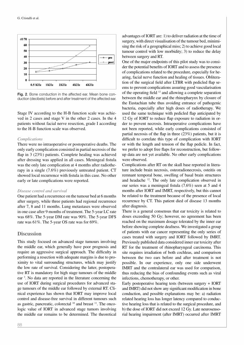
G. Cristalli et al.
88
Stage iV according to the h-B function scale was achie-ved in 2 cases and stage V in the other 2 cases. in the 4 patients without facial nerve resection, grade i according to the h-B function scale was observed.
ComplicationsThere were no intraoperative or postoperative deaths. The only early complication consisted in partial necrosis of the flap in 3 (23%) patients. Complete healing was achieved after dressing was applied in all cases. Meningeal fistula was the only late complication at 4 months after radiothe-rapy in a single (7.6%) previously untreated patient. CT showed local recurrence with fistula in this case. no other early or late complications were reported.
Disease control and survivalone patient had a recurrence on the tumour bed at 6 months after surgery, while three patients had regional recurrence after 7, 8 and 11 months. lung metastases were observed in one case after 9 months of treatment. The 5-year lC rate was 68%. The 5-year DM rate was 90%. The 5-year DFS rate was 61%. The 5-year oS rate was for 69%.
DiscussionThis study focused on advanced stage tumours involving the middle ear, which generally have poor prognosis and require an aggressive surgical approach. The difficulty in performing a resection with adequate margins is due to pro-ximity to vital surrounding structures, which may justify the low rate of survival. Considering the latter, postopera-tive rT is mandatory for high stage tumours of the middle ear 3. no data are reported in the literature concerning the use of iorT during surgical procedures for advanced sta-ge tumours of the middle ear followed by external rT. Cli-nical experience has shown that iorT may improve local control and disease-free survival in different tumours such as gastric, pancreatic, colorectal 10 and breast 11. The onco-logic value of iorT in advanced stage tumors involving the middle ear remains to be determined. The theoretical
advantages of iorT are: 1) to deliver radiation at the time of surgery, with direct visualisation of the tumour bed, minimi-sing the risk of a geographical miss; 2) to achieve good local tumour control with low morbidity; 3) to reduce the delay between surgery and rT.one of the major endpoints of this pilot study was to consi-der the potential benefits of iorT and to assess the presence of complications related to the procedure, especially for he-aring, facial nerve function and healing of tissues. oblitera-tion of the surgical field after lTBr with pedicled flap se-ems to prevent complications assuring good vascularisation of the operating field 12 and allowing a complete separation between the middle ear and the rhinopharynx by closure of the Eustachian tube thus avoiding entrance of pathogenic bacteria, especially after high doses of radiotherapy. We used the same technique with pedicled flap anticipated by 12 gy of iorT to reduce flap exposure to radiation in or-der to prevent necrosis. intraoperative complications have not been reported, while early complications consisted of partial necrosis of the flap in three (23%) patients, but it is difficult to correlate this type of complication with iorT or with the length and tension of the flap pedicle. in fact, we prefer to adopt free flaps for reconstruction, but follow-up data are not yet available. no other early complications were observed.Complications after rT on the skull base reported in litera-ture include brain necrosis, osteoradionecrosis, osteitis on remnant temporal bone, swelling of basal brain structures and headache 12. The only late complication observed in our series was a meningeal fistula (7.6%) seen at 5 and 4 months after iorT and iMrT, respectively, but this cannot be related to the treatment because of the presence of local recurrence by CT. This patient died of disease 13 months after diagnosis.There is a general consensus that ear toxicity is related to doses exceeding 50 gy; however, no agreement has been reached on the maximum dosage tolerated by the inner ear before showing complete deafness. We investigated a group of patients with ear cancer representing the only series of cases treated with surgery and iorT followed by iMrT. Previously published data considered inner ear toxicity after rT for the treatment of rhinopharyngeal carcinoma. This site requires irradiation of both cochleas, and comparison between the two ears before and after treatment is not possible. in our experience, only one side underwent iMrT and the contralateral ear was used for comparison, thus reducing the bias of confounding events such as viral infections, chemotherapy, or other.Early postoperative hearing tests (between surgery + iorT and iMrT) did not show any significant modification in bone conduction, and possible explanations may be: a) radiation related hearing loss has longer latency compared to conduc-tive hearing loss that is related to the surgical procedure, and b) the dose of iorT did not exceed 12 gy. late neurosenso-rial hearing impairment (after iMrT) occurred after iMrT
Fig. 2. Bone conduction in the affected ear. Mean bone con-duction (decibels) before and after treatment of the affected ear.

Intraoperative radiation therapy as adjuvant treatment in locally advanced stage tumours involving the middle ear
89
Fig. 3. Bone conduction in both ears. Median values of bone conduction before and after treatment at 0.5 kHz, 1 kHz, 2 kHz, 4 kHz and 6 kHz in the affected ear (left side) and the contralateral ear (right side).

G. Cristalli et al.
90
throughout the all frequencies and was statistically signi-ficant from 1 to 6 khz. late hearing loss after rT seems to be related to vascular injury of inner ear structures with progressive onset of fibrosis and ossification of the inner ear fluid space. At any rate, in this study, CT scan did not show any evidence of inner ear ossification after rT 4.in the literature, nerve toxicity has been observed in humans (lumbar tract) undergoing doses over 10 gy and in a canine model over 15 gy 13. nonetheless, our 4 cases did not show any facial nerve deficit after rT (12 gy of iorT + 50 gy iMrT). Furthermore, the results after neural graft repair we-re comparable with those reported in the literature where the rT regimen did not impact functional recovery 14.luc et al. described the prognostic factors for survival and recurrence in temporal bone cancer 3. yeung et Al. reported a 5-year local control rate of 67% for T3-T4 ear malignan-cies 15. Pfreunder et al. observed a 2-year local control rate of 85% for T1-2 and 50% for T3-T4 16. Bacciu et al. repor-ted 5-year disease specific survival of 86.2% and 48.7% for patients with T3 and T4 disease, respectively 17. our data confirmed the possibility to achieve a good control of di-sease with a lC rate of 68% and DFS rate of 61%, even in advanced stage 18. it is difficult to compare our results with those of other series given the heterogeneity of staging clas-sifications and treatment used. The low number of cases did not permit drawing any conclusion about the role of iorT in improving oncologic outcomes. This study represents the first for future studies with larger series in order to demon-strate the effectiveness of iorT in ear cancer.
ConclusionsA dose of 12 gy of iorT on the inner ear and surrounding tissues during advanced ear cancer surgery can be conside-red as safe. Early complications were not observed, while late complications such as mild neurosensorial hearing loss on middle-high frequencies can be expected after iorT followed by iMrT. Future studies are required to assess the oncologic value of iorT in local control of disease, confir-ming the hypothesis of this retrospective study.
AcknowledgmentsThe authors acknowledge Ms. Tania Merlino for editing the English language of the text.
References 1 Kinney SE. Tumours of the external auditory canal, middle ear, ma-
stoid and temporal bone. in: Comprehensive Management of Head
and Neck Tumours. Thawley SE, Panje Wr, Batsakis Jg, lindberg rD (eds). Philadelphia: WB Saunders Co.; 1987. p. 81-206.
2 Morris lgT, Mehra S, Shah JP, et al. Predictors of survival and recurrence after temporal bone resection for cancer. head and neck 2012;34:1231-9.
3 lassig AA, Spector ME, Soliman S, et al. Squamous cell carcino-ma involving the temporal bone: lateral temporal bone resection as primary intervention. otol neurotol 2013;34:141-50.
4 Jereczek-Fossa BA, Zarowski A, Milani F, et al. Radiotherapy induced ear toxicity. Cancer Treat rev 2003;29:417-30.
5 Marucci l, Pichi B, iaccarino g, et al. Intraoperative radiation therapy as an “early boost” in locally advanced head and neck cancer: preliminary results of a feasibility study. head neck 2008;30:701-8.
6 Arriaga M, Curtin h, Takahashi h, et al. Staging proposal for external auditory meatus carcinoma based on preoperative cli-nical examination and computed tomography findings. Ann otol rhinol laryngol 1990;99:714-21.
7 house JW, Brackmann DE. Facial nerve grading system. otola-ryngol head neck Surg 1985;93:146-7.
8 Conley J, novack AJ. The surgical treatment of malignant tumors of the ear and temporal bone. Arch otolarygol 1960;71:635-52
9 Soriani A, landoni V, Marzi S, et al. Setup verification and in vivo dosimetry during intraoperative radiation therapy (IORT) for prostate cancer. Med Phys 2007;34:3205-10.
10 Kusters M, Valentini V, Calvo FA, et al. Results of European pooled analysis of IORT-containing multimodality treatment for locally advanced rectal cancer: adjuvant chemotherapy prevents local recurrence rather than distant metastases. Ann oncol 2010;21:1279-84.
11 Veronesi U, orecchia r, luini A, et al. Full-dose intraoperative radiotherapy with electrons during breast-conserving surgery ex-perience with 590 cases. Ann Surg 2005;242;101-6.
12 nguyen MT, Billington A, habal MB. Osteoradionecrosis of the skull after radiation therapy for invasive carcinoma. J Craniofac Surg 2011;22:1677-81.
13 Sindelar WF, Kinsella TJ. Normal tissue tolerance to intraopera-tive radiotherapy. Surg oncol Clin n Am 2003;12:925-42.
14 leong SC, lesser Th. Long-term outcomes of facial nerve fun-ction in irradiated and nonirradiated nerve grafts. Ann otol rhinol laryngol 2013;122:695-700.
15 yeung P, Bridger A, Smee r, et al. Malignancies of the exter-nal auditory canal and temporal bone: a review. Anz J Surg 2002;72:114-20.
16 Pfreundner l, Schwagener K, Willner J, et al. Carcinoma of the external auditory canal and middle ear. int J radiation onco-logy Biol Phys 1999; 44:777-8.
17 Bacciu A, Clemente iA, Piccirillo E, et al. Guidelines for tre-ating temporal bone carcinoma based on long-term outcomes. otol neurotol 2013;34:898-907.
18 Cristalli g, Manciocco V, Pichi B, et al. Treatment and outcome of advanced external auditory canal and middle ear squamous cell carcinoma. J Craniofac Surg 2009;20:816-21.
received: november 11, 2014 - Accepted: July 24, 2015
Address for correspondence: giuseppe Mercante, Department of otolaryngology, head and neck Surgery, regina Elena national Cancer institute, via Elio Chianesi 53, 00144 rome, italy. E-mail: [email protected]

91
ACTA oTorhinolAryngologiCA iTAliCA 2016;36:91-96; doi: 10.14639/0392-100X-499
Head and neck
Post-operative pain management in head and neck cancer patients: predictive factors and efficacy of therapyDolore post-operatorio nei pazienti affetti da neoplasia testa-collo: fattori predittivi ed efficacia della terapia
C. BianChini1, M. Malagò1, l. CreMa1, C. aiMoni1, T. MaTarazzo2, S. BorTolazzi2, a. CiorBa1, S. PeluCChi1, a. PaSTore1
1 enT and audiology Department, 2 anaesthesiology Department, “S. anna” university hospital of Ferrara, italy
SummAry
There is increasing interest about all aspects of pain sensation for patients undergoing head and neck surgery, and efforts have been made to better assess, monitor and reduce the occurrence of pain. The aetiology of pain is considered to be “multifactorial”, as it is defined by several features such as personal experience, quality perception, location, intensity and emotional impact. The aim of this paper is: (i) to evaluate the efficacy of analgesic treatment in patients with head and neck cancer treated by surgery, and (ii) to study the variables and pre-dictive factors that can influence the occurrence of pain. A total of 164 patients, affected by head and neck cancer and surgically treated, be-tween December 2009 and December 2013, were included in this study. Data collected include age, gender, assessment of anaesthetic risk, tumour localisation, pathological cancer stage, Tnm stage, type of surgery performed, complexity and duration of surgery, post-operative complications, postoperative days of hospital stay and pain evaluation on days 0, 1, 3 and 5 post-surgery. We studied the appropriateness of analgesic therapy in terms of incidence and prevalence of post-operative pain; we also related pain to patient characteristics, disease and surgical treatment to determine possible predictive factors. The population studied received adequate pain control through analgesic therapy immediately post-surgery and in the following days. no associations between gender, age and post-operative pain were found, whereas pathological cancer stage, complexity of surgery and tumour site were significantly associated with the risk of post-operative pain. Adequate pain control is essential in oncological patients, and particularly in head and neck cancer patients as the prevalence of pain in this localisation is reported to be higher than in other anatomical sites. improved comprehension of the biological and psychological factors that characterise pain perception will help to enhance its control in the future.
Key WorDS: Pain • Head and neck cancer • Surgery • Quality of life • Pain therapy
riASSunTo
Negli anni è aumentata l’attenzione verso i molteplici aspetti associati alla “sfera” dolore, anche nei pazienti oncologici sottoposti a chirurgia testa-collo. Il dolore, definito infatti da diverse caratteristiche, quali l’esperienza personale, gli aspetti qualitativi della perce-zione, l’intensità, l’impatto emotivo, riconosce un’eziologia “multifattoriale”. Scopo del presente lavoro è stato: (i) valutare l’efficacia della terapia analgesica in pazienti affetti da tumore testa-collo e sottoposti a trattamento chirurgico; (ii) studiare le possibili variabili ed i fattori predittivi che possano influenzare l’insorgenza di dolore. Sono stati studiati 164 pazienti, affetti da neoplasia maligna del distretto testa-collo, trattati chirurgicamente tra il dicembre 2009 ed il dicembre 2013. I dati raccolti comprendono l’età, il sesso, la va-lutazione del rischio anestesiologico, la sede del tumore, la stadiazione TNM, il tipo di intervento effettuato, la complessità e la durata dell’intervento, le eventuali complicanze post-operatorie, i giorni di degenza post-intervento, la valutazione del dolore nei giorni 0, 1, 3 e 5 post-chirurgia. L’adeguatezza della terapia analgesica è stata espressa in termini di incidenza e prevalenza del dolore post-ope-ratorio, le variabili legate al paziente, alla malattia, al trattamento chirurgico e farmacologico, sono state poi associate all’insorgenza del dolore così da poter descrivere eventuali fattori predittivi. Dai dati ottenuti emerge che la popolazione studiata ha ricevuto un’ade-guata terapia antalgica, sia nell’immediato post-operatorio che nei giorni successivi. Non sono risultate associazioni statisticamente significative tra sesso, età ed incidenza del dolore post-chirurgico, mentre lo stadio del tumore, la complessità dell’intervento chirurgico e la sede della neoplasia hanno presentano correlazione significativa con il rischio di insorgenza di dolore post-operatorio. L’elevata prevalenza del dolore in ambito oncologico testa-collo, fa sì che un’appropriata ed attenta gestione del dolore risulti fondamentale. Nel futuro pertanto si auspica una sempre migliore comprensione dei fattori biologici, sociali e psicologici che caratterizzano la percezione del dolore ai fini di migliorarne il controllo.
PArole ChiAve: Dolore • Tumore testa-collo • Chirurgia • Qualità della vita • Analgesia
Acta Otorhinolaryngol Ital 2016;36:91-96

C. Bianchini et al.
92
Introductionover the past years, the definition of pain has become “multidimensional”, as it is considered to be not just a sensation, but has also been described by several features such as quality perception, location, intensity, emotional impact and frequency 1. Pain intensity is the most relevant clinical dimension of a painful experience; nonetheless, emotional, cognitive and behavioural aspects are part of the subjective perception of pain 1 2. The multifacto-rial nature of pain is particularly evident in oncological patients 3 4, as it can be accompanied by psychological, emotional and social problems 5 6. if poorly controlled, it can be associated with physical and mental deterioration, leading to increased morbidity, increased anxiety and de-pression, and as a result decreased quality of life.Pain is one of the most common anxieties before and af-ter surgery, and pain control after surgery still continues to receive little attention in the literature, particularly among head and neck patients 3 4 7-10. it has been reported that pain in head and neck cancer patients has a nociceptive origin due to the direct invasion and destruction of soft tissue and bone tissue by local invasion, but it can also be of neuro-pathic origin due to inflammation or compression of nerv-ous structures. Furthermore, its intensity can be exacerbated by actions such as speech, chewing and/or swallowing 3 4 8.The purpose of this study is: (i) to evaluate the efficacy of analgesic treatment in patients with head and neck cancer treated by surgery, and (ii) to understand the possible rela-tion between head and neck pain and predictive factors.
Materials and methodsA total of 164 patients affected by head and neck cancer and surgically treated in the period between December 2009 and December 2013 at the enT Department of the university hospital of Ferrara were included in this ret-rospective study.
Patient history, informed consent and pre-operative anaes-thesiologic evaluation data were collected for each patient. Prior to surgery, each patient received information about pain and its treatment, verbally and with paper-based in-formation sheets given by the enT and anaesthesiology team using a pain evaluation data sheet (incomplete or partially completed surveys were excluded).The pre-operative exams included: blood tests, electro-cardiography, chest X-ray and any further endoscopic, radiological, histopathological diagnostic investigations in order to correctly stage the tumour and to study as-sociated comorbidities. Data collected for each patient included: gender, age, tumour site, previous treatments (radiation therapy, chemotherapy, surgery), previous an-algesic therapy, assessment of anaesthetic risk (ASA), type of surgery performed, complexity of surgery ac-cording to the anaesthesiology protocols (Annex), dura-tion of surgery, post-operative complications, post-oper-ative days of hospital stay, drugs used in the immediate post-operative period, analgesic therapy administered during hospitalisation, pain evaluation twice a day (in the morning and in the evening) on days 0, 1, 3 and 5 post-op, definitive histological diagnosis, pTnm staging (according to uiCC/AJCC Classification of malignant Tumours 11), pathological cancer stage and any analgesic therapy prescribed at home.All patients received antibiotic and antithrombotic prophylaxis and gastric protection during hospitalisation. Pharmacological control of pain was obtained based on a preoperative established pain program, accordingly to the type of surgery performed (minor, medium and major surgery) (Table i). This consists of multimodal therapy: a combination of opioids, non-steroidal anti-inflammatory drugs and paracetamol. The anaesthesiologist usually pre-scribed postoperatively analgesic therapy infused by elas-tomeric pump for the first 24 hours, and sometimes for 48 or 72 hours post-operatively (Table i).
Table I. Post-operative analgesic therapy in relation to surgical complexity.
Surgical complexity Post-operative analgesic therapy
Minor (i.e. tracheotomy) A. Non-steroidal anti-inflammatory drugs (NSAIDs), at fixed times, every 6/8 hours1. ketoprofen 100 mg2. ketorolac 30 mg
B. Paracetamol 1 g iv every 6/8 hours; tramadol 100 mg iv every 8 hours.
Medium (i.e. thyroidectomy, parotidectomy, partial glossectomy; without neck dissection)
A. Morphine 2 mg/kg/24 hours in elastomeric pump;B. Morphine 2 mg/kg/24 hours in elastomeric pump + NSAIDs, at fixed times, every 8 hours;C. Morphine 2 mg/kg/24 hours in elastomeric pump + paracetamol 1 g iv every 6/8 hours;D. NSAIDs in elastomeric pump (i.e. ketoprofen 300 mg/24 hours).
Major (i.e. laryngectomy, neck dissections, pull-through, flap reconstructive surgery after extensive demolition)
A. Morphine 3 mg/kg/24 hours in elastomeric pump;B. Morphine 3 mg/kg/24 hours in elastomeric pump + NSAIDs, at fixed times, every 8 hours;C. Morphine 3 mg/kg/24 hours in elastomeric pump + paracetamol 1 gm iv every 6/8 hours;D. NSAIDs in elastomeric pump (i.e. ketoprofen 300 mg/24 hours) + paracetamol 1 gm iv every 6/8 hours.
Notes:• NSAIDsarenotindicatedincaseofcoagulopathy,hepatopathy,renalfailure,orhighriskofbleedingconditions.• Theuseofmorphinealoneorincombinationwithotherdrugsisrelatedtotheexpectedpain.• Morphineisnotindicatedinthoseaffectedbysuspected(orhistoryof)paralyticileus,psychiatricillness,orsevererespiratorydisease.

Pain management in head and neck cancer patients
93
Post-operative pain trend, in terms of number of episodes and intensity, during the entire hospital stay (on the day of surgery and then on the following days, until discharge) was recorded. Pain intensity during mornings (m) and evenings (e), as well as the maximum value of numeric rating scale (nrS) reached, was recorded each day.
Pain evaluation data sheetThe pain evaluation datasheet was completed on ad-mission for all patients, updated daily and after patient discharge, and was archived together with the patient’s medical records. Pain evaluation was performed through several methods. visual analogue scale (vAS) measured post-operative intensity pain immediately after surgery, while numeric rating Scale (nrS) was used during the remaining post-operatory hospital stay. The Behavioural Pain Scale (BPS) was used if patients were held in the intensive care unit in the immediate post-operative peri-od 12-15. Different sections composed the pain evaluation datasheet adopted for this study.Section i. includes patient personal data and medical diagno-sis. other information collected in this part include: pain at admission (assessed by nrS), type of pain (acute vs chron-ic); pain management/therapy at home; type of surgical pro-cedure and anaesthesia. expected pain and pain management plan proposed by the anaesthesiologist and type of surgical plan (with the aim of tailoring pain management for each pa-tient and its specific disease) is also reported in this section.Section ii. This part is dedicated to pain measurement and recording procedures. These measures were performed at different post-operative days. it was generally measured and recorded twice a day after surgery, and there is an extra section to report episodes of acute pain. only if nrS > 3 can extra analgesic drugs can be administrated to patients on the basis of the anaesthesiologist prescriptions.
Statistical analysisStatistical analysis was performed using contingency tables analysed by chi-square (χ2) or by Fisher’s test to compare small groups of data. To investigate the possible contribution of different variables in multivariate meth-ods, binary logistic processing procedures were used. The calculations were performed using the statistical system Systat v.5 (Systat inc. evanstone, il uSA).if not otherwise specified, p values < 0.05 were consid-ered statistically significant, while p values between 0.06 and 0.1 were considered of borderline significance.
ResultsA total of 164 patients were enrolled in this study. There were 101 males (61.5%) and 63 females (38.5%). Age range was 16 and 86 years, with a mean of 62.84 years. Patients were divided into two age groups: 0-60 years (56 patients, 34.1%) and > 60 years (108 patients, 65.9%; Table ii).
The primary cancer sites were very different and patients were grouped into four cohorts identified by a number: (1) pharyngeal and laryngeal tumours (64 patients, 39.1%); (2) thyroid, salivary gland and neck tumours (58 patients, 35.4%); (3) oral cavity tumours (32 patients, 19.5%); (4) tumours at other sites (i.e. head and neck skin or occult carcinomas to neck nodes; 10 patients, 6.1%; Table ii).Tnm stage was available in only 141 patients (as lym-phomas, skin tumours and neck metastases from occult cancers were excluded). Therefore, 2 patients were clas-sified as Tis (1.4%), 58 as T1 (41.1%), 28 as T2 (19.8%), 31 as T3 (22%) and 22 as T4 (15.6%). on the base of tumour staging, 2 patients (1.4%) had oncological stage 0,
Table II. Demographic and clinical characteristics of patients.
PatientsN (%)
Mean
Demographic data
Gender• Male• Female
101 (61.5)63 (38.4)
Age• 0-60 years• ≥ 60 years
56 (34.1)108 (65.9)
62.84
Clinical features
Tumour site• Pharynx-larynx• Neck-thyroid-salivary glands• Oral cavity
64 (39.1)58 (35.4)32 (19.5)10 (6.1)
Stage• Low (0-I-II)• High (III-IV)
52 (36.8)89 (63.2)
Previous therapies• No• Yes
114 (69.5)50 (30.5)
ASA• 1• 2• 3• 4
5 (3.1)59 (36.0)86 (52.5)14 (8.6)
Surgical complexity• Minor• Moderate• Major
1 (0.6%)56 (34.2)
107 (65.2)
Post-operative complications• No• Yes
138 (84.1)26 (15.9)
Hospital stay• ≤ 5 days• 6-20 days• ≥ 21 days
63 (38.5)77 (46.9)24 (14.6)
11.46
Post-op analgesic therapy• Opioids• Opioids+NSAIDs/paracetamol• NSAIDs
27 (16.5)121 (73.7)
16 (9.8)
Analgesic therapy at discharge• No• Yes• Data not available
69 (42.0)90 (54.9)
5 (3.1)
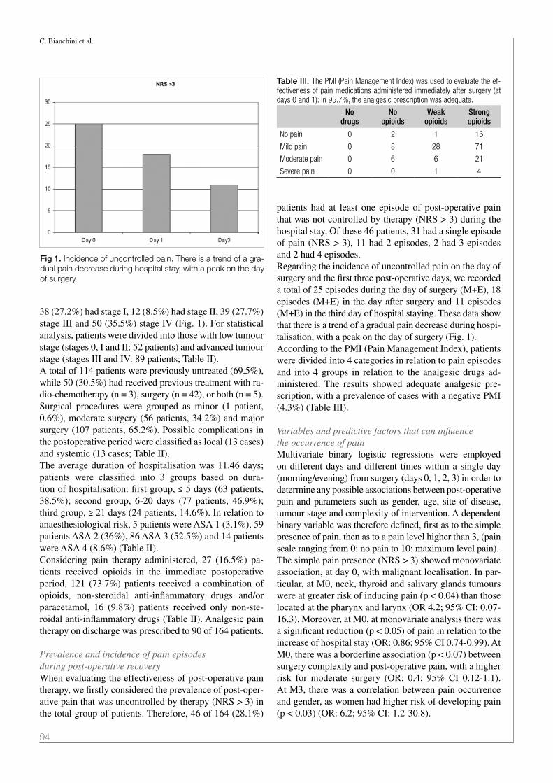
C. Bianchini et al.
94
38 (27.2%) had stage i, 12 (8.5%) had stage ii, 39 (27.7%) stage iii and 50 (35.5%) stage iv (Fig. 1). For statistical analysis, patients were divided into those with low tumour stage (stages 0, i and ii: 52 patients) and advanced tumour stage (stages iii and iv: 89 patients; Table ii).A total of 114 patients were previously untreated (69.5%), while 50 (30.5%) had received previous treatment with ra-dio-chemotherapy (n = 3), surgery (n = 42), or both (n = 5).Surgical procedures were grouped as minor (1 patient, 0.6%), moderate surgery (56 patients, 34.2%) and major surgery (107 patients, 65.2%). Possible complications in the postoperative period were classified as local (13 cases) and systemic (13 cases; Table ii).The average duration of hospitalisation was 11.46 days; patients were classified into 3 groups based on dura-tion of hospitalisation: first group, ≤ 5 days (63 patients, 38.5%); second group, 6-20 days (77 patients, 46.9%); third group, ≥ 21 days (24 patients, 14.6%). in relation to anaesthesiological risk, 5 patients were ASA 1 (3.1%), 59 patients ASA 2 (36%), 86 ASA 3 (52.5%) and 14 patients were ASA 4 (8.6%) (Table ii).Considering pain therapy administered, 27 (16.5%) pa-tients received opioids in the immediate postoperative period, 121 (73.7%) patients received a combination of opioids, non-steroidal anti-inflammatory drugs and/or paracetamol, 16 (9.8%) patients received only non-ste-roidal anti-inflammatory drugs (Table ii). Analgesic pain therapy on discharge was prescribed to 90 of 164 patients.
Prevalence and incidence of pain episodes during post-operative recoveryWhen evaluating the effectiveness of post-operative pain therapy, we firstly considered the prevalence of post-oper-ative pain that was uncontrolled by therapy (nrS > 3) in the total group of patients. Therefore, 46 of 164 (28.1%)
patients had at least one episode of post-operative pain that was not controlled by therapy (nrS > 3) during the hospital stay. of these 46 patients, 31 had a single episode of pain (nrS > 3), 11 had 2 episodes, 2 had 3 episodes and 2 had 4 episodes.regarding the incidence of uncontrolled pain on the day of surgery and the first three post-operative days, we recorded a total of 25 episodes during the day of surgery (m+e), 18 episodes (m+e) in the day after surgery and 11 episodes (m+e) in the third day of hospital staying. These data show that there is a trend of a gradual pain decrease during hospi-talisation, with a peak on the day of surgery (Fig. 1).According to the Pmi (Pain management index), patients were divided into 4 categories in relation to pain episodes and into 4 groups in relation to the analgesic drugs ad-ministered. The results showed adequate analgesic pre-scription, with a prevalence of cases with a negative Pmi (4.3%) (Table iii).
Variables and predictive factors that can influence the occurrence of painmultivariate binary logistic regressions were employed on different days and different times within a single day (morning/evening) from surgery (days 0, 1, 2, 3) in order to determine any possible associations between post-operative pain and parameters such as gender, age, site of disease, tumour stage and complexity of intervention. A dependent binary variable was therefore defined, first as to the simple presence of pain, then as to a pain level higher than 3, (pain scale ranging from 0: no pain to 10: maximum level pain).The simple pain presence (nrS > 3) showed monovariate association, at day 0, with malignant localisation. in par-ticular, at m0, neck, thyroid and salivary glands tumours were at greater risk of inducing pain (p < 0.04) than those located at the pharynx and larynx (or 4.2; 95% Ci: 0.07-16.3). moreover, at m0, at monovariate analysis there was a significant reduction (p < 0.05) of pain in relation to the increase of hospital stay (or: 0.86; 95% Ci 0.74-0.99). At m0, there was a borderline association (p < 0.07) between surgery complexity and post-operative pain, with a higher risk for moderate surgery (or: 0.4; 95% Ci 0.12-1.1). At m3, there was a correlation between pain occurrence and gender, as women had higher risk of developing pain (p < 0.03) (or: 6.2; 95% Ci: 1.2-30.8).
Table III. The PMI (Pain Management Index) was used to evaluate the ef-fectiveness of pain medications administered immediately after surgery (at days 0 and 1): in 95.7%, the analgesic prescription was adequate.
No drugs
No opioids
Weak opioids
Strong opioids
No pain 0 2 1 16
Mild pain 0 8 28 71
Moderate pain 0 6 6 21
Severe pain 0 0 1 4
Fig 1. incidence of uncontrolled pain. There is a trend of a gra-dual pain decrease during hospital stay, with a peak on the day of surgery.
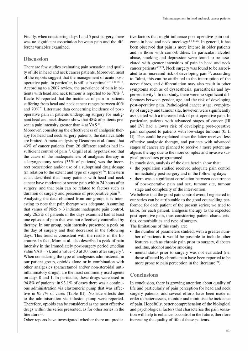
Pain management in head and neck cancer patients
95
Finally, when considering days 1 and 5 post-surgery, there was no significant association between pain and the dif-ferent variables examined.
DiscussionThere are few studies evaluating pain sensation and quali-ty of life in head and neck cancer patients. moreover, most of the reports suggest that the management of acute post-operative pain, in particular, is still sub-optimal 3 4 7-10 16-18. According to a 2007 review, the prevalence of pain in pa-tients with head and neck tumour is reported to be 70% 17. Keefe FJ reported that the incidence of pain in patients suffering from head and neck cancer ranges between 40% and 70% 7. literature data concerning incidence of post-operative pain in patients undergoing surgery for malig-nant head and neck disease show that 48% of patients pre-sent a pain intensity greater than 4 at vAS 18.moreover, considering the effectiveness of analgesic ther-apy for head and neck surgery patients, the data available are limited. A meta-analysis by Deandrea et al. found that 43% of cancer patients from 26 different studies had in-sufficient control of pain 19. orgill et al. hypothesised that the cause of the inadequateness of analgesic therapy in a laryngectomy series (35% of patients) was the incor-rect prescription and/or use of a suboptimal drug dosage (in relation to the extent and type of surgery) 20. inhestern et al. described that many patients with head and neck cancer have moderate or severe pain within 24 hours after surgery, and that pain can be related to factors such as duration of surgery and presence of preoperative pain 21.Analysing the data obtained from our group, it is inter-esting to note that pain therapy was adequate. Assuming that values of nrS > 3 indicate inadequate pain control, only 26.5% of patients in the days examined had at least one episode of pain that was not effectively controlled by therapy. in our group, pain intensity presented a peak on the day of surgery and then decreased in the following days. This trend is consistent with the results in the lit-erature. in fact, mom et al. also described a peak of pain intensity in the immediately post-surgery period (median value vAS = 7), and a value < 3 at 30 hours after surgery 9.When considering the type of analgesics administered, in our patient group, opioids alone or in combination with other analgesics (paracetamol and/or non-steroidal anti-inflammatory drugs), are the most commonly used agents on days 0 and 1. in particular, these drugs were used in 94.8% of patients: in 93.1% of cases there was a continu-ous administration via elastomeric pump that was effec-tive in 95.7% of cases (Table iii). no side effects due to the administration via infusion pump were reported. Therefore, opioids can be considered as the most effective drugs within the series presented, as for other series in the literature 22.other reports have investigated whether there are predic-
tive factors that might influence post-operative pain out-come in head and neck oncology 4 8 23-26. in general, it has been observed that pain is more intense in older patients and in those with comorbidities. in particular, alcohol abuse, smoking and depression were found to be asso-ciated with greater intensities of pain in head and neck cancer patients 4 23 24. neck surgery was found to be associ-ated to an increased risk of developing pain 25; according to Talmi, this can be attributed to the interruption of the nerve fibres, and differentiation may also result in other symptoms such as of dysaesthesia, paraesthesia and hy-persensitivity 3. in our study, there were no significant dif-ferences between gender, age and the risk of developing post-operative pain. Pathological cancer stage, complex-ity of surgery and tumour site, however, were significantly associated with a increased risk of post-operative pain. in particular, patients with advanced stages of cancer (iii and iv) had a lower risk of developing post-operative pain compared to patients with low-stage tumours (0, i, ii). This could be explained since the latter received less effective analgesic therapy, and patients with advanced stages of cancer are planned to receive a more potent an-algesic therapy due to the more complex and invasive sur-gical procedures programmed.in conclusion, analysis of the data herein show that:• the population studied received adequate pain control
immediately post-surgery and in the following days;• there was a significant correlation between occurrence
of post-operative pain and sex, tumour site, tumour stage and complexity of the intervention.
We believe that the good pain control overall registered in our series can be attributable to the good counselling per-formed for each patient of the present series; we tried to tailor, for each patient, analgesic therapy to the expected post-operative pain, thus considering patient characteris-tics, comorbidities and type of surgery.The limitations of this study are:• the number of parameters studied; with a greater num-
ber of patients it would be possible to include other features such as chronic pain prior to surgery, diabetes mellitus, alcohol and/or smoking.
• mental status prior to surgery was not evaluated (i.e. those affected by chronic pain have been reported to be more prone to pain perception in the literature 21).
Conclusionsin conclusion, there is growing attention about quality of life and particularly of pain perception for head and neck surgery patients, and several efforts have been made in order to better assess, monitor and minimise the incidence of pain. hopefully, better comprehension of the biological and psychological factors that characterise the pain sensa-tion will help to enhance its control in the future, therefore increasing the quality of life of these patients.

C. Bianchini et al.
96
AcknowledgementsThe authors would like to thank Dr. giuseppe gilli for his assistance with statistical analysis.
References1 merskey h, Bogduk n. Classification of Chronic Pain: De-
scriptions of Chronic Pain Syndromes and Definitions of Pain Terms. Second edition. Washington, DC: iASP Press; 1994. p. 209-214.
2 Frank-Stromborg m, olsen o. Instruments for Clinical Health-Care Research. Burlington, mA: Jones & Bartlett; 1999. p. 528-564.
3 Talmi yP, horowitz Z, Pfeffer mr, et al. Pain in the neck after neck dissection. otolaryngol head neck Surg 2000;123:302-6.
4 Chen SC, liao CT, Chang JT. Orofacial pain and predictors in oral squamous cell carcinoma patients receiving treat-ment. oral oncology 2011;47:131-5.
5 ghei A, Khot S. Pain management in patients with head and neck carcinoma. otorhinolaryngology Clinics: An interna-tional Journal 2010;2:69-75.
6 Portenoy rK, lesage P. Management of cancer pain. lancet 1999;353:1659-700.
7 Keefe FJ, manuel g, Brantley A, et al. Pain in the head and neck cancer patient: changes over treatment. head neck Surg 1986;8:169-76.
8 Chaplin Jm, morton rP. A prospective, longitudinal study of pain in head and neck cancer patients. head neck 1999;21:531-7.
9 mom T, Commun F, Derbal C, et al. Postoperative pain eval-uation in the surgery of head and neck cancers. rev laryn-gol otol rhinol (Bord) 1996;117:93-6.
10 Ziv g, Darryl B, nissim m, et al. Treatment of pain after head and neck surgeries: control of acute pain after head and neck oncological surgeries. otolaryngol head neck Surg 2006;135:182-8.
11 edge SB, Byrd Dr, Compton CC, et al. AJCC cancer stag-ing manual. new york: Springer; 2010.
12 Williamson A, hoggart B. Pain: a review of three commonly used pain rating scales. J Clin nurs 2005;14:798-804.
13 Collins SL, Moore RA, McQuay HJ. The visual analogue pain intensity scale: what is a moderate pain in millimeters. Pain 1997;72:95-7.
14 Chanques g, Payen JF, mercier g, et al. Assessing pain in non-intubated critically ill patients unable to self report: an adaptation of the Behavioral Pain Scale. intens Care med 2009;35:2060-7.
15 rodriguez CS, mcmillan S, yarandi h. Pain measurement in older adults with head and neck cancer and communication impairments. Cancer nurs 2004;27:425-33.
16 mitera g, Zeiadin n, Kirou-mauro A, et al. Retrospective assessment of cancer pain management in an outpatient pal-liative radiotherapy clinic using the Pain Management Index. J Pain Symptom manage 2010;39:259-67.
17 van den Beuken-van everdingen mh, De rijke Jm, Kes-sels Ag, et al. Prevalence of pain in patient with can-cer: a systematic review of the past 40 years. Ann oncol 2007;18:1437-49.
18 Sommer m, geurts JW, Stessel B, et al. Prevalence and pre-dictors of postoperative pain after ear, nose and throat sur-gery. Arch otolaryngol head neck Surg 2009;135:124-30.
19 Deandrea S, montanari m, moja l, et al. Prevalence of un-dertreatment in cancer pain. A review of published literature. Ann oncol 2008;19:1985-91.
20 orgill r, Krempl gA, medina Je. Acute pain management following laryngectomy. Arch otolaryngol head neck Surg 2002;128:829-32.
21 inhestern J, Schuerer J, illge C, et al. Pain on the first post-operative day after head and neck cancer surgery. eur Arch otorhinolaryngol 2015;272:3401-9.
22 Cavalcanti il, Carvalho AC, musauer mg, et al. Safety and tolerability of controlled-release oxycodone on postoperative pain in patients submitted to the oncologic head and neck surgery. rev Col Bras Cir 2014;41:393-9.
23 Shuman Ag, Terrell Je, light e, et al. Predictors of pain among patients with head and neck cancer. Arch otolaryn-gol head neck Surg 2012;138:1147-54.
24 Duffy SA, ronis Dl, valenstein m, et al. Depressive symp-toms, smoking, drinking, and quality of life among head and neck cancer patients. Psychosomatics 2007;48:142-8.
25 gellrich nC, Schimming r, Schramm A, et al. Pain, function, and psychologic outcome before, during, and after intraoral tumor resection. J oral maxillofac Surg 2002;60:772-7.
26 Bianchini C, maldotti F, Crema l, et al. Pain in head and neck cancer: prevalence and possible predictive factors. J Buon 2014;19:592-7.
Address for correspondence: Chiara Bianchini, enT and Audio-logy Department, “S. Anna” university hospital of Ferrara, via Al-do moro 8, 44124 Ferrara, italy. e-mail: [email protected]
received: november 12, 2014 - Accepted: June 6, 2015

97
ACTA oTorhinolAryngologiCA iTAliCA 2016;36:97-100; doi: 10.14639/0392-100X-410
Rhinology
The effects of inferior turbinoplasty on nasal airflow during cosmetic rhinoplastyGli effetti sul flusso aereo nasale della turbinoplastica inferiore in corso di rinosettoplastica
R. Zojaji1, M. KeshavaRZManesh2, M. BaKhshaee3, R. Behdani4, s. esMaeelZadeh4, M. MaZlouM FaRsi BaF5
1 otorhinolaryngology department, Mashhad Branch, islamic azad university, Mashhad, iran; 2 otolaryngologist, Mashhad, iran; 3 Rhinologist, associate professor of the sinus and surgical endoscopic Research Center, Faculty of Medicine, Mashhad university of Medical sciences, Mashhad, iran; 4 General Practitioner, sinus and surgical endoscopic Research Center, Faculty of Medicine, Mashhad university of Medical sciences, Mashhad, iran; 5 General Practitioner, Faculty of Medicine, Mashhad Branch, islamic azad university, Mashhad, iran
SummAry
rhinoplasty is one of the most common and challenging cosmetic procedures. one of the complications of rhinoplasty associated with dissat-isfaction is nasal obstruction, which is often due to narrowing of the nasal valve area. Application of certain procedures such as turbinoplasty can prevent and correct this problem. This study aim was to investigate the effect of inferior turbinoplasty in reduction of airflow resistance and nasal obstruction. using active anterior rhinomanometry, nasal airflow was measured in 50 patients who underwent cosmetic rhinoplasty and bilateral turbinoplasty before and 6 months after surgery. none of the patients subjectively complained of nasal obstruction before or after surgery. According to rhinomanometry results, improvement in nasal airflow was seen both in inspiration and expiration, although only expira-tion was significant (p = 0.034). Airflow changes in males and females and in different age groups was not significant (p > 0.05). it appears that rhinoplasty does not adversely affect nasal airflow when it is accompanied by simple adjuvant procedure inferior turbinoplasty.
Key wordS: Rhinoplasty • Rhinomanometry • Turbinoplasty
riASSunTo
La rinoplastica è una procedura chirurgica molto comune e al tempo stesso di elevata complessità. Uno degli aspetti più discussi di tale chi-rurgia sono gli insoddisfacenti esiti funzionali dovuti spesso ad un eccessivo restringimento dell’area della valvola nasale. Alcune procedure, come la turbinoplastica inferiore, possono favorire la prevenzione di tali problematiche ostruttive. L’intento del presente studio è quello di indagare gli effetti della turbinoplastica inferiore sulla resistenza nasale e sull’ostruzione respiratoria. In 50 pazienti è stata effettuata un’ana-lisi preoperatoria e sei mesi dopo rinoplastica con turbinoplastica inferiore mediante rinomanometria anteriore attiva. Nessuno dei pazienti ha lamentato sintomatologia ostruttiva nasale nel preoperatorio o nel corso del postoperatorio. In accordo con i dati rinomanometrici sia le resistenze inspiratorie che quelle espiratorie hanno presentato valori migliori nel postoperatorio, ma solo le resistenze espiratorie hanno pre-sentato una significatività statistica (p = 0,034). Le differenze in termini di flusso nasale fra maschi, femmine e differenti gruppi di età non sono state significative (p > 0,05). In conclusione dai dati del nostro studio emerge che la rinosettoplastica, associate a turbinoplastica parziale non ha un impatto negativo sulla funzionalità nasale ed in particolare sul flusso aereo attraverso le cavità nasali.
PArole ChiAve: Rinoplastica • Rinomanometria • Turbinoplastica
Acta Otorhinolaryngol Ital 2016;36:97-100
Introductionrespiratory problems following rhinoplasty are a major concern, and despite a favourable cosmetic outcome, can cause dissatisfaction for the patient. Some researchers have reported such breathing disturbances are as high as 70% following rhinoplasty, although scars and loss of mu-cosal sensation can also give the feeling of a “blocked” nose 1. The aetiology of nasal obstruction after rhinoplasty includes various factors that result from a combination of
both undiagnosed nasal disorders such as septal deviation, turbinate hypertrophy and mucosal diseases in addition to reduced nasal valve area following this type of surgery 2 3.on the other hand, the inferior nasal turbinate is an inde-pendent bone 4; forming a part of the nasal lateral wall, it is a major structure in the nose 5. The inferior turbinate causes re-sistance against the nasal airflow that is essential for normal respiration. moreover, as part of the valvular area it changes the lamellar and laminar flow into the turbinate state 6.

r. Zojaji et al.
98
rhinoplasty may cause reduction of nasal valve area and result in severe airflow obstruction and respiratory prob-lems. Accordingly, as for any type of surgery in this region it should be performed with utmost care and precision. The use of techniques such as inferior turbinate fracture or turbinoplasty and inferior turbinate size reduction by improving nasal airflow 7 can be beneficial in improving the post-operative results and increase patient satisfaction.we hypothesised that removing part of the inferior tur-binates (turbinoplasty) during rhinoplasty by increasing the internal nasal space might reduce airflow resistance and nasal obstruction and consequently improve patient breathing. inferior turbinoplasty is a procedure that re-duces the size of the inferior turbinates.Considering above hypothesis, this study aimed at investigat-ing the effect of inferior turbinoplasty in reducing the airflow resistance and nasal obstruction following rhinoplasty.
Materials and methodsThis prospective study was performed on patients referred to a private clinic for rhinoplasty between 2010 and 2012. Among patients referred for rhinoplasty, those with wide nose and large inferior turbinates and without symptomatic septal deviation, turbinate hypertrophy, and without history of chronic sinusitis or allergy were selected because the chance of developing airflow resistance and nasal obstruc-tion is higher in this group of patients after reduction of nose size. in other words, selected patients were candidates for elective rhinoplasty without septoplasty.informed consent was obtained from each patient. The study protocol was approved by the local institutional re-view board (irB).A total of 50 patients were randomly selected among eli-gible patients using a table of random numbers who un-derwent rhinomanometry and subsequent rhinoplasty. Ac-tive anterior rhinomanometry was performed by placing the nozzle into both nasal cavities. Patients were evalu-ated under similar circumstances (during certain periods of the day, no smoking and no exercise before the test). rhinomanometry was performed for all female patients
during the first week after menstruation. For all patients, closed rhinoplasty was performed by the same surgeon without any intervention or reconstruction of nasal valve. Bilateral high-low-high internal lateral osteotomy and in-ferior turbinate outfracture with preservation of webster’s triangle was performed for all patients.nasal airflow was measured by active anterior rhinoma-nometry before and 6 months after the operation. rhinoma-nometry was performed with a rino 4000 device (homoth medizinelektronik gmbh & Co. Kg, hamburg, ger-many). The airflow during inspiration and expiration was measured at 75, 150 and 300 Pascal, whereas in data analy-sis 150 Pascal was considered as the standard level of pres-sure. The results were reported as numbers and diagrams for each patient and were then analysed by a statistician.The patients were also evaluated for post-operative compli-cations such as respiratory problems after the operation.Based on rhinomanometry results, patients were divided into three groups. group 1: nasal airflow changes after surgery between -50 and 50 ml/sec that was considered as the default change in the airflow due to device error, moderator error or error in the patient’s respiration (un-changed condition); group 2: those with airflow changes less than -50 ml/sec were considered as patients with a worsened condition; group 3: patients with more than 50 ml/sec changes in the nasal airflow during the same time regarded as improved condition.
Statistical analysisStatistical analysis was performed using SPSS ver. 11.5 for windows. normally distributed quantitative vari-ables were demonstrated as mean ± standard deviation. The normality condition of the quantitative variables was investigated using the Kolmogorov-Smirnov test. mann-whitney u test was used to compare equality distribution of age in two gender and changes in nasal airflow follow-ing rhinoplasty in two groups. Chi-square and maximum likelihood ratio tests were used to assess relation between Changes in nasal airflow following rhinoplasty and ratio variant. A p value < 0.05 was considered as significant.
Table I. Changes in nasal airflow following rhinoplasty (Classification A).
Variable Group 1(Between -50 to 50 ml/sec)
n (%)
Group 2(Less than -50 ml/sec)
n (%)
Group 3(More than 50 ml/sec)
n (%)
Test Test P valuestatistic
Right nasal cavity inspiratory airflow 26 (52) 9 (18) 15 (30) 8.92 0.012*
Right nasal cavity expiratory airflow 25 (50) 8 (16) 17 (34) 8.68 0.013*
Left nasal cavity inspiratory airflow 26 (52) 11 (22) 13 (26) 7.96 0.019*
Left nasal cavity expiratory airflow 28 (56) 9 (18) 13 (26) 12.04 0.002*
Both nasal cavities inspiratory airflow 17 (34) 13 (26) 20(40) 1.48 0.477
Both nasal cavities expiratory airflow 21 (42) 8 (16) 21(42) 6.76 0.034*
*p < 0.05 significant

Turbinoplasty and nasal airflow
99
ResultsAmong the 50 cases, 8 (16%) were male and 42 (84%) fe-male. Their age ranged from 15 up to 59 years with mean age of 26.84 ± 8.81. The mean age of men and women was not significantly different (p = 0.055, Z = 1.921). Considering symptoms, none of the patients complained of nasal obstruction, either before or after surgery.mean inspiratory airflow before and after rhinoplasty was 670.32 ± 697.5 and 694.62 ± 715.5 ml/sec, respectively. in addition, mean expiratory airflow before and after rhi-noplasty were 676.38 ± 703 and 716.26 ± 718 ml/sec, re-spectively. Based on rhinomanometry results, the inspira-tory and expiratory airflows changed significantly at 6 months after rhinoplasty with turbinoplasty (Table i). The number of patients without airflow changes (group 1) was higher than those with a worsened condition (group 2); no significant difference was found in the number of pa-tients without airflow change (group 1) and those with improved condition (group 3) (Table ii).Both inspiratory and expiratory nasal airflows improved at 6 months after rhinoplasty and inferior turbinoplasty, but the improvement was significant only for expiratory airflow (p = 0.034). in addition, the level of airflow chang-es in males and females and in different age groups was not significant (p > 0.05).
Discussionnasal obstruction is a major complication of rhinoplasty that results in true problems and discomfort for the pa-tient 8. Since the airflow in each nasal cavity changes based on the prominence and lack of space caused by the inferior turbinate, changes in the turbinate structure can
thus affect nasal function, which is important in rhino-plasty surgeries and post-operative nasal obstruction.The importance and necessity of the present research is that if the efficacy of turbinoplasty during rhinoplasty is improved, common post-operative complications such as nasal obstruction are resolved, and further costs for revi-sion surgeries can be avoided.due to the increase in the demand for rhinoplasty, the re-sults of this study can have a major impact on its costs and patient satisfaction. herein, the role of fracture of the inferior turbinate or turbinoplasty was investigated in the prevention of post-operative nasal obstruction in those cases that had been referred for cosmetic rhinoplasty without respiratory complaints. in addition to qualitative and subjective studies by clinical history, quantitative and objective studies by anterior rhinomanometry were also performed which adds further value to our results.in a study by grymer, which was performed 6 months after rhinoplasty surgery using acoustic rhinometry, it was observed that the internal diameters of the nasal cavity, es-pecially in the anterior section, decreased after rhinoplas-ty surgery 9; this factor plays a major role in post-operative nasal obstruction.Kosh et al., in 2004, reported that rhinoplasty is the most important cause of nasal obstruction followed by nasal trauma (15%) and congenital anomaly (6%) 10. however, in the study by Courtiss, the findings suggested that rhino-plasty had no impact on nasal airflow 11. in another study regarding the complications of nasal obstruction follow-ing rhinoplasty surgery, revision osteotomy with turbino-plasty was proposed for overcoming this problem 3, a find-ing which is consistent with our results. in a similar study, the role of turbinectomy in cases of hypertrophic inferior and middle turbinates was highlighted as a solution for post-operative nasal breathing problems 12.mlynski et al. concluded that anterior turbinoplasty and tur-binectomy reduce nasal airflow resistance, but due to dis-ruption of the natural shape of the lateral nasal wall, worsen its respiratory function 13. in another study on cases with nasal airway obstruction, significant reduction in obstruc-tion was seen following inferior turbinectomy by carbon di-oxide laser in comparison with those receiving cryotherapy, turbinate submucosal resection and partial turbinectomy 14. This is also in agreement with our findings.moreover, the studies by ophir et al. on patients com-plaining from nasal obstruction due to inferior turbinate hypertrophy showed that total turbinectomy reduces na-sal obstruction in 84% of cases and also improves nasal-conditioning 15.Buyuklu et al., in a study on patients with mild and moder-ate inferior turbinate hypertrophies, showed that outfrac-ture of the inferior turbinate (turbinoplasty) is an effec-tive technique that can increase the nasal airway in these patients 16. moreover, in a recent study by Zhang et al. on 50 patients with chronic hypertrophic rhinitis and inferior
Table II. Changes in nasal airflow following rhinoplasty (Classification B).
Variable Comparing the unchanged
with worsened condition
(Group 1 & 2)
Comparing the unchanged
with improved condition
(Group 1 & 3)
Test statistic
P value
Test statistic
P value
Right nasal cavity inspiratory airflow
26.000 0.007* 26.000 0.118
Right nasal cavity expiratory airflow
25.000 0.005* 17.000 0.280
Left nasal cavity inspiratory airflow
11.000 0.021* 26.000 0.055
Left nasal cavity expiratory airflow
9.000 0.003* 28.000 0.029*
Both nasal cavities inspiratory airflow
13.000 0.584 17.000 0.742
Both nasal cavities expiratory airflow
21.000 0.026* 21.000 0.999
* p < 0.05.

r. Zojaji et al.
100
turbinate hypertrophy, it was found that inferior turbinate outfracture surgery is a proper method that can expand the nasal cavity and improve nasal ventilatory function 17. The findings of the two latest studies are consistent with our results, although with different patient cohorts (inferior turbinate hypertrophies vs rhinoplasty).The findings of our study are very similar to most previous reports on this issue, demonstrating that fracture of the infe-rior turbinate can be of major importance in improving nasal function, preventing post-operative obstructive symptoms and increase patient satisfaction following rhinoplasty surgeries. it also shows that turbinoplasty improves nasal airflow during both inspiration and more prominently expiration, with no as-sociation with age or sex. it also highlights that the number of patients with no significant change in nasal airflow is greater than those who experience significant worsening in airflow, meaning that due to the decreased nasal space following rhi-noplasty, no change can also be interpreted as improvement.The main limitation of this study is lack of a matched control arm that may limit the conclusions. Therefore, our findings should be interpreted with caution.As nasal obstruction following rhinoplasty is very com-mon, further studies using the unilateral turbinoplasty technique and larger case-control studies and in different countries are recommended.
ConclusionsBased on the evidence herein, turbinoplasty during cos-metic rhinoplasty in patients with large nose and large inferior turbinates may prevent nasal obstruction and ad-verse effects of surgery on nasal airflow.
References1 retinger g. Risks and complications in rhinoplasty. gmS
Curr Top otorhinolaryngol head neck Surg 2007;6:doc08.2 romo T, Pearson Jm, Presti P, et al. Rhinoplasty, Post rhi-
noplasty Nasal Obstruction. new york medscape; 2007 [up-dated 2007 12/10/2007; cited]; Available from: http://emedi-cine.medscape.com/article/841574-overview.
3 Pontell J, Slavit dh, Kern eB. The role of out fracture in cor-recting post-rhinoplasty nasal obstruction. ear nose Throat J 1998;77:106-8.
4 nurse lA, duncavage JA. Surgery of the inferior and middle turbinates. otolaryngol Clin north Am 2009;42:295-309.
5 hakan C, egeli e, Kutluhan A, et al. Pneumatization of the turbinate inferior as a cause of nasal obstruction. rhinology 2001;39:109-11.
6 hol mKS, huizing eh. Treatment of inferior turbinate pa-thology: a review and critical evaluation of the different tech-niques. rhinology 2000;38:157-66.
7 Chen XB, leong SC, lee hP, et al. Aerodynamic effects of inferior turbinate surgery on nasal airflow – a computational fluid dynamics model. rhinology 2010;48:394-400.
8 ghazipour A, dilmaghani nA. Post rhinoplastic na-sal obstruction in 180 patients. iran J otorhinolaryngol 2005;17;46-50.
9 grymer lF. Reduction rhinoplasty and nasal patency: change in the cross-sectional area of the nose evaluated by acoustic rhinometry. laryngoscope 1995;105:429-31.
10 Khosh mm, Jen A, honrado C, et al. Nasal valve reconstruc-tion: experience in 53 consecutive patients. Arch Facial Plast Surg 2004;6:167-71.
11 Courtiss eh, goldwyn rm. The effects of nasal surgery on airflow. Plast reconstr Surg 1983;72:9-21.
12 mahler d, reuven S. The role of turbinectomy in rhinoplas-ty. Aesthetic Plast Surg 1985;9:277-9.
13 mlynski g, grützenmacher S, mlynski B, et al. [Model studies of nasal turbinate surgery]. laryngorhinootologie 1993;72:614-7.
14 Selkin Sg. Laser turbinectomy as an adjunct to rhinosepto-plasty. Arch otolaryngol 1985;111:446-9.
15 ophir d. Resection of obstructing inferior turbinates follow-ing rhinoplasty. Plast reconstr Surg 1990;85:724-7.
16 Zhang QX, Zhou wg, Zhang hd, et al. Relationship be-tween inferior turbinate outfracture and the improvement of nasal ventilatory function. Chin J otorhinolaryngol head neck Surg 2013;48:422-5.
17 Buyuklu F, Cakmak o, hizal e, et al. Outfracture of the infe-rior turbinate: a computed tomography study. Plastic recon-struct Surg 2009;123:1704-9
Address for correspondence: ramin Zojaji, P.o. Box: 9185711111, mashhaad, Khorasan razavi, iran. Tel. +98 91 51113841. Fax +98 511 7620982. e-mail: [email protected]
received: April 22, 2015 - received: June 18, 2015

101
ACTA oTorhinolAryngologiCA iTAliCA 2016;36:101-106; doi: 10.14639/0392-100X-965
Rhinology
In vivo tissue response and durability of five novel synthetic polymers in a rabbit model Biocompatibilità e durata in vivo di cinque nuovi polimeri sintetici testati su coniglio
E. Sahin1, C. Cingi2, g. ESkiizmir3, n. altintoprak4, a. Calli5, C. Calli6, i. Yilgör7, E. Yilgör7
1 Bayindir icerenkoy hospital, Ent Clinic, istanbul; 2 Eskisehir osmangazi University, Department of otolaryngology-head and neck Surgery, Eskisehir, turkey; 3 Celal Bayar University, Department of otolaryngology-head and neck Surgery, manisa, turkey; 4 tuzla State hospital, Ent Clinic, istanbul, turkey; 5 izmir atatürk training and research hospital, Department of pathology, izmir, turkey; 6 Ekol Ent hospital, izmir, turkey, 7 koc University, Department of Chemistry, istanbul, turkey
SummAry
Alloplastic materials are frequently used in facial plastic surgeries such as rhinoplasty and nasal reconstruction. unfortunately, the ideal alloplastic material has not been found. This experimental study evaluates the tissue response and durability of five novel polymers devel-oped as an alloplastic material. in this experimental study involving a tertiary university hospital, six subcuticular pockets were formed at the back of 10 rabbits for the implantation of each polymer and sham group. Each pocket was excised with its adjacent tissue after three months, and collected for histopathological examination. Semi-quantitative examination including neovascularisation, inflammation, fibrosis, abscess formation, multinucleated foreign body giant cells was performed, and integrity of polymer was evaluated. A statistical comparison was performed. no statically significant difference was detected in neovascularisation, inflammation, fibrosis, abscess forma-tion and multinucleated foreign body giant cells when a paired comparison between sham and polymer ii, iii and iV groups was performed individually. nevertheless, the degree of fibrosis was less than sham group in polymer i (p = .027) and V (p = .018), although the other variables were almost similar. The integrity of polymers iii (9 intact, 1 fragmented) and iV (8 intact, 2 absent) was better than the other polymers. These novel synthetic polymers could be considered as good candidates for clinical applicability. All polymers provided satisfac-tory results in terms of tissue response; however, fibrovascular integration was higher in polymers ii, iii and iV. in addition, the durability of polymer iii and iV was better than the others.
Key woRds: Alloplastic material • Polymer • Rhinoplasty • Nasal reconstruction • Bioavailability
riASSunTo
I materiali alloplastici vengono frequentemente utilizzati negli interventi di chirurgia plastica sul volto, quali la rinoplastica e la chirurgia ricostruttiva del naso. Ad oggi non è stato ancora individuato un materiale alloplastico con caratteristiche ottimali. Il presente studio spe-rimentale si propone di valutare la risposta tissutale e la resistenza nel tempo di cinque nuovi polimeri proposti come materiali alloplastici. Il presente studio è stato condotto presso un ospedale universitario di terzo livello. Sono state ricavate sei tasche sottocutanee sul dorso di 10 conigli che sono state usate per l’impianto di ciascuno dei polimeri testati più una tasca di controllo. Ciascuna delle tasche è stata escissa congiuntamente al tessuto circostante dopo tre mesi, ed è stata sottoposta ad un esame istopatologico. È stata quindi condotta una valutazione semi quantitativa con focus su neo angiogenesi, infiammazione, fibrosi, formazione di ascessi, presenza di cellule giganti multinucleate contenenti corpi estranei e stato dei polimeri testati. E’ stata inoltre effettuata una valutazione statistica, che per quanto riguarda la comparazione diretta fra la tasca di controllo e i polimeri II, III e IV non ha mostrato differenze significative in merito alla neo vascolarizzazione, all’infiammazione, alla fibrosi, alla presenza di ascessi ed alla presenza di cellule giganti multinucleate. Il polimero I ha invece mostrato un grado di fibrosi inferiore rispetto alla tasca di controllo (p = .027) and V (p = .018), benché le altre variabili prese in considerazione fossero sostanzialmente uguali. L’integrità nel tempo dei polimeri III (9 intatti, uno frammentato) e IV (8 intatti, 2 assenti) è stata migliore di quella ottenuta con gli altri polimeri testati. Questo gruppo di nuovi polimeri può essere considerato interessante per future applicazioni cliniche. Tutti i polimeri hanno mostrato risultati accettabili in termini di risposta dei tessuti, tuttavia i fenomeni di integrazione fibrovascolare sono stati maggiori nel caso dei polimeri II, III e IV. Inoltre la durata nel tempo dei polimeri III e IV è stata la migliore in assoluto.
PaRole chiave: Materiali alloplastici • Polimeri • Rinoplastica • Ricostruzione nasale • Biocompatibilità
Acta Otorhinolaryngol Ital 2016;36:101-106

E. Sahin et al.
102
introductionAlloplastic materials play a significant role and are widely used in the field of facial plastic and reconstructive sur-gery, although autogenous tissues (e.g. cartilage, bone, skin/dermis, etc.) are generally preferred for most cases, if possible. They generally provide a significant decrease in operative time and prevent donor-site morbidity, espe-cially in revision cases in whom a second surgical site for harvesting a graft is almost always inevitable 1. A virtual explosion in the technologies of alloplastic materials has taken place; indeed, several types of different alloplas-tic materials such as expanded polytetrafluoroethylene (gore-Tex; W. l. gore and Associates, Flagstaff,Ariz), silicone rubber (such as silastic), polyethylene (such as Medpore; Porex, Fairburn, Ga), plastipore (Richards Ma-nufacturing Company, memphis, Tenn), polyesters and polyamides (such as Dacron; Ethicon inc., Somerville, nJ), mersilene (Ethicon inc), Supramid (S. Jackson inc, Alexandria, Va), Cooley Dacron knitted implant (me-adox; Boston Scientific, Quincy, Mass) have been used in different aspects of surgery in order to reconstruct or augment facial structures or improve deformities. unfor-tunately, most of these alloplastic materials have differ-ent amounts of potential risk for inflammatory reaction, extrusion, infection and resorption 2-5. Therefore, an ideal alloplastic material should be: (i) biocompatible, (ii) non-carcinogenic, (iii) non-mutagenic, (iv) non-antigenic, (v) resistant to infections, (vi) durable, (vii) easily carved, (viii) pliable, (ix) easily fixed and removed, (x) inexpen-sive and (xi) available in sufficient quantities.Biocompatibility has been recently defined as “the ability of a material to perform its desired function with respe-ct to a medical therapy, without eliciting any undesirable local or systemic effects in the recipient or beneficiary of that therapy, but generating the most appropriate benefi-cial cellular or tissue response in that specific situation, and optimizing the clinically relevant performance of that therapy”6. Therefore, the main component of biocompati-bility is tissue response. it is well known that once a tissue is injured for the implantation of an alloplastic material, a wound healing response which constitutes a sequence of complex events such as neovascularisation, inflammatory reaction, fibrosis and foreign body reaction take place in the adjacent tissues. Experimental and clinical studies have demonstrated that physical and chemical properties of alloplastic materials may influence and affect host re-sponse and lead to extrusion, overinflammation, infection and resorption or fragmentation of implants 2-5. Therefore, the search for an ideal alloplastic material still remains a challenge. in this study, five novel synthetic polymers were introduced as potential candidates for clinical appli-cation. moreover, durability of polymers and quality and intensity of tissue response against polymers were histo-logically examined in a rabbit model.
Materials and methodsThe experimental study was approved by the research Ethics Committee of the Eskişehir osmangazi university and DETAm (Eskişehir osmangazi university hospital Experimental Study Center), Eskişehir. All procedures were supervised by a veterinarian. Animals were placed in appropriate cages and had free access to water and a standardised commercial ration. Ten adult new Zealand Albino rabbits, weighting between 2.5 and 4 kg and aged between 15 to 18 months, were in-cluded and followed for three months. The pieces of poly-mers were prepared in a standardised fashion (0.5x0.5 cm in size). All pieces were packed separately and sterilised in a gas autoclave prior to surgery.
Polymer productionFive newly synthesised polymers were used in this study. The properties of these materials were as follows:1. ElASToSil lr3003/20 (shore hardness 30A, soft);2. ElASToSil lr3003/30 (shore hardness 37A, me-
dium soft); 3. IY-PO-03-149-B FTPU (fluorinated thermoplastic
polyurethane) (shore hardness 50A, medium soft);4. PTMO-1K/PDMS/50% EXTR (shore hardness 90A,
hard);5. PTMO1K/PDMS/40% EXTR (shore hardness 80A,
hard).
Physical and chemical properties of polymersELASTOSIL LR3003/20 and ELASTOSIL LR3003/30: Both polymers were highly elastic, cross-linked silicone rubbers supplied by Wacker Chemie. They were obtained by the platinum catalysed reactions of methylhydrogen-siloxane oligomers with methylvinylsiloxane oligomers. Elastosil rubbers were usually filled with small amounts of fumed silica and display good mechanical integrity.IY-PU-03-149-B (FTPU (fluorinated thermoplastic polyurethane): Poly(tetramethylene oxide) glycol (PT-mo-2000) with a <mn> value of 2040 g/mol was kindly provided by DuPont, USA. Fluorolink E10 H, which is an ethylene glycol terminated perfluoroether oligomer (E10 h) with a <mn> value of 1400 g/mol, is a product of Solvay Solexis, Belgium. Bis(4-isocyanatocyclohexyl)methane (HMDI) (99.5%) was supplied by Bayer. 2-me-thyl-1,5-diaminopentane (Dytek A) (DuPont) and reagent grade reaction solvents, isopropyl alcohol (IPA) (Merck) and tetrahydrofuran (ThF) (merck) were all used as re-ceived. Dibutyltindilaurate (DBTDL) catalyst was ob-tained from Air Products, USA. The polymerisation pro-cedure was conducted in a 3-neck, round-bottom Pyrex flask equipped with an overhead stirrer, addition funnel and thermometer. reaction was carried out by using a two-step procedure. PTMO-2000 2.283 g (1.119 mmol), E10 h 2.264 g (1.617 mmol) and hmDi 1.203 g (4.585

Bioavailability of novel polymers
103
mmol) were weighed into the reactor, stirred and heated to 700C. Next, 150 ppm of DBTDL in THF was added as the catalyst and the reaction was continued for 60 min to form the prepolymer. The mixture was then cooled to room temperature, dissolved in 15 g of ThF and diluted with 8 g of IPA. Chain extender, 0.215 g (1.849 mmol) Dytek A, was dissolved in 7 g IPA and added to the reac-tion mixture drop-wise, under strong agitation. The yield was quantitative. Polymer films were prepared by solu-tion casting into Teflon molds from THF/IPA. The solvent was first evaporated in an air oven at 500C overnight and then in a vacuum oven at 500C until constant weight was reached. Films obtained were kept in sealed polyethylene bags in a desiccator. PTMO-1K/PDMS/40% EXTR and PTMO1K/PDMS/50% EXTR: These polymers are polyurethaneurea elasto-mers based on PTMO-1000 and polydimethylsiloxane (PDMS-2000). They contained 40% and 50% by weight of PDMS, respectively, for improved biocompatibility. They were obtained by melt polymerisation in a twin-screw extruder.
Animals and implantation procedureAll experiments were performed under anaesthesia using intramuscular injection of xylazine (5 mg/kg) and keta-mine (50 mg/kg). Six surgical pockets for five polymer implantations and sham operation were generated to the dorsal area of rabbits after a skin incision of 1.5 cm in size, and undermining and elevation of subcutaneous tis-sue. All surgical pockets were performed approximately 2 cm apart from each other. Afterwards, pieces of pre-shaped and sterilised polymers were administered into the surgical pockets and placed just over muscles, under the subcutaneous tissue. Skin incisions were closed by simple interrupted sutures of mononylon 3-0 sutures. in the sham group, all surgical procedures were performed similarly except for polymer implantation.The animals were given a single injection of intramuscu-lar ceftriaxone (100 mg/kg) for five days, and followed for a period of three months. none of the polymers were extruded during the experimental period. All rabbits were sacrificed with anaesthetic (combination of xylazine and ketamine) overdose at the end of 3 months. The surgical pockets at the sites of polymer implantation and sham sites were dissected and excised. The integrity of polymer (absent, fragmented or intact) was evaluated and noted initially. Finally, all specimens were immediately fixed in neutralised 10% buffered formaldehyde for histopatho-logical examination.
Histopathological examinationSections of 5 μm in size were obtained from paraffin blocks and processed individually. Paraffin sections were submitted to deparaffinisation in xylene for a short time and followed by rehydration in decreasing alcohol solu-
tions. All sections were embedded. The paraffin sections were stained with haematoxylin-eosin and toluidine Blue for histopathological examination. The areas of tissue ad-jacent to the implants were first observed under low mag-nification and later scrutinised under high magnification. The tissue response was examined and graded with the following criteria: (i) vascular congestion (mild conges-tion, significant congestion with dilated vessels, highly dilated vessels with red blood cell extravasation), (ii) in-flammation (absent, mild, moderate, intense), (iii) fibrosis (absent, present of fibroblasts alone, reparative fibroblas-tic proliferation with thickness), (iv) abscess formation (absent, present), (v) foreign body giant cell (absent, pre-sent).All histopathological examinations were performed by a blinded board-certified pathologist.
Statistical analysisThe statistical analysis was performed using SPSS for Windows 17.0. A paired comparison between polymers and sham group was performed for each variable indi-vidually using the chi square test. A p value <0.05 was considered statistically significant.
Resultsnone of the animals was lost before the planned schedule. All polymers were tolerated well without causing gross infection, and no extrusion was observed. The distribution of tissue responses in polymer and sham groups are pre-sented in Figure 1. The degree of vascularisation is very similar to sham group in polymers ii and iii, although none of the polymers demonstrated a statistically signifi-cant difference compared with the sham group (Table i). The inflammatory reaction against all polymers was com-parable with sham group (Fig. 2A-C, Table i). no signifi-cant difference was observed when comparisons between polymer ii-sham group, polymer iii-sham group and polymer iV-sham group were performed according to the degree of fibrosis (Fig. 2A-C, Table i). on the other hand, the statistical comparison between polymer i-sham group and polymer V-sham group showed significant differenc-es in the favour of sham group (p = .027 and p = .018). Fi-nally, when polymer groups were individually compared with sham group according to the presence of abscess for-mation and multinucleated foreign body giant cell, none of the polymers showed a statistically significant differ-ence (Table i).The durability of polymer iii was considered excellent (90% intact) as shown in Figure 1. In addition, the dura-bility of polymer IV (80% intact) and I (70% intact) was acceptable. however, more than half of polymer ii was fragmented or lost at the end of the experiment. however, no significant difference was detected when polymer ii-iii, polymer ii-iV, polymer ii-i and polymer ii-V were

E. Sahin et al.
104
compared according to durability (p = 0.057, p = 0.069, p = 0.403 and p = 0.301).
discussion
Several alloplastic materials have been used in surgery. in general, they shorten the duration of surgery, reduce trauma to donor region and are readily available. how-ever, one of the main drawbacks of these alloplastic mate-
rials is tissue response, which may also lead to extrusion and/or poor resistance to infection. Therefore, the quest for finding an ideal alloplastic material still remains of wide interest. in this study, five new synthetic polymers, differing in physical structure and hardness, were intro-duced as potential candidates for clinical application. An in vitro experimental model was preferred for assessment of tissue response and durability of these polymers, and histopathological examination, a gold standard technique
Fig. 1. the distribution of tissue response [neovascularisation (a), inflammation (B), fibrosis (C), abscess formation (D), foreign body giant cell (E) and integrity of polymers (F)] in polymer and sham groups.
Fig. 2. photomicrograph showing the intact implant material (haemotoxylin and eosin, x40). (a), infiltration of the connective tissue capsule surrounding the implant by the inflammatory infiltrate which is mainly composed of lymphocytes and plasma cells (B), accompanied by eosinophils in some (C).

Bioavailability of novel polymers
105
for determining the degree of tissue response, was per-formed 3 5 7. The assessment of tissue response includes neovascularisation, inflammation, fibrosis, abscess forma-tion and multinucleated foreign body giant cells. Previous histopathological examination of explanted po-rous polyethylene implants demonstrated a significant decrease in fibrovascular invasion, increase in inflamma-tory reaction and presence of multinucleated foreign body cells8. in this study, none of the polymers was extruded, which considered a high tissue ingrowth and low inflam-matory response. in fact, implants that have a high capac-ity of fibrovascular integration are prone to behave more like natural tissue; thus, they can become more stable and more resistant to infections. in addition, naik et al. em-phasised the positive effect of vascularisation for reduc-tion of extrusion, migration and infection after polymer implantation9. in this experimental study, assessment of neovascularisation showed similar histological findings in all experimental groups (polymer implanted and sham groups) (Fig. 1 and Table i). on the other hand, the degree of fibrosis seems in favour of polymers ii, iii and iV, al-though no statistically significant difference was detected when compared with the sham group (Fig. 1 and Table i). however, the degree of fibrosis was less than sham group for polymer i (p = 0.027) and V (p = 0.018). Therefore, complete invasion by fibrovascular tissue at the site of polymer implantation, especially in polymers ii, iii and iV, was demonstrated. Sclafani et al. examined the tolera-bility of porous high-density polyethylene and nonporous silicone implants in an experimental study, and observed better fibrovascular integration with porous high-density
polyethylene2. moreover, they detected no inflammatory cells in the periphery of implant, even though several other studies have demonstrated a vibrant inflammatory response with porous polytetrafluoroethylene 10-12. This experimental study found no sign of increase in inflam-matory response at the adjacent sites of polymer implan-tation (Fig. 1 and Table i). moreover, the presence of mul-tinucleated foreign body giant cell, an important indicator of vigorous inflammatory response to implants, was not significantly different when individual comparison be-tween polymer and sham groups was performed (Table i). Therefore, high tolerability against all polymers was seen, although better results were observed with polymers iii and iV.The moulding and fashioning of an implant is crucial, es-pecially for facial reconstructive and aesthetic surgeries. Softer implants are generally preferred because they can be easily carved and structured, and have a more natural appearance. Polymers I, II and III are softer materials; the-refore, moulding and fashioning of these polymers is easi-er than with polymers iV and V. Finally, one of the most important characteristics of an ideal alloplastic material is the durability and/or firmness of an implant. An ideal implant should preserve its integrity, which is essential for long-term stability. in this experimental study, the du-rability of all synthetic polymers was acceptable, and no significant difference was seen when a paired comparison was performed (Fig. 1 and Table i). nevertheless, poly-mers III (90% intact) and IV (80% intact) demonstrated the highest reliability.
Table I. Statistical comparison between polymer and sham groups according to neovascularisation, inflammation, fibrosis, abscess formation and multinu-cleated foreign body giant cells.
Neovascularisation Inflammation Fibrosis Abscess formation Multinucleated foreign body giant cell
Polymer I-Sham group 0.301 0.779 0.027 0.305 0.136
Polymer I-Polymer II 0.041 .550 0.036 1.000 1.000
Polymer I-Polymer III 0.580 0.682 0.364 0.531 0.136
Polymer I-Polymer IV 0.327 0.912 0.148 0.305 1.000
Polymer I-Polymer V 0.301 0.450 0.470 0.305 0.606
Polymer II-Sham group 0.270 0.221 0.329 0.305 0.136
Polymer II-Polymer III 0.148 0.246 0.319 0.531 0.136
Polymer II-Polymer IV 0.118 0.680 0.587 0.305 1.000
Polymer II-Polymer V 0.494 0.099 0.043 0.305 0.606
Polymer III-Sham group 0.801 0.392 0.264 0.136 NS
Polymer III-Polymer IV 0.788 0.566 0.815 0.136 0.136
Polymer III-Polymer V 0.638 0.767 0.264 0.136 0.060
Polymer IV-Sham group 0.881 0.514 0.418 NS 0.136
Polymer IV-Polymer V 0.400 0.244 0.144 NS 0.606
Polymer V-Sham group 0.645 0.343 0.018 NS 0.060
NS: Not computed because parameter was a constant. Statistically significant (p<0.05).

E. Sahin et al.
106
conclusionsFive novel synthetic polymers have been developed and introduced as potential candidates for clinical application. in this experimental study, histopathological examination of tissue response against these polymers demonstrated high tolerability, especially with polymers ii, iii and iV. in addition, polymers iii and iV had a better durability and protected their integrity. Therefore, these synthetic polymers may be suitable for facial plastic and reconstruc-tive surgery; however, further studies are also required to evaluate their biocompatibility.
References1 Vuyk HD, Adamson PA. Biomaterials in rhinoplasty. Clin
otolaryngol Allied Sci 1998;23:209-17.2 Sclafani AP, Romo III T, Silver L. Clinical and histologic
behavior of exposed porous high-density polyethylene im-plants. Plast Reconstr Surg 1997;99:41-50.
3 Batniji RK, Hutchison JL, Dahiya R, et al. Tissue response to expanded polytetrafluoroethylene and silicone implants in a rabbit model. Arch Facial Plast Surg 2002;4:111-3.
4 Uysal A, Kayiran O, Karaaslan O, et al. Evaluation and man-agement of exposed high-density porous polyethylene implants: an experimental study. J Craniofac Surg 2006;17:1129-36.
5 Kolb CM, Pierce LM, Roofe SB. Biocompatibility com-parison of novel soft tissue implants vs commonly used biomaterials in a pig model. otolaryngol head neck Surg 2012;147:456-61.
6 Williams DF. On the mechanisms of biocompatibility. Bio-materials 2008;29:2941-53.
7 Amorim Wl, Costa ho, Souza FC, et al. Experimental study of the tissue reaction caused by the presence of cellulose pro-duced by Acetobacter xylinum in the nasal dorsum of rabbits. Braz J Otorhinolaryngol 2009;75:200-7.
8 Chuo JY, Dolman PJ, Ng TL, et al. Clinical and histopatho-logic review of of 18 explanted porous polyethylene orbital implants. ophthalmology 2009;116:349-54.
9 Naik MN, Murthy RK, Honavar SG. Comparison of vascu-larization of Medpor and Medpor-Plus orbital implants: a prospective, randomized study. Ophthal Plast Reconstr Surg 2007;23:463-7.
10 Spector M, Harmon SL, Kreutner A. Characteristics of tis-sue growth into Proplast and porous polyethylene implants in bone. J niomed mater res 1979;13:677-92.
11 maas CS, merwin gE, Wilson J, et al. Comparison of bioma-terials for facial bone augmentation. Arch otolaryngol head neck Surg 1990;116:551-6.
12 Wellisz T, Dougherty W, gross J. Craniofacial applications for the Medpor porous polyethylene flexblock implant. J Craniofac Surg 1992;101-7.
Address for correspondence: Cemal Cingi, Eskisehir osmangazi University, ENT Department, Meselik Kampusu, Eskisehir, Turkey. Tel. +90 532 2676616. E mail: [email protected]
received: november 15, 2015 - Accepted: January 15, 2016

107
ACTA oTorhinolAryngologiCA iTAliCA 2016;36:107-118; doi: 10.14639/0392-100X-647
Skull base
Endoscopic endonasal approach to the craniocervical junction: the importance of anterior C1 arch preservation or its reconstructionApproccio endoscopico endonasale alla giunzione craniocervicale: l’importanza di preservare o ricostruire l’arco anteriore dell’atlante
M. RE1, M. IACOANGELI2, L. DI SOMMA2, L. ALVARO2, D. NASI2, G. MAGLIULO3, F.M. GIOACCHINI1, D. FRADEANI1, M. SCERRATI21 Department of Otorhinolaryngology, Umberto I University General Hospital, Università Politecnica delle Marche, Ancona, Italy; 2 Department of Neurosurgery, Umberto I University General Hospital, Università Politecnica delle Marche, Ancona, Italy; 3 Organi di Senso Department, University ‘‘la Sapienza’’, Rome, Italy
SUMMAry
We report our experience with the endoscopic endonasal approaches (EEA) for different craniocervical junction (CCJ) disorders to analyse outcomes and demonstrate the importance and feasibility of anterior C1 arch preservation or its reconstruction. Between January 2009 and December 2013, 10 patients underwent an endoscopic endonasal approach for different CCJ pathologies at our institution. in 8 patients we were able to preserve the anterior C1 arch, while in 2 post-traumatic cases we reconstructed it. The CCJ disorders included 4 cases of irreducible anterior bulbo-medullary compression secondary to rheumatoid arthritis or CCJ anomalies, 4 cases of inveterate fractures of C1 and/or C2 and 2 tumours. Pre- and postoperative neuroradiological evaluation was always obtained by magnetic resonance imaging (Mri), computed tomographic (CT) scanning and dynamic cranio-vertebral junction x-ray. Pre- and postoperative neurologic disability assessment was obtained by ranawat classification for patients with rheumatoid arthritis and by nurick classification for the others. At a mean follow-up of 31 months (range: 14-73 months), an improvement of at least one ranawat or nurick classification level was observed in 6 patients, while in another 4 patients neurological conditions were stable. radiological follow-up revealed an adequate bulbo-medullary decompression in all patients and a regular bone fusion in cases of C1 and/or C2 fractures. in all patients spinal stability was preserved and none required subsequent posterior fixation. The endoscopic endonasal surgery provided adequate exposure and a low morbidity minimally invasive approach to the antero-medial located lesions of the CCJ, resulting in a safe, effective and well-tolerated procedure. This approach allowed preservation of the anterior C1 arch and the avoidance of a posterior fixation in all patients of this series, thus preserving the rota-tional movement at C0-C2 segment and reducing the risk of a subaxial instability development.
KEy WorDS: Endoscopic endonasal surgery • C2 odontoidectomy • Anterior C1 arch preservation • Spine instability
riASSUnTo
Riportiamo la nostra esperienza con l’approccio endoscopico endonasale (EEA) in una serie consecutiva di 10 pazienti affetti da lesioni anteriori della giunzione cranio-cervicale. L’obiettivo dello studio è analizzare l’outcome di questi pazienti focalizzando l’attenzione sulla possibilità di preservare o ricostruire l’arco anteriore di C1, quale importante elemento di stabilità della giunzione cranio-cervicale. Dal gennaio 2009 al dicembre 2013, 10 pazienti con patologia della giunzione craniocervicale sono stati operati mediante approccio endo-scopico endonasale. Le lesioni trattate includevano 4 casi di non riducibile compressione bulbo-midollare extradurale anteriore della giunzione (secondarie ad artrite reumatoide o anomalie della giunzione), 4 casi di fratture inveterate di C1 o del dente dell’epistrofeo e 2 casi lesioni tumorali. La valutazione clinica pre- e postoperatoria è stata effettuata mediante la scala di Ranawat per i casi di artrite reumatoide e di Nurick per gli altri. Il follow-up radiologico comprendeva invece RM, TC e RX con prove morfo-dinamiche per eventuale preesistente severa instabilità. Dopo l’approccio EEA puro alla giunzione craniocervicale, nessun paziente ha presentato un peggiora-mento neurologico, né si sono verificate significative complicanze. Al follow-up medio di 31 mesi (range 14-73 mesi), un miglioramento di almeno un livello della classificazione Ranawat o Nurick si è osservato in 6 pazienti mentre gli altri 4 sono rimasti stabili. Il follow-up neuroradiologico ha documentato in tutti i casi un’adeguata decompressione bulbo-midollare, mentre nei casi di frattura di C1 o C2 una regolare fusione ossea delle rime di frattura. Nessun paziente ha presentato segni di instabilità e non è stata pertanto necessaria alcuna procedura di stabilizzazione e fusione posteriore. L’approccio endoscopico endonasale garantisce un’adeguata esposizione delle lesioni antero-mediali della giunzione craniocervicale. Nella nostra serie di pazienti tale procedura ha permesso di preservare o ricostruire l’arco anteriore di C1, evitando quindi una sintesi posteriore e la relativa perdita di movimento rotazionale C0-C2 e l’instabilità subassiale.
PArolE ChiAVi: Chirurgia endoscopica endonasale • Odontoidectomia • Risparmio arco anteriore dell’atlante • Instabilità spinale
Acta Otorhinolaryngol Ital 2016;36:107-118

M. Re et al.
108
IntroductionThe craniocervical junction (CCJ) represents a surgical challenge because of the complexity of its anatomy and difficulty in accessing it in a minimally invasive manner. Anterior approaches for ventral pathology of the CCJ have traditionally involved open surgical procedures. The transoral-transpharyngeal approach is still the treatment of choice in a variety of diseases affecting the CCJ, in-cluding inflammatory (rheumatoid), post-traumatic atlan-toaxial disease and neoplastic diseases 1 2. This approach provides good exposure, but it includes some disadvantages such as a deep surgical corridor, a non-direct angle to the region, potential damages related to tongue retraction, palate splitting and upper airway swelling, risk of a postoperative oral flora contamination and prolonged intubation 3. recently, Kassam et al. 4 proposed a new approach: the endoscopic endonasal route (EEA). EEA provides a “nat-ural” anterior corridor that avoids many of the transoral approach-related morbidities 4-6. Furthermore, the endo-nasal endoscopic route allows a straightforward and better working angle with the possibility to preserve the anterior arch of C1 that is critical for the biomechanical stability of the occiput-C2 segment 7-8. We report our experience with the EEA for CCJ diseases to assess outcomes, advantages and limitations of this pro-cedure, and to further stress the importance and feasibility of the anterior C1 arch preservation or its reconstruction, in most cases.
Materials and methodsBetween January 2009 and December 2013, 27 patients underwent pure endoscopic endonasal approach for dif-ferent CCJ diseases at our institution. Among these, in 8 patients we were able to preserve the anterior C1, while in 2 patients the integrity of the anterior C1 arch was re-stored. The present series includes 3 males and 7 females. Mean age was 60.6 years (range: 18-81 yr). As reported in Table i, two patients had post-traumatic inveterate C2 An-derson-D’Alonso type ii fractures with pseudo-arthrosis, 3 had irreducible anterior bulbo-medullary compression related to rheumatoid arthritis (basilar invagination and/or rheumatoid pannus), 1 presented with an atlanto-occipital malformation with platibasia and basilar invagination, 1 had a C1-C2 meningioma and 1 harboured CCJ cancer metastasis. Finally, the last two patients with atlas frac-ture non-union after conservative treatment (the second in combination with Anderson-D’Alonso type ii odontoid fracture) were treated by a fully endoscopic endonasal anterior C1 arch reconstruction, in one patient combined along with the anterior transcervical screwing for the C2 fracture. informed consent had been obtained at the time of surgery from all patients included in the study.
Preoperative evaluation always included physical neuro-logical examination, magnetic resonance imaging (Mri), computed tomographic (CT) scanning with multiplane reconstruction and angiographic sequences, and dynamic cranio-vertebral junction X-ray, when feasible without excessive risks, to evaluate a potential instability of the CCJ. Pre- and post-operative neurologic disability assess-ment was obtained by ranawat classification for patients with rheumatoid arthritis and by nurick classification for the other ones 9 10. Postoperative neuroradiologic workup was scheduled by immediate CT scan and by Mri at 1-3 and 6 months depending on the pathology (neoplastic or not), Mri yearly (to check for the myelopathy), CT scan (to verify the completeness of the arthrodesis process) and dynamic X-ray (to assess spinal stability). Seriate office-based endoscopic controls were performed routinely after surgery. The first outpatient endoscopic control was car-ried out 3-4 days after surgery for removal of nasal tam-pons under endoscopic guidance. During this procedure, after removal of nasal tampons, nasal cavities and naso-pharyngeal mucosa was inspected. gentle suction and forceps were used to clean the nostrils and nasal cavities from any soft blood clot and/or nasal secretions up to the nasopharyngeal mucosa. in case of small mucosa dehis-cence defect, fresh autologous blood was injected over the nasopharyngeal mucosa to speed up healing. Before dis-charge, usually after 1 week, and 15 days, 1 months and 3 months thereafter, all patients underwent endoscopic follow-up while awake to evaluate the status of nasal mu-cosa. The mean follow-up was 31 months (range: 14-73 months).
Surgical techniqueAll the EEA were performed by a multidisciplinary team composed of a senior neurosurgeon and the senior Ear, nose and Throat (EnT) surgeon. Patients with anterior irreducible bulbo-medullary compression underwent en-doscopic endonasal odontoidectomy with anterior C1 arch preservation (Fig. 1 a-f). The principal steps of this procedure have already been described 11-15. Briefly, sur-gical procedures were carried out using rod lens endo-scopes (4 mm in diameter, 18 cm in length, 0° scope) with a high-definition camera. in all operations, we adopted the neuronavigation system and, in some cases of fractures, fluoroscopy was used to check the correct positioning of cannulated screws and plates. routinely, the posterior part of the hard palate was thinned by drilling out the outer bone layer, making the hard palate more flexible to en-hance the angle of ‘‘nasopalatine line’’ (Fig. 2a). Then, af-ter the identification of anterior C1 tubercle by anatomical landmarks and neuronavigation, a small linear incision (about 3 cm) was made in the midline of the nasopharyn-geal mucosa (Fig. 2b). After subperiosteal preparation, all efforts were made to preserve the anterior arch of the atlas by drilling the odontoid base which weakens in its api-

Anterior C1 arch preservation in endoscopic endonasal approaches to craniocervical junction
109
cal part so that it can be then easily pulled downward in the working area and removed along with any remaining compressive inflammatory lesions, using a combination of high-speed drill, ultrasonic bone curette and standard kerrison rongeurs. The wide surgical cavity was subse-quently inspected by angled lens endoscope and anatomi-cal limits of resection were verified by neuronavigation (Fig. 2c-h). Finally, the nasopharyngeal defect was su-tured (Fig. 2i). in the case of C1-C2 neoplastic lesions,
the surgery proceeded with the dural opening and tumour removal.For inveterate C2 Anderson-D’Alonso type ii fractures, the operation consists in an anterior transcervical odon-toid screw fixation combined, at the same session, with a transnasal endoscopic approach to the odontoid. in these cases, the EEA allows inflammatory pannus removal (re-lated to the pseudoarthrosis process) between the bony fragments and subsequent “in situ” anterior arthrodesis by
Fig. 1. Patient 2 (see Table I). A 65-year-old woman, with a long-lasting history of rheumatoid arthritis and recent onset of drop attacks. A, B. Pre-operative sagittal and axial T2 MRI images demonstrating basilar invagination and rheumatoid pannus with re-sulting severe bulbomedullary compression and associated myelopathy (white arrows). C. Pre-operative axial CT scan showing the peri-odontoid rheumatoid pannus (black arrow). D, E. Post-operative sagittal T1 and axial T2 MRI images showing the adequate spinal cord decompression after odontoidectomy and rheumatoid pannus removal, as highlighted by the increased cerebrospinal fluid space ventral to the bulbomedullary junction (black arrows). F. Postoperative axial CT scan illustrating the anterior C1 arch integrity (asterisk). G, H, I. One-year follow-up normal and dynamic cervical X-ray showing the absence of cranial settling and C1-C2 instability.

M. Re et al.
110
packaging the autologous bone and\or bone substitutes in the fracture space up to the underlying clivus. This com-bined approach may grant a minimally invasive direct ac-cess to the odontoid fractures for the removal of the pseu-doarthrosis cloth aiming at cleaning the bone borders up to the normal cancellous bone just before the subsequent anterior screw fixation by self-tapping screws for a better fracture healing and spinal realignment. in one patient, with a combination of anterior C1 arch fracture and odontoid fracture (Fig. 3ab), after anterior transcervical screwing and endoscopic endonasal arthro-desis of the odontoid fracture, we rebuilt the integrity of the anterior arch of C1 stumps by placing bone chips com-pressed between the bone under endoscopic control that were then fixed by three screws and one plate (Fig. 3cdef). This procedure was performed during EEA by insertion of
three guide wires under fluoroscopy and neuronavigation control into the edges of C1 fractures and into the tip of odontoid. The wires then guided the insertion of the can-nulated screws inserted under imaging control until they reached the posterior cortex of the odontoid (Fig. 4a-h). The length of the screw was determined pre-operatively on the basis of CT scan and intraoperatively by neuronav-igation supplemented by fluoroscopy. This step was use-ful just to guarantee temporary stabilisation of the autolo-gous bone during the arthrodesis process and to avoid the further displacement of C1 articular masses. Finally, the last patient with a non-union anterior atlas fracture after conservative treatment that developed C1 lateral masses displacement with cranial settling, was treated by a fully endoscopic endonasal anterior C1 arch reconstruction us-ing autologous bone graft and titanium mash.
Table I. Summary of patient profiles. RA = rheumatoid arthritis; CCJ = craniocervical junction; EEO = endoscopic endonasal odontoidectomy; EEA = endoscopic endonasal approach.
Patient no Age (sex)
Primary Disease Clinical status Preop/ Postop
Treatment C1 Arch Integrity Posterior Fusion
External Orthosis Follow-up (months)
Tracheostomy/ extubation
(days)
Re-start oral feeding (days)
1 54 (F) RA, irreducible bulbo-medullary compression secondary to basilar invagination and rheumatoid pannus
Ranawat Grade IIIa\II
EEO + RA pannus removal Yes No rigid cervical collar / 1 month
73 No / 0 1
2 65 (F) RA, irreducible bulbo-medullary compression secondary to basilar invagination and rheumatoid pannus
Ranawat Grade III\II
EEO + RA pannus removal Yes No rigid cervical collar / 2 months 49 No / 0 1
3 49 (M) Inveterate C1 fracture with cranial settling Nurick Grade 0\0 EE C1 arch reconstruction (with autologous bone and titamium mash)
Yes (following anterior C1 arch
reconstruction)
No rigid cervical collar / 1 month
37 No / 0 1
4 61 (F) RA, irreducible bulbo-medullary compression secondary to basilar invagination and rheumatoid pannus
Ranawat Grade III\II
EEO + RA pannus removal Yes No rigid cervical collar / 1 month
31 No / 0 1
5 59 (F) CCJ meningioma Nurick Grade II\II
Complete removal with EEA
Yes No no collar 27 No / 0 1
6 63 (F) CCJ osteolytic cancer metastasis Nurick Grade I\0 Subtotal removal with EEA
Yes No no collar 25 No / 0 1
7 77 (F) Inveterate C2 fracture with pseudarthrosis (Anderson-D’Alonso type II)
Nurick Grade IV\IV Anterior transcervical screwing + endoscopic anterior arthrodesis
Yes No rigid cervical collar / 2 months 21 No / 5 7
8 18 (M) Platibasia with basilar invagination and dens dislocation
Nurick Grade I\I
EEO Yes No rigid cervical collar / 2 months 19 No / 0 1
9 81 (F) Inveterate C2 fracture with pseudarthrosis (Anderson-D’Alonso type II)
Nurick Grade III\II
anterior transcervical screwing + endoscopic anterior arthrodesis
Yes No rigid cervical collar / 2 months 17 No / 0 1
10 79 (M) Inveterate C1 and C2 fracture (Anderson-D’Alonso type II) with pseudo-arthrosis
Nurick Grade IV\III anterior transcervical screwing + endoscopic anterior arthrodesis + Endoscopic endonasal C1 arch reconstruction (plate and screws)
Yes (following anterior C1 arch
reconstruction)
No rigid cervical collar / 3 months 14 No / 2 3

Anterior C1 arch preservation in endoscopic endonasal approaches to craniocervical junction
111
ResultsAdequate surgical exposure and working angle was achieved by the EEA in all 10 patients. in 8 patients we were able to preserve the anterior C1 arch, while in 2 post-traumatic cases it was reconstructed. The EEA allowed achievement of adequate decompression of the upper cervical medulla in all patients with basilar invagination and/or rheumatoid pannus and in the patient with a CCJ metastasis. The C1-C2 meningioma included in this series consisted in a relatively ventral small tumour (maximum diameter of 2.5 cm) determining symptomatic bulbo-medullary compression. The peculiar characteristics of this meningioma (antero-median location, small dimen-sion, soft consistency, presence of the so-called “cortical cuff” between the meningioma and neurovascular struc-tures and the limited dural tail) were particularly adequate
for the working angle granted by endoscopic endonasal approach and led to complete removal of the lesion with a limited osteo-dural dissection (see Fig. 5a-h). Thus, we were able to preserve the anterior C1 arch that represents in this case not only an element of stability, but also an important layer for reconstruction (see Fig. 5i). The re-construction of relatively small dural defect was done with a multilayer technique including the harvesting of autologous bone dust anchored to the C1 arch along with the tip of odontoid and the suture of the mucosa as an ad-ditional reinforcing layer between cranial and nasal cavity to speed up healing and reduce the incidence of CSF leak. A lumbar drain was placed in the operating theatre and maintained for 5 days. Ambulatory endoscopic controls performed at 4, 7 and 15 days after surgery no showed signs of CSF leak.
Table I. Summary of patient profiles. RA = rheumatoid arthritis; CCJ = craniocervical junction; EEO = endoscopic endonasal odontoidectomy; EEA = endoscopic endonasal approach.
Patient no Age (sex)
Primary Disease Clinical status Preop/ Postop
Treatment C1 Arch Integrity Posterior Fusion
External Orthosis Follow-up (months)
Tracheostomy/ extubation
(days)
Re-start oral feeding (days)
1 54 (F) RA, irreducible bulbo-medullary compression secondary to basilar invagination and rheumatoid pannus
Ranawat Grade IIIa\II
EEO + RA pannus removal Yes No rigid cervical collar / 1 month
73 No / 0 1
2 65 (F) RA, irreducible bulbo-medullary compression secondary to basilar invagination and rheumatoid pannus
Ranawat Grade III\II
EEO + RA pannus removal Yes No rigid cervical collar / 2 months 49 No / 0 1
3 49 (M) Inveterate C1 fracture with cranial settling Nurick Grade 0\0 EE C1 arch reconstruction (with autologous bone and titamium mash)
Yes (following anterior C1 arch
reconstruction)
No rigid cervical collar / 1 month
37 No / 0 1
4 61 (F) RA, irreducible bulbo-medullary compression secondary to basilar invagination and rheumatoid pannus
Ranawat Grade III\II
EEO + RA pannus removal Yes No rigid cervical collar / 1 month
31 No / 0 1
5 59 (F) CCJ meningioma Nurick Grade II\II
Complete removal with EEA
Yes No no collar 27 No / 0 1
6 63 (F) CCJ osteolytic cancer metastasis Nurick Grade I\0 Subtotal removal with EEA
Yes No no collar 25 No / 0 1
7 77 (F) Inveterate C2 fracture with pseudarthrosis (Anderson-D’Alonso type II)
Nurick Grade IV\IV Anterior transcervical screwing + endoscopic anterior arthrodesis
Yes No rigid cervical collar / 2 months 21 No / 5 7
8 18 (M) Platibasia with basilar invagination and dens dislocation
Nurick Grade I\I
EEO Yes No rigid cervical collar / 2 months 19 No / 0 1
9 81 (F) Inveterate C2 fracture with pseudarthrosis (Anderson-D’Alonso type II)
Nurick Grade III\II
anterior transcervical screwing + endoscopic anterior arthrodesis
Yes No rigid cervical collar / 2 months 17 No / 0 1
10 79 (M) Inveterate C1 and C2 fracture (Anderson-D’Alonso type II) with pseudo-arthrosis
Nurick Grade IV\III anterior transcervical screwing + endoscopic anterior arthrodesis + Endoscopic endonasal C1 arch reconstruction (plate and screws)
Yes (following anterior C1 arch
reconstruction)
No rigid cervical collar / 3 months 14 No / 2 3

M. Re et al.
112
in two patients with inveterate C2 Anderson-D’Alonso type ii fractures, treated by the anterior transcervical screwing combined with endoscopic trans-nasal anterior arthrodesis, radiological follow-up revealed regular bone fusion. Finally, in the two patients submitted to anterior C1 arch reconstruc-tion, satisfying radiological bone ossification was reached in about 3 months. in all patients, spinal stability was pre-served and none required a subsequent posterior fixation at a mean follow-up of 31 months (range: 14-73 months). no major complications occurred after surgery, no patients re-quired intra- or postoperative repair of CSF leaks, intensive
care unit (iCU) staying, if needed, was short and uneventful, and oral feeding was rapidly re-started. length of hospitali-sation was usually less than 1 week. no patient worsened or developed new neurological deficits postoperatively. An improvement of at least one ranawat or nurick classifica-tion level was observed in 6 patients, while in the remaining four patients neurological conditions were stable (Table i).
DiscussionThe most common disorders treated at the CCJ are degen-
Fig. 2. Patient 2 (see Table I). Intraoperative endoscopic transnasal images. A. The posterior part of the hard palate was thinned by drilling out the outer bone layer with ultrasonic bone dissector, making the hard palate more flexible to enhance the angle of ‘‘nasopalatine line’’. B, C. Linear incision in the midline of the nasopharyngeal mucosa and subperiosteal preparation of anterior C1 arch. D, E, F. Drilling out the base of odontoid process with preservation of the anterior C1 arch. This weakens the apex of dens that can be now easily pulled down by curette freed from ligaments and inflammatory tissue, and then removed with forceps. G. The rheumatoid pannus was removed by curette working behind the intact anterior C1 arch (asterisk). H. Pulsating surgical cavity after decompression with preservation of continuity of atlas ring. I. The suture of the mucosa in order to speed up the healing pro-cess. (SP: soft palate; HP: hard palate; ET: Eustachian tube, RPhx: rhinopharynx, OD: odontoid, AL: alar ligaments).

Anterior C1 arch preservation in endoscopic endonasal approaches to craniocervical junction
113
Fig. 3. Patient 10. A 79-year-old man with a previous cervical trauma (4 months ago) and inveterate anterior C1 arch and odontoid (Anderson-D’Alonso, type II) fractures with pseudoarthrosis. A. Preoperative sagittal CT scan revealing non-union type II odontoid fracture (black arrow). B. Preoperative axial CT scan revealing inveterate anterior C1 arch fracture (black arrow). C, D, E, F. Posto-perative sagittal and axial CT scans showed anterior odontoid screw fixation (white arrow) combined with endoscopic transnasal C1 arch reconstruction by placing autologous cancellous bone chips to bridge the osseous gap and then fixed by plate and screws for future arthrodesis (black arrows).

M. Re et al.
114
Fig. 4. Patient 10 (see Table I). A, B. Intraoperative endoscopic transnasal images showing sub-periosteal preparation of the ante-rior C1 arch and non-union fracture with pseudoarthrosis inflammatory pannus in between. C, D. Drilling of the edges of odontoid fracture below anterior C1 arch after verification with the neuronavigator. The EEA allows the inflammatory pannus (pseudoarthro-sis) removal and cleaning the bone borders up to the normal cancellous bone just before the subsequent anterior C2 screw fixation and C1 arch plating. E, F, G. Reconstruction of the anterior arch of C1 by positioning bone chips compressed between the bone edges and a plate fixed the insertion of cannulated screws. H. Final intra-operative fluoroscopy image showed the correct place-ment of plate (black arrow) and cervical screw (asterisk).

Anterior C1 arch preservation in endoscopic endonasal approaches to craniocervical junction
115
erative or posttraumatic atlantoaxial disease, meningiomas, chordomas and other rare bony tumours. rheumatoid ar-thritis is the most common inflammatory disease involv-ing the spine with predilection for the upper cervical spine. Surgery is usually reserved to the patients with symptomat-ic CCJ instability, basilar invagination with odontoid dis-location, or irreducible upper spinal cord compression by rheumatoid pannus. Treatment of anterior bulbo-medullary junction compressions can be performed by different surgi-cal procedures and is the type of the approach dictated by the nature and extension of the lesion 3 11-16 17-33. Transoral and transcervical approaches, and recently a transnasal approach to the CCJ, are the most widely used. The procedures have different trajectories, working an-gles and extents of exposure; therefore, several considera-tions should be taken into account to tailor the most ap-propriate surgical strategy for the different patients 3. The transoral-transpharyngeal technique is still considered the gold standard anterior approach and still represents the most experienced technique 1 2. however, this surgical technique is not properly minimally-invasive in the general sense of the respect of the anatomi-cal planes and\or its precise layer-by-layer reconstruction 13
14. in fact, this approach often involves the splitting of struc-tures such as the soft palate, mandible and maxilla. how-ever, Visocchi et al. proposed the use of a 30° endoscope for the transoral approach to avoid full soft-palate splitting, hard-palate splitting or extended maxillo/mandibulotomy. in fact, the use of the endoscope allows direct vision in all directions by rotating the instrument, and the authors con-cluded that with the aid of an endoscope, abnormalities as high as the mid-clivus can be visualised without extensive soft- or hard-palate manipulation 35-39.Furthermore, the transoral route is not a straightforward approach to the lesion and could present a deep surgical field with a small and asymmetric angle of working re-lated to the mouth opening and upper direction. The tran-soral approach also includes the risk of bacterial contami-nation secondary to oral cavity penetration, prolonged postoperative intubation and nasogastric tube feeding, along with potential effects on phonation. Finally, healing of the oropharyngeal incision can be difficult because of wound contamination by saliva and bacterial flora. All these issues have served as a stimulus to explore al-ternative and less invasive treatment options 4-6 11-16. Ad-vances in endoscopic technology and equipment 17-19 have allowed the use of minimally-invasive techniques in the CCJ. in 2005, Kassam et al. 4 first reported on EEA for resec-tion of an odontoid process and rheumatoid pannus in a 73-year-old woman. At present, few other similar cases have been reported in literature 4 11-14 20-33.The EEA is a more direct and straightforward approach with a shorter working distance in comparison with the transoral ones, offering good exposure and working area
from the clivus down to C2. All these concerns represent, in our opinion, a significant advancement in the manage-ment of irreducible compressive lesions of the anterior CCJ. The decompression is at least as effective as that obtained with the other approaches with a potential lower morbidity, and greater respect of the cervical spine bio-mechanics. The lower morbidity can be attributed to earlier extu-bation, prompt oral feeding and lower risk of bacterial wound contamination because the mucosal defect created by a transnasal approach is linear, smaller and above the level of the soft palate 4 11-14. Furthermore, by this approach, we were able to spare the hard and soft palate and to remove only a small portion of the posterior nasal septum preserving most of the na-sal mucosa, thus leaving the physiological mechanisms of breathing and phonation unaffected. Finally, the use of an endoscope has some advantages in itself, includ-ing the close-up vision and a larger field of vision in the depth, which are benefits enhanced by the use of dedicated angled lenses. however, it should be noted that there are some limitations to this approach. The le-sion must be located almost in the midline and above the ‘‘nasopalatine line’’ 4 and, occasionally, partial posterior drilling of the hard palate is needed to gain a more cau-dal access 11-14. regardless of the learning curve, similar to the transoral approach, some limitations still remain in terms of a narrow and deep surgical corridor; thus, these procedures should be performed only in selected centres by a multidisciplinary, well trained team in en-doscopic techniques. in approaching the CCJ we used the technique described by Kassam et al. 4, albeit with some modifications. These include: a midline linear incision of the nasopharyngeal mucosa rather than the creation of a flap, the non-open-ing of the sphenoid sinus and the attempt to preserve the anterior C1 arch continuity working above and below it. Furthermore, for fractures, we always tried to fix the frac-ture by screws and plates, even in a narrow space, and filling the gap by autologous bone and/or bone substitutes to guarantee the better future arthrodesis. in the present series, in 8 patients we were able to preserve the anterior C1 arch, while in 2 cases we reconstructed it. in our experience and according to the literature, ante-rior C1 arch preservation plays an important role for the biomechanical stability of the CCJ, as it could avoid two dreadful conditions: the phenomenon of cranial settling and the need for a posterior occipitocervical or C1-C2 fixation, with its related risk of subaxial subluxation de-velopment 7 8 11 12 14. on the contrary, in the transoral ap-proach, for the angled working corridor, the anterior ring of C1 and the base of the odontoid process are almost always completely resected as well as in the transcervi-cal approach, where C1 ring removal is essential to gain access to the lower clivus 3. The last controversial issue
M. Re et al.
116
is represented by literature reports that describe the risk of spine instability secondary to disruption of craniocer-vical junction ligamentous attachments, even in case of bony preservation (i.e. anterior C1 arch). So far, all our cases did not present such complications even in the long term 11-14. We can hypothesise on some possible explana-tions based on our experience and literature evidence. At-las ring integrity could prevent C1-C2 subluxation even in cases of transverse ligament disruption thanks to the important role of second stabilisers (capsular ligaments, paraspinal muscle, tectorial membrane, anterior longi-tudinal ligament, and ligamentum flavum) that provide a relevant restraint to C1-C2 segment motion 28-34. Fur-thermore, analysing our cases of rheumatoid arthritis we found that in many cases, because of the inflammatory process of the synovial capsule and joints, the articulation between C0-C1 and C1-C2 already present some grade of fusion that limits movement and dislocations. Moreover, in most cases, in order to spare the anterior C1 arch, we do not remove the base of the odontoid process but only the dislocated tip to relieve brainstem compression. in these cases, the transverse ligament with its attachment to the bone, most likely, is almost entirely preserved and we noted after a few months a sort of fusion between the re-sidual odontoid process and the posterior border of the C1 arch. Keeping this concept in mind, in the last cases we intentionally fused C1 to the residual C2 dens by screws and bone substitutes to enhance future spinal stability. Finally, management of odontoid and atlas fractures is still a matter of debate. Stable atlas fractures are treated conservatively, while unstable ones are surgically man-aged, usually by posterior occipito-cervical or C1-C2 fixations 29-34. These procedures provide CCJ stability, but their serious disadvantage are the elimination of the rotational mobility at C1-C2 segment and the abolition of flexion-extension movement at C0-C1, resulting in an increased risk of lower cervical spine degeneration and instability 29. As demonstrated in two of our cases of od-ontoid fracture, a possible minimally-invasive alternative to posterior approaches is represented by the combined approach through the classic anterior cervical screwing supplemented by an EEA to clean up the bony borders, put bone chips in between to enhance the future arthro-desis and eventually reinforce the stability by C1 to C2 screws. All patients submitted to this combined approach presented radiologic evidence of fusion of the bone seg-ments at follow-up without any clinical and radiological evidence of cervical instability. This is, in our opinion, the key point because the arthrodesis is the most impor-tant objective of the surgeon in these situations. This is without doubt obtained thanks to the direct contact be-tween surfaces of the bony fragments and the bone chips that in the fracture gap replace the pseudoarthrosic tissue. The importance of C1-ring reconstruction, even after an anterior transoral or transcervical approach, was recently
stressed by other authors to restore CCJ stability with motion preservation 28 31 40 41. Pure endoscopic endona-sal anterior C1 arch reconstruction could represent an innovative and function preserving option in the surgi-cal treatment of atlas fractures. in our opinion, this ap-proach is really minimally-invasive from an anatomical point of view, and considering patient discomfort pro-vides the shortest way to the target and offers the best working angle with the possibility to gain a bilateral control of the entire anterior C1 arch. in all patients treated for post-traumatic atlantoaxial disease, neck pain quickly disappeared, no major complications oc-curred and oral feeding was re-started the day after sur-gery. At the last follow-up, the range of motion of the cervical spine was only slightly reduced (15% in rota-tion) and CT demonstrated the anterior C1 arch recon-struction with arthrodesis and anterior atlanto-occipital fusion between the atlas and lower clivus that further reinforces spine stability. Posterior occipito-cervical or C1-C2 fixation was not required.
ConclusionsAn EEA may represent an innovative and complementary option for treatment of complex anteromedial located CCJ lesions, avoiding many of the morbidities associated with the transoral or transcervical approaches, resulting, in a safe, effective and well tolerated procedure. EEA allowed, in properly selected patients, preservation of the anterior C1 arch and avoidance of posterior fixation, thus preserv-ing the rotational movement at C0-C2 segment and reduc-ing the risk of development of subaxial instability. The main limitation of this approach consists of the narrow and deep surgical corridor; thus, these procedures should be performed only in selected centres by a multidiscipli-nary team that is well trained in endoscopic techniques. obviously, long-term clinical studies will be needed to better understand the indications, clinical advantages and disadvantages of this technique.
References1 Crockard hA. The transoral approach to the base of the brain
and upper cervical cord. Ann r Coll Surg Engl 1985;67:321-5. 2 Blazier CJ, hadley Mn, Spetzler rF. The transoral surgi-
cal approach to craniovertebral pathology. J neurosci nurs 1986;18:57-62.
3 Baird CJ, Conway JE, Sciubba DM, et al. Radiographic and anatomic basis of endoscopic anterior craniocervical decompression: a comparison of endonasal, transoral, and transcervical approaches. neurosurgery 2009;65 (Suppl 6):158-63.
4 Kassam AB, Snyderman C, gaedner P, et al. The expanded endonasal approach: a fully endoscopic endonasal approach and resection of the odontoid process: technical case report. neurosurgery 2005;57(Suppl 1):E213.

Anterior C1 arch preservation in endoscopic endonasal approaches to craniocervical junction
117
5 Alfieri A, Jho hD, Tschabitscher M. Endoscopic endonasal approach to the ventral craniocervical junction: anatomical study. Acta neurochir 2002;144:219-25.
6 Cavallo lM, Messina A, Cappabianca P, et al. Endoscopic en-donasal surgery of the midline skull base: anatomical study and clinical considerations. neurosurg Focus 2005;19:E2.
7 naderi S, Crawford nr, Melton MS, et al. Biomechanical analysis of cranial settling after transoral odontoidectomy. neurosurg Focus 1999;6:E7.
8 Steinmetz MP, Mroz TE, Benzel EC. Craniovertebral junction: biomechanical considerations. neurosurgery 2010;66(Suppl 3):7-12.
9 revanappa KK, rajshekhar V. Comparison of Nurick grad-ing system and modified Japanese Orthopaedic Association scoring system in evaluation of patients with cervical spon-dylotic myelopathy. Eur Spine J 2011;20:1545-51.
10 ranawat CS, o’leary P, Pellicci P, et al. Cervical spine fusion in rheumatoid arthritis. J Bone Joint Surg Am 1979;61:1003-10.
11 iacoangeli M, gladi M, Alvaro l, et al. Endoscopic endo-nasal odontoidectomy with anterior C1 arch preservation in elderly patients affected by rheumatoid arthritis. Spine J 2013;13:542-8.
12 gladi M, iacoangeli M, Specchia, et al. Endoscopic transnasal odontoid resection to decompress the bulbo-medullary junction: a reliable anterior minimally invasive technique without posterior fusion. Eur Spine J 2012;21 (Supp. 1):55-60.
13 iacoangeli M, Di rienzo A, di Somma lg, et al. Improving the endoscopic endonasal transclival approach: the impor-tance of a precise layer by layer reconstruction. Br J neuro-surg 2014;28:241-6.
14 iacoangeli M, Di rienzo A, Alvaro, et al. Fully endoscopic endonasal anterior C1 arch reconstruction as a function preserving surgical option for unstable atlas fractures. Acta neurochir 2012;154:1825-6.
15 roselli r, iacoangeli M, Pompucci A, et al. Anterior cervical epidural abscess treated by endoscopy-assisted minimally in-vasive microsurgery via posterior approach. Minim invasive neurosurg 1998;41:161-5.
16 Wolinsky JP, Sciubba DM, Suk i, et al. Endoscopic image-guided odontoidectomy for decompression of basilar invagi-nation via a standard anterior cervical approach. Technical note. J neurosurg Spine 2007;6:184-91.
17 re M, Magliulo g, romeo r, et al. Risks and medicolegal aspects of endoscopic sinus surgery. A review. Eur Arch otorhinolaryngol 2014;271:2103-17.
18 re M, Massegur h, Ferrante l, et al. Traditional endonasal and microscopic sinus surgery complications versus endo-scopic sinus surgery complications. A meta-analysis. Eur Arch otorhinolaryngol 2012;269:721-9.
19 Wu JC, Mummaneni PV, Ei-Sayed ih. Diseases of the odontoid and craniovertebral junction with management by endoscopic approaches. otolaryngol Clin north Am 2011;44:1029-42.
20 Mazzatenta D, Zoli M, Mascari C, et al. Endoscopic endona-sal odontoidectomy: clinical series. Spine 2014;39:846-53.
21 Wu JC, huang WC, Cheng h, et al. Endoscopic transnasal transclival odontoidectomy: a new approach to decompres-
sion: technical case report. neurosurgery 2008;63(Suppl 1):onSE92-4.
22 hankinson TC, grunstein E, gardner P, et al. Transnasal od-ontoid resection followed by posterior decornpression and occipitocervical fusion in children with Chiari malforma-tion tvpe I and ventral brainstem compression. J neurosurg Pedìatr 2010;5:549-53.
23 gempt J, lehmberg J, Meyer B, et al. Endoscopic transnasal resection of the odontoid in a patient with severe brainstem compression. Acta neurochir 2010;152:559-60.
24 gempt J, lehmberg J, grams AE, et al. Endoscopic transna-sal resection of the odontoid: case series and clinical course. Eur Spine J 2011;20:661-6.
25 leng, lZ, Anand VK, hartl r, et al. Endonasal endoscopic resection of an os odontoideum to decompress the cervi-comedullary junction: a minimal access surgical technique. Spine 2009;34:E139-43.
26 Cornelius JF, Kania r, Bostelmann r, et al. Transnasal en-doscopic odontoidectomy after occipitocervical fusion dur-ing the same operative setting - technical note. neurosurg rew 2011;34:115-21.
27 Magrini S, Pasquini E, Mazzatenta D, et al. Endoscopic en-donasal odontoidectomy in a patient affected by Down syn-drome: technical case report. neurosurgery 2008;63:E373-4.
28 ito h, neo M, Sakamoto T, et al. Subaxial subluxation after atlantoaxial transarticular screw fixation in rheumatoid pa-tients. Eur Spine J 2009;18:869-76.
29 Panjabi MM. The stabilizing system of the spine. Part I. Function, dysfunction, adaptation, and enhancement. J Spi-nal Disord 1992;5:383-9.
30 Kakarla UK, Chang SW, Theodore n. Atlas fractures. neu-rosurgery 2010;66(Suppl 3):60-7.
31 ruf M, Melcher r, harms J. Transoral reduction and osteo-synthesis C1 as a functionpreserving option in the treatment of unstable Jefferson fractures. Spine 2004;29:823-7.
32 Wang J, Zhou y, Zhang ZF, et al. Direct repair of displaced anterior arch fracture of the atlas under microendoscopy: experience with seven patients. Eur Spine J 2012;21:347-51.
33 Koller h, resch h, Tauber MA. A biomechanical rationale for C1-ring osteosynthesis as treatment for displaced Jef-ferson burst fractures with incompetency of the transverse atlantal ligament. Eur Spine J 2010;19:1288-98.
34 Agrawal A, reyes PM. A novel technique of odontoidoplasty and C1 arch reconstruction: anatomical and biomechanical basis. neurosurgery 2011;68(Suppl 1 operative):103-13.
35 Visocchi M, Della Pepa gM, Doglietto F, et al. Video-assist-ed microsurgical transoral approach to the craniovertebral junction: personal experience in childhood. Childs nerv Syst 2011;27:825-31.
36 Visocchi M, Doglietto F, Della Pepa gM, et al. Endoscope-assisted microsurgical transoral approach to the anterior craniovertebral junction compressive pathologies. Eur Spine J 2011;20:1518-25.
37 Visocchi M, la rocca g, Della Pepa g, et al. Anterior videoas-sisted apporach to the craniovertebral junction: tansnasal or transoral? A cadaveric study. Acta neurochir 2004;156:285-92.
38 Visocchi M, Pietrini D, Tufo T, et al. Preoperative irreduc-ible C1-C2 dislocations: intra-operative reduction and pos-

M. Re et al.
118
terior fixation. The “always posterior strategy”. Acta neuro-chir 2009;151:551-9.
39 Visocchi M, Di Martino A, Maugeri r, et al. Videoassisted ante-rior surgical approaches to the craniocervical junction: ration-ale and clinical results. Eur Spine J 2015;24:2713-23.
40 Mendes gA, Dickman CA, rodriguez-Martinez ng, et al.
Endoscopic endonasal atlantoaxial transarticular Screw fix-ation technique: an anatomical feasibility and biomechani-cal study. J neurosurg Spine 2015 22:470-7.
41 Duntze J, Eap C, Kleiber JC, et al. Advantages and limita-tions f endoscopic endonasal odontoidectomy. A series of nine cases. orthop Traumatol Surg res 2014;100:775-8.
received: March 16, 2015 - Accepted: october 19, 2015
Address for correspondence: Massimo re, Department of otorhi-nolaryngology, ospedali riuniti di Ancona, Università Politecnica delle Marche, via Conca 71, 60020 Ancona Torrette, italy. Tel. +39 071 5964625. Fax +39 071 5964415. E-mail: [email protected], [email protected]

119
ACTA oTorhinolAryngologiCA iTAliCA 2016;36:119-126; doi: 10.14639/0392-100X-587
Audiology
Idiopathic sensorineural hearing loss in the only hearing earIpoacusia neurosensoriale idiopatica nell’unico orecchio udente
S. BERRETTINI1, 2, A. DE VITO1, L. BRUSCHINI1, S. FORTUNATO1, F. FORLI11 ENT, Audiology and Phoniatrics Universitary Unit of “Azienda Universitaria Ospedaliera Pisana” Pisa, Italy; 2 Guest Professor at Division of ENT Diseases, Karolinska Institutet, Stockholm, Sweden
SUMMARY
A retrospective chart review was used for 31 patients with sudden, progressive or fluctuating sensorineural hearing loss (SHL) in the only hearing ear who had been consecutively evaluated at the ENT, Audiology and Phoniatrics Unit of the University of Pisa. The group of patients was evaluated with a complete history review, clinical evaluation, imaging exam (MRI, CT), audiologic tests (tone and speech au-diometry, tympanometry, study of stapedial reflexes, ABR and otoacoustic emission) evaluation. In order to exclude genetic causes, patients were screened for CX 26 and CX30 mutations and for mitochondrial DNA mutation A1555G. Patients with sudden or rapidly progressive SHL in the only hearing ear were treated with osmotic diuretics and corticosteroids. In patients who did not respond to intravenous therapy we performed intratympanic injections of corticosteroid. Hearing aids were fitted when indicated and patients who developed severe to profound SHL were scheduled for cochlear implant surgery. The aim of this study is to report and discuss the epidemiology, aetiopathogen-esis, therapy and clinical characteristic of patients affected by SHL in the only hearing hear and to discuss the issues related to the cochlear implant procedure in some of these patients, with regard to indications, choice of the ear to implant and results.
KEY WORDS: Sensorineural hearing loss • Only hearing ear • Progressive sensorineural hearing loss • Cochlear implant
RIASSUNTO
Uno studio retrospettivo è stato condotto su 31 pazienti, giunti all’osservazione della cinica ORL Audiologia e Foniatria dell’Università di Pisa dal 2007 al 2013, affetti da ipoacusia neurosensoriale improvvisa, fluttuante o progressiva nell’unico orecchio udente. L’intero gruppo di pazienti è stato valutato con una adeguata anamnesi, otomicroscopia, esami di imaging (TC RMN), test audiologici (audiome-tria tonale e vocale, impedenziometria, potenziali evocati uditivi e otoemissioni acustiche). Questo gruppo di pazienti è stato sottoposto anche a una valutazione genetica (ricerca mutazioni CX 26, CX 30 e DNA mitocondriale A1555G) e a test di laboratorio. I pazienti con ipoacusia improvvisa o rapidamente progressiva sono stati trattati con diuretici osmotici e corticosteroidi (endovena o intratimpanici). I pazienti che non hanno recuperato in maniera adeguata dopo il trattamento sono stati avviati a un percorso di protesizzazione. Nei pazienti con insufficiente resa protesica sono stati sottoposti ad impianto cocleare. Lo scopo di questo studio è quello di discutere l’epidemiologia, l’eziopatogenesi e le caratteristiche cliniche di pazienti affetti da ipoacusia neurosensoriale nell’unico orecchio udente e inoltre discutere le indicazioni e la scelta dell’orecchio da impiantare in questa categoria di pazienti.
PAROLE CHIAVE: Ipoacusia neurosensoriale • Unico orecchio udente • Ipoacusia progressiva • Impianto cocleare
Acta Otorhinolaryngol Ital 2016;36:119-126
IntroductionPatients suffering from severe to profound unilateral senso-rineural hearing loss (SHL) in one ear can develop idiopath-ic SHL in the contralateral ear after a period varying from months to years. This condition may be sudden, progressive or fluctuating. The development of SHL is a dramatic event leading patients with an only hearing ear to be confused, worried, anxious and isolated from the surrounding world, and thus worthy of special consideration. This small sub-population of patients becomes practically deaf, so that SHL in the only hearing ear is an audiologic emergency. So far, it has not been possible to provide any specific therapy, while
steroids, vasodilators and diuretics are frequently prescribed. Cochlear implants are often the last resort to rehabilitate these patients when medical therapy and hearing aids do not result in satisfactory hearing performance. We believe it is of interest to investigate the clinical char-acteristics of this specific group of patients because the scientific literature describing this “disease” is relatively limited. To our knowledge, the aetiopathogenesis and in-cidence of this clinical condition have never been previ-ously reported. There are only a few studies reporting on sudden SHL 1-4 in the only hearing ear, but none describ-ing the progressive forms of the disease. In this regard, delayed endolymphatic hydrops (DEH) is
S. Berrettini et al.
120
a clinical entity that may present with features similar to SHL in the only hearing ear. This disease, first reported by Nadol et al. in 1975 5, is characterised by the early onset of profound or total SHL in one ear. After a prolonged period of stagnation, a late phase of the disease appears with different otologic symptoms that can be described as two main types of DEH: ipsilateral and contralateral variants. Ipsilateral DEH appears with episodic vertigo in the deaf ear, while contralateral DEH appears as fluctuat-ing hearing loss and/or episodic vertigo in the previously normal ear. In contralateral DEH, the SHL first affects the low-tone frequencies and is always fluctuating 6.In this paper, we report on a retrospective case series of 34 patients (collected over 6 years) with one deafened ear – as a result of various causes – who subsequently developed progressive, fluctuating or sudden SHL in the contralateral ear. The aim is to present and discuss epide-miology, aetiopathogenesis and clinical characteristics of patients affected by SHL in the only hearing hear and to discuss the issues related to the cochlear implant proce-dure in these patients with regards to indications, choice of the ear to implant and results.
Materials and methodsA retrospective chart review was used for 34 patients with SHL in the only hearing ear who had been consecutively evaluated at the Ear, Nose and Throat (ENT) Audiology and Phoniatrics Unit of the University of Pisa (tertiary referral centre for audiological disease) between January 2007 and January 2013. The sample was composed of 19 men and 15 women. All data were collected after re-ceiving informed consent by all patients involved and ac-cording to the Declaration of Helsinki. All patients were evaluated with a complete history review (with particular attention to family history and other systemic diseases). Patients underwent otomicroscopy, pure tone audiometry, speech audiometry, tympanometry and study of stapedial reflexes. All patients were also evaluated with auditory brainstem responses and otoacoustic emissions. Patients who complained of dizziness underwent vestibular ex-amination with videonystagmography. Several blood tests were carried out to exclude any known cause of hearing loss (complete blood cell count, general chemistry screen, VES, PCR, mucoproteins, fibrinogen, urine test, total and fractionated protein, bilirubin, νGT, LDH, SGOT, SGPT, CPK, TSH, T3, T4, serological tests for toxoplasmosis, syphilis, borrelia burgdorferi, antibod-ies ANA ENA, AMA, ASMA, CLIF test, ANCA). In or-der to exclude genetic causes, patients were screened for connexin 26 and connexin 30 mutations and for the most frequent mitochondrial DNA mutation related to deafness (A1555G). Patients were also evaluated with 1.5-3.0 Tesla contrast-enhanced magnetic resonance imaging (MRI) of the brain, cerebellopontine angle and inner ear. The study
protocol of the inner ear includes a thin slice heavily T2W 3D sequence (FIESTA) to stress the signal difference between the cerebrospinal fluid (CSF) and other tissues, and thin axial and coronal SE or FSE 2D T1W with and without gadolinium administration. Axial FLAIR imag-ing was performed, which covered the entire brain. All patients underwent CT scan of petrous bone to exclude advanced otosclerosis, perilymphatic fistulas or other bone pathologies. Patients with a history of hypertension or cardiovascular disease were also evaluated with colour Doppler ultrasound of neck vessels. Patients with known autoimmune diseases, vascular dis-eases, diabetes and other metabolic systemic diseases were excluded. Out of 34 patients three were excluded: one because of the presence of a mutation in the connexin 26 gene (M34T), another for an enlarged bilateral vestibular aqueduct (EVA) and one because of the presence of superficial he-mosiderosis of the central nervous system. This patient experienced unilateral profound deafness after a head trauma with a temporal bone fracture and some years later developed fluctuating progressive SHL in the contralat-eral ear 7. In 18 of the 31 patients included, the aetiology of SHL in the first deafened ear was unknown and was defined as idiopathic; in 6 patients SHL was due to ear surgery (1 patient with translabyrinthine surgery for acoustic neu-roma; in 5 patients deafness was due to tympanoplasty for cholesteatoma or middle ear chronic otitis with postop-erative severe or profound SHL). In another patient SHL was subsequent to a head trauma with a temporal bone fracture. An infectious aetiopathogenesis (bacterial men-ingitis, acute otitis media, systemic viral infection etc.) was recognised in another 6 cases.Idiopathic SHL occurring in the second affected ear was classified as follows:• progressive: SHL ≥15 dB HL (PTA at 0.5-1-2-4 kHz)
occurring in a 10-year period 8;• sudden: abrupt hearing deterioration of ≥30 dB HL in
at least 3 consecutive frequencies occurring in a period no longer than 3 days 9;
• progressive-fluctuating: recurrent episodes of SHL of any entity that recover rapidly.
The entire group of patients was evaluated by the same audiological team and submitted to the battery of audio-logical tests that we routinely use to evaluate patients with progressive, fluctuating or sudden SHL (Table I). Patients with sudden or rapidly progressive SHL in the only hearing ear were treated with glycerol 10% 500 ml/day i.v. for 7 days, methylprednisolone 250 mg/day for 3 days, and then tapered for a 15-day period and a proton pump inhibitor (lansoprazole 30 mg/die) was added. In patients who did not respond to intravenous therapy we performed intratympanic injections of dexamethasone (4 mg/ml 3 injections for 10 days). Hearing aids were fitted

Idiopathic sensorineural hearing loss in the only hearing ear
121
when indicated and patients who developed severe to pro-found SHL were scheduled for cochlear implant surgery.
ResultsThe mean age of patients when SHL occurred in the first ear was 28.09 years (age range 0-62 years). The mean pure tone average (PTA between 0.5 kHz and 1 kHz, and 2 kHz) in the first deafened ear was 82.03 dB (range 70 dB-120dB). SHL occurred in the contralateral ear after a mean period of 25.09 years (age range 2-57 years). The onset of SHL in the contralateral ear was sudden in 12 pa-tients, progressive in 13 cases and progressive with fluc-tuations in another 6 cases.With regards to the hearing threshold curve in the second affected ear, 18 patients developed downsloping SHL, 4 patients upsloping SHL and the last 9 patients a flat curve SHL. The mean mean pure tone audiometry (PTA) in the second ear that developed a hearing loss was 42.03 dB (range 30-120 dB) (Table II). Eleven of 31 patients complained monolateral tinnitus (only in the first ear interested by SHL), while 20 of 31 complained of bilateral tinnitus. Four of 31 patients com-plained of occasional and non-recurrent vertigo, not tem-porally related to the development of SHL. Of the 18 patients with sudden or rapidly progressive SHL in the second ear, 5 (# 1, 7, 18, 25, 26) were treated with the above-mentioned mentioned protocol; the remaining
13 patients referred to our clinic over 6 months after the onset of SHL and with no indications to medical treat-ment. In these patients, the mean PTA before therapy was 59.6 dB and 47.6 dB after therapy with a mean improve-ment of 12.0 dB.Hearing aids fitting in the best hearing ear, or bilaterally when indicated, was proposed to 25 of 31 patients. In 17 of 25 patients, hearing performance were good: mean open-set speech-recognition score in silence was 78.8% (range 60-100%). While in 8 of 25 patients (# 3, 8, 11, 27, 28, 29, 30, 31) the hearing performance with hearing aids was very poor (mean open-set speech-recognition score in silence 25.6%, range 0-45%), and cochlear implant sur-gery was proposed. Patients 3 and 11 declined the cochle-ar implant (CI) procedure. The remaining 6 patients were implanted by using a Nucleus Contour Advance CI24RE (Cochlear) (Table III). Of a group of 184 patients (both children and adults), 6 were implanted in the same period (2007-2013). All patients were operated by the same sur-gical team and the final insertion of the CI was performed by the same senior surgeon. The array of electrodes was fully inserted in all cases. A post-operative X-ray of the skull was performed to confirm the correct placement of the implant. Five of six patients were implanted in the sec-ond deafened ear, while one patient (# 8) was implanted in the first deafened ear. The mean preoperative open-set speech-recognition score in silence was 20% (range 0-40%). The mean post-operative score (Table III), meas-ured 1 year after the switch-on of the implant, was 83% (range 70-100%) with a significant improvement in hear-ing performance.
DiscussionEpidemiologyTo our knowledge, there are no papers in the literature re-porting epidemiologic data of idiopathic SHL in the only hearing ear. Few reports 1 2 10 11 on sudden SHL in the only hearing ear have been published. Stahl and Cohen 2 reported that 20% of patients with sudden SHL were deaf in the contralateral ear (9 of 45 patients), while Lee et al. 1 reported a percent-age of 11.5% (25 out of 217 patients). Fetterman et al. 12 found that 1.7% of patients (14/823 patients) with sud-den SHL had bilateral loss. Shaia and Sheehy 11 reported 1,220 cases of sudden SHL observed at the House Ear Clinic from 1964 to 1972, 4% of which were bilateral. Half of their cases occurred simultaneously while the oth-ers were sequential. Therefore, the occurrence of sudden SHL seems to be a rather common event in patients with only one hearing ear, and more frequent than sudden SHL in the normal population, which is reported to be 5 to 160 per 100,000 13-14. Concerning the involvement of the only hearing ear, patients with progressive or fluctuating SHL (19/31 or 61% in our sample) should be added to patients
Table I. Protocol of clinical evaluation for SHL in the only hearing ear.
Auditory test Tonal and speech audiometry
Tympanometry study of stapedial reflex
ABR and otoacustic emission
Auditory skills test
Laboratory test Blood count
Glucose, cholesterol, triglycerides
Creatinine, blood urea nitrogen, electrolytes
Total protein and fractionated
Bilirubin, νGT, LDH, SGOT, SGPT, CPK
TSH, T3, T4
Urine test
Serological tests for toxoplasmosis, syphilis, Borrelia burgdorferi
ESR, CRP, mucoproteins, fibrinogen
Antibodies: ANA, ENA, AMA, ASMA, CLIF test ANCA, anti-collagen type II, antiphospholipids
Genetic test Mutation of CX 26 and 30
Mitochondrial DNA (A1555G)
Imaging test 3.0 Tesla MRI
CT
Colour Doppler of neck vessels
Specialist evaluation Neurological
Rheumatologic

S. Berrettini et al.
122
Table II. Patients and clinical characteristics.
Age Age of first SHL
Aetiology first SHL
Age SHL contralateral
ear
PTA first ear
PTA second
ear
Development SHL in the second ear
Audiometric curve
1 45 18 Idiopathic 42 73 80 Sudden Downsloping
2 50 28 Idiopathic 50 78.3 60 Fluctuating Downsloping
3 76 61 Surgery for neuroma 63 120 80 Progressive Downsloping
4 32 5 Infective 30 120 70 Fluctuating Downsloping
5 62 38 Idiopathic 52 87 90 Progressive Downsloping
6 70 60 Idiopathic 69 70 30 Progressive Upsloping
7 69 55 Infective (OMC) 57 120 50 Sudden Downsloping
8 57 41 Idiopathic 52 120 80 Progressive Downsloping
9 36 15 Idiopathic 34 120 32.5 Fluctuating Upsloping
10 75 14 Surgery for OMC 65 95 65 Progressive Downsloping
11 75 14 Trauma 65 120 100 Progressive Pantonal
12 60 5 Infective 60 70 57 Sudden Downsloping
13 72 62 Surgery for OMC 72 73 63 Sudden Downsloping
14 66 9 Infective 65 120 50 Progressive Upsloping
15 72 33 Surgery for OMC 70 110 45 Progressive Downsloping
16 71 47 Surgery for OMC 52 90 86.25 Progressive Pantonal
17 61 6 Surgery for OMC 54 120 47.5 Fluctuating Downsloping
18 53 1 Infective 38 112.5 40 Sudden Downsloping
19 64 62 Idiopathic 64 82.5 70 Sudden Downsloping
20 45 1 Idiopathic 38 120 58.75 Sudden Downsloping
21 65 48 Idiopathic 57 120 77.5 Sudden Downsloping
22 29 2 Infective 16 120 35 Sudden Upsloping
23 22 0 Idiopathic 21 120 46.7 Progressive Pantonal
24 58 4 Idiopathic 29 116.7 50 Progressive Pantonal
25 68 48 Idiopathic 63 90 35 Fluctuating Pantonal
26 44 0 Idiopathic 42 105 33 Fluctuating Downsloping
27 74 14 Idiopathic 71 120 73.3 Sudden Downsloping
28 65 60 Idiopathic 65 81.6 120 Sudden Pantonal
29 69 40 Idiopathic 66 120 78.3 Progressive Pantonal
30 70 60 Idiopathic 69 113.3 76.6 Sudden Pantonal
31 65 20 Idiopathic 58 98.3 101.6 Progressive Pantonal
32 52 20 M34T 41 115 45 Progressive Pantonal
33 45 6 EVA 15 80 40 Fluctuating Pantonal
34 75 50 Trauma _ SS 70 120 52.5 Progressive Downsloping
Table III. Patient who underwent a CI procedure.
Patient n Ear implanted:1st ear or 2nd ear
Auditory skills test recognition open-set
bisyllabic word in silence
Use of bimodal hearing stimulation
Time from onset of HL in the implanted
ear
Use of hearing aid in the ear implanted before
surgery
8 1st 70% (100% IC+HA) Yes 16 years Yes (for 5 years)
27 2nd 85% No 3 years Yes (for 2 years)
28 2nd 80% Yes 3 months No
29 2nd 75% No 3 years Yes (for 1 year)
30 2nd 100% No 2 months No
31 2nd 90% No 6 years Yes (for 4 years)

Idiopathic sensorineural hearing loss in the only hearing ear
123
affected by sudden SNHL. This has led us to think that the development of SHL in the two ears is not an independ-ent event, but that it could be related to aetiopathogenetic factors. We believe that the possibility of developing SHL in the only hearing ear is not a negligible fact and that further epidemiologic studies should be conducted in this par-ticular group of patients to better understand the risks for patients with an only hearing ear to develop SHL in the contralateral ear.
AetiologyThe aetiological study of hearing loss is greatly important for both comprehensive treatment of disease and progno-sis. The aetiology of this condition has been the subject of numerous reports in the literature, but to our knowledge no data are available concerning the aetiology of hearing loss in patients with an only hearing ear. In the past years our group has made a great effort to investigate the aetiol-ogy of hearing loss, in particular of progressive SHL and, as previously described 8, we have defined the following causes: genetic mutations, inner ear malformations, infec-tious diseases, autoimmune disease, neoplastic, trauma, etc. Patients with hearing loss, especially those with an only hearing ear should be submitted to accurate and comprehensive aetiologic evaluation to investigate a pos-sible correlation between the aetiology and the risk of de-veloping a contralateral SHL.Despite the comprehensive aetiological study in our pa-tients, the aetiology of SHL in the only hearing ear re-mains unknown in a high percentage of these patients (31/34 patients, 91%). Therefore, we defined the disease as idiopathic and can only make some hypotheses on its origins. A first hypothesis is genetic: unknown genetic mutations might lead to the development of SHL, possibly with a time lapse between the two ears. A second hypothesis is the presence of microscopic osse-ous or membranous labyrinth malformations that are not visible at the resolution of our diagnostic tools. The most recent imaging techniques have revealed inner ear mal-formations in a relevant percentage of patients affected by SHL. The data, reported in the scientific literature, mainly concern the paediatric population with a reported prevalence of inner ear malformations in children with profound SHL between 14-30% 15. However, minor mal-formations or malformations limited to the membranous labyrinth, not detectable with common diagnostic tools, may be responsible for some cases of SHL of unknown origin. A third hypothesis is the viral one. Some authors have hypothesised two different possibilities in the develop-ment of SHL by viral damage. Schuknecht et al. 16 ex-plained the genesis of DEH, and hypothesised a praecox viral labyrinthopathy leading to early cochleo-vestibular
damage in one ear with subsequent delayed hydrops in the contralateral ear due to an alteration of endolymph production and resorption. Other authors 17 have consid-ered that a praecox viral infection could be responsible for SHL in the first ear and then a delayed reactivation of the same virus could cause SHL in the second ear. They found the genome cytomegalovirus (CMV) within the cochlea of a deaf patient with no evidence of acute infection. The presence of this virus suggested a possible role in inner ear injury through reactivation of the latent virus within the cochlea. A final possible hypothesis is the autoimmune one. To explain the genesis of DEH, our group 18 hypothesised that contralateral hydrops could be mediated by an auto-immune process. We found an non-specific immunologi-cal pattern that was altered in 50% of patients with DEH and was significantly higher than in patients affected by Ménière’s disease. A similar hypothesis is that SHL in the contralateral ear may be caused by an autoimmune mechanism involving recirculating memory cells sensi-tised against cochlear tissues. A comparable mechanism is described in ophthalmology, which is called sympathet-ic ophthalmia 19 in which there is an inflammatory reac-tion in the healthy eye after a traumatic destruction of the other eye. The pathologic mechanism could be explained as follows: during an infection, trauma, or surgery there is exposure of anatomically sequestered proteins of the inner ear. These proteins, recognised as ‘foreign’, serve as antigens, and result in the induction of lymphocytes. These cells re-circulate as memory cells and reach the intact contralateral cochlea, thus leading to immune re-sponse and damage of the organ. In 1994, Gloddek et al. 20 described an animal model of this “sympathetic cochleo-labyrinthitis”. They found a high percentage of sensitised lymphocytes on the apical turn of the cochlea that is not in agreement with the clinical findings herein, since the patients examined in this study (18/31) mainly showed a downsloping audiometric curve.
Clinical issues and therapiesIn reviewing the literature, we found a number of studies reporting patients with sudden SHL in the only hearing ear and describing their clinical features, treatment and results (Table IV) 1-4. The samples 1-4 (Table IV) were het-erogeneous and the therapy protocols were different, so that it was difficult to compare the results and efficacy of the therapies. In 2006, Stahl and Cohen 2 reported 9 cases of patients affected by sudden SHL in the only hearing ear, and de-scribed the clinical characteristics of patients and features of the hearing threshold curve. The authors treated pa-tients with prednisolone 60-80 mg/day, who achieved a mean improvement of 9 ± 8.7 dB in the three main affect-ed frequencies. Stahl and Cohen 2 concluded that patients affected by a sudden SHL in the only hearing ear might

S. Berrettini et al.
124
receive the same treatment with corticosteroids as other patients affected by sudden SHL. Pykko et al. 4 reported on 10 cases of patients, 6 of whom were affected by Mènière’s disease, one by Cogan’s syn-drome and 3 by idiopathic SHL; they treated patients with corticosteroids and immunosuppressants, and hypoth-esised an autoimmune genesis of SHL in the contralateral ear. Patients were treated with azathioprine (25 mg tid) and prednisolone (5-15 mg/day) after an initial dose of 20-40 mg and reported a mean improvement of 22.4 dB. Lee et al. 1 disposed of a larger case series of 24 patients and were the only authors reporting on patients who had undergone cochlear implantation. They treated patients with prednisolone 1-1.15 mg/kg/day tapered, MgSO4 (4 g/day), Dextran (10 ml/kg in 5% dextrose), Carbogen inhalation and corticosteroid intratympanic injection if there was no improvement. A recovery rate of 64% was reported. We treated our group of patients with the following thera-peutic protocol: glycerol 10% 500 ml/day i.v. for 7 days, methylprednisolone 250 mg/day for 3 days then tapered for a 15-day therapy and a proton pump inhibitor (lan-soprazole 30 mg/day) 21. Patients who did not respond to intravenous therapy after 7 days were submitted to 3 in-tratympanic injections of dexamethasone (4 mg/ml) in 10 days.We treated only 5 (# 1, 7, 18, 25, 26) of 18 patients with sudden or rapidly progressive SHL because the remaining 13 patients had referred to our clinic with a time-lapse that was longer than 6 months from the onset of SHL in the second ear, and there were no indications about medi-cal treatment. Mean PTA improvement in treated patients was 12.0 dB [range 0-24 dB]. These results are similar to those found by other authors 2.The treatment of sudden idiopathic SHL is debated in the literature and there is currently no evidence of its efficacy.
Recent clinical guidelines 22 suggest to use only corti-costeroids (oral, iv, or intratympanic) as first line therapy and eventually hyperbaric oxygen therapy. Guidelines discourage clinicians from using pharmacologic agents (antivirals, thrombolytics, vasodilators, vasoactive sub-stances, antioxidants) that may have side effects and no documented efficacy. The guidelines also recommend the use of intratympanic injection of corticosteroids as a salvage therapy. In this specific group of patients, we de-cided to add glycerol to the therapeutic protocol, owing to the analogies between idiopathic SHL in the only hearing ear and contralateral-type DEH. Diuretics significantly improve the hearing of patients with this type of DEH 23.
Outcomes of CI procedure in patients with SHL in the only hearing earCI is a viable option for patients whose hearing deficit becomes bilaterally severe to profound and who no longer benefit from hearing aids.Only one paper 1 reported on patients with idiopathic SHL in the only hearing ear that underwent a CI procedure. Six of 25 patients were submitted to CI; the results in terms of reaching the top of the category of auditory performances (CAP) defined as ‘use of telephone with known speaker’ were good. All these patients were implanted in the sec-ond ear that developed SHL. The time necessary to reach CAP was linked to the time of auditory deprivation in the second ear. The authors concluded that a treatment like CI might be considered as early as 3 months so that the patient can return to daily verbal communication. In our group, 6 of 31 patients underwent a CI procedure; 5 of 6 were implanted in the second ear that developed SHL, and consequently with a shorter deprivation. The hearing performances were very good in all these patients with a mean of 87.5% [range 75-100%] of post-operative-ly disyllabic word recognition scores.
Table IV. Studies reporting patients with sudden SHL in the only hearing ear.
Article Year Patients Aetiology of 1st ear HL
Development of 2nd ear HL
Type of audiometric curve
in the 2nd ear
Treatment Cochlear implant
Stahl and Cohen
2006 9 - Sudden 4 Downsloping5 Upsloping
Prednisolone 60-80 mg/day
-
Lee et al. 2010 25 12 idiopathic7 Inflammatory
2 Trauma1 Acoustic
Schwannoma2 AVL
Sudden - Prednisolone 1-1.15 mg/kg/day tapered
MgSO4 (4 g/day)
Dextran (10 ml/kg in 5% dextrose)
Carbogen InhalationIntratympanic injection
if no recovery
6
Hawkings 2008 1 Congenital Sudden Pantonal Oral steroids -
Pykko et al. 1997 10 6 Ménière1 Cogan
3 Idiopathic
2 Sudden1 Fluctuating7 Progressive
- Azathioprine (25 mg tid)Prednisolone (5-15 mg/day) after initial dose of
20-40 mg

Idiopathic sensorineural hearing loss in the only hearing ear
125
Only one patient was implanted in the first ear that devel-oped SHL. This choice was due to the fact that the hearing threshold was significantly better in the second deafened ear (so as to allow the use of a hearing aid, even if with partial results), and also to the fact that the first ear had a history of slight progressive SHL with long term use of a hearing aid. The auditory performances with CI only were good and satisfactory even in this patient, but lower than in the previously reported 6 patients (70% recognition of bisyllabic words). However, with bimodal stimulation the patient reached 100% of the recognition score. Moreover, he reported good satisfaction with the implant and ben-efits in the quality of life.In patients with bilateral severe to profound SHL, can-didates for a CI procedure, the criteria employed for the choice of the ear to implant has changed over the years. If residual hearing in one ear is suitable for hearing aid fitting, implantation in the worse ear is preferred to al-low bimodal stimulation. If none of the ears is good for HA fitting, it is usually recommended to implant the ear with less hearing deprivation. Hearing deprivation is one of the stronger predictors of CI outcome in adults with post-verbal hearing loss. However, in the literature there is no clear agreement on the choice of the side to implant in patients with monoau-ral sound deprivation. Several authors 24-25 have reported that implanting the ear with a longer deprivation did not appear to have a negative impact on CI outcome. Notwith-standing, the UK Cochlear Implant Study Group 26 argued that CI was less effective in the ear with a longer depriva-tion even if residual hearing is better. Recently, Boisvert et al. 2012 27 examined speech recog-nition results in 30 adults with bilateral SHL using only one hearing aid. Fifteen received the implant in the sound-deprived ear and 15 in the aided ear. The authors con-cluded that there was no significant difference in speech recognition results for the 2 groups when the patient in the group with CI in the sound deprived ear were tested with bimodal stimulation.Among the 6 patients of our sample submitted to CI, 5 were not suitable for HA fitting, and the second deafened ear was implanted with good results. We decided to im-plant the remaining one patient on the first deafened ear, because the second ear was suitable for HA fitting.
ConclusionsHerein, we focus our attention on progressive, sudden or fluctuating SHL in the only hearing ear. SHL occurrence is not negligible and it would be important to better un-derstand the risk for patients with an only hearing ear to develop SHL in the contralateral one. We believe a comprehensive diagnostic protocol is man-datory to investigate all known causes of hearing loss (ear malformations, genetic anomalies, superficial siderosis,
etc.) and, if possible, to prevent contralateral involvement. Further studies should be conducted in the aetiology, epi-demiology and aetiopathogenesis, because at present only hypotheses (genetic, micro-malformation, autoimmune, viral) can be made regarding the genesis of SHL in the only hearing ear.HA fitting may not be simple for the progression or fluc-tuation of hearing loss over time, and therefore CI may be indicated in patients that developed a bilateral severe to profound SHL. According to literature data, the results of our patients submitted to CI are satisfactory. Our data (even if the sample is small) seem to indicate that bet-ter results can be expected in patients implanted in the ear with a shorter deprivation. Good results can be also achieved in patients implanted in the first deafened ear by using bimodal stimulation.
References1 Lee SS, Ho Cho H, Ho Jang C, et al. Fate of sudden deaf-
ness occurring in the only hearing ear: outcomes and tim-ing to consider cochlear implantation. J Korean Med Sci 2010;25:283-6.
2 Stahl N, Cohen D. Idiopathic sudden sensorineural hear-ing loss in the only hearing ear: patient characteristics and hearing outcome. Arch Otolaryngol Head Neck Surg 2006;132:193-5.
3 Hawkins DB. Hearing rehabilitation in a patient with sud-den sensorineural hearing loss in the only hearing ear. Am Acad Audiol 2008;19:267-74.
4 Pyykkö I, Ishizaki H, Peltomaa M. Azathioprine with corti-sone in treatment of hearing loss in only hearing ear. Acta Otolaryngol Suppl 1997;529:83-5.
5 Nadol JB Jr, Weiss AD, Parker SW. Vertigo of delayed onset after sudden deafness. Ann Otol Rhinol Laryngol 1975;84:841-6.
6 Giannoni B, Pagnini P, Vannucchi P. Delayed endolymphatic hydrops. [Article in Italian]. Acta Otorhinolaryngol Ital Sup-pl 1998;59:66-70.
7 Berrettini S, De Vito A, Bruschini L, et al. Cochlear im-plantation in patients affected by superficial hemosiderosis of the central nervous system. Eur Arch Otorhinolaryngol 2012;269:25-31.
8 Berrettini S, Piragine F. Progressive sensorineural hear-ing loss: definition, classification, main etiological fac-tors. [Article in Italian]. Acta Otorhinolaryngol Ital Suppl 1998;59:9-12.
9 Zadeh MH, Storper IS, Spitzer JB, et al. Diagnosis and treat-ment of sudden-onset sensorineural hearing loss: a study of 51 patients. Otolaryngol Head Neck Surg 2003;128:92-8.
10 Kirikae I, Matsuzaki T. Clinical observations of bilateral sudden deafness. Audiol Jpn 1967;10:320-5.
11 Shaia FT, Sheehy JL. Sudden sensori-neural hear-ing impairment: a report of 1,220 cases. Laryngoscope 1976;86:389-98.
12 Fetterman BL, Luxford WM, Saunders JE, et al. Sud-den bilateral sensorineural hearing loss. Laryngoscope 1996;106:1347-50.

S. Berrettini et al.
126
13 Byl FM Jr. Sudden hearing loss: eight years’ experience and suggested prognostic table. Laryngoscope 1984;94:647-61.
14 Klemm E, Deutscher A, Mosges R. A present investigation of the epidemiology in idiopathic sudden sensorineural hearing loss [in German]. Laryngorhinootologie 2009;88:524-7.
15 Agarwal SK, Singh S, Ghuman SS, et al. Radiological as-sessment of the Indian children with congenital sensorineu-ral hearing loss. Int J Otolaryngol 2014;2014:808759.
16 Schuknecht HF, Gulya AJ. Endolymphatic hydrops. An over-view and classification. Ann Otol Rhinol Laryngol Suppl 1983;106:1-20.
17 Di Nardo W, Cattani P, Lopizzo T, et al. Multiple viral ge-nome search in endolabyrinthic fluids of profoundly deaf pa-tients: possible cytomegalovirus intracochlear reactivation. Audiol Neurootol 2009;14:290-5.
18 Casani A, Fattori B, Berrettini S, et al. Delayed endolym-phatic hydrops: an analysis of 12 cases]. [Article in Italian] Acta Otorhinolaryngol Ital 1993;13:297-303.
19 Dreyer WB, Zegarra H, Zakov ZN, et al. Sympathetic oph-thalmia. Am J Ophthalmol 1981;92:816-9.
20 Gloddek B, Rogowski M, Arnold W. Adoptive transfer of an autoimmunological labyrinthitis in the guinea pig; animal model for a sympathetic cochleolabyrinthitis Clin Exp Im-munol 1994;97:133-7.
21 Berrettini S, Ravecca F, Forli F, et al. Diagnostic and ther-apeutic approach to progressive sensorineural hearing loss. [Article in Italian]. Acta Otorhinolaryngol Ital Suppl 1998;59:87-94.
22 Stachler RJ, Chandrasekhar SS, Archer SM, et al. Clinical practice guideline: sudden hearing loss. Otolaryngol Head Neck Surg 2012;146:S1-35.
23 Fujino K, Naito Y, Endo T, et al. Clinical characteristics of delayed endolymphatic hydrops: long-term results of hear-ing and efficacy of hyperbaric oxygenation therapy. Acta Otolaryngol Suppl 2007;557:22-5.
24 Francis HW, Yeagle JD, Brightwell T, et al. Central effects of residual hearing: implications for choice of ear for cochlear implantation. Laryngoscope 2004;114:1747-52.
25 Chen JM, Shipp D, Al-Abidi A, et al. Does choosing the “worse” ear for cochlear implantation affect outcome? Otol Neurotol 2001;22:335-9.
26 UK Cochlear Implant Study Group. Criteria of candidacy for unilateral cochlear implantation in postlingually deaf-ened adults I: theory and measures of effectiveness. Ear Hear 2004;25:310-35.
27 Boisvert I, Lyxell B, Mäki-Torkko E, et al. Choice of ear for cochlear implantation in adults with monaural sound-deprivation and unilateral hearing aid. Otol Neurotol 2012;33:572-9.
Address for correspondence: Andrea De Vito, ENT, Audiology and Phoniatrics Universitary Unit of “Azienda Universitaria Ospedalie-ra Pisana” Ospedale Cisanello, via Paradisa 2, 56124 Pisa, Italy. Fax +39 050 997521. E-mail: [email protected]
Received: February 1, 2015 - Accepted: September 3, 2015

127
ACTA oTorhinolAryngologiCA iTAliCA 2016;36:127-134; doi: 10.14639/0392-100X-666
Otology
Early and late surgical site infections in ear surgeryComplicanze infettive locali precoci e tardive nella chirurgia otologica
P.L. BASTIER1, C. LEROYER2, A. LASHéRAS2, A.-M. ROGUES2, V. DARROUZET1, V. FRANCO-VIDAL1
1 ENT and Head and neck surgery department, Bordeaux University Hospital, Bordeaux, France; 2 Infection control unit, Bordeaux University Hospital, Bordeaux, France
SUMMAry
A retroauricular approach is routinely used for treating chronic otitis media. The incidence of surgical site infections after ear surgery is around 10% in contaminated or dirty procedures. This observational prospective study describes surgical site infections after chronic otitis media surgery with the retroauricular approach and investigated their potential predictive factors. This observational prospective study included patients suffering from chronic otitis media and eligible for therapeutic surgery with a retroauricular approach. During follow-up, surgical site infections were defined as “early” if occurring within 30 days after surgery or as “late” if occurring thereafter.The data of 102 patients were analysed. Concerning early surgical site infections, four cases were diagnosed (3.9%) and a significant asso-ciation was found with preoperative antibiotic therapy, wet ear at pre-operative examination, class iii (contaminated) in the surgical wound classification, nniS (national nosocomial infection Surveillance) index > 1, and oral post-operative antibiotic use. Seven late surgical site infections were diagnosed (7.1%) between 90 and 160 days after surgery and were significantly correlated to otorrhoea during the 6 months before surgery, surgery duration ≤60 minutes, canal wall down technique and use of fibrin glue. Surgical site infections after chronic otitis media surgery seem to be associated with factors related to the inflammatory state of the middle ear at the time of surgery in early infections and with chronic inflammation in late infections.
KEy WorDS: Otitis media • Otologic surgical procedure • Surgical wound infection • Nosocomial infection
riASSUnTo
La via retroauricolare rappresenta l’approccio di scelta nel trattamento dell’otite media cronica. Nelle procedure “sporche” l’incidenza della complicanza infettiva locale è del 10%. Il presente studio analizza le infezioni del sito chirurgico dopo la chirurgia dell’otite media cronica e ne investiga i potenziali fattori predittivi. Il presente studio, dal design osservazionale prospettico, ha incluso pazienti affetti da otite media cronica e candidati alla chirurgia mediante approccio retroauricolare. Sono state definite precoci le complicanze postope-ratorie insorte entro i 30 giorni e tardive quelle insorte oltre i 30 giorni. Sono stati analizzati i dati di 102 pazienti. Sono stati registrati 4 casi (3,9%) di infezione precoce, per la quale è stata evidenziata un’associazione significativa con l’antibioticoterapia preoperatoria, l’orecchio in fase secernente all’esame otoscopico preoperatorio, una classe III (contaminato) nella classificazione delle ferite chirurgiche, indice NNIS (National Nosocomial Infection Surveillance) >1 e assunzione di antibiotici per OS nel postoperatorio. Sono state inoltre regi-strate 7 complicanze tardive (7,1%), occorse fra i 90 e i 160 giorni dall’intervento, significativamente correlate alla presenza di otorrea nei sei mesi precedenti la chirurgia, una durata del tempo chirurgico inferiore o uguale a 60 minuti, una tecnica aperta e all’uso della colla di fibrina. L’infezione del sito chirurgico sembra essere associata ai fattori correlati allo stato infiammatorio dell’orecchio medio al momento della chirurgia nelle infezioni precoci e all’infiammazione cronica nelle infezioni tardive.
PArolE ChiAVE: Otite media • Procedura chirurgica • Infezione della breccia chirurgica • Infezione nosocomiale
Acta Otorhinolaryngol Ital 2016;36:127-134
IntroductionThe term chronic otitis media (CoM) covers a wide range of otological pathologies characterised by the presence of chronic inflammation of the middle ear and mastoid cavities mucosa 1. Dry or suppurative tympanic perfora-tions, cholesteatomas and inflammatory mediated os-sicular lesions are the main presentations of CoM 2. The retroauricular approach, which is routinely performed for treating CoM, allows access to the tympanic membrane, middle ear cleft and mastoid through a retroauricular inci-sion. The goals of CoM surgery differ according to the
type of lesions: removal of infective and devitalized tis-sues within the mastoid, cholesteatoma excision, opening of all the air cells in a common cavity, and repair of the tympanic membrane or sound-conducting mechanisms 3 4.As infectious complications may arise spontaneously in CoM both intra- and extra-cranially, there is a potential for surgical site infection (SSi) after intervention 5. The incidence of SSi after ear surgery is around 10% in con-taminated or dirty procedures 6. The three main sources of contamination are flora of the normal middle ear and rhi-nopharynx, flora of the skin through the external auditory

P.L. Bastier et al.
128
canal (EAC) or retroauricular incision, and pathogenic bacteria within infected mastoid cells 6.local infection after tympanoplasty may require antibi-otic treatment, hospitalisation and even re-intervention. Moreover, infection may impair tympanic graft uptake and long-term functional results of hearing and mastoid cell ventilation 7.The aim of this work was to study potential predictive fac-tors for SSi after CoM surgery performed with a retroau-ricular approach.
Materials and methodsThis observational prospective study was conducted by the EnT department of a tertiary referral centre in asso-ciation with an infection control unit. This study protocol was approved by the local ethics committee.All the patients suffering from CoM and eligible for therapeutic surgery with a retroauricular approach were consecutively included. Exclusion criteria were: presence of another active otologic disease including acute otitis media or external, retroauricular cutaneous infection at the time of surgery, history of previous head and neck ra-diotherapy, previous SSi of the operated ear 30 days be-fore surgery. Patients receiving intradermal corticosteroid injections for the treatment of a retro-auricular keloid at the same time were also excluded. Patients were admitted on the evening before or day of sur-gery in ambulatory or conventional hospitalisation. The day before and the morning of surgery, patients took a povidone-iodine shower including shampoo. retroauricular hair was clipped at admission. All the patients were operated on by one of two experienced senior surgeons. The operative field was scrubbed with povidone-iodine scrub solution then painted with povidone-iodine solution associated with a povidone-iodine ear bath. Subcutaneous tissues were infiltrated using adrenalinated lidocaine solution from the hypoderma to the periosteum. The retroauricular approach was performed in every patient by a retroauricular cutaneous incision to reach the middle ear by dissection along the osseous EAC and/or by drilling through the mastoid cavities. According to the disease, several other procedures may have been per-formed, such as antromastoidectomy using the canal wall up (CWU) or canal wall down (CWD) method, cholesteatoma removal, ossiculoplasty, or tympanoplasty. These techniques may have used autologous (auricular cartilage, ossicle, fascia temporalis) or foreign materials (titanium ossicular prosthe-sis, myringotomy tube, fibrin glue, gelatine sponge). After the procedure, single interrupted suturing was used with ny-lon monofilament in adults and polymeric absorbable suture in children. Ear packing was then set into the EAC using silicone sheets and ear wicks. retroauricular incision care included daily cleaning, disinfection with an antiseptic and dressing by a nurse at the patient’s home. According to applicable standards 8, antibiotic prophylaxis
was not given systematically. intravenous intra-operative and oral post-operative antibiotic treatments were given in the event of an active infectious or inflammatory state discovered in the middle ear during surgery. however, all patients received local antibiotics (auricular ofloxacin) during packing and for 10 days after its removal.Packing duration and delay between total packing remov-al and first control visit depended on the characteristics of the patients and the interventions performed.Follow-up ended at the first control visit (one month or more later after total packing removal depending on the type of surgery) or in the event of an SSi diagnosis. SSi were defined by one or more of the following criteria: in-flammation of the retroauricular scar, retroauricular pu-rulent discharge, purulent otorrhoea or otitis media. All SSi diagnoses were validated by the surgeons responsible for patient care. SSi were defined as “early SSi” if occur-ring within 30 days after surgery or as “late SSi” if occur-ring thereafter. Early SSi patients were excluded from the analysis of late SSi.Pre-, intra- and post-operative data were recorded by a member of the surgical team at each follow-up. Param-eters were patient age and sex, tobacco use, existence of previous surgery on the same ear, ear discharge during the six months before surgery, rhinosinusitis or antibiotic treatment (systemic or topical use) during the two weeks before surgery, results of pre-operative ear examination, month of surgery, type of hospitalisation (ambulatory or conventional), order of the intervention in the day’s plan-ning of the operating theatre, duration of surgery, pres-ence of cholesteatoma in the middle ear cavities, need for bone drilling, use of CWU or CWD technique, need for autologous graft (bone, cartilage, fascia), foreign mate-rial (gelatine sponge, fibrin glue, non-resorbable implant), resorbable suture, use of intravenous intra-operative or post-operative oral antibiotics, and ear packing duration.According to the pre-operative otoscopic examination, “wet ear” patients were differentiated from “dry ear” patients. Wet ears were defined as a non-purulent otorrhoea of the EAC. This state was different from infected ears, which were ex-cluded, and was considered as a sign of active CoM. We also determined the American Society of Anesthesiologists (ASA) score and the surgical wound contamination class for each patient in order to calculate the national nosocomial infection Surveillance (nniS) index 9-11. Contaminated sur-gery included wet ears and ears where an inflammatory state was diagnosed after opening the middle ear. other interven-tions were classified as clean-contaminated. Analyses were done with Microsoft Access 2007 for Win-dows and graphPad Prism V6.01 for Windows (graphPad Software, inc., San Diego, CA). Associations between SSi and characteristics of patients and surgery were ana-lysed using Fisher’s exact test. results were are expressed as hazard ratio (95% confidence interval) [hr (95% Ci)]). A P <0.05 was considered as statistically significant.

Surgical site infection in ear surgery
129
ResultsBetween november 2011 and June 2012, 111 patients were included. of these, three were excluded because the inclusion criteria were not respected. Five patients were lost to follow-up after complete packing removal. one pa-tient was excluded for acute otitis media diagnosed dur-ing surgery. Analyses were performed on the remaining 102 patients. There were 62 men and 40 women with a mean age of 34.5 ± 19.5 years. A canal wall up approach was needed in 94% of interventions and 44% needed bone drilling. Cholesteatoma was found in middle ear cavities in 62 patients. Mean follow-up was 124.7 ± 83.2 days.Eleven SSi were diagnosed during the follow-up (overall SSi rate 10.8%). Characteristics of patients and SSis are described shown in Table i.
Early SSIFour early SSi were diagnosed (early SSi rate 3.9%) be-tween 3 and 30 days after surgery. The symptoms were mainly purulent otorrhoea and scar inflammation. in uni-variate analysis, the following factors were significantly correlated with early SSi: pre-operative antibiotic therapy, wet ear at pre-operative examination, class iii (contami-nated) in surgical wound classification, nniS score >1 and, oral post-operative antibiotic use (Table ii).
Late SSISeven late SSi were diagnosed amongst the 98 remaining patients (late SSi rate 7.1%) between 90 and 160 days after surgery. The most common presentation of late SSi was purulent otorrhoea. in univariate analysis, the follow-ing factors were significantly correlated with late SSi: ot-
Table I. Characteristics of SSI patients. Eleven patients were diagnosed with SSI during the follow-up. (CWU: Canal Wall Up, CWD: Canal Wall Down, Scar infl.: scar inflammation, Purulent otor.: purulent otorrhoea, Atb: antibiotic therapy, Hosp.: hospitalisation). Early SSI Late SSI
Case number 1 2 3 4 5 6 7 8 9 10 11
Age (y) 53 39 59 16 48 31 23 68 12 17 40
Sex F M M M M F F F M M M
Otorrhoea during the previous 6 months
No Yes Yes No Yes Yes Yes No No No Yes
Pre-operative antibiotic therapy
No Local Oral, Local
No No No No No No No No
Pre-operative ear examination
Wet Wet Wet Dry Wet Dry Dry Dry Dry Dry Wet
Cholesteatoma No No Yes Yes No No Yes Yes No No Yes
Drilling No No Yes Yes No Yes Yes Yes No No Yes
Technique CWU CWU CWD CWU CWU CWU CWU CWD CWU CWU CWD
Autologous graft Cartilage Cartilage, fascia, bone
Cartilage No Fascia Cartilage, fascia
Cartilage, fascia, bone
Cartilage Cartilage, fascia
Cartilage Cartilage, fascia
Foreign material No No Fibrin Glue
No Gelatine Sponge
Gelatine Sponge
No Fibrin Glue
No No Fibrin Glue
Resorbable suture No No No No No No Yes No Yes No No
Intravenous intra-operative antibiotic administration
No Yes No No Yes No No No No No Yes
Oral post-operative antibiotic administration
Yes Yes No Yes Yes No No No No No Yes
NNIS score a 2 2 2 1 1 0 1 1 1 1 2
Clinical examination at SSI diagnosis
Scar infl., purulent
otor.
Scar infl. Purulent otor.
Purulent otor.
Purulent otor.
Purulent otor.
Purulent otor.
Purulent otor.
Pain, purulent
otitis media
Purulent otor.
Pain, purulent
otor.
Time to SSI diagnosis after surgery (days)
3 7 11 30 90 100 116 122 150 157 160
Treatment of SSI Atb, hosp.,
surgery
Atb, hosp.
Atb Atb Atb Atb Atb Atb Atb Atb Atb
a NNIS score: one point given for each of the following criteria: ASA score of 3-5, contaminated or dirty-infected surgery and surgery lasting longer than 60 minutes.

P.L. Bastier et al.
130
Table II. Univariate analysis of early SSI of patients and intervention characteristics. The statistical association between early SSI and patients or interven-tions characteristics was studied using Fisher’s exact test. P values in bold are statistically significant. (HR: Hazard Ratio, CI: Confidence interval; CWU: Canal Wall Up, CWD: Canal Wall Down).
Total SSI No SSI HR [95% CI] p =
n = 102 n = 4 n = 98
Age
≤ 15 32 0 32 1.13 [0.35; 3.6] 1.00
> 15 70 4 66
Gender
M 62 3 59 1.93 [0.2; 17.97] 1.00
F 40 1 39
Tobacco use Yes 28 1 27 0.88 [0.1; 8.12] 1.00
No 74 3 71
Number of interventions on same ear First 54 1 53 0.34 [0.03; 2.76] 0.34
Second or more 48 3 45
Otorrhoea during previous six months Yes 22 2 20 3.64 [0.54; 24.38] 0.20
No 80 2 78
Infectious episodes during previous 15 days Yes 5 0 5 0 [+∞; -∞] 1.00
No 97 4 93
Pre-operative antibiotic therapy Yes 10 2 8 9.2 [1.45; 58.43] 0.047
No 92 2 90
Pre-operative ear examination Wet 12 3 9 22.5 [2.54; 199.53] 0.005
Dry 90 1 89
Day off surgery Yes 31 0 31 0 [+∞; -∞] 0.31
No 71 4 67
Surgery duration (min) ≤ 60 46 2 44 1.22 [0.18; 8.31] 1.00
> 60 56 2 54
Surgical wound classification III 19 4 15 48.48 [2.48; 947.3] 0.0009
II 83 0 83
NNIS score > 1 17 4 13 57 [2.9; 1120] 0.0006
≤ 1 85 0 85
Cholesteatoma Yes 62 2 60 0.65 [0.09; 4.4] 0.64
No 40 2 38
Drilling Yes 45 2 43 1.27 [0.19; 8.64] 1.00
No 57 2 55
Technique CWU 96 3 93 0.19 [0.02; 1.54] 0.22
CWD 6 1 5
Autologous graft Yes 97 3 94 0.15 [0.02; 1.24] 0.18
No 5 1 4
Foreign material Yes 48 1 47 0.38 [0.04; 3.49] 0.62
No 54 3 51
Fibrin glue Yes 5 1 4 6.47 [0.81; 51.64] 0.18
No 97 3 94
Non-resorbable foreign material Yes 10 0 10 0 [+∞; -∞] 1.00
No 92 4 88
Resorbable suture Yes 33 0 33 0 [+∞; -∞] 0.30
No 69 4 65
Intravenous intra-operative antibiotic administration Yes 16 1 15 1.79 [0.2; 16.17] 0.50
No 86 3 83
Oral post-operative antibiotic administration Yes 18 3 15 14 [1.54; 127.1] 0.02
No 84 1 83
Ear packing duration (d) ≤ 7 56 2 54 0.82 [0.12; 5.61] 1.00
> 7 46 2 44

Surgical site infection in ear surgery
131
orrhoea during the 6 months before surgery, surgery dura-tion ≤60 minutes, canal wall down technique and use of fibrin glue (Table iii).The number of SSi cases in each group was insufficient to perform multivariate analysis.
DiscussionEarly and late SSi after surgery for CoM with the retroau-ricular approach were associated with various predictive factors, underlining the fact that the two types of infection do not share the same physiopathology.Early SSi are linked to the inflammatory state of the ear at the time of surgery. Surgical procedures on inflamma-tory tissues seem to promote the proliferation of bacteria already present in the middle ear or on the patient’s skin. The five predictive factors we found significantly associ-ated with early SSi are related and reflect the inflamma-tory state of the middle ear in the days before or during the surgery. Some authors distinguish active vs. inactive CoM 4 12. The difference is based on the presence or ab-sence of ear discharge at pre-operative examination 12 or within one month in the pre-operative period 4. Wet ear in CoM can be caused by mucosal oedema or myringitis along the edge of the perforation 4. The distinction be-tween wet and dry ear is based on the surgeon’s assess-ment just before the intervention and may be more reli-able than patient anamnesis and more related to CoM activity on the day of surgery. our definition of early SSi matches with that of nosocomial SSi given by the Center for Disease Control (CDC) 13, and can also be considered as being due to pathogens brought to the surgical site by various manoeuvres in spite of the preventive measures taken pre-, intra- and post-operatively. nosocomial SSi has received much attention in the lit-erature, although data on SSi in ear surgery are relatively scarce and lack specific or standardised definitions and in-dices. For example, the delay used to consider an infection as nosocomial varies from 2 weeks to 3 months, whereas the CDC consider it as 30 days 6 14-18. Moreover, the CDC definitions of nosocomial SSi differentiate incisional in-fections (superficial and deep) and specific organ/space infections 13. The CDC considers the ear and mastoid as a specific organ/space. owing to communication created between the mastoid cavities and the subcutaneous plane by the retroauricular approach, the distinction between incisional and organ/space infection may be difficult in the event of retroauricular purulent discharge. it is also difficult to specify the origin of purulent otorrhoea. it may be due to superficial incisional infection of the EAC skin, to mechanical discharge of middle ear cavity infection through the separation planes, or to residual tympanic per-foration. Exact diagnosis may require mastoid exploration surgically or by CT scan. however, the recent tendency is to apply a more helpful and clinically relevant definition
that distinguishes only wound infection from middle ear infection 14.The determination of Altemeier’s surgical wound con-tamination class is a matter of debate in ear surgery. The middle ear is covered by a respiratory epithelium and communicates with the pharynx through the auditory tube. it can be considered as a part of the respiratory tract and thus middle ear surgery should be considered at least as clean-contaminated surgery according to Altemeier’s terms. however, some authors feel that ear surgery can be classified as clean surgery in the absence of purulent ear discharge and outside the context of CoM 14. in our study, both wet ears and dry ears with a intra-operative di-agnosis of local inflammation were classified as contami-nated, while other ears i.e. dry ears without middle ear inflammation, were considered as clean-contaminated. The pre-operative distinction between dry and wet ears has some advantages compared to the exact determination of Altemeier contamination class. The latter can be deter-mined only during surgery after opening the middle ear. in the study by Mills et al., mucosal disease was found in 28% of ears classified as inactive CoM after otoscopic examination compared to 83% in active ears 12. Calcula-tion of the nniS index requires the determination of the T cut point, which differentiates short and long operations. in the initial description of the nniS index, the T time for EnT interventions (except head and neck surgery) was defined as the 75th percentile of the distribution of duration of surgery, rounded to the nearest whole num-ber of hours. The authors obtained a T time equal to 3 hours from a pool of 1,061 ear, nose, mouth and pharynx interventions 11. in our study, we used a T time equal to one hour, as recommended by the French SSi surveillance program (rAiSin). This value is also applicable to a wide range of non-otologic interventions. in our study on 102 interventions, the calculated T time was 105.75 minutes rounded to 2 hours. it may be useful to determine a more specific T time in a large population of ear interventions in order to increase the accuracy of the nniS index in this specific context. our sample size was too small to determine the interde-pendence between antibiotic use, wet ear, surgical wound classification and nniS score. nevertheless, it appears that wet ear is the most clinically relevant factor predic-tive of early SSi. however, in the event that wet ear is diagnosed before surgery, there is no consensus as to whether the intervention should be postponed or wheth-er antibiotic prophylaxis should be administered. Some authors have found that healing and functional results of tympanoplasty in ears with active CoM are poorer than those in ears with inactive CoM 19 20. The wet/dry distinc-tion has already been included in prognostic scores used in middle ear surgery and for predicting anatomical and functional long-term outcome 3. other authors found no difference between wet and dry ears with regards to the

P.L. Bastier et al.
132
Table III. Univariate analysis of late SSI of patients and intervention characteristics. The statistical association between late SSI and patients or intervention characteristics was studied by Fisher’s exact test. P values in bold are statistically significant. (HR: Hazard Ratio, CI: Confidence interval; CWU: Canal Wall Up, CWD: Canal Wall Down).
Total SSI No SSI HR [95%CI] p =
n = 98 n = 7 n = 91
Age ≤ 15 32 1 31 0.34 [0.04; 2.74] 0.42
> 15 70 6 60
Gender M 62 4 55 0.88 [0.21; 3.73] 1.00
F 40 3 36
Tobacco use Yes 28 2 25 1.05 [0.22; 5.1] 1.00
No 74 5 66
Number of interventions on same ear First 54 3 50 0.64 [0.15; 2.7] 0.70
Second or more 48 4 41
Otorrhoea during previous six months Yes 22 4 16 5.2 [1.26; 21.39] 0.03
No 80 3 75
Infectious episodes during previous 15 days Yes 5 0 5 0 [+∞; -∞] 1.00
No 97 7 86
Pre-operative antibiotic therapy Yes 10 0 8 0 [+∞; -∞] 1.00
No 92 7 83
Pre-operative ear examination Wet 12 2 7 3.96 [0.89; 17.55] 0.124
Dry 90 5 84
Day off surgery Yes 31 3 28 1.62 [0.39; 6.81] 0.68
No 71 4 63
Surgery duration (min) ≤ 60 46 6 38 7.36 [0.92; 58.93] 0.043
> 60 56 1 53
Surgical wound classification III 19 2 13 2.21 [0.47; 10.38] 0.29
II 83 5 78
NNIS score >1 17 1 12 1.09 [0.14; 8.34] 1.00
≤1 85 6 79
Cholesteatoma Yes 62 3 57 0.48 [0.11; 2.01] 0.43
No 40 4 34
Drilling Yes 45 4 39 1.71 [0.4; 7.22] 0.70
No 57 3 52
Technique CWU 96 5 88 0.13 [0.03; 0.53] 0.04
CWD 6 2 3
Autologous graft Yes 97 7 87 0.77 [0.04; 15.75] 1.00
No 5 0 4
Foreign material Yes 48 4 43 1.45 [0.34; 6.13] 0.71
No 54 3 48
Fibrin glue Yes 5 2 2 9.4 [2.56; 34.47] 0.025
No 97 5 89
Non-resorbable foreign material Yes 10 0 10 0 [+∞; -∞] 1.00
No 92 7 81
Resorbable suture Yes 33 2 31 0.79 [0.16; 3.84] 1.00
No 69 5 60
Intravenous intra-operative antibiotic administration Yes 16 2 13 2.21 [0.47; 10.38] 0.29
No 86 5 78
Oral post-operative antibiotic administration Yes 18 2 13 2.21 [0.47; 10.38] 0.29
No 84 5 78
Ear packing duration (d) ≤ 7 56 5 49 2.04 [0.41; 10] 0.45
> 7 46 2 42

Surgical site infection in ear surgery
129
outcome of tympanoplasty and affirmed that ear discharge was not a reason to postpone surgery 12. however, no in-dication was given in that study concerning the causes of failure, including the rates of SSi. Functional outcome (auditory status, tympanic closure) was not collected dur-ing our study and the occurrence of SSi ended the fol-low-up. Data concerning the effects of SSi on functional outcome could have helped in the decision to postpone surgery in the event of a wet ear. This was especially true in the event of minor SSi such as delayed purulent ear discharge accessible to local antibiotic therapy without the need for surgery or rehospitalisation. Ears with active CoM are usually treated medically before surgery to stop the discharge and lower mucosal inflammation 12. The on-ly active CoM ears that we operate are those that are re-sistant to medical treatment and where surgery may help in controlling the inflammatory process by removing any infective tissues and re-establishing normal middle ear cavity ventilation. Concerning antibiotic prophylaxis, a recent meta-analysis showed that there was no significant evidence that antibiotic prophylaxis is helpful in reducing SSis after ear surgery 14. The same authors also found a bias in some studies where surgical wound class was de-termined pre-operatively according to the type of surgery without taking intra-operative findings into account 14. The late SSi diagnosed in this study were not due to no-socomial infection in view of their time to onset. Unlike early SSi, late SSi mechanisms seem to involve anatomi-cal modifications of middle ear cavities due to surgery and chronic inflammatory activity of CoM before surgery. otorrhoea during the 6 months before surgery could be a marker of this chronic inflammation and is associated with late SSi onset, while wet ear at the time of surgery seems only to predict early SSi. The anatomical goals of CoM surgery to avoid recurrence are the removal of all inflammatory tissue and the readjust-ment of the V/S ratio where V is for ventilation (the volume of air circulating in the middle ear) and S is for surface (the area of the walls of the middle ear cavities) 21. in some cases of common tympanoplasty, tympanic membrane re-pair may impair ventilation of the middle ear and decrease the elimination of inflammatory secretions. This can lead to SSi even if the normal anatomy has been restored. Massive cholesteatoma or recurrent CoM need a wide opening of the antromastoid cavities using the CWD method, and per-fect ventilation is difficult to achieve in such cases, leading to recurrent infections. This kind of surgery, especially in multi-operated ears, might be more rapid than simple tym-panoplasty in ears with fewer extended lesions. This might partly explain the paradoxical correlation between late SSi and shorter duration of surgery. long duration of surgery is usually associated with SSi, a parameter that is included in the nniS score 11 22. however, duration of surgery does not seem to influence retroauricular wound infection rates at 3 weeks after tympanomastoid surgery 4. The associa-
tion between fibrin glue usage and late SSi remains unclear and has not been reported to date either in either ear sur-gery or in interventions involving other areas of the body. The small number of patients in our study in whom fibrin glue was used could have introduced a bias. The number of cases with fibrin glue use may also have been biased. Fibrin glue is often used with bone powder or hydroxyapatite to fill mastoid cavities, but these materials are to be avoided in the event of active inflammation. Further studies are needed to confirm this association. A limitation in of the present study was the absence of ex-haustive bacteriological analysis. only a few sterile bacte-riological analyses were performed for every case of wet ear and for each case of SSi. The data obtained were not sufficient to draw conclusions about important questions such as the difference between pre- and post-operative bacteriological flora or the pre-operative identification of germs significantly implicated in SSi.
ConclusionsThe abovementioned data highlight the need for specific SSi surveillance procedures and for risk indices in oto-logic surgery 22. larger studies would help to confirm our results and the use of long-term functional parameters and bacteriological analysis would help aid in guiding the cli-nician when confronted by the different kinds of CoM that may be encountered.
AcknowledgementsThe authors thank the Drs o. Bellec, C. nagouas, D. Dunion, M. Cauchois, i. larget, D. Bonnard, F. Porez, D. Fonmarty and J. nodimar for their valuable help in caring for the patients included in this study.
References1 Verhoeff M, van der Veen El, rovers MM, et al. Chronic
suppurative otitis media: a review. int J Pediatr otorhi-nolaryngol 2006;70:1-12.
2 Pulec Jl, Deguine C. Classification of chronic suppura-tive otitis media. oper Tech otolaryngol-head neck Surg 1995;6:2-4.
3 Becvarovski Z, Kartush JM. Smoking and tympanoplasty: implications for prognosis and the Middle Ear Risk Index (MERI). laryngoscope 2001;111:1806-11.
4 Bidkar Vg, Jalisatigi rr, naik AS, et al. Perioperative only versus extended antimicrobial usage in tympanomastoid sur-gery: a randomized trial. laryngoscope 2014;124:1459-63.
5 Panda nK, Sreedharan S, Mann SB, et al. Prognostic factors in complicated and uncomplicated chronic otitis media. Am J otolaryngol 1996;17:391-6.
6 govaerts PJ, raemaekers J, Verlinden A, et al. Use of antibiotic prophylaxis in ear surgery. laryngoscope 1998;108:107-10.

P.L. Bastier et al.
132
7 Pignataro l, grillo Della Berta l, Capaccio P, et al. Myringo-plasty in children: anatomical and functional results. J lar-yngol otol 2001;115:369-73.
8 Société Française d’Anesthésie-réanimation. Antibioprophylaxie en chirurgie et medecine interventionnelle (patients adultes) [in-ternet]. 2010 [cité 2014 juill 25];Available from: http://www.sfar.org/_docs/articles/Antibioprophylaxieversion2010.doc.pdf
9 Dripps r. New classification of physical status. Anesthesiol-ogy 1963;24:111.
10 Altemeier W A, Burke J F, Pruitt B A, et al. Manual on Con-trol of Infection in Surgical Patients. 2nd Ed. Philadelphia, PA, USA: JB lippincott 1984.
11 Culver Dh, horan TC, gaynes rP, et al. Surgical wound in-fection rates by wound class, operative procedure, and pa-tient risk index. National Nosocomial Infections Surveillance System. Am J Med 1991;91:152S-157S.
12 Mills r, Thiel g, Mills n. Results of myringoplasty opera-tions in active and inactive ears in adults. laryngoscope 2013;123:2245-9.
13 horan TC, gaynes rP, Martone WJ, et al. CDC definitions of nosocomial surgical site infections, 1992: a modifica-tion of CDC definitions of surgical wound infections. infect Control hosp Epidemiol off J Soc hosp Epidemiol Am 1992;13:606-8.
14 Verschuur hP, de Wever WWh, van Benthem PPg. Antibiot-ic prophylaxis in clean and clean-contaminated ear surgery. Cochrane Database Syst rev online 2004;CD003996.
15 hester To, Jones ro. Prophylactic antibiotics in surgery for chronic ear disease. laryngoscope 1998;108:1334-7.
16 Kattipattanapong W, isaradisaikul S, hanprasertpong C. Sur-gical site infections in ear surgery: hair removal effect; a preliminary, randomized trial study. otolaryngol head neck Surg 2013;148:469-74.
17 Borgstein J, de Zwart g, Bruce iA. Ear packing after ear sur-gery: is it really necessary? J laryngol otol 2008;122:253-4.
18 Winerman i, Segal S, Man A. Effectiveness of prophy-lactic antibiotic treatment in mastoid surgery. Am J otol 1981;3:65-7.
19 Denoyelle F, roger g, Chauvin P, et al. Myringoplasty in children: predictive factors of outcome. laryngoscope 1999;109:47-51.
20 onal K, Uguz MZ, Kazikdas KC, et al. A multivariate analysis of otological, surgical and patient-related factors in determining success in myringoplasty. Clin otolaryngol 2005;30:115-20.
21 Portmann M, Portmann D. Management of open and closed cavities in otology. rev laryngol otol rhinol 1990;111:181-3.
22 Mangram AJ, horan TC, Pearson Ml, et al. Guideline for Prevention of Surgical Site Infection, 1999. Centers for Disease Control and Prevention (CDC) Hospital Infection Control Practices Advisory Committee. Am J infect Control 1999;27:97-132; quiz 133-4; discussion 96.
received: March 31, 2015 - Accepted: July 25, 2015
Address for correspondence: Pierre-louis Bastier, EnT and head and neck Surgery Department, Bordeaux University hospital, Pla-ce Amélie raba-leon, 33000 Bordeaux, France. Tel. +33 5 57 82 02 84. Fax +33 5 56 79 87 68. E-mail: [email protected]

135
ACTA oTorhinolAryngologiCA iTAliCA 2016;36:135-138; doi: 10.14639/0392-100X-599
Otology
Can the learning curve in stapes surgery predict future functional outcome?L’analisi della curva di apprendimento della chirurgia dell’otosclerosi può aiutare a predirre i risultati funzionali?
B. Sergi, g. PaludettiClinic of Otorhinolaryngology; università Cattolica di roma, italy
SummAry
over the last 20 years, the number of stapes operations performed has decreased steadily. This inadequate exposure to stapes surgery poses problems for both trainees and trainers. We retrospectively reviewed the outcomes of stapedotomy performed by a young physician at the EnT Clinic of the “A. gemelli” hospital of rome. We used the technique of “one-shot” Co
2 laser stapedotomy using a titanium-Teflon
piston. For data analysis, we considered the audiograms obtained 24 hours preoperatively and at the last follow-up examination (mean 45 months). Air conduction (AC) and bone conduction (BC) PTA were calculated for 0.25, 0.5, 1, 2 and 4 khz thresholds. Air bone gap (ABg) were obtained from ACPTA and BCPTA. Postoperative hearing gain was calculated from the ABg before the operation minus the ABg at late follow-up examination. Analysis of outcomes did not show a clear endpoint for the learning curve; complete closure of the ABg was obtained in a large number of patients at the beginning followed by patients who showed a higher ABg. Fortunately, we did not observed any “dead ear”. The study supports a learning curve in stapes surgery, but the results can vary widely among surgeries with excellent results followed by others that are not fully satisfactory. Stapes surgery should not be one of the first ear surgeries performed by a young otologist due to the functional outcome expected by patients and the lack of necessary surgical skills.
KEy WordS: Otosclerosis • Stapedotomy • CO2 laser • Learning curve • Hearing threshold
riASSunTo
Il numero di interventi per otosclerosi è progressivamente diminuito nel corso degli ultimi 20 anni. Questa riduzione crea difficoltà sia al chi-rurgo esperto ma soprattutto a quello giovane che inizia il suo percorso di formazione. Abbiamo analizzato in maniera retrospettiva i risultati funzionali ottenuti dopo stapedotomia effettuati da un giovane chirurgo presso la Clinica Otorinolaringoiatrica del Policlinico “A. Gemelli” di Roma. La tecnica impiegata è quella della stapedotomia con laser CO
2 e utilizziamo una protesi in teflon e titanio. I risultati funzionali
sono stati valutati come riduzione della ipoacusia trasmissiva sulle frequenze comprese tra 250 e 4000 Hz all’ultimo esame eseguito durante il follow-up. L’analisi dei dati non ha evidenziato un momento in cui la curva di apprendimento possa essere considerata conclusa poiché ottimi risultati con una riduzione pressoché completa della ipoacusia trasmissiva si sono alternati con altri i cui risultati funzionali non sono stati altrettanto ottimi. In nessun caso si è comunque registrata una coclearizzazione dell’ipoacusia. Questa analisi supporta l’esistenza di una curva di apprendimento della chirurgia dell’otosclerosi senza però individuare un punto dopo il quale si possa prevedere che i risultati funzionali saranno tutti ottimi. La chirurgia dell’otosclerosi non dovrebbe essere effettuata all’inizio della pratica della chirurgia otologica, data l’aspettativa funzionale che il paziente ripone nell’intervento e la mancanza delle capacità chirurgiche necessarie.
PArolE ChiAvE: Otosclerosi • Stapedotomia • Laser CO2 • Curva di apprendimento • Soglia uditiva
Acta Otorhinolaryngol Ital 2016;36:135-138
IntroductionStapes surgery is one of the most highly satisfying but tech-nically challenging otologic procedures, and several studies have documented that clinical outcomes are dependent on surgical experience 1 2. during the last years, the number of stapes surgeries seems to be reduced, and some reasons can explain this trend: fluoridation of water supplies, improvement of quality of hearing aids and increasing number of surgeons.it follows that few cases of otosclerosis present to each surgeon per year leading to problems for acquiring the needed surgical skills for training. on the other hand, the
surgical technique and the technological tools have been improved over the decades rendering some tricky points more simple: stapedectomy surgeons changed to the small fenestra technique thus reducing the risk of an inner ear damage; microdrills and lasers allowed to perform safer platinotomy; the self crimping prosthesis have eliminated this dangerous part of the procedure.We report and analyse the outcomes of our first surgeries for otosclerosis to determine if the functional results can be related to our learning curve and if we can draw some conclusions regarding future teaching activities.

B. Sergi, g. Paludetti
136
Materials and methodsFrom January 2008 to december 2011, 171 ears affected by otosclerosis were operated on at the Clinic of otorhi-nolaryngology of the università Cattolica del Sacro Cuore of rome. There were 125 females and 46 males. The age at the time of surgery ranged between 18 and 74 years, with an average of 45 years (standard deviation 12 years).A diagnosis of otosclerosis was based on a clinical his-tory of progressive hearing loss, normal otoscopic find-ings, an audiogram showing a mean conductive hearing loss of 20 dB in the range of 0.5 to 4 khz and absence of cochleostapedial reflexes.Surgery was performed under local anaesthesia with ad-equate preoperative sedation. in a few cases, upon the pa-tient’s request, general anaesthesia was used. A transcanal approach through an ear speculum was standard. After having visualised and evaluated the ossicular chain, cut the stapedial tendon, separated the incudostapedial joint and removed the stapes arch previously fractured by a hook, perforation of the footplate was obtained with a Co2
laser using the SurgiTouch system (lumenis Co., Tel Aviv, is-rael) and the “one-shot technique”. in all cases, the desired perforation diameter was achieved by a single shot, and in 12 cases, we rounded off the edges with the smallest hook. usually, we use a power setting of 20 to 22 W with an ex-posure time of 0.03 to 0.05 second per pulse and a diameter of the shot of 0.6 mm. A 0.4 mm titanium Teflon prosthe-sis was then inserted in the perforation and crimped to the long process of the incus. The prosthesis diameter is 0.1 to 0.2 mm smaller than the perforation diameter. Finally, the oval window niche was sealed with connective tissue or a blood clot, the tympanomeatal flap was replaced and the ear canal was packed with merocel.Audiograms were obtained at least 24 hours preopera-tively. Functional results were recorded at the last follow-up examination (mean 45 months; standard deviation 13 months; range 24-70 months). Audiologic evaluation was performed using a tonal audiometric test according to the guidelines of the Committee on hearing and Equilibrium (1995; American Academy of otolaryngology head and neck Surgery Foundation). Air-conduction and BC pure-tone average (PTA) values were calculated as the mean of 0.25, 0.5, 1, 2 and 4 khz thresholds. Air-bone gaps (ABg) were obtained from ACPTA and BCPTA thresholds. Fi-nally, the postoperative hearing gain was calculated from the ABg before the operation minus the ABg of the last follow-up examination. Patients were queried about oc-currence of nystagmus, vertigo and tinnitus.data distribution was preliminarily assessed with a Kolmog-orov-Smirnov test. After verifying the normal distribution of data, statistical analysis was performed using a Student’s t test when 2 sets of data were compared and a paired, 2-way analysis of variance when more than 2 data sets were com-pared. The level of significance was set at p < 0.05.
ResultsAnalysis of the data involved the consecutive surgeries per-formed by a single surgeon younger than 40 years at the beginning of his experience with stapes surgery. Among all the stapedotomies performed, 31 patients were operated on by the same surgeon. Two patients were excluded by the analysis because one patient was lost to follow-up and another was not considered because a stapedectomy was accidentally performed when fracturing the stapes super-structure.no patient complained of tinnitus, nystagmus, or vertigo.The results are summarised in Figures 1 to 5.The mean preoperative ACPTA was 55 dB, and the mean preoperative BCPTA was 23 dB. The mean pre-operative ABg was 32 dB (Fig. 1).At the last follow-up, ACPTA was 31 dB, and BCPTA was 22 dB. Considering each frequency, the postoperative ACP-TA improvement was greater at low and middle frequencies than at high frequencies: the ACPTA improvement was 33,
Fig. 1. Histograms show the main outcome of the study.
Fig. 2. Mean aC thresholds before and after surgery.

Stapes surgery and its learning curve
137
34, 24, 17 and 12 dB at 0.25, 0.5, 1, 2 and 4 khz, respec-tively (Fig. 2). Bone conduction PTA remained almost sta-ble at 0.25, 0.5 and 1 khz, improved by 4 dB at 2 khz and worsened by 2 at 8 khz (Fig. 3). The mean post-operative ABg was 9 dB: 18 patients (62%) had an ABg of 10 dB or less; 10 patients (34,4%) presented an ABg between 11
and 20 dB; 1 patient (3,4%) showed an ABg between 21 and 30 dB (Fig. 4). The post-operative ABg for the 29 con-secutive patients is reported in Figure 5.Analysis of variance showed a statistically significant dif-ference between preoperative and postoperative ACPTA (p < 0.001); the postoperative ABg also improved sig-nificantly compared with preoperative ABg (p < 0.001).
DiscussionWhen learning a new procedure, performance tends to improve with experience, and graphically plotting perfor-mance against experience produces a learning curve. The challenge becomes providing enough time and employing teaching strategies that facilitate universal achievement. learning differs from surgeon to surgeon, and some sur-geons will reach a peak faster than others. The key ele-ments in learning are: unequivocal definition of what is to be learned and how it will be evaluated; allowing trainees to learn at their own pace; assessment of progress with appropriate feedback; and testing that the expert phase has been achieved. learning a surgical technique is a pro-cess that goes through some stages. didactic phase: study, watch videos and training with instruments and tech-niques on artificial and cadaver models; training phase: under staff supervision; practice phase: perform the pro-cedure with increasing degrees of independence.Stapes surgery is regarded as the ideal operation to investi-gate the learning curve of surgery. it is conceptually simple, but technically difficult. once the technique is mastered, the results of stapes surgery are predictably good. Further-more, the outcome of the operation is easily measured and can be compared with gold standard results of stapes sur-gery that are well documented. however, due to the few cases operated on each year, some otolaryngologists only perform their first complete stapes operation when they be-come consultants. it is natural that they go through a learn-ing curve before they can achieve good results.many authors have reported on the learning curve in stapes surgery: some focused the analysis on the results obtained by residents 3 4, others on the use of laser 5 and others on the decreasing number of surgeries performed with following impact for training 6. Some authors quantified the learning curve analysing their results: hughes 1 indicates that it took 50 stapes operations over a 9 year period to obtain a postop-erative air-bone gap of 10 dB or better in 90% of patients. yung reported that the peak on his learning curves appears to be at 60 to 70 cases over a 10 year period: there was no cochlear damage, and 90% of the cases had a postoperative air-bone gap of less than 10 (mean 6) dB. however, such favourable results were not maintained in subsequent cases. oates, analysing his learning curve, noted two peaks, one at 30 to 40 cases (mean air-bone gap of 5.5 dB) and the other at 70 to 80 cases (mean air-bone gap of 5.2 dB) over a 6 year period. Both authors had a “dead” ear at the early
Fig. 3. Mean BC thresholds before after surgery.
Fig. 5. Post-operative aBg for all patients.
Fig. 4. distribution of patients by the level of aBg after surgery.

B. Sergi, g. Paludetti
138
stage of their learning curve: case 13 for yung and case 5 for oates 7. This volume of surgical experience stands in stark contrast to the number of surgeries performed by resi-dents during training.The analysis of our first surgeries showed that we achieved a good functional outcome in 29 patients over a 4 year pe-riod: the median air-bone gap was 6 dB, 62% of patients showed an air-bone gap less than 10 dB and we had no “dead” ears. however, if we analyse the post-operative air-bone gap of each patients, as shown in Figure 5, we noted that all results except one (patient 18) were accept-able, and that this unsatisfactory result was not at the be-ginning of our series but later on in the series.As noted by yung and oates, we believe that it is wrong to set a clear endpoint in a functional technique such as stapes surgery, and that it would be better to speak of important milestones during the learning curve. moreover, it is almost impossible to select the easiest cases for training in stapes surgery: in our series, we had an unintentional stapedec-tomy (not included in the analysis) and faced complications as intraoperative bleeding and some anatomical anomalies such as a narrow window niche and dehiscent facial nerve protruding over the stapes and the oval window. no preop-erative technique, including high resolution CT, can help the young surgeon in diagnosis allowing referral of difficult cases to a more experienced surgeon. nevertheless, we ob-tained good results since each surgery was performed under control of a well trained surgeon with immediate feedback. in this way, the young surgeon can feel more comfortable because the trainer can intervene at any time in the opera-tion, if necessary; this is recommended because the well-being of patients is the first priority.moreover, the use of laser simplifies the procedure by avoiding manual fenestration of the oval window, which is one of the most challenging and difficult steps of sta-pes surgery: a small mistake can lead to disastrous results and complete sensorineural hearing loss. We use the Co2
laser with the SurgiTouch system (lumenis Co., Tel Aviv, israel): in this device, the microprocessor-controlled ro-tating mirrors are synchronised to the laser, guiding the laser beam over the surface enabling a one-shot footplate perforation of a preselected diameter; it also reduces the generation of heat and induction of pressure waves in the scala vestibuli. one shot Co
2 laser stapedotomy can
shorten the learning curve for a young surgeon: indeed, the laser allows to perforate the platina avoiding danger-
ous manipulation, permitting the young surgeon to feel more confident. however, the surgeon has to be aware of the target of the laser beam to avoid damage to surround-ing structures. on the other hand, surgeons should know and apply more conventional techniques because the sur-gery must be concluded even in case of device failure.
ConclusionsStapedotomy is a highly satisfying but technically chal-lenging otologic procedure, and several studies have docu-mented that clinical outcomes are dependent on surgical experience. There has been an overall decline in volume of stapes surgery, and thought must be given to how to provide trainees with adequate training to perform this sur-gery safely and effectively. The reality is that the majority of current residency programs are not providing adequate training for stapedotomy. Surgical observation, graded sur-gical responsibility and temporal bone laboratory experi-ence have remained the mainstay of otological training. For these reasons, we do not recommend to treat otosclerosis at the beginning of the surgical experience: it is desirable to achieve optimal surgical skills in middle ear surgery before dealing with a stapedotomy. We perform surgeries knowing that our first dead ear could be just around the corner, and we believe that the number of surgeries performed is not enough to ensure good hearing results.
References1 hughes gB. The learning curve in stapes surgery. laryngo-
scope 1991;101:1280-4.2 Sargent EW. The learning curve revisited: stapedotomy. oto-
laryngol head neck Surg 2002;126:20-5.3 Backous dg, Coker nJ, Jenkins hA. Prospective study of
resident-performed stapedectomy. Am J otol 1993;14:451-4.4 vernick dm. Stapedectomy results in a residency training
program. Ann otol rhinol laryngol 1986;95:477-9.5 garin P, Watelet JB. Lasers used by a young surgeon for sta-
pes surgery. rev laryngol otol rhinol 1998;119:55-8.6 ruckenstein mJ, Staab JP. Who is performing stapedectomy
surgery? Implications for residency and fellowship training. laryngoscope 2008;118:1224-7.
7 yung mW, oates J, vowler Sl. The learning curve in sta-pes surgery and its implication to training. laryngoscope 2006;116:67-71.
Address for correspondence: Bruno Sergi, university School of medicine and Surgery, department head and neck Surgery, institu-te of otorhinolaryngology, “A. gemelli” hospital, largo A. gemelli 1, 00168 rome, italy. Tel. +39 06 30155544. Fax +39 06 3051194. E-mail: [email protected]
received: december 12, 2014 - Accepted: January 8, 2015

139
ACTA oTorhinolAryngologiCA iTAliCA 2016;36:139-143; doi: 10.14639/0392-100X-890
Clinical techniques and technology
Oral cavity reconstruction with the masseter flapRicostruzione del cavo orale con lembo massetere
R. Mahieu1 2, S. RuSSo3, T. GualTieRi1, G. ColleTTi4, a. DeGanello1
1 Department of Surgery and Translational Medicine, university of Florence, italy; 2 university Medical Center, Groningen, The netherlands; 3 national Cancer institute Giovanni Paolo ii, Bari, italy; 4 Department of Maxillo-facial Surgery, university of Milan, italy
SummAry
The purpose of this report is to highlight how an unusual, outdated, unpopular and overlooked reconstructive method such as the masseter flap can be a reliable, straightforward and effective solution for oral reconstruction in selected cases. We report the transposition of the mas-seter crossover flap in two previously pre-treated patients presenting a second primary oral squamous cell carcinoma; excellent functional results with satisfactory cosmetic appearance were obtained in both cases. in the literature, only 60 cases of oral cavity and oropharyngeal reconstructions using the masseter flap have been reported. The possible clinical utility of this flap, even in modern head and neck recon-structive surgery, is presented and discussed. We believe that the masseter flap should enter in the armamentarium of every head and neck surgeon and be kept in mind as a possible solution since it provides an elegant and extremely simple procedure in suboptimal cases for microvascular reconstruction.
Key words: Oral cavity reconstruction • Masseter flap • Second primary tumor • Vessel depleted neck • Pedicled flap
riASSunTo
Lo scopo di questo lavoro è quello di evidenziare come una metodica ricostruttiva inusuale, datata ed impopolare come il lembo di mas-setere possa invece rappresentare, per casi selezionati, una soluzione affidabile, semplice ed efficace nelle ricostruzioni del cavo orale. Riportiamo di seguito l’utilizzo del lembo di massetere in due pazienti che presentavano un secondo tumore del cavo orale e che in prece-denza erano già stati sottoposti ad intervento chirurgico nel distretto testa collo; in entrambi i casi sono stati ottenuti eccellenti risultati funzionali e soddisfacenti risultati estetici. In letteratura, fino ad oggi, sono stati riportati solo 60 casi di ricostruzione del cavo orale e dell’orofaringe con il lembo di massetere. L’utilità clinica del lembo di massetere, anche nell’ambito di un approccio moderno alle rico-struzioni del distretto testa collo, viene discussa approfonditamente in questo articolo. Riteniamo che il lembo di massetere debba far parte del bagaglio culturale di ogni chirurgo del testa-collo ed essere considerato fra le alternative proponibili, in quanto ha dimostrato di essere una metodica elegante ed estremamente semplice in casi in cui sussistono delle perplessità sulle procedure microvascolari.
Parole Chiave: Ricostruzione cavo orale • Lembo di massetere • Secondo tumore primitivo • Lembo peduncolato
Acta Otorhinolaryngol Ital 2016;36:139-143
introduction
The most popular method for the management of oral cav-ity and oropharyngeal defects following cancer ablation is nowadays represented by the transposition of microvascu-lar flaps 1-3, and free flaps, in fact, offer the head and neck surgeon a broad variety of available tissues (bone, muscle, skin, etc.) for optimal restoration of form and function. however, not every defect strictly requires a free flap to achieve good functional results 4 5, and not every patient is an optimal candidate for a microvascular procedure 6. Therefore, alternative pedicled flaps 7-12 may have an im-portant role even in the free flap era when dealing with elderly patients suffering from severe comorbidities 6 or with pretreated patients presenting recurrences 13 or sec-ond primary tumours 14.
The masseter muscle has been widely used for reanima-tion in facial nerve palsy; on the other hand, it has been seldomly reported for oral cavity and oropharyngeal re-construction. Conley and gullane in 1978 first introduced the masseter muscle flap as a reconstructive method for the management of oropharyngeal defects 15. This report remained isolated and was followed 10 years later by papers from Tiwari and Snow 16 and langdon 17. These authors highlighted the usefulness and reliability of this flap, the ease and rapidity of its harvest and the minimal technical support required for the procedure. The main reported complication, of both superiorly and inferior-ly based masseter flaps, was postoperative reduction of mouth opening 18.This paper presents two clinical cases treated at the De-partment of Surgery and Translational medicine of the

R. Mahieu et al.
140
university of Florence in which the defect resulting from tumour resection was reconstructed by the transposition of the masseter crossover flap; the senior author (AD) per-formed both procedures. The advantages of this unusual reconstructive method over other more popular solutions are discussed in the light of a personalised and tailored surgical approach.
Clinical technique and cases The masseteric branches of the maxillary artery (mbmA), facial artery (mbFA), transverse facial artery (mbTFA) and superficial temporal artery (mbSTA) supply the mas-seter. Based on its diameter, frequency of occurrence and distribution area, the mbTFA can be considered to be the main branch supplying the masseter muscle. This artery is never encountered during standard comprehensive or selective neck dissection, which makes the harvest of the flap perfectly reliable even after previous or concomitant neck dissection as long as the external carotid artery is not transected. Venous drainage of the masseter muscle is provided by the facial vein which flows into the inter-nal jugular vein; in case of previous neck dissection, the pterygoid venous plexus will provide venous drainage as long as the internal jugular vein is preserved.The flap can be harvested as a crossover flap by maintain-ing the superior zygomatic attachments, or as an island flap by transecting both insertions. The only careful step is elevation of the parotid gland and terminal branches of the facial nerve from the superficial aspect of the muscle. This step, however, is easily performed with adequate ex-posure; the fascia of the masseter just above the angle of the mandible is incised and dissected free along with the cheek flap to preserve the branches of the facial nerve, and the muscle is freed along its posterior margin from the parotid gland. The detachment of the mandibular or zygomatic insertions is very quickly obtained with elec-trocautery and the muscle is ready to be transposed.
Patient 1At routine follow-up consultation a second primary tu-mour in the retromolar trigone/posterior alveolar ridge on the right side was detected in a 64-year-old man. Bi-opsy revealed adenosquamous cell carcinoma, contrast enhanced CT scan was acquired and preoperative staging was cT1n0m0. Five years previously he had undergone full thickness resection of the cheek and labial commis-sure at the right hand side with bilateral neck dissection (levels I-V ipsilateral, and I-III contralateral) and post-operative radiotherapy for a cT4an2bm0/pT4an1 oral squamous cell carcinoma. reconstruction at that time was achieved with an Abbé-Estlander flap from the upper lip and a facial artery musculomucosal flap (Fig. 1).The area of the second primary tumour was approached via lateral visor flap, and tumour resection included mar-
ginal mandibulectomy; reconstruction was performed by transposition of the masseter crossover flap. healing was uncomplicated and the flap epithelialised within 3 weeks. The pathological report confirmed adenosquamous cell carcinoma pT1, which was radically removed. The patient remains free of disease at 23 months follow-up (Fig. 2).
Patient 2At routine follow-up consultation a second primary squa-mous cell carcinoma of the right superior retromolar trigo-ne was detected in a 56-year-old woman. Biopsy revealed a squamous cell carcinoma, contrast enhanced CT scan was acquired and preoperative staging was cT4an0m0.nine years previously she had undergone resection of a cT2n0m0 squamous cell carcinoma of the right inferior alveolar ridge and floor of mouth via inferior labiotomy and lower cheek flap approach, together with selective neck dissection (levels i-iii); the defect was closed pri-marily. The pathological report confirmed a pT2n0 squa-mous cell carcinoma with negative surgical margins, r0. The area of the second primary tumour was approached through the previous inferior labiotomy and harvesting a
Fig. 1. Full thickness resection of the right cheek and labial commissure, bilateral neck dissection and reconstruction with abbé-estlander + FaMM flap.
Fig. 2. Resection of the second primary tumour (arrows) and reconstruction with the masseter crossover flap, complete re-epithelisation of the muscle and full coverage of the remaining mandible was obtained.

Masseter flap
141
lower cheek flap. The resection of the coronoid process of the mandible provided lateral access to the pterygoid plates to ensure an adequate posterior margin, and inferior posterior maxillectomy with wide macroscopic margins was performed. The resection resulted in a class ib post-maxillectomy defect (okay classification) with extension to a full thickness resection of the lateral quarter of the soft palate; reconstruction was very easily achieved by transposition of a masseter crossover flap (Fig. 3). The pathological report confirmed bony involvement of the squamous cell carcinoma, pT4a, with negative surgi-cal margins (mucosal and bony, r0), with an indication for adjuvant radiotherapy. The postoperative course was complicated by the onset of a sialocele, which was man-aged by weekly transcutaneous needle evacuations for 3 weeks. Despite this minor complication, healing was un-eventful, the defect underwent spontaneous epithelisation and the patient completed postoperative radiotherapy.
discussionThe masseter flap has been seldomly reported for oral cavity and oropharyngeal reconstructions, and in the liter-ature only the results of 60 cases are available (Table i) 16-
18. in the series reported by Tiwari and Snow 16, the flap survived in 23 of 24 cases. in two cases, there was a tem-porary cutaneous fistula in the neck. Three patients had temporary trismus. one patient had persistent trismus. Two patients had unexplained postoperative pain over the temporomandibular area in the first week, but it improved with time until complete recovery. in the series by langdon 17, there were no complications related to the flap and in all cases the bare muscle epithe-lialised spontaneously with no breakdown of the suture margins. no complications were reported either with pre-vious or adjuvant radiotherapy.in the series reported by Antoniades et al. 18, the viability of the flap was excellent in all patients and epithelisation was completed within 3 weeks. The authors stated how the island masseter muscle flap was more flexible and pli-able than the crossover version. The island masseter flap is free to pivot around its pedicle with increased mobil-ity and is useful for oropharyngeal defects; in the cross-over masseter flap, the superior zygomatic insertions are maintained with obvious limitations in the mobility, and it is therefore used for more adjacent defects, mainly the retromolar trigone. in these series, the masseter flap was a safe one-stage procedure, which does not require elabo-rate techniques or postoperative care, and results in ac-ceptable aesthetic loss. in our opinion, the disadvantages of the masseter muscle flap, which restricted its wider use, are represented by the
Fig. 3. inferior-posterior maxillectomy with masseter crossover flap reconstruction, complete re-epithelisation and tight sepa-ration between the oral cavity and maxillary sinus was obtained.
Table I. Overview of previously-reported cases.
Author No. of flaps
Site* Stage of tumour
Previous neck RT
Masseter Muscle Flap†
Adjuvant treatment‡
Complications after surgery
Patients requiring further surgery**
Tiwari 1988 24 RTr: 10LFM: 6PF: 3LBMT: 3AFM: 2SP: 1
T2: 16T3: 8
NR NR None: 16RT: 8
None: 14Fistula: 2Trismus: 2PO-pain: 2BoA: 1WI: 1
VP: 6
Langdon1989
14 EA: 1PFM+RTr:1SP+HP: 1NR: 11
NR NR NR NR Fistula: 1Trismus: 1None: 13
CF: 1None: 2NR: 11
Antoniades2005
22 NR T2: 2T3: 13T4: 7
None: 22 SBMF: 12IMF: 10
CT+RT: 15RT: 7
SI: 2SH: 1Trismus: NR
NR
NR: Not Reported*RTr: Retromolar trigone LFM: lateral floor of mouth PF: palatoglossal fold LBMT: lateral border of the middle third of the tongue AFM: anterior floor of the mouth SP: soft palate EA: edentulous alveolus PFM: posterior floor mouth HP: hard palate† SBMF: superiorly based masseter muscle flap IMF: island masseter muscle flap‡ RT: radiotherapy CT+RT: chemo radiation therapy¶ PO-pain: postoperative pain BoA: Breakdown of anastomoses WI: Wound infection SI: Superficial infection SH: small haematoma** VP: Vestibuloplasty CF: closure fistula

R. Mahieu et al.
142
close vicinity to the primary tumour, which often results in its inclusion with the resected specimen, and by the dimensional limitations and limited mobility that make it inadequate for large or complex defects. in the cases presented, the masseter cross-over flap was chosen instead of a fasciocutaneous free flap or a temporal myofascial flap for several reasons. Both cases were con-sidered suboptimal for a microvascular procedure; in fact, both had an ipsilateral vessel depleted neck which raised some concerns about recipient vessels. Furthermore, since both second primaries were ipsilateral to the previously dissected neck and distant from the midline, there were no indications for an additional neck dissection. reconstruc-tion by means of an alternative pedicled flap was sought, and the masseter cross-over flap was favoured over the temporal flap in both cases. By approaching the tumour through a lateral visor flap and a lower cheek flap, the masseter muscle could be harvested immediately and very easily in both cases, without the need for an additional in-cision. Both surgical procedures were conducted without temporary tracheotomy, and hospitalisation lasted for 9 and 8 days, respectively; no further reduction in mouth opening was recorded for patient #1 and no postoperative trismus was seen in patient #2.
ConclusionsThe masseter flap offers a reliable method for oral cavity and oropharyngeal reconstruction in selected cases; it is a
safe, single stage procedure, which does not require elab-orate technique or postoperative care. Especially advan-tageous are the low postoperative morbidity, low rate of postoperative complications and good functional results with acceptable cosmetic donor site morbidity (Fig. 4).
references1 Tarsitano A, Ciocca l, Cipriani r, et al. Mandibular recon-
struction using fibula free flap harvested using a custom-ised cutting guide: how we do it. Acta otorhinolaryngol ital 2015;35:198-201.
2 Berrone m, Crosetti E, Succo g. Repositioning template for mandibular reconstruction with fibular free flaps: an alter-native technique to pre-plating and virtual surgical plan-ning. Acta otorhinolaryngol ital 2014;34:278-82.
3 Pellini R, Pichi B, Marchesi P, et al. External monitor for buried free flaps in head and neck reconstructions. Acta otorhinolaryngol ital 2006;26:1-6.
4 Deganello A, Manciocco V, Dolivet G, et al. Infrahyoid fascio-myocutaneous flap as an alternative to free radial forearm flap in head and neck reconstruction. head neck 2007;29:285-91.
5 Deganello A, Gitti G, Parrinello G, et al. Cost analysis in oral cavity and oropharyngeal reconstructions with mi-crovascular and pedicled flaps. Acta otorhinolaryngol ital 2013;33:380-7.
6 Deganello A, Gitti G, Parrinello G, et al. Infrahyoid flap re-construction of oral cavity and oropharyngeal defects in el-derly patients with severe general comorbidities. head neck 2012;34:1299-305.
7 Deganello A, leemans Cr. The infrahyoid flap: a compre-hensive review of an often overlooked reconstructive method. oral oncol 2014;50:704-10.
8 Deganello A, gitti g, Struijs B, et al. Palliative combined treatment for unresectable cutaneous basosquamous cell carcinoma of the head and neck. Acta otorhinolaryngol ital 2013;33:353-6.
9 Deganello A, gallo o, De Cesare Jm, et al. Surgical man-agement of surgery and radiation induced peristomal neck ulcerations. B-EnT 2008;4:169-74.
10 giordano l, Bondi S, Toma S et al. Versatility of the supra-clavicular pedicle flap in head and neck reconstruction. Acta otorhinolaryngol ital 2014;34:394-8.
11 leite AK, de matos ll, Belli m, et al. Pectoralis major myocutaneous flap for head and neck reconstruction: risk factors for fistula formation. Acta otorhinolaryngol ital 2014;34:389-93.
12 Bussu F, Gallus R, Navach V et al. Contemporary role of pec-toralis major regional flaps in head and neck surgery. Acta otorhinolaryngol ital 2014;34:327-41.
13 Deganello A, Franchi A, Sardi i, et al. Genetic altera-tions between primary head and neck squamous cell car-cinoma and recurrence after radiotherapy: recurrence, genetically related cancer, or second primary? Cancer 2010;116:1291-7.
14 Deganello A, gitti g, mannelli g, et al. Risk factors for mul-tiple malignancies in the head and neck. otolaryngol head neck Surg 2013;149:105-11.
Fig. 4. Pre- and postoperative appearance of both patients; the aesthetic deformity following a masseter flap reconstruction is minimal.

Masseter flap
143
15 Conley J, Gullane PJ. The masseter muscle flap. laryngo-scope 1978;88:605-12.
16 Tiwari rm, Snow gB. Role of masseter crossover flap in oropharyngeal reconstruction. J laryngol otol 1989;103:298-301.
17 langdon JD. The masseter muscle cross-over flap: a versa-
tile flap for reconstruction in the oral cavity. Br J oral max-illofac Surg 1989;27:124-31.
18 Antoniades K, Lasaridis N, Vahtsevanos K, et al. Supe-riorly based and island masseter muscle flaps for re-pairing oropharyngeal defects. J Craniomaxillofac Surg 2005;33:334-9.
Address for correspondence: Alberto Deganello, Department of Surgery and Translational medicine, Azienda ospedaliero-univer-sitaria Careggi, largo Brambilla 10, 50134 Firenze, italy. Tel/Fax +39 055 7947054. E-mail: [email protected]
received: november 28, 2015 - Accepted: December 2, 2015

144
ACTA oTorhinolAryngologiCA iTAliCA 2016;36:144-148; doi: 10.14639/0392-100X-674
Case series and reports
Endonasal endoscopic resection of ossifying fibroma involving the ethmoid sinus, orbit and anterior skull base: case report and literature reviewResezione endoscopica di un fibroma ossificante interessante il seno etmoidale, l’orbita e il basicranio anteriore: case report e revisione della letteratura
M. JURLINA1, N. SkItAReLIć2, D. PASSALI3, F.M. PASSALI4, R. MLADINA1 1 Department of Otolaryngology Head and Neck Surgery, University Hospital Rebro, Zagreb, Croatia; 2 Department of Otolaryngology Head and Neck Surgery, General Hospital Zadar, Zadar, Croatia; 3 Department of ORL, University of Siena, Italy; 4 Department of ORL University of Rome tor Vergata, Italy
SUMMAry
ossifying fibroma is a benign fibro-osseous tumour that rarely involves the ethmoid sinuses and orbit. it is classified as a benign fibroos-seous lesion, a term that is synonymous with a variety of lesions reported in the literature. recurrence rate with deleterious effects in cases of extramandibular ossifying fibroma is the impetus for open en bloc resection of the tumour. Continuously evolving techniques in endo-nasal endoscopic sinus surgery has rendered resection of large benign sinonasal and cephalonasal tumours possible. The authors report a case of ossifying fibroma involving the ethmoid sinus, orbit and anterior skull base in a 65-year-old previously healthy woman completely resected by endonasal endoscopic sinus surgery. The patient was free from postoperative complications and was dismissed from hos-pital on the sixth postoperative day. At present, the patient is disease-free at a regular five-year postoperative follow-up. Endonasal endoscopic resection of sinonasal ossifying fibromas is an excellent therapeutic option when performed by a surgeon experienced in endoscopic sinonasal surgery. The advantages of an endonasal endoscopic approach include direct visualization, enhanced visibility and magnification resulting in decreased intraoperative and postoperative morbidity. Aesthetic outcome is excellent in the absence of facial scars.
KEy WorDS: Ossifying fibroma ethmoid • Endonasal endoscopic sinus surgery
riASSUnTo
Il fibroma ossificante è un tumore fibro-osseo benigno che solo raramente interessa il seno etmoidale e l’orbita. Viene classificato come una lesione fibro-ossea benigna, una dicitura che raggruppa una discreta varietà di lesioni riportate in letteratura. Una tendenza alla recidiva con importanti sequele ha rapresentato la spinta verso una resezione open en bloc nelle forme extramandibolari di questo tipo di lesione. La continua evoluzione delle tecniche di endoscopia endonasale ha reso possibile la resezione delle grandi lesioni benigne nasali e cefalo-nasali. Gli autori descrivono l’asportazione completa di un voluminoso fibroma ossificante interessante seno etmoidale, orbita e basicranio anteriore in una paziente di 65 anni in buone condizioni generali. La paziente non ha avuto complicanze postoperatorie ed è stata dimessa in sesta giornata. La paziente è al momento al quinto anno di follow-up e si presenta libera da malattia. L’asportazione endoscopica del fibroma ossificante endonasale è un’ottima scelta terapeutica nelle mani del chirurgo esperto. I vantaggi della tecnica includono la visualizzazione diretta della neoformazione e la sua maggiore magnificazione, che portano a una riduzione delle complicanze intra e postoperatorie. L’outcome estetico è ovviamente eccellente per l’assenza di cicatrici.
PArolE ChiAVE: Fibroma ossificante • Etmoide • Chirurgia endoscopica nasale
Acta Otorhinolaryngol Ital 2016;36:144-148
Introduction Ossifying fibroma (OF) is a rare, benign fibro-osseous tumour typically found in the mandible, and therefore has been reported mainly by oral surgeons 1 2. About 10% of all cases are found in the maxilla 3. OF rarely involve the ethmoid sinuses and orbit 1 4 5. The authors present an unusual case of OF involving the left ethmoid sinus, or-
bit and anterior skull base occurring in an elderly woman who was completely resected by endonasal endoscopic sinus surgery.
Case reportA 65-year-old previously healthy woman who presented with the sensation of pressure in the left orbit. She did not

Endoscopic resection of ossifying fibroma from skull base
145
refer any disturbances of smell or vision. The patient was not taking any medications and had no known drug aller-gies. Ophthalmologic examination showed no pathologic findings. There was no conjunctival oedema or haemor-rhage. The showed any pathologic changes. Endoscopic evaluation of the left nasal cavity showed oedema within the left middle meatus. Multi-slice com-puted tomography (MSCT) of the paranasal sinuses dem-onstrated an ovoid mass expanding within the left ethmoid complex. On coronal MSCT, the tumour was seen to oc-cupy the left nasal cavity and eroding the lamina papyra-cea. It was attached to the anterior skull base by a narrow, stalk like base (Fig. 1). Based on the physical examination findings and imaging studies, the patient was admitted to the hospital and pre-pared for surgery. The patient was operated under general, hypotensive anaesthesia. During the procedure, the tumour was found to be attached to the anterior skull base and erod-ing the left lamina papyracea. A 30° endoscope was used to proceed to delicate preparation of the anterior skull base. The tumour was completely resected endoscopically using straight powered drill bits with clear intraoperative presen-tation of cribriform plate. The intraoperative tumour is pre-sented in Figure 2. The left periorbita and skull base were not surgically violated and orbital fat did not protrude into the surgical field at the end of procedure. There were no postoperative complications. Routine post-operative endoscopic examination of the left sinonasal cavity was performed and no evidence of cerebrospinal fluid leaks from the anterior cranial fossa was present.
The histopathological finding was consistent with an OF. The tumour mainly consisted of two components: a fibrous stroma, rich in fibroblasts and small vessels sur-rounded with bony lamellae that were rimmed by osteo-blasts and occasionally by osteoclasts. A postoperative coronal MSCT scan after one year post-operative follow-up is shown in Figure 3. The patient is presently completely free of disease at a regular five-year follow-up.
DiscussionOF holds a tenuous place in the classification of bony lesions. It was first mentioned by Menzel in 1872, who considered it to be a form of osteoma 6. OF is classified
Fig. 1. Preoperative coronal MSCT scan showing the tumour occupying the left nasal cavity and eroding the lamina papyra-cea. It was attached to the anterior skull base by a narrow, stalk-like base (black arrow).
Fig. 3. Postoperative coronal MSCT scan at one year posto-perative follow-up.
Fig. 2. Intraoperative tumour specimen.

M. Jurlina et al.
146
as a benign fibro-osseous lesion, a term that is synony-mous with a variety of lesions reported in available litera-ture (Table I). The origin of OF is debated with a predominant theory claiming the tumour originates from periodontal roots be-cause of their capacity to produce cementum and osteoid tissue 6. The tumour has the capability of producing ce-ment, lamellar bone and fibrous tissue in widely differing proportions. The periodontal membrane is a mesodermal germ layer product. The normal migration of the medial part of nasal anlage occurs through the ethmoid sinus re-gion, and a small portion of this mesenchyme differenti-ates into the peridontal membrane 6. The ethmoidal locali-sation of OF could therefore be explained by incomplete migration and maturation of the peridontal membrane. Marvel et al. stated that OF originates from primitive mes-enchymal cells that are believed to produce cementum at sites distant from odontogenic tissue 7. Both benign and malignant fibro-osseous lesions should be considered in the differential diagnosis of OF. These include well-differentiated osteosarcoma, and the spectrum of fibro-osseous lesions of the head and neck 8. Regardless of the theory of origin, fibro-osseous lesions and OF differ from fibrous dysplasia not only histologi-cally, but also in their distinct clinical behaviour. Some authors have suggested that sharply defined calcifying spherulation is an important differential finding not seen in fibrous dysplasia, but most pathologists agree that it is very difficult to differentiate between these two en-tities using histological criteria alone. No hereditary tendencies have been observed, and no pigmentary or endocrine changes have been reported associated with ossifying fibromas. Moreover, abnormal serum calcium, phosphorous or alkaline phosphatase levels have not been reported.OF is usually well circumscribed rather than diffuse and most often involves a single bone. The tumour produces moderate expansion of the tables of the involved bone, but usually leaves a thin “egg shell” boundary on either side with no periosteal reaction. It has definite bounda-
ries but is not truly encapsulated 9. All previously men-tioned features were present in our case. OF typically presents in the mandible (75%) and thus is usually reported and treated by oral surgeons. Other re-ported locations of OF are maxilla, frontal bone, sphe-noid bone, ethmoid bone, temporal bone, orbit, anterior cranial fossa and auricula 1 5 6 8-12. The otolaryngologist’s concern lies with the extramandibular presentation be-cause OF is believed to behave more aggressively than its mandibular counterpart and requires complete surgi-cal resection 13. Presenting symptoms of OF depend on the location of the tumour and range from nasal obstruction to disfigu-ration. Individual patients may present with ophthal-mologic symptoms, such as proptosis and diplopia. In-tracranial extension of OF fibroma itself or associated mucoceles may give rise to neurological symptoms 1 4.
Patients with OF range from newborns 14 to those in their eighth decade of life, with more aggressive behav-iour at an earlier age 15-17. The highest incidence of OF is reported between the ages of 20 to 40 years with a female predilection 7 18 19.Although malignant transformation in OF has not been re-ported 1, surgery is the treatment of choice 20. Radiothera-py is contraindicated for OF because it may increase the possibility of malignant transformation. Reported rates of malignant transformation range from 4% to 40% 4.
Mandibular OF is traditionally treated with curettage because of the favourable results in this particular ana-tomical location. Recurrence rate with deleterious ef-fects when OF is located in extramandibular regions is the impetus for open en bloc resection of the tumour 3 7
21 22. Complete resection of extramandibular OF is cu-rative, and the exact surgical approach depends on the location and extent of the tumour. For the accomplish-ment of that task a variety of open approaches have been reported including: Caldwell-Luc operation, curettage, with peripheral ostectomy when the tumour is located in the floor of the maxillary sinus; lateral rhinotomy with medial maxillectomy when the tumour is located in the medial wall of the maxillary sinus; external ethmoidec-tomy for recurrent ethmoid tumour, transcranial/subcra-nial approach, and subfrontal approach 1 3 4 7 23 24. Continuously evolving techniques in endonasal endo-scopic sinus surgery have made resection of large benign sinonasal and cephalonasal tumours possible 25 26. A total of 15 cases of sinonasal OF endoscopically treated have been published in the available literature 12 27-30. The ad-vantages of endonasal endoscopic approach include di-rect visualisation, enhanced visibility and magnification resulting in decreased intraoperative trauma and postop-erative morbidity. The advent of 3D navigation devic-es further enhances previously mentioned advantages. Aesthetic outcome is excellent in the absence of facial scars. Postoperative surveillance is accurate and flaw-
Table I. Various terms reported in the literature for different histological variations of ossifying fibromas. Ossifying fibroma
Cementifying fibroma
Cemento-ossifying fibroma
Desmo-osteoblastoma
Psammo-osteoid fibroma
Psammonmatoid ossifying fibroma
Juvenile ossifying fibroma
Juvenile aggressive ossifying fibroma
Juvenile active ossifying fibroma

Endoscopic resection of ossifying fibroma from skull base
145
less. All these features make an endoscopic approach a preferable option wherever possible. Complications of the endoscopic technique include injury to the skull base with resultant cerebrospinal fluid leak, infection, bleeding and injury to orbital structures 27 31.
ConclusionsIn this report, we present a case of a 18 mm OF that was completely resected endoscopically without com-plications or recurrence after five-year follow-up. The tumour’s well-demarcated borders allowed for com-plete resection and ensured assurance of tumour-free margins. The unusual feature of OF in our case was its adherence to the anterior skull base in the form of a nar-row stalk and without dural involvement. It significantly complicated the operating procedure because of immi-nent endocranial complications. The tumour was suc-cessfully and completely drilled off from the skull base and further extracted through the choanae and mouth. Endonasal endoscopic resection of sinonasal ossifying fibromas is an excellent therapeutic option when per-formed by a surgeon experienced in endonasal endo-scopic sinus surgery. The endoscopic examination also provides excellent postoperative surveillance for early detection of recurrence. It should be anticipated as a treatment of choice in case of sinonasal OF with hesi-tation only in cases of extensive orbital or intracranial extension.
References 1 Nakagawa K, Takasato Y, Ito Y, et al. Ossifying fibroma in-
volving the paranasal sinuses, orbit, and anterior cranial fossa: case report. Neurosurgery 1995;36:1192-5.
2 Godt A, Gulicher D, Kalwitzki M, et al. Dislocation of an upper third molar by an ossifying fibroma – case report. J Craniomaxillofac Surg 2008;36:360-4.
3 Cox VS, Rimell FL, Marenttete LJ, et al. Ethmoidal cemen-to-ossifying fibroma: The transglabellar/subcranial ap-proach. Otolaryngol Head Neck Surg 1996;114:335-8.
4 Vaidya AM, Chow JM, Goldberg K, et al. Juvenile aggres-sive ossifying fibroma presenting as an ethmoid sinus mu-cocele. Otolaryngol Head Neck Surg 1998;119:665-8.
5 Oukabli M, Akhaddar A, Qamouss O, et al. Nasoethmoidal psammomatoid cemento-ossifiying fibroma with intraorbital extension. Rev Stomatol Chir Maxillofac 2010;111:43-5.
6 Brademann G, Werner JA, Janig U, et al. Cemento-ossi-fying fibroma of the petromastoid region: case report and review of the literature. J Laryngol Otol 1997;111:152-5.
7 Marvel JB, Marsh MA, Catlin FI. Ossifying fibroma of the mid-face and paranasal sinuses: diagnostic and therapeutic considerations. Otolaryngol Head Neck Surg 1991;104:803-8.
8 Kaufmann RA, Padmore RF, Ridge JA, et al. Primary extraosseous cemento- ossifying fibroma of the auricle. Head Neck 1999;21:578-81.
9 Thomas GK, Kasper KA. Ossifying fibroma of the frontal bone. Arch Otolaryn 1966;83:69-72.
10 Sterling KM, Stollman A, Sacher M, et al. Ossifying fibroma of sphenoid bone with coexistent mucocele: CT and MRI. J Comput Assist Tomogr 1993;17:492-4.
11 Shetty SD, Salib RJ, Nair SB, et al. Ossifying fibromyxoid tu-mour of the sphenoid sinus. J Laryngol Otol 2010;124:437-40.
12 Singhal A, Ram R, Singhal P, et al. Cemento-ossifying fi-broma of maxillary antrum in a young female patient. Indian Soc Pedod Prev Dent 2011;29:44-7.
13 London SD, Schlosser RJ, Gross CW. Endoscopic manage-ment of benign sinonasal tumors: a decade of experience. Am J Rhinol 2002;16:221-7.
14 Al-Mazrou KA, Mansoor A, Payne M, et al. Ossifying fi-bromyxoid tumor of the ethmoid sinus in a newborn: re-port of a case and literature review. Int J Pediatr Otorhi-nolaryngol 2004;68:225-30.
15 Johnson LC. Yousefi M, Vinh TN, et al. Juvenile active ossifying fibroma: its nature, dynamics and origin. Acta Otolaryngol Suppl 1991;488:1-40.
16 De Riu G, Meloni SM, Raho MT, et al. Complications of mandibular reconstruction in childhood: report of a case of Juvenile Aggressive Fibromatosis. J Craniomaxillofac Surg 2006;34:168-72.
17 Bohn OL, Kalmar JR, Allen CM, et al. Trabecular and psam-momatoid juvenile ossifying fibroma of the skull base mim-icking psammomatoid meningioma. Head and Neck Pathol 2011;5:71-5.
18 MacDonald-Jankowski DS, Li TK. Ossifying fibroma in a Hong Kong community: the clinical and radiological features and out-comes of treatment. Dentomaxillofac Radiol 2009;38:514-23.
19 Efune G, Perez CL, Tong L, et al. Paranasal sinus and skull base fibro-osseous lesions: when is biopsy indicated for di-agnosis? Int Forum Allergy Rhinol 2012;2:160-5.
20 Wenig BM, VinthTN, Smirniotopoulos JG, et al. Aggressive Psammomatoid Ossifying Fibromas of the Sinonasal Re-gion. Cancer 1995;76:1155-65.
21 MacDonald-Jankowski DS. Ossifying fibroma: a systematic review. Dentomaxillofac Radiol 2009;38:495-13.
22 Verma P, Rarthore PK, Mrig S, et al. Cemento-ossifying fi-broma of the maxilla: a case report. Indian J Otolaryngol Head Neck Surg 2011;63:38-40.
23 Chong VFH, Tan LHC. Maxillary sinus ossifying fibroma. Am J Otolaryngol 1997;18:419-24.
24 Shekhar MG, Bokhari K. Juvenile aggressive ossify-ing fibroma of the maxilla. J Indian Soc Pedod Prev Dent 2009;27:170-4.
25 Fei Z, Zhang X, Jiang XF, et al. Removal of large benign cephalonasal tumours by transbasal surgery combined with endonasal endoscopic sinus surgery and neuronavigation. J Craniomaxillofac Surg 2007;35:30-4.
26 Ooi EH, Glicksman JT, Vescan AD, et al. An alternative management approach to paranasal sinus fibro-osseous le-sions. Int Forum Allergy Rhinol 2011;1:55-63.
27 Brodish BN, Morgan CE, Sillers MJ. Endoscopic resec-tion of fibroosseous lesions of the paranasal sinuses. Am J Rhinol 1999;13:111-6.
28 Choi YC, Jeon EJ, Park YS. Ossifying fibroma arising
M. Jurlina et al.
146
in the right ethmoid sinus and nasal cavity. Int J Pediatr Otorhinolaryngol 2000;54:159-62.
29 Garrett P, Stilianos MS, Kountakis E. Endoscopic resec-tion of large sinonasal ossifying fibroma. Am J Otolaryn-gology 2005;26:54-6.
30 Selmani Z, Anttila J, Mertakorpi J, et al. Cemento-ossify-
ing fibroma of the ethmoidal sinus in a child presenting with isolated pain in the nasal region. J Craniofac Surg 2004;15:215-7.
31 May M, Levine HL, Mester SJ, et al. Complications of en-doscopic sinus surgery. analysis of 2018 patients – Incidence and prevention. Laryngoscope 1994;104:1080-3.
Received: Februray 1, 2015 - Accepted: June 1, 2015
Address for correspondence: Neven Skitarelić, Department of Otorhinolaryngology Head and Neck Surgery, General Hospital Zadar, Bana Josipa Jelačića 13, 23 000 Zadar, Croatia. Fax +38 5 23 312724. E-mail: [email protected]

149
ACTA oTorhinolAryngologiCA iTAliCA 2016;36:149-152; doi: 10.14639/0392-100X-192913
Case series and reports
Traumatic intra-sphenoidal pseudoaneurysm lodged inside the fractured sphenoidal sinusPseudoaneurisma di origine traumatica localizzato in un seno sfenoidale fratturato
P. PELLICCIA1,2, M. BARTOLOMEO1, G. IANNETTI2, A. BONAFÉ3, M. MAKEIEFF1
1 Department of Head and Neck Surgery, CHU, Montpellier, France; 2 Department of Maxillo-Facial Surgery, University of Rome La Sapienza, Rome, Italy; 3 Department of Neuroradiology, CHU, Montpellier, France
SUMMAry
We describe a case of traumatic intra-sphenoidal right internal carotid artery pseudoaneurysm lodged inside the fractured sphenoidal sinus that developed in a patient with a previous history of frontal and skull base fractures involving the sphenoid sinus and walls of the carotid canal, but with normal intracranial findings at early CT angiography. The patient presented two episodes of massive life-threatening de-layed epistaxis before successful endovascular treatment combining the use of coils and an uncovered stent was instituted. This case report highlights that patients with head trauma who present sphenoid sinus fractures with or without massive epistaxis should be evaluated for the development of traumatic internal carotid artery pseudoaneurysm as soon as possible. if the first angiographic evaluation reveals normal findings, repeated epistaxis should prompt a second angiographic evaluation because psudoaneurysm takes time to develop. Early treatment with uncovered stent of the aneurysm can be a life-saving therapeutic approach.
KEy WorDS: Epistaxis • Delayed epistaxis • Pseudoaneurysm • Head injuries • Sphenoidal sinus fracture • Endovascular procedures
riASSUnTo
Descriviamo il caso di un pseudoaneurisma di origine traumatica dell’arteria carotide interna destra localizzato all’interno di un seno sfenoidale fratturato che si è sviluppato in un paziente che aveva riportato delle fratture dell’osso frontale e della base cranica che coin-volgevano le pareti del seno sfenoidale e il canale della carotide malgrado l’angio-TC precoce fosse negativa. Il paziente ha presentato due episodi di epistassi ritardata massiva potenzialmente letale prima di essere trattato con successo con tecniche endovascolari utilizzanti spirali metalliche e uno stent non ricoperto. Questo caso sottolinea il fatto che i pazienti con trauma cranico che presentano fratture nel seno sfenoidale con o senza epistassi massiva dovrebbero essere studiati il più presto possibile alla ricerca dello sviluppo di uno pseudo-aneurisma postraumatico della carotide interna. Se la prima angio-TC è negativa, un’epistassi ricorrente dovrebbe condurre alla realiz-zazione di una seconda angio-TC poiché lo pseudoaneurisma richiede tempo per svilupparsi. Un trattamento endovascolare precoce con uno stent non ricoperto può impedire il decesso del paziente.
PAROLE CHIAVE: Epistassi • Epistassi ritardata • Pseudoaneurisma • Trauma cranico • Frattura del seno sfenoidale • Procedure endovascolari
Acta Otorhinolaryngol Ital 2016;36:149-152
IntroductionA false aneurysm (pseudoaneurysm) corresponds histo-logically to an external lumen that develops into a sur-rounding haematoma following disruption of all layers of the vessel wall 1-5.Post-traumatic pseudoaneurysms of the intracranial por-tion of the internal carotid artery (iCA) are uncommon 2. They represent a rare complication of head injury, espe-cially in adults 3 4 7. Mortality is high (up to 50%) 2 4 6 8. These lesions may appear in the petrous, cavernous, or supraclinoid portions of the internal carotid artery. Most frequently, they are located in the cavernous portion 1-5.The patients may present with massive epistaxis due to the rupture of the pseudoaneurysm into the sphenoid si-
nus. Epistaxis appears at variable times after trauma, usu-ally less than 6 months afterwards; however, in a previous review of 100 cases from the literature, in 7 cases epistax-is appeared later than 6 months after trauma 5. in some cases, recurrent episodes of epistaxis can occur before diagnosis is reached 5. The likelihood of exsanguination increases with each subsequent episode of epistaxis 5.
CaseA 22-year-old man suffered from non-penetrating head and neck injury with loss of consciousness due to a car ac-cident. he was admitted to the Emergency Department of the nearest primary care hospital in France. At admission, the patient had regained consciousness and complained of

P. Pelliccia et al.
150
headache and neck pain. neurological examination was normal. CT scan of the head and the neck showed frontal and skull base fractures involving the right sphenoid sinus and walls of the right carotid canal (Fig. 1). Early vascu-lar study performed by CT angiography showed normal findings on the vessels of the right side, whereas the left iCA was dissected and occluded in the neck. however, the patient did not develop cerebral ischaemic symptoms. The patient was referred to the Teaching hospital of Montpellier, where conservative management of iCA dis-section was preferred.
During the hospital stay, he presented two episodes of massive life-threatening delayed epistaxis. The first oc-curred 20 days after trauma and was self-limiting. Ap-parently the hemorrhage was from both nostrils. Anterior rhinoscopy performed between the two episodes failed to identify an anterior source. nasal endoscopy with a rigid endoscope found a septal deviation on the left side; clots were suctioned and no active bleeding was found. labora-tory tests (complete blood count with differential, bleed-ing time, prothrombin time, activated partial thrombo-plastin time) identified no underlying medical problems.
Fig. 3. Second CT angiography performed after the first episode of mas-sive self-limiting delayed epistaxis. Sagittal view showed the saccular traumatic pseudoaneurysm of the right ICA lodged inside the fractured sphenoidal sinus.
Figu. 4. Processed image of second CT angiogram with volume rendering reconstruction (anterior view) showing the right ICA pseudoaneurysm. The left ICA was dissected and occluded.
Fig. 1. CT scan of the head performed the day of the car accident (axial view) showing frontal and skull base fractures involving the right sphenoid sinus and walls of the right carotid canal.
Fig. 2. Second CT angiography performed after the first episode of massive self-limiting delayed epistaxis, 20 days after the trauma. Axial view revealed a saccular traumatic pseudoaneurysm of the right ICA. The dissected left ICA was occluded, but the patient did not develop cerebral ischaemic symptoms.

Traumatic intra-sphenoidal pseudoaneurysm lodged inside the fractured sphenoidal sinus
151
Another CT angiography was performed just before the second episode (Figs. 2 and 3: axial and sagittal views, respectively). Angiographic evaluation revealed a saccu-lar traumatic pseudoaneurysm of the right internal carotid artery (iCA) lodged inside the fractured sphenoidal sinus. Figure 4 shows a processed image of a CT angiogram with volume rendering reconstruction (anterior view). Few minutes after the exam the patient presented a mas-sive haemorrhage that required bilateral nasal packing with inflatable balloon devices. The patient was then treated using a stent-assisted endovascular technique un-der general anaesthesia. The right iCA pseudoaneurysm sac was outlined in multiple projections using a diagnos-tic biplane and interventional angiography imaging sys-tem (Neurostar T.O.P.; Siemens, Erlangen, Germany). It measured 30x20x18 mm, was pear-shaped and had an an-terior, inferior and medial orientation. The neck was situ-ated a few mm below the ophthalmic artery. Most of the sac opacification was observed in late arterial and early venous phases, with marked contrast stagnation. An Eche-lonTM 0.014-inch microcatheter (ev3 Inc., Plymouth, MN, USA) was used to selectively catheterize the pseudoaneu-rysm. next, two concentric nitinol auto-expandable stents (EnterpriseTM 4.5x28 mm and 4.5x22 mm, Cordis Corp., Warren, nJ, USA) were deployed in the targeted arte-rial segment. The pseudoaneurysm was treated with 15 detachable coils in a single session: three MicroPlex-10 Compass Complex and 12 helical 0.014-inch hydroCoil-sTM (MicroVention Inc., Tustin, CA, USA). The final an-giogram showed a subtotal exclusion with residual opaci-fication of the proximal sac near the iCA orifice (Fig. 5).
There was no procedure-related complication and the epistaxis definitively stopped.
DiscussionFracture of the lateral wall of the sphenoid may lead to the formation of a pseudoaneurysm in the sphenoid sinus. rarely, traumatic iCA pseudoaneurysms present similar to an asymptomatic sphenoid sinus mass lesion. Massive bleeding can occur if a biopsy of the mass by nasal endos-copy is attempted 9.Traumatic pseudoaneurysms are fragile and prone to rup-ture. Death is three times less likely if the pseudoaneu-rysm is diagnosed before it has ruptured, compared with diagnosis after rupture 10. Consequently, early diagnosis with cerebral angiography and prompt treatment are es-sential.Unilateral blindness (absent in this case) is often present and its association with massive delayed epistaxis after severe craniofacial trauma should indicate diagnosis. of this classic clinical triad, epistaxis was a constant find-ing in a previous review of 100 cases from the literature, whereas unilateral blindness appeared in 73 patients; a history of fracture occurred in 77 of 88 patients 5.The loss of unilateral vision that occurs immediately after trauma is often the result of either a direct lesion to the optic nerve, injury to the ophthalmic artery or compres-sion by a lesion such as a pseudoaneurysm. in some cases, the loss of vision may appear late. Traumatic pseudoaneu-rysms of the cavernous portion of the carotid artery may also produce symptoms related to compression of the cra-nial nerves in the cavernous sinus or symptoms second-ary to a carotid cavernous fistula (proptosis, loss of visual acuity, or homonomous hemianopsia) 10.Traumatic carotid cavernous fistula concomitant with pseudoaneurysm in the sphenoid sinus is extremely rare but has been documented; the cases reported in the lit-erature mostly resulted from penetrating head trauma and motor vehicle accidents 10. in this setting, the intracavern-ous high pressure of carotid cavernous fistula is presumed to further destroy the fractured sphenoid sinus wall 10.Since angiography is no longer a routine examination for head injury, traumatic ICA pseudoaneurysms tend to be underdiagnosed 1 4 6. A high level of suspicion is neces-sary to detect such lesions early on. CT or conventional angiography may be used, but the latter remains the gold standard for exploration. The typical image is an irregular-shaped dilatation of the injured branch without a well-de-fined neck and located elsewhere than at a branch point 2 4 it is best seen on late arterial or early venous phases of the angiogram and empties slowly 3 4.if a diagnostic angiogram is performed early, it may or may not reveal the presence of a pseudoaneurysm, be-cause it takes time to develop. Chambers et al. have indi-cated that a second angiogram, which must be performed
Fig. 5. Final angiogram performed after endovascular treatment of the pseudoaneurysm showing subtotal exclusion with residual opacification of the proximal sac near the ICA orifice.

P. Pelliccia et al.
152
in 2 to 3 weeks, may then show the traumatic aneurysm 5. however, it is not safe to postpone angiography in pa-tients with a high risk of traumatic intracranial pseudoa-neurysm, such as the one reported in the present report. The high mortality associated with this entity underlines the importance of early angiography to confirm diagnosis. A follow-up angiogram should be obtained if the initial study was negative 2.Many methods have been reported to treat intracavernous pseudoaneurysm, including endovascular stent and coils, endovascular covered stent, detachable balloons, direct surgical repair the internal carotid, trapping procedures, etc. 2-6. in our institution, we prefer to treat these lesions by combining the use of coils and an uncovered stent. in conclusion, pseudoaneurysm of internal carotid artery is an uncommon, but potentially fatal cause of epistax-is. optimal management demands rapid recognition and treatment to give the best functional outcome. high sus-picion and carotid arteriography are essential for diagno-sis. in differential diagnosis of patients with intractable epistaxis and isolated sphenoid sinus lesions, iCA pseu-doaneurysm should be considered, even in patients with normal findings at urgent vascular study. Early treatment with an uncovered stent of the aneurysm can be a life-saving therapeutic approach.Patients with head trauma who present with sphenoid sinus fractures with or without massive epistaxis should be evaluated for the development of traumatic iCA pseu-doaneurysms as soon as possible. if the first angiographic evaluation reveals normal findings, recurrent epistaxis should prompt a second angiographic evaluation. Current treatment of traumatic iCA pseudoaneurysm in-volves the occlusion of the main artery or exclusion of the pseudoaneurysm through the use of endovascular tech-
niques. Patients who do not tolerate test occlusion require extracranial-to-intracranial bypass surgery.
References1 Britz gW, newell DW, West gA. Traumatic cerebral an-
eurysms secondary to penetrating intracranial injuries. in: richard Winn h, edS. Youmans Neurological Surgery. 5th ed. Philadelphia: Saunders Elsevier; 2004. p. 2131-5.
2 Dubey A, Sung WS, Chen yy, et al. Traumatic intracranial aneurysm: a brief review. J Clin neurosci 2008;15:609-12.
3 guillaume DJ, haddad FS, haddad gF, et al. Diagnosis and management of traumatic intracranial aneurysms. in: Schmidek hh, Sweet Wh, eds. Operative Neurosurgery: Indications, Methods and Results. 5th ed. Philadelphia: Saun-ders Elsevier; 2005. p. 1224-32.
4 holmes B, harbaugh rE. Traumatic intracranial aneurysms: a contemporary review. J Trauma 1993;35:855-60.
5 Chambers EF, rosenbaum AE, norman D, et al. Traumatic aneurysms of cavernous internal carotid artery with second-ary epistaxis. AJnr Am J neuroradiol 1981;2:405-9.
6 Cohen JE, gomori JM, Segal r, et al. Results of endovascu-lar treatment of traumatic intracranial aneurysms. neuro-surgery 2008;63:476-86.
7 En-nouali h, Akhaddar A, Mahi M, et al. Les faux anévrys-mes artériels traumatiques intracrâniens. A propos d’un cas. Journal de neuroradiologie 2002;29:281-4.
8 Larson PS, Reisner A, Morassutti DJ, et al. Traumatic intrac-ranial aneurysms. neurosurg Focus 2000;8:e4.
9 Tseng yy, yang ST, yeh yS, ET Al. Traumatic internal ca-rotid artery pseudoaneurysm mimicking sphenoid sinus tu-mor. rhinology 2007;45:332-4.
10 huai rC, yi Cl, ru lB, ET Al. Traumatic carotid cavern-ous fistula concomitant with pseudoaneurysm in the sphe-noid sinus. interv neuroradiol 2008;14:59-68.
received: September 18, 2013 - Accepted: September 23, 2013
Address for correspondence: Pierfrancesco Pelliccia, Service ORL et Chirurgie Cervico-Faciale ChU Montpellier, 80, Avenue Augu-stin Fliche, 34295 Montpellier, France. Tel. +33 650071394. Fax +33 490492682. E-mail: [email protected]

153
ACTA oTorhinolAryngologiCA iTAliCA 2016;36:153
Calendar of events – Italian and International Meetings and Courses
Acta Otorhinolaryngol Ital 2016;36:153
Information, following the style of the present list, should be submitted to the Editorial Secretariat of Acta Otorhinolaryngologica Italica ([email protected]). In accordance with the Regulations of S.I.O. and Ch.C.-F. (Art. 8) Members of the Society organis-ing Courses, Congresses or other scientific events should inform the Secretary of the Association (A.U.O.R.L., A.O.O.I.) within the deadlines set down in the respective Statutes and Regulations.
MAY-deceMber 2016
EndOChICAgO 7th WORLd COngRESS FOR EndOSCOpIC SURgERy OF thE SkULL BASE And BRAIn • May 15-18, 2016 • Chicago (IL) – USA
Course Directors: Amin B. Kassam, Martin Corsten, Ricardo L. Carrau, Daniel M. Prevedello, Vijay Anand, Theodore H. Schwartz – Website: www.endoworld.org/d-1_7TH_WORLD_CONGRESS
103° COngRESSO nAzIOnALE SIO SOCIEtA ItALIAnA dI OtORInOLARIngOLOgIA E ChIRURgIA CERvICO-FACCIALE • May 25-28, 2016 • Rome – Italy
President: Roberto Filipo – Website: www.sioechcf.it
hEAL (hEARIng ACROSS thE LIFESpAn): “EARLy IntERvEntIOn: thE kEy tO BEttER hEARIng CARE” • June 2-4, 2016 • Lake Como – Italy
Website: www.heal2016.org – E-mail: [email protected] – Tel. +39 049 8601818 – Fax +39 0498602389
CORSO pRAtICO dI AnAtOMIA ChIRURgICA E dISSEzIOnE SpERIMEntALE OtOLOgICA 2° LIvELLO - XXvII EdIzIOnE • June 6-10, 2016 • Sanremo (IM) – Italy
A cura di: A. Tombolini, F. Baricalla – Coordinato da: A. Tombolini
COngRESSO nAzIOnALE dEL CEnACOLO ItALIAnO dI AUdIOvEStIBOLOgIAJune 23-25, 2016 • Chieti – Italy
President: Adelchi Croce. Scientific Secretariat: Giampiero Neri. E-mail: [email protected]
26th COngRESS OF thE EUROpEAn RhInOLOgIC And thE 35th IntERnAtIOnAL SyMpOSIUM OF InFECtIOn And ALLERgy OF thE nOSE & 17th COngRESS OF thE IntERnAtIOnAL RhInOLOgIC SOCIEty (ERS 2016) • July 3-7, 2016 • Stockholm – Sweden
President: Pär Stjärne – Website: www.ers-isian2016.com
InStRUCtIOnAL WORkShOp EUROpEAn ACAdEMy OF OtOLOgy And nEURO-OtOLOgy September 28 - October 1, 2016 • İzmir, Turkey
President: O. Nuri Ozgirgin – Website: www.eaono.org
jAnuArY-deceMber 2017CORSO dI dISSEzIOnE OtOLOgICA, OtOnEUROLOgICA E IMpLAntOLOgIA UdItIvA, dISSEzIOnE EndOSCOpICA dELL’ORECChIO MEdIO E IntERnO • January 10-12, 2017 • Paris, France
Directors: Olivier Sterkers and Daniele Bernardeschi
9th MILAnO MAStERCLASS • March 24-28, 2017 • Milan – Italy
Charimen: Paolo Castelnuovo and Pietro Palma – Website: www.milanomasterclass.it
2nd WORLd COngRESS On EndOSCOpIC EAR SURgERy • April 27-29, 2017 • Bologna – Italy
Chairmen: Livio Presutti, Muaaz Tarabichi, Daniele Marchioni
IFOS pARIS 2017 - Ent WORLd COngRESS • June 24-28, 2017 • Paris – France
Telephone +33 (0)1 44 64 15 15 – E-mail: [email protected] – Website: www.ifosparis2017.org