copas fp instruments - union bio which then falls directly below the exit nozzle. ... the copas fp...
TRANSCRIPT

Flow sorting systems for the automated analysis and sorting of viable multicellular organisms, cells,
and other 20-1500 micron-sized objects
Large Particle Analysis and Sorting
COPAS FP™ Instrumentsfor Large Particle Flow Cytometry

Many objects are too large or too fragile for conventional flow cytometry. Alternatively, manual microscopic manipulation of small multicellular organisms is tedious and subjective thereby limiting the size and scope of experiments. To fill this void, in 1998 the first COPAS™ (Complex Object Parametric Analyzer and Sorter) was introduced as a tool for high throughput manipulation of C.elegans in drug screening and the platform became known informally within the research community as “the worm sorter.”
Over the next 10 years Union Biometrica expanded the COPAS platform into a family of fully-automated systems for high throughput analysis and sorting/dispensing of objects ranging from 20-1500 microns in diameter. More recently, Union Biometrica has introduced the more powerful COPAS FP™ platform using its own FlowPilot™ software to integrate various hardware enhancements with intuitive user control for improved optical sensitivity, fluidics regulation, and quantitative discrimination. Each of the four size-optimized platforms is capable of sorting objects by length, optical density and three channels of fluorescence detection.
By automating time consuming manual sorting processes, the time required for experiments is dramatically reduced, human error is eliminated and large-scale screens that previously could not be considered are now possible.
Briefly, the process begins with a sheath reservoir pressurized to produce a constant flow rate of fluid through the flow channel. The sample is contained in a continuously mixing sample cup and is pressurized just enough to penetrate the laminar sheath stream. The result is a narrow core sample stream carried by the surrounding sheath flow and centered in the flow stream where it is illuminated by at least one visible laser, typically 488 nm. Systems can be configured with up to three lasers to create a customized solution to meet your specific application requirements.
The system’s detectors measure time of flight (length of signal), optical density, and fluorescence emissions (colors customizable across the visible spectrum) that are analyzed as optical characteristics that can be used as sort criteria. By default, all fluid exiting the flow channel is diverted by an air stream to a ‘waste/recovery container’ unless a ‘sort’ signal is produced. In that case the air diverter is briefly turned off to generate a droplet of fluid containing the sortable object which then falls directly below the exit nozzle.
The system is equipped with an X-Y stage and can be configured to dispense to a multiwell plate (up to 96 or 384 wells) or to a bulk collection container utilizing FlowPilot’s customizable plate template. Those objects which were diverted to the sample recovery container may be retrieved for viability assessment or further experiments.
COPAS FP™ Instruments • for Large Particle Flow Cytometry
Large Particle Analysis and Sorting
Large Particle Flow Cytometry

The COPAS FP platform is based on the basic principles of flow cytometry, however they differ from traditional flow cytometers in three important design areas:
• First, the large-bore fluidics and COPAS flow cells can accommodate objects as large as 20 – 1500 microns, which is much larger than traditional flow cytometers.
• Second, COPAS systems operate at slower flow rates and lower pressures thereby avoiding the potentially disruptive high shear forces inherent in standard flow cytometers.
• The third difference is the heart of the COPAS technology. A patented pneumatic sorting mechanism, located downstream of the flow cell, utilizes an air diverter to dispense organisms and large cells in a fluid drop. Comparatively, traditional cytometers typically rely on mechanical sorting or application of a large electrostatic charge. Both of these have limitations when large particle samples are involved.
Taken together, these COPAS design features permit high speed analysis and gentle sorting of large objects. This gentle handling maintains viability while delivering high recoveries of purified biological materials.
Patented Gentle Sorting Mechanism
WASTE / SAMPLE RECOVERY
SORT COLLECTION:microwells or bulk container
FLUORESCENCEDETECTION
OPTICAL DENSITY& SIZE DETECTION
LASER EXCITATION - multiple lasers
SAMPLE INTRODUCTION
sheath flowsheath flow
flow convergence
microwells: 24, 48, 96, or 384
AIRSORTING
Some Examples of Application Areas
Large Cells/ Cell Clusters Small Plant Models Small Multicellular
Organisms Beads & Particles
• Adipocytes
• Cardiomyocytes
• Duct Cells (Kidney, Pancreatic, etc.)
• Pancreatic Islets
• Stem Cell Clusters / EBs
• Spheroids & Organoids (mammary, neurospheres, intestinal, tumorspheres)
• Arabidopsis seeds
• Calli
• Fungi
• Pollen
• Protoplasts
• C. elegans
• D. melanogaster
• Marine Plankton
• Medaka
• Mosquito
• Zebrafish (D. rerio)
• Bead Based Assays
• Cells in & on beads
• Encapsulated samples
• Microspheres
COPAS FP™ Instruments • for Large Particle Flow Cytometry

FlowPilot™ Software for System Control & Data Analysis
Profiler II/FlowPilot-PRO™ SoftwareProfiler II, unique to Union Biometrica, takes data collection to the next level by simultaneously recording up to a maximum of 8,000 measurements along each object’s time of flight (TOF) for each of the four optical parameters: extinction and three fluorescence channels. The software then graphically and numerically displays variations of those signals along the length of an object as a succession of peaks and valleys that directly trace the fluorescence intensity and optical density of the object as it passes through the laser in the flow cell. The result is an “optical profile” of each object graphically mapping the location and intensity of all four optical parameters plus determining values for peak height, peak width, peak count and relative position – all of which can be used as sort criteria.
Profiling GFP-expressing seam cells in nematodes using FlowPilot-Pro
software. (courtesy of Appleford at the Woolard lab, U. of Oxford.)
Union Biometrica’s FlowPilot software was developed for BioSorter® and COPAS FP instruments with the demanding flow cytometry user in mind. But the software is intuitive and easy-to-use so you don’t have to be an expert to begin using FlowPilot equipped instruments.
The dynamic Flow Pilot desktop allows the user to easily access or hide instrument control, data acquisition and dispensing panels based on personal preference. Users can define and manipulate multiple independent graphical and statistical displays of acquired data including multiple regions per plot, custom scaling and logical gating options. Retrievable experiment and sample template files as well as options included for data review (on-instrument or off-line) provide powerful tools for post-acquisition analysis. The user can create custom output receptacle templates for dispensing with well to well dimensions as tight as 384 well standards. Data is also stored in standard flow cytometry format so it can be analyzed later with other flow cytometry software that may be available in your laboratory.
Another profiling feature is Partial Profiling. By zeroing in on one region of the profile, Partial Profiling allows you to strategically identify optical or fluorescence characteristics from that area alone. With Partial Profiling active, profile features (peak height, width or count) as well as integrated values over that limited portion are now analyzed and graphed as their own customized parameter (pp). Partial Profiling can be configured to analyze extinction and fluorescence measurements exclusively to the organism’s head, tail, middle, or end regions.
For example, presence of greenpp[head] HEAD NEURONS vs redpp[center] OVARY regardless of any other green or red fluorescence in the animal). (*pp indicates partial profiling is active in the head area [head] or center of object [center]).
[email protected] • www.unionbiometrica.com
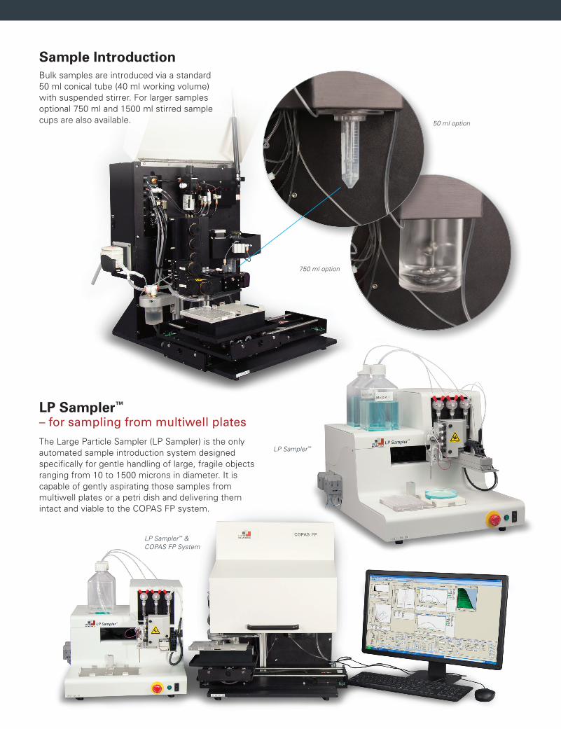
Bulk samples are introduced via a standard 50 ml conical tube (40 ml working volume) with suspended stirrer. For larger samples optional 750 ml and 1500 ml stirred sample cups are also available.
Sample Introduction
The Large Particle Sampler (LP Sampler) is the only automated sample introduction system designed specifically for gentle handling of large, fragile objects ranging from 10 to 1500 microns in diameter. It is capable of gently aspirating those samples from multiwell plates or a petri dish and delivering them intact and viable to the COPAS FP system.
LP Sampler™ – for sampling from multiwell plates
50 ml option
750 ml option
LP Sampler™
LP Sampler™ & COPAS FP System
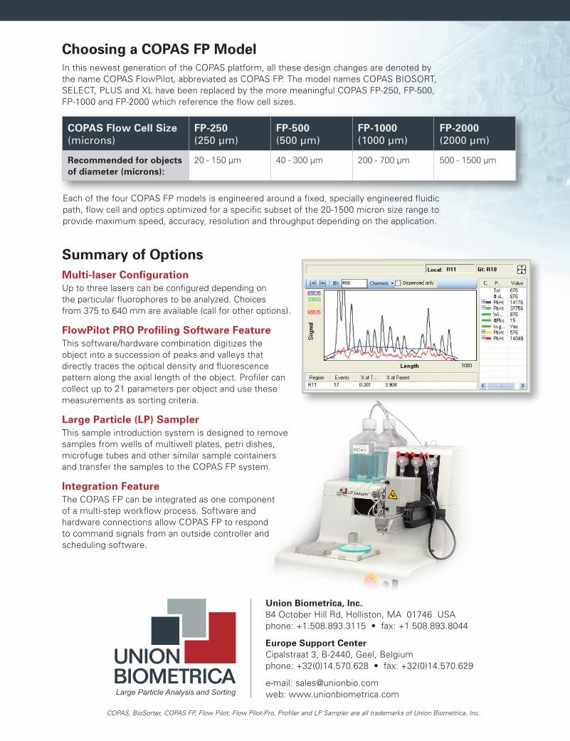
Choosing a COPAS FP Model
Summary of Options
Each of the four COPAS FP models is engineered around a fixed, specially engineered fluidic path, flow cell and optics optimized for a specific subset of the 20-1500 micron size range to provide maximum speed, accuracy, resolution and throughput depending on the application.
In this newest generation of the COPAS platform, all these design changes are denoted by the name COPAS FlowPilot, abbreviated as COPAS FP. The model names COPAS BIOSORT, SELECT, PLUS and XL have been replaced by the more meaningful COPAS FP-250, FP-500, FP-1000 and FP-2000 which reference the flow cell sizes.
Multi-laser ConfigurationUp to three lasers can be configured depending on the particular fluorophores to be analyzed. Choices from 375 to 640 mm are available (call for other options).
FlowPilot PRO Profiling Software FeatureThis software/hardware combination digitizes the object into a succession of peaks and valleys that directly traces the optical density and fluorescence pattern along the axial length of the object. Profiler can collect up to 21 parameters per object and use these measurements as sorting criteria.
Large Particle (LP) SamplerThis sample introduction system is designed to remove samples from wells of multiwell plates, petri dishes, microfuge tubes and other similar sample containers and transfer the samples to the COPAS FP system.
Integration FeatureThe COPAS FP can be integrated as one component of a multi-step workflow process. Software and hardware connections allow COPAS FP to respond to command signals from an outside controller and scheduling software.
COPAS Flow Cell Size (microns)
FP-250 (250 µm)
FP-500 (500 µm)
FP-1000 (1000 µm)
FP-2000 (2000 µm)
Recommended for objects of diameter (microns):
20 - 150 µm 40 - 300 µm 200 - 700 µm 500 - 1500 µm
COPAS, BioSorter, COPAS FP, Flow Pilot, Flow Pilot-Pro, Profiler and LP Sampler are all trademarks of Union Biometrica, Inc.
Large Particle Analysis and Sorting
Europe Support CenterCipalstraat 3, B-2440, Geel, Belgiumphone: +32(0)14.570.628 • fax: +32(0)14.570.629
Union Biometrica, Inc.84 October Hill Rd, Holliston, MA 01746 USAphone: +1.508.893.3115 • fax: +1.508.893.8044
e-mail: [email protected]: www.unionbiometrica.com