crossed fused renal ectopia multidetector … · crossed fused renal ectopia multidetector computed...
TRANSCRIPT

Int J Anat Res 2014, 2(2):305-09. ISSN 2321-4287 305
Original Article
CROSSED FUSED RENAL ECTOPIA MULTIDETECTOR COMPUTEDTOMOGRAPHY STUDYSharma V 1, Ramesh Babu C.S *2, Gupta O.P 3.
ABSTRACT
Address for Correspondence: C.S.Ramesh Babu, Associate Professor of Anatomy, MuzaffarnagarMedical College, N.H.58, Opp. Beghrajpur Industrial Area, Muzaffarnagar (UP) India.Mobile: +91-9897249202. E-Mail: [email protected]
Access this Article online
Quick Response code Web site:
1 Assistant Professor of Anatomy, Muzaffarnagar Medical College, Muzaffarnagar. India.*2 Associate Professor of Anatomy, Muzaffarnagar Medical College, Muzaffarnagar, India.3 Dr.O.P.Gupta Imaging Center, Sumer Bhawan, Bachcha Park, Meerut, India.
Crossed renal ectopia is one of the rarest congenital malformations where a kidney is located on the sideopposite to the side of its ureteral insertion into the urinary bladder and is generally fused with the normallylocated ipsilateral mate. Generally this anomaly remains as a silent clinical entity and is incidentally detectedduring evaluation for other conditions. We report here three such cases of crossed fused renal ectopia de-tected by multidetector row contrast enhanced computed tomography. Crossed fused renal ectopia of inferiortype was observed in a male on the right side with the ureter of the ectopic left kidney crossing the midline. Intwo female patients, L-shaped or tandem kidney was noted, one on the right and another on the left side. Overall in two cases the left kidney was ectopic and in one the right kidney. No renal pathologies like urinary tractinfection, nephrolithiasis or hematuria were found in our patients.KEYWORDS: Crossed fused renal ectopia, Tandem kidney, L-shaped kidney, Renal ectopia, Multidetectorcomputed tomography, Renal fusion anomaly.
INTRODUCTION
International Journal of Anatomy and Research,Int J Anat Res 2014, Vol 2(2):305-09. ISSN 2321- 4287
Received: 22 March 2014Peer Review: 22 March 2014 Published (O):30 April 2014Accepted: 10 April 2014 Published (P):30 June 2014
International Journal of Anatomy and ResearchISSN 2321-4287
www.ijmhr.org/ijar.htm
Congenital malformations of the urinary systemare not uncommon and crossed renal ectopia(CRE) is one of the rare positional and fusionanomalies of the kidney. Crossed renal ectopiaoccurs when a kidney is located on the sideopposite from which its ureter enter into theurinary bladder [1]. In about 90 % of cases, thecrossed ectopic kidney fuses with its ipsilateralmate. Crossed fused renal ectopia is the secondmost common renal fusion anomaly after thehorse-shoe kidney with an estimated incidenceof 1: 2000 to 1: 7500 autopsies.[1,2]. Theprevalence of the crossed renal ectopia withfusion was estimated to be 1 in 1000 live births[3]. In a review of 400 children evaluated byDMSA renal scan, crossed fused renal ectopia
was found in 7 cases (1.75 %) [4]. In an anotherretrospective review, the incidence of CRE wasreported as 1 out of 3078 CT scans and horse-shoe kidney in 1 out of 474 scans [5]. The trueincidence of this anomaly is not known becausea large majority of the patients having thisanomaly remain asymptomatic and undetected.Though CRE remains as a silent entity, in somecases it may be associated with recurrent urinarytract infections, nephrolithiasis, vesicoureteralreflux, uretero-pelvic junction obstruction,hydronephrosis and multicystic renal dysplasiaand hence its importance to nephrologists,surgeons and radiologists. Moreover, thecondition may also be associated with congenitalmalformations affecting skeletal, cardiovascular,
Int J Anat Res 2014, 2(2):305-09. ISSN 2321-4287 306
genitourinary and gastrointestinal systems[6,7,8,9]. In this case series, we review theradiological features of crossed fused renalectopia detected by multidetector rowcomputed tomographic (MDCT) examination inthree patients. It is recently suggested thatMDCT urography is the modality of choicecomprehensively evaluating anatomical featuresof this renal fusion anomaly in a singleexamination [2].MATERIALS AND METHODS
Three cases of crossed fused renal ectopiapresented here were evaluated in a singlediagnostic center during the period fromOctober, 2012 to February, 2014 and theanomaly was detected incidentally when thepatients were examined for other suspectedconditions. The diagnostic center routinelyobtains written informed consent from thepatients before contrast injection. All patientsunderwent contrast enhanced computedtomography (CECT) by a 64 channel scanner (GEOptima-60) and received 85 – 100 ml of non-ioniccontrast (Omnipaque, 300 mg I/ml) at the rateof 5 ml/s intravenously. Scans were obtainedfrom diaphragm to upper part of thigh anddelayed phase scans were also obtained. Thescans were analyzed in a separate work station(AW volume share 4.5) with multiplanarreformatting and maximum intensity projection(MIP) and volume rendered (VR) imagesobtained.
resulting in a L-shaped crossed fused renalectopia (Fig.2). The right ureter crosses themidline at L-5 vertebra. Hilum of the right kidneyis directed anteriorly whereas that of left kidneyanterolaterally. Slight dilatation of leftpelvicalyceal system is seen. The ectopic rightkidney receives its arterial supply from arecurrent branch having a curved course arisingfrom the left common iliac artery and crossinganterior to aortic bifurcation. A short right renalvein and a longer left renal vein drain into leftcommon iliac vein.
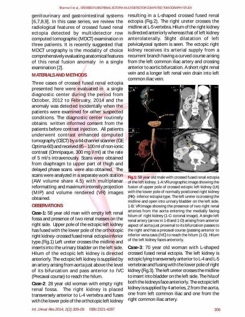
Fig.1: 58 year old male with crossed fused renal ectopiaof the left kidney. 1-A:VR urographic image showing thefusion of upper pole of crossed ectopic left kidney (LK)with the lower pole of normally positioned right kidney(RK)- inferior ectopia type. The left ureter is crossing themidline and open into urinary bladder on the left side.1-B: VR image showing the presence of two right renalarteries from the aorta entering the medially facinghilum of right kidney (1-C- coronal image). A single leftrenal artery (arrow in 1-B and 1-D) arising from anterioraspect of aorta just proximal to its bifurcation passes tothe right and has a precaval course (passing anterior toinferior vena cava (IVC) to reach the hilum (1-D). Hilumof the left kidney faces anteriorly.
OBSERVATIONSCase-1: 58 year old man with empty left renalfossa and presence of two renal masses on theright side. Upper pole of the ectopic left kidneyhas fused with the lower pole of the orthotopicright kidney- crossed fused renal ectopia inferiortype.(Fig.1) Left ureter crosses the midline andinserts into the urinary bladder on the left side.Hilum of the ectopic left kidney is directedanteriorly. The ectopic left kidney is supplied byan artery arising from aorta just above the levelof its bifurcation and pass anterior to IVC(Precaval course) to reach the hilum.Case-2: 28 year old woman with empty rightrenal fossa. The right kidney is placedtransversely anterior to L-4 vertebra and fuseswith the lower pole of the orthotopic left kidney
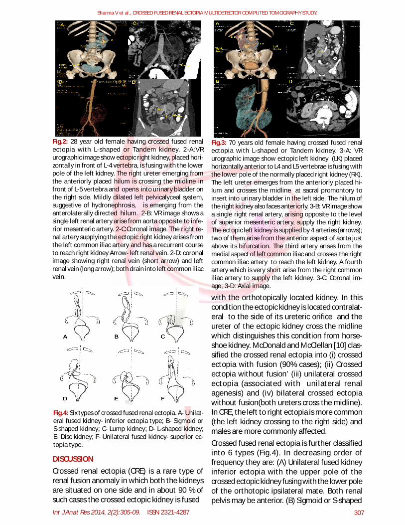
Case-3: 70 year old woman with L-shapedcrossed fused renal ectopia. The left kidney isectopic lying transversely anterior to L-4 and L-5vertebrae and fusing with the lower pole of rightkidney (Fig.3). The left ureter crosses the midlineto insert into bladder on the left side. The hila ofboth the kidneys face anteriorly. The ectopic leftkidney is supplied by 4 arteries, 2 from the aorta,one from left common iliac and one from theright common iliac artery.
Sharma V et al., CROSSED FUSED RENAL ECTOPIA MULTIDETECTOR COMPUTED TOMOGRAPHY STUDY.
Int J Anat Res 2014, 2(2):305-09. ISSN 2321-4287 307
Fig.2: 28 year old female having crossed fused renalectopia with L-shaped or Tandem kidney. 2-A:VRurographic image show ectopic right kidney, placed hori-zontally in front of L-4 vertebra, is fusing with the lowerpole of the left kidney. The right ureter emerging fromthe anteriorly placed hilum is crossing the midline infront of L-5 vertebra and opens into urinary bladder onthe right side. Mildly dilated left pelvicalyceal system,suggestive of hydronephrosis, is emerging from theanterolaterally directed hilum. 2-B: VR image shows asingle left renal artery arise from aorta opposite to infe-rior mesenteric artery. 2-C:Coronal image. The right re-nal artery supplying the ectopic right kidney arises fromthe left common iliac artery and has a recurrent courseto reach right kidney Arrow- left renal vein. 2-D: coronalimage showing right renal vein (short arrow) and leftrenal vein (long arrow); both drain into left common iliacvein.
Fig.3: 70 years old female having crossed fused renalectopia with L-shaped or Tandem kidney. 3-A: VRurographic image show ectopic left kidney (LK) placedhorizontally anterior to L4 and L5 vertebrae is fusing withthe lower pole of the normally placed right kidney (RK).The left ureter emerges from the anteriorly placed hi-lum and crosses the midline at sacral promontory toinsert into urinary bladder in the left side. The hilum ofthe right kidney also faces anteriorly. 3-B: VR image showa single right renal artery, arising opposite to the levelof superior mesenteric artery, supply the right kidney.The ectopic left kidney is supplied by 4 arteries (arrows);two of them arise from the anterior aspect of aorta justabove its bifurcation. The third artery arises from themedial aspect of left common iliac and crosses the rightcommon iliac artery to reach the left kidney. A fourthartery which is very short arise from the right commoniliac artery to supply the left kidney. 3-C: Coronal im-age; 3-D: Axial image.
DISCUSSION
Fig.4: Six types of crossed fused renal ectopia. A- Unilat-eral fused kidney- inferior ectopia type; B- Sigmoid orS-shaped kidney; C- Lump kidney; D- L-shaped kidney;E- Disc kidney; F- Unilateral fused kidney- superior ec-topia type.
Crossed renal ectopia (CRE) is a rare type ofrenal fusion anomaly in which both the kidneysare situated on one side and in about 90 % ofsuch cases the crossed ectopic kidney is fused
with the orthotopically located kidney. In thiscondition the ectopic kidney is located contralat-eral to the side of its ureteric orifice and theureter of the ectopic kidney cross the midlinewhich distinguishes this condition from horse-shoe kidney. McDonald and McClellan [10] clas-sified the crossed renal ectopia into (i) crossedectopia with fusion (90% cases); (ii) Crossedectopia without fusion’ (iii) unilateral crossedectopia (associated with unilateral renalagenesis) and (iv) bilateral crossed ectopiawithout fusion(both ureters cross the midline).In CRE, the left to right ectopia is more common(the left kidney crossing to the right side) andmales are more commonly affected.Crossed fused renal ectopia is further classifiedinto 6 types (Fig.4). In decreasing order offrequency they are: (A) Unilateral fused kidneyinferior ectopia with the upper pole of thecrossed ectopic kidney fusing with the lower poleof the orthotopic ipsilateral mate. Both renalpelvis may be anterior. (B) Sigmoid or S-shaped
Sharma V et al., CROSSED FUSED RENAL ECTOPIA MULTIDETECTOR COMPUTED TOMOGRAPHY STUDY.

Int J Anat Res 2014, 2(2):305-09. ISSN 2321-4287 308
CONCLUSION
kidney in which the crossed kidney lies inferiorlywith the renal pelvis directed laterally and thenormally positioned kidney lies superiorly withthe pelvis directed medially. (C) Unilateral Lumpkidney with fusion occurring over a wide marginand both renal pelvis directed anteriorly; locatedmore inferiorly. (D)L-Shaped or Tandem kidneyin which the crossed kidney lies inferiorly andtransversely fusing with the lower pole of thenormal kidney. (E) Unilateral disc kidney in whichthe fusion occurs along the medial borders and(F) Unilateral fused kidney superior ectopia typeis the least common type; the ectopic kidney isplaced superiorly with its lower pole fusing withthe upper pole of the normal kidney. Both renalpelvis are anterior.The precise mechanism of occurrence of crossedrenal ectopia is not fully understood and severaltheories have been put forward to explain thisanomaly. Among them are the mechanicaltheory (abnormally placed umbilical arteriesmechanically obstructing cephalad migration),the ureteral theory (wandering of the ureteralbud to the opposite side), the teratogenic theory,the genetic theory (observation of the anomalyin families) and theory of abnormal rotation ofthe caudal end of the fetus (increased prevalenceof this anomaly with scoliosis).CRE is sporadically reported in the literaturebecause this anomaly may remain as a silentclinical entity without producing any signs andsymptoms and this is supported by several casereports in cadavers. [11–14]. In these four casereports, male to female ratio and left to rightratio is 3:1. Case studies of patients investigatedfor nephrolithiasis and pyelonephritis withinferior type of crossed fused renal ectopia ofthe left kidney, have been reported [15-17].Inferior ectopia type of CRE is the most commontype. Only in one case inferior type of crossedfused renal ectopia of the right kidney wasdetected [18]. Sigmoid type of kidney, which issecond common type of CRE with fusion,associated with staghorn calculus was reportedby Amin et al. [19]. Superior ectopia, the raresttype of CRE with fusion, was reported in a femalepatient by Patel and Singh [20]. In our study L-shaped kidneys were found in two femalepatients and inferior type of CRE in one malepatient.
A number of case series of CRE with fusion havebeen published. [21-24]. Analysis of thesestudies indicate that the CRE with fusion occursmore commonly in males and the left kidney isaffected more than the right kidney. Manycongenital anomalies are associated with CREwith fusion such as vaginal agenesis [6],VACTERL association [8], TAR syndrome [9], renaldyaplasia [25] and a single ureter [26]. Kulkarnietal detected intestinal malrotation associatedwith a lump kidney in a male cadaver. [7].Crossed fused left renal ectopia with left sidedpolydactyly was found in a 24 week aborted malefetus [27]. We did not find any congenitalanomaly in our cases.
Crossed fused renal ectopia is an uncommoncongenital anomaly which can remain asymp-tomatic throughout life and hence undetected.It is generally found incidentally when patientsare investigated for other abdominal patholo-gies. In some cases it may be associated withnephrolithiasis, recurrent infections , hydroneph-rosis and congenital malformations affectingskeletal, gastrointestinal and urogenital systems.We have reported two cases of a rare type ofL-shaped or tandem kidneys, both found infemales, though CRE is more common in males.Multidetector computed tomographic (MDCT)evaluation provides excellent anatomical detailsof this anomaly in a single examinationimportant for surgeons, nephrologists andradiologists for proper management.
Acknowledgement
Conflicts of Interests: None
REFERENCES
Technical assistance provided by Mr. Arjun Singhin preparation of CT images, Mr.Sushil Kumar andMr.Tithender in preparation of photographs andMr.Chandra Prakash, Artist is sincerelyacknowledged.
[1]. Bauer SB. Anomalies of the upper urinary tract. In:Walsh PC, Retik AB, Vaughan ED, Wein AJ, editors“ Campbell’s Urology”, 8th. Ed. Philadelphia,W.B.Saunders Co., 2002; p 1898-1902.
[2]. Türkvatan A, Ölçer T, Cumhur T. Multidetector CTUrography of renal fusion anomalies. Diagn IntervRadiol., 2009; 15: 127-134.
[3]. Abeshouse BS, Bhisitkul I . Crossed renal ectopiawith and without fusion. Urol .Int. 1959; 9:63.
Sharma V et al., CROSSED FUSED RENAL ECTOPIA MULTIDETECTOR COMPUTED TOMOGRAPHY STUDY.

Int J Anat Res 2014, 2(2):305-09. ISSN 2321-4287 309
Sharma V et al., CROSSED FUSED RENAL ECTOPIA MULTIDETECTOR COMPUTED TOMOGRAPHY STUDY.
[4]. Halaseh M, Alkhawaldeh K, Al-Ibraheem A, Al-Adwan H, Al-Kaylani H. Detection of congenitalrenal anomalies in children being investigated byTc 99m- DMSA renal scan. J. Royal Med Serv., (JRMS)2011; 18(2): 36-42.
[5]. Glodny B, Petersen J, Hofmann KJ, Schen KC, HerwigR, et al. Kidney fusion anomalies revisited: clinicalradiological analysis of 209 cases of crossed fusedectopia and horseshoe kidney. BJU International,2008; 103: 224-235.
[6]. Suthar KD, Mewada BN. Crossed fused renal ectopiawith vaginal agenesis- a case report. Asian J MedRes. 2012; 1(4): 132-133.
[7]. Kulkarni R, Appaji AC, Kulkarni RN. Crossed renalectopia associated with malrotation of intestine: Arare case report. Int J Anat Res.,2013; 1(2): 53-56.
[8]. Padma S, Sundaram PS, Sonik B. A case of VACTERLand non-VACTERL association without the “V andL”. Indian J Nucl Med., 2014; 29: 46-49.
[9]. Ahmad R. A rare association of crossed fused renalectopia. BMC Nephrol., 2007;8:5.
[10]. McDonald JH, McClellan DS. Crossed renal ectopia. Am J Surg 1957; 93: 995.
[11]. Palit S, Datta AK, Tapadar A. A rare presentation ofrudimentary ectopic right kidney fused to the lowerpole of the left with multiple aberrant renal vessels:A case report. J Ant Soc Ind. 2008; 57(2): 146-150.
[12]. Potu BK, Subramaniam B, Cheng PS. Crossed fusedrenal ectopia: a case report. Eur J Anat. 2012; 16(1):79-81.
[13]. Rajaram V, Govindarajan M. Crossed fused renalectopia: a case report. Int J Anat Sci., 2011; 2(2):19-21.
[14].Karambelkar RR, Nikumbh RD, Nikumbh DB,Shewale AD. A rare case study: crossed fused renalectopia. Inferior ectopia type with brief review ofliterature. J Anat Photon. 2013; 113: 127-129.
[15]. Jimenez Pacheco A, Arrabal Polo MA, Arrabal MartinM, Zuluaga Gomez A. Pyelonephritis in crossed-fused renal ectopia. Nefrologia. 2009; 29(3): 277-278.
[16]. Hochwald O, Shaoul R. Crossed fused ectopic leftkidney. Arch Dis Child. 2004; 89:704.
How to cite this article:Sharma V, Ramesh Babu C.S, Gupta O.P. CROSSED FUSED RENALECTOPIA MULTIDETECTOR COMPUTED TOMOGRAPHY STUDY.Int J Anat Res 2014;2(2):305-09.
[17]. Zamora-Varela FR, Gonzalez-Tejedal VM, Gonzalez-Ambriz A. Crossed renal ectopia with fusion andmultiple renal calculi managed with nephrectomythrough anterior paramedian approach. Rev MexUrol., 2013; 73 (4): 200-203.
[18]. Sharma R, Bargotra R. Crossed fused renal ectopia-Inferior ectopia type. J K Science, 2009; 11 (4): 202-203.
[19]. Amin MU, Khan S, Nafees M. Crossed fused renalectopia with staghorn calculus and grosshydronephrosis. J Coll Phys Surg Pakistan, 2009; 19(1): 69-70.
[20]. Patel TV, Singh AK. Crossed fused ectopia of thekidneys. Kidney International. 2008; 73:662.
[21]. Solanki S, Bhatnagar V, Gupta AK, Kumar R. Crossedfused renal ectopia: challenges in diagnosis andmanagement. J Indian Assoc Pediatr Surg. 2013;18(1): 7-10.
[22]. Boyan N, Kubat H, Uzum A. Crossed renal ectopiawith fusion: report of two patients. Clin Anat. 2007;20(6): 699-702.
[23]. deOliveira CMC, deOliveira Santos DC, Gomes DM,Choukroun G, Kubrusly M. Crossed renal ectopiawith fusion: report of two cases and review ofliterature. J Bras Nefrol. 2012; 34 (3).
[24].Turkvatan A, Olcer T, Cumhur T, Akdur PO.Multideetector computed tomographic urographyfor evaluation of crossed fused renal ectopia. JAnkara Univ Faculty of Medicine, 2008; 61 (3): 149-154.
[25]. Birmole BJ, Borwankar SS, Vaidya AS, Kulkarni BK.Crossed renal ectopia. J Postgrad Med., 1993; 39:149.
[26]. Kaur N, Saha , Mriglani R, Saini P, Gupta A. Crossedfused renal ectopia with a single ureter: A rareanomaly. Saudi J Kidney Dis Transpl. 2013; 24(4):773-776.
[27]. Thyagaraju K, Subhadra Devi V. Crossed fused leftrenal ectopia (CRE) in a fetus with left sidedpolydactyly- A case report. Int J Basic Appl Med Sci.,(IJBAMS). 2013; 3 (1): 161-164.