crystal engineering of analogous and homologous organic compounds ... · pdf fileorganic...
TRANSCRIPT

Page 1 of(page number not for citation purposes)
8
Crystal engineering of analogous and homologousorganic compounds: hydrogen bonding patterns intrimethoprim hydrogen phthalate and trimethoprim
hydrogen adipatePackianathan Thomas Muthiah*1, Savarimuthu Francis1,
Urszula Rychlewska2 and Beata Warżajtis2
Full Research Paper Open Access
Address:1Department of Chemistry, Bharathidasan University, Tiruchirappalli-620 024, India and 2Department of Chemistry, Adam MickiewiczUniversity, Grunwaldzka 6, 60–780 Poznañ, Poland
Email:Packianathan Thomas Muthiah* - [email protected];Savarimuthu Francis - [email protected]; Urszula Rychlewska [email protected]; Beata Warżajtis - [email protected]
* Corresponding author
Beilstein Journal of Organic Chemistry 2006, 2, No. 8.doi:10.1186/1860-5397-2-8
Received: 06 December 2005Accepted: 07 April 2006Published: 07 April 2006
© 2006 Muthiah et al; licensee Beilstein-Institut.License and terms: see end of document.
AbstractBackgroundTrimethoprim [2,4-diamino-5-(3',4',5'-trimethoxybenzyl)pyrimidine] is an antifolate drug. It selectively inhibits the bacterial
dihydrofolate reductase (DHFR) enzyme.
ResultsIn the crystal structures of trimethoprim (TMP)-hydrogen phthalate (1) and trimethoprim-hydrogen adipate (2), one of the N atoms
of the pyrimidine ring is protonated and it interacts with the deprotonated carboxylate oxygens through a pair of nearly parallel
N-H...O hydrogen bonds to form a fork-like interaction. In the compound 1, the pyrimidine moieties of the TMP cations are
centrosymmetrically paired through a pair of N-H...N hydrogen bonds involving 4-amino group and the N (N3) atom of the
pyrimidine rings to form a 8-membered hydrogen bonded ring [R22(8)]. The 4-amino group of one TMP moiety and 2-amino
group of another TMP moiety (both moieties are members of a base pair) are bridged by the carbonyl oxygen of the phthalate
moiety through N-H...O hydrogen bonds forming 8-membered hydrogen-bonded ring [R22(8)]. The characteristic hydrogen-
bonded rings observed in the structure aggregate into a supramolecular ladder consisting of a pair of chains, each of which is built
up of alternate TMP and hydrogen phthalate ions. In the compound 2, two TMP cations and two hydrogen adipate anions are
arranged about an inversion center so that the complementary DDAA (D = donor, A = acceptor) arrays of quadruple hydrogen-
bonding patterns are formed. The head-to-tail arrangement of the hydrogen adipate ions leads to a hydrogen-bonded supra-
molecular chain. From crystal engineering point of view, it is interesting to note that the compound 1 has a hydrogen-bonded

Beilstein Journal of Organic Chemistry 2006, 2, No. 8.
Page 2 of(page number not for citation purposes)
8
network remarkably identical with its aliphatic analogue, trimethoprim hydrogen maleate. Similarly the compound 2, resembles its
homolog trimethoprim hydrogen glutarate.
ConclusionIn the crystal structure of trimethoprim hydrogen phthalate, the hydrogen-bonded network is remarkably identical with its aliphatic
analogue, trimethoprim hydrogen maleate. Similarly in the crystal structure of trimethoprim hydrogen adipate the hydrogen bonded
network resembles its homolog trimethoprim hydrogen glutarate.
Beilstein Journal of Organic Chemistry 2006, 2, No. 8.
Page 2 of(page number not for citation purposes)
8
Table 1: Crystallographic parameters for 1 and 2
Properties 1 2
Formula C14 H19 N4 O3+ C14 H18 N4 O3
+
C8 H5 O4- C6 H10 O4
-
M.wt 456.45 436.46Crystal System Triclinic TriclinicSpace group P-1 P-1a/A° 4.6510(10) 8.172(2)b/A° 11.700(2) 9.4744(8)c/A° 20.362(4) 13.8162(8)α/° 76.21(3) 85.741(6)β/° 86.23(3) 87.430(10)γ/° 84.03(3) 88.680(10)V/A°3 1069.3(4) 1065.5(3)Z 2 2Radiation α/A° 1.54718 1.54718Dc/g cm-3 1.418 1.360T/K 293(2) 294(2)μ/mm-1 0.900 0.870F(000) 480 464Reflection collected 4420 3706Observed data[I>2σ(I)]
3137 3062
Parameters refined 395 404Final R1 onobserved data
0.0421 0.0439
Final wR2 onobserved data
0.1246 0.1270
Structure solution SHELXS97 [36] SHELXS97Structure refinement SHELXL97 SHELXL97Graphics PLATON97 [37] PLATON97
IntroductionNon-covalent interactions are the essential tool for both crystal
engineering and supramolecular chemistry [1-4]. Supra-
molecular synthons are the building motif for these fields [5].
Hydrogen bonding is the most important non-covalent interac-
tions. It plays a vital role in biological structure and functions,
molecular design, etc [6]. Recently Sijbesma and Meijer have
investigated the role of quadruple hydrogen bonded network in
the various heterocyclic compounds [7]. Pyrimidine derivatives
offer multiple metal binding modes and have remarkable
hydrogen bonding potential. Trimethoprim [2,4-diamino-5-
Figure 1: The schematic diagram for the various hydrogen-bondedmotifs observed in compound (1).
(3',4',5'-trimethoxybenzyl)pyrimidine] (TMP) is an antifolate
drug. In the protonated form, it exerts its activity through the
inhibition of the enzyme dihydrofolate reductase (DHFR) [8].
In most of the trimethoprim-carboxylate salts, one of the
nitrogen atoms of the pyrimidine ring is protonated and it inter-
acts with the carboxylate group through a nearly parallel
N-H...O hydrogen bonds to form a cyclic bimolecular hydrogen
bonded motif(fork-like interaction) [9-15]. These motifs self
assemble in combination with other hydrogen-bonding groups
leading to base-pairing, quadruple hydrogen bonded arrays,

Beilstein Journal of Organic Chemistry 2006, 2, No. 8.
Page 3 of(page number not for citation purposes)
8
Table 2: Hydrogen bonding geometry for the compounds 1 and 2
Compound D-H...A H...A D...A D-H...A
1 N4 - H3... O4a 2.19(2) 2.939(2) 139.8(17)N2 - H6... O6b 2.08(2) 2.954(2) 164(2)N4 - H9... N3c 2.11(2) 2.986(2) 171(2)N2 - H10... O4d 2.05(3) 3.008(2) 174(2)N2 - H10... O5 2.53(2) 3.109(2) 118.8(18)N1 - H11... O7 1.76(2) 2.678(2) 164.4(19)O5 - H16... O6 1.35(3) 2.399(2) 174(3)C21 - H5 ... O7 2.24(2) 2.670(2) 104.5(15)C24 - H8 ... O4 2.28(2) 2.679(3) 103.0(13)C15 - H25... O1 2.56(3) 3.037(3) 110(2)
2 N1 - H5O ...O4e 1.73(2) 2.682(2) 175(2)O7 - H7O... O4f 1.65(4) 2.555(2) 169(3)N2 - H21... O5 1.95(2) 2.841(2) 179(3)N2 - H22...O5 2.04(3) 2.833(2) 143(2)N4 - H42...O6 2.08(3) 2.951(3) 168(2)
Symmetry Codes : a = -1+x, 1+y, z, b = 1+x, y, z, c = -x, 1-y, 1-z, d = 1-x, -y, 1-z, e = 2-x, 1-y, -z, f = -1+x, y, z
Figure 2: The schematic diagram for the various hydrogen-bondedmotifs observed in compound (2).
DDAA and DADA (D- donor, A = acceptor), etc. C-H...π, π-π
stacking, etc are further stabilizing the crystal structures. The
quadruple hydrogen bonded arrays have been observed in the
crystal structures of TMP m-chlorobenzoate, [11] TMP-sorbate
dihydrate [12], TMP-trifluoroacetate [15], TMP-formate [16],
TMP-hydrogen glutarate [17], TMP-nitrate [18], TMP-sali-
cylate methanol solvate [19] etc,. In the crystal structures of
TMP-terephthalate-terephthalic acid [13], TMP-3-carboxy-4-
hydroxybenzenesulfonate dihydrate [14] and TMP-sulfate
Figure 3: The ORTEP 3 view of the asymmetric unit of the compound1.
trihydrate [20] other types of hydrogen-bonded networks are
present.
The hydrogen-bonding networks in the crystal structures of
TMP/pyrimethamine salts of various dicarboxylic acids have
been investigated in our laboratory [13,21]. Recently we have
also reported a novel isomorphism [21]. The crystal structures
of pyrimethamine hydrogen maleate [21] and pyrimethamine
hydrogen succinate [21] are isomorphous since the hydrogen

Beilstein Journal of Organic Chemistry 2006, 2, No. 8.
Page 4 of(page number not for citation purposes)
8
Figure 4: The ORTEP 3 view of the asymmetric unit of the compound 2.
succinate is the saturated analogue of hydrogen maleate. The
hydrogen succinate ion adopts a folded conformation with an
intramolecular hydrogen bond (mimicking the hydrogen
maleate ion) leading to identical hydrogen-bonded networks in
both the crystal structures. In the present work, crystal struc-
tures of TMP hydrogen phthalate and TMP hydrogen adipate
have been investigated in order to identify the hydrogen
bonding networks and compare them with those in the aliphatic
analogue, TMP hydrogen maleate [22] and the homolog, TMP
hydrogen glutarate [17] respectively.
Results and discussionThe schematic diagram of the hydrogen-bonded motifs
observed in these crystal structures (see Supporting
Information File 1) is shown in Figure 1 and Figure 2. An
ORTEP 3 view of the compounds 1 & 2 is shown in Figure 3
and Figure 4. In the compounds 1 (trimethoprim hydrogen
phthalate) and 2 (trimethoprim hydrogen adipate)(see
Supporting Information File 2) one of the nitrogen atoms (N1)
of the pyrimidine ring is protonated. The protonated pyrimidine
ring interacts with the carboxylate oxygens through a pair of
parallel N-H...O (Table 2) hydrogen bonds to form a fork-like
in te rac t ion (mot i f I ) . This i s reminiscent of the
trimethoprim(TMP)-carboxylate interaction observed in the
DHFR-TMP complexes [24]. This hydrogen bonded motif is
one among the 24 most frequently observed bimolecular cyclic
hydrogen-bonded motifs in organic crystal structures [25]. This
has also been observed in the crystal structures of trimethoprim
carboxylates such as trimethoprim salicylate monohydrate [26],
trimethoprim acetate [9], trimethoprim salicylate methanol
solvate [19], trimethoprim benzoate [10] etc,. This fork-like
hydrogen-bonded interaction (motif I) has further self
assembled in combination with other hydrogen-bonded motifs
to form different types of networks. The planes of the
carboxylate group and the pyrimidine ring (involved in the fork-
like interaction) make a dihedral angle of 9.8° in compound 1
and 6.3° in compound 2 respectively.
In the compound 1 (Table 1), the pyrimidine moieties of
trimethoprim cations are centrosymmetrically paired through a
couple of N-H...N hydrogen bonds involving the 4-amino
group and the N3 atom (motif II). One of the O atoms (O4) at
the carboxyl group of the hydrogen phthalate ion bridges the
2-amino and 4-amino groups on either side of the paired TMP
cations, forming 8-membered hydrogen-bonded ring (motif
III), with graph-set notation [R22(8)] [27]. The atom O5 of the
carboxyl group of the hydrogen phthalate ion forms an
intramolecular O-H...O hydrogen bond with the O6 atom of the
carboxylate group. The hydrogen-bonding patterns formed
upon the association of pyrimidine moieties of TMP molecules
via self-pairing and carboxylate bridging resemble those
observed in the crystal structure of TMP-hydrogen maleate
[22]. The same type of DADA array has also been observed in
the other crystal structures of trimethoprim-salicylate methanol
solvate [18], trimethoprim-trifluoroacetate [15], pyrimethamine-
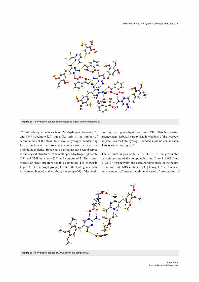
hydrogen phthalate [21] etc,. The characteristic hydrogen-
bonded rings observed in the structure aggregate into a supra-
molecular ladder consisting of a pair of chains, each of which
is built up of alternate TMP and hydrogen phthalate ions (motif
III & IV) as shown in Figure 5 [28]. The one of the hydrogen
atoms of the 2-amino group is also involved in bifurcated
hydrogen-bonding with the carboxyl O atoms (O4 & O5) to
form a 4-membered hydrogen bonded ring [R21(4)] [27].
In the compound 2 (Table 1), in motif V, two TMP cations and
two hydrogen adipate anions are arranged about an inversion
center so that the complementary DDAA arrays of quadruple
hydrogen-bonding patterns are formed. This has also been
observed in TMP m-chlorobenzoate [11], TMP-hydrogen glut-
arate [17] and TMP succinate [29]. In motif VI, the hydrogen
atoms of 2- and 4-amino groups of the TMP cation are
hydrogen-bonded to the carboxylate and carboxyl ends, respect-
ively, of the same hydrogen adipate ion. Thus, the hydrogen
adipate bridges the 2-amino and 4-amino groups of TMP.
These hydrogen-bonded interactions are almost identical with
Beilstein Journal of Organic Chemistry 2006, 2, No. 8.
Page 5 of(page number not for citation purposes)
8
Figure 5: The hydrogen-bonded supramolecular ladder in the compound 1.
Figure 6: The hydrogen-bonded DDAA array in the compound 2.
TMP-dicarboxylate salts such as TMP-hydrogen glutarate [17]
and TMP-succinate [29] but differ only in the number of
carbon atoms of the chain. Such cyclic hydrogen-bonded ring
formation blocks the base-pairing interaction between the
pyrimidine moieties. Hence base-pairing has not been observed
in the crystal structures of trimethoprim-hydrogen glutarate
[17] and TMP-succinate [29] and compound 2. The supra-
molecular sheet structure for this compound 2 is shown in
Figure 6. The carboxyl group (O7-H) of the hydrogen adipate
is hydrogen-bonded to the carboxylate group (O4) of the neigh-
bouring hydrogen adipate ion(motif VII). This head-to-tail
arrangement (carboxyl-carboxylate interaction) of the hydrogen
adipate ions leads to hydrogen-bonded supramolecular chain.
This is shown in Figure 7.
The internal angles at N1 (C2-N1-C6) in the protonated
pyrimidine ring of the compounds 1 and 2 are 119.9(1)° and
119.6(2)° respectively, the corresponding angle in the neutral
trimethoprim(TMP) molecule [31] being 115.5°. Such an
enhancement of internal angle at the site of protonation of

Beilstein Journal of Organic Chemistry 2006, 2, No. 8.
Page 6 of(page number not for citation purposes)
8
Figure 7: The supramolecular chain made up of hydrogen adipate in the compound 2.
pyrimidine ring is very characteristic. In the compounds 1 and
2 the dihedral angles between the plane of the pyrimidine and
phenyl rings are 74.0(7)° and 88.8° respectively. These values
are closer to the crystal structures of TMP-sulfate trihydrate
[20] (75.8(9)°) and TMP 4-hydroxybenzoate dihydrate [30]
(89.1(1)°).
The major (77%) and minor (23%) components in the
disordered hydrogen adipate molecule adopt quite unusual bent
carbon chain conformations: the gauche-gauche-trans (ggt)
and the gauche-trans-trans (gtt) forms, respectively. Of the 46
adipic acid fragments present in the Cambridge Crystallo-
graphic Data Base [32] there is only one example of the ggt
conformation [33] and two cases in which the acid adopts the
gtt form [34,35]. The adoption of the bent carbon chain
conformation by adipic acid seems necessary in order to place
the two terminal carboxyl functions in mutual syn orientation
so that they can fasten the 2- and 4-amino groups of the TMP
molecule. The disorder, on the other hand, might result from
incompatible dimensions between the adipic acid and the two
amino groups of the TMP molecule. Much better fit between
the 2- and 4-amino groups of the TMP molecule on one side,
and aliphatic dicarboxylic acid on the other side is achieved in
the case of glutaric acid [17]. This is for two reasons: firstly, in
the energetically preferred extended carbon chain conformation
an odd number of carbon atoms in a chain implicates the syn
orientation of the terminal carboxyl functions and, secondly,
the carbon chain is identical in length as the N2-C2-N3-C4-N4
fragment of the TMP. The observation that, irrespective on the
number of carbon atoms constituting the dicarboxylic acid
chain, the TMP/dicarboxylic acid interactions are represented
by the same motif VI is quite unusual.
Figure 8: Scatterplot illustrating the distribution of the two torsionangles (C4-C5-C7-C8 (TOR1) and C5-C7-C8-C9 (TOR2)) thatdescribe the mutual orientation of pyrimidine and phenyl rings in theTMP molecule. The torsion angle values were obtained from the July2003 release of the 5.24 version of the CSD [32]. The values of TOR2have been restricted to the range from -90 to +90°.
The TMP molecule can be regarded as having a rigid frame,
built on the methyl group, on which the substituted phenyl and
pyrimidine six-membered rings are free to rotate. An arbitrary
conformation of this molecule can be described by the torsion
angles of the two rings to the frame. We define these torsion
angles as C4-C5-C7-C8 and C5-C7-C8-C9, i.e. with respect to
one of the rings the other can rotate around the C5-C7 or
around the C7-C8. Figure 8 shows the distribution of these

Beilstein Journal of Organic Chemistry 2006, 2, No. 8.
Page 7 of(page number not for citation purposes)
8
torsion angles in 37 TMP fragments deposited in the
Cambridge Structural Data Base [32]. The points mostly cluster
around the plus/minus (80°, 30°) and (160°, 70°) regions. The
points representing the (80°, 30°) combination predominantly
lie in the region where both torsion angles have the same sign,
which is the condition for a propeller conformation. In the
presented crystal structures 1 and 2 the corresponding torsion
angles adopt the values -161.4(1) and 63.5°, and 69.1(2) and
36.5(3)°, respectively. Hence the observed TMP conformations
match the two most densely populated conformations observed
in other crystal structures containing the TMP moieties.
In the compound 1, the ionized and non-ionized carboxyl
groups are inclined at an angles of 6.0(1)° and 9.0(1)° respect-
ively to the plane of the phenyl ring. The bond angles of C19-
C17-O4, C19-C17-O5 in the carboxyl group are 119.1(2)° and
120.7(17)° respectively. The similar angles at the carboxylate
group C20-C18-O6 and C20-C18-O7 are 120.4(2)° and
117.4(2)° respectively. These values are comparable with the
crystal structure of pyrimethamine hydrogen phthalate [21]. In
the compound 2, the bond angles at carboxylate group, C18-
C17-O5 and C18-C17-O4 are 120.1(2)° and 116.5(2)° respect-
ively, whereas the angle at the carboxyl group C21-C22-O6,
C21-C22-O7 are 123.1(2)° and 113.1(2)° respectively.
The crystal structure of compound 2 is further stabilized by two
C-H...π interactions [38] [C16-H161...Cg1(atoms N1-C6)
(2.923Å, 137°) and C20-H201...Cg2 (atoms C8-C13) (2.779Å,
156°)] and the pyrimidine stacking interactions. The interplanar
and centroid to centroid distances are 3.381Å and 3.738Å,
respectively, and the angle between the centroid vector and
normal to the plane is 25.3°.
Supporting Information
Supporting Information File 1the CIF information
[http://www.beilstein-journals.org/bjoc/content/
supplementary/1860-5397-2-8-S1.cif]
Supporting Information File 2experimental details
[http://www.beilstein-journals.org/bjoc/content/
supplementary/1860-5397-2-8-S2.doc]
AcknowledgmentsSF thanks the Council of Scientific and Industrial Research
(CSIR), New Delhi, India, for the award of a Senior Research
Fellowship (reference No. 9/475(109)2002 EMR-I).
References1. Desiraju, G. R. Curr. Sci. 2001, 81, 1038–1055.2. Desiraju, G. R. Acc. Chem. Res. 2002, 35, 565–573. doi:10.1021/
ar010054t3. Aakeroy, C. B.; Beatty, A. M. Aust. J. Chem. 2001, 54, 409–421.
doi:10.1071/CH011334. Rao, C. N. R. Curr. Sci. 2001, 81, 1030–1037.5. Thalladi, V. R.; Goud, B. S.; Hoy, V. J.; Allen, F. H.; Howard, J. A. K.;
Desiraju, G. R. Chem. Commun. 1996, 401–402. doi:10.1039/cc9960000401
6. Steiner, T. Angew. Chem., Int. Ed. 2000, 41, 48–76. doi:10.1002/1521-3773(20020104)41:1<48::AID-ANIE48>3.0.CO;2-U
7. Sijbesma, R. P.; Meijer, E. W. Chem. Commun. 2003, 5–16.doi:10.1039/b205873c
8. Hitching, G. H.; Kuyper, L. F.; Baccananari, D. P. In Design of EnzymeInhibitors as Drugs; Sandler, M.; Smith, H. J., Eds.; Oxford UnversityPress: New York, 1988; pp 343 ff.
9. Bryan, R. F.; Haltiwanger, R. C.; Woode, M. K. Acta Crystallogr., Sect.C 1987, 43, 2412–2415. doi:10.1107/S0108270187087584
10. Bettinetti, G. P.; Giordano, F.; Mana, A. L. Acta Crystallogr., Sect. C1985, 41, 1249–1253. doi:10.1107/S0108270185007314
11. Raj, S. B.; Muthiah, P. T.; Rychlewska, U.; Warzajtis, B.CrystEngComm 2003, 5, 48–53. doi:10.1039/b211312k
12. Raj, S. B.; Stanley, N.; Muthiah, P. T.; Bocelli, G.; Olla, R.; Cantoni, A.Cryst. Growth Des. 2003, 3, 567–571. doi:10.1021/cg020043m
13. Hemamalini, M.; Muthiah, P. T.; Bocelli, G.; Cantoni, A. ActaCrystallogr., Sect. E 2003, 59, o14–o17. doi:10.1107/S1600536802021670
14. Raj, S. B.; Sethuraman, V.; Francis, S.; Hemamalini, M.; Muthiah, P.T.; Bocelli, G.; Cantoni, A.; Rychlewska, U.; Warzajtis, B.CrystEngComm 2003, 5, 70–76. doi:10.1039/b300126a
15. Francis, S.; Muthiah, P. T.; Bocelli, G.; Righi, L. Acta Crystallogr., Sect.E 2002, 58, o717–o719. doi:10.1107/S1600536802009509
16. Umadevi, B.; Prabakaran, P.; Muthiah, P. T. Acta Crystallogr., Sect. C2002, 58, o510–o512. doi:10.1107/S0108270102011150
17. Robert, J. J.; Raj, S. B.; Muthiah, P. T. Acta Crystallogr., Sect. E 2001,57, o1206–o1208. doi:10.1107/S1600536801018001
18. Murugesan, S.; Muthiah, P. T. Acta Crystallogr., Sect. C 1997, 53,763–764. doi:10.1107/S0108270196015636
19. Panneerselvam, P.; Stanley, N.; Muthiah, P. T. Acta Crystallogr., Sect.E 2002, 58, o180–o182. doi:10.1107/S1600536802000909
20. Muthiah, P. T.; Umadevi, B.; Stanley, N.; Shui, N.; Eggleston, D. S.Acta Crystallogr., Sect. C 2001, 57, o1179–o1182.
21. Sethuraman, V.; Stanley, N.; Muthiah, P. T.; Sheldrick, W. S.; Winter,M.; Luger, P.; Weber, M. Cryst. Growth Des. 2003, 3, 823–828.doi:10.1021/cg030015j
22. Prabakaran, P.; Robert, J. J.; Muthiah, P. T.; Bocelli, G.; Righi, L. ActaCrystallogr., Sect. C 2001, 57, 459–461. doi:10.1107/S0108270101000269
23. Kuma, KM-4; Kuma Diffraction: Wroclaw, Poland, 1991.24. Kuyper, L. F. In Crystallographic and modeling methods in Molecular
Design; Bugg, C. E.; Ealick, S. E., Eds.; Springer-Verlag: New York,1990; pp 56 ff.
25. Allen, F. H.; Raithby, P. R.; Shields, G. P.; Taylor, R. Chem. Commun.1998, 1043–1044. doi:10.1039/a801424h
26. Murugesan, S.; Muthiah, P. T. Academy Discussion Meeting onFrontiers in Structural Chemistry, I. I. T., Chennai, India,, 1996; .[Abstract No. 3.4].
27. Etter, M. C. Acc. Chem. Res. 1990, 23, 120–126. doi:10.1021/ar00172a005

Beilstein Journal of Organic Chemistry 2006, 2, No. 8.
Page 8 of(page number not for citation purposes)
8
28. Nguyen, V. T.; Ahn, P. D.; Bishop, R.; Scudder, M. L.; Craig, D. C. Eur.J. Org. Chem. 2001, 4489–4499. doi:10.1002/1099-0690(200112)2001:23<4489::AID-EJOC4489>3.0.CO;2-Z
29. Sethuraman, V. Ph.D. Thesis, Department of Chemistry,Bharathidasan University: Tiruchirappalli, India, 2002.
30. Robert, J. J. Ph.D. Thesis, Department of Chemistry, BharathidasanUniversity: Tiruchirappalli, India, 1998.
31. Koetzle, T. F.; Williams, G. J. B. J. Am. Chem. Soc. 1976, 98,2074–2078. doi:10.1021/ja00424a009
32. Allen, F. H.; Kennard, O. Chem. Des. Autom. News 1993, 8, 31.33. McCann, M.; Casey, M. T.; Devereux, M.; Curran, M.; McKee, V.
Polyhedron 1997, 16, 2741–2748. doi:10.1016/S0277-5387(97)00037-5
34. Zheng, Y.-Q.; Lin, J.-L.; Pan, A.-Y. Z. Anorg. Allg. Chem. 2000, 626,1718–1720. doi:10.1002/1521-3749(200008)626:8<1718::AID-ZAAC1718>3.0.CO;2-A
35. Zheng, Y.-Q.; Sun, J.; Lin, J.-L. Z. Anorg. Allg. Chem. 2001, 627,90–94. doi:10.1002/1521-3749(200101)627:1<90::AID-ZAAC90>3.0.CO;2-O
36. SHELXL97; University of Göttingen: Germany, 1997.37. PLATON97; Utrecht University: The Netherlands, 1997.38. Desiraju, G. R.; Steiner, T. The weak hydrogen bond in Structural
Chemistry and Biology; Oxford University Press: Oxford, 1999.
License and TermsThis is an Open Access article under the terms of the
Creative Commons Attribution License
(http://creativecommons.org/licenses/by/2.0), which
permits unrestricted use, distribution, and reproduction in
any medium, provided the original work is properly cited.
The license is subject to the Beilstein Journal of Organic
Chemistry terms and conditions:
(http://www.beilstein-journals.org/bjoc)
The definitive version of this article is the electronic one
which can be found at:
doi:10.1186/1860-5397-2-8