ctos board of directors 1999± 2000
TRANSCRIPT

1357± 714X print/1369± 1643 online/01/010035± 43 ½ 2001 Taylor & Francis LtdDOI: 10.1080/13577140120048935
Sarcoma (2001) 5, 35± 79
CTOS Board of Directors 1999± 2000
Brian O’Sullivan, MD (President)Jaap Verweij, MD (Vice President)Lee Helman, MD (Secretary)Mark Gebhardt, MD (Treasurer)Nicola Baldini, MD (past President)Thor Alvegard, MD, PhD (Medical Oncology & Radiotherapy, 1997± 2000)Robert S. Benjamin, MD (Medical Oncology, 1997± 2000)J. Sybil Biermann, MD (1998± 2001)Vivien Bramwell-Wesley, MD (1998± 2001)Franco Gherlinzoni, MD (1999± 2002)Michael Simon, MD (1999± 2002)Ian Judson, MD (1998± 2001)Scott Nelson, MD (1999± 2002)Martine van Glabbeke MD (1999± 2002)Claude Turc-Carel, M.D (1998± 2001)Frits Van Coevorden, MD (1998± 2001)Sharon Weiss, MD (Pathology, 1997± 2000)
36 CTOS abstracts
6th Annual Scientific Meeting, 2± 4 November 2000, Hotel Okura, Amsterdam, The Netherlands
Program Committee:
Ole S. Nielsen (Chairman)Claude S. Turc-Carel (Basic Science Program Chairman)Mark C. Gebhardt (past Chairman)Karen Antman (next Chairman) Brian O’Sullivan (President)
Scientific Committee:
Thor A. Alvegård (Sweden), radiation oncologyNicola Baldini (Italy), biologyRobert Benjamin (USA), medical oncologyRobert Bell (Canada), surgeryVivien Bramwell (Canada), medical oncologyCharles Catton (Canada), radiation oncologyFrits van Coevorden (The Netherlands), surgeryGeorge Demetri (USA), medical oncologyChristopher Fletcher (USA), pathologyAllen M. Goorin (USA), pediatric oncologyLee Helman (USA), pediatric oncologyPeter R. Hohenberger (Germany), surgeryIan Judson (UK), medical oncologyJonathan J. Lewis (USA), surgeryOle Steen Nielsen (Denmark), radiation oncologyBrian O’Sullivan (Canada), radiation oncologyPiero Picci (Italy), medical oncologyPeter Pisters (USA), surgeryJaap Verweij (The Netherlands), medical oncology
CTOS abstracts 37
Program Schedule
Thursday, 2 November 2000
1:00± 5:00 p.m. Registration, Set Up Posters Ð Hotel Lobby6:00± 7:00 p.m. Welcome Reception Ð Meerman Room
Friday, 3 November 2000
7:00 a.m. Registration, breakfast for meeting attendees Ð Foyer Okura8:00 a.m. Welcome, Opening Announcements Ð Heian Room8:15 a.m. CASE DISCUSSION: `Treatment of Gynecological Sarcomas’
Moderators & discussants: Vivien Bramwell (London, Ont.) & Nicholas Reed (Glasgow)9:00 a.m. NINA AXELRAD KEYNOTE LECTURE:
Supported by Nina Axelrad Sarcoma FundIntroduction/moderator: Brian O’Sullivan (Toronto)Murray Brennan (Memorial Sloan-Kettering Cancer Center, New York): `Soft Tissue Sar-coma: 25 Years of Achievements, Failures and Challenges’.
9.45 a.m. Coffee Break, Poster Viewing Ð Foyer Okura, Foyer Amstel & Otter, Esperance
Room
10:15 a.m. PROFFERED PAPERS: `Surgical Treatment of Sarcomas’ Ð Heian Room
Moderators: Antonie Tamineau (Leiden) & Frits van Coevorden (Amsterdam)10:15 a.m. Chondrosarcoma of bone: analysis of factors related to prognosis in 108
cases with a minimum of two years follow-up Ð Michelle Ghert10:25 a.m. The effect of re-resection in extremity soft tissue sarcoma Ð Jonathan
Lewis10:35 a.m. Classification of positive margins after resection of extremity soft tissue
sarcoma predicts the risk of local recurrence Ð Craig Gerrand10:45 a.m. Combined modality management of retroperitoneal sarcomas: phase 1
trial of pre-operative doxorubicin-based concurrrent chemoradiation sur-gical resections, and intraoperative electron-beam radiation therapy(IORT) Ð Peter Pisters
10:55 a.m. Peritoneal sarcomatosis treated by cytoreductive surgery and intraperito-neal hyperthermic perfusion Ð Marcello Deraco
11:05 a.m. Frits van Coevorden: `Review of session related posters’11.15 a.m. Antonie Tamineau: `Review of session (state of the art)’
11:30 a.m. MINI SYMPOSIUM: `Local Recurrence of Soft Tissue Sarcomas’
Moderators: Jonathan Lewis (New York) & Martin Robinson (Sheffield)
11:30 a.m. Surgery Ð Murray Brennan11:38 a.m. Radiation Oncology Ð Martin Robinson11:46 a.m. Medical Oncology Ð Robert Benjamin11:54 a.m. `General discussion’
12:15 p.m. Lunch, Poster Viewing Ð Foyer Okura, Foyer Amstel & Otter, Esperance Room
1:15 p.m. PROFFERED PAPERS: `Radiation Oncology’ Ð Heian Room
Moderators: Charles Catton (Toronto) & Thor Alvegaard (Lund)
1:15 p.m. Complete resection and intra-operative radiation therapy improve out-come of retroperitoneal sarcomas Ð Jean-Pierre Pierie
1:25 p.m. Radiation morbidity two years post treatment: results from a randomizedtrial of pre- versus post-operative radiotherapy Ð Aileen Davis
38 CTOS abstracts
1:35 p.m. Real-time radiotherapy review of a randomized trial of pre-and post-oper-ative radiotherapy for localized soft-tissue sarcoma of the extremity ÐCharles Catton
1:45 p.m. Thor Alvegaard: `Review of session related posters’1.55 p.m. Charles Catton: `Review of session (state of the art)’
2:10 p.m. PROFFERED PAPERS: `Basic Science/Biology’
Moderators: Claude S. Turc-Carel (Nice) & Jay S. Wunder (Toronto)
2:10 p.m. Differential expression of EZRIN in a high and low metastatic osteosar-coma model Ð Chand Khanna
2:20 p.m. Molecular cloning of putative oncogene and antioncogen involved in thedevelopment of osteosarcoma Ð Junya Toguchida
2:30 p.m. Clinico-pathological and biological analyses of the chop-related fusiongenes in myxoid liposarcomas Ð Taisuki Hosaka
2:40 p.m. Cytokine levels in serum of the patients with soft tissue sarcomas and theirrelationship to alterations of routine blood tests Ð Piotr Rutkowski
2:50 p.m. Wild-Type P53 sensitizes soft tissue sarcoma cells to doxorubicin bydownregulation of MDR1 expression Ð Raphael Pollock
3:00 p.m. Jay Wonder: `Review of session related posters’3:10 p.m. Claude Turc-Carel: ̀ Review of session (state of the art)’
3:25 p.m. Coffee Break, Poster Viewing Ð Foyer Okura, Foyer Amstel & Otter, Esperance
Room
3:55 p.m. CTOS YOUNG INVESTIGATOR AWARD PRESENTATIONS Ð Heian Room
Moderator: Nicola Baldini (Bologna)
3:55 p.m. Nicola Baldini: `Short introduction’4:00 p.m. Award Presentation 1: Regulation of osteosarcoma (OS) metastasis by
IGF-I receptor signaling: a novel therapeutic target Ð Kristy Weber
4:20 p.m. Award Presentation 2: Results of two consecutive phase II studies andinterim analysis of a phase III intergroup study of neoadjuvant treatmentincluding regional hyperthermia (RHT) in high risk soft tissue sarcoma(HR-STS) Ð Clemens Wendtner
4:40 p.m. PROFFERED PAPERS: `Medical Oncology’
Moderators: Robert Benjamin (Houston) & Ian Judson (London)
4:40 p.m. Expression of the trail receptor dr4 in human soft tissue sarcomas Ð RudyKomdeur
4:50 p.m. Dominant negative IkBa Potentiates in anti-tumor activity of doxorubicinin a rat hind limb isolated perfusion model Ð Robert Davidson
5:00 p.m. A pilot study of short course intensive multiagent chemotherapy for poorrisk osteosarcoma Ð Jim Janinis
5:10 p.m. A phase 1 trial of intraperitoneal hyperthermic chemotherapy for the treat-ment of sarcomatosis Ð Malcolm Bilimoria
5:20 p.m. Ian Judson: `Review of session related posters’5:30 p.m. Robert Benjamin: `Review of session (state of the art)’
5:45 p.m. Adjourn
6:15 p.m. Reception and Boat Tour
7:15 p.m. Dinner Banquet Ð Ballroom I & II
Saturday, 4 November 2000
7:00 a.m. Registration, breakfast for meeting attendees Ð Foyer Okura
CTOS abstracts 39
8:00 a.m. WORKSHOP: `Management of osteosarcomas’
Moderators: Piero Picci (Bologna) & Robert Grimer (Birmingham)
Suggested topics: 8:00 a.m. Histopathological problems Ð Pancras Hogendoorn8:20 a.m. Surgical aspects. New developments Ð Robert Grimer8:40 a.m. Role of high dose Methotrexate Ð Kirsten Sundby Hall8:55 a.m. Treatment of high risk patients Ð Piero Picci9:15 a.m. Late effects of the treatment of bone tumors Ð Juliet Hale9:30 a.m. Long-term problems after surgery Ð Per-Ulf Tunn9:45 a.m. `General discussion’
10:00 a.m. Coffee Break, Poster Viewing Ð Foyer Okura, Foyer Amstel & Otter, Esperance
Room
10:30 a.m. COOPERATIVE GROUP SESSION: `How do we improve Intergroup collabora-
tion?’ Ð Heian Room
Moderators: Peter Pisters (Houston) & Peter R. Hohenberger (Berlin)
10:30 a.m. How to moderate intergroup studies Ð M. van Glabbeke10:45 a.m. Panel discussion: `How to improve International Collaboration and
Research?’
Panel members:
Peter Pisters: `ACOSOG’ & `RTOG’Lee Helman: `Pediatric Coop. Group’John Edmonson: ̀ ECOG’Ian Judson: ̀ EORTC’Bihn N Bui: `French SG’Piero Picci: `Italian SG’Vivien Bramwell: `NCIC-CTG/CSG’Thor Alvegaard: `SSG’
11:45 a.m. Peter Pisters & Peter R. Hohenberger: `Summary of discussion and con-clusions (how to proceed?)’
12:00 noon Lunch, Poster Viewing Ð Foyer Okura, Foyer Amstel & Otter, Esperance Room
1:00 p.m. PROFFERED PAPERS: `Pediatric Oncology’ Ð Heian Room
Moderators: Lee Helman (Bethesda) & Axel Le Cesne (Paris)
1:00 p.m. CD99 engagement: an effective therapeutic strategy for Ewing tumors ÐKatia Scotlandi
1:10 p.m. Induction of chemoresistence to doxorubicin in cells carrying a P53 germ-line mutation detected in a Li± Fraumeni family Ð Luca Sangiorgi
1:20 p.m. Study of age as major prognostic factor in localised Ewing’s sarcoma ÐNicole Delepine
1:30 p.m. Lee Helman: `Review of session and session related posters’
1:50 p.m. PROFFERED PAPERS: `Diagnostic Imaging/Pathology’
Moderators: Pancras Hogendoorn (Leiden) & Laurence Baker (Ann Arbor)
1:50 p.m. Monitoring the effect of isolated limb perfusion in soft tissue sarcoma withdynamic contrast-enhanced MR imaging Ð C.S.P. van Rijswijk
2:00 p.m. Upregulation of PTHrP and BCL-2 expression characterizes early malig-nant transformation of osteochondroma towards peripheral chondrosar-coma and is a late event in central chondrosarcoma Ð JVMG Bovee

40 CTOS abstracts
2:10 p.m. Does the histologic subtype of high-grade central osteosarcoma influencethe response to treatment with chemotherapy and does it affect overall sur-vival Ð E. Hauben
2:20 p.m. Round-cell and myxoid liposarcoma of the extremities. A clinicopatho-logic study of 102 cases Ð Andrea Pellacani
2:30 p.m. Laurence Baker: `Review of session related posters’2:40 p.m. P. Hoogendoorn: ̀ Review of session (state of the art)’
3:05 p.m. Coffee break Ð Foyer Okura, Foyer Amstel & Otter, Esperance Room
3:35 p.m. MINI SYMPOSIUM: `New Drugs in Sarcomas’ Ð Heian Room
Moderators: George Demetri (Boston) & Jaap Verweij (Rotterdam)
3:35 p.m. Troglitazone and newer PPAR-gamma receptor ligands in liposarcomasÐ George Demetri
3:50 p.m. ET-743 in soft tissue sarcomasÐ Jaap Verweij4:05 p.m. New emerging concepts Ð Ian Judson4:20 p.m. `General discussion’
4.30 p.m. Closing. Summary of Scientific Meeting. Next Meeting.
Reviewer/presenter: Karen Antman (New York)
4:45 p.m. Member’s Business Meeting
6:00 p.m. Adjournment

CTOS abstracts 41
Proffered Papers Ð Surgical Treatment of Sarcomas
Chondrosarcoma of Bone: Analysis of Factors Related to
Prognosis in 108 Cases With a Minimum of Two Years
Follow-up
Rizzo M, Ghert MA, Harrelson JM, Scully SP
Duke University Medical Center, Durham, NC 27710, USA
Introduction: Chondrosarcoma is unresponsive to existing adju-
vant therapies and is primarily a surgical disease. There is an estab-
lished relationship between the histologic grade of these tumorsand prognosis. The purpose of this study was to review our institu-
tion’ s experience with chondrosarcoma and assess factors related
to prognosis and outcome.Methods: The medical records of 108 patients were retrospec-
tively reviewed. Data was evaluated with respect to patient demo-
graphics, tumor location, histologic grade, tumor size, surgicalmargins, metastases and recurrence. The tumors were sub-classi-
fied based on histologic grade with grade 1 lesions defined as low-
grade, and grade 2 and 3 lesions (as well as dedifferentiatedlesions) defined as high-grade. All patients were followed for a
minimum of 2 years. Statistical analysis was performed using uni-
variate, multivariate, and Kaplan± Meier survival analysis.Results: There were 68 males and 40 females with a mean age at
presentation of 53 years (range, 26± 70 years). Clinical follow-up
averaged 97 months (range, 3± 314). The most common tumorlocations included the femur (46), pelvis (22) and humerus (10).
There were 31 low-grade and 77 high-grade chondrosarcomas.
One hundred and one patients underwent surgical resection. Widemargins were achieved in 78 patients, 11 underwent marginal
resection and 12 tumor resections had positive margins (intrale-
sional). Seventy-two patients remained alive at the time of thisstudy with no evidence of disease, 23 have died of disease, six died
without disease and seven remain alive with recurrent disease. The
high-grade tumors had a significantly increased rate of death dueto disease (p < 0.01), development of metastases (p < 0.01), and
local recurrence (p < 0.01). There was a significant relationship
between local recurrence and positive margins (p < 0.03), andbetween metastases and positive margins (p < 0.03). Patient demo-
graphics, tumor location and size did not correlate significantly
with outcome.Conclusions: This study supports previous findings that tumor
grade in chondrosarcoma has prognostic significance, and that
adequate surgical margins are essential to maximize survival. Aschondrosarcoma does not respond to standard chemotherapy or
radiation protocols, our findings emphasize the need for molecular
markers and novel biologic adjuvant therapies.
The Effect of Re-resection in Extremity Soft Tissue
Sarcoma
Lewis JJ, Leung D, Espat J, Woodruff JM, Brennan MF
Memorial Sloan-Kettering Cancer Center, New York, NY 10021,
USA
Introduction: This study was undertaken to determine if re-resec-
tion impacts on disease-specific survival in patients with inade-quately resected, primary extremity soft tissue sarcoma. We
analyzed two groups of patients: those who underwent a single
definitive radical resection at a specialist cancer center versus thosewho underwent an incomplete excisional resection in the commu-
nity, followed by a second definitive radical re-resection at a spe-
cialist cancer center.Methods: Patients who underwent treatment for primary tumors
(from July 1982 to June 1999) at a single institution were the subject
of study. Two groups of patients were analyzed: those who under-went one definitive radical resection (one operation) and those who
were previously resected and then referred for subsequent radical
re-resection (two operations). Survival was determined with the
Kaplan± Meier actuarial method. Statistical significance was evalu-
ated using log-rank testing and Cox model stepwise regression.
Results: During this time, we resected 1092 patients with primary
extremity soft tissue sarcoma. Of these, 685 underwent definitive
radical resection and 407 underwent re-resection after undergoing
excisional resection elsewhere. The median follow-up was 4.8
years. The 5-year disease-specific survival of the definitive resec-
tion (one operation) group was 68% and that of the re-resection
(two operations) group was 84% (p = 0.0001). On multivariate
analysis, re-resection was adjusted and controlled for age, grade,
depth, size, histology and margins. Re-resection (two operations)
remained a significant predictor of improved disease-specific sur-
vival (p = 0.003), even after these adjustments. In order to further
determine whether this difference was stage or referral biased, we
divided the patient population by AJCC stage. In all stages there
was a trend to improved outcome, and this was most marked and
statistically significant (p = 0.005) for those with AJCC Stage III
disease (> 5 cm, high-grade and deep).
Conclusions: These data suggest that patients with extremity soft
tissue sarcoma who undergo re-resection with two `primary’ oper-
ations have an improved survival compared with those who
undergo one operation. The most plausible explanation, referral
and selection bias is questionable given the significance of re-resec-
tion as a variable, even after adjusting for stage and other high-risk
factors. This suggests that where possible, re-resection (two oper-
ations) should be liberally applied in patients with primary extrem-
ity soft tissue sarcoma.
(Ann Surg, in press). Paper will be presented by Murray F. Bren-
nan, and we would like consideration for Jonathan J. Lewis (non-
member) to present.
Classification of Positive Margins After Resection of
Extremity Soft Tissue Sarcoma Predicts The Risk of Local
Recurrence
Gerrand CH, Wunder JS, Griffin A, Kandel RA, O’Sullivan B,
Catton CN, Bell RS, Davis AM
Mount Sinai Hospital, Toronto, Ont., Canada M5G 1X5, and
Princess Margaret Hospital, Toronto, Ont., Canada M5G 2C1
Introduction: Local failure after combined treatment of soft tissue
sarcoma by surgery and radiotherapy is highly associated with a
positive resection margin. We a priori hypothesized that patients
who have a positive margin can be classified on the basis of their
clinical features into groups that are at low or high risk of local
recurrence. Four groups were defined.
Group 1, low grade liposarcomas (low risk). The positive margin
followed an intentionally marginal excision of a low-grade liposar-
coma.
Group 2, planned positive margin against a critical structure (low
risk). In order to preserve a functional extremity, a positive margin
was accepted against a critical structure (nerve, vessel or bone).
Group 3, prior unplanned excision (high risk). An intralesional
unplanned excision was performed prior to referral. A positive
margin was found after resection of the residual tumour in our cen-
tre.
Group 4, unplanned positive margin (high risk). A positive margin
was unexpectedly found during primary resection of tumour, usu-
ally following surgical error.
Methods: To test this hypothesis, we used a prospectively col-
lected database, containing 537 patients who underwent surgical
excision of an extremity soft tissue sarcoma in our hospital and had
the potential for 3 years of follow-up. There were positive margins
in 112 cases: 87 of these had undergone a standard treatment
regime of surgery and adjuvant radiotherapy. Twenty-five patients
were excluded because they did not receive radiotherapy (13), they
received chemotherapy (7), or the procedure was not intended to
be curative (5). Mean follow-up was 4.0 years (0.2± 9.3). Patients
42 CTOS abstracts
were assigned to groups by one investigator who reviewed the clin-
ical records and was blinded to outcome.
Results: Group 1 tumours were low grade by definition. Groups
2, 3 and 4 did not differ from each other significantly by histologi-
cal grade, length of follow-up, patient age, gender, or anatomical
location.
The rate of local recurrence in group 2 was significantly less than
group 3 (p = 0.01) and group 4 (p = 0.01). There was no significant
difference between groups 3 and 4. Twenty-two patients died or
were lost to follow-up before 2 years. When these patients were
excluded from the analysis, the differences in local recurrence
between the groups remained significant.
Conclusion: Planned positive margins against critical structures
by experienced surgical oncologists represent a low risk for tumour
recurrence. Classifying patients with positive margins into groups
according to clinical setting provides a useful indication of the risk
of local recurrence following local treatment of soft tissue sarcoma.
Combined Modality Management Of Retroperitoneal
Sarcomas: Phase I Trial Of Pre-operative Doxorubicin-
based Concurrent Chemoradiation, Surgical Resection,
And Intraoperative Electron-beam Radiation Therapy
(Iort).
Pisters PWT, Patel SR, Crane C, Feig BW, Hunt KK, Burgess
MA, Papadopoulos NE, Plager C, Benjamin RS, Pollock RE,
Janjan NE
University of Texas M.D. Anderson Cancer Center, Houston, TX
77030, USA
Background: Patterns of failure for patients with retroperitoneal
sarcomas (RPS) demonstrate that the majority of patients develop
local recurrence. Strategies to enhance to efficacy/intensity of local
therapies are needed. One approach involves the use of pre-opera-
tive chemoradiotherapy (chemoXRT). This protocol explores the
use of pre-operative doxorubicin given by protracted venous infu-
sion (PVI) with concurrent external beam radiotherapy (EBRT).
This approach takes advantage of the benefits of pre-operative
radiotherapy, the activity of doxorubicin in STS, and the radiosen-
sitizing properties of doxorubicin. When chemo XRT is combined
with surgical resection and intraoperative electron-beam radiation
therapy (IORT), local therapy is maximized. Objectives of this
phase I trial included: (1) define the toxicities of pre-operative PVI
doxorubicin and concurrent EBRT followed by surgical resection
with IORT; and (2) establish the maximum tolerated dose (MTD)
of EBRT when combined with PVI doxorubicin.
Methods: Patients with localized, resectable, grade II or III pri-
mary or recurrent RPS are eligible. Pre-operative continuous infu-
sion doxorubicin is administered (4 mg/m2 over 24 hours x 4 days/
week for 4 weeks) with concomitant escalating doses of EBRT (1.8
Gy/fraction). The dose escalation scheme (and number of patients
treated) for successive cohorts of patients has been: 18 Gy (three
patients), 30.6 Gy (three patients), 36 Gy (three patients), 41.4 Gy
(three patients), 46.8 Gy (12 patients), and 50.4 Gy (two patients).
Results: Twenty-six patients have been treated. The median
tumor size was 12 cm (range, 6± 31 cm). Histologies included leio-
myosarcoma (n = 8), liposarcoma (n = 5), malignant fibrous histi-
ocytoma (n = 5), unclassified soft tissue sarcoma (n = 5), and other
RPS (n = 3). The MTD has not yet been defined. Only one patient
experienced grade IV neutropenia at 18 Gy and there were no epi-
sodes of febrile neutropenia. Grade III gastrointestinal toxicities
have included diarrhea (one patient each at 18, 30.6, and 50.4 Gy)
and nausea (one patient each at 46.8 and 50.4 Gy); no patients
have experienced grade IV gastrointestinal toxicities. Twenty-one
patients have undergone surgical resection. IORT (15 Gy) was
provided to 15 patients with no identifiable toxicities. No major
wound complications have been observed.
Conclusions: (1) Doxorubicin-based concurrent chemoradiation
can be given to a dose of 46.8 Gy with minimal grade III/IV toxic-
ities; (2) MTD has not yet been defined; and (3) no identifiable
toxicities are attributed to IORT. This combined modality
approach appears to have an acceptable overall toxicity profile and
capitalizes on all of the advantages of pre-operative/intraoperative
treatment to enhance the therapeutic ratio of surgery and radio-
therapy in the management of RP STS.
Peritoneal Sarcomatosis Treated By Cytoreductive Surgery
And Intraperitoneal Hyperthermic Perfusion
Deraco M, Gronchi A, Pennacchioli E, Baratti D, Bertulli R,
Casali PG, Rasponi A, Dileo P, Pilotti S, Vaglini M, Azzarelli A
Istituto Nazionale Tumori, Milan, Italy
Intervention: Peritoneal sarcomatosis (PS) is a very aggressive
condition with a poor prognosis. We propose to investigate the
effect of an aggressive surgery followed by intra peritoneal drugs
delivery and local hyperthermia.
Patients and methods: In a phase II clinical study, 21 patients
(eight men and 13 women) with PS were treated by cytoreductive
surgery (CRS) and intraperitoneal hyperthermic perfusion
(IPHP). The median age was 52.3 years (range, 29± 74 years). The
mean follow-up was 15.6 months (range, 2± 44 months). Twelve
patients (57%) presented retroperitoneal sarcomas and nine
(43%) patients had visceral ones. Nine, three and nine patients
presented grade 1, 2 and 3, respectively. Nine out of 21 (43%) and
four out of 21 (19%) patients were pre-treated with systemic
chemotherapy and radiotherapy, respectively. According to the
Japanese classification of intraperitoneal disease extension for gas-
tric cancer, two (10%), 11 (52%) and eight (38%) cases presented
P1, P2, and P3 dissemination, respectively. Eighty percent of the
patients were rendered optimally cytoreduced (cc-0/cc-1). The
IPHP was carried out with the closed abdomen technique, using a
preheated polysaline perfusate containing CDDP + MMC or
CDDP + DX through a heart-lung pump at a mean flow of 700 ml/
min for 60 minutes from the hyperthermic phase (42.5Ê C).
Results: The overall treatment toxicity and surgical morbidity rates
were 14 and 15%, respectively. The treatment related mortality
was 0%. Median survival and median progression free survival
were 26 and 6.7 months, respectively. Median time to local pro-
gression was 16.3 months.
Conclusions: The results of our study are promising and a ran-
domised controlled clinical trial should be addressed for confirma-
tion.
Proffered Papers Ð Radiation Oncology
Complete Resection And Intra-operative Radiation
Therapy Improve Outcome Of Retroperitoneal Sarcomas
Pierie JPEN, Betensky RA, Choudry U, Willett CG, Souba WW,
Ott MJ
Massachusetts General Hospital Cancer Center, Boston, MA 02114,
USA
Objective: The assessment of long-term outcomes of patients with
retroperitoneal sarcomas (RS) undergoing resection and external
Group 1 Group 2 Group 3 Group 4
Number of patients 24 28 19 16Number of local
recurrences1 (4%) 1 (4%) 6 (32%) 6 (38%)
CTOS abstracts 43
beam radiation therapy (EBRT) with or without intra-operative
electron beam radiation therapy (IOERT). Summary and background data: Despite improved imaging, surgi-
cal techniques, and technical innovations in radiation therapy, the
survival of patients with RS is still poor. Survival might beenhanced with improved local control, when IOERT is added to
the treatment regimen.
Methods: One hundred and three consecutive patients treated forprimary RS were studied. The median follow-up was 27 months
(range, 1± 193 months). Demographic features, clinical presenta-
tion, stage and histology of the tumor, the type of surgical treat-ment, and the addition of EBRT and IOERT were analyzed to
determine their impact on survival and recurrence.
Results: The mean age at presentation was 55 ± 17 years (range,10± 93 years), with a slight female preponderance (56:47). Sixty-six
percent of the patients presented with of pain or discomfort, 30%
with a palpable mass, 23% with distant disease, and 5% with lymphnode metastases. The mean tumor size was 15 ± 6 cm (range, 3± 34
cm). The most common histologic type was leiomyosarcoma (27%)
with predominately high-grade tumors (86%). Complete grossresection of the tumor was possible in 61% of patients and this
increased survival versus both debulking (hazard ratio [HR] = 0.30,
p = 0.0005) and biopsy (HR = 0.22, p < 0.0001). The 5- and 10-year survival rates were 62 and 52%, respectively, for those with
complete resection versus 29 and 20% after incomplete resection.
In all 103 patients, IOERT plus EBRT enhanced survival as com-pared with EBRT alone (HR = 0.40, p = 0.058). Five- and 10-year
survival rates were 70% after the use of IOERT. In a multivariate
model including all 103 patients, male gender, increasing size ofthe tumor, a more advanced stage of the tumor, the resection of
more than one organ, a histology of malignant Schwannoma,
incomplete resection of the tumor and the absence of IOERT,were associated with a decreased survival.
Among the 62 patients undergoing a complete resection, there was
a trend for IOERT to further augment survival as compared withEBRT alone (HR = 0.38, p = 0.13), leading to 5- and 10-year sur-
vival rates of 77%. IOERT increased the time to both local and dis-
tant recurrence as compared with EBRT alone (HR = 0.27, p =0.036) in this group.
Conclusions: Complete gross resection remains the most effective
treatment for retroperitoneal sarcomas. The addition of IOERT toEBRT is more effective than EBRT alone in increasing survival
and decreasing both local and distant recurrence after complete
tumor resection.
Radiation Morbidity Two Years Post Treatment: Results
From A Randomized Trial Of Pre- Versus Post-operative
Radiotherapy
Davis AM, O’Sullivan B, Catton CN, Chabot P, Hammond A,
Benk V, Turcotte R, Bell RS, Wunder JS, Goddard K, Day A,
Sadura A, Pater J, and Zee BCanadian Sarcoma Group and the National Cancer Institute of
Canada-Clinical Trials Group, Canada
Purpose: The objectives of this study were: (1) to determine if there
was a difference between patients treated with pre-operative (pre-
op) versus post-operative (post-op) radiotherapy in the secondaryendpoints of the SR.2 trial, specifically RTOG skin and subcutane-
ous tissue, bone toxicity, joint stiffness and oedema; and (2) to eval-
uate the relationship of these endpoints to function as measured bythe Musculoskeletal Tumor Rating Scale (MSTS) and the Toronto
Extremity Salvage Score (TESS), at 2 years post-treatment.
Methods: The sample analyzed included a subgroup of 113patients from the SR.2 study who were randomized to pre-op
versus post-op radiotherapy and who had primary wound closure.
The morbidity endpoints dichotomized at a score of < 2 or ³ 2were evaluated by treatment arm using the Chi-square test. Multi-
variate step-wise logistic regression was used to evaluate the effect
of treatment arm, radiation field size, maximal radiation dose,
MSTS and TESS scores on skin and subcutaneous tissue, bone
toxicity, joint stiffness and oedema.Results: There was no difference in skin, bone, or joint toxicity in
the two treatment arms. Twenty-six of 46 in the post-op arm com-
pared with 19 of 67 in the pre-op arm had grade 2 or greater sub-cutaneous fibrosis (p = 0.003). Oedema grade 2 or greater was
more frequent in the post-op arm (11 of 46) versus five of 67 in the
pre-op arm (p = 0.014). In univariate analysis, dmax dose andTESS score were associated with subcutaneous fibrosis, bone and
joint toxicity. In multivariate analysis, only field area was associ-
ated with skin toxicity (p = 0.14); field area (p = 0.0002) and max-imum radiation dose (p = 0.0563) were associated with
subcutaneous fibrosis (with treatment arm confounded by field
area); bone toxicity was associated with the TESS score (p =0.004); maximal radiation dose (p = 0.0052) and MSTS (p =
0.0001) were associated with joint toxicity; and, field area was
associated with oedema (p = 0.0054).Summary and conclusions: Patients treated with post-op radio-
therapy have greater subcutaneous tissue toxicity and oedema.
However, these effects are confounded by larger radiation fieldarea and maximum dose in the post-op treatment arm. Bone and
joint toxicity have a significant impact on patient function.
Real-time Radiotherapy Review Of A Randomized Trial Of
Pre- And Post-operative Radiotherapy For Localized Soft-
tissue Sarcoma Of The Extremity
Catton CN, Goddard K, O’Sullivan B, James K
Departments of Radiation Oncology, The Princess Margaret Hospital,
Toronto, and The Cancer Control Agency of British Columbia.
National Cancer Institute of Canada (NCIC) Clinical Trials Group,
Kingston, Ont., Canada
Purpose: To evaluate the process and results of real-time radiother-
apy (RT) review of the NCIC SR-2 randomized trial of pre- and
post-operative RT for localized extremity soft-tissue sarcoma (STS).Material and methods: The trial opened in 1994 and closed in 1997
after the planned interim analysis showed a significant difference in
outcome for the primary endpoint between the two treatment arms.Ten centers entered 189 patients. Review of RT plans was required
within three fractions from the start of therapy. Copies of simulator
films, isodose distributions, prescription and dose calculations, set-up photo and diagnostic images were required for the review, and
were to be couriered to the review center before treatment. Plans
were evaluated for compliance to dose and fractionation, and that theclinical target volume (CTV) margins met minimum requirements,
and were covered by the 95% isodose line, and that dose distributions
were homogeneous to ± 5%. A report of protocol compliance or non-compliance with recommendations for plan modification was faxed
to the submitting center and mailed to the central trial office (CTO).
The final decision for any changes was left to the treating oncologist.Records of the reviews and completed treatments were analyzed to
determine protocol compliance for RT given during the trial, and to
determine the effectiveness of the real-time review process.Results: Five patients did not receive RT, leaving 184 for analysis.
The trial review rate was 161/184 (87%), as 23 eligible cases were
not reviewed. One hundred and fifty-three of 161 treatmentsreviewed (95%) met protocol standards, including five plans modi-
fied because of the review. Eight cases (5%) were not modified as
recommended. Reasons were: unknown (five cases), treatmentcompleted before the review (two cases), disagreement about the
location of the gross tumour volume (GTV) (one case). Review was
completed within the first three fractions in 90/161 cases (56%).Reasons for review not performed in real time were: sufficient data,
but submitted late, 56/71 (78%); incomplete data submitted, but
updated late, 6/71 (8%); reviewer late in submitting review, 9/71(12%). Review was not performed because data was not submitted,
or was lost en route (15/23) or incomplete data was never subse-
quently updated (8/23).
44 CTOS abstracts
Conclusions: Extremity STS is one of the most difficult sites to
plan for RT, often requiring complex, and individualized plans,and RT review was an essential quality control component for the
SR-2 trial. The trial achieved 87% of treatments reviewed, with
95% of these meeting protocol requirements. The real-time review
rate was only 56%. Failure was usually due to the short time avail-
able for the collection and couriering of data between simulationand treatment. For future trials, on-line data transmission should
improve efficiency and reduce the risk of data being lost in transit.
Thirteen percent of cases were not reviewed, and notification from
CTO to reviewers and centers of upcoming reviews might lessenthe rate of forgotten and lost submissions. CTO should ascertain
that non-compliant plans are modified to meet protocol, or that an
explanation for non-compliance is recorded.
Proffered Papers Ð Young Investigator
Regulation Of Osteosarcoma (Os) Metastasis By Igf-i
Receptor Signaling: A Novel Therapeutic Target
Weber K, Tsan R, Doucet M, MacEwen E, Radinsky R
MD Anderson Cancer Center, Houston, TX 77030, USA
Patients with metastatic OS continue to have a survival of ~20% at 5
years despite aggressive chemotherapy and multiple thoracotomies.
Increased understanding of the biology of OS progression, metasta-
sis, and its resistance to chemotherapy will uncover new molecular
targets. OS overexpresses insulin-like growth factor I receptor (IGF-I-R), and recent data suggests that OS progression and metastasis
requires a functional IGF-I-R. The purpose of this study was to abol-
ish IGF-I-R activity in highly metastatic OS cells, and to test its
effects on growth and metastatic properties in vitro and in an ortho-topic nude mouse model. Cellular proliferation assays of the human
SAOS-2 and metastatic variant SAOS-LM2 OS cells revealed a 40%
increase in proliferation subsequent to IGF-I treatment, compared
with controls. A 30% inhibition of growth was observed following
treatment with IGF-BP-3, a negative regulator of IGF-I. A chemore-sistance/survival advantage was also observed for SAOS-LM2 OS
cells in the presence of IGF-I as shown by reduction in doxorubicin-
induced apoptosis from 62% (doxorubicin alone) to 21% (doxoru-
bicin + IGF-I) (p < 0.05). Enforced expression of a dominant nega-tive truncated IGF-I-R (952-STOP) in these cells resulted in a 50±
85% decrease in IGF-I-R autophosphorylation compared with con-
trols following IGF-I treatment. There was also a corresponding
decrease in downstream AKT and MAP kinase activity. OS 952-
STOP clones had a 30% longer doubling time under anchorage-dependent conditions, and failed to form colonies under anchorage-
independent conditions versus controls. In vivo experiments in an
orthotopic nude mouse model are ongoing to test the contribution of
IGF-I-R to OS growth, metastasis and response to therapy. Prelimi-nary results show that mice injected with the parental OS cells form
lung metastasis, whereas those injected with the dominant negative
IGF-I-R transfectant cells do not.
Results Of Two Consecutive Phase Ii Studies And Interim
Analysis Of A Phase Iii Intergroup Study Of Neoadjuvant
Treatment Including Regional Hyperthermia (Rht) In High
Risk Soft Tissue Sarcoma (Hr-sts) Patients
Wendtner CM, Abdel-Rahman S, Falk MH, Lang NK, Krych M,
Hiddemann W, Issels RD
Med. Klinik III/Grosshadern, Ludwig-Maximilians-Univer sity and
KKG Hyperthermie, GSF, D-81377 Munich, Germany
We report on 113 patients with HR-STS (non-resectable primary/
S1, recurrent/S2, inadequately resected/S3) located within
extremities, trunk or abdomen who were treated within a neoad-
juvant phase II protocol (RHT-91 or RHT-95). HR-criteria were:tumor grade II/III + tumor size (> 8 cm for RHT-91; > 5 cm for
RHT-95) + extracompartmental extension. The neoadjuvant
RHT-91 protocol (59 patients) included four cycles of pre-opera-tive chemotherapy (XT) plus RHT, followed by surgery and four
cycles of adjuvant XT plus RHT. In addition, R1/R2-resected
patients received radiation. The RHT-95 protocol (54 patients)was identical except that patients after surgery obtained XT with-
out RHT and adequate radiation regardless of resection status.
XT of both studies consisted of etoposide (125 mg/m2) on day 1+ 4, ifosfamide (1500 mg/m2) on day 1± 4, and adriamycin (50 mg/
m2) on day 1 (EIA). RHT (1 h at 42.5°C) was given on day 1 + 4.
Radiographic response in 52 evaluable patients of the RHT-91(42%) and in 36 assessable patients of the RHT-95 study (33%)
included 1 + 2 CR, 8 + 6 PR, 13 + 4 MR, 17 + 10 SD and 13 +
14 PD, respectively. Among 74 patients undergoing surgery, theamputation rate was < 15%. After different median observation
times (RHT-91, 58 months/RHT-95, 30 months), probability of
overall survival (42% versus 48%; p = 0.392) and distant progres-sion free survival (51% vesrus 64%; p = 0.357) are quite similar
for both studies. Subgroup analysis (S3 versus S1, S2) for overall
survival revealed also no significant difference (47% versus 48%;p = 0.616). Interestingly, local relapse free survival was in favour
of the RHT-91 study, which included pre- and post-operative
RHT (58% vesrus 57%; p = 0.021).Based on these results, a randomized prospective phase III inter-
group study (EORTC 62961/ESHO RHT-95) with transatlantic
participation is ongoing comparing EIA ± RHT in previouslydefined (S1± S3) risk groups, and includes pre- and post-operative
RHT in the experimental treatment arm with local relapse free sur-
vival as the main study endpoint. Since July 1997, more than 100patients have been randomized. Feasibility of pre-operative XT
was 95% (270 of 284 cycles in 71 patients evaluable) and that of
post-operative XT 74% (115 of 156 cycles in 39 patients evalua-ble), respectively. In all patients assessed so far, no grade IV hema-
tological toxicity was observed. Three patients died of acute non-
hematological toxicity (4%). Feasibility of pre-operative RHT was90% (133 of 148 cycles in 37 patients evaluable) and of post-oper-
ative RHT 59% (45 of 76 cycles in 19 patients evaluable), respec-
tively, while almost no severe reactions directly attributed tohyperthermia were reported. Taken together, treatment within this
international phase III protocol is a feasible and safe approach for
HR-STS patients, while impact on local disease control and sur-vival has to be awaited.
Proffered Papers Ð Basic Science/Biology
Differential Expression Of Ezrin In A High And Low
Metastatic Osteosarcoma Model
Khanna C, Prehn J, Khan J, Nguyen P, Trepel J, Meltzer P,
Helman L
Pediatric Oncology and Medicine Branches, National Cancer Institute,
and Cancer Genetics Section, National Human Genome Research
Institute, National Institutes of Health, Bethesda, MD 20892, USA
Osteosarcoma (OSA) is the most common primary tumor of bone.
Despite successful control of the primary tumor and adjuvant
chemotherapy, relapse of OSA in the lungs occurs in over 30% ofpatients within 5 years. In order to understand the complex proc-
ess of metastasis from appendicular tumors to the lungs, we have
used cDNA microarray to define differences in gene expressionbetween clonally related murine model variants of osteosarcoma
that differ in pulmonary metastatic potential. The murine osteosa-
rcoma models are characterized by orthotopic growth at appendic-ular sites in balb/c mice, spontaneous pulmonary metastasis to the
lungs, and clonally related variants (K7M2 and K12) that differ in
pulmonary metastatic potential.
CTOS abstracts 45
A 4000 gene cDNA microarray was used to compare gene expres-
sion between the primary tumors of the more aggressive (K7M2)and less aggressive (K12) models. Differentially expressed genes
were defined by a red to green ratio not equal to 1.0 with 99% con-
fidence (> 1.0 or < 1.0 ± 99% confidence interval), a mean maxi-mum signal intensity greater than 2000, and concordant
differential expression in two separate microarray hybridizations.
Eighty genes were defined as differentially expressed betweenK7M2 and K12. Forty-two genes were over-expressed in K7M2
compared with K12, and 38 genes were over-expressed in K12
compared with K7M2.A functional approach to the analysis of the differentially expressed
genes was taken, by assigning each gene to six non-mutually exclu-
sive metastasis-associated categories (using the PubMed andOMIM databases): proliferation and apoptosis, motility and
cytoskeleton, invasion, immune-surveillance, adherence and ang-
iogenesis. The high and low metastatic variants were then evalu-ated within each of these metastasis-associated processes.
Functional studies demonstrated significant differences in motil-
ity, adherence, and angiogenesis that favored the aggressive behav-ior in K7M2 compared with K12. For this reason, the differentially
expressed genes with motility, adherence, and angiogenesis associ-
ated functions were considered more likely to explain differencesin the metastatic behavior of K7M2 and K12 than genes associated
with proliferation and apoptosis, invasion and immune-surveil-
lance.This approach brought attention to ezrin, a motility and adherence
gene not previously described in OSA, that was found over-
expressed in K7M2 compared with K12 tumors. We have con-firmed differential expression of ezrin by Northern analysis. Immu-
nocyostaining for ezrin has confirmed increased levels of ezrin
protein and demonstrated the enhanced localization of ezrin at thecell membrane in K7M2 compared with K12 cells. Using North-
ern analysis, we have demonstrated the expression of ezrin in dogs
with naturally occurring osteosarcoma and in 5/5 human osteosar-coma cell lines.
Ongoing work will attempt to define the biological role of ezrin in
osteosarcoma and the relevance of ERM proteins (ezrin, radixin,and moesin) in human cases of osteosarcoma.
Molecular Cloning Of Putative Oncogene And
Antioncogene Involved In The Development Of
Osteosarcoma
Toguchida J. Murakami H, Nakamata T, Nakayama T. Nakamura T
Institute for Frontier Medical Sciences and Department of Orthopaedic
Surgery, Kyoto University, Kyoto, Japan
Mutations of the Rb and p53 genes were found in approximately
60% of osteosarcomas, and hereditary mutations of either genespredispose individuals for the risk of osteosarcomas, suggesting the
major role of these tumor suppressor genes in osteosarcoma. How-
ever, it is not yet clear whether mutations of both genes will be suf-ficient or other genetic alterations will be necessary for the
development of osteosarcoma. To address this issue, we have
undertaken the in vitro transformation experiment. First, we haveestablished an osteoblast-like cell line, MMC2, from the p53 ( ± /± )
mice. MMC2 showed several phenotypes as differentiated osteob-
lasts such as the ability to produce calcified nodules in vitro. Toinactivate the Rb gene in MMC2, HPV16E7 was introduced by
the retrovirus vector, and one cell line was established and desig-
nated as MMC2-E7. MMC2-E7 per se seemed to be a non-trans-formed cell line, because MMC2-E7 failed to make colonies in the
soft agar and no constant tumor formation was observed in vivo.
These results suggested that genetic alterations other than themutation of the p53 and Rb genes will be involved in the develop-
ment of osteosarcoma. After a prolonged latent period, however,
MMC2-E7 produced tumors in vivo, and a cell line (MMC2-TC)
was established from tumor tissues, which showed several pheno-
types as a fully transformed cell. To isolate the genes responsiblefor the final transformation step, the differential display method
was performed and two fragments, designated DDM23 and
DDM36, were isolated. The expression of DDM23 was detected
only in MMC2-TC, but not in MMC2 or MMC2-E7, and there-
fore it was considered to be a new oncogene involved in the processof malignant transformation. The expression of the other frag-
ment, DDM36, was lost during the progression from MMC2-E7
to MMC2-TC, suggesting that it was a candidate for the new
tumor suppressor gene in osteosarcoma. Functional and muta-tional analyses of these putative osteosarcoma-involved genes will
provide the clue to understand the molecular mechanism of oste-
osarcoma development.
Clinico-pathological And Biological Analyses Of The
Chop-related Fusion Genes In Myxoid Liposarcomas
Hosaka T1,2, Kanoe H1,2, Nakamura T2, Toguchida J1,2
1Institute for Frontier Medical Science and 2Department of Orthopaedic
Surgery, Kyoto University, Kyoto 606-8507, Japan
The characteristic t(12;16)(q13;p11) or t(12;22)(q13;q12) chro-mosomal translocations, which lead to gene fusions that encode
the TLS-CHOP or EWS-CHOP chimeric protein, respectively,
are associated with human myxoid/round cell liposarcomas. How-
ever, the role of those chimeric proteins in the development ofliposarcomas still remains to be solved. To address this issue, we
have performed the mutation analysis using clinical specimens,
and also conducted the transformation experiment using fusion
genes detected in the mutation analysis.
First, we have performed a reverse transcription-polymerase chainreaction to detect the TLS-CHOP or EWS-CHOP fusion tran-
scripts in 26 liposarcomas that were diagnosed as either myxoid or
round cell type. TLS-CHOP fusion transcrips were detected in 17
cases with four different subtypes, EWS-CHOP fusion transcrips
were detected in three cases with two different subtypes, and nofusion transcript was detected in six cases. There was no significant
correlation between any clinical or pathological findings and the
subtypes of TLS-CHOP fusion transcripts. However, two cases
with a novel type of EWS-CHOP fusion transcript, which was cre-ated by the fusion between EWS exon 10 and CHOP exon 2, dem-
onstrated enormously huge tumors at the diagnosis, and both
tumors were treated effectively by chemotherapy. As in vitro stud-
ies, we have cloned entire cDNAs of four different TLS-CHOP
transcripts and introduced them to 3T3-L1 preadipocytes by ret-roviral transduction. These cells had no apparent growth advan-
tage, and no colonies were found in the soft agar. However, the
adipogenic differentiation was unable to be induced in vitro.
DOL54, one of the downstream targets of TLS-CHOP genes, wasupregulated in these cells. These results suggested that the func-
tion of TLS-CHOP gene in the development of liposarcomas is
other than growth progression.
Cytokine Levels In Serum Of The Patients With Soft Tissue
Sarcomas And Their Relationship To Alterations Of
Routine Blood Tests
Rutkowski P1, Kaminska J2, Rysinska A2, Steffen J3, Ruka W1
1Soft Tissue/Bone Sarcomas Department, 2Tumor Markers
Department, and 3Department of Immunology, MSklodowska-Curie
Memorial Cancer Center-Institute, Roentgena 5, Warsaw, Poland
Introduction: It has been demonstrated that several cytokines may
be synthesized and released by many lymphoid and epithelial
malignant tumors, and that raised serum level of various cytokine
46 CTOS abstracts
may affect the prognosis. It has been also reported that cytokine
levels may influence some changes in the blood tests, which wasalso found as negative prognostic factors in patients with malig-
nancy. There are only a few reports about cytokine production in
such a rare group of malignant solid tumors as soft-tissue sarco-mas. The aim of the study was to evaluate the profile of the selected
cytokines in serum of untreated patients with malignant soft-tissue
tumors. Then we attempted to correlate these results with the pre-treatment routine blood tests (RBC, hemoglobin level (HGB),
WBC, platelets count, white blood differential count Ð neutrocyte
count, lymphocyte count (LY), monocyte count).Materials and Methods: One hundred and forty-four patients (74
males, 70 female; mean age, 50.1 ± 16.9 years) with histologically
proven soft-tissue sarcomas before treatment were enrolled intothe study. In the study, we evaluated serum level of 13 types of
cytokines and their soluble receptors (IL-IRA, IL-2SR, IL-6, IL-8,
IL-10, EL-6SR, TNFRI, TNFRII, TNFA, GCSF, MCSF, bFGF,VEGF) with an ELISA method. Peripheral blood samples from 45
healthy volunteers were collected as controls. Statistical analysis
was performed using Kolmogorov± Smirnov and Mann± WhitneyU tests (p < 0.05).
Results: Significant differences in 11/13 cytokine productions (IL-
IRA, 11-2SR, IL-6, IL-8, IL-10, TNFRI, TNFRII, TNFA, MCSF,bFGF, VEGF) (p < 0.001) were observed between malignant soft
tissue tumors and the healthy subjects group. Elevated serum level
of a complex of several cytokines (particularly, IL-IRA, IL-2SR, IL-6, IL-8, EL-10, MCSF, TNFRI) correlated significantly (p <
0.001) with the most frequently founded alterations in the blood
tests: neutrophilia (29.5% of cases), decreased HGB (22.3%),monocytosis (22.3%) and thrombocytosis (14.5%). In 10% of
patients, we found lymphocytopenia (LY < 1.0), which demon-
strated the relationship to serum levels of IL-6, IL2SR, MCSF.Additionally, it was detected that increased sera several cytokin
levels and also blood test abnormalities correlated significantly with
more advanced stage of the primary tumor (size and grade).Conclusions: The results of this study suggest that cytokine pro-
duction is probably involved in soft tissue sarcoma progression,
what is implied by their increased sera levels in soft-tissue sarcomasversus controls. The results of the analysis may also suggest that
important biological effects and immunological implications of the
particular cytokines can be reflected in routine blood test abnor-malities. Sera levels of selected cytokines may be useful matker in
the diagnosis of soft tissue sarcomas.
Wild-type P53 Sensitizes Soft Tissue Sarcoma Cells To
Doxorubicin By Downregulation Of Mdr1 Expression
Zhan M, Yu D, Lang A, Li L, Pollock RE
MD Anderson Cancer Center, Houston, Texas, USA
p53 mutations occur in almost one-half of all soft issue sarcomas
(STS) and may be a contributing factor in their chemoresistanceto most of chemotherapeutic agents including Doxorubicin (Dox),
the most active single agent in this disease. To examine whether
introduction of wild-type (wt) p53 might increase chemosensitivityof STS cells harboring p53 mutations, two wt p53 stable transfect-
ants of SKLMS-1 sarcoma cells, SKp53-1 and SKp53-3, and a p53
temperature-sensitive mutant transfectant of SKLMS-1 cells,SKAla-14, were used and compared with parental cells SKLMS-1
for their sensitivity to Dox. MTT assays showed that the IC50 of
Dox decreased from 2.5 mm for SKLMS-1 to 0.25 mm forSKp53-1 and 0.18 mm for SKp53-3 cells. Clonogenetic assays
showed that the IC50 decreased from 27.5 mg/ml for SKLMS-1 to
2.4 mg/ml for SKp53-1 and SKp53-3 cells. In tumorigenic assays,cells were injected subcutaneously (s.c.) into SCID mice. When
the resulting tumor volume reached 62.5 mm3, mice were given
Dox (1 mg/kg) s.c. weekly. Consequently, Dox treatment inhibitedtumor growth more effectively in mice bearing SKp53-1 and
SKp53-3 tumors than in mice bearing SKLMS-1 tumors. Western
blot, northern blot, and immunohistochemical analyses showed
that the mdr1 gene encoded p-glycoprotein expression decreased
in wt p53 transfectants compared with SKLMS-1 cells. Higher
levels of intracellular accumulations of Dox were found in wt p53
transfectants than that in SKLMS-1 cells. No difference in DNAfragmentation, Bax or Bcl-2 expression was detected. Taken
together, these results suggest that introduction of wt p53 into STS
cells harboring p53 mutation can enhance their chemosensitivity to
Dox by inhibiting mdr1 expression. These results also suggest that
combination of p53 gene therapy and chemotherapy might
increase the therapeutic efficacy in the treatment of STS.
Proffered Papers Ð Medical Oncology
Expression Of The Trail Receptor Dr4 In Human Soft
Tissue Sarcomas
Komdeur R, de Jong S, Hoekstra HJ, Molenaar WM, Plaat BEC,
Van den Berg E, Van der Graaf WTA
University Hospital Groningen, 9700 RB, The Netherlands
Introduction: Nowadays, limb salvage is feasible in more than
75% of patients with a primarily irresectable soft tissue sarcoma
(STS) of the extremity after treatment with hyperthermic isolated
limb perfusion with TNF a and melphalan (HILP-TM). The TNF
related apoptosis inducing ligand (TRAIL) is a recently described
member of the TNF family of cytokines that rapidly induces apop-
tosis in tumor cells. TRAIL appears to be non-toxic to normal tis-
sues when administered in non-human primates. Six distinctreceptors for TRAIL have been identified, of which only DR4 and
DR5 can initiate cell-death. Adding TRAIL to the treatment
schedule might further improve limb salvage rate for locally
advanced extremity STS. HILP offers the unique possibility to
introduce TRAIL in humans since unforeseen toxicity would be
limited. The objectives of the current study are: (1) to investigate
the expression of DR4 in human STS, and (2) to determine
whether the expression of DR4 is changed after HILP-TM.
Patients and methods: Twenty-five patients with a primarily irresect-
able extremity STS underwent HILP with TNF a (3± 4 mg) and
melphalan (10± 13 mg/l limb volume), followed by a delayed resec-
tion. The median period between HILP and resection of the tumor
remnant was 61 days (range, 12± 81 days). Tumor samples were
obtained from the diagnostic pre-HILP specimen and from the
post-HILP resection specimen. Immunohistochemical detection
of DR4 was scored as described in Table 1.
Results: Of the 25 samples obtained before HILP-TM, 16 scored
positive for DR4 expression (64%) (Fig. 1). After HILP, 18 sam-
ples were evaluable: 12 scored positive (76%) (Figure 2). Statisti-
cal analysis of paired (pre- and post-HILP) samples revealed no
significant change in DR4 expression. Interestingly, all synovial
sarcomas (n = 4) were scored negative before HILP-TM, whereas
three were positively stained afterwards.
Conclusions: The majority of these human STS expresses theDR4 receptor. Of interest, all four synovial sarcomas scored DR4-
negative before HILP; three of them were positive afterwards. DR4
expression did not significantly change in paired samples after
HILP. However, acute effects may be missed due to the long
period between HILP and tumor resection. The high percentage of
initial DR4 positive tumors make TRAIL an interesting agent for
STS when it becomes available for clinical studies.
Dominant Negative IkBa Potentiates In Anti-tumor Activity
Of Doxorubicin In A Rat Hind Limb Isolated Perfusion Model
Davidson R, Lu X, Chiao P, Pollock R, Feig B
University of Texas M.D. Anderson Cancer Center, Houston, TX
77030, USA
Introduction: Inhibition of NF-kB activity has been shown to
potentiate apoptotic killing secondary to chemotherapeutic and
CTOS abstracts 47
biologic agents. We hypothesize that direct gene transfer of the
dominant negative inhibitor (IkBaM) would potentiate the in vivo
tumor response to doxorubicin delivered via isolated perfusion in
a rat hind limb fibrosarcoma model.
Methods: Viable tumors were established in the hind limb ofFisher rates weighing 150± 250 g using a methylcholanthrene
induced rat fibrosarcoma (RFS). When tumors reached 5± 10 mm
in greatest dimension, direct intra-tumoral injection with 6 x 10¹⁰pfu of either Ad5IkBaM, empty Ad5 vector (EV) or a similar
volume of phosphate buffered saline (PBS) was performed serially
for 3 days. Rats were then perfused with doxorubicin 1.0 mg/gbody weight/cm3 for 10 minutes at a perfusion rate of 2.4 cm3/min.
Animals were subsequently observed for 21 days with serial record-
ing of tumor volumes. Results: There was a reduction in tumor volume from baseline to
day 21 in the Ad5IkBaM treated group ( ± 21%) as opposed to con-
tinued tumor growth in the EV (+58%) and PBS (+457%) treatedgroups. Overall, ANOVA was significant for the three groups (p <
0.001). The graph below shows the percent volume change for the
three groups.Conclusions: The addition of Ad5IkBaM potentiates the effect of
doxorubicin in this isolated lower extremity perfusion model in the
rat. We believe that inhibition of NF-kB activity leads to increasedapoptotic sarcoma cell killing in concert with doxorubicin or other
effectors that lead to apoptosis. IkBaM may represent an important
adjunct to therapies based on the induction of apoptosis in thefuture.
A Pilot Study Of Short Course, Intensive, Multiagent
Chemotherapy For Poor Risk Osteosarcoma
Janinis J, McTiernan A, Cassoni AM, Whelan JS
The London Bone and Soft Tissue Tumour Service, Meyerstein Institute
of Oncology, Middlesex Hospital, London W1N 8AA, UK
Aim: To assess the feasibility, toxicity and response to shortcourse, multiagent chemotherapy culminating in peripheral stem
cell supported high-dose chemotherapy (HDC) in patients with
poor risk osteosarcoma (OS).Patients and methods: Between April 1995 and April 1999, 30
patients entered the study. Median age, 24 years (range, 9± 46
years). Median age for extremity OS, 17 years; for pelvic/axial OS,30 years. Male to female ratio, 1.7:1. Primary site: extremity OS,
14; pelvic OS, 12; other, 4. Metastases at presentation, 15/30.
Chemotherapy consisted of five blocks given consecutively. Block1: 100 mg/m2 cisplatin, 75 mg/m2 q 14 days x 2 doxorubicin. Block
2 (x 1): 50 mg/m2 cisplatin, 4 g/m2 ifosfamide, and 500 mg/m2
etoposide (stem cell harvest post Block 2). Block 3: 18 g/m2 q 21days x 2 ifosfamide. Block 4: 12 g/m2 q 7± 10 days x 3 methotrex-
ate. Block 5 (administered around week 21): AUC8 carboplatin
and 400 mg/m2 etoposide on days ± 8, ± 6, and ± 4, 60 mg/kg cyclo-phosphamide on days ± 6 and ± 4. On day 0, patients underwent
reinfusion of their harvested stem cells.
Results: A total of 226 cycles of chemotherapy (blocks 1± 5) wereadministered. A significantly higher number of patients with
extremity OS received more than 75% of the intended dose of
chemotherapy or the intended number of cycles for blocks 2, 3,and 4 compared with those with pelvic/axial OS. HDC was admin-
istered to 11 patients (10 with extremity OS and one with a pelvic
OS). Grade 3 or 4 toxicity (blocks 1± 4): neutropenia, 49% ofcycles; thrombocytopenia, 26%. There were 59 episodes of febrile
neutropenia. There were two treatment-related deaths: one post-
HDC from sepsis, and one during surgery. Responses (30 patientsevaluable): PR, 30%; mR, 17%; SD, 47%; PD, 6%. Histologic
response (22/26 evaluable patients) > 90% tumor necrosis, 23%.
Twenty-seven patients underwent primary surgery. Limb salvageoperation, 20. Eight patients underwent pulmonary metastasec-
tomy. The median survival time for the whole group was 16
months (95% CI, 13± 19 months). The 2-year survival rate for the
whole group of patients was 33% (50% for extremity tumors and
19% for pelvic/axial tumors); median follow-up, 16 months(range, 7± 57 months).
Conclusion: Dose intensive multiagent chemotherapy is feasible
in the group of patients with extremity OS but not those withpelvic/axial primaries. A number of factors may account for this
such as higher age group and poor performance status. Inferior
survival rates in the pelvic/axial group are attributed to poor localtumor control by surgery and less dose intensive treatment.
A Phase I Trial Of Intraperitoneal Hyperthermic
Chemotherapy For The Treatment Of Sarcomatosis
Bilimoria MM, Feig B, Mansfield P, Pisters PWT, Pollock RE, Patel
S, Plager C, Benjamin R, Burgess M, Chase J, Murphy A, Griffin J,
Mirza, N, and Hunt K
UT MD Anderson Cancer Center, Houston, TX 77030-4095, USA
The appropriate therapeutic interventions for patients with intra-
abdominal disseminated sarcoma (sarcomatosis) remain unclear.We have previously reported that these patients have a median sur-
vival of 13 months irrespective of the current adjuvant therapy
available (CTOS abstract #0002, 1999). A phase I study usingtumor debulking coupled with hyperthermic peritoneal perfusion
with cisplatin was initiated to determine the toxicity, operative
complications, and effects on time to tumor progression.Methods: A total of 25 patients were enrolled in the study, of
which 19 underwent complete tumor debulking followed by intra-
peritoneal hyperthermic perfusion with cisplatin. Patients with lowvolume liver metastases were eligible for perfusion. The dose of
cisplatin used was modified from an initial 150 to 90 mg/m2. Per-
fusate time was modified from 120 to 90 minutes. Seventeenpatients received a 90 mg/m2 dose with a perfusion time of 90 min-
utes. Inlet temperature of perfusate was decreased from 44 to
41°C. All changes were secondary to toxicity.Results: Two patients were treated with the initial parameters
(150 or 120 mg/m2 cisplatin; 44°C inlet temperature; 120 minute
dwell time). A total of 17 patients were treated with the modifieddose of cisplatin (90 mg/m2), the modified perfusate time (90 min-
utes), and the modified inlet temperature (41°C). The median age
of the patients studied was 52 years (range, 24± 77 years). Themedian number of separate tumor nodules removed was 100
(range, 6± 1000+). Median time on mechanical intubation was 1
day (range, 1± 39 days) with a median hospital stay of 15 days(range, 9± 69 days). Median platelet nadir was 85 K (range, 9± 176
K) necessitating a median of 0 (range, 0± 83) platelet transfusions
in these patients. The median hemoglobin nadir was 8.2 g/dl(range, 7.1± 13.4 g/dl), although the median number of periopera-
tive RBC transfusions was 5 U (range, 0± 34 U). Four patients
experienced major complications (24%) in this group. One patientexperienced adult respiratory distress syndrome associated with
sepsis, another patient experienced pulmonary edema requiring
prolonged intubation, and two others experienced renal failurerequiring temporary hemodialysis (both patients received an initial
dose of cisplatin of 150 or 120 mg/m2). There was one reoperation
for post-operative bleeding and there were no perioperative deaths.Three patients died as a result of metastases to the liver 7, 11, and
16 months after the procedure. Four patients are alive with no evi-
dence of recurrence 4± 8 months following the procedure. Theremaining 12 patients are alive with recurrent disease (five with
recurrent peritoneal disease and seven with primary liver metas-
tases). The median time to local recurrence was 5 months (range,2± 9 months), while the median time to distant recurrence was 4
months (range, 1.5± 12 months).
Conclusions: This pilot study of tumor debulking and intraperito-neal hyperthermic perfusion with cisplatin in patients with sarco-
matosis reveals that the procedure can be performed without
mortality and with significant morbidity in only one-quarter of the
48 CTOS abstracts
patients. Although the median time to local recurrence was 5
months, 24% of the patients remain disease-free at a median of 6months follow-up. All patients who died following the procedure
died of metastases to the liver suggesting that, although the proce-
dure can control peritoneal disease, patients are still at risk for fail-
ure from liver disease. A phase II investigation of this aggressive
therapy is needed better define response rates and progression-freein a larger cohort of patients.
Proffered Papers Ð Pediatric Oncology
CD99 Engagement: An Effective Therapeutic Strategy For
Ewing Tumors
Katia Scotlandi1, Nicola Baldini1, Vanessa Cerisano1, Maria
Cristina Manara1, Stefania Benini1, Massimo Serra1, Pier-Luigi
Lollini2, Patrizia Nanni2, Giordano Nicoletti3, Ghislaine
Bernard4, Alain Bernard4, Piero Picci1
1Laboratorio di Ricerca Oncologica, Istituti Ortopedici Rizzoli,
Bologna, Italy, 2Istituto di Cancerologia, Universit à di Bologna, Italy, 3IST, Istituto Nazionale per la Ricerca sul Cancro-Genova, Unit à
Satellite di B iotecnologie, Bologna, Italy, and 4Unitù INSERM 343,
Hôspital de l’Archet, Nice Cedex 3, France
CD99, a cell surface protein encoded by the MIC2 gene, is
broadly distributed on many cell types, with a particularly strong
expression on immunopoietic cells and on Ewing’ s sarcoma cells.
Within the hemopoietic system, CD99 appears to have a role inthe differentiation process of T lymphocytes, by mediating adhe-
sion properties and apoptosis of immature thymocytes and prolif-
eration of mature T cells. In Ewing’ s sarcoma, CD99 has long
been considered only as an important marker for the diagnosis ofthese lesions. In this paper, we demonstrate that engagement of
CD99 significantly inhibits the in vitro and in vivo growth ability
of Ewing’ s sarcoma cells. In particular, ligation of CD99 with
specific MAbs resulted in a significant induction of apoptosis and
of the homotypic adhesion potential of these cells as well as in asignificant inhibition of the ability of Ewing’ s cells to grow in
semisolid medium and in athymic mice. The growth inhibition
was significantly related with the level of expression of CD99 on
the surface of the cells and was time and dose dependent. Analy-sis of apoptosis and of the proliferation rate revealed that CD99
engagement significantly induced apoptosis of Ewing’ s sarcoma
cells but lacked to affect their proliferation ability. Moreover, we
show that anti-CD99 MAbs may be advantageously used in asso-
ciation with conventional anticancer agents. These results pro-vide a novel entry site for therapeutic intervention, which may
have application in the care of patients with Ewing tumor, and
warrant further studies to clarify the molecular mechanisms acti-
vated by CD99 engagement.
Induction Of Chemoresistence To Doxorubicin In Cells
Carrying A P53 Germline Mutation Detected In A Li-
fraumeni Family
Sangiorgi L1, Cerone MA2, Soddu S2, Gobbi G1, Lucarelli E1, Brach del Prever A3, Picci P1, Helman LJ4
1Rizzoli Orthopedic Institute, Bologna, 2Regina Elena Cancer Institute,
Rome, 3University of Turin, Turin, Italy, and 4NCI, NIH, Bethesda,
MD, USA
A phenotypic Li± Fraumeni family (mother died of breast carci-noma at the age of 35, brother died of rabdomyosarcoma at the age
of 3, two sisters died of osteosarcoma at the ages of 10 and 14) was
investigated for the presence of germline p53 mutations. SSCP for
exons 4± 11 of the gene on tumor DNA from the two sisters
revealed an abnormal conformer in exon 6. DNA sequence analy-
sis showed a transversion from adenine to cytosine at codon 220
(amino acid change from tyrosine to serine). The same pattern was
observed on microdissected paraffin-embedded tissue of both the
mother and the brother. We generated, by site directed mutagene-
sis, a plasmid encoding the p53SER220 mutation (pLp53-S220).
We transfected fibroblasts from p53 ± /± mice (F10) with pLp-S220
and pLp53-H175 (a plasmid encoding the p53His175 mutant).
The presence of the two mutations did not increase the prolifera-
tion rate of F10 fibroblast. Drug sensitivity (assessed by IC50
value) of the fibroblasts carrying the mutations was evaluated for
doxorubicin, cisplatin and 5-florouracil. Fibroblasts carrying the
p53SER220 and p53His175 mutations showed a selective resist-
ance for doxorubicin with, respectively, a 3.5- and a 2.9-fold
increase. These data were confirmed by the evaluation of the clo-
nogenic ability of the fibroblasts carrying the different transfect-
ants. In conclusion, the p53SER220 germ-line mutation seems to
induce a gain of function in term of chemoresistency for the
mutated p53 protein.
Study Of Age As Major Pronostic Factor In Localised
Ewing’s Sarcoma
Delepine F, Delepine N, Guikov E, Delepine G
Oncologic Paediatric Unit, Avicenne University Hospital, 125 Bd de
Stalingrad, 93009 Bobigny Cedex, France
Introduction: In Ewing’s sarcoma, the prognostic value of age is
debated. Most early monocentric studies published disease free
survival rate between 10 and 30% for adult patients compared with
20± 60% for children. But other multicentric trials (IESS, CESS or
SFOP) did not find such a difference. We imagined that the
observed differences could be correlated with the given drug inten-
sities, and analysed our data to prove it.
Material: From January 1986 to January 1999, 48 patients with
localised Ewing’ s sarcoma of bone have been treated by our team.
There were 29 males and 19 females with a median age of 18 years
(range, 5± 35 years). Chemotherapy started with a short bi-drug
induction (6 weeks of cyclophosphamide± doxorubicin) surgery in
all cases (en bloc resection when feasible, curettage for vertebral and
sacral locations). Post-operative chemotherapy used five or six
drugs (Vincristine± Dactinomycine± Ifosfamide± Cyclophospha-
mide± Doxorubicin or Etoposide± Cisplatinium) for 10 months.
All patients have been followed-up with physical examination,
plain RX-rays, bone scan, computed tomographies of the lungs
and primary site, every 3 months for 2 years, then every 6 months
for 2 years, and yearly then after.
Results: With a median follow up of 7 years and 6 months, 37 (77
%) patients are event-free survivors.
In this series, the site of the tumor and the tumoral volume had no
impact on disease free survival but only age, body surface area and
response to pre-operative chemotherapy.
The life expectancy of patients under 19 years is 96% (24/25), but
only 56% (13/23) for patients aged 19 or older (p < 0.001). The
disease-free survival of patients with body surface area under 1.3
m2 is 100% compared with 55% for patients with larger surface (p
< 0.001).
The univariate analysis shows that received drug intensities of vin-
cristine and dactinomycin are the only independent therapeutic
prognostic factors (both correlated with age and body surface
area). With the total dose limit of 2 mg vincristine and 2 mg dac-
tinomycin, patients with larger surface area (> 1.3 m2) received
less drugs per square meter than younger patients.
In multivariate analysis, age had no prognostic value, but only the
received drug intensities of Vincristine and Dactinomycin.
Conclusion: In Ewing’ s sarcoma, ageis not an independent prognos-
tic factor, but only underlines the importance of given dose inten-
sities of vincristine and dactinomycin.
CTOS abstracts 49
Proffered Papers Ð Diagnostic Imaging/pathology
Monitoring The Effect Of Isolated Limb Perfusion In Soft
Tissue Sarcoma With Dynamic Contrast-enhanced Mr
Imaging
van Rijswijk CSP1, Smid-Geirnaerdt MJA2, van Coevorden F3,
Kroon BBR3, Peterse JL4, Tollenaar RAEM5, Taminiau
AHM6,Hogendoorn PCW7, Bloem JL1
1Department of Radiology, 5Department of Surgery, 6Department of
Orthopedic Surgery, and 7Department of Pathology, Leiden University
Medical Center, Leiden (2300 RC), The Netherlands, and 2Department of Radiology, 3Department of Surgery, and 4Department
of Pathology, Netherlands Cancer Institute, Antoni van Leeuwenhoek
Hospital, Amsterdam (1066 CX), The Netherlands
Purpose: To assess whether magnetic resonance (MR) imaging,
with the emphasis on dynamic contrast-enhanced MR, can deter-mine tumor response after isolated limb perfusion with recom-
binant tumor necrosis factor-alpha in order to plan the moment of
resection.Material and methods: We prospectively included a pilot of eight
patients, with proven high-grade soft tissue sarcoma, who were
treated with isolated limb perfusion prior to resection. T1- and T2-weighted, static and dynamic contrast-enhanced MR images were
acquired prior to and following isolated limb perfusion (immedi-
ately before surgery). We evaluated tumor volume, signal intensity, and start and pro-
gression of tumor enhancement. Early and rapidly progressive
enhancing areas versus late or non-enhancing areas seen ondynamic contrast-enhanced images were correlated with the his-
topathologic findings of the resected specimens, except for one
patient in which tumor resection was postponed because of meta-static disease.
Pathologic response was defined as complete response (CR) if
100% tumor necrosis was present, partial remission (PR) if 50%necrosis was present, and no change (NC) if < 50% tumor necrosis
was present.
Results: None of the patients showed pathologic CR, four of eightpatients showed pathologic PR, and three of eight patients showed
pathologic NC. In one patient, only clinical examination was avail-
able, which showed NC. Tumor volume response did not correlate with pathologic
response.
On dynamic contrast-enhanced images, early and rapidly progres-sive enhancing areas corresponded to residual viable tumor. Late
and gradual enhancing areas or non-enhancing areas corresponded
to (therapy-induced) necrosis, degeneration or fibrosis. Discussion: Dynamic contrast-enhanced MR imaging seems
accurate in classifying response to isolated limb perfusion in soft
tissue sarcoma. Its potential role in planning the moment of resec-tion of high-grade soft tissue sarcoma of the extremities merits fur-
ther evaluation.
Upregulation Of Pthrp And Bcl-2 Expression Characterizes
Early Malignant Transformation Of Osteochondroma
Towards Peripheral Chondrosarcoma And Is A Late Event
In Central Chondrosarcoma
Bovee JVMG, Van den Broek LJCM, Cleton-Jansen AM,
Hogendoorn PCWDepartment of Pathology, Leiden University Medical Center, Leiden,
The Netherlands
Chondrosarcomas are malignant cartilage-forming tumors arising
centrally in bone (central chondrosarcoma), or within the cartilag-
inous cap of osteochondroma (peripheral chondrosarcoma). Forhereditary multiple osteochondromas, two responsible genes,
EXT1 and EXT2, have been cloned. Their recently elucidated role
in heparan sulfate biosynthesis and Hedgehog (Hh) diffusion leads
to the hypothesis that EXT inactivation affects fibroblast growth
factor (FGF) and Indian Hedgehog (IHh)/PTHrP signaling, twoimportant pathways in chondrocyte proliferation and differentia-
tion. We investigated the immunohistochemical expression of mol-
ecules involved in IHh/PTHrP (PTHrP, PTHrP-receptor, Bcl-2)and FGF (FGF2, FGFR1, FGFR3 and p21) signaling in osteo-
chondromas (n = 24), and peripheral (n = 29) and central (n = 20)
chondrosarcomas. IHh/PTHrP and FGF signaling molecules aremostly absent in osteochondromas. Although no somatic EXT
mutations were found in sporadic osteochondromas, the putative
EXT downstream targets are affected similarly in sporadic andhereditary tumors. In chondrosarcomas, re-expression of FGF2,
FGFR1, PTHrP, Bcl-2 and p21 is found. Expression levels
increase with increasing histological grade. Upregulation ofPTHrP and Bcl-2 characterizes malignant transformation of oste-
ochondroma since PTHrP and Bcl-2 expression is significantly
higher in borderline and grade I peripheral chondrosarcomas ascompared with osteochondromas. In contrast, upregulation of
PTHrP and Bcl-2 seems to be a late event in central cartilaginous
tumorigenesis since expression is mainly restricted to high gradecentral tumors.
Does The Histologic Subtype Of High-grade Central
Osteosarcoma Influence The Response To Treatment With
Chemotherapy And Does It Affect Overall Survival? A
Study Based On The Material Of Two Consecutive Trials Of
The European Osteosarcoma Intergroup (Eoi) Consisting
Of 570 Patients
Hauben E, Weeden S, Pringle J, Van Marck E, Hogendoorn PCWUniversity of Antwerp, Antwerp, B -2560 Belgium, and Leiden
University Medical Centrum, 2300 RC Leiden, The Netherlands
Large randomised trials are mandatory when one wants to examine
the effect of different aspects (clinical, treatment modality or
other) of pathological condition on overall outcome. This is espe-cially true when studying a disease in which there is a multi-facto -
rial influence on progression and outcome.
The data on 570 patients with biopsy proven primary central oste-osarcoma of an extremity, included in two consecutive studies of
the European Osteosarcoma Intergroup, were analysed to evalu-
ate: if the histological subtype of the biopsy specimen correlateswith the subtype of the resected specimen, if there is a relation
between histological subtype and overall survival, and if there is a
relation between histological subtype and histological response tochemotherapy.
High-grade osteosarcoma, as defined by established criteria, was
subtyped as either conventional, chondroblastic, teleangiectatic,small cell, fibroblastic, osteoclast rich, anaplastic and sclerotic/
osteoblastic well differentiated.
A panel of experienced pathologists was appointed to review thehistological diagnosis and to assess the tumour response to chem-
otherapy on the resected specimen of each patient entered into the
trials. Subtyping on the biopsy specimen proved to be highly repre-
sentative for the subtype of the whole tumour. In 102 patients
for which subtyping was performed on the biopsy and theresected specimen, there were only two discrepancies. Of the
568 patients for whom subtype is available, 404 (71%) were of
the common type, 54 (10%) were chondroblastic, 53 (9%) hadfibroblastic tumours, and the remaining consisted of rare sub-
types.
Good response to pre-operative chemotherapy was defined as greaterthan 90% necrosis. The proportion of patients responding well to
chemotherapy differed significantly between subtypes (chi-square
test statistics = 11.44, p = 0.01 on 3 degrees of freedom (df)). In com-parison with the common subtype, there was a higher proportion of
good responders in the fibroblastic group and a lower in the chond-
roblastic. Good responders had significantly better survival than
50 CTOS abstracts
patients who responded poorly to pre-operative chemotherapy (log-
rank statistic = 25.20, p < 0.01 on 1 df). Survival did not differ sig-
nificantly according to subtype (log-rank statistic = 2.72, p = 0.44 on
3 df), although there is a clear suggestion that patients with chond-
roblastic tumours experience better long-term survival.
This large set of prospectively collected data provides important
information on the relationship between pathological subtype, his-
tological response and survival. Histological response has a known
prognostic effect on survival, and we have shown that rates of
response differ by subtype. There is limited evidence that some his-
tological subtypes experience better survival. Despite this large
multi-institutional study, we have insufficient numbers of non-
common tumours to examine this unambiguously.
Round-cell And Myxoid Liposarcoma Of The Extremities.
A Clinicopathologic Study Of 102 Cases
Pellacani A, Pignatti G, Bertoni F, Giunti A, Baldini N
Istituti Ortopedici Rizzoli, 40136 Bologna, Italy
We evaluated the clinical and pathologic features of 102 patients
with biopsy proven myxoid or round cell liposarcoma examined at a
single institution between 1977 and 1999. Routine hematoxylin and
eosin stained slides of all cases were reviewed. Morphologic variables
evaluated included percent of round cell differentiation, percent of
lipoblast differentiation, and presence of tumor necrosis. Clinical
follow-up was available for all patients. Flow cytometry for determi-
nation of DNA ploidy was performed on fresh tissue available from
28 cases. Survival was determined by the Kaplan± Meier test using
the approximate chi-square statistic for the log-rank test.
Age at diagnosis ranged from 12 to 78 years (median, 46 years),
with a significant difference between males (median, 35 years) and
females (median, 66 years). The single most common location was
the thigh (70 cases). Histologically, round cell differentiation was
present in 35 cases. Spontaneous tumor necrosis was noted in 11
cases. By flow cytometry, 27 tumors were diploid and one was ane-
uploid. Twenty-four patients developed metastases at a mean of 22
months after diagnosis.
With statistical analysis, age (> 45 years), male sex (p < 0.05), per-
cent of round cell differentiation (> 25%; p < 0.05), and the pres-
ence of spontaneous tumor necrosis were associated with a poor
prognosis. No correlation was observed between DNA ploidy and
percent of round cell differentiation or clinical outcome.
Posters Ð Surgical Treatment Of Sarcomas
025 The Predictive Value Of Tissue Tranglutaminase In
Malignant Fibrous Histiocytoma
Heiner JP, Hughes EM, Aeschlimann D, Hafez R, Kehoe R
University of Wisconsin, Madison, WI 53792, USA
041 Pathologic Fracture In Osteosarcoma: Prognostic
Significance And Treatment Implications
Ghert MA, Zurakowski D, Gebhardt MC, Thompson RC, Scully SP
Duke University Medical Center, Durham, NC 27710, USA
044 Thallium 201 Uptake In Chondroid Lesions
Templeton KT, Raveill T, Tawfik O
University of Kansas Medical Center, Kansas City, KS 66160, USA
045 Complications And Morbidity Of Adjuvant Therapy For
Soft Tissue Sarcoma
Templeton KT
University of Kansas Medical Center, Kansas City, KS, USA
046 Osteochondroma Of The Femoral Neck
Siebenrock KA, Ganz R
Orthopaedic Surgery, University of Berne, 3010 Berne, Switzerland
047 Surgical Margins Influence Local Recurrence Of Soft
Tissue Sarcomas In The Thigh, But Not Survival In 152
Patients
Vraa S, Keller J, Nielson O, Jurik AG, Jenson OM
Center for Bone and Soft-Tissue Sarcomas, University Hospital of
Aarhus, DK-8000 Arhus C, Denmark
048 Soft Tissue Sarcomas After 80 Years Of Age. Study Of A
Series Of 22 Patients
Turcotte R, Barabas D, Isler M, Doyon J, Normandin D
Division of Orthopaedic Surgery, Maisonneuve-Rosemont Hospital,
Montreal, Que., Canada H1T 4B3
049 Primary Leiomyosarcoma Of Bone. A
Clinicopathologic Study Of 10 Patients
Barabas D, Turcotte R, Isler M, Doyon J, Normandin
Division of Orthopaedic Surgery, Maisonneuve-Rosemont Hospital,
Montreal, Que., Canada H1T 4B3
050 Giant Cell Tumor Of The Pelvis And Sacrum: Review
Of Seventeen Cases And Meta-analysis
Leggon RE, Berry BH, Zlotecki R, Reith J, Wood D, Vlak R,
Scarborough MT
University of Florida, Gainesville, FL 32610-0246, USA
051 Recent Experience With The Compress ¾ Device For
Compliant Fixation Of Oncologic Endoprostheses
O’Donnell RJ1,2, Martin DL2, Silverstein RM2, Johnston JO1
1Department of Orthopaedic Surgery, Orthopaedic Oncology Service,
University of California, San Francisco, 1701 Divisadero St., Suite
280, San Francisco, CA 94115-1652, USA, and 2Department of
Musculoskeletal Oncology, The Permanente Medical Group, Inc., 1200
El Camino Real, South San Francisco, CA 94080-3299, USA
052 How Could The Treatment Of Soft Tissue Sarcoma Be
Improved On A Regional Basis?
Glencross J1, Silcocks P2, Robinson MH3
1Leicestershire Health Authority, 2Trent Cancer Registry, and 3YCR
Department of Clinical Oncology, Weston Park Hospital, Sheffield, S10
2SJ, UK
CTOS abstracts 51
053 Indications And Advantages Of Free Flaps In The
Management Of Soft Tissues Sarcomas: A Retrospective
Study Of 22 Cases
Bonvalot S, Mamlouk K, Kolb F, Le Pù choux C, Le Cesne A
Department of Surgery, Institut Gustave Roussy, Villejuif, France
054 Aggressive Fibromatosis. A Retrospective Study Of 72
Patients
Sù rensen A, Keller J, Nielsen OS, Jensen OM
Centre for Bone and Soft Tissue Sarcomas, University Hospital of
Aarhus, Aarhus, DK-8000, Denmark
055 Adult Pelvic Sarcomas
Dekker C1, Houtkarnp R1, Peterse JL2, van Coevorden F1
1Department of Surgical Oncology and 2 Department of Pathology, The
Netherlands Cancer Institute/Antoni van Leeuwenhoek ziekenhuis, The
Netherlands
056 Liver Resection For Metastatic Soft-tissue Sarcomas
van Ruth S, Mutsaerts E, van Coevorden F
Department of Surgery, Netherlands Cancer Institute / Antoni van
Leeuwenhoek Hospital, Amsterdam, The Netherlands
057 Surgery For Sarcoma Of The Spine: Outcome Analysis
Of 59 Patients Over A 12-year Period
Patrick Boland, Mark Bilsky, Murray Brennan, Robert Woodruff,
John Healey
Memorial Sloan Kettering Cancer Center, NY, NY, USA
063 The Use Of Cemented Allografts For Reconstruction Of
Segmental Bone Defects After Tumour Resection
Gerrand CH, Griffin AM, Davis AM, Wunder JS, Bell RS, Gross
AE
University Musculoskeletal Oncology Unit, Mount Sinai Hospital,
Toronto, Ont., Canada M5G 1X5
068 Treatment Of Non-resectable Soft Tissue Sarcoma Of
The Limbs By Isolated Limb Perfusion
Vaglini M, Gronchi A, Pennacchioli E, Deraco M, Baratti D,
Bertulli R, Casali PG, Lozza L, Rasponi A, Dileo P, Pilotti S,
Azzarelli A
Istituto Nazionale Tumori, Milan, Italy
069 Soft Tissue Sarcomas Of The Hand And Foot
Lin PP, Guzel VB, Pisters PWT, Zagars GK, Yasko AW
University of Texas M.D. Anderson Cancer Center, 1515 Holcombe
Blvd., Houston TX 77030, USA
070 Long-term Follow-up Of Giant Cell Tumor Of The
Sacrum Treated With Selective Arterial Embolization And
Other Modalities
Lin PP, Guzel VB, Moura MF, Morello FA Jr, Benjamin RS, Gokaslan ZL, Weber KL, Yasko AW
University of Texas MD Anderson Cancer Center Box 106, Houston,
TX 77030, USA)
071 Survivorship Of Segmental Prosthetic Arthroplasty For
Limb Salvage Following Bone Sarcoma Resections
Yasko AW, Lin PP, Weber KL
The University of Texas M. D. Anderson Cancer Center, Houston, TX
77030, USA
072 Pathologic Fractures In Osteosarcoma
Saghieh SS, Lin PP, Weber KW, Jaffe N, Patel SR, Benjamin RS,
Yasko AW
The University of Texas M. D. Anderson Cancer Center, Houston, TX
77030, USA
080 Combined Surgery, Brachytherapy And External
Irradiation Of Locally Advanced Soft Tissue Sarcomas: A
Feasible Approach
Pedersen JG1, Krarup-Hansen A2, Daugaard S3, Höjlund B4,
Rosendal F2, Larsen jS2, Lund B1, Engelholm SA2
1Department of Orthopaedic Surgery, 2Department of Radiation
Oncology, 3Department of Pathology, and 4Department of Radiology,
National University Hospital (Rigshospitalet), DK-2 100 Copenhagen,
Denmark
065 Combined Therapy For Retroperitoneal Soft Tissue
Sarcomas: Pre-operative Radiation And Surgery
Gronchi A, Azzarelli A, Baratti D, Lombardi F, Gandola L, Navarria P, Bertulli R, Casali PG, Pennacchioli E, Rasponi A,
Dileo P, Pilotti S
Istituto Nazionale Turnori, Milan, Italy
066 Chordoma: Natural History And Treatment Results In
55 Patients
Gronchi A, Azzarelli A, Baratti D, Lozza L, Bertulli R, Casali PG, Pennacchioli E, Rasponi A, Dileo P, Pilotti S
Istituto Nazionale Tumori, Milan, Italy
087 PERIOSTEAL CHONDROSARCOMA: EXPERIENCE
OF THE ISTITUTO ORTOPEDICO RIZZOLI
Fabbri N, De Paolis M, Vanel D, Mercuri M, Picci P
Department of Musculoskeletal Oncology, Istituto Ortopedico Rizzoli ±
Bologna, Italy
088 Desmoplastic Fibroma: Experience Of The Istituto
Ortopedico Rizzoli
Fabbri N, Trentani F, Vanel D, Mercuri M, Picci P, Bertoni F
Department of Musculoskeletal Oncology, Istituto Ortopedico Rizzoli ±
Bologna, Italy

52 CTOS abstracts
089 HEMANGIOPERICYTOMA OF BONE: THE RIZZOLI
EXPERIENCE
Campanacci L, Fabbri N, Vanel D, Mercuri M
Department of Musculo Skeletal Oncology, Istituto Ortopedico Rizzoli,
Bologna, Italy
090 SOFT-TISSUE SARCOMA (STS) Ð ADHERENCE TO
GUIDELINES
Nijhuis PHA1, Schaapveld M2, Otter R2, Hoekstra HJ1
1Department of Surgical Oncology, Groningen University Hospital,
Groningen, The Netherlands, and 2Comprehensive Cancer Center
North-Netherlands, Groningen, The Netherlands
091 Pre-operative Radiation Therapy In The Treatment Of
Soft Tissue Sarcomas
Virkus, Mollabashy A, Reith JD, Berrey HB, Zlotecki RA,
Scarborough MTUniversity of Florida, Gainesville, FL 32610, USA
095 Outcome Following Local Recurrence In Osteosarcoma
Grimer RJ
The Royal Orthopaedic Hospital Oncology Service, Bristol Road South,
Birmingham, UK
107 Combined Surgery, Brachytherapy And External
Irradiation Of Locally Advanced Soft Tissue Sarcomas: A
Feasible Approach
Pedersen JG1, Krarup-Hansen A2, Daugaard S3, Hojlund B4,
Rosendal F1, Larsen JS2, Lund B1, Engelholm SA2
Departments of 1Orthopaedic Surgery, 2Radiation Oncology, 3Pathology, and 4Radiology, National University Hospital
(Rigshospitalet), DK-2100 Copenhagen, Denmark
109 Unplanned Excision For Soft Tissue Sarcoma:
Identification Of A Subgroup With Bad Prognosis
van Geel AN, Eggermont AMM, Schmitz PIM
Department of Surgical Oncology and Statistics, University Hospital
Rotterdam/Daniel den Hoed Cancer Center, Rotterdam, The
Netherlands
Radiation Oncology
029 Potential Impact Of High Technology Radiation
Treatment For Sarcomas To Improve Patient Outcome
Herman Suit, Karen Doppke, Jong Kung, J. Michael Collier, Ira
Spiro, Thomas Delaney
Department of Radiation Oncology, Massachusetts General Hospital,
Harvard Medical School, Boston, MA, USA
031 Effects Of Irradiation And Radioprotectant On Rat
Growth Plate Morphology
Damron TA, Spadaro JA, Farnum CE, Margulies BS, Strauss J
Upstate Medical University Department of Orthopedic Surgery,
Syracuse, NY 13224, USA
032 Bone Density Effects Of Radiation And The
Radioprotectant Drug Amifostine In A Rat Model
Morgan H, Damron TA, Margulies B, Spadaro J
Upstate Medical University Department of Orthopaedic Surgery,
Syracuse, NY 13224, USA
033 Pattern Of Local Recurrence After Conservative
Surgery And Radiotherapy For Soft Tissue Sarcoma
Cleator SJ, Cottrill C, Harmer CL
Sarcoma Unit, Royal Marsden Hospital NHS Trust, London SW3
6JJ, UK
036 Treatment Outcome In Patients Radiated For Spinal
And Paraspinal Tumors
DeLaney TF, Ancukiewicz M, Hornicek FJ, Spiro IJ, Harmon
DC, Suit HD
Massachusetts General Hospital, Boston, MA 02114, USA
038 Pre-operative Radiotherapy In Adult Soft-tissue
Sarcomas
Morysinski T, Ruka W, Rutkowski P
Soft Tissue/Bone Sarcomas Department, M. Sklodowska-Curie
Memorial Cancer Center Institute, Warsaw, Roentgena 5, Poland
098 Malignant Peripheral Nerve Sheat Tumors (Mpnst): A
Retrospective Analysis On 125 Patients
Voican D, Billard C, Terrier P, Bonvalot S, Vanel V, Le Pù choux
C, Missenard G, Le Deley MC, Le Cesne A
Soft Tissue Sarcoma Unity, Institut Gustave Roussy (IGR), Villejuif
94805, France
099 Is Adjuvant Radiation Therapy (Rt) Indicated After
Optimal Resection For Soft Tissue Sarcoma (Sts) Of The
Extremities?
Le Cesne A, Khanfir K, Terrier P, Alzieu L, Bonvalot S, Vanel D,
Le Pù choux C
Soft Tissue Sarcoma Unity, Institut Gustave Roussy, Villejuif 94805,
France
Basic Science/biology
001 Insulin And Igfs Signaling In Soft Tissue Sarcomas: Role
Of Irs-1 And Irs-2 Proteins
Bogetto L, Cervi M, Spessotto P, Mucignat MT, Canzonieri V, De
Paoli A, Perris R, Colombatti A
Centro di Riferimento Oncologico Ð National Cancer Institute, Aviano
33081, Italy
CTOS abstracts 53
002 Ecm Interactions And Production Of Proteolytic
Enzymes In Malignant Fibrous Histiocytomas
Spessotto P, Mucignat MT, Wasserman B, Burino I, Bogetto L,
Cigana A, Frustaci S, Perris R, Colombatti A
Centro di Riferimento Oncologico, National Cancer Institute, Aviano
33081, Italy
008 Doxorubicin And Valspodar (Psc 833) Combined
Therapy In Canine Osteosarcoma According To P-
glycoprotein Expression
Cagliero E1, Scotlandi K2, Morello E3, Manara MC2, Buracco P3, Baroetto R1, Ferracini R1
1Istituto per la Ricerca e la Cura del Cancro, Candiolo 10060, Italy, 2Istituti Ortopedici Rizzoli, Bologna 40136, Italy, and 3Dipartimento
di Patologia Animale,Torino 10126, Italy
104 P16INK4A Gene And Osteosarcoma
Benassi MS, Molendini L, Gamberi G, Merli M, Ragazzini P,
Magagnoli G, Picci P
Laboratory of Cancer Research, Rizzoli Institute, 40100 Bologna, Italy
Intramural Oxygen Status In Soft Tissue Sarcoma:
Prognostic Value With Respect To Different Treatment
Modalities
Peter Hohenberger, Mathias Schlafke, Birger Bida
Divison Of Surgery and Surgical Onocolgy, Charite, Humboldt
University of Berlin, Germany
Medical Oncology
010 Results Of A T10-derived Protocol Based On Pre-
operative High-dose Methotrexate With Individual Dose
Adjustment In The Treatment Of Primitive Osteosarcoma
Duffaud F, Digue L, Volot F, Bouvier C, Curvale G, Poitout D,
Favre RHôpital La Timone, Marseille 13385, France
013 Preliminary Results Of A Phase 2 Trial Of Oral
Piritrexim In Patmients With Malignant Fibrous
Histiocytoma (Mfh)
Patel SR, Jenkins J, Hays C, Beech J, Burgess MA, Papadopoulos NE, Plager C, Pisters PWT, Feig BW, Hunt K, Pollock RE,
Benjamin RS
The Sarcoma Center, University of Texas M.D. Anderson Cancer
Center, 1515 Holcombe Blvd., Houston, TX 77030, USA
014 Extraskeletal, Adult Ewing Family Of Tumors (Eft):
Combined Treatment In 42 Patients
Casali PG, Bertulli R, Santoro A, Lombardi F, Spreafico C, Dileo
P, Gandola L, Navarria P, Gronchi A, Quagliuolo V, Valente M,
Brega Massone P, Mastore M, Rolfo CD, Rasponi A, Cataldo I,
Pilotti S, Azzarelli A
Istituto Nazionale Tumori, Milan, Italy
015 Aggressive Fibromatosis: A Conservative Approach To
Inoperable Disease Based On Low-dose Chemotherapy
With Methotrexate + Vinblastine
Azzarelli A, Gronchi A, Bertulli R, Casali PG, Lozza L, Baratti D,
Pennacchioli E, Rasponi A, Dileo P, Rolfo CD, Pilotti S
Istituto Nazionale Tumori, Milan, Italy
016 Metastatic Angiosarcoma ± Recent Treatment Results
Harmon DC
Massachusetts General Hospital, Boston, MA 02114, USA
078 Expression Of Fas Ligand And Fas In Human Soft
Tissue Sarcomas Before And After Hyperthermic Isolated
Limb Perfusion With Tnf a And Melphalan
Komdeur R, Molenaar WM, de Jong S, Hoekstra HJ, Plaat BEC,
Van den Berg E, Van der Graaf WTA
University Hospital Groningen, 9700 RB, The Netherlands
085 Influence Of Locally Advanced And Recurrent Disease
On Survival Of Patients With Advanced Soft Tissue
Sarcomas. A Retrospective Analysis Of The Eortc Soft
Tissue And Bone Sarcoma Group (Stbsg)
Reichardt P, Van Glabbeke MM, Mouridsen HT, Verweij J,
Radford J, Rodenhuis S, Azzarelli A, Le Cesne A, Buesa J, Keizer
HJ, van Oosterom A, Kirkpatrick AL, Nielsen OS
EORTC Soft Tissue and Bone Sarcoma Group and EORTC Data
Center, Brussels, Belgium
086 Chemotherapy For Metastatic Chondrosarcoma Of
Bone: Single Institution Experience In 14 Patients
Pink D, Tilgner J, Dörken B, Reichardt P
Department of Hematology, Oncology and Tumorimmunology, Robert-
Rössle-Klinik, Universitätsklinikum Charitù , Humboldt-University,
13122 Berlin, Germany
097 Osteosarcoma Over The Age Of 40
Grimer RJ
on behalf of the European Musculoskeletal Oncology Society, The Royal
Orthopaedic Hospital Oncology Service, Bristol Road South,
B irmingham B31 2AP, UK
54 CTOS abstracts
110 Feasibility Of Pre Or Post-operative Combined
Chemotherapy And Radiation-therapy In Adult Soft Tissue
Sarcomas Of Extremities
Frustaci S1, Buonadonna A1, Boz G1, Berretta M1, Rupolo M1,
Bertola G1, Innocente R1, Gherlinzoni F2, De Paoli A1
1CRO-IRCCS, National Cancer Institute, Aviano, and 2Orthopedic
Division, General Hospital, Gorizia
Pediatric Oncology
081b Strategy Of Treatment In Desmoid Tumors
(Aggressive Fibromatosis In Children And Adults): To
Avoid Absolutely Radiotherapy
Delepine N, Alkallaf S, Markowska R, Cornifie H, Belarbi L,
Delepine G
Oncologic Paediatric Unit, Avicenne University Hospital, 125 Bd de
Stalingrad, 93009 Bobigny Cedex, France
081 Treatment Of Pelvic Ewing’s Sarcoma With
Multidisciplinary Treatment
Delepine N1, Delepine G1, Delepine F2, Alkallaf S1, Cornille H1,
Sokolov T3
1Oncologic Paediatric Unit, Avicenne University Hospital, 125 Bd de
Stalingrad, 93009 Bobigny Cedex, France, and 2 Bone Tumor Center,
Sofia, Bulgarie, and 3CHU de Rouen, Orthopaedic Department,
France
Diagnostic Imaging/pathology
024 Biological Characterisation Of Soft Tissue Sarcomas
Nielsen MM1, Hansen O1, Starklint H2, Knoop A1, Rose C3
1Department of Oncology, Odense University Hospital, DK-5000
Odense C, Denmark, 2Department of Pathology, Vejle Hospital, and 3Department of Oncology, Lund University Hospital, Sweden
017 Good Pathological Response To Neoadjuvant
Chemotherapy In Patients Showing Clinical Or
Radiological Progression Of Disease ± 3 Clincal Cases
Plummer R, Lucraft H, Murray S, Malcolm A, Dildey P, Grainger
A, Baudouin C, Verrill M
North of England Bone and Soft tissue Tumour Service, Freeman
Hospital, Newcastle Upon Tyne, NE7 7DN, UK
018 Malignant Fibrous Histiocytoma Arising Within A
Solitary Osteochondroma
Carluke I, A Grainger, Murray SA, Malcolm AJ
The North of England Bone and Soft Tissue Tumour
Service, The Freeman Hospital, Newcastle upon Tyne NE7
7DN, UK
019 Sacrococcygeal Soft Tissue Ependymoma
Carluke I, Murray SA, Malcolm AJ
The North of England Bone and Soft Tissue Tumour Service, The
Freeman Hospital, Newcastle upon Tyne NE7 7DN, UK
020 Tissue Microarray Blocks: An Efficient Method For
Screening Soft Tissue And Bony Sarcomas
Thomas D, Baker L, Giordano T
University of Michigan Comprehensive Cancer Center, Ann Arbor, MI
48109, USA
021 Myofibroblast Modulation In Desmoid Tumors
Baldini N, Barbanti-Brodano G, Zini N, Bertoni F, Giunti A
Istituti Ortopedici Rizzoli, 40136 Bologna, Italy
023 Her2/neu Expression In Osteosarcomas And Other
Bony Sarcomas
Thomas D, Giordano T, Sanders D, Arrowsmith P, Baker L
University of Michigan Comprehensive Cancer Center, Ann Arbor, M1
48109, USA
074 Matrix Gene Expression And Cellular Phenotyping In
Chondroid Chordoma Reveals Focal Maturation Of
Neoplastic Chordoid Cells Mimicking Histogenesis Of
Developing Nucleus Pulposus
Gottschalk D, Fehn M, Patt S, Saeger W, Kirchner T, Aigner T
Institute of Pathology, FAU Erlangen-Nürnberg, Germany
093 Host Immune Response In Osteosarcoma
Casanova J, Reith JD, Berrey BH, Enneking WF, Scarborough
MT
University of Florida, Gainesville, FL 32610, USA
094 Molecular Predictors Of Outcome In Patients With
Osteosarcoma
Reith JD, Casanova J, Berrey BH, Enneking WF, Scarborough
MT
University of Florida, Gainesville, FL 32610, USA
103 A Case Of Gastro Intestinal Sarcoma Tumor (Gist)
Revealed By A Mediterranean Kaposi’s Sarcoma In An Hiv-
negative Patient: Casual Association Or Not?
Vincent Baty1, Isabelle Ray-Coquard1, Eric Fontaumard1,
Dominique Ranchere-Vince2, David Tavan1, Jean-Yves Blay2
1Clinique Eugene Andre, and 2Centre Leon Berard, Lyon, France
Oncology Research
009 Hypoxia In Human Soft Tissue Sarcomas: Adverse
Impact On Survival And No Association With P53
Mutations
Nordsmark M1,2, Alsner J1, Keller J3, Nielsen OS2, Jensen OM4,
OvergaardJ1
1Department of Experimental Clinical Oncology, 2Department of
Oncology, 3Department of Orthopaedic Surgery and 4Department of
Pathology, Danish Cancer Society, Aarhus University Hospital.
Nörrebrogade 44, bldg 5, DK-8000 Aarhus C, Denmark
CTOS abstracts 55
101 The Functional And Local Results Of Limb Sparing
Procedures In Upper Girdle Neoplasms Treatment
Dardzinski R, Ruka W, Koziol A, Rutkowski P Maria
Sklodowska± Curie Memorial Cancer center, Warshaw, Poland
102 Lymphonpenia (Ly) As An Independent Prognostic
Factor For Survival In Advanced Soft Tissue Sarcomas As
Well As In Lymphomas, And Metastatic Breast And Renal
Carcinoma
Blgy J, Ray-Coquard I, van Glabbeke M, Nù grier S, Saghatchian M, Verweij J, Sebban C, Escudler B, Guastalla J-P, Judson I,
Nielsen OS, Biron P, Le Cesne A, Centre L. Bù rard, H.Ed.
Herriot, Lyon, I.G. Roussy, Villejulf, France, and EORTC STSSG
Bruxelles, Belgium
Posters Ð Surgical Treatment Of Sarcomas
025 The Predictive Value Of Tissue Tran-glutaminase In
Malignant Fibrous Histiocytoma
Heiner JP, Hughes EM, Aeschlimann D, Hafez R, Kehoe R
University of Wisconsin, Madison, WI 53792, USA
Introduction: Transglutaminase is a family of calcium dependent
enzymes that act by catalyzing the cross-linking of proteins during
the formation of N-glutamyl-lysyl bond between two peptidechains, and through G protein mediated activation in normal tis-
sues. Great interest in tTGase has come about because of its impli-
cated role in apoptosis, cell adhesion, tumor growth anddifferentiation and invasive/metastatic behavior.
Malignant fibrous histiocytoma (MFH) is thought to be the most
common soft tissue sarcoma of late adult life. Although severalother factors have also been investigated, prognosis for these
tumors has been largely based on size of tumors and grade. Initial
treatment is typically wide surgical resection and radiation therapy.The aim of our study was to determine if tTGase has predictive
value for metastatic disease or local recurrence in MFH to better
identify those patients that may benefit from adjuvant chemother-apy.
Methods: This is a retrospective study using pathologic specimens
previously obtained from 1988± 1999 in patients diagnosed with
MFH. Twenty-three patients in this time period for which we haveadequate non-irradiated tissue are included in the study. Thirteen
subjects were male, nine were female. Paraffin-fixed pathologic
specimens were collected, underwent deparafination and immuno-histochemical staining as previously described by Aeschliman.1
After staining with the monoclonal antibody was complete, the
stains for each tumor were graded on a 0± 3 scale by two separateobservers, blinded to each other’ s results.
A retrospective review was performed of patients’ charts as well as
through telephone contact for those patients that had limitedfollow-up. Information on size of tumor, histologic grade, dates of
recurrence of metastatic disease and death were recorded. Biosta-
tistical analysis was performed to evaluate the relationship of size,grade, age and tTGase average rating to disease free survival. This
was performed in a multivariate and univariate fashion utilizing
Kaplan ± Meier regression curves.
Results: Average follow-up was 40 months (range, 4± 126months). Eight out of 23 had no local recurrence or metastatic dis-
ease, 2/23 had local recurrence, and 14/23 had metastatic disease.
Univariate analysis demonstrated no statistically significant differ-ence for disease free survival based on age or histologic grade. Dis-
ease free survival for those with a size of 10 cm was significantly
shorter (p < 0.01) with a median disease free survival of ~3 months
versus ~3 years for those < 10 cm. Additionally, those with a higher
(> 1) tTGase average score had a significantly shorter (p ~ 0.02)disease free survival of ~4 months versus 2 years for those £ 1.
However, when tTgase was corrected for tumor size, a trend (p £0.09) was seen for tTgase score in multivariate analyses.Discussion: Tissue transglutaminase has been shown to be involved
in programmed cell death, cell matrix stabilization and cell adhe-
sion. We found that a higher tTgase (p ~ 0.02) may be associatedwith a poorer outcome. These people may benefit from adjuvant
chemotherapy but otherwise would not have previously been iden-
tified to be in a higher risk group. A larger study with more statis-tical power is needed to determine if the association found here is
significant.
041 Pathologic Fracture In Osteosarcoma: Prognostic
Significance And Treatment Implications
Ghert MA, Zurakowski D, Gebhardt MC, Thompson RC, Scully SP
Duke University Medical Center, Durham, NC 27710, USA
Introduction: The presence of a pathologic fracture in an osteosa-
rcoma has been considered a poor prognostic factor and an indica-
tion for immediate amputation in many tumor centers. Thepurpose of this study was to determine, in the current era of neo-
adjuvant chemotherapy, if the presence of a pathologic fracture in
an osteosarcoma has prognostic significance, and if limb salvagecan be safely performed in these patients without compromising
clinical outcome.
Methods: In a cooperative effort of the Musculoskeletal TumorSociety (MSTS), members from eight institutions provided ret-
rospective data on 58 patients treated for a pathologic fracture in
an osteosarcoma, and 55 patients treated for an osteosarcomawithout pathologic fracture. The two groups were matched for
tumor location and grade. The following variables were analyzed:
age, gender, stage of disease, anatomic location, tumor size, path-ological fracture, fracture union, fracture displacement, tumor
management (limb salvage versus amputation), chemotherapy,
surgical margin, vascular invasion and percent necrosis. Survivalanalysis was performed with the Kaplan ± Meier product-limit
method with variables having a p value < 0.10 based on the log-
rank test entering into the multivariate Cox proportional-hazardsregression model. Outcomes examined were survival, disease-
free survival, local recurrence and distant metastasis. Multiple
stepwise logistic regression analysis was used to compare the out-come of limb salvage versus amputation with respect to local
recurrence.
Results: The median age of the 113 patients was 17.4 years (range,2± 69 years). Mean follow-up was 4.0 years (range, 6 months± 12.7
years). Ninety-five percent of the patients presented with MSTS
stage IIB disease. All but eight patients underwent induction chem-otherapy. Of the 58 patients with a pathologic fracture, 32 were
managed with immobilization, six with internal fixation, and 16 with
amputation. Overall mortality was 35%, with 14 patients (12%) suf-fering a local recurrence. All patients who experienced a local recur-
rence died of the disease process. The presence of a pathologic
fracture was a significant univariate factor associated with decreasedoverall survival (p = 0.03), disease-free survival (p = 0.03), increased
local recurrence (p = 0.005) and distant metastasis (p = 0.03). Mul-
tivariate analysis revealed that the presence of a pathologic fracturewas a significant risk factor for local recurrence (p = 0.003). In the
group with a pathologic fracture, 10/41 (24%) treated with limb sal-
vage and 1/17 (6%) treated with an amputation suffered a localrecurrence. Stepwise multiple logistic regression revealed that limb
salvage was a multivariate risk factor for local recurrence in the path-
ologic fracture group, with those patients undergoing limb salvageestimated to be nine times more likely to develop local recurrence
than those undergoing amputation (odds ratio, 9.0; 95% confidence
interval, 5.0± 35.5; p = 0.01).

56 CTOS abstracts
Discussion and conclusion: The presence of a pathologic fracture
in an osteosarcoma is an independent predictor of local recur-rence with limb salvage significantly increasing the risk. Given
the fact that recurrence in an osteosarcoma has a uniformly fatal
outcome, our data indicate that the presence of a pathologic
fracture may be a contraindication to limb salvage in thesepatients.
044 Thallium 201 Uptake In Chondroid Lesions
Templeton KT, Raveill T, Tawfik O
University of Kansas Medical Center, Kansas City, KS 66160, USA
Heterogeneity seen in cartilagenous lesions can make grading andsurgical planning difficult. Low-grade lesions may potentially be
treated with less aggressive surgery. However, if higher-grade
lesions are missed, the local recurrence rate can be high. In con-
junction with standard imaging modalities, thallium 201 scanshave been utilized in the evaluation of tumors to distinguish benign
from malignant lesions. In chondroid lesions, thallium 201 may
help to differentiate low- from high-grade lesions.
Methods: Thallium 201 scans were utilized to evaluate 25patients (15 females, 10 males) who had cartilagenous lesions
noted on standard radiographs. Four patients were felt to have
low-grade lesions on open biopsy. Thallium 201 scans were
obtained to evaluate for the presence and location of higher-gradeareas in these lesions. thallium 201 (5 mCi) was injected, with
images obtained early (20 minutes) and late (3 hours). SPECT
imaging was used for pelvic lesions. Eighteen patients underwenteither surgical excision/wide resection if they had positive scans or
curettage if their scans were negative but they remained sympto-
matic. The remainder of the patients was followed with routine
clinical and radiographic exams.
Results: The areas noted on histologic exam of the operatedlesions to be benign or grade I malignancies demonstrated an aver-
age thallium 201 uptake (compared with the normal contralateral
side) of 1.005 ± 0.125 (range, 0.90± 1.10). This was unchanged ondelayed images. All have been followed radiographically and clini-
cally for over 12 months, without evidence of recurrence. In com-
parison, those patients found to have grade II or III lesions
histologically had mean uptake ratios of 1.485 ± 0.255 (range,1.26± 1.64) cts/pixel compared with an equivalent anatomic loca-
tion on the contralateral normal side (p = 0.0001). All four patients
who were felt to have low-grade lesions on initial biopsy had thal-
lium 201 ratios of greater than 1.3 in some area of their tumors. Allunderwent wide resection and were found to have grade II ± III
lesions. In addition, seven other patients with chondroid lesions
diagnosed radiographically and having negative thallium scanshave been followed with serial clinical exams and roentgenograms.
All of these patients remain asymptomatic and with no change in
their radiographs (follow-up, 4 months± 5 years).
Conclusion: Thallium 201 uptake, mediated through the Na-K
ATPase dependent pump, is dependent not only on blood flow, butalso on tumor cell viability and activity. A greater degree of uptake is
seen in more active regions of tumors. With the heterogeneity seen
in chondroid lesions, Thallium 201 may help elucidate thosetumors, or regions within tumors, that are more aggressive and,
therefore, need more aggressive surgical management.
045 Complications And Morbidity Of Adjuvant Therapy For
Soft Tissue Sarcoma
Templeton KT
University of Kansas Medical Center, Kansas City, KS, USA
Although adjuvant therapy, combined with surgery, can lead to
local tumor control and salvage of extremity, post-operative com-
plications can delay further treatment and, potentially, lead to loss
of the extremity. This study was designed to analyze those factors
that may contribute to post-operative morbidity.Methods: The records of 45 patients presenting with extremity
soft tissue sarcomas were reviewed retrospectively. Five patients
were unable to undergo limb salvage, leaving 40 patients that formthe basis of this study. These patients all underwent surgical resec-
tion of their tumors in combination with radiation (pre- or post-
operatively) and/or chemotherapy. They were then followed for atleast 3 months to evaluate for post-operative complications. The
complications were defined as major (i.e. requiring further sur-
gery) or minor (managed non-operatively).Results: There were 24 female and 16 male patients with an aver-
age age of 55 years. The most common diagnoses were MFH and
liposarcoma. The most common locations for the tumor werethigh and forearm. Thirty-one patients received radiotherapy, 21
pre-operatively (six with a post-operative boost) and 10 post-oper-
atively only. There were four minor complications, usually sloughof a skin graft, and 16 major complications. The most frequent
major complications were seromas, primarily in those patients
treated initially with a free tissue transfer into the resection bed,and dehiscence, seen in those patients closed primarily. Two free
tissue transfers underwent necrosis, presumably due to the anasto-
moses being performed into abnormally large vessels adjacent tothe tumor, which eventually decreased in caliber. No complication
resulted in an amputation. Patient factors that were analyzed and
found not to be related to major wound complications were age,gender, obesity (measured by body mass index), nutrition (meas-
ured by white cell count and serum albumin), and smoking. Treat-
ment factors that were not related to complications were the use ofchemotherapy, radiation dose, and interval between pre-operative
radiation and surgery. However, those receiving pre-operative
radiation had significantly more complications than those treatedwith post-operative RT alone. Furthermore, those with a larger
estimated volume of resection, especially if treated with pre-oper-
ative RT, had a higher incidence of complications. Other surgicalfactors that correlated with wound complications were the width of
resection (most likely affecting the vascularity of the operative
bed), estimated volume of resection (although modified if freetissue transfer was performed), and estimated blood loss. Total
operative time was not significant. Another independent risk factor
for complications was an incisional or excisional biopsy performedat another institution prior to presentation (56% complications
versus 29%). This was not due to increased resection volume as
the volume of tissue ultimately resected was lower (944.7cm3
versus 1888 cm3) for those initially treated elsewhere.
Conclusions: Post-operative morbidity appears to be relatively
independent of the patient factors analyzed herein. Morbidity ismore likely to be determined by treatment and tumor-related fac-
tors, such as tumor and therefore, resection volume and width,
estimated blood loss, and prior surgery. These factors may lead torelative hypoxia in the resection bed.
046 Osteochondroma Of The Femoral Neck
Siebenrock KA, Ganz R
Orthopaedic Surgery, University of Berne, 3010 Berne, Switzerland
Osteochondromas of the femoral neck represent rare intraarticu-
lar lesions. The difficulty to obtain adequate surgical access,
especially when located at the inferior and posterior femoralneck, has been expressed already in the literature. The authors
have approached these lesions with a new, versatile approach that
offers the advantage of complete tumor visualization, circumfer-ential access to the femoral neck combined with the possibility of
entire hip joint inspection, and intraoperative control of adequate
tumor resection. The surgical approach includes a digastric tro-chanteric osteotomy and subluxation or even dislocation of the
femoral head, thereby respecting the crucial blood supply to the
femoral head. This approach has been used in four patients with

CTOS abstracts 57
osteochondroma of the femoral neck presenting with pain,
restricted range of motion and limping. Femoro-acetabular
impingement of the bulky osteochondroma against the acetabular
rim could be verified in all cases. In two patients, labral lesions
were found at the impingement site. All four patients had com-
plete or significant relief of symptoms at a mean follow-up of 34
months (18± 48 months) without signs of progressive osteoarthri-
tis or avascular necrosis.
047 Surgical Margins Influence Local Recurrence Of Soft
Tissue Sarcomas In The Thigh, But Not Survival In 152
Pateents
Vraa S, Keller J, Nielson O, Jurik AG, Jenson OM
Center for Bone and Soft-Tissue Sarcomas, University Hospital of
Aarhus, DK-8000 Arhus C, Denmark
In a 19-year period (1979± 1998), 152 consecutive patients with
soft tissue sarcomas in the thigh were surgically treated in the Sar-
coma center in Aarhus. Prognostic clinicopathologic factors for
local recurrence and survival were studied by use of multivariate
statistical analysis. Twenty-seven patients (18%) had a low-grade
tumor, 26 (17%) an intermediate-grade tumor, and 99 (65%) a
high-grade tumor.1 Twenty-seven patients (18%) were amputated
and 125 (82%) had local resection. Twenty-one patients (14%)
underwent a marginal resection, 82 patients (54%) had a wide
resection, and 49 (32%) had a compartmental resection. Fourteen
patients (9%) developed local recurrence. All patients but one had
the local recurrence surgically removed. The patients with local
recurrence had a significantly poorer survival compared with
patients without local recurrence. Forty-nine patients (32%)
developed distant metastases. The 5-year recurrence-free rate was
91%. The multivariate analysis selected marginal resection (versus
wide and compartmental resection) and histological-high grade
(versus intermediate and low-grade) as unfavorable prognostic fac-
tors for local recurrence. The 5-year survival rate was 68%. High
age (versus age less than median age) and hitological high grade
(versus intermediate and low-grade) were selected as unfavorable
prognostic factors for survival in a multivariate analysis. In the
present study, surgical margin influenced local recurrence but not
the overall survival in sarcomas in the thigh.
Reference
1. Jensen OM, et al. Histopathologica grading of soft tissue
tumours, prognostic significance in a prospective study of 278
consecutive patients. Pathology 1991; 163:19± 24.
048 Soft Tissue Sarcomas After 80 Years Of Age. Study Of A
Series Of 22 Patients
Turcotte R, Barabas D, Isler M, Doyon J, Normandin D
Division of Orthopaedic Surgery, Maisonneuve-Rosemont Hospital,
Montreal Que., Canada H1T 4B3
Aim of the study: The management of older patients who have soft
tissue sarcomas (STS) is one of the most challenging problems in
oncology. The aim of this retrospective study was to report the
results of the treatment for the STS in 22 patients older than 80
years of age.
Patients and methods: Twenty-two patients (10 men and 12
women) with STS treated between 1991 and 1999 in our depart-
ment were studied. The histologic appearance and grade were cor-
related with subsequent treatment and clinical behavior. Based on the type of therapy, four groups were analyzed: in the first
group, patients were treated exclusively by surgical resection (four
patients); in the second group, patients were treated only by radi-otherapy (two patients); in the third group, surgical resection was
completed by radiotherapy (12 patients); and, finally, the fourth
group patients without therapy (four patients).Results: The patients age at diagnosis ranged from 80 to 91 years
(average, 84.8 years). Tumors were located in the lower limb in 14
patients, in the upper limb in seven patients, and in the back in onepatient. The histologic types of tumor were the following: malignant
fibro-histiocytoma (10), leiomyosarcoma (7), liposarcoma (3),
synovia-sarcoma (1), and malignant schwannoma (1). Nineteenpatients had a histologically high-grade tumor (grade III 15/22 and
grade IV 4/22). Three patients had a low-grade tumor (grade II).
The mean survival of patients after diagnosis was 26.3 months(range, 1± 113 months). Ten patients died during the follow-up,
four deaths were related to the disease and six were not. Metastases
were found in six patients, in four of them at the time of diagnosis. In the first group (surgically treated patients), the mean survival was
15.5 months and one patient died of unrelated cause. Local recur-
rence occured in one patient. In the second group (two patients withradiotherapy only), the mean survival was 5.5 months, both patients
developped diffuse metastases and both died. In the third group (12
patients with surgical resection and radiotherapy), the mean survivalwas 31.3 months. Six patients died: one of lung metastasis and five
death were not related with the tumor. Three local reccurrences
occurred. In the fourth group (four patients without treatment), allpatients died; three of them related to the disease and one of unre-
lated cause. The mean survival was 3 months.
Conclusion: Patients older than 80 years with STS remain a high-risk group. Nevertheless, age is not a limitating factor in the surgi-
cal treatment of STS. These data suggest that an aggressive
approach including surgical resection and radiotherapy is appro-priate in management of older patients with STS.
049 Primary Leiomyosarcoma Of Bone. A
Clinicopathologic Study Of 10 Patients
Barabas D, Turcotte R, Isler M, Doyon J, Normandin D
Division of Orthopaedic Surgery, Maisonneuve-Rosemont Hospital,
Montreal, Que., Canada H1T 4B3
Backround and objectives: Primary leiomyosarcoma of bone(PLMSB) is a very rare malignant tumor with uncertain pathogenic-
ity. In order to improve our understanding of pathological behav-
iour, prognostical predictors and treatment of this entity, weproposed to review all PLMSB surgically treated in our department.
Methods and results: We retrospectively reviewed 10 patients (five
men and five women) with PLMSB treated between 1991 and 1999in our department. The mean age was 57.2 years (30± 79 years). The
long bones were preferentially affected; tumor was localized in the
lower limb in eight patients and in the upper extremity in two patients.At histology high-grade tumor was found in six patients. Surgical
resection was performed in all patients, a limb salvage surgery
being possible in nine patients. Surgical resection was completedby pre- and post-operative chemotherapy in two patients and by
post-operative radiotherapy in three patients. One patient had pre-
operative radiotherapy. The mean follow-up was 31.3 months (12±72 months). Five patients are alive with no evidence of disease,
four patients died with disease and one patient is alive with lung
metastasis. Metastases occured in four patients, mean time tometastasis was 16 months after surgery. In the four patients who
died, survival averaged 25 months.
Conclusions: Our experience concerning the diagnosis of PLMSBis similar to the reported data. There is an unclear relationship
between histology and prognosis. If metastases occur early, they
are a common cause of death. Survival at 2 years is 60%. Surgical
58 CTOS abstracts
treatment is the therapy of choice. Multiagent chemotherapy and
radiotherapy did not seem to provide an improved prognosis overa simple ablative procedure.
050 Giant Cell Tumor Of The Pelvis And Sacrum: Review
Of Seventeen Cases And Meta-analysis
Leggon RE, Berry BH, Zlotecki R, Reith J, Wood D, Vlasak R, Scarborough MT
University of Florida, Gainesville, FL 32610-0246, USA
Seventeen patients with giant cell tumor (GCT) of the pelvis andsacrum were reviewed. The mean patient age was 33 years and
average follow-up was 8.0 years. Sixteen of 17 lesions were stage 3
(benign, aggressive). Symptom duration averaged 12 months. The
average largest tumor dimension was 11 cm, and one developedbenign pulmonary metastases. The treatment was radiation ther-
apy (RT) alone in nine patients, surgery with intralesional margins
alone (S(IL)) in two, and surgery with wide margins S (W) in four.Primary RT in eight sacral lesions resulted in one local recurrence
of tumor, four cases in which the tumor involuted and the bone
reformed, and three cases that were stable but without ossification.Wide surgical margins for pelvic lesions resulted in no local recur-
rences. Two of 12 patients treated with RT developed radiation-
induced sarcoma (RIS). Local recurrence occurred in 23% of
patients in this series.
Meta-analysis yielded 239 lesions (73 pelvis, 166 sacrum). Recur-
rence rates were 49% for RT (32 of 65 patients), 47% for S(IL) (32
of 68 patients), 46% for S(IL)± RT (38 of 83 patients), 60% for
S(IL) + cryosurgery (six of 10 patients), and 0% for S(W) (0 of 23patients). Benign lung metastases developed in 6%, secondary
malignancies in 2%, perioperative death in 2%, and multicentricity
in < 1%. The average dose of RT was 4780 cGy. Radiation dosagestudies of 122 cases, including 34 cases of primary RT alone, were
reviewed. Increasing doses of RT (< 4500 cGy versus 4500± 5500
cGy versus > 5500 cGy) did not appear to decrease the rate of local
recurrence. RIS developed in 11% of patients with > 5-year follow-up. There did not appear to be a benefit to RT in addition to int-
ralesional surgery. Disease status was worse for sacral GCT
(ANED, 90%; AWD, 1%; DOD, 6%; DOC, 3%) at an averagefollow-up of 8.7 years.
Surgery with wide margins should be considered for GCT of the
ilium, pubis, or lower sacrum to decrease the chance of local recur-
rence and the potential for radiation-induced sarcoma. Treatmentof acetabular lesions must be individualized. Primary RT alone is
most strongly indicated for large lesions of the sacrum.
051 Recent Experience With The Compress ¾ Device For
Compliant Fixation Of Oncologic Endoprostheses
O’Donnell RJ1,2, Martin DL2, Silverstein RM2, Johnston JO1
1Department of Orthopaedic Surgery, Orthopaedic Oncology Service,
University of California, San Francisco, 1701 Divisadero St., Suite
280, San Francisco, CA 94115-1652, USA, and 2Department of
Musculoskeletal Oncology, The Permanente Medical Group, Inc., 1200
El Camino Real, South San Francisco, CA 94080-3299, USA
Purpose: To determine the early survivorship of the ComPreSs ¾(CPS) prosthesis for compliant endoprosthetic fixation of massive
oncologic defects.
Methods: A retrospective review of CPS implants was under-
taken. Results with respect to prosthetic failure, local recurrence,metastatic disease, and death were studied. Radiographs were ana-
lyzed to ascertain evidence of prosthetic loosening.
Results: Twenty-eight consecutive cases comprising the entire
experience of a single surgeon with the ComPreSs ¾ prosthesis
were reviewed. Follow-up averaged 1.43 years (range, 0.25± 3.92
years). There were 18 male and 10 female patients with averageage of 30 years (range, 12± 68 years). There were 21 primary onco-
logic cases, six revision oncologic cases, and one post-traumatic
case. Osteosarcoma (12 cases) was the most common diagnosis.Prosthetic locations included: distal femur (18), proximal tibia (5),
distal humerus (3), and proximal femur (2).
There was only one (1/28, 3.6%) mechanical distal femoral pros-thetic failure at 3 months leading to successful revision CPS sur-
gery. There was one deep infection treated with debridement,
antibiotics, and prosthetic retention. There were two local recur-rences (osteosarcoma, dedifferentiated chondrosarcoma with
pathologic fracture) requiring amputation at 13 and 20 months
post-operatively. Both implants were firmly fixed; both patientsdeveloped metastatic disease. There were no other metastases.
There were no deaths. All remaining prostheses have continuous
radiographic evidence of stable osteointegration.Discussion: The ComPreSs ¾ implant has proven to be versatile,
durable, and effective for primary and revision oncologic indica-
tions. The CPS design attempts to avoid problems of looseningand stress shielding encountered with both cemented and porous-
coated stems conventionally used in limb salvage reconstructions.
Rigid fixation at the prosthetic± bone interface prevents motion,allowing osteointegration and sealing the canal from particulate
debris. High compliance afforded by the washers works to avert
stress shielding. This is expected to enhance long-term prostheticsurvival. A multi-center FDA IDE study of the distal femoral CPS
prosthesis is now underway.
052 How Could The Treatment Of Soft Tissue Sarcoma Be
Improved On A Regional Basis?
Glencross J1, Silcocks P2, Robinson MH3
1Leicestershire Health Authority, 2Trent Cancer Registry, and 3YCR
Department of Clinical Oncology, Weston Park Hospital, Sheffield, S10
2SJ, UK
Background: Bone and soft tissue sarcomas are a rare and heter-
ogeneous group of tumours, rarely seen by most clinicians duringtheir careers. Early recognition, appropriate and timely investiga-
tions, and treatment are shown to markedly affect outcomes. In the
Trent Region in the UK, a soft tissue sarcoma interest group haddeveloped outline consensus based guidelines for investigation and
management. The Trent Cancer Registry was asked to undertake
a retrospective baseline audit of the care of such patients in Trent.Results: Soft tissue sarcomas registered with Trent Cancer Regis-
try in 1995± 1997 and meeting ICD-O criteria for site and mor-
phology were selected. Data were collected from clinical recordsagainst a set of criteria outlined in the protocol in all registering
hospitals. Data included: recording of clinical details, specialities
involved in management, diagnostic and staging investigations,completeness of surgery, histological reporting, use of adjuvant
therapy and outcome (death or known recurrence)
From 257 registrations, 204 cases were included. Ten percent ofnotes were unobtainable; 50% were initially referred to general
surgeons, although definitive surgery was performed by surgeons
(40%) and orthopaedic surgeons (33%); 74% saw an oncologist;55% had a diagnostic CT or MRI scan. Only 52% had adequate
staging investigations; 63% had a biopsy; but 13% were `shelled
out’ at biopsy. Thirty-five percent had wide or adequate excision;in 48%, primary surgery was incomplete or marginal. Outcome
was poorer in these patients. Histological reporting was variable.
No consistent grading system was used; details on tumour marginswere available for 66% of cases; tumour size stated in 73% of cases;
52% received adjuvant therapy; 10% being considered for
EORTC trial entry. Follow-up was generally by a specialist oncol-ogy team, although 10% had no recorded follow-up.
A comparison of patients referred to a sarcoma `expert’ prior to
surgical exploration with those not referred was undertaken. From
CTOS abstracts 59
the patients having surgery, the following pre-operative investiga-
tions were made: CT or MRI before definitive surgery: expert, 23/25 (92%); non-expert, 51/135 (37%); CT scan to include metas-
tases: expert,18/25 (72%); non-expert,72/179 (40.2%); Biopsy
taken: expert, 24/25 (96%); non-expert, 98/179 (55.9%); Norecord of tumour size in notes: expert, 7/25 (28%); non-expert, 37/
179 (20.7%).
Conclusions: The audit demonstrates that optimal care as definedby the soft tissue sarcoma group in the Trent Region was not pro-
vided over this period within the region. All clinicians should
follow management guidelines and proposals to set up specialistreferral centres should be considered.
053 Indications And Advantages Of Free Flaps In The
Management Of Soft Tissues Sarcomas: A Retrospective
Study Of 22 Cases
Bonvalot S, Mamlouk K, Kolb F, Le Pù choux C, Le Cesne A
Department of Surgery, Institut Gustave Roussy, Villejuif, France
Objective: The medical charts of patients from the same institu-
tion presenting locally advanced soft tissues sarcomas of the limbsand trunk and reconstructed with free flaps were reviewed.
Patients and methods: From October 1997 to March 2000, 22
patients presenting locally advanced soft tissue sarcomas weretreated with large local resection and free flap reconstruction. A
neoadjuvant chemotherapy was instituted in 10 cases and post-
operative radiation was performed in 16 cases. The average age ofpatients was 46 years (SD, 17). There were 14 females and eight
males. Fifteen tumors were located on the limbs and seven on the
trunk. The tumor mean size was 11.5 cm with five grade I, fivegrade II, and 12 grade III according to the FNLCC classification.
Two patients presented radio-induced sarcomas. The treatment
was primary in 12 cases and 10 patients presented a recurrenttumor, two of whom had previous external beam radiation. Free
tissue transfer to cover the excision site was necessary after: large
skin defects secondary to direct superficial extension in 16 cases ordictated by an aberrant previous incision in two cases, recurrence
after pedicled flap in two cases, filling of dead space in one case,
and exposition of major vessels in irradiated areas in two cases.Major vessels and nerves resections were performed concomitantly
in five cases. A total of 23 free flaps were performed with 19 latis-
simus dorsi and four transversal rectus abdomini musculo-cutane-ous flaps. To insure the vitality of the free tissue transfer when a
vital organ exposition was programmed, such as in trans-thoracic
resection, four flaps were elevated in two stages according to the`chausson aux pommes’ technique advocated by Servant.
Results: Mean follow-up was 14 months. Margin’ s quality was:
R0 in 17 cases ( ³ 5 mm, n = 11; < 5 mm, n = 6), three of whomdeveloped pulmonary metastasis; R1 in four cases, two of whom
developed local recurrence after 5 and 6 months; and R2 in one
patient who had no local control post-operatively.Flap failure was experienced in two cases (one in a 78-year-old
patient and one in a previously irradiated field; a successful second
free flap was performed in this latter patient). Twenty patients(91%) had an uneventful healing that allowed undelayed adjuvant
radiotherapy when indicated.
Conclusion: Free flap transfer is a safe and reliable technique(with a 91% success rate in our study), which enables broadening
of limb salvage indications, full thickness thoracic resection and
improvement of margin status. It provides well-vascularized tissue to cover exposed vital struc-
tures and allow precocious adjuvant radiotherapy.
The use of a distant donor site does not compromise the functionof an already damaged limb as would a local flap and may enhance
motor results in cases of neuro-muscular reanimation.
Tissue transferred from a distant site of the excision area decreaseslocal dissection and the potential area of local recurrence.
Performing flap transfer before tumor excision allows safe proce-
dure in the case when vital structures exposition is programmed.
054 Aggressive Fibromatosis. A Retrospective Study Of 72
Patients
Sù rensen A, Keller J, Nielsen OS, Jensen OM
Centre for Bone and Soft Tissue Sarcomas, University Hospital of
Aarhus, Aarhus, DK-8000, Denmark
Introduction: Aggressive fibromatosis (or desmoid) has a high
recurrence rate as a result of a strong infiltrative growth pattern.
The purpose of this study was to evaluate outcome and influence
of possible prognostic factors following surgical treatment of
aggressive fibromatosis, and in an immunohistochemical project to
evaluate expression of estrogen receptors.
Materials and methods: Seventy-two primary tumours treated in
the period January 1970± September 1998 were analysed; 19 men
and 53 women. Median age was 31 years (range, 1 month± 77
years). Fifty patients had an extra abdominal tumour and 22 an
abdominal. Median tumour size was 4 cm (range, 1± 27 cm). Log-
rank and Cox regression analyses were used for statistical analyses.
For immunohistochemical determination, deparaffinized slides
were used.
Results: The overall and relapse-free 5-year survival were 98 and
73%, respectively. A univariate analysis identified age, tumour size
and surgical compartmentalisation as prognostic factors for local
recurrence. Furthermore, radiotherapy was found to have an effect
in extra abdominal tumours. A multivariate analysis identified age
> 31 years, tumour size £ 4 cm and intracompartmental location as
independent positive prognostic factors for local recurrence. No
tumours expressed estrogen receptors.
Conclusions: Aggressive fibromatosis has a high local recurrence
rate. Age, tumour size and surgical compartmentalisation seem to
affect the local recurrence rate. The tumours do not express estro-
gen receptors.
055 Adult Pelvic Sarcomas
Dekker C1, Houtkarnp R1, Peterse JL2, van Coevorden F1
1Department of Surgical Oncology and 2 Department of Pathology, The
Netherlands Cancer Institute/Antoni van Leeuwenhoek ziekenhuis, The
Netherlands
Introduction: Soft tissue sarcomas in the pelvis, excluding uter-
ine sarcomas, are rare malignancies. In a retrospective study in
our national cancer referral center, 33 patients (21 males and 12
females) were seen in a 25-year period. In 26 patients, either a
leiomyosarcoma (18) or a rhabdomyosarcorna (8) was seen.
Thirty-one out of the 33 sarcomas were intermediate or high
grade.
Recurrent disease: Unlike soft tissue sarcomas in extremities, a
high rate of local and distant recurrence (66%) was seen. In 7/22
(32%) of cases with recurrent disease, only local recurrence was
seen, while in the other 15 cases (68%) recurrence presented as
distant disease, synchronous with local recurrence in only one of
these 15. At first presentation, nine patients already had metastatic
disease. Unlike in extremity sarcoma, metastatic disease presented
as pulmonary metastases in 5/22 patients (23%) only.
Survival: All patients with primary metastatic disease eventually
died of disease, as did eight of the other 24 patients. Four of the 24
have evidence of disease, one died of unrelated causes and the
remaining 11 patients are alive and well. Adults (adolescents) with
rhabdomyosarcoma have a 25% NED survival versus those with
leiomyosarcoma (33%). Among the others, patients with liposar-
coma did best.
In this retrospective study, multimodality approach of these
tumors did not appear to be more effective than single modality
treatment.
Conclusion: Pelvic sarcomas differ from most other sarcomas in pres-
entation and outcome. In the management of these pelvic sarcomas,
these particular characteristics should be taken into consideration.
60 CTOS abstracts
056 Liver Resection For Metastatic Soft-tissue Sarcomas
van Ruth S, Mutsaerts E, van Coevorden F
Department of Surgery, Netherlands Cancer Institute / Antoni van
Leeuwenhoek Hospital, Amsterdam, The Netherlands
Introduction: Involvement of the liver as a metastatic site of
soft-tissue sarcomas (STS) is found in less than 10% of the all STS
patients. Usually it concerns visceral or retroperitoneal STS.
Untreated, the median survival is about 12 months. Chemother-
apy, especially for GIST tumors, shows disappointing results. In
the literature, resection of STS liver metastases shows prolonged
survival. Five-year survival is 10± 20%. We selected the data of
patients undergoing surgery in our institute for metastatic STS.
Aims of the study: The aims of this study are to evaluate the out-
come of liver resection performed on patients with STS liver
metastases and compare them with results of colorectal and other
non-colorectal liver metastases. Endpoints are perioperative mor-
tality and morbidity, post-operative hospital stay, disease free and
overall survival.
Patients and methods: From our prospective hepatic surgery data-
base, the files of patients with metastatic disease were selected.
Pathological and surgical reports of the STS cases were analyzed.
Of all patients, follow-up data were obtained. Survival was calcu-
lated according to the Kaplan± Meier method.
Results: Six patients with a mean age of 59 years (range, 45± 65
years) underwent hepatic resection and one patient underwent
repeated metastasectomy for recurrent hepatic disease 4 years
later. Primary localization of the STS was the lower extremity (n =
1, pleomorphic sarcoma), intestines (stomach (n = 1, GIST),
rectum (n = 3, GIST)) and retroperitoneurn (n = 1, leiomyosar-
coma). The median disease free interval between primary STS and
liver metastases was 48 months (range, 0± 144 months). Surgical
procedures included right hemiliepatectomy (n = 1), right hemilie-
patectomy combined with segment I resection (n = 1) and five seg-
mental resections (V/Vl, IV, IV, VII, VIII). All metastasectornies
had clear margins. There was no mortality. One major complica-
tion occurred: relaparotomy was necessary because of p.o. hemor-
rhage. The median post-operative hospital stay was 14 days (range,
8± 24 days). The median post resection survival was 32 months
(range, 9± 68 months). Two patients died of their malignancy (new
hepatic metastases, n = 2, one of them with concomitant extrahe-
patic metastases), four patients are still alive, two with evidence of
disease (hepatic metastases, n = 1; extrahepatic metastases, n = 1)
and two of them without evidence of disease, one only but after
repeated hepatic surgery. The survival data was well comparable
with the survival data for resection of colorectal metastases (n =
120), but better than the survival of other noncolorectal metastases
in this database (n = 17).
Conclusion: Liver resection of metastases of soft tissue sarcomas
can be performed safely and may prolong survival in patients with
STS and reach at least as good overall survival figures as in patients
with colorectal carcinomas.
057 Surgery For Sarcoma Of The Spine: Outcome Analysis
Of 59 Patients Over A 12-year Period
Patrick Boland, Mark Bilsky, Murray Brennan, Robert Woodruff,
John Healey
Memorial Sloan Kettering Cancer Center, NY, NY, USA
Introduction: Sarcomas may present in the spine, arising as pri-
mary tumors in the bone or neural elements, or as metastatic dis-
ease. While sarcomas are uncommon tumors relative to
carcinomas, patients with these tumors more often come to surgery
for spine disease because of their relative resistance to irradiation
and chemotherapy. Surgery is often employed in order to preserve
neurologic function, achieve spinal stability and improve local dis-
ease control. This retrospective study examines the role of surgery
in patients with primary, metastatic or recurrent sarcoma of the
spine.
Methods: Fifty-nine patients underwent 99 surgical procedures
for sarcomas involving the spine between 1985 and 1997. The
most common histologic diagnoses were leiomyosarcoma (14) andosteogenic sarcoma (9). Fifty-three tumors resected were high
grade. The indications for surgery were: neurologic compromise
(69), oncologic (25), and mechanical instability (5). The initialtype of surgery included laminctomy (29), vertebrectomy (17),
and spondylectomy (17). Three patients underwent enbloc verte-
brectomy. Fifty-one patients underwent radiation therapy and 39underwent chemotherapy.
Results: The mean survival from the time of initial diagnosis in all
patients was 45 months. Greater than 90% of patients maintainedthe ability to ambulate and 30% of patients regained the ability to
ambulate.
Conclusions: Spine surgery for sarcoma is valuable as a palliative
measure to preserve neurologic function and improve quality of life.En bloc spondylectomy should be undertaken in patients with no evi-
dence of metastatic disease in whom there is a potential for cure.
063 The Use Of Cemented Allografts For Reconstruction Of
Segmental Bone Defects After Tumour Resection
Gerrand CH, Griffin AM, Davis AM, Wunder JS, Bell RS, Gross AE
University Musculoskeletal Oncology Unit, Mount Sinai Hospital,
Toronto, Ont., Canada M5G 1X5
Introduction: Sterilization of allograft bone by irradiation may
increase the risk of fracture. We routinely reinforce large-segmentirradiated allograft bone with pressurized intramedullary cement.
The purpose of this study was to review our experience of this tech-
nique and the outcomes and complications associated with it.
Methods: The prospectively collected records of all patients whounderwent reconstruction of an extremity after tumour resection
with a cement-reinforced allograft and potential for 3 years follow-
up were reviewed.
Results: Thirty-nine patients met the criteria for inclusion. The
diagnosis was primary bone sarcoma in 25 cases, metastatic disease
in 10, soft-tissue sarcoma involving bone in two, multiple myelomain one, and osteofibrous dysplasia in one. The allograft was inter-
calary in 17 cases, osteochondral in 14 and used for arthrodesis in
eight cases.
Twenty-three patients were alive at a mean of 5.4 years (range,
3.0± 10.1 years). Fifteen patients died at a mean of 2.3 years. One
case was lost to follow-up. There were fractures of three allografts,three became infected and seven patients required secondary bone
grafting for non-union. Five allografts were removed. There were
two amputations for local recurrence.
Discussion and conclusion: Intramedullary cementing mayimprove the mechanical properties of irradiated bone and is asso-
ciated with an acceptably low fracture rate. If cement is excluded
from the graft± host junction and the junction is supplemented withautograft, the non-union rate is comparable or better than that
reported elsewhere. Intramedullary cement fixation may be partic-
ularly useful with irradiated allografts since cementing likelyreduces some of the harmful mechanical effects of radiation.
068 Treatment Of Non-resectable Soft Tissue Sarcoma Of
The Limbs By Isolated Limb Perfusion
Vaglini M, Gronchi A, Pennacchioli E, Deraco M, Baratti D,
Bertulli R, Casali PG, Lozza L, Rasponi A, Dileo P, Pilotti S,
Azzarelli A
Istituto Nazionale Tumori, Milan, Italy
Intervention: Isolated limb perfusion (ILP) is a sophisticated
technique with theoric advantages. In difficult cases, it is one of the
CTOS abstracts 61
most effective neoadjuvant treatments, able to implement conserv-
ative surgery and local control. A real impact on survival has yet tobe demonstrated.
Patients: From August 1982 to February 2000, we observed 949
patients affected by extremity soft tissue sarcoma (girdlesexcluded): 68 (36 males and 32 females; mean age, 48.3 years)
were judged non-resectable and were then treated by ILP. The
mean follow-up was 30.5 months (range, 0± 174 months). Site, his-tology and grade were normally represented. The ILP was carried
out with antiblastic drug alone in 45 (64%) patients and antiblastic
+ recombinant tumor necrosis factor a (rTNFa ) in 25 (36%)patients. A delayed resection of the residual tumor was performed
1± 2 months after ILP.
Results: There was no treatment related mortality. Regional tox-icity, according to the Wieberdink scale, was observed in 25 (36%)
patients grade II, seven (10%) grade III, five (7%) grade IV, and
three (4%) grade V. The later group underwent amputation or dis-articulation. Ten (14%) patients presented nerve injury. Systemic
toxicity was observed in five (7%) cases. Limb salvage was
achieved in 61/70 (87%). An overall (complete + partial) immedi-ate response was obtained in 55 (79%) patients. After resection of
the residual tumor, the pathological response rates was 24 (34%)
CR, 31 (44.3%) PR, six (9%) NR, one (1%) PRO and eight (11%)data not available. Six (9%) no-responder patients underwent
amputation or disarticulation. In the TNF group, CR was achieved
in 64%, while in the no-TNF group CR was observed in 18% ( c 2,p < 0.05). In the TNF group, the overall response (CR + PR) was
achieved in 88%, while in the no-TNF group CR + PR was
obtained in 73% ( c 2, p = 0.15). Five-year OS in the TNF and no-TNF group were 65 and 46%, respectively (p = 0.0047).
Conclusions: ILP is an effective treatment with acceptable mor-
bidity and toxicity. The addition of rTNF a seems to improve therate of CR and subsequent survival. Nevertheless, the high limb
salvage rate, observed in our experience, is still the main determi-
nant to define this approach as first choice treatment for non-resectable or marginally resectable soft tissue limb sarcomas.
069 Soft Tissue Sarcomas Of The Hand And Foot
Lin PP, Guzel VB, Pisters PWT, Zagars G.K., and Yasko AW
University of Texas M.D. Anderson Cancer Center, 1515 Holcombe
Blvd., Houston, TX 77030, USA
Introduction: Soft tissue sarcomas of the hand and foot pose a spe-
cial surgical challenge because of the difficulty in obtaining widesurgical margins. This study was designed to evaluate event-free
outcome in a cohort of patients with hand and foot sarcomas
treated with predominantly limb-sparing approaches. Methods: A retrospective study was performed on 115 patients
with a soft tissue sarcoma of the hand or foot between 1980 and
2000 who were evaluated, treated, and followed at this institution.The medical records were reviewed to evaluate clinicopathologic
prognostic factors, treatment, and event-free outcome. Kaplan±
Meier analysis was used to assess survival, and the log-rank testwas used to compare different curves.
Results: Most patients (95%) were treated after previous, pre-
referral surgery at an outside institution. Most tumors (75%) wereT1 lesions (< 5 cm), and most (80%) were intermediate or high
grade. Patients who were treated by wide re-excision had a 10-year
local relapse-free survival of 88%, and this was significantly betterthan the corresponding rate of 58% for patients who did not have
re-excision (p = 0.05). Radiation improved local control for
patients who did not undergo re-excision (p = 0.02). However,radiation did not improve local control for patients who had wide
re-excision. The overall survival rates for patients with localized
disease were 76 and 65%, respectively. Survival was significantlyworse for patients who had regional metastasis. Radical amputa-
tion as initial surgical treatment did not decrease the likelihood of
regional metastasis and did not improve survival.
Conclusion: Limb sparing surgery is possible in most patients with
soft tissue sarcomas of the hand and foot. Aggressive, wide re-exci-sion is an effective method of achieving a high rate of local control.
Radiation seems appropriate when margins are close or positive.
There does not appear to be a survival benefit to immediate radical
amputation
070 Long-term Follow-up Of Giant Cell Tumor Of The
Sacrum Treated With Selective Arterial Embolization And
Other Modalities
Lin PP, Guzel VB, Moura MF, Morello FA Jr, Benjamin RS, Gokaslan ZL, Weber KL, Yasko AW
University of Texas MD Anderson Cancer Center Box 106, Houston,
TX 77030, USA
Introduction: Giant cell tumor of the sacrum is difficult to man-
age. Standard treatment, including surgery and radiation, are asso-
ciated with significant complications and high relapse rates. The
goal of this study is to evaluate the long-term outcome of arterialembolization.
Methods: From 1975 to 2000, 21 patients were treated for giant
cell tumor of the sacrum, of which 10 had failed previous treat-
ment. Eighteen patients underwent selective arterial embolization,of which eight had bland embolization and 10 had chemoemboli-
zation with intra-arterial cis-platin. One patient did not have
embolization because of hypovascularity. Two patients underwent
total sacrectomy. The mean number of embolizations was 3.8(range, 1± 7). The median follow-up time was 105 months (range,
6± 205 months).
Results: Of the patients who underwent embolization, four failed
to show a positive radiographic response (decrease in vascularity
and increase in peripheral bone formation). Three additionalpatients had late failure of treatment. In the most favorable model,
the rate of local control with embolization was 64% at 5 years and
51% at 10 years by Kaplan± Meier analysis. Patients undergoingchemoembolization with intra-arterial cis-platin did not have sig-
nificantly better local control than bland embolization without
chemotherapy, but this was not randomized, and in certain cases a
positive effect was noted with cis-platin. Radiation provided long-term control in two patients after failure of other treatments. Three
patients developed radiation sarcomas. Aggressive surgical exci-
sion was effective in obtaining local control, but functional results
varied widely.
Discussion and conclusion: The treatment of sacral giant cell tumor
continues to be problematic. Both surgery and radiation can result
in significant morbidity. Arterial embolization has some efficacy,
but one-half of the patients will ultimately fail treatment. Never-theless, it is a potentially useful modality. A minority of patients
can have their disease controlled by embolization alone. Many
patients show a partial response and this may make surgical exci-
sion safer and less morbid. A multimodality approach may provideoptimal results.
071 Survivorship Of Segmental Prosthetic Arthroplasty For
Limb Salvage Following Bone Sarcoma Resections
Yasko AW, Lin PP, Weber KL
The University of Texas M. D. Anderson Cancer Center, Houston, TX
77030, USA
Introduction: Prosthetic arthroplasty is the most common methodof reconstruction of segmental bone defects following resection of
bone sarcomas. The purpose of this study was to determine the
survivorship of the reconstructions in short- and long-term follow-
up.
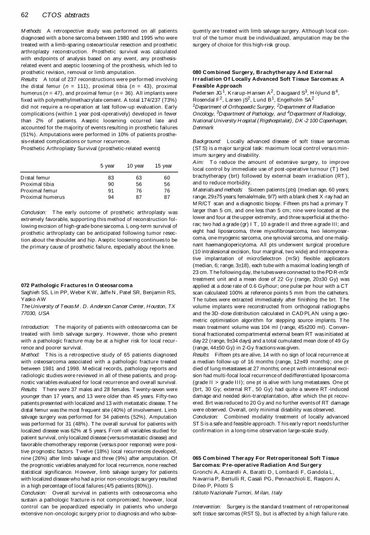
62 CTOS abstracts
Methods: A retrospective study was performed on all patients
diagnosed with a bone sarcoma between 1980 and 1995 who were
treated with a limb-sparing osteoarticular resection and prosthetic
arthroplasty reconstruction. Prosthetic survival was calculated
with endpoints of analysis based on any event, any prosthesis-
related event and aseptic loosening of the prosthesis, which led to
prosthetic revision, removal or limb amputation.
Results: A total of 237 reconstructions were performed involving
the distal femur (n = 111), proximal tibia (n = 43), proximal
humerus (n = 47), and proximal femur (n = 36). All implants were
fixed with polymethylmethacrylate cement. A total 174/237 (73%)
did not require a re-operation at last follow-up evaluation. Early
complications (within 1 year post-operatively) developed in fewer
than 2% of patients. Aseptic loosening occurred late and
accounted for the majority of events resulting in prosthetic failures
(51%). Amputations were performed in 10% of patients prosthe-
sis-related complications or tumor recurrence.
Prosthetic Arthroplasty Survival (prosthetic-related events)
Conclusion: The early outcome of prosthetic arthroplasty was
extremely favorable, supporting this method of reconstruction fol-
lowing excision of high-grade bone sarcoma. Long-term survival of
prosthetic arthroplasty can be anticipated following tumor resec-
tion about the shoulder and hip. Aseptic loosening continues to be
the primary cause of prosthetic failure, especially about the knee.
072 Pathologic Fractures In Osteosarcoma
Saghieh SS, Lin PP, Weber KW, Jaffe N, Patel SR, Benjamin RS,
Yasko AW
The University of Texas M. D. Anderson Cancer Center, Houston, TX
77030, USA
Introduction: The majority of patients with osteosarcoma can be
treated with limb salvage surgery. However, those who present
with a pathologic fracture may be at a higher risk for local recur-
rence and poorer survival.
Method: This is a retrospective study of 65 patients diagnosed
with osteosarcoma associated with a pathologic fracture treated
between 1981 and 1998. Medical records, pathology reports and
radiologic studies were reviewed in all of these patients, and prog-
nostic variables evaluated for local recurrence and overall survival.
Results: There were 37 males and 28 females. Twenty-seven were
younger than 17 years, and 13 were older than 45 years. Fifty-two
patients presented with localized and 13 with metastatic disease. The
distal femur was the most frequent site (40%) of involvement. Limb
salvage surgery was performed for 34 patients (52%). Amputation
was performed for 31 (48%). The overall survival for patients with
localized disease was 62% at 5 years. From all variables studied for
patient survival, only localized disease (versus metastatic disease) and
favorable chemotherapy response (versus poor response) were posi-
tive prognostic factors. Twelve (18%) local recurrences developed,
nine (26%) after limb salvage and three (9%) after amputation. Of
the prognostic variables analyzed for local recurrence, none reached
statistical significance. However, limb salvage surgery for patients
with localized disease who had a prior non-oncologic surgery resulted
in a high percentage of local failures (4/5 patients (80%)).
Conclusion: Overall survival in patients with osteosarcoma who
sustain a pathologic fracture is not compromised; however, local
control can be jeopardized especially in patients who undergo
extensive non-oncologic surgery prior to diagnosis and who subse-
quently are treated with limb salvage surgery. Although local con-
trol of the tumor must be individualized, amputation may be the
surgery of choice for this high-risk group.
080 Combined Surgery, Brachytherapy And External
Irradiation Of Locally Advanced Soft Tissue Sarcomas: A
Feasible Approach
Pedersen JG1, Krarup-Hansen A2, Daugaard S3, Höjlund B4,
Rosendal F2, Larsen jS2, Lund B1, Engelholm SA2
1Department of Orthopaedic Surgery, 2Department of Radiation
Oncology, 3Department of Pathology, and 4Department of Radiology,
National University Hospital (Rigshospitalet), DK-2 100 Copenhagen,
Denmark
Background: Locally advanced disease of soft tissue sarcomas
(STS) is a major surgical task: maximum local control versus min-
imum surgery and disability.
Aim: To reduce the amount of extensive surgery, to improve
local control by immediate use of post-operative turnour (T) bed
brachytherapy (brt) followed by external beam irradiation (RT),and to reduce morbidity.
Materials and methods: Sixteen patients (pts) (median age, 60 years;
range, 29± 75 years; female/male, 9/7) with a blank chest X-ray had an
MR/CT scan and a diagnostic biopsy. Fifteen pts had a primary T
larger than 5 cm, and one less than 5 cm; nine were located at the
lower and four at the upper extremity, and three superficial at the tho-
rax; two had a grade (gr) I T, 10 a grade II and three a grade III; and
eight had liposarcoma, three myxofibrosarcoma, two leiomyosar-coma, one myogenic sarcoma, one synovial sarcoma, and one malig-
nant haemangiopericytoma. All pts underwent surgical procedure
(10 intralesional excision, four marginal, two wide) and intraoperera-
tive implantation of microSelectron (mSr) flexible applicators
(median, 6; range, 3± 18), each tube with a maximal loading length of
23 cm. The following day, the tubes were connected to the PDR-mSr
treatment unit and a mean dose of 22 Gy (range, 20± 30 Gy) was
applied at a dose rate of 0.6 Gy/hour; one pulse per hour with a CTscan calculated 100% at reference points 5 mm from the catheters.
The tubes were extracted immediately after finishing the brt. The
volume implants were reconstructed from orthogonal radiographs
and the 3D-dose distribution calculated in CADPLAN using a geo-
metric optimisation algorithm for stepping source implants. The
mean treatment volume was 104 ml (range, 45± 200 ml). Conven-
tional fractionated compartmental external beam RT was initiated atday 22 (range, 9± 34 days) and a total cumulated mean dose of 49 Gy
(range, 44± 50 Gy) in 2-Gy fractions was given.
Results: Fifteen pts are alive, 14 with no sign of local recurrence at
a median follow-up of 16 months (range, 12± 49 months); one pt
died of lung metastases at 27 months; one pt with intralesional exci-
sion had multi-focal local recurrence of dedifferentiated liposarcoma
(grade II > grade III); one pt is alive with lung metastases. One pt
(brt, 30 Gy; external RT, 50 Gy) had quite a severe RT-induceddamage and needed skin-transplantation, after which the pt recov-
ered. Brt was reduced to 20 Gy and no further events of RT damage
were observed. Overall, only minimal disability was observed.
Conclusion: Combined modality treatment of locally advanced
STS is a safe and feasible approach. This early report needs further
confirmation in a long-time observation large-scale study.
065 Combined Therapy For Retroperitoneal Soft Tissue
Sarcomas: Pre-operative Radiation And Surgery
Gronchi A, Azzarelli A, Baratti D, Lombardi F, Gandola L, Navarria P, Bertulli R, Casali PG, Pennacchioli E, Rasponi A,
Dileo P, Pilotti S
Istituto Nazionale Turnori, Milan, Italy
Intervention: Surgery is the standard treatment of retroperitoneal
soft tissue sarcomas (RSTS), but is affected by a high failure rate.
5 year 10 year 15 year
Distal femur 83 63 60Proximal tibia 90 56 56Proximal femur 91 76 76Proximal humerus 94 87 87
CTOS abstracts 63
Adjuvant radiation may improve local control and possibly sur-
vival. We report the results of a phase I ± II study on radiationtherapy (RT) delivered in a pre-operative setting, to determine the
treatment feasibility and its possible benefit on surgical quality.
Patients and methods: From September 1996 to December 1999, 41patients affected by RSTS were referred to our institution.
Twenty-one of them, affected by primary or recurrent disease,
were eligible for the combined therapy. As regards to the histology,14 were liposarcoma, five were leiomiosarcoma, one rhabdomiosa-
rcoma and one abdominal gastro-intestinal stromal tumor. Seven-
teen were low grade and four high grade. A CT-basedcomputerized treatment plan was obtained in all patients to deter-
mine treatment volume. Personalized shields were realized to con-
firm dose distribution. The scheduled dose was 50 Gy. Surgerywas then planned between the 30th and the 60th day after the end
of RT.
Results: One patient received only 32 Gy for gastro-intestinalintolerance. All the other patients completed the treatment without
significant toxicity. One patient progressed soon after RT and was
not operated. All the other 20 patients underwent surgery 32± 46days after the end of RT. A complete removal of the tumor was
possible in 15 cases: resection was extended to viscera with 10
nephrectomies, seven colectomies, two distal pancreatectomies,one splenectomy, one gastric resection and one resection of the
inferior vena cava. No major surgical difficulties were encountered.
In the remaining five cases not completely resected, a sarcomatosiswas found at laparotomy. One patient died 3 months later due to
related abdominal complications. One patient died 12 months
later due to local abdominal recurrence, and two died 22 and 32months later, respectively, due to distant metastasis. The remain-
ing 11 patients are all alive with a median follow-up of 25 months
(range, 6± 40 months), eight of whom free of disease.Conclusions: Pre-operative radiation is feasible and safe. A control-
led study with strict inclusion criteria is now recommended to
investigate the possible benefit of the combined treatment on sur-vival.
066 Chordoma: Natural History And Treatment Results In
55 Patients
Gronchi A, Azzarelli A, Baratti D, Lozza L, Bertulli R, Casali PG,
Pennacchioli E, Rasponi A, Dileo P, Pilotti SIstituto Nazionale Tumori, Milan, Italy
Intervention: Chordoma is a rare neoplasm with poor prognosis asalready observed by several authors. Our experience reports one of
the largest series in the literature and confirms the need of new
adjuvant treatments.Patients: We observed at our institution 55 patients affected by
chordoma. The neoplasm was located in the sacrum in 49 cases, at
the base of the skull in three and in the vertebral bodies in three.The median age at the time of the diagnosis was 58 years (range,
2± 77 years); 34 patients were male and 21 female; median diame-
ter was 13 cm (range, 2± 30 cm). During the 1970s, advances inpre-operative staging, more reliable surgical techniques and a clear
definition of oncological adequacy were the basis of a new rationale
treatment of sacral chordoma. Since 1977, patients underwent sur-gery with intent of radicality. The technique of operation per-
formed in 29 patients was high amputation of the sacrum by
posterior approach, with the aim to perform an uncontaminatedremoval of the entire lesion and surrounding normal tissue. Radi-
otherapy was considered in case of marginal or intralesional oper-
ations.Results: Surgical margins were rated as wide in nine patients, mar-
ginal in 17 and macroscopically intralesional in three. Post-operative
radiation therapy was performed in nine patients with marginal/int-ralesional surgery; one patient underwent pre-operative RT. Sacral
chordoma recurred in 17 out of these 29 recent operated patients.
Local failure, mainly located in the ischiorectal area, developed in 13
cases, distant isolated metastases (lung) in one patient, and both
local and distant failure in three, with involvement of lung in two
cases and distant subcutaneous dissemination in one. As regard to
resection margins definition, local ± distant failure developed in sixout of nine (66.7%) patients with wide margins and in 11 out of 17
(64.7%) patients with marginal operation.
Conclusions: Although chordoma is considered a low-grade
lesion, surgical treatment to obtain effective local control is diffi-
cult, due to late diagnosis and anatomical site. In the present study,
wide surgical margins were obtained only in 31% of the operated
patients and recurrence rate was high. Distant metastases, isolated
or concurrent to local failure, were detected only in four out of 29patients (13.7%). Therefore, strong effort in achieving complete
surgical excision is mandatory, as well as in developing more effec-
tive adjuvant therapies.
087 Periosteal Chondrosarcoma: Experience Of The Istituto
Ortopedico Rizzoli.
Fabbri N, De Paolis M, Vanel D, Mercuri M, Picci P
Department of Musculoskeletal Oncology, Istituto Ortopedico Rizzoli ±
Bologna, Italy
Periosteal chondrosarcoma is a rare primary malignant tumor of
the bone surface. Due to its rarity, the differential diagnosis with
other primary tumor of the bone surface can be difficult and the
potential for metastasis remains unclear. Studies reporting series ofpatients homogeneously managed at the same institution are scant
in the literature. From 24,855 bone tumors filed at our institution,
26 cases of periosteal chondrosarcoma were identified; two cases
were excluded for incomplete imaging studies. A retrospective
study of the remaining 24 cases was undertaken. Clinico-patho-
logic features were reviewed and analyzed along with follow-up in
order to define diagnostic criteria and understand prognosis of this
condition.
There were 18 males and six females, and average age was 33 years(range, 17± 65 years). A palpable mass and dull pain averaging
more than 18 months of duration were the most common symp-
toms at presentation.
Tumor location was the femur in 14 cases (12 distal, two proxi-
mal), humerus in seven cases (five proximal, two shaft), tibia in
two cases (two proximal) and iliac wing in one case. Imaging stud-
ies showed a frequently lobulated juxtacortical mass, usually sug-
gestive of cartilaginous matrix on both plain X-rays and CT orMRI scan. Although abnormalities of the cortex were present in all
the cases, medullary involvement could be detected and histologi-
cally confirmed only in two cases.
Information regarding the surgical margin and a minimum follow-
up longer than 3 years were available in 18 patients (average, 11
years; range 3± 30 years). Surgical margin was adequate (wide) in
10 cases and inadequate (wide/contaminated or marginal) in the
remaining eight cases. All the patients were disease free; three local
recurrences were managed with further surgery. None of thepatients developed metastasis. There were no local recurrences
when the surgical margin was adequate. In our experience, prog-
nosis is excellent after adequate surgical management and risk of
metastasis is very low.
088 Desmoplastic Fibroma: Experience Of The Istituto
Ortopedico Rizzoli
Fabbri N, Trentani F, Vanel D, Mercuri M, Picci P, Bertoni F
Department of Musculoskeletal Oncology, Istituto Ortopedico Rizzoli ±
Bologna, Italy
Desmoplastic fibroma is a rare benign tumor of bone. Despite its
benign nature, a marked tendency for local recurrence has been
64 CTOS abstracts
reported in the past. However, studies reporting series of patients
homogeneously managed at the same institution are scant in therecent literature.
From 24,855 bone tumors filed at our institution, 16 cases of
desmoplastic fibroma were identified. A retrospective study of thisgroup of patients was undertaken; clinico-pathologic features were
reviewed and analyzed along with follow-up in order to define bio-
logic aggressiveness and potential for local recurrence of this disor-der.
There were nine males and seven females, and average age was 22
years (range, 9± 36 years). Swelling and pain usually lasting morethan 6 months were the most common presenting symptoms.
Tumor location was the femur in five cases (three proximal, two
distal), tibia in three cases (two proximal, one distal), humerus intwo cases (one proximal, one shaft), fibula in two cases (one prox-
imal, one distal), and calcaneus, periacetabular region, sacrum and
L2 body in one case each.Imaging studies constantly showed a purely lytic defect, sharply
marginated towards the bone and contained by paper thin cortex
towards the surrounding soft tissues. Staging of the tumor accord-ing to MSTS was stage 2 in 12 cases, stage 3 in three cases, and
stage 1 in one case.
Information regarding the surgical margin and a minimum follow-up longer than 3 years were available in 14 patients (average, 5
years; range, 3± 22 years). Surgical management consisted of wide
resection in nine cases and intralesional curettage in five cases; inthree cases, the use of phenol or liquid nitrogen was added to the
curettage as a local adjuvant.
All the patients were disease free. None of the patients developedmetastasis. In our experience, prognosis is excellent after adequate
surgical management, either wide resection or aggressive curet-
tage.
089 Hemangiopericytoma Of Bone: The Rizzoli Experience
Campanacci L, Fabbri N, Vanel D, Mercuri M
Department of Musculo Skeletal Oncology, Istituto Ortopedico Rizzoli,
Bologna, Italy
Hemangiopericytoma is a rare malignant vascular tumor, originat-ing from the perivascular cells. Usually arising in the soft tissues, it
is extremely rare in the skeleton. We coud not find large series
reports in the literature.Treatment is surgery, sometimes associated with radiation and/or
chemotherapy. At the Rizzoli Institute, since September 1900 to
date, 24,855 cases of bone and soft tissue tumors and pseudotu-moral lesions are filed. Out of 6434 cases of malignant bone tumors
(excluding metastases), only 13 cases of hemangiopericytoma pri-
mary of the bone were observed (two of which were consultations).There were four males and nine females, aged 8± 79 years (average,
41 years). Pelvic bones and/or lumbo-sacral spine were involved in
six cases, a rib in one, upper and lower limbs in three cases each(femur in two, fibula in one, and humerus in three). In two cases,
the lesion involved two adjacent bones.
Symptoms were always present since a long period of time (from 6 to60 months; average, 25.5 months). Radiologically, all cases presented
as purely ostelitic, containing trabecular septae, with expanded corti-
cal bone; in 10 cases, a soft tissue mass was also present. In two cases,small osteolitic images distal to the primary lesion were observed; two
cases presented with a pathological fracture.
Among the 11 cases treated at our institution, five were managedwith surgery alone, one with surgery and radiation-therapy; three
patients were treated by chemotherapy and radiation therapy (sur-
gery was not possible at presentation). One case (8-year-old girl)was cured with chemotherapy alone.
Six patients died of the disease (all developed lung metastases, one
a local recurrence) after 5 months to 6 years; five are continuousdisease free at 4± 21 years of follow-up.
Hemangiopericytoma still represent a difficult issue for diagnosis
and treatment, its course can be rapidly aggressive, and lung
metastases can appear even after years; the role of chemotherapy is
still uncertain in improving the outcome. Delaied diagnosis has an
ominous prognosis.
090 Soft-tissue Sarcoma (Sts) Ð Adherence To Guidelines
Nijhuis PHA1, Schaapveld M2, Otter R2, Hoekstra HJ1
1Department of Surgical Oncology, Groningen University Hospital,
Groningen, The Netherlands, and 2Comprehensive Cancer Center
North-Netherlands, Groningen, The Netherlands
Introduction: Because many general surgeons are unfamiliar withthese rare tumors, a reasonable number is found by chance, often
after inadequate resection, leading to complex definitive treat-
ment. Therefore, guidelines for diagnosis and treatment were
developed. As the diagnostic management is essential for definitive
treatment, adherence to these guidelines is important.
Methods: Primary STS, registered by the Comprehensive Cancer
Center North-Netherlands from January 1989± January 1996, wereanalyzed retrospectively with regard to adherence to the diagnostic
guidelines. Urogenital STS, gastro-intestinal STS, and Kaposi’ s
sarcomas were excluded.
Results: Three hundred and fifty-one STS were analyzed. In the spe-
cialized center, 69% of patients were younger than 60 years, whereas
in district hospitals 63% were older than 60 years. With increase of
age, referral to the center declined in a linear fashion. For all guide-lines, adherence was significantly better in the center (Table 1).
Conclusions: In many aspects of the diagnostic process of STS,
existing guidelines were not followed, especially in community
hospitals. Adherence to all individual guidelines was significantly
better in the specialized center. In order to improve compliance
with future STS guidelines, appropriate guideline development,
dissemination, and implementation programs should be devel-oped. Concentration of STS in a limited number of hospitals and
intensified collaboration with specialized centers seem advisable.
Special attention should be paid to older patients, which were sig-
nificantly more often not referred to a specialized center.
091 Pre-operative Radiation Therapy In The Treatment Of
Soft Tissue Sarcomas
Virkus, Mollabashy A, Reith JD, Berrey HB, Zlotecki RA,
Scarborough MT
University of Florida, Gainesville, FL 32610, USA
This report presents the results of a cohort of patients with a soft
tissue sarcoma treated with a standard protocol including pre-
operative radiation therapy (PRT) and surgical resection, assessing
wound complications, local recurrence, and oncologic outcome.
A standard protocol of PRT followed by en bloc resection was per-formed in 218 patients with a primary soft tissue sarcoma from
1984 to 1999. Wound complications were defined as minor (local
wound care), moderate (operative I&D), major (STSG or flap),
and amputation. Wound complications are reported for the entire
patient population. Oncologic results are reported on a subset of
patients that excluded patients who were stage III at the time ofresection, or received chemotherapy. There were 114 males, 104
females, with an average age of 56 years (range, 8± 86 years).
Thirty-six patients received chemotherapy. At the time of resec-
tion, 13 patients were stage IA, 16 were stage IB, 63 were stage
IIA, 111 were stage IIB, and 15 were stage III. Ten patients had an
intralesional surgical margin, 91 marginal, 115 wide, and two rad-
ical. Primary wound closure was obtained in 187 patients, nineneeded a split-thickness skin graft, 20 a rotational flap, and two a
free flap for closure. Only six patients underwent primary amputa-
tion. Mean oncologic follow-up was 53 months.
The overall wound complication rate was 32.6% (minor, 10%;
moderate, 13%; major, 7%; amputation, 1%). If minor wound
problems treatable with short-term local wound care are excluded,
CTOS abstracts 65
the wound complication rate was 22.9%. When stage III and chem-
otherapy patients were removed, 172 patients remained for onco-
logic outcome analysis. Seventy-three (42%) developed late
metastases, predominantly to the lungs (82%). Eighteen patients
developed a local recurrence, which was 15.1% of those patients
with a minimum of 2 years of follow-up. The recurrence rate in
patients with prior surgery at an outside institution was 24%, versus
12% if initial surgery was at our institution. At latest follow-up, 80
patients (46%) were continuously disease free, six (3%) ANED,
eight (4%) alive with disease, and 65 (38%) dead of disease. The 2
and 5-year survival rates were 77 and 66%, respectively.
The ideal timing of adjuvant radiation therapy remains to be deter-
mined. PRT allows for a smaller radiation dose and field to be
delivered, theoretically decreasing the morbidity of radiation. Our
local control rate is similar to that seen in other studies where a
high percentage of adequate surgical margins is obtained. Twenty-
two percent of our patients required a return to the operating room
for wound complications. This is similar to the wound complica-
tion rates in studies where post-operative XRT or no XRT is used.
Distant control continues to be a difficult problem in the treatment
in soft tissue sarcomas.
095 Outcome Following Local Recurrence In Osteosarcoma
Grimer RJ
The Royal Orthopaedic Hospital Oncology Service, Bristol Road South,
Birmingham, UK
Purpose: Local recurrence of osteosarcoma is generally thought to
be a death sentence, with few patients surviving. This paper
addresses this question and attempts to identify possible prognos-
tic factors and treatment options for patients with local recurrence.
Method: All patients who had developed a local recurrence fol-
lowing treatment of a non-metastatic high-grade osteosarcoma
were included in this review. Patients with metastases at the time
of diagnosis were excluded. Eighty-one patients were found to ful-
fill the above criteria. LR arose at an average of 16 months (range,
3± 49 months). Sixteen of the patients were already known to have
lung metastases and a further 27 were found to have metastatses
within 3 months of treatment of the LR. Another 25 developed
metastases later, leaving only eight patients who did NOT subse-
quently develop metastases.
Results: Overall survival was most clearly related to whether or
not metastases were present at the time of presentation with LR. In
patients with known metastases or ones that appeared within 3
months, there were no survivors past 30 months. For those with no
metastases within 3 months, the survival was 40% at 5 years from
the time of LR. Even in those 25 who developed metastases later,
the survival was 30% at 3 years and 23% at 5 years. Disease free
interval was not significant.
Conclusion: LR accompanied by metastases is currently a death
sentence. Experimental treatments for these patients may be justi-
fied. In patients with solitary LR, aggressive treatment is justified
and has a similar outlook to that for lung metastases alone.
107 Combined Surgery, Brachytherapy And External
Irradiation Of Locally Advanced Soft Tissue Sarcomas: A
Feasible Approach
Pedersen JG1, Krarup-Hansen A2, Daugaard S3, Hojlund B4,
Rosendal F1, Larsen JS2, Lund B1, Engelholm SA2
Departments of 1Orthopaedic Surgery, 2Radiation Oncology, 3Pathology, and 4Radiology, National University Hospital
(Rigshospitalet), DK-2100 Copenhagen, Denmark
Background: Locally advanced disease of soft tissue sarcomas
(STS) is a major surgical task: maximum local control versus min-
imum surgery and disability.
Aim: To reduce the amount of extensive surgery, to improve
local control by immediate use of post-operative turnour (T) bed
brachytherapy (brt) followed by external beam irradiation (RT),
and to reduce morbidity.
Materials and methods: Sixteen patients (pts) (median age, 60
years; range, 29± 75 years; female/male, 9/7) with a blank chest
X-ray had an MR/CT scan and a diagnostic biopsy. Fifteen pts had
a primary T larger than 5 cm, and one less than 5 cm; nine were
located at the lower and four at the upper extremity, and three
superficial at the thorax; two had a grade (gr) I T, 10 a grade II and
three a grade III; and eight had liposarcoma, three myxofibrosar-
coma, two leiomyosarcoma, one myogenic sarcoma, one synovial
sarcoma, and one malignant haemangiopericytoma. All pts under-
went surgical procedure (10 intralesional excision, four marginal,
two wide) and intraopererative implantation of microSelectron
(mSr) flexible applicators (median, 6; range, 3± 18), each tube with
a maximal loading length of 23 cm. The following day, the tubes
were connected to the PDR-mSr treatment unit and a mean dose
of 22 Gy (range, 20± 30 Gy) was applied at a dose rate of 0.6 Gy/
hour; one pulse per hour with a CT scan calculated 100% at refer-
ence points 5 mm from the catheters. The tubes were extracted
immediately after finishing the brt. The volume implants were
reconstructed from orthogonal radiographs and the 3D-dose dis-
tribution calculated in CADPLAN using a geometric optimisation
algorithm for stepping source implants. The mean treatment
volume was 104 ml (range, 45± 200 ml). Conventional fractionated
compartmental external beam RT was initiated at day 22 (range,
9± 34 days) and a total cumulated mean dose of 49 Gy (range, 44±
50 Gy) in 2-Gy fractions was given.
Results: Fifteen pts are alive, 14 with no sign of local recurrence
at a median follow-up of 16 months (range, 12± 49 months); one pt
died of lung metastases at 27 months; one pt with intralesional
excision had multi-focal local recurrence of dedifferentiated liposa-
rcoma (grade II > grade III); one pt is alive with lung metastases.
One pt (brt, 30 Gy; external RT, 50 Gy) had quite a severe
RT-induced damage and needed skin-transplantation, after which
the pt recovered. Brt was reduced to 20 Gy and no further events
of RT damage were observed. Overall, only minimal disability was
observed.
Conclusion: Combined modality treatment of locally advanced
STS is a safe and feasible approach. This early report needs further
confirmation in a long-time observation large-scale study.
109 Unplanned Excision For Soft Tissue Sarcoma:
Identification Of A Subgroup With Bad Prognosis
van Geel AN, Eggermont AMM, Schmitz PIM
Department of Surgical Oncology and Statistics, University Hospital
Rotterdam/Daniel den Hoed Cancer Center, Rotterdam, The
Netherlands
Introduction: Unplanned excision (UE) for soft tissue sarcoma
(STS) is a frequent procedure. Unclear is the influence on progno-
sis.
Material and methods: From 1986 to 1993, 86 patients (32.7% of
all referred patients) were treated after UE of STS elsewhere (sub-
fascial; limbs, girdles and trunk, > 5 CMS). All patients had CT or
MRI before definitive treatment; minimal follow-up was 5 years.
Results: Thirty-one patients (36.1%) had complications
(hematoma) after UE. In 46 patients (53.5%), residual tumor was
seen on CT/MRI and found at pathological examination in 66
patients (76.7%). In one-half of the patients, definitive treatment
was considered to be more complex, resulting in increased morbid-
ity.
Conclusion: After multivariate analysis, a group of patients (35
patients = 40%) with a significant bad prognosis was found: older
than 60 years with a second treatment with increased complexity
and morbidity independent of this treatment is radical or not. This
66 CTOS abstracts
group can be considered as a high-risk group eligible for adjuvantchemo therapy trials.
Posters Ð Radiation Oncology
029 Potential Impact Of High Technology Radiation
Treatment For Sarcomas To Improve Patient Outcome
Herman Suit, Karen Doppke, Jong Kung, J. Michael Collier, Ira
Spiro, Thomas Delaney
Department of Radiation Oncology, Massachusetts General Hospital,
Harvard Medical School, Boston, MA, USA
The available radiation treatment techniques employed in the
management of the mesenchymal tumors cause substantial quan-
tities of non-involved tissues/structures to receive important doselevels. The consequence is that there is a non-negligible frequency
of treatment associated morbidity. This is in the form of: excess
fibrosis, wound healing delay or breakdown, late appearingdamage in major nerves and vessels, pathological fracture and the
rare radiation induced malignant neoplasm. There are several new
techniques, which will achieve full coverage of the defined targettissue for each treatment session, but with major reductions in the
volume of normal tissues/structures included in the high dose vol-
ume. As complications of treatment cannot develop in unirradi-ated tissues, there is predicted a major lowering of the frequency
and severity of treatment related morbidity. These new techniques
are principally intensity modulated X-ray therapy, intensity modu-lated proton beam therapy, on-line diagnostic quality imaging,
Monte Carlo based dose calculations, etc. The presentation will
assess the impact of these developments on three relativelycommon clinical problems. These are lesions located in the: (1)
medial proximal thigh, (2) thoracic vertebral body, and (3) retro-
peritoneal region.
031 Effects Of Irradiation And Radio-protectant On Rat
Growth Plate Morphology
Damron TA, Spadaro JA, Farnum CE, Margulies BS, Strauss J
Upstate Medical University Department of Orthopedic Surgery,
Syracuse, NY 13224, USA
Introduction: Previous work in our laboratory has shown a benefi-
cial effect on rat limb growth from administration of the radiopro-tectant amifostine. The histological correlates of these quantitative
measurements of limb length have not previously been described.
Purpose: The purpose of this paper is to detail the histological
changes over time in the rat growth plate (GP) following irradia-
tion with and without the radioprotectant amifostine.
Methods: Histological specimens were examined from 84Sprague± Dawley male rats, 4 weeks old, previously included in
reports detailing limb length effects. Groups of n = 6 included for
analysis were `cage controls’ at 6 weeks, irradiation only; (singledose, 12.5 and 17.5 Gy) at 6 weeks, ̀ radioprotectant pretreatment’
(100 and 300 mg/kg amifostine for the 12.5 Gy groups, and 100
mg/kg for the 17.5 Gy groups) at 6 weeks, and `early timing’ (0.5,1, 2, 3, and 4 weeks post 17.5 Gy with and without 100 mg/kg ami-
fostine). Irradiation was administered to the right distal femur and
proximal tibia; the left leg served as the control.
Results: Morphology of the irradiated GP changed consistently
over time. At 0.5 weeks, a subtle decrease in cellular profiles with
maintenance of overall zonal architecture was evident. At 1 week,
cellular profiles were notably reduced, and zonal architecture was
nearly lost. At 2 weeks, the GP was composed of sparsely situated
terminal, hypertrophic-appearing chondrocytes. At 3 weeks, cellu-
lar profiles increased via clonal bodies. At 4 and 6 weeks, clonal
bodies predominated.
The growth plate cellular profile area following irradiation hit a
nadir at 2 weeks and rebounded at the third week toward normal.
TRAP staining showed an increase in osteoclasts between the 2
and 3 week time periods that corresponded to an relative decrease
in GP height over the same interval.
Epifluorescent growth rates reached a nadir 1 week following irra-
diation and slowly returned toward normal thereafter.
Growth plate overall height was greater for controls than irradiated
specimens at all time periods, but these effects were much greater cen-
trally than peripherally. Amifostine effects, however, were observed
more consistently at the periphery rather than in the central GP.
Discussion: Irradiation results in a predictable pattern of histo-
logic changes, with the greatest effects observed at 1 week in the rat
proximal tibia. Prereatment with amifostine is unable to preserve
normal GP morphology despite its maintenance of longitudinal
growth, but many parameters are statistically improved compared
with the irradiated specimens.
032 Bone Density Effects Of Radiation And The
Radioprotectant Drug Amifostine In A Rat Model
Morgan H, Damron TA, Margulies B, Spadaro J
Upstate Medical University Department of Orthopaedic Surgery,
Syracuse, NY 13224, USA
Purpose: The purpose of this project was to examine the effects on
the bone density measurements of irradiated rat femurs with and
without the radioprotective drug amifostine (AMF).
Methods: Seventy-two weanling Spague± Dawley rats were rand-
omized into treatment groups. In all treated animals, the right knee
(distal femur and proximal tibia) was irradiated with single-frac-
tion 17.5 Gy, while the left leg was used as an internal control. Ani-
mals were sacrificed at 0.5, 1, 2, 3, 4, and 6 weeks following
irradiation, 12 per time period. One-half of the animals in each of
the groups received 100 mg/kg amifostine 20 minutes prior to irra-
diation. For the bone density analysis, the skeletonized femurs
were placed into a cylindrical holder and suspended in deionized
water. Bone density (g/cm) was determined using peripheral quan-
titative computed tomography (pQCT; Norland XCT 2000, Fort
Atkinson, WI), resolution 90 microns.
Results: The irradiated slices maintained a significantly higher
BMD than controls through 6 weeks in the most proximal meta-
physis and through 3 weeks in the distal metaphysis (Fig. 1). The
slice closest to the metaphysical side of the physis demonstrated a
unique early peak BMD at 2 weeks, which decreased dramatically
by 3 weeks. The BMD for rats that received amifostine was signif-
icantly lower through 3 weeks than for rats that received radiation
alone (p < 0.02 for 0.5, 2, and 3 weeks).
Discussion: Three conclusions are drawn from the current data.
First, at early time periods following irradiation, BMD within the
irradiation field is greater than control in this animal model Sec-
ond, a 2-week early peak in BMD occurs in the juxta-physeal met-
aphysis. Third, amifostine has a significant effect in holding BMD
close to normal in the juxta-physeal region. We hypothesize that
the sensitive marrow elements, particularly in this case the mono-
cyte precursor of osteoclasts, are more sensitive to irradiation
damage than the osteoblasts, given the slower turnover of the latter
cells. In addition, we hypothesize that the early 2-week peak in
BMD occurs only in the distal metaphysis due to the greater rela-
tive contribution of provisional calcification to BMD there, since
this calcification is a passive process that is likely to continue
despite radiation effects, particularly when unchecked by resorp-
Survival Overall Loc. rec. free Dist. rec. free
2 years 86.0 85.8 84.6 5 years 74.3 72.1 70.6
CTOS abstracts 67
tive activity of chondroclasts and osteoclasts. These hypotheses are
supported by histological observations.
033 Pattern Of Local Recurrence After Conservative
Surgery And Radiotherapy For Soft Tissue Sarcoma.
Cleator SJ, Cottrill C, Harmer CL
Sarcoma Unit, Royal Marsden Hospital NHS Trust, London SW3
6JJ, UK
Purpose: Over the past two decades, our centre has adopted a
policy of conservative surgery followed by adjuvant radical-dose
radiotherapy for all medium- and high-grade soft tissue sarcomas.From the patients treated in this manner at the Royal Marsden Hos-
pital over the past 15 years, we analyzed 17 who had relapsed locally.
We discuss technical factors that may have contributed to localrelapse in some cases, and the geographical relationship between
sites of recurrence and the phase 1 and 2 radiotherapy volumes.
Patients and methods: We examined those cases recorded on our
soft tissue sarcoma database between January 1986 and September
1999 who had recurred locally following surgery and radical post-operative radiotherapy. We excluded patients with residual macro-
scopic disease following surgery and any patients with metastatic dis-
ease at presentation. We recorded details relating to original tumourstage, histological type, grade and surgical margins. The cohort
included eight patients with T1 (47%) and nine patients with T2
tumours (53%). The tumours were grade 1, 2 and 3 in two (12%),
six (35 %) and nine (53%) number of cases, respectively. The major-ity of patients were treated with a phase I volume corresponding to
the entire muscle compartment that received 50 Gy in 25 fractions
over 5 weeks. The phase II was a reduced volume corresponding to
the tumour bed and received 10 Gy in five fractions during the sixthweek of treatment. Four of the patients were treated according to a
hyperfractionated regimen consisting of 72 Gy in 60 fractions twice
daily over 6 weeks with volume reduction after 60 Gy.
Results: Mean time to local recurrence was 26 months. Four (23%)patients recurred within the phase I volume, nine (53%) recurred
within the phase II volume, and three (18%) outside the irradiated
volume. One recurrence was marginal. Eight patients who relapsed
had positive surgical excision margins originally. In six patients,there had been deviations from our radiotherapy protocol, usually
unavoidable, which may have accounted for treatment failure; these
included all three out of field recurrences. Of the 11 patients for
whom it was possible to adhere strictly to protocol, two recurred inthe phase I volume and nine in the phase II volume. With a mean
follow-up time since completion of radiotherapy of 63 months,
seven patients have died and, of these, six were disease related.
Conclusion: A dose greater than 60Gy may be necessary despiteinitial complete surgical resection. Prospective, multi-centre data
collection and ideally a randomized trial are required to formulate
an improved treatment policy.
036 Treatment Outcome In Patients Radiated For Spinal
And Paraspinal Tumors
DeLaney TF, Ancukiewicz M, Hornicek FJ, Spiro IJ, Harmon
DC, Suit HD
Massachusetts General Hospital, Boston, MA 02114, USA
Objective: Tumors of the spine and paraspinal soft tissues can
present difficult management challenges because of the proximity
of the spinal cord, which limits resection margins and radiationdosage. A retrospective review of treatment outcome in a cohort of
these patients managed in a single institution was undertaken to
assess prognostic factors and treatment techniques important for
favorable treatment outcome.
Materials and methods: Fifty-seven patients with spinal and par-
aspinal tumors treated with radiotherapy between 1971 and 1998were identified in our sarcoma database. Nine patients with
Ewing’ s sarcoma/PNET were identified for separate analysis, leav-
ing 48 patients for analysis for this study. Outcome was assessedaccording to the following tumor and treatment variables: site (spi-
nal/paraspinal), tumor grade, surgical margin status, and radiation
dose.Results: Twenty-four lesions were in the spine and 24 were in par-
aspinal soft tissue. Forty-two patients had malignant tumors with
the following histologies: neurofibrosarcoma/malignant schwan-noma (9), malignant fibrous histiocytoma (7), chondrosarcoma
(5), chordoma (5), osteosarcoma (4), fibrosarcoma (3), and others
(9). Six patients had benign lesions: desmoid (2), giant cell tumor(2), aneurysmal bone cyst (1), and hemangioma (1). Median
tumor size was 8.5 cm (range, 1.2± 18 cm). Local treatment con-
sisted of combined surgery and radiotherapy in 45 patients andradiotherapy alone in three patients. Median radiation dose was
60.2 Gy (range, 22.4± 78.6 Gy). Five patients received brachyther-
apy as a component of treatment, with a median implant dose of28.7 Gy, while two patients received intra-operative radiotherapy.
Chemotherapy was administered to 15 patients. Overall 5-year
survival was dependent upon tumor grade: 0± 1, 89%; 2, 48%; 3±4, 36% (p = 0.10). Overall 5-year survival was similar for spinal
(72%) and paraspinal (63%) lesions (p = 0.96). Local control was
85% for spinal lesions and 66% for paraspinal lesions (p = 0.59).There was a trend towards improved local control in patients with
negative surgical margins (81%) compared with patients in whom
the margins were positive (55%) (p = 0.30). No clear relationshipcould be demonstrated between radiation dose and treatment out-
come.
Conclusions: Similar to other sites, tumor grade is of prognosticimportance in patients with spinal and paraspinal tumors. In spite
of the large tumor size (median, 8.5 cm) seen in this patient popu-
lation and the limits on surgical resection and radiation doseimposed by proximity of the spinal cord, combined modality treat-
ment with surgery and radiotherapy produced local control in the
majority of patients. Improved therapeutic strategies are necessaryfor patients with positive surgical margins and paraspinal tumors,
in whom local control remains suboptimal.
038 Pre-operative Radiotherapy In Adult Soft-tissue
Sarcomas
T. Morysinski, W. Ruka, P. RutkowskiSoft Tissue/Bone Sarcomas Department, M. Sklodowska-Curie
Memorial Cancer Center Institute, Warsaw, Roentgena 5, Poland
Introduction: Soft-tissue sarcomas are characterized by high local
recurrence potential after inadequate treatment. The aim of the
study was to analyze the results of the surgical treatment of thesoft-tissue sarcomas combined with pre-operative radiotherapy.
Materials and methods: Between 1984 and 1999, 167 patients (84
male, 83 female; mean age, 49.2 years) treated in our institutionwere enrolled into the study. Twenty-five patients (15%) repre-
sented stage IA,B (according to UICC/AJCC 1987 classification),
and 142 (85%) represented stage IIA,B + IIIA,B. All patients with-out distant metastases (MO) were included into the study consec-
utively. Primary tumors represented 64 (38%) cases, and local
recurrence or scar after non-radical excision 103 (62%). Thetumors were mainly located in lower limbs (121 patients; 72%).
Most of the tumors were > 10 cm in diameter (75%). Tumor char-
acteristics according to histological type were: 38 (23%) synovialsarcoma, 33 (20%) malignant fibrous histiocytoma, 27 (16%)
liposarcoma, 23 (14%) malignant schwannoma, 20 (12%) fibrosa-
rcoma, and 26 (15%) others. Irradiation fields contained thetumor (primary, recurrent or scar) with drain sites and the margins
~ 5 cm. The patients were divided into two groups: group I, 134
patients underwent pre-operative radiation in total dose of 2000
68 CTOS abstracts
cGy (400 cGy/fraction) followed by surgical resection 3± 5 days
later; and group II, 33 patients underwent ̀ conventional’ radiationin total dose of 4000± 5000 cGy (200 cGy/fraction) with operation
6 weeks later. Median follow-up time was 36 months for survivals.
The Kaplan± Meier method and the log-rank test were used for sta-tistical analyses.
Results: The 5-year overall survival in group I was 69% and that
in group II was 47% (p < 0.05). The 5-year local-recurrence freesurvival in group I (75%) was significantly better than that in group
II (56%) (p = 0.006).
Conclusions: Pre-operative radiotherapy in adult soft-tissue sarco-
mas used five fractions to a total dose of 2000 cGy regimen applied3± 5 days before the operation is connected with better tumor local
control rates and demonstrates benefit for overall survival in com-
parison with a conventional pre-operative radiotherapy group.
098 Malignant Peripheral Nerve Sheat Tumors (Mpnst): A
Retrospective Analysis On 125 Patients
Voican D, Billard C, Terrier P, Bonvalot S, Vanel V, Le Pù choux C, Missenard G, Le Deley MC, Le Cesne A
Soft Tissue Sarcoma Unity, Institut Gustave Roussy (IGR), Villejuif
94805, France
Purpose: To evaluate the prognostic factors in adult patients (pts)
with initial localized MPNST.
Patients: between 1980 and 1997, 125 pts were treated at IGR.
Surgery of primary tumor was performed in all cases. A completeresection was achieved in 84% of pts. Adjuvant chemotherapy and
radiotherapy were given in 33 and 42% of pts, respectively. The
median age of pts was 39 years (range, 15± 77 years). Tumor siteswere as follows: extremity, 31%; thorax, 21%; abdomen, 22%;
head and neck, 18%; and pelvis, 9%. The median tumor size was
9 cm (range, 1± 30 cm). Neurofibromatosis type I (NFI) is presentin 20% of pts.
Results: Median follow-up time was 9 years (range, 3± 25 years).
Relapses were observed in 69% of pts (local, 56%; distant, 19%;both, 25%). The 5- and 8-year overall survival were 50 and 42%,
respectively. High histologic grade, tumor size > 10 cm, NF1 asso-
ciation, non-extremity tumors and primary incomplete resectionare unfavorable prognostic factors for overall survival in univariate
analysis. High-grade MPNST, non-extremity tumors and NFI
phenotype are correlated to overall survival in multivariate analy-sis. A high treatment-related morbidity was observed in NFI pts
with MPNST.
Conclusion: Optimal resection seems to be the best predictive
parameter for a favorable outcome in localized MPNST. Adjuvanttreatments are recommended in pts with high-grade locally
advanced MPNST.
099 Is Adjuvant Radiation Therapy (Rt) Indicated After
Optimal Resection For Soft Tissue Sarcoma (Sts) Of The
Extremities?
Le Cesne A, Khanfir K, Terrier P, Alzieu L, Bonvalot S, Vanel D, Le Pù choux C
Soft Tissue Sarcoma Unity, Institut Gustave Roussy, Villejuif 94805,
France
Background: The impact of adjuvant RT on both local control
and distant relapse is not clearly established after wide excision of
extremity STS.
Purpose: We performed a retrospective analysis on behavior ofpatients (pts) who underwent a large resection in our institution
(first or second resection in case of incomplete surgery) and
received or not adjuvant RT. All histological specimens were care-
fully analyzed and only pts with free tumoral margins (ftm) were
retained for analysis. Histopathological classification was as fol-lows: minimal RO (mRO) resection (ftm < 10 mm) and optimal
RO (oRO) resection (ftm ³ 10 mm).
Patients: From 1975 to 1996, 133 pts with a median age of 44years were operated at IGR. The median tumor size was 6 cm
(range, 1± 18 cm). Ninety-three pts (70%) primary resected in
other centers were reoperated, and residual tumor cells were foundin 54% of pts. Sixty-nine pts (17 oRO and 52 mRO) received adju-
vant RT and 64 pts did not (54 oRO and 10 mRO). Characteristics
of pts (age, tumor size, grade, adjuvant chemotherapy) were simi-lar in both RT and no-RT groups.
Results: Median follow-up time was 10 years (range, 3± 25 years).
Thirty-three pts had a local relapse: 11 in the RT group and 22 ptsin the control group (p = 0.01). Grade and ftm are correlated to
overall survival, and adjuvant RT is correlated to relapse free sur-
vival in univariate analysis. A positive impact of RT on local con-trol was only seen in pts with a mRO resection (p = 0.005) and in
pts with residual tumor cells after re-excision (p = 0.001). RT has
no influence on 5- and 10-year overall survival. Conclusion: Optimal resection seems to be the best predictive
parameter for a favorable outcome in terms of local control in
localized STS. Adjuvant RT is indicated in mRO resections and incase of residual tumor cells after definitive surgery, but its role after
oRO resection has to be validated by a prospective randomized
trial
Posters Ð Basic Science/Biology
001 Insulin And Igfs Signaling In Soft Tissue Sarcomas: Role
Of Irs-1 And Irs-2 Proteins
Bogetto L, Cervi M, Spessotto P, Mucignat MT, Canzonieri V, De
Paoli A, Perris R, Colombatti ACentro di Riferimento Oncologico -National Cancer Institute, Aviano
33081, Italy
Insulin is a pleiotrophic hormone with both metabolic and
mitogenic effects on cells. Insulin receptor substrate 1 and 2 pro-
teins (IRS-1/2) are the principal intracellular substrates of insulin(and IGFs) receptor(s) that undergo tyrosine phosphorylation
immediately after ligand stimulation. They act as docking proteins
transmitting the receptor signal to downstream effector proteinscontaining SRC homology 2 domains, such as the p85 regulatory
subunit of phosphatidyl-inositol 3-kinase (PI 3K), and the growth
factor receptor binding protein 2 (Grb2). Together, these interme-diate signals stimulate a variety of different biological effects,
including glucose transport, glycogen synthesis, gene expression,
cell migration, and cell proliferation. Although IRS-1 and IRS-2contain common functional units and share some unique signal-
ling pathways, it is not yet clear if they play redundant or selective
roles in insulin action in different tissues, even if data provide evi-dence that they are not functionally interchangeable substrates.
These two proteins show differential expression in several carci-
noma tissues and cell lines: IRS-1 results highly expressed in hepa-tocellular and breast carcinomas, whereas pancreatic carcinomas
overexpress IRS-2. Little is known about the role of IRS-1 and 2
in soft tissue sarcomas (STS) as well as about the insulin and IGFssignalling in the biology of these tumours. We have analyzed the
expression pattern of IRS-1, IRS-2, IR, and IGF-1R in a number
of cell lines derived from our patients, and have selected two malig-nant fibrous histiocytoma-like (MFH) sarcoma cell lines (97441
and 98337) for their differential expression of IRS-1 and IRS-2
molecules and their different behaviour in vitro under basal condi-tions or under stimulation with insulin. From each tumour, three
morphologically different sub-lines, named A, B and C, were
obtained. 97441 A and 98337 A cells show high expression of IRS-1, whereas IRS-2 predominates in 97441 B cells; 98337 B cells
lack both IRS-1 and IRS-2. The C populations of 97441 and
98337 show only a weak IRS-1 expression and no IRS-2. The
CTOS abstracts 69
IGF-1R expression is almost the same among the different sub-
lines, whereas a slight increase of IR appears only in 97441 B cells.
Extending our study on in vitro migration and invasion of these
cells, we found that insulin does not modify the migration activity
of 98337 A and B (on different ECM molecules), whereas it con-
siderably increases the invasive capability of 98337 A. These
results indicate a possible role of insulin in dissociating the migra-
tion and the invasion signalling pathways. To study the effect of
IRS molecules that are differentially expressed, we are transfecting
these cell lines with vectors containing sense or antisense IRS-1/2.
002 Ecm Interactions And Production Of Proteolytic
Enzymes In Malignant Fibrous Histiocytomas
Spessotto P, Mucignat MT, Wasserman B, Burino I, Bogetto L,
Cigana A, Frustaci S, Perris R, Colombatti A
Centro di Riferimento Oncologico, National Cancer Institute, Aviano
33081, Italy
We have established several novel cell lines from surgical specimens
of recurrent metastases of different type of soft tissue sarcomas,
focusing our attention on malignant fibrous histiocytomas (MFH),
which were found to undergo spontaneous growth behaviour-con-
version in vitro, loosing progressively their anchorage dependence
without incurring into apoptotic processes. From each of two of this
type of sarcoma, three sub-lines different for morphological criteria
were obtained: we defined large and big spindle cells as `sub-line A’ ,
smaller and less adherent fibroblastic cells as s̀ub-line B’ , and cells
growing in suspension as ̀ sub-line C’ . The malignancy of these cells,
evaluated by a colony forming unit assay, was higher for sub-lines B
and C than A in both cell lines. In order to verify whether these sar-
coma cells could represent a useful model to study three different
steps in the process of metastasis, we looked for their binding activity
to extracellular matrix (ECM) molecules, capacity to invade and to
produce proteolytic enzymes. As regards ECM static and dynamic
interactions, we found that the three sub-lines differed for the ability
to adhere to a variety of ECM substrates and for migratory/invasive
behaviour, but these differences were not the same for the two
MFHs. The distinct behaviour of the sub-lines was also observed for
the production of degradative enzymes. Zymographic analyses were
performed for the production of the metalloproteinases 2 and 9
(MMP-2 and MMP-9), and of the urokinase-type activator of plas-
minogen (uPA), which are highly correlated with malignancy in car-
cinomas. The sub-lines of the two sarcomas displayed a production
pattern different either under normal culture conditions or after acti-
vation; it seemed that the induced production of MMP-9 and the
increase in uPA secretion were correlated with a more aggressive
phenotype. Extending our study on other proteolytic enzymes with
a RT-PCR approach, we observed that the MMP expression of
MFH sub-lines was identical for MT1-MMP, MMP-3 and MMP-
13 but completely different for MMP-7 and MMP-11. In conclu-
sion, our results suggest that interactions with ECM and MMP
expression and production could be consistent `markers’ to distin-
guish apparently similar sarcomas in different categories of biologi-
cal aggressiveness/malignancy.
008 Doxorubicin And Valspodar (Psc 833) Combined
Therapy In Canine Osteosarcoma According To P-
glycoprotein Expression
Cagliero E1, Scotlandi K2, Morello E3, Manara MC2, Buracco P3,
Baroetto R1, Ferracini R1
1Istituto per la Ricerca e la Cura del Cancro, Candiolo 10060, Italy, 2Istituti Ortopedici Rizzoli, Bologna 40136, Italy, and 3Dipartimento
di Patologia Animale,Torino 10126, Italy
Background: Osteosarcoma of the appendicular skeleton is one of
the most life-threatening canine neoplasms. Only 10% of dogs
treated with surgery alone survive 1 year after treatment. Even with
the current most effective treatment, surgery combined with adju-
vant chemotherapy, 65% of dogs die within 1 year and 85% within
2 years. In human osteosarcoma, it has been shown that P-glyco-protein (Pgp), which confers multidrug resistance (MDR) to some
anticancer agents, including doxorubicin, might be a major cause
of failure of disease control. We evaluated Pgp expression in oste-osarcoma of dogs treated with combined administration of doxo-
rubicin and the MDR modulator PSC 833, and examined the
pharmacokinetic between the two drugs.
Patients and methods: Dogs presenting appendicular osteosarcoma
without detectable lung metastaseis at the time of diagnosis under-went surgery consisting of amputation or of limb sparing tech-
niques when applicable. Post-operatively, dogs received
doxorubicin alone in the first cycle, and in the following four cyclesthey received a combination of doxorubicin and PSC 833. Accord-
ing to predictable pharmacokinetic interactions, doxorubicin dose
was reduced of 30%. Several blood samples were collected for thedetermination of doxorubicin and PSC 833 levels. Pgp expression
was monitored on tumor tissue samples from surgical excision
prior to treatment using immuno-histochemistry with anti-Pgpmonoclonal antibodies.
Results: All the dogs treated were tested for Pgp expression priorto filing for treatment. Pgp was found to be expressed in 13/15
(87%) of the samples. At the time of doxorubicin administration,
adequate blood concentrations (i.e. 2.5 mM) of PSC 833, whichare needed to reverse MDR, were achieved at the dose of 7.5 mg/
kg/day. Dogs did not show clinical or laboratory findings of toxic-
ity. In order to maintain the same exposure and to control dose-related toxicity of doxorubicin, we verified that similar plasma
levels of doxorubicin were achieved after both the standard full
dose and with the reduced dose in animals given PSC 833.
Discussion: Current treatment of canine osteosarcoma using sur-
gery and adjuvant therapy is not satisfactory. Combination therapy
with doxorubicin and PSC 833 allows a decrease in doxorubicindose infusions with equivalent therapeutic exposure. The high
incidence of Pgp expression in tumor samples prior to treatment
confers to the study a rationale for downmodulation of MDR. Fur-ther investigations are needed to evaluate concentrations of doxo-
rubicin in the tumor tissues and to validate the efficacy of the
treatment in ongoing clinical trials.
104 P16INK4A Gene And Osteosarcoma
Benassi MS, Molendini L, Gamberi G, Merli M, Ragazzini P,
Magagnoli G, Picci P
Laboratory of Cancer Research, Rizzoli Institute, 40100 Bologna, Italy
Introduction: The p16INK4A putative tumor suppressor geneexpresses two different mRNA transcripts, alpha and beta. The
alfa transcript p16INK4a, a cyclin D/cdk4 inhibitor, prevents inac-
tivation of the retinoblastoma protein (pRb). The beta transcriptp14ARF, protects p53 protein by binding with MDM2, and
upregulates the p21 protein.
Aim: In order to define the role of the p16INK4A gene in human
osteosarcoma (OS), we studied gene status and expression of the
molecules involved in p16 regulatory system.
Materials and methods: Thirty-five high-grade formalin-fixed,
paraffin embedded and frozen OS samples were studied by immu-nohistochemistry (IHC) and immunoblot. Exons 1 and 2, 5 ¢ CpG
island methylation of the p16INK4A gene and semiquantitative
analysis of the CDK4 gene were performed by specific PCR.
Results: IHC analysis revealed an increased expression of p16
protein in 14/35 OS. p16 staining intensity ranged from moderate
to strong. Of these, six with pRb positive expression simultane-ously showed cdk4 overexpression (6 x) and CDK4 gene amplifi-
cation (8± 10 x). No deletions or mutations were found in
p16INK4A gene, but nine p16-negative OS showed 5 ¢ CpG meth-ylation. A weak to moderate expression of p14ARF protein was
detected in 15/35 OS; of these, 11 had active p53 and 10 immuno-
reacted to p21protein.
Conclusions: These results suggest that two important cell cycle
regulatory pathways are involved in OS pathogenesis.
70 CTOS abstracts
Intramural Oxygen Status In Soft Tissue Sarcoma:
Prognostic Value With Respect To Different Treatment
Modalities
Peter Hohenberger, Mathias Schlafke, Birger Bida
Divison Of Surgery and Surgical Onocolgy, Charite, Humboldt
University of Berlin, Germany
Purpose: Intramural oxygen partial pressure (pO2) was reportedto be predictive for the likelihood of distant metastases in human
soft tissue sarcoma if pO2 was < 10 mmHg (Brizel, Cancer Res
1996; 56:941). However, those patients were treated by 50 Gyirradiation, and it is well known that oxygenation interferes with
the efficacy of radiation therapy. We were interested to know
whether pO2 is also of predictive value in patients treated surgicallyand represents an independent prognostic marker.
Methods: In 73 patients suffering from soft tissue sarcoma of
the limb or trunk, intratumoral oxygen partial pressure wasmeasured polarographically. We used an O2-sensitive fine
needle probe (KIMOC, Eppendorf, Germany; n = 41). The
mean of pO2, range and proportion of the hypoxic tissue fraction(< 5 mmHg) was evaluated and depicted in histogramas of 200
measurements per tumor. The tissue temperature was recorded
along the puncturing canal. Another 32 patients were analysedby use of an intratumoral implant probe p(ti)O2 with a sensitive
surface of 7.1 mm2 (LICOX; Med. Systems Corp., Greenvale,
NY, USA) during neoadjuvant combined modality therapy.Tumor vascularisation was divided into hyper-, iso-, hypo-, and
avascular pattern according to angiogram and compared with
the oxygenation status. DFS was measured by the Kaplan ± Meiermethod, and the log-rank technique was used to compare DFS
of different groups of patients. Spearman correlation coefficients
were calculated to examine the relationship of tumor oxygena-tion and vascularity.
Results: The mean pO2 values were from 0.6 to 69 mmHg. G1
tumors showed a markedly higher pO2 (51.4 mmHg) in contrastto G2 and G3 tumors with 32 mand 29 mmHg, respectively. The
proportion of hypoxic fractions was significantly lower in G1
tumors (6.3%) versus G2/3 tumors (15 and 18%, respectively; p= 0.01) Liposarcomas showed the highest pO2 values and there
was no correlation to vascularity as determined by angiogram.
There were no significant differences in the proportion of patientsdeveloping distant metastases whether their primary tumors
showed mean pO2 exceeding or being less than 10 mmHg. Meas-
urements of pO2 obtained during neoadjuvant treatment, how-ever, showed considerable differences between histological
subtypes but also within the same group.
Conclusions: Tumor pO2 is not a general marker of risk of distantmetastases in soft tissue sarcoma if patients are treated without radi-
otherapy. However, extreme differences between tumors of identical
type may severely interfere with drug uptake f.e. during neoadjuvanttreatment. Intratumoral pO2 might be very useful tool to control for
effects of systemic or regional drug treatment of sarcomas.
Acknowledgement: Part of this work was supported by grant ofDeutsche Forschungsgemeinschaft SFB 273/C 13.
Posters Ð Medical Oncology
010 Results Of A T10-derived Protocol Based On Pre-
operative High-dose Methotrexate With Individual Dose
Adjustment In The Treatment Of Primitive Osteosarcoma
Duffaud F, Digue L, Volot F, Bouvier C, Curvale G, Poitout D,
Favre RHôpital La Timone, Marseille 13385, France
From January 1987 to December 1997, 42 adolescents andadults with high-grade osteosarcoma were treated at La Timone
University Adults Hospital with a T10-derived protocol using
high-dose methotrexate (HDMTX) with individual dose adjust-
ment. The male/female ratio was 3:1 and the median age 24
years (range, 14± 53 years). Primary tumor sites includedextremities (90.4%), axial bones (7.2%), and head and neck
(2.4%). Seven patients (16.7%) presented with initial lung
metastases. Pre-operative chemotherapy included 4-weeklycycles of HDMTX with individual dose adjustment followed by
citrovorum factor rescue. HDMTX was administered in 8-hour
perfusion to achieve a serum MTX concentration of 1.000mmol/l (Cmax) at the end of the infusion. Definitive surgery was
performed 10± 14 days after the last HDMTX cycle. Twenty-
nine patients (72.5%) had conservative surgery and 11 patients(27.5%) underwent amputation. The histologic response to pre-
operative chemotherapy evaluated according to the method of
Huvos grading system was rated `good’ ( ³ 90% tumor necrosis)in 11.2% of patients or `poor’ (< 90% tumor necrosis) in 88.8%
of patients. `Good’ responders received post-operative chemo-
therapy based on HDMTX, pirarubicin and BCD (Bleomycin-Cyclophosphamide-Dactinomycin). whereas `poor’ responders
received a `salvage’ regimen including ifosfamide, pirarubicin,
cisplatin ± etoposide during 18 weeks. No toxic death occurredand severe toxicities (WHO grade III/IV) due to chemotherapy
were moderate. Individual MTX dose adjustment according to
the patient’ s pharmacokinetic profile was optimum (mean Cmax-obtained was 1001 ± 75.4 m mol/l). Mean delivered dose of
HDMTX was higher than that recommended by Rosen (13.1 g/
m2 versus 8± 12 g/m2). With a median follow-up of 55 months(range, 11± 139 months), the 5- and 8-year overall survival (OS)
rates were 65.7 and 61.3% for the entire group. For the 35
patients with localized disease at diagnosis, the 5- and 8-year OSrates were 75.8 and 70.4%. For the same group, event-free sur-
vival (EFS) rates were 64.6 and 55.5% at 5 and 8 years. Four-
teen patients (33.3%) relapsed: eight patients developed lungmetastases, two developed a local recurrence and four presented
both. The difference in OS and EFS rates at 5 years between
`good’ and ̀ poor’ responders was not significant. In multivariateanalysis, tumor size, dose intensity and pre-operative MTX dose
intensity were independent prognostic factors for OS (p =
0.0004, 0.04, 0.007). Our OS and EFS rates are similar to thosereported in recent studies despite the low rate of `good’ respond-
ers to pre-operative chemotherapy obtained. Our post-operative
`salvage’ chemotherapy regimen is effective for `poor’ respond-ers yielding identical EFS and OS rates as those of `good’
responders.
013 Preliminary Results Of A Phase 2 Trial Of Oral
Piritrexim In Patmients With Malignant Fibrous
Histiocytoma (Mfh)
Patel SR, Jenkins J, Hays C, Beech J, Burgess MA, Papadopoulos
NE, Plager C, Pisters PWT, Feig BW, Hunt K, Pollock RE,
Benjamin RSThe Sarcoma Center, University of Texas M.D. Anderson Cancer
Center, 1515 Holcombe B lvd., Houston, TX 77030, USA
Piritrexim is a Epid-soluble antifol with a very high affinity for
dihydrofolate reductase. It enters the cell by passive diffusion
and has been shown to inhibit growth of methotrexate-resistantcells in vitro. A phase 1 trial of the intravenous preparation
revealed a response in a patient with MFH. We studied the oral
preparation in patients with metastatic MFH who had receivedone prior chemotherapy regimen. Patients were required to have
histologic confirmation, measurable/evaluable disease, ECOG
PS £ 2 and reasonable organ function. Piritrexim was adminis-tered at a starting dose of 25 Ing p.o. TID x 5 consecutive days
each week for 3 weeks followed by 1 week rest off treatment. A
total of 10 patients have been treated at our center and are eval-uable for response and toxicity. The median age is 55 years
(range, 29± 77 years). There were five males and five females, all
with ECOG PS = 1. All patients had one prior chemotherapy
CTOS abstracts 71
regimen, five patients had prior radiation and 10 patients had
prior surgery. Two patients have shown a radiographic response.
One patient with a primary tumor in the right thigh (and lung
metastases) had a 53% reduction in the measurement of the pri-
mary tumor, thus qualifying for a partial response. Another
patient with metastatic disease in the lungs had a 26% reduction
in the size of the lung nodule after 5 months of treatment. Unfor-
tunately, his treatment had to be discontinued since the sponsor
terminated their plans for further development of the drug.
Follow-up CT scan of the chest on this patient 6 weeks off treat-
ment prior to planned surgery reveals a near complete response
with only one tiny residual abnormality. A total of 26 cycles are
evaluable for toxicity. One patient each in one cycle each expe-
rienced grade 3 nausea, vomiting and fatigue. Overall, the treat-
ment was well tolerated. In conclusion, oral piritrexim does
seem to have some biologic activity in patients with MFH.
Unfortunately, our trial has been terminated as per the sponsor’ s
request, and therefore no more data will be available to truly
assess the activity of this drug.
014 Extraskeletal, Adult Ewing Family Of Tumors (Eft):
Combined Treatment In 42 Patients
Casali PG, Bertulli R, Santoro A, Lombardi F, Spreafico C, Dileo
P, Gandola L, Navarria P, Gronchi A, Quagliuolo V, Valente M,
Brega Massone P, Mastore M, Rolfo CD, Rasponi A, Cataldo I,
Pilotti S, Azzarelli A
Istituto Nazionale Tumori, Milan, Italy
Background: Only small series of `extraskeletal’ Ewing’ s sarcoma
or pPNET are available, after first report in 1975. Both skeletal
and extraskeletal cases in adults are also rare.
Intervention: From 1988, over 10 years, 42 chemoradiotherapy-
naive patients in the adult age (range, 16± 58 years; median, 24
years) with diagnosis of extraskeletal Ewing’ s sarcoma or pPNET
were prospectively treated with: primary chemotherapy (Ifosfa-
mide + Epirubicin + Vincristine, x 4± 7); surgery, if feasible, in
non-metastatic patients; radiotherapy + Cisplatin and Vincristine;
consolidation chemotherapy (Ifosfamide + Dacarbazine + Actino-
mycinD, or Etoposide + Cisplatin or Ifosfamide, or high-dose Ifos-
famide, x 2± 6).
Patient characteristics: Of 42 patients, 36 had only local disease:
31 had visible disease, five were NED at entry after diagnostic sur-
gery; tumor originated from thorax (12), limbs (11), paravertebral
region (7), pelvis (3), and head and neck (3). All ED patients are
evaluable for response to primary chemotherapy, save for one who
received symptomatic concurrent chemo-radiotherapy. Six
patients had evaluable metastatic disease (to lungs ± liver ± bone).
Tumor size was > 10 cm in 55% of isolated local lesions. Median
follow-up for all patients is 86 months.
Results: Response to chemotherapy was achieved in 72% of 36
evaluable patients. Among 30 patients with local disease, com-
plete responses were 30% (6/9 were pathologically detected). The
10-year actuarial RFS was 46% in patients with local disease.
Patients with limb and head and neck lesions had a RFS of 70%,
versus 31% in patients with thoracic, paravertebral and pelvic dis-
ease. None of six patients with metastatic disease stayed disease-
free.
Conclusions: (1) In this series of adult patients with extraskeletal
EFT, long-term RFS was superimposible to childhood series of
skeletal origin accrued in the same years, particularly if tumor size
is considered. (2) The complete response rate seems lower. This
might be relevant in mutuating treatments from protocols for skel-
etal disease. (3) A subgroup of patients with worse prognosis, due
to origin from thorax, paravertebral region, and pelvis, was identi-
fied in this series. This is worth confirming. If so, these high-risk
patients would need, and certainly metastatic patients do need
other/new approaches.
015 Aggressive Fibromatosis: A Conservative Approach To
Inoperable Disease Based On Low-dose Chemotherapy
With Methotrexate + Vinblastine
Azzarelli A, Gronchi A, Bertulli R, Casali PG, Lozza L, Baratti D,
Pennacchioli E, Rasponi A, Dileo P, Rolfo CD, Pilotti SIstituto Nazionale Tumori, Milan, Italy
Intervention: Over a 10-year span from 1989, 30 patients (pts)with inoperable aggressive fibromatosis were conservatively treated
with chemotherapy alone, in the framework of a pilot study on
Methotrexate + Vinblastine.Patients: Thirty patients (median age, 27 years; M/F, 13/17),
with primary (20%) or recurrent (80%) inoperable aggressive
fibromatosis (arising from scapular girdle in 12 pts, limbs ineight, head and neck in three, pelvis in three, paravertebral
regions in two, chest wall in two), were treated conservatively, by
administering Methotrexate 30 mg/m2 + Vinblastine 6 mg/m2,given every 7± 10 days for a median interval of 1 year (range, 4±
27 months). Only two pts underwent surgery, with a debulking
intent in both cases, followed by radiation therapy. Radiotherapywas given to another two pts as well. All the other pts received
chemotherapy alone.
Results: Eighteen patients (60%) showed stable disease orminor tumor shrinkage with symptom relief. Partial response
was detected in 12 patients (40%). While no complete response
was observed, no patient had tumor progression during treat-ment. After a median follow-up of 75 months, the 10-year actu-
arial progression-free interval is 67%. Toxicity was mild,
including neutropenia, increase in transaminases, paraestesias.Subjective intolerance to such a long-term treatment was of
major concern: in 15 pts, the decision was shared to stop their
treatment after less than 40 courses. However, progression wasdetected in four out of six pts who stopped after less than 20
courses.
Conclusions: A low-dose chemotherapy regimen like Methotrex-ate + Vinblastine allowed us to conservatively manage aggressive
fibromatosis in two-thirds of pts with inoperable disease. Optimal
treatment length is left to be determined, although it seems thatsome threshold for effectiveness does exist. Tolerability might
increase with recently proposed similar regimens, like Methotrex-
ate + Vinorelbine. Low-dose chemotherapy stands amongst cur-rently available choices in aggressive fibromatosis, and may
constitute a reasonable option in the subset of pts with inoperable
disease.
016 Metastatic Angiosarcoma ± Recent Treatment Results
Harmon DCMassachusetts General Hospital, Boston, MA 02114, USA
Metastatic angiosarcoma is often viewed as an unfavorable formof sarcoma. We were surprised to find two long-term remissions
among patients treated aggressively as well as a string of
responders to milder chemotherapy, so we reviewed the chartsof all patients evaluated at MGH in the past 8 years. Of 20
patients seen in consultation for metastatic angiosarcoma, four
declined any therapy, primarily because of older age or compli-cating medical illnesses. Two patients were treated elsewhere
and follow-up is not available. The 14 remaining patients had
17 courses of treatment. There were 11 major responses, seventhat were complete or near-complete, with two possible cures.
One patient treated with carboplatinum/taxol for undifferenti-
ated tumor got a 4-month near-CR before dying of toxicityshortly after a pathology review at MGH showed angiosarcoma.
Of 10 patients who received MAID chemotherapy (mesna, dox-
orubicin, ifosfamide, dacarbazine), three had CRs (completeresponses) including two continuous remissions of over 6 years.
One of these long-term survivors was treated with radiation to
his partially resected maxillary sinus primary and consolidated
72 CTOS abstracts
with MAID and a carboplatin/cyclophosphamide stem cell
transplant after resection of a lymph node metastasis. The othersurvivor had two brain metastases resected at diagnosis, then
radiation to a hard-to-diagnose scalp primary, then MAID for
lymph node metastases. The third CR lasted only 4 monthsafter MAID alone, but another patient with a PR (partial
response) to MAID alone at 5+ months is approaching CR.
Another patient, who got MAID followed by stem cell trans-plant, relapsed and then got a 1-year near-CR to single-agent
vinorelbine, relapsed and got a second 1-year near-CR to vinor-
elbine. Thereafter, one patient, who had no response to MAID,got a 5-month PR to vinorelbine. Three patients too old to get
MAID received vinorelbine alone instead, and one got a near-
CR of 6+ months and two had PRs of 13 and 7+ months.Aggressive multi-agent, multi-modality treatment for those who
can tolerate it and milder single-agent vinorelbine for those who
cannot may be an appropriate policy for dealing with metastaticangiosarcoma.
078 Expression Of Fas Ligand And Fas In Human Soft
Tissue Sarcomas Before And After Hyperthermic Isolated
Limb Perfusion With Tnf a And Melphalan
Komdeur R, Molenaar WM, de Jong S, Hoekstra HJ, Plaat BEC, Van den Berg E, Van der Graaf WTA
University Hospital Groningen, 9700 RB, The Netherlands
Introduction: Hyperthermic isolated limb perfusion with TNF aand melphalan (HILP-TM) has improved the limb salvage rate for
primarily irresectable soft tissue sarcomas (STS) of the extremityto > 75%. Apart from the effect of TNF a on the tumor-associated
vasculature, it may also act as a cytotoxic agent either directly or
indirectly. TNF a can upregulate the expression of Fas Ligand(FasL) and Fas; FasL is a member of the TNF family and rapidly
induces apoptosis after binding to its receptor Fas. TNF a and
cytostatics may (partially) exert their anticancer effect via the Fas±FasL pathway.
Objectives: (1) To determine whether the expression of FasL and
Fas is changed after HILP-TM. (2) To evaluate the relationshipbetween FasL and Fas expression, tumor response and apoptotic
index.
Patients and methods: Thirty-five patients with a primarily irre-sectable extremity STS underwent HILP with TNF a (3± 4 mg)
and melphalan (10± 13 mg/l limb volume). The median period
between HILP and resection of the tumor remnant was 60 days(range, 12± 103 days). Tumor samples were collected from the
diagnostic pre-HILP specimen and from the post-HILP resection
specimen. For assessment of tumor response and immunohisto-chemical analysis of FasL and Fas, see Tables 1 and 2. Using the
TUNEL method, the apoptotic index was quantified as the per-
centage of apoptotic tumor cells.Results: Six patients had CR (17%), leaving no viable tumor
tissue for immunohistochemistry; 25 patients revealed a PR
(71%); four patients had NC (11%). Before HILP, 15/33 (45%)and 15/19 (79%) samples scored positive for FasL and Fas, respec-
tively (Figs. 1 and 3). After HILP, the samples for FasL and Fas
stained positively in 11/28 (39%) and 13/15 (87%), respectively(Figs. 2 and 4). For evaluable paired samples taken before and
after HILP, no significant changes in FasL or Fas were seen. The
mean percentage of apoptotic cells increased from 0.91 to 2.09%after HILP (p < 0.05). Statistical analysis revealed no correlation
between FasL/Fas expression and tumor response or apoptotic
index. Conclusions: No significant changes were found in FasL and Fas
expression after HILP-TM. FasL and Fas expression did not pre-
dict the tumor response to HILP-TM or apoptosis. However,these results may be influenced by the protracted period between
HILP and tumor resection and absence of evaluable tumor tissue
after HILP-TM in patients with CR.
085 Influence Of Locally Advanced And Recurrent Disease
On Survival Of Patients With Advanced Soft Tissue
Sarcomas. A Retrospective Analysis Of The Eortc Soft
Tissue And Bone Sarcoma Group (Stbsg)
Reichardt P, Van Glabbeke MM, Mouridsen HT, Verweij J, Radford J, Rodenhuis S, Azzarelli A, Le Cesne A, Buesa J, Keizer
HJ, van Oosterom A, Kirkpatrick AL, Nielsen OS
EORTC Soft Tissue and Bone Sarcoma Group and EORTC Data
Center, Brussels, Belgium
To establish the influence of locally advanced, recurrent and met-astatic disease on survival of patients with advanced soft tissue sar-
comas, a retrospective analysis using the database of the EORTC
STBSG was conducted. A total of 1622 patients treated in five dif-ferent studies with anthracycline based chemotherapy as first-line
treatment of advanced soft tissue sarcomas were included in the
analysis. Patients were grouped into five categories: locallyadvanced, recurrent disease, metastatic disease, locally advanced
plus metastases, and recurrent disease plus metastases. Informa-
tion on age, sex, performance status, histology by pannel diagnosis,grade, and time of recurrence or metastases since first diagnosis are
available on all patients.
Overall response rates in the five categories were 26, 32, 28, 24,and 19%, respectively, which was significant with a p value of
0.016 (overall chi-square test).
The median overall survival in the five categories was 416, 345,388, 275, and 303 days, respectively, which was significant with an
overall p value of 0.0001 (log-rank test). Univariate comparison of
patients with locally advanced disease and patients with recurrentdisease showed a significant difference with a p value of 0.0127.
Comparing patients with local recurrence alone with patients with
metastases ± local or recurrent disease did not reveal a statisticallysignificant difference (p = 0.8397). Patients with locally advanced
disease versus all other patients had a significantly superior survival
with a p value of 0.0001. In the multivariate analysis, using thepublished model for risk factors (Van Glabbeke et al., J Clin Oncol
1999; 17:150± 157), the independent prognostic value of three
additional variables was tested: presence of locally advanced dis-ease, presence of recurrent disease, and presence of metastases. As
expected, age, grade, liver involvement and performance status
remained highly significant. Looking at the new variables, onlypresence of recurrent disease was an independently statistically sig-
nificant negative factor for survival with a p value of 0.0325.
Conclusions: Response rates are the lowest in patients with locallyadvanced disease plus metastases and especially recurrent disease
plus metastases, probabely due to higher tumor burden. Presence
of recurrent disease represents an independent risk factor associ-ated with inferior survival and should therefore be considered as a
stratification factor in trials where the principal end-point is sur-
vival.
086 Chemotherapy For Metastatic Chondrosarcoma Of
Bone: Single Institution Experience In 14 Patients
Pink D, Tilgner J, Dörken B, Reichardt P
Department of Hematology, Oncology and Tumorimmunology, Robert-
Rössle-Klinik, Universitätsklinikum Charitù , Humboldt-University,
13122 Berlin, Germany
Chondrosarcoma is a rare malignant tumor of cartilage producingcells and the second most common primary neoplasia of bone after
osteosarcoma. The majority of patients are between 30 and 60
years of age. The pelvic girdle is the predominant site of primarydisease. Most tumors are well differentiated and surgery is the only
established treatment option. High-grade chondrosarcomas, how-
ever, tend to develop distant metastases mainly in the lungs.Chemotherapy for metastatic disease has not been investigated to
any extent and published data do not exist beyond occasional case
reports.
CTOS abstracts 73
From 1993 to 1999, 14 patients with metastatic chondrosarcoma
of bone not amenable to surgical resection of metastases recievedchemotherapy in our institution. The median age of the patients
(seven males and seven females) was 54 years (range, 31± 71 years).
Primary tumors were mainly located in the pelvis (five patients).Other primary locations were equally distributed between thorax,
upper and lower limb (three patients each). Histology showed
high-grade lesions in nine patients, low grade in two patients anddedifferentiated chondrosarcoma in three patients. Metastases
were predominantely located in the lungs (13 cases). One patient
presented with liver metastases only. Additional locations werebone, skin, lymph nodes and brain (one case, respectively).
Median time interval from first diagnosis to metastases was 10
months (range, 0± 32 months).Chemotherapy consisted of dose-intensive epirubicin/ifosfamide
combination in eight patients, high-dose ifosfamide in one patient,
doxorubicin, ifosfamide, cisplatin ± methotrexat in four patientsand another anthracyclin-based combination in one patient. Six
patients recieved second-line chemotherapy with high-dose ifosfa-
mide (three patients) and cisplatin/gemcitabine (three patients).Three patients were treated with oral trofosfamide as maintenance
therapy.
Treatment results of first-line chemotherapy were a partialresponse in three patients, stable disease in five patients and pro-
gression in six patients. Response duration was 6, 6 and 12
months. Disease stabilisation lasted from 4 to 12 months. Medianoverall survival from diagnosis of metastases was 20 months for PR
and SD patients (range, 6± 80+) and 10.5 months (range, 4± 34) for
patients with progressive disease.Conclusions: Chemotherapy resulted in a response rate of 21%
and an overall progression arrest rate of 57%. Response to treat-
ment resulted in a doubling of the overall survival time from 10.5to 20 months. Chemotherapy should be considered for patients
with metastatic chondrosarcoma with inoperable disease or as part
of a multimodality treatment regimen.
097 Osteosarcoma Over The Age Of 40
Grimer RJon behalf of the European Musculoskeletal Oncology Society. The Royal
Orthopaedic Hospital Oncology Service, Bristol Road South,
Birmingham B31 2AP, UK
Purpose: To ascertain whether patients over the age of 40 have a
different disease pattern and prognosis to younger patients withosteosarcoma.
Background: Osteosarcoma is principally a disease of adolescents,
but 13% of all patients with osteosarcoma will be over the age of40 and, to date, have been excluded from most national trials of
treatment of osteosarcoma. A collaborative study from EMSOS
has collected data on these patients.Material: Data on 486 patients have been contributed from 13
centres. The ages ranged from 40 to 89 with a mean of 54; 275
were male and 211 female. The most common sites were femurfollowed by pelvis and tibia, and 84 had a secondary osteosarcoma:
42 related to Paget’s disease and 42 secondary to radiation. Thirty
patients had `low grade’ osteosarcoma, the remainder having highgrade tumours, of which 57 had metastases at presentation.
Almost half the patients had chemotherapy. Surgery consisted of
amputation in 118 and limb salvage surgery in 252, with 113having endoprostheses and 15 having allografts. One hundred and
eighty-four were felt to have had adequate surgical margins and
102 had close or inadeqaute margins. Eighty-five patients had radi-otherapy, of which 41 were palliative.
Results: The overall survival rates were 37% at 5 years falling to
27% at 10 years. The 5- and 10-year survival rates for stages 1,2and 3 turnours were, respectively: 78 and 62%, 41 and 28%, 9 and
9%. Patients over the age of 60 and patients who did not have ade-
quate treatment, not surprisingly, fared worse, as did all patients
with secondary osteosarcoma. The median survival time for
patients with Paget’ s osteosarcoma was 7 months and that for radi-
ation sarcoma was 12 months. For patients with stage 2 tumoursn
the effect of age was such that the 5-year survival rates were 45%
for those younger than 60 and 28% for those over 60. There was
little information about effectiveness of chemotherapy but there
was a trend for those with a good response to fare better than those
without. Local recurrence arose in 7% of those having amputations
and 22% of those having limb salvage surgery. It was closely
related to margins of excision with 27% of those having inadequate
margins developing local recurrence compared to 12% of those
with adequate margins.
Conclusion: Osteosarcoma over the age of 40 requires just as
much care and multidisciplinary working as osteosarcoma in the
younger age group. Patients with secondary osteosarcoma have a
dismal outlook. Chemotherapy and effective surgery remain the
mainstay of treatment.
110 Feasibility Of Pre Or Post-operative Combined
Chemotherapy And Radiation-therapy In Adult Soft Tissue
Sarcomas Of Extremities
Frustaci S1, Buonadonna A1, Boz G1, Berretta M1, Rupolo M1,
Bertola G1, Innocente R1, Gherlinzoni F2, De Paoli A1
1CRO-IRCCS, National Cancer Institute, Aviano, and 2Orthopedic
DivISION, General Hospital, Gorizia
Adjuvant chemotherapy (CT) impacts favorably on DFS and OS.
Pre or post-operative radiation therapy (pre-, post-op RT) and
wide excision allow limb preservation with local control success
comparable to radical (ablative) surgery. However, the long inter-
val between diagnosis and start of CT could be responsible for the
appearance of resistant cellular clones and possibly micro-metas-
tases. Therefore, the anticipation and combination of CT with RT
could be worthwhile in terms of prognosis. Based on this hypothe-
sis, we started a pilot study to evaluate the feasibility and toxicity
of this integration.
Methods: Eligibility: high grade (grade 3± 4 Broder), polymor-
phous, sub-fascial spindle cell sarcomas of extremities and/or gir-
dles, size > 5 cm or any dimension if a relapse; age > 16 and < 65
years; PS < 2 ECOG; no previous CT or RT. Chemotherapy: Epi-
rubicin, 60 mg/m2, days 1± 2; Ifosfamide, 3 g/m2, days 1± 3, with
fractionated equivalent dose of Mesna; G-CSF, 300 mg/die s.c.
from day +9 to day +16. Treatment was given every 21 days. Radi-
ation therapy: Conventional treatment divided in two phases in the
pre-operative setting: 22 Gy/11 Fr between cycles 1 and 2 of CT;
22 Gy/11 Fr between cycles 2 and 3 of CT; and a third phase of 16
Gy/Fr between cycles 4 and in the post-operative setting.
Results: From September 1996, 14 patients have been enrolled,
seven as neo-adjuvant therapy and seven as adjuvant therapy. Tox-
icity: On 41 evaluable cycles of CT: leucopenia grade 3± 4, 51%;
thrombocytopenia grade 3± 4, 14%; anemia grade 3± 4, 9%; nonhe-
matological toxicity grade 3± 4, 32% (stomatitis 4%); cutaneous
(erithema 21%), mucositis (proctitis 7%). All seven patients
treated in pre-op setting underwent conservative surgery; one
patient had major complication (infected seroma). No complica-
tions were noted in post-op group.
Feasibility: The average median dose intensity of CT was 84%
(60± 100%). The RT was performed in foreseen doses and time
interval in 13 of 14 patients. In one patient, RT was delayed due
to thrombocytopenia (field of RT included great vessels). The
median time interval between biopsy and start of chemotherapy in
our adjuvant previous study was 92 days (range, 17± 288 days),
while in this experience it was 9 days (range, 5± 34 days).
Preliminary Conclusions: Integration of CT with RT allowed a
preservation of dose intensity of 84% with a compliant toxic pro-
file. The accrual is ongoing in order to obtain sufficient data on
toxicity to define a new neo-adjuvant cooperative randomized trial.

74 CTOS abstracts
Posters Ð Pediatric Oncology
081b Strategy Of Treatment In Desmoid Tumors
(Aggressive Fibromatosis In Children And Adults): To
Avoid Absolutely Radiotherapy
Delepine N, Alkallaf S, Markowska R, Cornifie H, Belarbi L,
Delepine G
Oncologic Paediatric Unit, Avicenne University Hospital, 125 Bd de
Stalingrad, 93009 Bobigny Cedex, France
Desmoid tumor is a histologic benign tumor. Nevertheless, fre-
quent relapses are able to threaten life or conservation of limb. To
point out the best indications of treatment, we reviewed our cases.
From 1981 to 1998, we treated 50 patients with fibromatosis
(mean age, 28 years; range, 1± 70 years). Locations were inferiorlimb (19), superior limb (17), axial (9), head and neck (5). Seven-
teen patients were seen at first hand, 33 with relapses. Treatment
was adapted to each patient, in function of age, history, and risksof spontaneous evolution.
En bloc extratumoral resection was performed each time when
surgery did not expose to heavy functional sequellaes (22). The
other patients were treated by large resection, but never invalid-ant. Fourteen patients received pre- or/and post-operative chem-
otherapy. TEn received Interferon cc, and nine received
tamoxifen.
Results: Mean follow-up is 5 years 3 months. Thirty-four
patients are in complete remission, nine in stable disease, six evo-lutive disease, and one died with thoracic complications. Follow-
ing repeated surgery, functional sequellaes are numerous: eight
nerve palsies, eight articular stiffnesses. Major functionalsequellaes came from surgery with radiotherapy. In one case,
radiotherapy of a cervical lesion led to impossibility of surgery and
to death.
The analysis of our series leads to schematising the indications oftreatment.
1. If the desmoid tumor does not threaten life or limb, only surgery
is needed as large as possible, avoiding functional sequellaes.
2. When tumoral relapses appear, treatment with Tamoxifen
must be discussed.
3. When the tumor invades noble structures that could be hurt bysurgery and lead to dramatic sequellaes, pre-operative chemother-
apy is useful, in order to soften the tumor and to help surgery.
Conclusion: In this unforeseeable illness, backgrounds of indica-
tions are respective risks of spontaneous evolution and of treat-ment. Besides surgery, needed fast all cases but often insufficient,
the value of interferon, tamoxifen and/or chemotherapy must be
considered.
The most important concept is the necessity to adapt a well-bal-anced treatment avoiding sequellaes and particularly radiotherapy.
This last attitude leads to most heavy sequellaes and therapeutic
deadlocks.
081 Treatment Of Pelvic Ewing’s Sarcoma With
Multidisciplinary Treatment
Delepine N1, Delepine G1, Delepine F2, Alkallaf S1, Cornille H1, Sokolov T3
1Oncologic Paediatric Unit, Avicenne University Hospital, 125 Bd de
Stalingrad, 93009 Bobigny Cedex, France, and 2 Bone Tumor Center,
Sofia, Bulgarie, and 3CHU de Rouen, Orthopaedic Department,
France
Introduction: Despite the improved survival of patients with
Ewing’ s sarcoma, pelvic location remains a bad prognostic factor.
This retrospective analysis tries to point out the reasons of such asituation, and to evaluate the impact of modem comprehensive
approach on prognosis.
Material and methods: From February 1977 to June 1998, 53
patients have been treated by our group for Ewing’s sarcoma of
pelvic bones. Thirty-two were males, 21 females, aged 6± 35 years
(median, 16.3 years). At first screening, 15 patients had alreadymetastases and 38 presented with localised disease.
Treatment included chemotherapy for all patients according to the
current protocol at the time of presentation: four drugs (Vincris-tine± Dactinomycin± Cyclophosphamide± Doxorubicin), five drugs
(Vincristine± Doxorubicin± Cyclophosphamide± Dactinomycine +
ifosfamide) or six drugs association (Ifosfamide± Vincristine± Dox-orubicin± Cyclophosphamide± Dactinomycine + Etoposide or Cis-
platinium). Local treatment used radiotherapy alone for 24
patients, surgery alone in 18 and a combination in 11.All patients have been followed up every 3 months for 2 years,
every 6 months for 2 other years, and then yearly.
Results: With a median follow-up of 10 years, the 5-year actuarialevent free survival rate for all patients is 31%; 13% for primary
metastatic patients and 40% for patients seen with localised disease
(p < 0.001). In primary localised tumor, the major prognostic fac-tors are the adequacy of surgical resection (p < 0.01) and the high
dose intensity of chemotherapy, particularly during the induction
(p < 0.05).Patients treated by radiotherapy had a 44% risk of local recurrence,
17% life expectancy, and a 13 month median survival compared
with an 80% life expectancy and 80 month median survival forpatients after wide resection.
Conclusion: (1) Primary metastatic patients require a new
approach. (2) Early wide resection of the primary and adequatedose intensity of a six-drug chemotherapy gives the best results in
pelvic Ewing’ s despite large tumoral volume or even incomplete
response to pre-operative chemotherapy.
Posters Ð Diagnostic Imaging/Pathology
024 Biological Characterisation Of Soft Tissue Sarcomas
Nielsen MM1, Hansen O1, Starklint H2, Knoop A1, Rose C3
1Department of Oncology, Odense University Hospital, DK-5000
Odense C, Denmark, 2Department of Pathology, Vejle Hospital, and 3Department of Oncology, Lund University Hospital, Sweden
Introduction: In an effort to identify better prognostic factorsconcerning patients with soft tissue sarcomas (STS), the impor-
tance of specific biological characteristics (changes in tumour
suppressor genes, oncogene expression, and growth rate) hasbeen addressed in a number of prognostic studies. Unfortunately,
no consensus can be drawn from these studies, mainly due to lim-
ited number of studies and lack of proper methodology. There-fore, we decided to study the two suppressor gene products, p53
and the pRb1, the oncogene product mdm2 Ð all three involved
in the cell cycle control Ð and the proliferation marker MIB1.Aim: To analyse the prognostic significance of the clinical factors
and the markers p53, pRb1, mdm2 and MIB1 in patients with
STS.Material: All patients with primary STS of the extremities, trunc,
retroperitoneum, and head and neck were recruited from a prede-
fined geographical area during the period 1 January 1970± 31December 1994. A total of 383 patients were included, of these 28
patients had metastatic disease, leaving 355 patients available for
the survival analysis with a median follow-up of 13 years and 9months; 154 patients had either a relapse or a progression of the
tumour. Two hundred and sixteen patients died.
Methods: The clinical data was retrospectively collected by reviewof medical files. The expression of p53, pRb1, mdm2, and MIB1
in the tumours were detected using immuno-histochemistry. The
survival analyses were performed using both univariate and multi-variate methods with overall survival as end-point.
Results: We were able to create a prognostic model based on age
at diagnosis, large size, high grade, deep sited tumours, retroperito-neal location that could identify four significantly different risk
groups of patients with localised STS, with four different survival
probabilities. The immunohistochemical markers MIB1 and pRb1
CTOS abstracts 75
had independent prognostic value when adjusted for the effect of
the clinical prognosticator, while p53 and mdm2 did not. Highexpression of MIB1 and pRb1 was correlated to poor prognosis.
However, none of the indexes including the immunohistochemical
markers predicted patient outcome better than the index including
only clinical data.
Conclusion: The already known clinical prognosticators are stillconsidered to be the best variables in predicting overall survival of
patients with STS.
017 Good Pathological Response To Neoadjuvant
Chemotherapy In Patients Showing Clinical Or
Radiological Progression Of Disease ± 3 Clincal Cases
Plummer R, Lucraft H, Murray S, Malcolm A, Dildey P, Grainger
A, Baudouin C, Verrill M
North of England Bone and Soft tissue Tumour Service, Freeman
Hospital, Newcastle Upon Tyne, NE7 7DN, UK
Neoadjuvant chemotherapy has an established role in the treat-ment of head and neck carcinoma, breast cancer and osteosar-
coma. As well as an increase in the limb/breast conservation rate,
pathological response to neoadjuvant chemotherapy is an inde-
pendent prognostic marker in osteosarcoma1 and breast carci-noma.2 We report three cases of soft tissue sarcoma treated with
neoadjuvant chemotherapy.
Case 1: A 50-year-old man presented with malignant fibrous his-
tiocytoma of the right thigh. After two cycles of neoadjuvant dox-
orubicin (75 mg/m2), there was clinical and radiological diseaseprogression, and he proceeded to immediate surgery. There was
90% tumour cell necrosis in the operative tumour specimen. Fur-
ther doxorubicin was given as adjuvant therapy.
Case 2: A 47-year-old woman presented with a high grade liposa-rcoma of the right thigh. She received one cycle of neoadjuvant
doxorubicin (75 mg/m2). There was clinical progression of the pri-
mary tumour, and she went forward to surgery immediately. A
high degree of tumour necrosis was seen in the pathological speci-
men.
Case 3: A 44-year-old man presented with a right thigh high
grade malignant fibrous histiocytoma. He received three cycles of
ifosfamide (5 g/m2)/adriamycin (50 mg/m2). MRI scan showed
enlargement of the tumour, with central necrosis. The surgicalpathological specimen showed evidence of chemotherapeutic
damage with scattered tumour cells in a background of reactive
collagen and extensive haemorrhage.
Each of these patients received pre-operative chemotherapy with
the aim of enabling limb conservation. In each case, there wasclinical and/or radiological disease progression within three
cycles of chemotherapy, and patients had early surgery. Histo-
logic examination of each operative specimen revealed extensive
tumour cell necrosis.
These favourable pathological responses were at odds with the clin-ical and radiological evidence of progressive disease by standard
response criteria. Two of these patients were in clinical trials of
chemotherapy and were recorded to have progressive disease when
there was clearly an antitumour effect. In cases of early progressionof a primary soft tissue sarcoma on chemotherapy, we recommend
that investigators should consider either biopsy or dynamic gadolin-
ium enhanced MRI scanning to detect pathological responses, which
are unexpected on clinical and conventional radiological assessment
References
1. Davis AM, Bell RS, Goodwin PJ. Prognostic factors in oste-osarcoma: a critical review. J Clin Oncol 1994; 12(2):423± 31.
2. Fisher B, Bryant J, Wolmark N, Namounas E, Brown A,
Fisher ER, Wickerham DL. Effect of pre-operative chemo-
therapy on the outcome of women with operable breast can-
cer. J Clin Oncol 1998; 16:2672± 85.
018 Malignant Fibrous Histiocytoma Arising Within A
Solitary Osteochondroma
Carluke I, A Grainger, Murray SA, Malcolm AJ
The North of England Bone and Soft Tissue Tumour Service, The
Freeman Hospital, Newcastle upon Tyne NE7 7DN, UK
Malignant change of exostoses in patients who have multiple oste-
ochondromas is well recognised. Chondrosarcoma is most com-
monly seen, although osteosarcoma and other soft tissue sarcomashave been described. Malignant fibrous histiocytoma arising
within an exostosis in a patient who had multiple osteochondromas
has been reported in the literature. We report an almost uniquecase of MFH occuring within a solitary osteochondroma arising
from the proximal tibia of a 21-year-old male.
This was treated by en bloc excision, with allografting of the result-ant defect. Pathological examination and immunohistochemical
staining showed a focus of hypercellular spindle cell tumour
embedded in a collagenous matrix, with marked positivity to SMA,within co-existent osteochondroma and bursa.
019 Sacrococcygeal Soft Tissue Ependymoma
Carluke I, Murray SA, Malcolm AJ
The North of England Bone and Soft Tissue Tumour Service, The
Freeman Hospital, Newcastle upon Tyne NE7 7DN, UK
Ependymomas are glial derived tumours arising mostly intracrani-
ally or within the spinal cord, cauda equina or filum terminale.
Extra spinal ependymomas do occur, although rarely, and have
been described arising within the pelvis and mediastinum, as wellas the sacrococcygeal region. Sacrococcygeal ependymomas repre-
sent about 5% of all primary ependymomas. Metastatic behaviour
is not predictable either clinically or histologically, although bothrecurrence and metastasis occur more frequently than in the intra-
dural variety. Recommended treatment is wide excision.
We describe four cases of sacrococcygeal ependymoma treatedwith wide excision. Histological appearances were typical with
immunocytochemical staining for glial fibrillary acidic protein
(GFAP) strongly positive.
020 Tissue Microarray Blocks: An Efficient Method For
Screening Soft Tissue And Bony Sarcomas
Thomas D, Baker L, Giordano T
University of Michigan Comprehensive Cancer Center, Ann Arbor, MI
48109, USA
As the human genome project reaches conclusion, the number ofoncology-related genes and corresponding antibodies and nucleic
add probes will continue to proliferate. Evaluation of these new
markers using conventional tissue-based assays will require signif-icant resources. The recently developed tissue microarray tech-
nique allows for the efficient screening of large numbers of tumor
specimens with multiple probes. To date, no one has applied thistechnique to the screening of soft tissue and bony sarcomas. Basi-
cally, the technique involves the cutting of several core tissue biop-
sies (diameter, 0.6 mm; height, 3± 4 mm) taken from representative`donor’ paraffin embedded tumor blocks and precisely arrayed into
a `recipient’ paraffin block. Several tissue microarray blocks con-
taining multiple cores from soft tissue and bony sarcomas wereproduced containing over 100 sarcoma specimens.
Sections cut from these arrays allow parallel detection of DNA or
RNA by nonradioactive in situ hybridization and protein by immu-nohistochemistry. At least 200 consecutive 5 m m thick sections can
be cut from each tumor block. This allows consecutive analysis of
a large number of molecular markers.
76 CTOS abstracts
In order to validate this approach of using small representative
cores, the original donor blocks were also examined retrospectivelyfor the expression of oncology-related markers such as telomerase
reverse transcriptase (by in situ hybridization and immunohisto-
chemistry), p53, Mb-l, ErbB2 (by immunohistochemistry) and
apoptosis (by TUNEL assay), and were shown to correlate wellwith the results obtained with the tissue microarray blocks.
This technique represents an effective means of screening large
numbers of specimens with multiple probes and antibodies.
Acknowledgements: Kindly supported by the Walther Cancer
Institute.
021 Myofibroblast Modulation In Desmoid Tumors
Baldini N, Barbanti-Brodano G, Zini N, Bertoni F, Giunti A
Istituti Ortopedici Rizzoli, 40136 Bologna, Italy
Myofibroblasts play a role in several conditions, including fibroma-
toses. During granulation tissue contraction, a high proportion ofmyofibroblasts develop the expression of a-smooth muscle actin.
When contraction stops, myofibroblasts containing a-smooth
muscle actin disappear, and the scar becomes less cellular and com-
posed of typical fibroblasts. Similarly, during the development offibrocontractive diseases, fibroblasts acquire contractile features and
produce the centripetal force leading to retraction. For this purpose,
myofibroblasts develop connections to the surrounding extracellular
matrix. The mechanisms leading to the modulation of myofibrob-lasts remain to be explored. We evaluated a retrospective series of 24
cases of desmoid tumors in order to analyze the expression of a-
smooth muscle actin and of CD68 antigen, which identifies reactive
mononuclear cells, on paraffin-embedded tissue sections by immu-nohistochemistry. A positive staining for a-smooth muscle actin,
identifying myofibroblasts among the spindle cell component of the
lesion, was found in 11/24 cases (46%), whereas the occurrence of
CD68-positive mononuclear cells was detected in 19/24 cases(79%). Ultrastructurally, CD68-positive cells were found to corre-
spond to two cell types, i.e. macrophages and mast cells. It may be
hypothesized that these elements modulate myofibroblasts through
a paracrine mechanism. It is well known that mast cell proliferationis accompanied by fibrotic changes, possibly through the release of
heparin, a molecule that is capable to increase a-smooth muscle
actin expression. Moreover, macrophages release growth factors,
such as GM-CSF, that are implicated in the modulation of myofi-broblasts. It is conceivable that therapeutic agents that interfere with
the functional status of spindle cells may be used as a tool for the
inhibition of growth of desmoid tumors, as previously demonstrated
in other fibrocontractive diseases.
023 Her2/neu Expression In Osteosarcomas And Other
Bony Sarcomas
Thomas D, Giordano T, Sanders D, Arrowsmith P, Baker L
University of Michigan Comprehensive Cancer Center, Ann Arbor, M1
48109, USA
Osteosarcoma and skeletal Ewing’s sarcoma are the most common
primary bone tumors in the pediatric age group. Both are aggressive
malignancies with a tendency for early and rapid pulmonary metasta-sis. Advances in surgical oncology and adjuvant pre-operative chem-
otherapy have increased the overall 5-year survival to approximately
70%. However, although this represents the majority of patients,
those who relapse rarely respond to salvage therapy. Clearly, there isa need for alternate adjuvant chemotherapy for these patients.
Recently, there have been conflicting reports about the expression of
c-erb B2 proto-oncogene product in osteosarcomas and Ewing’s sar-
coma. The c-erb B2 proto-oncogene encodes the human epidermal
growth factor receptor 2 (Her2/Neu); a membrane bound tyrosine
kinase, which when over expressed in rodent fibroblasts causes malig-
nant transformation. Several groups of authors have claimed that a
high percentage of osteosarcomas express Her2/Neu and that either
this portends a poor prognosis or was associated with a decreased risk
of relapse. Archival cases of osteosarcomas (n = 38, including pre-
and post-treatment samples; age range, 6± 27 years) and Ewing’s sar-
coma (n = 11; age range, 16 months± 22 years) were retrieved and the
diagnosis confirmed. Specimens were assessed for Her2/Neu onco-
gene expression by standard immunohistochernical techniques. Sev-
eral cases demonstrated cytoplasmic staining, but none showed the
membranous staining characteristic of over-expression by breast car-
cinomas. To validate the negative immunostains, reverse tran-
scriptase polymerase chain reaction of RNA extracted from archival
material also failed to demonstrate the presence of mRNA for c-
ErbB2, even though appropriate internal controls were positive. Our
results demonstrate that ErbB2 expression in either osteosarcomas
and Ewing’s sarcoma is not common and thus not likely to be an
important prognostic factor, and further that therapy with recom-
binant humanized anti-HER2 monoclonal antibodies may not be
appropriate therapy for these patients.
Acknowledgements: Kindly supported by the Walther Cancer
Institute.
074 Matrix Gene Expression And Cellular Phenotyping In
Chondroid Chordoma Reveals Focal Maturation Of
Neoplastic Chordoid Cells Mimicking Histogenesis Of
Developing Nucleus Pulposus
Gottschalk D, Fehn M, Patt S, Saeger W, Kirchner T, Aigner T
Institute of Pathology, FAU Erlangen-Nürnberg, Germany
Introduction: Beside conventional chordoma (cchor), so-called
chondroid chordoma (chon chor) is described as a specific sub-
type. However, since its first description, conflicting results have
been reported on the existence of this cartilaginous tumor variant,
and several studies suggested chon chor being in fact low-grade
chondrosarcomas. In the present study, we used molecularbiolog-
ical in situ localization techniques for mRNAs of different collagen
types and the proteins as marker gene products, indicating differ-
ent phenotypic or developmental stages of chondrocytes, to eluci-
date further the origin and histogenesis of the chondroid tumor
compartment in chon chor.
Materials and methods: Seven specimen of chon chor and 14 of
cchor were routinely fixed immediately after removal and embed-
ded in paraffin. Additionally, four samples of fetal vertebral col-
umns with remnants of chorda dorsalis (cd) tissue were included.
Histomorphological evaluation was performed with HE, GAGs
were visualized by toluidine blue, and the presence of collagens by
Masson± Goldner’ s staining.
Immunohistochemistry: Deparaffinized sections were enzymati-
cally pretreated and stained with primary antibodies against colla-
gen type I, II, III, VI and X, aggrecan proteoglycan, S-100,
vimentin, EMA, CK-19 and Pan-CK. In situ hybridization was
performed as described by our group elsewhere.
Results: We could clearly demonstrate the multifocal deposition
of the cartilage-specific type II collagen protein and mRNA in
cchor and chon chor, and thus clearly demonstrate the chondro-
genic potential of all chor irrespective the appearance of ouvert car-
tilage formation. Aggrecan expression was present throughout all
chor and, thus, a very characteristic gene product of these neo-
plasms also in the absence of chondroid differentiation. There was
a clear biochemical resemblance of cchor and the chordoid tumor
compartment of chon chor to the cd. Respectively, the chondroid
tumor compartment of chon chor resembled the adult, matured
nucleus pulposus (np). Noteworthy, no type X collagen could be
demonstrated in any of our chor, explaining the lack of chondroid
calcification in these neoplasms. Further studies will show whether
CTOS abstracts 77
type X-collagen could be used as a marker gene product to differ-
entiate between cartilaginous entities.
Discussion: In the present study, we investigated the biochemicalcomposition of the extracellular tumor matrix as well as the matrix
gene expression pattern in conventional and chondroid chordomas
in comparison with cell and tissue morphology as well as the cyto-protein profile of the neoplastic cells. Herein, the analysis of the
matrix gene expression pattern allows one to identify and charac-
terize cartilaginous cell differentiation within the neoplasms, whichis not unequivocally possible by histomorphological, ultrastruc-
tural or cytoprotein analysis. Using this approach, we could iden-
tify and trace the cellular differentiation pattern in chordomasincluding the chondroid variants and could identify chondroid dif-
ferentiation sui generis as a characteristic facette of a subset of chor-
domas, here mimicking the histogenesis of the developing np.
093 Host Immune Response In Osteosarcoma
Casanova J, Reith JD, Berrey BH, Enneking WF, Scarborough MT
University of Florida, Gainesville, FL 32610, USA
Background: Few previous studies document the role and signifi-
cance of the host immune response in high-grade osteosarcoma.
This retrospective analysis examines the nature of the inflamma-tory response found in and around osteosarcomas and the relation-
ship to disease free and overall survival.
Patients and methods: All patients had stage IIB osteosarcomas
about the knee (distal femur, proximal tibia). Group A included 20patients who were biopsied and treated with neoadjuvant chemo-
therapy and surgical resection. Group B included 73 patients who
underwent surgical resection of their tumor and were treated withpost-operative chemotherapy. All histologic slides examined
included tumor and the adjacent reactive zone. Each case was eval-
uated with antibodies to CD3, CD4, CD8, CD20, and TIA-1using standard avidin± biotin complex methods. The number of
CD3-, CD8-, and CD20-positive inflammatory cells was further
quantified by histomorphometry.
Results:
Conclusions: An increased number of inflammatory reactionaround osteosarcomas appears to be related to better survival, par-
ticularly when patients are given neoadjuvant chemotherapy. Pres-
ence of high numbers of T-cell tumor infiltrating lymphocytes (CD-3, CD4-, CD8-positive cells) correlates with improved survival. B
cells and natural killer cells (CD20- and TIA-1-positive, respec-
tively) appear to play a less significant role in the immune response.
094 Molecular Predictors Of Outcome In Patients With
Osteosarcoma
Reith JD, Casanova J, Berrey BH, Enneking WF, Scarborough MT
University of Florida, Gainesville, FL 32610, USA
Background: A variety of molecular markers related to survival,including bcl-2, p53, p-glycoprotein, CD95 (Fas), CD95-ligand
(Fas-L), CD44, and CD44v, have been studied in a variety of
human neoplasms, particularly carcinomas. Their significance in
patients with osteosarcoma is largely unknown. The purpose of
this archival study was to determine if there is a correlationbetween expression of these factors and disease free and overall
survival for patients with osteosarcoma.
Patients and methods: All patients had stage IIB osteosarcomasoriginating around the knee (distal femur, proximal tibia).
Group A included 20 patients who underwent biopsy and were
treated with neoadjuvant chemotherapy and surgical resection.Group B included 73 patients who underwent primary surgical
resection of their tumor followed by post-operative chemother-
apy. Tumors were evaluated with antibodies to bcl-2, p53, Fas,Fas-L, CD44, CD44v6, and p-glycoprotein using standard
avidin ± biotin complex methods. Expression of the various anti-
gens was compared with disease-free and overall survival,accounting for percent necrosis (Group A) and margin status
(Groups A & B).
Results:
Conclusions: CD95 appears to have a `protective’ function in
patients with osteosarcoma, probably by allowing tumor cells toproceed through apoptosis pathways to cell death. Although
CD44v6, a vascular adhesion molecule, was identified in only 14%
of the total cases, its expression correlated with subsequent devel-opment of metastases and death (11 of 13 patients developed pul-
monary metastases, 10 dying of disease). Although p-glycoprotein
did not reach statistical significance, there was a trend towarddeath from disease in patients expressing it.
103 A Case Of Gastro Intestinal Sarcoma Tumor (Gist)
Revealed By A Mediterranean Kaposi’s Sarcoma In An Hiv-
negative Patient: Casual Association Or Not?
Vincent Baty1, Isabelle Ray-Coquard1, Eric Fontaumard1,
Dominique Ranchere-Vince2, David Tavan1, Jean-Yves Blay2
1Clinique Eugene Andre, and 2Centre Leon Berard, lyon, France
Mediterranean Kaposi’ s sarcoma (KS) typically runs a chroniccourse in elderly patients and may be associated with secondary
malignancies. HHV-8 has been well documented to be associated
with all forms of KS. Gastrointestinal stromal tumors (GIST) are arecently described entity whose etiology remains unknown. The pos-
sible role of human herpes-virus 8 (HHV-8) in this disease has not
been reported. We hereafter report the first case of an association ofa colonic GIST with KS in an HIV-negative patient. A 72-year-old
Spanish woman was admitted with a few months history of flat skin
lesions and nodules on her left knee. The histopathological study ofskin biopsies demonstrated Cutaneous Kaposi’ s sarcoma (CKS).
Endoscopic explorations of the gastrointestinal tract were performed
as a standard staging procedure and revealed a stenotic tumor on thetransverse colon. The histopathological and immunochemistry stud-
ies of colonic tumor enabled identification of a GIST. A segmental
surgical resection was subsequently performed. The tumor size was 4x 1.5 x 1.5 cm3. The association of Kaposi’ s sarcoma and smooth
muscle tumors of the gastrointestinal tract is extremely rare since only
one case has been reported in an HIV-positive patient. Infection with
Antibody Total Group A Group B p value
CD3 82/93 (88%) 20/20 (100%)
62/73 (85%) 0.04 (group B)
Mean, 123 cells
Mean, 88 cells
CD4 74/93 (80%) 17/20 (85%) 57/73 (78%) < 0.01CD8 75/93 (81%) 18/20 (90%) 57/73 (78%) < 0.002CD20 49/93 (53%) 10/20 (50%) 39/73 (53%) Not
significantTIA-1 30/93 (32%) 9/20
(45%)21/73 (29%) Not
significant
AntibodyTotal
positive Group A Group B p value
bcl-2 0/93 0/20 0/73 Not significant
p53 17/93 (18%) 6/20 (30%) 13/73 (15%) Not significant
CD95 9/93 (10%) 1/20 (5%)
8/73 (11%) < 0.05
CD95-L 52/93 (56%)16/20 (80%)36/73 (49%) Not significant
CD44 28/93 (30%)11/20 (55%)17/73 (23%) Not significant
CD44v6 13/93 (14%) 6/20 (30%) 7/73 (10%) < 0.03p-Glycoprotein 46/93 (49%) 8/20 (40%) 38/73 (52%) 0.10

78 CTOS abstracts
HHV-8 is identified in more than 95% of KS and has been demon-
strated in other skin cancer in immunosuppressed patients, and forangiosarcoma in a HIV-negative patient. Conversely, latent EBV
infection has been demonstrated in low-grade leiomyosarcoma (LS)
in HIV-positive patients but neither in adjacent Kaposi s sarcoma
lesions nor in LS occuring in immunocompetent hosts. Thus, ourcase is remarkable since the association of LS and CKS has not been
reported previously in a non-HIV patient, and suggests a possible
association between HHV-8 infection and GIST. The presence ofEpstein± Barr virus and HHV-8 DNA sequences is investigated on
both CKS and GIST tissue specimens, and the results will be availa-
ble for presentation at CTOS.
Posters Ð Oncology Research
009 Hypoxia In Human Soft Tissue Sarcomas: Adverse
Impact On Survival And No Association With P53
Mutations
Nordsmark M1,2, Alsner J1, Keller J3, Nielsen OS2, Jensen OM4,
OvergaardJ1
1Department of Experimental Clinical Oncology, 2Department of
Oncology, 3Department of Orthopaedic Surgery and 4Department of
Pathology, Danish Cancer Society, Aarhus University Hospital.
Nörrebrogade 44, bldg 5, DK-8000 Aarhus C, Denmark
Pre-clinical studies have suggested that cells with mutations in the
tumour suppressor gene, p53 have a reduced apoptotic potential
under hypoxic conditions and that this may lead to progression ofneoplastic cells into a more malignant phenotype. The present
study correlated tumour oxygenation and p53 status in human soft
tissue sarcomas (STS) and compared oxygenation status with
treatment outcome after 5 years follow-up. Pretreatment tumouroxygenation measurements were performed in 33 patients with
STS using polarographic needle electrodes (Eppendorf, Ger-
many), and status of the p53 gene was determined in 32 of those
by PCR using DNA extracted from paraffin-embedded (n = 2) sec-tions or frozen biopsies (n = 30). Overall median of the tumour
median pO2 was 15 mmHg (range, 1± 58 mmHg). Only six
tumours had p53 mutations and no association was found betweenmutant p53 and hypoxia. Twenty-eight cases could be evaluated
by actuarial survival estimates. At a median follow-up of 74
months, 12 patients had died. When stratifying into well-oxygen-
ated and hypoxic tumours by overall median pO2, patients withhypoxic tumours had a poorer disease specific survival probability
(log-rank, p = 0.05) and a poorer overall survival (log-rank, p =
0.01) at 5 years. In conclusion, we demonstrated that hypoxia was
a marker for metastatic disease and poorer overall survival inhuman STS. Moreover, tumour oxygenation and p53 status were
not associated.
Acknowledgements: Supported by grant form the Danish Cancer
Society.
101 The Functional And Local Results Of Limb Sparing
Procedures In Upper Girdle Neoplasms Treatment
Dardzinski R, Ruka W, Koziol A, Rutkowski P Maria
Sklodowska± Curie Memorial Cancer center, Warshaw, Poland
Thirty-one patients (15 females and 16 males) were operated on in
Maria Sklodowska-Curie Memorial Cancer Centre from 1980 to
1999 (altogether 33 operations were performed). Mean age at
operation was 37.0 years (range, 13± 68 years). Fifteen tumorsinvolved the scapula, while in 16 cases neoplasms were located in
proximal humerus.
In three cases, soft tissue sarcoma was diagnosed, and all other
cases were primary bone tumours. All patients with osteosarcoma
received both pre- and post-operative chemotherapy. In cases of
soft tissue sarcomas, neoadjuvant chemotherapy was also intro-duced. The follow-up periods of patients ranged from 6 to 236
months (mean, 42 months).
All surgical procedures were classified according to the systemelaborated by Malawer. There were six operations of the V1 type
and 11 resections of the V type. Less extensive procedures were
performed in 16 patients Ð three, seven and six operations in typesI, II, and III, respectively. Only in one case was internal prosthesis
inserted; all other patients were operated without bone length
reconstruction. External orthopaedic devices were used instead. The functional results were assessed according to the MSTS eval-
uation system including the analysis of six factors. The patient’ s
occupation and recreational abilities were particularly emphasized.Simplified, a three-factor evaluation scale was also introduced and
compared with the MSTS system. Analysis of local recurrence as
the main drawback of limb sparing surgery was performed. At themoment, 22 patients are alive free of disease. Out of nine patients
who died, four deceased with symptoms of local recurrence. In two
cases, local recurrence was accompanied or preceeded by systemicdissemination. One patient is free of disease 84 months after reop-
eration of his recurrence. Together, local recurrence was observed
in five out 31 patients (18.5). No evident correlation between localrecurrence and the extent of the free margin was found. All failures
concerned advanced cases Ð II B according to the TNM staging
system. Non-oncological complications were infrequent (6/31). None of
them resulted in permanent disability. Only one temporary palsy of
the radical nerve was observed. The mean functional score in accordance with MSTS rating
system was 72.6% (range, 50± 90%). This outcome corresponds to
the data published by the others. The lack of prosthetic reconstruc-tion resulted in inferior hand positioning, but due to the applica-
tion of external devices, does not seem to effect significantly
patients’ ability.
102 Lymphopenia (Lyp) As An Independent Prognostic
Factor For Survival In Advanced Soft Tissue Sarcomas As
Well As In Lymphomas, And Metastatic Breast And Renal
Carcinoma
Blgy J, Ray-Coquard I, van Glabbeke M, Nù grier S, Saghatchian M, Verweij J, Sebban C, Escudler B, Guastalla J-P, Judson I,
Nielsen OS, Biron P, Le Cesne A, Centre L. Bù rard, H.Ed.
Herriot, Lyon, I.G. Roussy, Villejulf, France, and EORTC STSSG
Bruxelles, Belgium
Lyp is frequently observed in patients (pts) with advanced cancers
and is a powerful predictor of the toxicity of chemotherapy (J Clin
Oncol 14:636; J Clin Oncol 17:2W; Blood 92:405), Here, we ana-lysed the prognostic value of lyp on survival after chemotherapy
or immunotherapy in four distinct tumor types. The prognostic
value of lyp was tested in databases of previously reported multi-centric prospective studies of systemic therapies in untreated pts:
(1) of CYVADIC in advanced soft tissue sarcomas (ASTS) (n =
191) (Cancer 1984; 53:1825); (2) of CEF chemotherapy in met-astatic breast carcinoma (MBC) (n = 280) (Bull Cancer 1990;
77:941); (3) of IL-2 ± IFN a in metastatic renal cell carcinoma
(MPCC) (n = 481) (N Engl J Med 1998; 338:1272); and in a pro-spective monocentric serie of agressive nonHodgkin’ s lymphomas
(NHL) treated in consecutive phase III studies in the CLB
between 1987 and 1993 (n = 322). The incidence of lyp < 1000/m l was remarkably constant among these four series, i.e. 240/a,
24%, 20% and 27%, respectively. Lymphopenia was significantly
more frequent (p < 0.05) in pts with performance status (PS) > lin ASTS, MBC, and MRCC series, and in pts aged > 60 (p <
0.05) with NHL. Lyp < 1000/ m l was not significantly correlated
to response to chemotherapy of inummotherapy in multivariate
CTOS abstracts 79
analysis using logistic regression in any series tested. In contrast,
lyp < 1000/ m l significantly correlated to overall survival in univar-iate analysis (median survival (MS)): in ASTS, 6 months versus
12 months (p < 0.001); in MBC, MS 10 months versus 14
months (p < 0.01); in NHL, MS 11 months versus 94 months (p< 0.001)’ and in MPCC (lyp < 1500/ m l), MS 11 months versus
18 months (p = 0.01). In multivariate analysis using Cox model,
lyp was found to be an independent prognostic factor for overallsurvival in ASTS (RR, 1.46; 95% CI, 1.001± 2.142) along with
liver metastases (mets) and PS; in MBC (RR, 1.75; 95% CI, 1.3±
2.4) along with mets and PS; in MPCC (RR, 1.2; 95% CI, 0.97±1.51) along with met free interval, number of met sites, loss >
10% of body weight and PS > O; and in NHL (RR, 1.47; 95%
CI, 1.02± 2-1, along with prognostic factors of the InternationalPrognostic Index). We conclude that lymphopenia is a general
and yet unrecognised prognostic factor for overall survival in wide
variety of frequent cancers including advanced soft tissue sarco-mas.