current understanding of polymyxin b applications in bacteraemia
TRANSCRIPT

Anti-Infective Agents in Medicinal Chemistry, 2009, 8, 367-385 367
1871-5214/09 $55.00+.00 © 2009 Bentham Science Publishers Ltd.
Current Understanding of Polymyxin B Applications in Bacteraemia/ Sepsis Therapy Prevention: Clinical, Pharmaceutical, Structural and Mechanistic Aspects
Patrick Garidel1 and Klaus Brandenburg2,*
1Martin-Luther-Universität Halle-Wittenberg, Physikalische Chemie, Halle/Saale, Germany; 2Forschungszentrum Borstel, Leibniz-Zentrum für Medizin und Biowissenschaften, Borstel, Germany
Abstract: Polymyxin B (PMB) belongs to a class of antibiotics discovered more than six decades ago. PMB was used for various bacterial infection threatening, in particular to sepsis. Its use, however, was abandoned because of the observation of severe side effects. In the last years this view changed due to the appearance of multi-drug resistant Gram-negative pathogens, which were resistant to most available antibiotics, leading to a re-evaluation of the polymyxin antibiotics (PMB and PME).
Although there is a large market potential for the development of drugs to fight sepsis, the available successful clinical strategies are very limited. The cause for this lies in the clinical failures of a number of drug candidates, which were tested in the last years. This was attributed to some extent to our elementary understanding of the pathophysiology of sepsis, to not optimally designed clinical trials and a lack of appropriate pre-clinical models to establish the proof of concept (POC). At that time there were just humble knowledge about the structural mechanisms involved in the advantageous aspects of PMB-endotoxin interactions to increase the knowledges outcome in sepsis therapy.
Therefore, the current paper describes the clinical aspects of PMB application in bacteraemia and sepsis therapy. How-ever, the focus of the presented paper lies in the structural and mechanistic aspects of PMB-endotoxin (LPS: lipopolysac-charide) recognition and how this knowledge can be applied for the development or improvement of new clinical drug candidates to support sepsis therapies. Due to chemical similarities between PME and PMB, certain aspects of the use of PME as an antimicrobial agent and in sepsis therapy are considered and compared to PMB.
Keywords: Polymyxin, PMB, PME, endotoxin, lipopolysaccharide, LPS, sepsis, anti-infective agent, biophysics
INTRODUCTION
Polymyxins are a class of antibiotics (Fig. 1) discovered more than 60 years ago. It contains five different compounds (polymyxin A-E) from which only two have clinical rele-vance, namely polymyxin B (PMB) and polymyxin E (PME, also known as colistin) [1]. Polymyxin B was derived from Bacillus polymyxa in 1947. Two years later, using Bacillus polymyxa susp. colistinus, PME was made available [2]. The antimicrobial activity of polymyxins is focused against non-fermentative Gram-negative bacteria, which is summarized in Table 1. The PMB mechanism of action will be described below. PMB is used since more than five dec-ades in topical and ophthalmic antibiotic preparations [4], but parenteral applications were not further considered be-cause of early concerns about the toxicological potentials of PMB. The main side effects were nephrotoxicity and neuro-toxicity [5]. However, in the last years the polymyxins were re-evaluated as antimicrobial agents. The main driving forces for this are a dramatic increase of problems encountered in the appearance of multi-drug resistance among clinically important Gram-negative bacteria and the absence of new
*Address correspondence to this author at the Forschungszentrum Borstel, LG Biophysik, Parkallee 10, D-23845 Borstel, Germany; Tel: +49-(0)4537-188235; Fax: +49-(0)4537-188632; E-mail: [email protected]
antibiotic drugs acting against these bacteria. The polymyx-ins are now more and more used via the parenteral route to treat infections caused by Gram-negative strains like Pseu-domonas aeruginosa, Acinetobacter baumannii, or Kleb-siella pneumoniae, especially because they show the re-quested therapeutic effect and because the resistance of these bacteria against PMB and colistin is currently low [4, 6, 7]. However, the last issue is probably due to the limited application of polymyxins in the last years. Zavascki and coworkers (2007) [8] have recently pre-sented a critical review on the PMB treatment of multi-drug resistant pathogens and have summarised the main resistance mechanisms [9] to polymyxins in Gram-negative bacteria (Table 2) PMB as sulphate salt has a bactericidal action against almost all Gram-negative bacilli (see Table 1) except the Proteus group. All Gram-positive bacteria, fungi, and the Gram-negative cocci N. gonorrhoeae and N meningitidis are resistant [18]. Because polymyxins are seen as a “last option therapy” it is very important to apply polymyxins with great care to avoid any kind of generated bacterial resistance against these drugs. As a parenteral drug product, PMB is applied as water soluble PMB sulphate salt, whereas colistin is applied as a sodium salt of the prodrug colistin methanesulfonate (CMS).

368 Anti-Infective Agents in Medicinal Chemistry, 2009, Vol. 8, No. 4 Garidel and Brandenburg
Fig. (1). Chemical structure of Polymyxin derivatives. Table 1. Antimicrobial Activity of PMB Against Non-Fermentative Gram-Negative Bacteria and Enterobacteriaceae Isolates
(Adapted from Gales et al. 2006 [3])
Minimal inhibitory concentration / mg·l-1
Organism 50 % 90 % Range % Susceptible / resistant
Non-fermentative Gram-negative bacteria
Acinetobacter spp. (2621) 1 2 1 to > 8 97.9 / 2.1
Aeromonas spp. (368) 1 > 8 1 to > 8 71.7 / 28.3
Alcaligens spp. (121) 2 > 8 1 to > 8 63.6 / 36.4
Burkholderia cepacia (153) > 8 > 8 0.5 to > 8 11.8 / 88.2
Pseudomonas aeruginosa (8705) 1 2 1 to > 8 98.7 / 1.3
Pseudomonas spp. (non-aeruginosa; 282) 1 4 1 to > 8 88.3 / 11.7
Stenotrophomoas maltophilia (1256) 1 8 0.12 to > 8 72.4 / 27.6
Other non-enteric Gram-negative bacilli (302) 4 > 4 < 1 to > 8 44.4 / 55.6
Enterobacteriaceae
Citrobacter spp. (895) 1 1 1 to > 8 99.1 / 0-9
Enterobacter spp. (4693) 1 > 8 1 to > 8 88.3 to 16.7
Escherichia coli (18 325) 1 1 1 to > 8 99.5 / 0.5
Klebsiella spp. (8188) 1 1 1 to > 8 98.2 / 1.8
Indole-positive Proteus spp. (895)a > 8 > 8 1 to > 8 1.3 / 98.7
Proteus mirabilis (1931) > 8 > 8 1 to > 8 0.7 / 99.3
Salmolella spp. (2909) 1 4 1 to > 8 76.0 / 24.0
Shigella spp. (828) 1 1 1 to > 8 99.0 / 1.0
Serratia spp. (1919) > 8 > 8 0.25 to > 8 5.4 / 94.6
Other enteric Gram-negative bacilli (340) 1 8 1 to > 8 75.9 / 24.1
aIncludes: Morganella morganii (n = 507), Proteus spp. (n = 64), Proteus vulgaris (n = 179), Providencia alcalifaciens (n = 1), Providencia rettgeri (n = 41), Providencia spp. (n = 18) and Providencia stuartii (n = 85).
Current Understanding of Polymyxin B Applications Anti-Infective Agents in Medicinal Chemistry, 2009, Vol. 8, No. 4 369
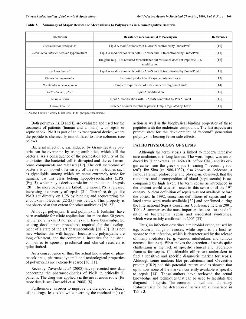
Table 2. Summary of Major Resistance Mechanisms to Polymyxins in Gram-Negative Bacteria
Bacterium Resistance mechanism(s) to Polymyxin References
Pseudomonas aeruginosa Lipid A modifications with L-Ara4N controlled by PmrA/PmrB [10]
Salmonella enterica serovar Typhimutium Lipid A modification with both L-Ara4N and PEtn controlled by PmrA/PmrB [11]
The gene mig-14 is required for resistance but resistance does not implicate LPS modification
[12]
Escherichia coli Lipid A modification with both L-Ara4N and PEtn controlled by PmrA/PmrB [11]
Klebsiella pneumoniae Increased production of capsule polysaccharide [13]
Burkholderia cenocepacia Complete requirement of LPS inner core oligosaccharide [14]
Helicobacter pylori Lipid A modification [15]
Yersinia pestis Lipid A modification with L-Ara4N controlled by PmrA/PmrB [16]
Vibrio cholerae Presence of outer membrane protein OmpU regulated by ToxR [17]
L-Ara4N: 4-amino-4-deoxy-L-arabinose; PEtn: phosphoethanolamine Both polymyxins, B and E, are evaluated and used for the treatment of patients (human and animals) with sepsis or septic shock. PMB is part of an extracorporeal device, where the peptide is chemically immobilized to fibre columns (see below). Bacterial infections, e.g. induced by Gram-negative bac-teria can be overcome by using antibiotics, which kill the bacteria. As a consequence of the permeation activity of the antibiotics, the bacterial cell is disrupted and the cell mem-brane components are released [19]. The cell membrane of bacteria is composed of a variety of diverse molecules such as glycolipids, among which are some extremely toxic for humans. To this class belong lipopolysaccharides (LPS) (Fig. 2), which play a decisive role for the induction of sepsis [20]. The more bacteria are killed, the more LPS is released increasing the severity of sepsis. [21]. Therefore, drugs like PMB act directly on LPS by binding and sequestering the endotoxin molecules [22-25] (see below). This property is not observed at that extent for other antibiotics [26, 27]. Although polymyxin B and polymyxin E (colistin) have been available for clinic applications for more than 50 years, neither polymyxin B nor polymyxin E have been subjected to drug development procedures required for the develop-ment of a state of the art pharmaceuticals [28, 29]. It is not sure whether this will happen, because the polymyxins are long off-patent, and the commercial incentive for industrial companies to sponsor preclinical and clinical research is quite limited. As a consequence of this, the actual knowledge of phar-macokinetic, pharmacodynamic and toxicological properties of polymyxins are extremely scarce [30, 31]. Recently, Zavascki et al. (2008) have presented new data concerning the pharmacokinetics of PMB in critically ill patients. The drug was applied via the intravenous route (for more details see Zavascki et al. 2008) [8]. Furthermore, in order to improve the therapeutic efficacy of the drugs, less is known concerning the mechanism(s) of
action as well as the biophysical binding properties of these peptides with the endotoxin compounds. The last aspects are prerequisites for the development of “second” generation polymyxins bearing fewer side effects.
PATHOPHYSIOLOGY OF SEPSIS
Although the term sepsis is linked to modern intensive care medicine, it is long known. The word sepsis was intro-duced by Hippokrates (ca. 460-370 before Chr.) and its ori-gin came from the greek (meaning “ becoming rot-ten”). Ibn Sina (ca. 980-1037), also known as Avicenna, a famous Iranian philosopher and physician, observed, that the rottenness and decomposition of blood (septicaemia) is ac-companied by high fever. The term sepsis as introduced in the ancient world was still used in this sense until the 19th
century. A clear definition of sepsis was not available before the 90ties. In 1992, consensus definitions of sepsis and re-lated terms were made available [32] and confirmed during the International Sepsis Consensus Conference held in 2001. Table 3 summarises the most important features for the defi-nition of bacteraemia, sepsis and associated syndromes, which were mainly confirmed in 2005 [33]. Thus, an infection describes a microbial event, caused by e.g. bacteria, fungi or viruses, while sepsis is the host re-sponse to that infection, which is characterised by the release of many mediators (e. g. various interleukins and tumour necrosis factor- ). What makes the detection of sepsis quite challenging is the lack of specific clinical and laboratory features for sepsis. Considerable efforts are undertaken to find a sensitive and specific diagnostic marker for sepsis. Although some markers like procalcitonin and C-reactive protein (CRP) had this potential, recent studies showed that up to now none of the markers currently available is specific to sepsis [34]. These authors have reviewed the actual knowledge and techniques that can be used to facilitate the diagnosis of sepsis. The common clinical and laboratory features used for the detection of sepsis are summarised in Table 3.
370 Anti-Infective Agents in Medicinal Chemistry, 2009, Vol. 8, No. 4 Garidel and Brandenburg
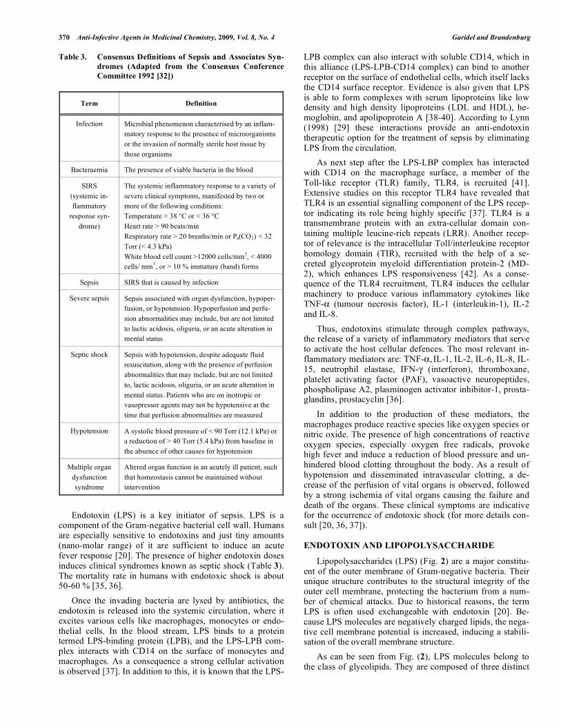
Table 3. Consensus Definitions of Sepsis and Associates Syn-dromes (Adapted from the Consensus Conference Committee 1992 [32])
Term Definition
Infection Microbial phenomenon characterised by an inflam-matory response to the presence of microorganisms or the invasion of normally sterile host tissue by those organisms
Bacteraemia The presence of viable bacteria in the blood
SIRS (systemic in-flammatory
response syn-drome)
The systemic inflammatory response to a variety of severe clinical symptoms, manifested by two or more of the following conditions: Temperature > 38 °C or < 36 °C Heart rate > 90 beats/min Respiratory rate > 20 breaths/min or Pa(CO2) < 32 Torr (< 4.3 kPa) White blood cell count >12000 cells/mm3, < 4000 cells/ mm3, or > 10 % immature (band) forms
Sepsis SIRS that is caused by infection
Severe sepsis Sepsis associated with organ dysfunction, hypoper-fusion, or hypotension. Hypoperfusion and perfu-sion abnormalities may include, but are not limited to lactic acidosis, oliguria, or an acute alteration in mental status
Septic shock Sepsis with hypotension, despite adequate fluid resuscitation, along with the presence of perfusion abnormalities that may include, but are not limited to, lactic acidosis, oliguria, or an acute alteration in mental status. Patients who are on inotropic or vasopressor agents may not be hypotensive at the time that perfusion abnormalities are measured
Hypotension A systolic blood pressure of < 90 Torr (12.1 kPa) or a reduction of > 40 Torr (5.4 kPa) from baseline in the absence of other causes for hypotension
Multiple organ dysfunction syndrome
Altered organ function in an acutely ill patient, such that homeostasis cannot be maintained without intervention
Endotoxin (LPS) is a key initiator of sepsis. LPS is a component of the Gram-negative bacterial cell wall. Humans are especially sensitive to endotoxins and just tiny amounts (nano-molar range) of it are sufficient to induce an acute fever response [20]. The presence of higher endotoxin doses induces clinical syndromes known as septic shock (Table 3). The mortality rate in humans with endotoxic shock is about 50-60 % [35, 36]. Once the invading bacteria are lysed by antibiotics, the endotoxin is released into the systemic circulation, where it excites various cells like macrophages, monocytes or endo-thelial cells. In the blood stream, LPS binds to a protein termed LPS-binding protein (LPB), and the LPS-LPB com-plex interacts with CD14 on the surface of monocytes and macrophages. As a consequence a strong cellular activation is observed [37]. In addition to this, it is known that the LPS-
LPB complex can also interact with soluble CD14, which in this alliance (LPS-LPB-CD14 complex) can bind to another receptor on the surface of endothelial cells, which itself lacks the CD14 surface receptor. Evidence is also given that LPS is able to form complexes with serum lipoproteins like low density and high density lipoproteins (LDL and HDL), he-moglobin, and apolipoprotein A [38-40]. According to Lynn (1998) [29] these interactions provide an anti-endotoxin therapeutic option for the treatment of sepsis by eliminating LPS from the circulation. As next step after the LPS-LBP complex has interacted with CD14 on the macrophage surface, a member of the Toll-like receptor (TLR) family, TLR4, is recruited [41]. Extensive studies on this receptor TLR4 have revealed that TLR4 is an essential signalling component of the LPS recep-tor indicating its role being highly specific [37]. TLR4 is a transmembrane protein with an extra-cellular domain con-taining multiple leucine-rich repeats (LRR). Another recep-tor of relevance is the intracellular Toll/interleukine receptor homology domain (TIR), recruited with the help of a se-creted glycoprotein myeloid differentiation protein-2 (MD-2), which enhances LPS responsiveness [42]. As a conse-quence of the TLR4 recruitment, TLR4 induces the cellular machinery to produce various inflammatory cytokines like TNF- (tumour necrosis factor), IL-1 (interleukin-1), IL-2 and IL-8. Thus, endotoxins stimulate through complex pathways, the release of a variety of inflammatory mediators that serve to activate the host cellular defences. The most relevant in-flammatory mediators are: TNF- , IL-1, IL-2, IL-6, IL-8, IL-15, neutrophil elastase, IFN- (interferon), thromboxane, platelet activating factor (PAF), vasoactive neuropeptides, phospholipase A2, plasminogen activator inhibitor-1, prosta-glandins, prostacyclin [36]. In addition to the production of these mediators, the macrophages produce reactive species like oxygen species or nitric oxide. The presence of high concentrations of reactive oxygen species, especially oxygen free radicals, provoke high fever and induce a reduction of blood pressure and un-hindered blood clotting throughout the body. As a result of hypotension and disseminated intravascular clotting, a de-crease of the perfusion of vital organs is observed, followed by a strong ischemia of vital organs causing the failure and death of the organs. These clinical symptoms are indicative for the occurrence of endotoxic shock (for more details con-sult [20, 36, 37]).
ENDOTOXIN AND LIPOPOLYSACCHARIDE
Lipopolysaccharides (LPS) (Fig. 2) are a major constitu-ent of the outer membrane of Gram-negative bacteria. Their unique structure contributes to the structural integrity of the outer cell membrane, protecting the bacterium from a num-ber of chemical attacks. Due to historical reasons, the term LPS is often used exchangeable with endotoxin [20]. Be-cause LPS molecules are negatively charged lipids, the nega-tive cell membrane potential is increased, inducing a stabili-sation of the overall membrane structure. As can be seen from Fig. (2), LPS molecules belong to the class of glycolipids. They are composed of three distinct
Current Understanding of Polymyxin B Applications Anti-Infective Agents in Medicinal Chemistry, 2009, Vol. 8, No. 4 371
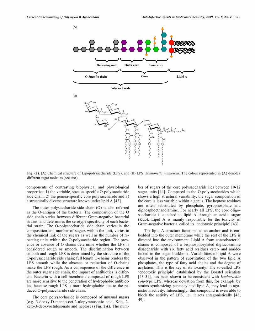
components of contrasting biophysical and physiological properties: 1) the variable, species-specific O-polysaccharide side chain, 2) the genera-specific core polysaccharide and 3) a structurally diverse structure known under lipid A [43]. The outer polysaccharide side chain (O) is also referred as the O-antigen of the bacteria. The composition of the O side chain varies between different Gram-negative bacterial strains, and determines the serotype specificity of each bacte-rial strain. The O-polysaccharide side chain varies in the composition and number of sugars within the unit, varies in the chemical link of the sugars as well as the number of re-peating units within the O-polysaccharide region. The pres-ence or absence of O chains determine whether the LPS is considered rough or smooth. The discrimination between smooth and rough LPS is determined by the structure of the O-polysaccharide side chain; full length O-chains renders the LPS smooth while the absence or reduction of O-chains make the LPS rough. As a consequence of the difference in the outer sugar side chain, the impact of antibiotics is differ-ent. Bacteria with a cell membrane composed of rough LPS are more sensitive to the penetration of hydrophobic antibiot-ics, because rough LPS is more hydrophobic due to the re-duced O-polysaccharide side chain. The core polysaccharide is composed of unusual sugars (e.g. 3-deoxy-D-manno-oct-2-ulopyranosonic acid, Kdo, 2-keto-3-deoxyoctulosonate and heptose) (Fig. 2A). The num-
ber of sugars of the core polysaccharide lies between 10-12 sugar units [44]. Compared to the O-polysaccharides which shows a high structural variability, the sugar composition of the core is less variable within a genus. The heptose residues are often substituted by phosphate, pyrophosphate and diphosphoethanolamine. For nearly all LPS, the core oligo-saccharide is attached to lipid A through an acidic sugar (Kdo). Lipid A is mainly responsible for the toxicity of Gram-negative bacteria, called its ‘endotoxic principle’ [43]. The lipid A structure functions as an anchor and is em-bedded into the outer membrane while the rest of the LPS is directed into the environment. Lipid A from enterobacterial strains is composed of a bisphosphorylated diglucosamine disaccharide with six fatty acid residues ester- and amide-linked to the sugar backbone. Variabilities of lipid A were observed in the pattern of substitution of the two lipid A phosphates, the type of fatty acid chains and the degree of acylation. This is the key of its toxicity. The so-called LPS ‘endotoxic principle’ established by the Borstel scientists [43-51], has been shown to be consistent with Escherichia coli-type LPS, whereas deviation from this, for example by strains synthesizing pentaacylated lipid A, may lead to ago-nistic inactivity. Interestingly, this compound is even able to block the activity of LPS, i.e., it acts antagonistically [48, 49].
Fig. (2). (A) Chemical structure of Lipopolysaccharide (LPS), and (B) LPS: Salmonella minnesota. The colour represented in (A) denotes different sugar moieties (see text).
372 Anti-Infective Agents in Medicinal Chemistry, 2009, Vol. 8, No. 4 Garidel and Brandenburg
This lipid A (Fig. 2A) adopts a cubic inverted aggregate structure from which a conical shape of the molecule can be deduced, whereas the tetraacyl lipid A precursor IVa adopts a cylindrical shape and is endotoxically inactive, but antago-nizes active LPS [52]. These super-structural assemblies play a key role for the induction of the cellular responses [43, 52].
CLINICAL MANAGEMENT OF SEPSIS
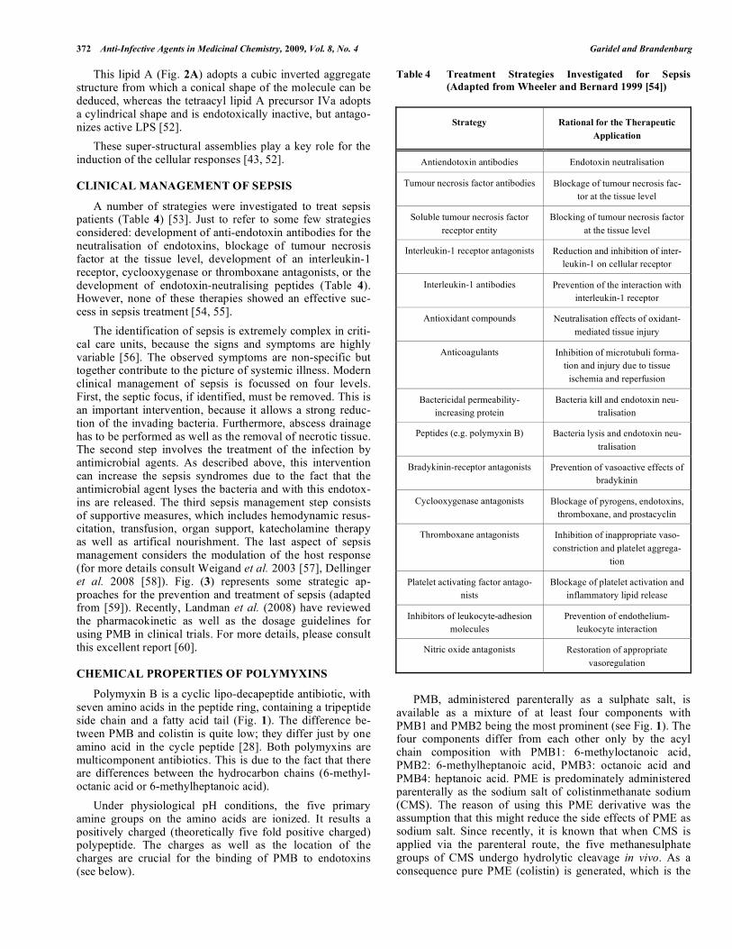
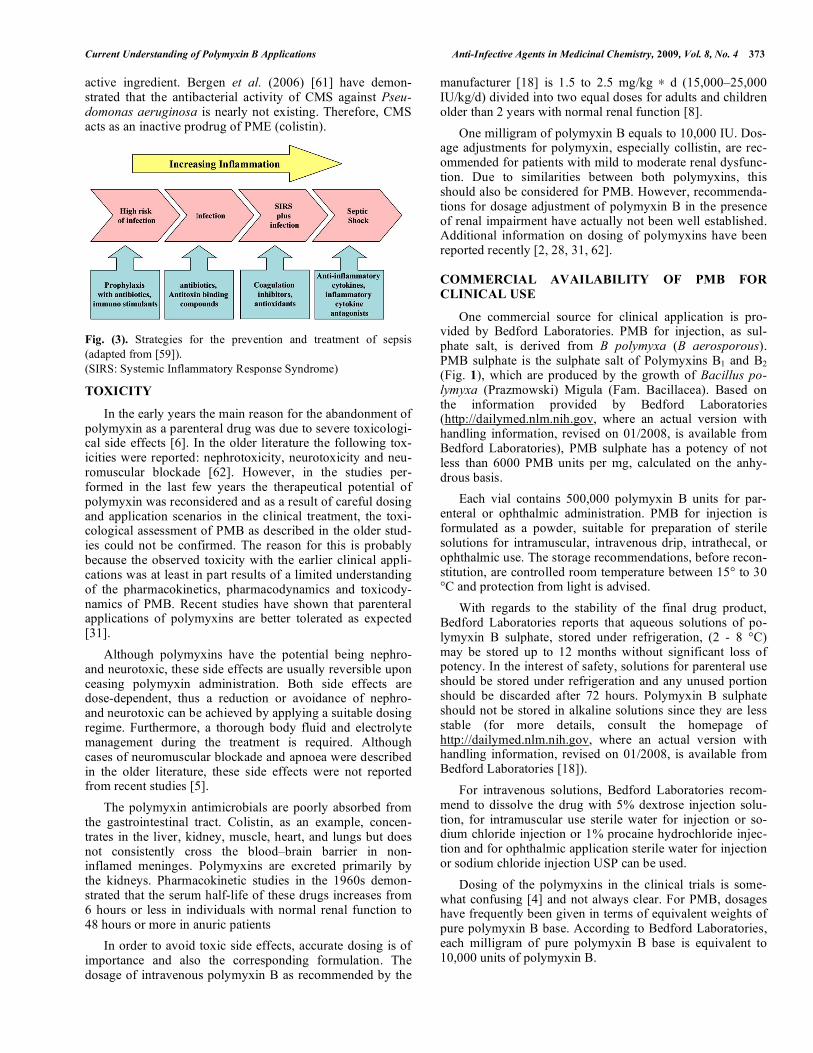
A number of strategies were investigated to treat sepsis patients (Table 4) [53]. Just to refer to some few strategies considered: development of anti-endotoxin antibodies for the neutralisation of endotoxins, blockage of tumour necrosis factor at the tissue level, development of an interleukin-1 receptor, cyclooxygenase or thromboxane antagonists, or the development of endotoxin-neutralising peptides (Table 4). However, none of these therapies showed an effective suc-cess in sepsis treatment [54, 55]. The identification of sepsis is extremely complex in criti-cal care units, because the signs and symptoms are highly variable [56]. The observed symptoms are non-specific but together contribute to the picture of systemic illness. Modern clinical management of sepsis is focussed on four levels. First, the septic focus, if identified, must be removed. This is an important intervention, because it allows a strong reduc-tion of the invading bacteria. Furthermore, abscess drainage has to be performed as well as the removal of necrotic tissue. The second step involves the treatment of the infection by antimicrobial agents. As described above, this intervention can increase the sepsis syndromes due to the fact that the antimicrobial agent lyses the bacteria and with this endotox-ins are released. The third sepsis management step consists of supportive measures, which includes hemodynamic resus-citation, transfusion, organ support, katecholamine therapy as well as artifical nourishment. The last aspect of sepsis management considers the modulation of the host response (for more details consult Weigand et al. 2003 [57], Dellinger et al. 2008 [58]). Fig. (3) represents some strategic ap-proaches for the prevention and treatment of sepsis (adapted from [59]). Recently, Landman et al. (2008) have reviewed the pharmacokinetic as well as the dosage guidelines for using PMB in clinical trials. For more details, please consult this excellent report [60].
CHEMICAL PROPERTIES OF POLYMYXINS
Polymyxin B is a cyclic lipo-decapeptide antibiotic, with seven amino acids in the peptide ring, containing a tripeptide side chain and a fatty acid tail (Fig. 1). The difference be-tween PMB and colistin is quite low; they differ just by one amino acid in the cycle peptide [28]. Both polymyxins are multicomponent antibiotics. This is due to the fact that there are differences between the hydrocarbon chains (6-methyl-octanic acid or 6-methylheptanoic acid). Under physiological pH conditions, the five primary amine groups on the amino acids are ionized. It results a positively charged (theoretically five fold positive charged) polypeptide. The charges as well as the location of the charges are crucial for the binding of PMB to endotoxins (see below).
Table 4 Treatment Strategies Investigated for Sepsis (Adapted from Wheeler and Bernard 1999 [54])
Strategy Rational for the Therapeutic Application
Antiendotoxin antibodies Endotoxin neutralisation
Tumour necrosis factor antibodies Blockage of tumour necrosis fac-tor at the tissue level
Soluble tumour necrosis factor receptor entity
Blocking of tumour necrosis factor at the tissue level
Interleukin-1 receptor antagonists Reduction and inhibition of inter-leukin-1 on cellular receptor
Interleukin-1 antibodies Prevention of the interaction with interleukin-1 receptor
Antioxidant compounds Neutralisation effects of oxidant-mediated tissue injury
Anticoagulants Inhibition of microtubuli forma-tion and injury due to tissue
ischemia and reperfusion
Bactericidal permeability-increasing protein
Bacteria kill and endotoxin neu-tralisation
Peptides (e.g. polymyxin B) Bacteria lysis and endotoxin neu-tralisation
Bradykinin-receptor antagonists Prevention of vasoactive effects of bradykinin
Cyclooxygenase antagonists Blockage of pyrogens, endotoxins, thromboxane, and prostacyclin
Thromboxane antagonists Inhibition of inappropriate vaso-constriction and platelet aggrega-
tion
Platelet activating factor antago-nists
Blockage of platelet activation and inflammatory lipid release
Inhibitors of leukocyte-adhesion molecules
Prevention of endothelium-leukocyte interaction
Nitric oxide antagonists Restoration of appropriate vasoregulation
PMB, administered parenterally as a sulphate salt, is available as a mixture of at least four components with PMB1 and PMB2 being the most prominent (see Fig. 1). The four components differ from each other only by the acyl chain composition with PMB1: 6-methyloctanoic acid, PMB2: 6-methylheptanoic acid, PMB3: octanoic acid and PMB4: heptanoic acid. PME is predominately administered parenterally as the sodium salt of colistinmethanate sodium (CMS). The reason of using this PME derivative was the assumption that this might reduce the side effects of PME as sodium salt. Since recently, it is known that when CMS is applied via the parenteral route, the five methanesulphate groups of CMS undergo hydrolytic cleavage in vivo. As a consequence pure PME (colistin) is generated, which is the
Current Understanding of Polymyxin B Applications Anti-Infective Agents in Medicinal Chemistry, 2009, Vol. 8, No. 4 373
active ingredient. Bergen et al. (2006) [61] have demon-strated that the antibacterial activity of CMS against Pseu-domonas aeruginosa is nearly not existing. Therefore, CMS acts as an inactive prodrug of PME (colistin). Fig. (3). Strategies for the prevention and treatment of sepsis (adapted from [59]). (SIRS: Systemic Inflammatory Response Syndrome)
TOXICITY
In the early years the main reason for the abandonment of polymyxin as a parenteral drug was due to severe toxicologi-cal side effects [6]. In the older literature the following tox-icities were reported: nephrotoxicity, neurotoxicity and neu-romuscular blockade [62]. However, in the studies per-formed in the last few years the therapeutical potential of polymyxin was reconsidered and as a result of careful dosing and application scenarios in the clinical treatment, the toxi-cological assessment of PMB as described in the older stud-ies could not be confirmed. The reason for this is probably because the observed toxicity with the earlier clinical appli-cations was at least in part results of a limited understanding of the pharmacokinetics, pharmacodynamics and toxicody-namics of PMB. Recent studies have shown that parenteral applications of polymyxins are better tolerated as expected [31]. Although polymyxins have the potential being nephro- and neurotoxic, these side effects are usually reversible upon ceasing polymyxin administration. Both side effects are dose-dependent, thus a reduction or avoidance of nephro- and neurotoxic can be achieved by applying a suitable dosing regime. Furthermore, a thorough body fluid and electrolyte management during the treatment is required. Although cases of neuromuscular blockade and apnoea were described in the older literature, these side effects were not reported from recent studies [5]. The polymyxin antimicrobials are poorly absorbed from the gastrointestinal tract. Colistin, as an example, concen-trates in the liver, kidney, muscle, heart, and lungs but does not consistently cross the blood–brain barrier in non-inflamed meninges. Polymyxins are excreted primarily by the kidneys. Pharmacokinetic studies in the 1960s demon-strated that the serum half-life of these drugs increases from 6 hours or less in individuals with normal renal function to 48 hours or more in anuric patients In order to avoid toxic side effects, accurate dosing is of importance and also the corresponding formulation. The dosage of intravenous polymyxin B as recommended by the
manufacturer [18] is 1.5 to 2.5 mg/kg d (15,000–25,000 IU/kg/d) divided into two equal doses for adults and children older than 2 years with normal renal function [8]. One milligram of polymyxin B equals to 10,000 IU. Dos-age adjustments for polymyxin, especially collistin, are rec-ommended for patients with mild to moderate renal dysfunc-tion. Due to similarities between both polymyxins, this should also be considered for PMB. However, recommenda-tions for dosage adjustment of polymyxin B in the presence of renal impairment have actually not been well established. Additional information on dosing of polymyxins have been reported recently [2, 28, 31, 62].
COMMERCIAL AVAILABILITY OF PMB FOR CLINICAL USE
One commercial source for clinical application is pro-vided by Bedford Laboratories. PMB for injection, as sul-phate salt, is derived from B polymyxa (B aerosporous). PMB sulphate is the sulphate salt of Polymyxins B1 and B2 (Fig. 1), which are produced by the growth of Bacillus po-lymyxa (Prazmowski) Migula (Fam. Bacillacea). Based on the information provided by Bedford Laboratories (http://dailymed.nlm.nih.gov, where an actual version with handling information, revised on 01/2008, is available from Bedford Laboratories), PMB sulphate has a potency of not less than 6000 PMB units per mg, calculated on the anhy-drous basis. Each vial contains 500,000 polymyxin B units for par-enteral or ophthalmic administration. PMB for injection is formulated as a powder, suitable for preparation of sterile solutions for intramuscular, intravenous drip, intrathecal, or ophthalmic use. The storage recommendations, before recon-stitution, are controlled room temperature between 15° to 30 °C and protection from light is advised. With regards to the stability of the final drug product, Bedford Laboratories reports that aqueous solutions of po-lymyxin B sulphate, stored under refrigeration, (2 - 8 °C) may be stored up to 12 months without significant loss of potency. In the interest of safety, solutions for parenteral use should be stored under refrigeration and any unused portion should be discarded after 72 hours. Polymyxin B sulphate should not be stored in alkaline solutions since they are less stable (for more details, consult the homepage of http://dailymed.nlm.nih.gov, where an actual version with handling information, revised on 01/2008, is available from Bedford Laboratories [18]). For intravenous solutions, Bedford Laboratories recom-mend to dissolve the drug with 5% dextrose injection solu-tion, for intramuscular use sterile water for injection or so-dium chloride injection or 1% procaine hydrochloride injec-tion and for ophthalmic application sterile water for injection or sodium chloride injection USP can be used. Dosing of the polymyxins in the clinical trials is some-what confusing [4] and not always clear. For PMB, dosages have frequently been given in terms of equivalent weights of pure polymyxin B base. According to Bedford Laboratories, each milligram of pure polymyxin B base is equivalent to 10,000 units of polymyxin B.
374 Anti-Infective Agents in Medicinal Chemistry, 2009, Vol. 8, No. 4 Garidel and Brandenburg
PME is mostly used as sodium colistinmethanate (for more details see Horton et al. 1982 [63]).
ANTIMICROBIAL ACTIVITY
The reason for the use of the polymyxin antibiotics were due to their broad spectrum of activity. Table 1 summarises the antimicrobial activity of PMB [3]. Hogardt et al. (2004) [64] showed that PMB exhibits good activity against P. aeruginosa at MIC90 4 mg/l which is in accordance to the data published by Gales et al. 2006 [3]. So far, most investigations on the pharmacodynamics of the polymyxins have focused on colistin, and less is known about the pharmacodynamics of PMB [65]. Better under-standing of the pharmacodynamics of polymyxin B may help to determine its exact dose rationale to optimize patient out-comes and avoid resistance. Recently, Tam et al. (2005) [30] examined the pharmacodynamics of polymyxin B against P. aeruginosa and suggested that the bactericidal activity of this regimen was concentration-dependent and seemed to be re-lated to the ratio of the area under the concentration-time curve to the MIC. Investigations for the determination of the optimal dosage of these regimens in different subpopulations of patients are, however, urgently required. In the last years antimicrobial resistance was observed for both polymyxins, since Gram-negative bacteria have devel-oped various resistance mechanisms to overcome the antimi-crobial activity (for more details see Table 2 and references cited therein) [66]. Ko and coworkers (2007) [67] have in-vestigated the antimicrobial resistance of PMB and PME in clinical isolates of Acinetobacter spp. They have collected 265 isolates of Acinetobacter spp., which were identified to species level using partial rpoB gene sequences. About 81 % of the isolates were Acinetobacter baumannii. Out of these about 18 % and 28 % were resistant to PMB and PME, re-spectively. Antoniadou et al. (2007) [68] have also reported about PME resistant isolates of Klebsiella pneumonia which were encountered in intensive care unit patients.
APPLICATION OF POLYMYXINS IN BACTERA-EMIA/SEPSIS
Michalopoulos and Falagas (2008) [4] have recently summarised the use of PME and PMB in critical care units, because these peptides were mainly administered for the treatment of life-threatening nosocomial acquired infections caused by multiple-drug resistant Gram-negative pathogens, like Acinetobacter sp, P. aeruginosa, Klebsiella sp, and En-terobacter sp. in adult patients, due to the lack of other therapeutically efficient agents [19, 69]. Recent reports have also demonstrated the use of these agents, especially PME as intravenous administration in children [28, 70, 71]. The clinical and microbiologic efficacy and safety profile of PMB in the treatment of multidrug-resistant Gram-negative bacterial infections of the respiratory tract was re-examined retrospectively by Sobieszczyk et al. (2004) [72]. They presented a combination therapy study with PMB for the treatment of multidrug-resistant Gram-negative respira-tory tract infections. In their study, 25 critically ill patients were treated. The patients received a total of 29 courses of
PMB, which was administered in a combination study with another antimicrobial agent. They used two administration routes. The patients were treated with intravenous and aero-solized PMB, at a mean PMB duration therapy of 19 days. At the end of the treatment a mortality of 21 % was ob-served, and overall mortality at discharge was 48 %. Nephro-toxicity was observed in 3 patients (10 %). However, this side effect did not result in discontinuation of the therapy. The outcome of the study presented by Sobieszczyk and col-leagues (2004) [72] was that PMB combined with other an-timicrobials can be considered as a reasonable and safe treatment option for multidrug-resistant Gram-negative res-piratory tract infections in the setting of limited therapeutic options. The study published by Holloway et al. (2006) [73] in-vestigated the administration of intravenous PMB in 29 criti-cally ill patients with infections caused by multidrug-resistant A. baumannii. The observed clinical cure was 76 %, whereas crude mortality rate was 27 %. Sarria and col-leagues (2004) [70] reported their experience from the use of intravenous PMB in a patient with A. baumannii sepsis and acute renal failure that required continuous hemodialysis and were able to present a successful treatment. Recently, an intravenous PMB application was presented for the treatment of 74 patients infected by hospital-acquired multidrug-resistant Pseudomonas aeruginosa [74]. A fa-vourable outcome for 48 % of the treated patients was ob-served, which led to the conclusion that PMB is a reliable antimicrobial drug, but only as salvage therapy, for nosoco-mial pneumonia caused by multidrug-resistant Pseudomonas aeruginosa. The data presented by Pastewski et al. (2008) [75] sug-gest that PMB may be effective for multidrug-resistant Gram-negative bacteraemia and urinary tract infections for patients with limited therapeutic options, but they pointed out that precautions should be taken by the correct dosing in order to avoid toxicity, especially nephrotoxicity. Examples are also known where a therapeutic difference was observed between PME and PMB. Systemic colistin (PME) has shown efficacy against multidrug-resistant Pseu-domonas aeruginosa and Acinetobacter spp., but it has pre-sented poor results in pneumonia. Aerosolized polymyxin (PMB) in cystic fibrosis patients led to good results. Pereira et al. (2007) [76] used inhaled PMB to treat respiratory in-fections by multidrug-resistant Gram-negative bacilli. Nine-teen patients were treated with inhaled polymyxin B: 14 had pneumonia, most of which had previously failed treatment with intravenous PMB, and 5 tracheobronchitis. Inhaled PMB was given at a dose of 500,000 IU twice a day after an aerosolized 2-agonist. In pneumonia, inhaled and intrave-nous PMB was administered together. 89 % of the patients were in the intensive care unit. Sixteen infections (84 %) were caused by P. aeruginosa, while Klebsiella pneumoniae, Alcaligenes xylosoxidans, and Burkholderia sp. caused one infection each. In the 14 pneumonia cases, the median of previous use of intravenous polymyxin B was 20 days (range 0-32), whereas inhaled polymyxin B was used for a mean of 14 days (range 4-25). Cure occurred in 10 (53 %) patients, improvement in 8 (42 %), and failure in 1. Nine patients died during hospitalization (all with pneumonia). Adverse events
Current Understanding of Polymyxin B Applications Anti-Infective Agents in Medicinal Chemistry, 2009, Vol. 8, No. 4 375
occurred in 4 patients without interruption of inhalation. The study published by Pereira et al. (2007) [76] is the largest report using inhaled PMB to treat nosocomial pneumonia by multidrug-resistant Gram-negative bacilli that had failed intravenous polymyxin B. It was also effective alone in P. aeruginosa tracheobronchitis. This report highlights that the route of application also determines the outcome of the ther-apy. Studies are known, where both polymyxins are applied for the treatment of bacteraemia and sepsis [31]. Zhou et al. (2007) [77] have demonstrated that polymyxin was effective in the management of multidrug-resistant Pseudomonas aeruginosa pulmonary infections in patient with the highly pathogenic avian influenza (H5N1)infection. In this study both polymyxins were used. PMB was subsequently admin-istered intramuscularly or intravenously combined with PME aerosol therapy. The use of colistin in bacteraemia and sepsis therapy can be summarised as follows. Colistin applied intravenously in intensive care units patients with sepsis showed in 73 % of the treated patients a clinical response [78]. They reported a deterioration of renal function in ca. 14 % of patients, whereas survival at 30 days was 57.7%. Karabinis et al. (2004) [79] described the successful management of a pa-tient with bacteraemia/septic shock caused by K. pneumo-niae. Again PME was administered via the intravenous route at a dosage of 9 million IU/d (2.5 mg/kg, divided into three doses). Michalopoulos and colleagues (2005) [69] used a continuous intravenous application of PME in a critically ill patient with bacteraemia caused by a multiresistant A. bau-mannii strain susceptible only to PME. With the used ap-proach, they were able to cure the patient from this life-threatening infection. The same group [80] also reported that the mortality rate was acceptable (35.7%) in critically ill patients with ICU-acquired bacteraemia caused by MDR A. baumannii who received intravenously colistin and mero-penem, based on sensitivity tests and MICs. On the contrary, the mortality rate was 56% in the group of patients with bac-teraemia caused by A baumannii strains susceptible only to PME (see also Michalopoulos and Falagas 2008 [4]). Michalopoulos and Falagas (2008) [4] have recently mentioned for polymyxin E that „that the selective pressure caused by extensive or inadequate colistin use may have contributed to the emergence of colistin resistance among P. aeruginosa, A. baumannii, and K. pneumoniae isolates”, potentially increasing morbidity and mortality rates in criti-cally ill patients and necessitating prudent use of colistin [68]. For this reason, the empiric use of colistin should be limited to institutions in which there is recognized infection caused by multidrug-resistant Gram-negative bacilli [79]. This is also true for PMB. It is very crucial to use these antimicrobial agents with care and only where it is really appropriate in order to avoid multi-drug resistance [81].
APPLICATION OF POLYMYXINS IN AN EXTRACO-RPOREAL MEDICAL DEVICE
An extracorporeal hemoperfusion device (TORAY-MYXIN) based on PMB was first developed in 1994. It is designed for the selective blood purification from endotoxins
via direct hemoperfusion. Toraymyxin is made up of poly-styrene-derivative fibres to which the peptide is covalently bound on the surface of an insoluble carrier material inside the cartridge [82-84]. According to Teramoto et al. (2002) [85] Toraymyxin is composed of an islands-in-a-sea-type composite fibre with the island ingredient comprising poly-propylene, and the sea ingredient comprises a polystyrene derivative. It is prepared from the corresponding polystyrene fibre, which is amidomethylated with N-methylol- -chloroacetamide, and the resulting fibre is incubated subse-quently in the PMB aqueous strongly alkaline solution (pH 9 to 13). As a consequence, PMB is chemically bonded to -carbon of the acetyl residue in poly(4-chloroacetami-domethyl styrene) of PMX-F by replacing chlorine. PMB is covalently bounded at a weight ratio of 0.5 w% [86]. The advantage of this extracorporal device is due to the fact that the endotoxin can be inactivated in the blood with-out exerting its toxicity on the brain and kidney. Direct hemoperfusion using such polymyxin B-immobilized fiber column (PMX; Toray Industries Inc., To-kyo, Japan) has been tested and used since about 15 years for the treatment of septic shock. Ruberto et al. (2007) [87] have recently analysed the efficacy, safety and clinical effects of direct hemoperfusion with an immobilized polymyxin-B fibre column (DHP-PMX) in solid organ transplanted patients with severe sepsis or septic shock. 15 patients (mean age 55 years old) were considered which developed severe sepsis or septic shock after kidney or liver transplantation. For all patients, Gram-negative bacteria were detected. They were treated using the conventional approach, namely: antibiotic therapy, vasopres-sive or inotropic agents, and ventilation support. Addition-ally, each patient was treated three times using DHP-PMX treatment. Ruberto and coworkers (2007) [87] observed no adverse events for the 15 treated patients and they concluded that the use of DHP-PMX in association with conventional therapy may be an important aid in patients with sepsis. A beneficial outcome was also described for patients with severe sepsis or septic shock which underwent a liver transplantation by applying a direct DHP-PMX hemoperfu-sion [88]. DHP-PMX as a treatment option is used in a number of clinical cases. Kakugawa et al. (2008) [89] recently reported on the successful DHP-PMX treatment outcome of a patient undergoing a rapidly progressive interstitial pneumonia as-sociated with clinically amyopathic dermatomyositis. Murakami et al. (2007) [90] observed that it is important to initiate early in suspected septic shock patients the simul-taneous DHP-PMX and drug treatment in order to facilitate hemodynamic improvement, which is beneficial for the pa-tient. A number of molecular factors affect the clinical out-come during sepsis or septic shock. Using the DHP-PMX approach after a continuous hemodiafiltration with po-lymethylmethacrylate membrane hemofilters for the treat-ment of septic shock patients, also a beneficial improvement of critical laboratory parameters like systolic blood pressure and an improved Pao2/Fio2 ratio after treatment has been shown [91, 92].

376 Anti-Infective Agents in Medicinal Chemistry, 2009, Vol. 8, No. 4 Garidel and Brandenburg
The activity of proapoptotic circulating factors play a crucial role for patient which were injured by a Gram-negative sepsis-induced acute renal failure. Cantaluppi et al. (2008) [93] tested the hypothesis that the extracorporeal therapy with DHP-PMX may prevent Gram-negative sepsis-induced acute renal failure by reducing the activity of proapoptotic circulating factors. Using cultured renal cells, a significant decrease of plasma-induced proapoptotic activity was observed after DHP-PMX treatment, which confirmed their approach. Another factor of relevance during sepsis, is the activa-tion of neutrophils that injures tissues and organs. From lit-erature it is known that blood purification therapies such as continuous veno-venous hemofiltration (CVVH) and direct hemoperfusion with polymyxin-immobilized fibre (DHP-PMX) have been used for the treatment of sepsis and septic shock, however, the effects of such therapies on neutrophil activation have been poorly understood. Naka et al. (2006) [94] have therefore focused on this aspect, and have evalu-ated neutrophil reactive oxygen species, in particular the H2O2 production, in the pathophysiology of sepsis or septic shock and the effect of continuous veno-venous hemofiltra-tion or direct hemoperfusion with polymyxin-immobilized fibre on neutrophil reactive oxygen species. First of all, Naka and coworkers (2006) [94] found that patients with sepsis or septic shock had significantly higher levels of neutrophil reactive oxygen species compared with normal volunteers. Neutrophil reactive oxygen species did not change over time in patients treated either with continuous veno-venous hemofiltration or without this treatment. However, applying direct hemoperfusion with polymyxin-immobilized fibres to patients with septic shock, they were able to demonstrate a significant inhibition of neutrophil reactive oxygen species of ca. 30 %. Another factor of relevance is 8-hydroxy-2 -deoxyguanosine (8-OHdG). In healthy volunteers the con-centrations of 8-OHdG were 5.5 ng/mg creatinine, whereas in septic shock patients it was 38.0 ng/mg [95]. The study demonstrates that urinary 8-OHdG levels correlated signifi-cantly with plasma endotoxin levels, the Acute Physiology and Chronic Health Evaluation score and the Sepsis-related Organ Failure Assessment score (all with p > 0.01). Naka-mura et al. (2006) [95] were able to provide evidence that urinary 8-OHdG levels, which are believed to be associated with septic shock, decreased effectively using polymyxin B-immobilized fibre (PMX-F) haemoperfusion. Beyond the binding of endotoxins to PMB, also other molecules such as endocannabinoids bind strongly to PMB. From the last class arachidonylethanolamide (AEA) and 2-arachidonylglycerol (2-AG) are of interest because they are involved in septic shock. The prostaglandin 8-epi prosta-glandin F2 (F2-isoprostane) is used as a biomarker of oxi-dative stress in biological systems. Kase and co-workers (2008) [96] have therefore evaluated whether a DHP-PMX hemoperfusion therapy would decrease serum levels of en-docannabinoids. Twenty-six patients with septic shock, in-cluding those with septic shock induced by peritonitis, un-derwent laparotomy for drainage. Two groups of patients were formed: patients whose mean arterial blood pressure had increased more than 2.6 kPa) (=20 mm Hg) (responder
group; N = 13); and patients whose mean arterial blood pres-sure did not increase or had increased no more than 2.6 kPa (non-responder group; N = 13). They observed no changes of arachidonylethanolamide after DHP-PMX in either the non-responder or responder groups. However, the levels of 2-arachidonylglycerol decreased significantly after DHP-PMX hemoperfusion therapy in the responder group, but not in the non-responder group. F2-isoprostane gradually increased after DHP-PMX treatment; on the other hand, levels of F2-isoprostane re-mained constant in the responder group. Patients with septic shock are under considerable oxidative stress, and 2-arachidonylglycerol plays an important role in the cardiovas-cular status of these patients. Kase et al. (2008) [96] con-cluded that the removal of 2-arachidonylglycerol by DHP-PMX benefits patients with septic shock by stabilizing car-diovascular status and decreasing long-term oxidative stress. The efficacy of PMX has been proven in studies with a uniform case definition and without any other blood purifica-tion techniques. However, based on the knowledge that in some studies favourable effects of direct hemoperfusion with polymyxin-B-immobilized fibre columns (PMX) for the treatment of septic shock have been reported, Shimizu and collaborators (2008) [97] have recently demonstrated that a direct hemoperfusion with PMX improves septic hypoten-sion and reduces inflammatory mediators in septic patients with colorectal perforation. In their investigation, 52 patients with severe sepsis or septic shock secondary to colorectal perforation were treated with DHP-PMX. A number of clini-cal parameters were considered (hemodynamic alterations and plasma concentrations of endotoxin, interleukin (IL)-1 , IL-1 receptor antagonist (IL-1Ra), IL-6, IL-8, and IL-10) following PMX treatment. Shimizu et al. (2008) [97] ob-served a significant reduction in plasma endotoxin in the survivors immediately after DHP-PMX treatment compared to before treatment. Furthermore, inflammation factors like IL-1 , IL-1Ra, and IL-8 were significantly reduced during a 2-h interval of PMX. They concluded from their results that DHP-PMX treatment appears to adsorb endotoxin and addi-tionally a modulation of circulating cytokine is observed, both effects being beneficial for the treatment of the patients. Kushi and colleagues (2008) [98] investigated the effects of DHP-PMX treatment with regards to the gastric intramu-cosal pH (pHi) for patients who underwent early goal-directed therapy within 6 h of a diagnosis of sepsis or septic shock. Their findings suggest that DHP-PMX improves gas-tric intramucosal pH with normalized values from 48 h on-wards. The studies presented above have clearly shown that di-rect hemoperfusion using a polymyxin B-immobilized fibre column has been used successfully for the treatment of septic shock. Septic shock is a condition associated with diffuse coagulopathy and multiple organ failure, and frequently ends in death. On the other hand, the effectiveness of continuous hemodiafiltration using a polymethylmethacrylate membrane hemofilter (PMMA- CHDF) for critically ill patients has also been reported [92]. 27 septic shock patients were treated using the DHP-PMX approach. The patients, except for the nine in whom continuous hemodiafiltration (CHDF) was not performed after DHP-PMX, were divided into two groups:
Current Understanding of Polymyxin B Applications Anti-Infective Agents in Medicinal Chemistry, 2009, Vol. 8, No. 4 377
namely, a group in which PMMA-CHDF therapy was added after DHP-PMX (11 cases), and a group in which continuous hemodiafiltration using a polyacrylonitrile membrane hemofilter (PAN-CHDF) therapy was added after DHP-PMX (7 cases). The outcomes in the two groups were com-pared. The average Acute Physiology and Chronic Health Evaluation (APACHE) II score and the average sepsis-related organ failure assessment (SOFA) score were not sig-nificantly different between the two groups. The PMMA-CHDF group showed significantly better outcomes, with significant improvements of the serum PAI-1, protein C, IL-6 and N-arachidonoylethanolamine (AEA) levels. Based on their results Sakamoto et al. (2008) [92] concluded that PMMA-CHDF may be more effective than PAN-CHDF in the management of septic shock. A number of studies are still devoted to investigate this extracorporal medical device for the removal of endotoxins from the blood [99-102].
VETERINARY APPLICATIONS
PMB and DHP-PMX has been tested and evaluated in a number of animals models like rats [103, 104], dogs [105, 106], pigs [107], sheep [108] and horses [109], to inhibit LPS-induced fever, because PMB binds to the lipid A of bacterial endotoxin. These data are of relevance for the pre-clinical evaluation of the therapy. However, there is a certain interest to develop a therapy against endotoxemia in horses [108-110], because there is a lack of a safe, affordable and effective treatment for endo-toxemia in horses in order to reduce the incidence of this potentially fatal condition. Results, presented by various studies [111, 112] suggest that PMB is a safe, effective inhibitor of endotoxin-induced inflammation in healthy horses. MacKay et al. (1999) [113] determined the efficacy of polymyxin B-dextran 70 (PBD) formulation for the treatment of endotoxemic horses and found that when used in combi-nation with a cyclooxygenase-inhibiting drug, PBD has po-tential for treatment of horses with endotoxemia. PME has also been tested in various animals like dogs. Sentürk (2005) [114], as an example, found that PME has an anti-endotoxic effect and can be used safely for dogs with endotoxemia.
PHARMACEUTICAL CONSIDERATIONS
Systemic applications of PMB have been strongly lim-ited, especially for the parenteral route, due to its high toxic potential. The entrapment of antibiotics in liposomes or other carrier systems is known to enhance their antimicrobial ac-tivities while minimizing their toxic effects. This is also known for oral application, because Coppi et al. (2008) [115] showed that the use of alginate/chitosan microparticles to target the lymphatic system could improve safety when administering PMB orally (see also [116]). Alipour and colleagues (2008) [117] have recently inves-tigated the formulation and incorporation of PMB into liposomes. They have prepared different pseudobinary lipid
systems composed of a phosphatidylcholine and cholesterol. The entrapment efficiency of sonicated liposomes were de-pendent on the acyl chain composition of the phospholipids, being six-fold higher for saturated compared to unsaturated phospholipids acyl chain composition. It was also observed, that the entrapment efficiency was dependent on the liposo-mal preparation, because for extruded liposomes, the en-trapment efficiency was higher for lipids containing unsatu-rated acyl chains. The drug release was also different when saturated or unsaturated lipids were used, with a faster release kinetics for the saturated phospholipids formulation. Alipour et al. (2008) [117] focussed on the antimicrobial activities of the PMB liposome formulations, showing the minimal inhibitory concentrations of sonicated saturated liposomes against Gram-negative strains were generally lower when compared to free polymyxin B. This is explained by the fact that the penetration of PMB into a resistant strain of Pseudomonas aeruginosa was higher following its administration as a liposomal formulation as compared to its conventional form. The combination of free PMB and with liposomal containing PMB formulations had an antibacterial activity similar to that of free antibiotic. The author concluded that an “incor-poration of polymyxin B in liposomes could be useful in the management of Gram-negative infections induced by these microorganisms.” Another study presented by McAllister et al. (1999) [118] were focused on the observation that the pulmonary residence time of PMB is substantially increased when ad-ministered as a liposomal formulation. The main criteria for the use of liposomal PMB formulations to improve the treatment of cystic fibrosis lung infections, is an unaffected antimicrobial activity of PMB for the liposomal formulation. Therefore, McAllister et al. investigated whether the micro-bial activity against Pseudomonas aeruginosa, was retained for liposomal preparation versus the free drug. They were also interested in the role of liposomal surface characteristics in determining interactions with bacterial cell surfaces. They showed that the encapsulation efficiency was dependent on the charge of the used excipients. Using positively charged amphiphiles, PMB encapsulation was reduced compared to neutral or negatively charged lipids. Concerning PMB anti-microbial activity, it was found that antimicrobial activity was retained after encapsulation. Using a PMB concentration of 0.3 mg/l, McAllister et al. (1999) [118] showed that posi-tively as well as negatively charged liposomal PMB formula-tions, and the free drug, killed all cells after 1 h, whereas the use of neutral liposome formulations did not significantly decrease the surviving cell fraction. The reason for the dif-ferences in the minimal inhibitory concentrations of various liposomal formulation could be correlated to the free drug concentration, which was obtained through the release of entrapped PMB. Enhanced activity was only observed for formulation composed of positively charged excipients, which may be related to favourable electrostatic interactions between the cells and the liposomes. In summary, they con-clude that “liposome encapsulation of PMB was not detri-mental to antimicrobial activity, and liposome surface prop-erties and release characteristics were important in determin-ing interactions with bacterial cells”. Other drug delivery
378 Anti-Infective Agents in Medicinal Chemistry, 2009, Vol. 8, No. 4 Garidel and Brandenburg
livery systems of interest for the formulation of PMB are lipid nanoparticles and nanoemulsions [119]. For ophthalmic PMB formulations the choice of the cor-rect preservative is of high relevance to maintain PMB activ-ity [120, 121]. Additional aspects of pharmaceutical relevance are related to analytics, stability and purity of the drug product (for more details see [122]).
STRUCTURAL AND MECHANISTIC INTER- PRETATIONS
Dispersed in water, LPS molecules form down to ex-tremely low concentrations supramolecular aggregates. The critical micellar concentration, i.e., the concentration at which monomers start to form aggregates, can be estimated to be in the nM range or lower [123, 124]. Thus the concen-tration of LPS monomers are extremely low and the formed aggregates play a major role in the activation of a number of processes [50]. Furthermore, it has been shown that only LPS aggregates are biologically active and not the monomers [51]. The formed supramolecular structures, cubic or lamel-lar structure, depend on the polar to apolar surface area ratio, and determine the ability of LPS to be active or inactive [43]. The solution structure of the two polymyxins (PMB and PME) were studied by Pristov ek and Kidri (1999) [125] using homo-nuclear magnetic resonance (NMR) results combined to a molecular modelling study. It came out that the free peptides exist in an equilibrium of fast exchanging conformations; only local conformational preferences can be deduced from NMR data and NOE restrained structure cal-culations, namely a distorted type II’ beta-turn extending from residues 5-8 and/or a gamma-turn in residue 10. Using photon correlation spectroscopy, Wiese (2001) [126] ob-served that above peptide concentrations of 10 mM peptide aggregation may occur. As mentioned above, lipopolysaccharide are highly nega-tively charged molecules. Peptide that act as antimicrobial agents have in a first step to solubilise and lyse the bacterial cell membrane, but they have also to neutralise the LPS by peptide association [23]. Therefore, peptides with high af-finities for LPS have to be developed with the ability to bind and thus neutralise free endotoxin molecules. Biomolecules bearing these properties are e.g. the bactericidal/ permeabil-ity-increasing protein (BPI), cathelidins CAP18), Limulus antilipopolysaccharide factor (LALF), cyclic peptides de-rived from the LPS-binding domain [127], and polymyxin [23, 27, 127]. In order to induce a strong interaction of the peptide with LPS, most relevant peptides bear positive charges. Under physiological conditions, PMB bears 5 positive charges. Thomas and co-workers (1999) [128] used surface plas-mon resonance studies to elucidate the endotoxin-neutralising activity of PMB. Based on their data they tried to discriminate between the 3 possible ways by which PMB can act as a detoxification agent: (i) by altering the organiza-tion of the endotoxin in the lamellar phase, (ii) by coating the LPS lamellar phase, and/or (iii) by solubilizing and removing it from the LPS assembly. The SPR (surface plasmon reso-
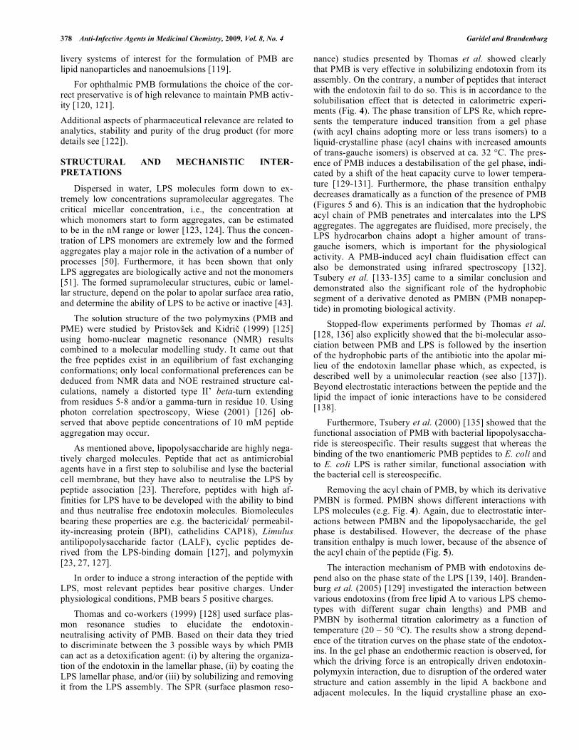
nance) studies presented by Thomas et al. showed clearly that PMB is very effective in solubilizing endotoxin from its assembly. On the contrary, a number of peptides that interact with the endotoxin fail to do so. This is in accordance to the solubilisation effect that is detected in calorimetric experi-ments (Fig. 4). The phase transition of LPS Re, which repre-sents the temperature induced transition from a gel phase (with acyl chains adopting more or less trans isomers) to a liquid-crystalline phase (acyl chains with increased amounts of trans-gauche isomers) is observed at ca. 32 °C. The pres-ence of PMB induces a destabilisation of the gel phase, indi-cated by a shift of the heat capacity curve to lower tempera-ture [129-131]. Furthermore, the phase transition enthalpy decreases dramatically as a function of the presence of PMB (Figures 5 and 6). This is an indication that the hydrophobic acyl chain of PMB penetrates and intercalates into the LPS aggregates. The aggregates are fluidised, more precisely, the LPS hydrocarbon chains adopt a higher amount of trans-gauche isomers, which is important for the physiological activity. A PMB-induced acyl chain fluidisation effect can also be demonstrated using infrared spectroscopy [132]. Tsubery et al. [133-135] came to a similar conclusion and demonstrated also the significant role of the hydrophobic segment of a derivative denoted as PMBN (PMB nonapep-tide) in promoting biological activity. Stopped-flow experiments performed by Thomas et al. [128, 136] also explicitly showed that the bi-molecular asso-ciation between PMB and LPS is followed by the insertion of the hydrophobic parts of the antibiotic into the apolar mi-lieu of the endotoxin lamellar phase which, as expected, is described well by a unimolecular reaction (see also [137]). Beyond electrostatic interactions between the peptide and the lipid the impact of ionic interactions have to be considered [138]. Furthermore, Tsubery et al. (2000) [135] showed that the functional association of PMB with bacterial lipopolysaccha-ride is stereospecific. Their results suggest that whereas the binding of the two enantiomeric PMB peptides to E. coli and to E. coli LPS is rather similar, functional association with the bacterial cell is stereospecific. Removing the acyl chain of PMB, by which its derivative PMBN is formed. PMBN shows different interactions with LPS molecules (e.g. Fig. 4). Again, due to electrostatic inter-actions between PMBN and the lipopolysaccharide, the gel phase is destabilised. However, the decrease of the phase transition enthalpy is much lower, because of the absence of the acyl chain of the peptide (Fig. 5). The interaction mechanism of PMB with endotoxins de-pend also on the phase state of the LPS [139, 140]. Branden-burg et al. (2005) [129] investigated the interaction between various endotoxins (from free lipid A to various LPS chemo-types with different sugar chain lengths) and PMB and PMBN by isothermal titration calorimetry as a function of temperature (20 – 50 °C). The results show a strong depend-ence of the titration curves on the phase state of the endotox-ins. In the gel phase an endothermic reaction is observed, for which the driving force is an entropically driven endotoxin-polymyxin interaction, due to disruption of the ordered water structure and cation assembly in the lipid A backbone and adjacent molecules. In the liquid crystalline phase an exo-
Current Understanding of Polymyxin B Applications Anti-Infective Agents in Medicinal Chemistry, 2009, Vol. 8, No. 4 379
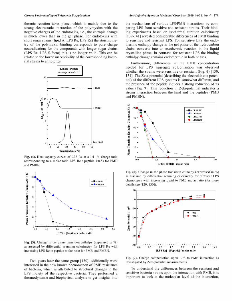
thermic reaction takes place, which is mainly due to the strong electrostatic interaction of the polymyxins with the negative charges of the endotoxins, i.e., the entropic change is much lower than in the gel phase. For endotoxins with short sugar chains (lipid A, LPS Re, LPS Rc) the stoichiome-try of the polymyxin binding corresponds to pure charge neutralization; for the compounds with longer sugar chains (LPS Ra, LPS S-form) this is no longer valid. This can be related to the lower susceptibility of the corresponding bacte-rial strains to antibiotics. Fig. (4). Heat capacity curves of LPS Re at a 1:1 -/+ charge ratio (corresponding to a molar ratio LPS Re : peptide 1:0.8) for PMB and PMBN. Fig. (5). Change in the phase transition enthalpy (expressed in %) as assessed by differential scanning calorimetry for LPS Re with increasing LPS Re to peptide molar ratio for PMB and PMBN. Two years later the same group [130], additionally were interested in the now known phenomenon of PMB resistance of bacteria, which is attributed to structural changes in the LPS moiety of the respective bacteria. They performed a thermodynamic and biophysical analysis to get insights into
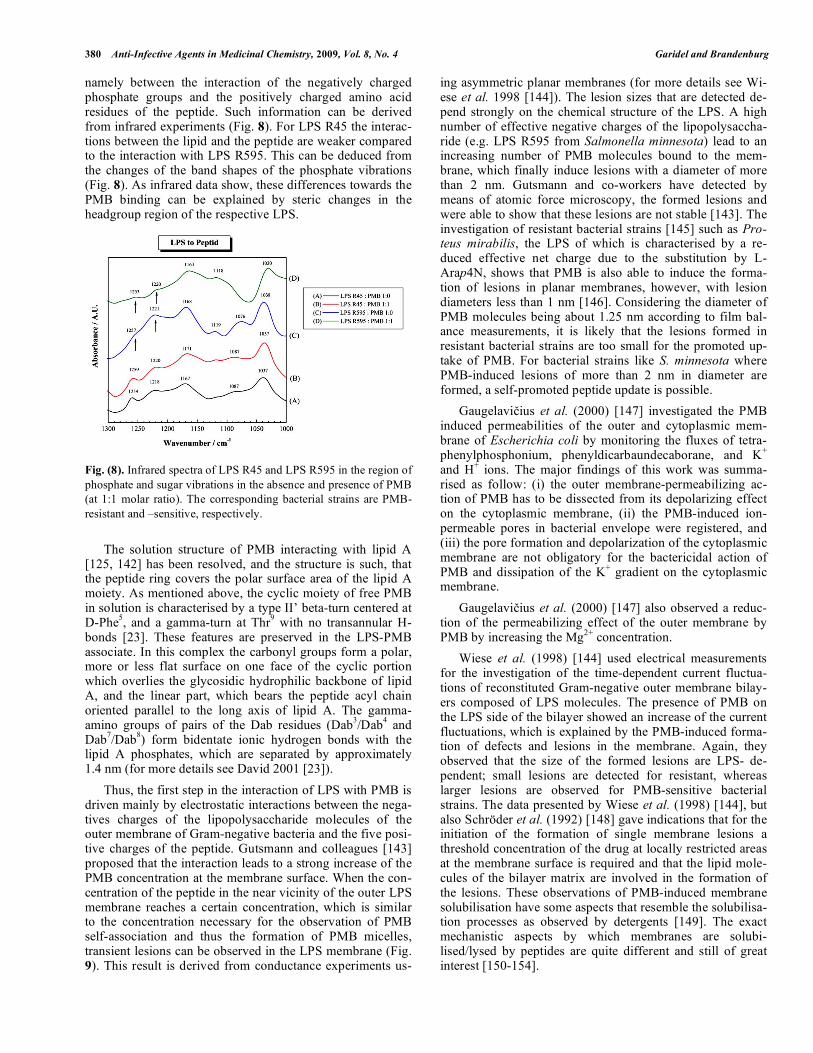
the mechanisms of various LPS/PMB interactions by com-paring LPS from sensitive and resistant strains. Their bind-ing experiments based on isothermal titration calorimetry [139-141] revealed considerable differences of PMB binding to sensitive and resistant LPS. For sensitive LPS the endo-thermic enthalpy change in the gel phase of the hydrocarbon chains converts into an exothermic reaction in the liquid crystalline phase. In contrast, for resistant LPS the binding enthalpy change remains endothermic in both phases. Furthermore, differences in the PMB concentration needed for LPS aggregate solubilisation was observed whether the strains were sensitive or resistant (Fig. 6) [130, 131]. The Zeta-potential (describing the electrokinetic poten-tial) of the different LPS systems is somewhat different, and the presence of the peptide induces a strong reduction of its value (Fig. 7). This reduction in Zeta-potential indicates a strong interaction between the lipid and the peptides (PMB and PMBN).
Fig. (6). Change in the phase transition enthalpy (expressed in %) as assessed by differential scanning calorimetry for different LPS chemotypes with increasing Lipid to PMB molar ratio (for more details see [129, 130]).
Fig. (7). Charge compensation upon LPS to PMB interaction as investigated by Zeta-potential measurements.
To understand the differences between the resistant and sensitive bacteria strains upon the interaction with PMB, it is important to look at the molecular level of the interaction,
380 Anti-Infective Agents in Medicinal Chemistry, 2009, Vol. 8, No. 4 Garidel and Brandenburg
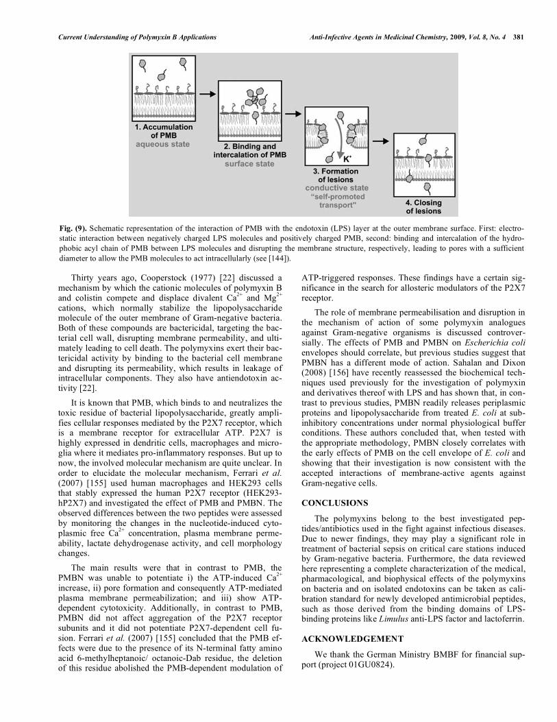
namely between the interaction of the negatively charged phosphate groups and the positively charged amino acid residues of the peptide. Such information can be derived from infrared experiments (Fig. 8). For LPS R45 the interac-tions between the lipid and the peptide are weaker compared to the interaction with LPS R595. This can be deduced from the changes of the band shapes of the phosphate vibrations (Fig. 8). As infrared data show, these differences towards the PMB binding can be explained by steric changes in the headgroup region of the respective LPS. Fig. (8). Infrared spectra of LPS R45 and LPS R595 in the region of phosphate and sugar vibrations in the absence and presence of PMB (at 1:1 molar ratio). The corresponding bacterial strains are PMB-resistant and –sensitive, respectively. The solution structure of PMB interacting with lipid A [125, 142] has been resolved, and the structure is such, that the peptide ring covers the polar surface area of the lipid A moiety. As mentioned above, the cyclic moiety of free PMB in solution is characterised by a type II’ beta-turn centered at D-Phe5, and a gamma-turn at Thr9 with no transannular H-bonds [23]. These features are preserved in the LPS-PMB associate. In this complex the carbonyl groups form a polar, more or less flat surface on one face of the cyclic portion which overlies the glycosidic hydrophilic backbone of lipid A, and the linear part, which bears the peptide acyl chain oriented parallel to the long axis of lipid A. The gamma-amino groups of pairs of the Dab residues (Dab3/Dab4 and Dab7/Dab8) form bidentate ionic hydrogen bonds with the lipid A phosphates, which are separated by approximately 1.4 nm (for more details see David 2001 [23]). Thus, the first step in the interaction of LPS with PMB is driven mainly by electrostatic interactions between the nega-tives charges of the lipopolysaccharide molecules of the outer membrane of Gram-negative bacteria and the five posi-tive charges of the peptide. Gutsmann and colleagues [143] proposed that the interaction leads to a strong increase of the PMB concentration at the membrane surface. When the con-centration of the peptide in the near vicinity of the outer LPS membrane reaches a certain concentration, which is similar to the concentration necessary for the observation of PMB self-association and thus the formation of PMB micelles, transient lesions can be observed in the LPS membrane (Fig. 9). This result is derived from conductance experiments us-
ing asymmetric planar membranes (for more details see Wi-ese et al. 1998 [144]). The lesion sizes that are detected de-pend strongly on the chemical structure of the LPS. A high number of effective negative charges of the lipopolysaccha-ride (e.g. LPS R595 from Salmonella minnesota) lead to an increasing number of PMB molecules bound to the mem-brane, which finally induce lesions with a diameter of more than 2 nm. Gutsmann and co-workers have detected by means of atomic force microscopy, the formed lesions and were able to show that these lesions are not stable [143]. The investigation of resistant bacterial strains [145] such as Pro-teus mirabilis, the LPS of which is characterised by a re-duced effective net charge due to the substitution by L-Arap4N, shows that PMB is also able to induce the forma-tion of lesions in planar membranes, however, with lesion diameters less than 1 nm [146]. Considering the diameter of PMB molecules being about 1.25 nm according to film bal-ance measurements, it is likely that the lesions formed in resistant bacterial strains are too small for the promoted up-take of PMB. For bacterial strains like S. minnesota where PMB-induced lesions of more than 2 nm in diameter are formed, a self-promoted peptide update is possible. Gaugelavi ius et al. (2000) [147] investigated the PMB induced permeabilities of the outer and cytoplasmic mem-brane of Escherichia coli by monitoring the fluxes of tetra-phenylphosphonium, phenyldicarbaundecaborane, and K+ and H+ ions. The major findings of this work was summa-rised as follow: (i) the outer membrane-permeabilizing ac-tion of PMB has to be dissected from its depolarizing effect on the cytoplasmic membrane, (ii) the PMB-induced ion-permeable pores in bacterial envelope were registered, and (iii) the pore formation and depolarization of the cytoplasmic membrane are not obligatory for the bactericidal action of PMB and dissipation of the K+ gradient on the cytoplasmic membrane. Gaugelavi ius et al. (2000) [147] also observed a reduc-tion of the permeabilizing effect of the outer membrane by PMB by increasing the Mg2+ concentration. Wiese et al. (1998) [144] used electrical measurements for the investigation of the time-dependent current fluctua-tions of reconstituted Gram-negative outer membrane bilay-ers composed of LPS molecules. The presence of PMB on the LPS side of the bilayer showed an increase of the current fluctuations, which is explained by the PMB-induced forma-tion of defects and lesions in the membrane. Again, they observed that the size of the formed lesions are LPS- de-pendent; small lesions are detected for resistant, whereas larger lesions are observed for PMB-sensitive bacterial strains. The data presented by Wiese et al. (1998) [144], but also Schröder et al. (1992) [148] gave indications that for the initiation of the formation of single membrane lesions a threshold concentration of the drug at locally restricted areas at the membrane surface is required and that the lipid mole-cules of the bilayer matrix are involved in the formation of the lesions. These observations of PMB-induced membrane solubilisation have some aspects that resemble the solubilisa-tion processes as observed by detergents [149]. The exact mechanistic aspects by which membranes are solubi-lised/lysed by peptides are quite different and still of great interest [150-154].
Current Understanding of Polymyxin B Applications Anti-Infective Agents in Medicinal Chemistry, 2009, Vol. 8, No. 4 381
Thirty years ago, Cooperstock (1977) [22] discussed a mechanism by which the cationic molecules of polymyxin B and colistin compete and displace divalent Ca2+ and Mg2+ cations, which normally stabilize the lipopolysaccharide molecule of the outer membrane of Gram-negative bacteria. Both of these compounds are bactericidal, targeting the bac-terial cell wall, disrupting membrane permeability, and ulti-mately leading to cell death. The polymyxins exert their bac-tericidal activity by binding to the bacterial cell membrane and disrupting its permeability, which results in leakage of intracellular components. They also have antiendotoxin ac-tivity [22]. It is known that PMB, which binds to and neutralizes the toxic residue of bacterial lipopolysaccharide, greatly ampli-fies cellular responses mediated by the P2X7 receptor, which is a membrane receptor for extracellular ATP. P2X7 is highly expressed in dendritic cells, macrophages and micro-glia where it mediates pro-inflammatory responses. But up to now, the involved molecular mechanism are quite unclear. In order to elucidate the molecular mechanism, Ferrari et al. (2007) [155] used human macrophages and HEK293 cells that stably expressed the human P2X7 receptor (HEK293-hP2X7) and investigated the effect of PMB and PMBN. The observed differences between the two peptides were assessed by monitoring the changes in the nucleotide-induced cyto-plasmic free Ca2+ concentration, plasma membrane perme-ability, lactate dehydrogenase activity, and cell morphology changes. The main results were that in contrast to PMB, the PMBN was unable to potentiate i) the ATP-induced Ca2+ increase, ii) pore formation and consequently ATP-mediated plasma membrane permeabilization; and iii) show ATP-dependent cytotoxicity. Additionally, in contrast to PMB, PMBN did not affect aggregation of the P2X7 receptor subunits and it did not potentiate P2X7-dependent cell fu-sion. Ferrari et al. (2007) [155] concluded that the PMB ef-fects were due to the presence of its N-terminal fatty amino acid 6-methylheptanoic/ octanoic-Dab residue, the deletion of this residue abolished the PMB-dependent modulation of
ATP-triggered responses. These findings have a certain sig-nificance in the search for allosteric modulators of the P2X7 receptor. The role of membrane permeabilisation and disruption in the mechanism of action of some polymyxin analogues against Gram-negative organisms is discussed controver-sially. The effects of PMB and PMBN on Escherichia coli envelopes should correlate, but previous studies suggest that PMBN has a different mode of action. Sahalan and Dixon (2008) [156] have recently reassessed the biochemical tech-niques used previously for the investigation of polymyxin and derivatives thereof with LPS and has shown that, in con-trast to previous studies, PMBN readily releases periplasmic proteins and lipopolysaccharide from treated E. coli at sub-inhibitory concentrations under normal physiological buffer conditions. These authors concluded that, when tested with the appropriate methodology, PMBN closely correlates with the early effects of PMB on the cell envelope of E. coli and showing that their investigation is now consistent with the accepted interactions of membrane-active agents against Gram-negative cells.
CONCLUSIONS
The polymyxins belong to the best investigated pep-tides/antibiotics used in the fight against infectious diseases. Due to newer findings, they may play a significant role in treatment of bacterial sepsis on critical care stations induced by Gram-negative bacteria. Furthermore, the data reviewed here representing a complete characterization of the medical, pharmacological, and biophysical effects of the polymyxins on bacteria and on isolated endotoxins can be taken as cali-bration standard for newly developed antimicrobial peptides, such as those derived from the binding domains of LPS-binding proteins like Limulus anti-LPS factor and lactoferrin.
ACKNOWLEDGEMENT
We thank the German Ministry BMBF for financial sup-port (project 01GU0824).
Fig. (9). Schematic representation of the interaction of PMB with the endotoxin (LPS) layer at the outer membrane surface. First: electro-static interaction between negatively charged LPS molecules and positively charged PMB, second: binding and intercalation of the hydro-phobic acyl chain of PMB between LPS molecules and disrupting the membrane structure, respectively, leading to pores with a sufficient diameter to allow the PMB molecules to act intracellularly (see [144]).
382 Anti-Infective Agents in Medicinal Chemistry, 2009, Vol. 8, No. 4 Garidel and Brandenburg
REFERENCES [1] Storm, D.R.; Rosenthal, K.S.; Swanson, P.E. Polymyxin and re-
lated peptide anti-biotics. Ann. Rev. Biochem., 1977, 46, 723. [2] Evans, M. E.; Feola, D. J.; Rapp, R. P. Polymyxin B sulfate and
colistin: old antibiotics for emerging multiresistant gram-negative bacteria. Ann. Pharmacother., 1999, 33, 960.
[3] Gales, A. C.; Jones, R. N.; Sader, H. S. Global assessment of the antimicrobial activity of polymyxin B against 54 731 clinical iso-lates of Gram-negative bacilli: report from the SENTRY antimi-crobial surveillance programme (2001–2004). Clin. Microbiol. In-fect., 2006, 12, 315.
[4] Michalopoulos, A.; Falagas, M. E. Colistin and Polymyxin B in Crit. care. Crit. Care Clin., 2008, 24, 377.
[5] Falagas, M. E.; Kasiakou, S. K. Toxicity of polymyxins: a system-atic review of the evidence from old and recent studies. Crit. Care, 2006, 10, R27.
[6] Falagas, M. E.; Kasiakou, S. K. Colistin: the revival of polymyxins for the management of multidrug-resistant Gram-negative bacterial infections. Rev. Anti-infect. Agents, 2005, 40, 1333.
[7] Zavascki, A.P.; Goldani, L.Z.; Cao, G.; Superti, S.V.; Lutz, L.; Barth, A.L.; Ramos, F.; Boniatti, M.M.; Nation, R.L.; Li, J. Phar-macokinetics of intravenous polymyxin B in Crit.ly ill pa-tients. Clin. Infect. Dis., 2008, 47 , 1298.
[8] Zavascki, A. P.; Goldani, L. Z.; Li, J.; Nation, R.L. Polymyxin B for the treatment of multidrug-resistant pathogens: A Crit. review. J. Antimicrob. Chemother., 2007, 60, 1206.
[9] Hornef, M. W.; Wick, M. J.; Rhen, M.; Normark, S. Bacterial strategies for overcoming host innate and adaptive immune re-sponses. Nat. Immunol., 2002, 3; 1033.
[10] Moskowitz, S. M.; Ernst, R. K.; Miller, S. I. PmrAB, a two compo-nent regulatory system of Pseudomonas aeruginosa that modulates resistance to cationic antimicrobial peptides and addition of ami-noarabinose to lipid A. J. Bacteriol., 2004, 186, 575.
[11] Winfield, M. D.; Groisman, E. A. Phenotypic differences between Salmonella and Escherichia coli resulting from the disparate regu-lation of homologous genes. Proc. Natl. Acad. Sci. USA, 2004, 101, 17162.
[12] Brodsky, I. E.; Ernst, R. K.; Miller, S. I. Mig-14 is a Salmonella gene that plays a role in bacterial resistance to antimicrobial pep-tides. J. Bacteriol., 2002, 184, 3203.
[13] Campos, M. A.; Vargas, M. A.; Regueiro V. Capsule polysaccha-ride mediates bacterial resistance to antimicrobial peptides. Infect. Immun., 2004, 72, 7107.
[14] Loutet, S. A.; Flannagan, R. S.; Kooi, C. A complete lipopolysac-charide inner core oligosaccharide is required for resistance of Burkholderia cenocepacia to antimicrobial peptides and bacterial survival in vivo. J. Bacteriol., 2006, 188, 2073.
[15] Tran, A. X.; Whittomore, J. D.; Wyrick, P. B. The lipid A 1-phosphatase of Helicobacter pylori is required for resistance to the antimicrobial peptide polymyxin. J. Bacteriol., 2006, 188, 4531.
[16] Winfield, M. D.; Latifi, T.; Groisman, E. A. Transcriptional regula-tion of the 4-amino-5-deoxy-L-arabinose biosynthetic genes in Yersinia pestis. J. Biol. Chem., 2005, 280, 14765.
[17] Mathur, J.; Waldor, M. K. The Vibrio cholera ToxR-regulated porin OmpU confers resistance to antimicrobial peptides. Infect. Immun., 2004, 72, 3577.
[18] Bedford Laboratories. Polymyxin B for injection (package insert), OH 44146. Bedford Laboratories, 2004, at http:// dailymed.nlm.nih.gov.
[19] Garidel, P.; Andrä, J.; Howe, J.; Gutsmann, T.; Brandenburg, K. Novel anti-inflammatory and antiinfective agents. Anti-Infect. Agents Med. Chem., 2007, 6, 185.
[20] Beutler, B.; Rietschel, E. Th. Innate immune sensing and its roots: the story of endotoxin. Nat. Rev., 2003, 3, 169.
[21] Evans, M. E., Pollack, M. Effect of antibiotic class and concentra-tion on the release of lipopolysaccharide from Escherichia coli. J. Infect. Dis. , 1993, 1336.
[22] Cooperstock, M.S. Inactivation of endotoxin by polymyxin B. Antimicrob. Agents Chemother., 1974, 6, 422.
[23] David, S. A. Towards a rational development of anti-endotoxin agents: novel approaches to sequestration of bacterial endotoxins with small molecules. J. Mol. Recognit., 2001, 14; 370.
[24] Nahra, R.; Dellinger, R. P. Targeting the lipopolysaccharides: still a matter of debate? Curr. Opin. Anesthesiol., 2008, 21, 98.
[25] Cunha, B. A. New uses for older antibiotics: Nitrofurantoin, Ami-kacin, Colistin, Polymyxin B, Doxycycline, and Minocycline revis-ited. Med. Clin. North Am., 2006, 90, 1089.
[26] Fenner, T. Therapie von Infektionen. Erregerspektrum, Diagnostik, Behandlungsschemata. 2. Auflage, Schattauer GmbH: Stuttgart, 2003.
[27] Andrä, J.; Gutsmann, T.; Garidel, P.; Brandenburg, K. Mechanisms of endotoxin neutralization by synthetic compounds. J. Endotox. Res., 2006, 12, 261.
[28] Kwa, A.; Kasiakou, S. K.; Tam, V. H.; Falagas, M. E. Polymyxin B: Similarities to and differences from colistin (polymyxin E). Ex-pert Rev. Anti-Infect. Ther., 2007, 5 (5), 811.
[29] Lynn, W. A. Anti-endotoxin Ther.eutic options for the treatment of sepsis. J. Antimicrob. Chemother., 1998, 41, (Suppl. A), 71.
[30] Tam, V.; Schilling, A.; Vo, G. Pharmacodynamics of polymyxin B against Pseudomonas aeruginosa. Antimicrob. Agents Chemother., 2005, 49, 3624.
[31] Nation, R. L.; Li, J. Optimizing use of colistin and polymyxin B in the Crit.ly ill. Semin. Respir. Crit. Care Med., 2007, 28, 604.
[32 Consensus Conference Committee. American College of Chest Physicians / Society of Crit. Care Medicine consensus conference: definitions for sepsis and organ failure and guidelines for the use of innovative Ther.ies in sepsis. Crit. Care Med., 1992, 20, 864.
[33] Levy, M. M.; Fink, M. P.; Marshall, J. C.; Abraham, E.; Angus, D.; Cook, D.; Opal, S. M.; Vincent, J. L.; Ramsay, G. 2001 SCCM/ESICM/ACCP/ATS/SIS International Sepsis Definitions Conference. Crit. Care Med., 2003, 31; 1250 and published in In-tensive Care Med., 2003, 29, 530.
[34] Vincent, J. L.; Habib, A. M.; Verdant, C.; Bruhn, A. Sepsis diagno-sis and management: work in progress. Minerva Anestesiol., 2006, 72, 87.
[35] Moerer, O.; Schmid, A.; Hofmann, M.; Herklotz, A.; Reinhart, K.; Wedan, K.; Schneider, H.; Burchardi, H. Direct costs of severe sep-sis in three German intensive care units based on retrospective elec-tronic patient record analysis of resource use. Intensive Care Med., 2002, 28, 1440.
[36] Remick, D. G. Pathophysiology of sepsis. Biological perspectives. Am. J. Pathol., 2007, 170, 1435.
[37] Jacobi, J. Pathophysiology of sepsis. Am. J. Health-Syst. Pharm., 2002, 59, S3.
[38] Brandenburg, K.; Jürgens, G., Andrä, J.; Lindner, B.; Koch, M .H. J.; Blume, A.; Garidel, P. Biophysical characterization of the inter-action of high-density lipoprotein (HDL) with endotoxins. Eur. J. Biochem., 2002, 269, 5972.
[39] Brandenburg, K.; Garidel, P.; Andrä, J.; Jürgens, G.; Müller, M.; Blume, A.; Koch, M. H. J.; Levin, J. Cross-linked hemoglobin con-verts endotoxically inactive pentaacyl endotoxins into a physio-logically active conformation. J. Biol. Chem., 2003, 278, 47660.
[40] Jürgens, G.; Müller, M.; Garidel, P.; Koch, M. H. J.; Nakakubo, H.; Blume, A.; Brandenburg, K. Investigation into the interaction of recombinant human serum albumin with Re-lipopolysaccharide and lipid A. J. Endotox. Res., 2002, 8, 115.
[41] Hornef, M. W.; Henriques-Normark, Normak, S. The function and biological role of toll-like receptors in infectious diseases: an up-date. Current Opinion Infect. Dis., 2008, 21, 304.
[42] Bhor, V. M.; Thomas, C. J.; Surolia, N.; Surolia, A. Polymyxin B: An ode to an old antidote for endotoxic shock. Mol. BioSyst., 2005, 1, 213.
[43] Brandenburg, K.; Andrä, J.; Müller, M.; Koch, M. H. J.; Garidel, P. Physicochemical properties of bacterial glycopolymers in relation to bioactivity. Carbohydrate Res., 2003, 338, 2477.
[44] Holst, O. The structures of core regions from enterobacterial lipopolysaccharides - An update. FEMS Microbiol. Lett., 2007, 271, 3.
[45] Rietschel, E. Th.; Seydel, U.; Zähringer, U.; Schade, U. F.; Brade, L.; Loppnow, H.; Feist, W.; Wang, M.-H.; Ulmer, A. J.; Flad, H.-D.; Brandenburg, K.; Kirikae, T.; Grimmecke, D.; Holst, O.; Brade, H. Bacterial endotoxin: Molecular relationships between structure and activity. Infect. Dis. Clin. North Am., 1991, 5, 753.
[46] Seydel, U.; Hawkins, L.; Schromm, A. B.; Heine, H.; Scheel, O.; Koch, M. H. J.; Brandenburg, K. The generalized endotoxic princi-ple. Eur. J. Immunol., 2003, 33, 1586.
[47] Brandenburg, K.; Hawkins, L.; Garidel, P.; Andrä, J.; Müller, M.; Heine, H.; Koch, M. H. J.; Seydel, U. Structural polymorphism and endotoxic activity of synthetic phospholipid-like amphiphiles. Bio-chemistry, 2004, 43, 4039.
Current Understanding of Polymyxin B Applications Anti-Infective Agents in Medicinal Chemistry, 2009, Vol. 8, No. 4 383
[48] Schromm, A. B.; Howe, J.; Ulmer, A. J.; Wiesmüller, K.-H.; Sey-berth, T.; Jung, G.; Rössle, M.; Koch, M. H. J.; Gutsmann, T.; Brandenburg, K. Physicochemical and biological analysis of syn-thetic bacterial lipopeptides: Validity of the concept of endotoxic conformation. J. Biol. Chem., 2007, 282, 11030.
[49] Schromm, A. B.; Brandenburg, K.; Loppnow, H.; Zähringer, U.; Rietschel, E. Th.; Carroll, S. F.; Koch, M. H. J.; Kusumoto, S.; Seydel, U. The charge of endotoxin molecules influences their con-formation and interleukin-6 inducing capacity, Eur. J. Biochem., 2000, 161, 5464.
[50] Schromm, A. B.; Brandenburg, K.; Rietschel, E. T.; Seydel, U. Do endotoxin aggregates intercalate into phospholipid membranes in a nonspecific, hydrophobic manner? J. Endotox. Res., 1995, 2, 313.
[51] Mueller, M.; Lindner, B.; Kusumoto, S.; Fukase, K.; Schromm, A. B.; Seydel, U. Aggregates are the biologically active units of endo-toxin. J. Biol. Chem., 2004, 279, 26307.
[52] Brandenburg, P.; Garidel, P.; Gutsmann, T. In: Physicochemical properties of microbial glycoploymers. Moran, A.; Holst, O.; Bren-nan, P.; von Itzstein, M. Eds, Microbial Glykobiology, Elsevier 2009, Chapter 38, pp. 759-779.
[53] Schürholz, T.; Marx, G. Management of sepsis. Minerva Anesthe-siol., 2008, 74, 181.
[54] Wheeler, A.P.; Bernard, G.R. Treating patients with severe sepsis. N. Engl. J. Med., 1999, 340, 207.
[55] Marx, G.; Schuerholz, T.; Reinhart, K. Intensivmedizinische As-pekte bei Sepsis. Chirurg, 2005, 76, 845.
[56] Mackenzie, I.; Lever, A. Management of sepsis. Br. Med. J., 2007, 335, 929.
[57] Weigand, M. A.; Bardenheuer, H. J.; Böttiger, B. W. Klinisches Management bei Patienten mit Sepsis. Der Anaesthesist, 2003, 52, 3.
[58] Dellinger, R.; Levy, M. M.; Carlet, J. M.; Bion, J.; Parker, M. M.; Jaeschke, R.; Reinhart, K.; Angus, D. C.; Brun-Buisson, C.; Beale, R.; Calandra, T.; Dhainaut, J. F.; Gerlach, H.; Harvey, M.; Marini, J. J.; Marshall, J.; Ranieri, M.; Ramsay, G.; Sevransky, J.; Thomp-son, T.; Townsend, S.; Vender, J. S.; Zimmerman, J. L.; Vincent, J. L. Surviving Sepsis Campaign: International guidelines for man-agement of severe sepsis and septic shock. Crit. Care Med., 2008, 36, 296.
[59] Bernard, G. R. Sepsis trials: intersection of investigation, regula-tion, funding, and practice. Am. J. Respir. Crit. Care Med., 1995, 152, 4.
[60] Landman, D.; Georgescu, C.; Martin, D. A.; Quale, J. Polymyxins revisited. Clin. Microbiol. Rev., 2008, 21, 449.
[61] Bergen, P. J.; Rayner, C. R.; Nation, R. L. Colistin methanesul-fonate is an inactive prodrug of colistin against Pseudomonas aeruginosa. Antimicrob. Agents Chemother., 2006, 50, 1953.
[62] Yuan, Z.; Tam, H. Polymyxin B: a new strategy for multidrug-resistant Gram-negative organisms. Expert Opin. Investig. Drug, 2008, 17, 661.
[63] Horton, J.; Pankey, G. A. Polymyxin B, colistin, and sodium colis-timethate. Med. Clin. North Am., 1982, 66, 135.
[64] Hogardt, M.; Schmoldt, S.; Götzfried, M.; Adler, K.; Heesemann, J. Pitfalls of polymyxin antimicrobial susceptibility testing of Pseudomonas aeruginosa isolated from cystic fibrosis patients. J. Antimicrob. Chemother., 2004, 54, 1057.
[65] Li, J.; Turnidge, J.; Milne, R.; R. L. Nation; K. Coulthard. In vitro pharmacodynamic properties of colistin and colistin methanesul-fonate against Pseudomonas aeruginosa isolates from patients with cystic fibrosis. Antimicrob. Agents Chemother., 2001, 45, 781.
[66] Seltmann, G.; Lindner, B.; Holst, O. Resistance of Serratia marcescens to polymyxin B: A comparative investigation of two S-form lipopolysaccharides obtained from a sensitive and a resistant variant of strain 111. J. Endotox. Res., 1996, 3, 497.
[67] Ko, K. S.; Suh, J. Y.; Kwon, K. T.; Jung, S.-I.; Park, K.-H.; Kang, C. I.; Chung, D. R.; Peck, K. R.; Song, J.-H. High rates of resis-tance to colistin and polymyxin B in subgroups of Acinetobacter baumannii isolates from Korea. J. Antimicrob. Chemother., 2007, 60, 1163.
[68] Antoniadou, A.; Kontopidou, F.; Poulakou, G. Colistin-resistant isolates of Klebsiella pneumoniae emerging in intensive care unit patients: first report of a multiclonal cluster. J. Antimicrob. Che-mother., 2007, 59, 786.
[69] Michalopoulos, A.; Kasiakou, S. K.; Rosmarakis, E. S.; Falagas, M. Cure of multidrug-resistant Acinetobacter baumannii bacterae-
mia with continuous intravenous infusion of colistin. Scand. J. In-fect. Dis., 2005, 37, 142.
[70] Sarria, J. C.; Angulo-Pernett; F.; Kimbrough, R. C.; McVay, C. S.; Vidal, A. M. Use of intravenous polymyxin B during continuous venovenous hemodialysis. Eur. J. Clin. Microbiol. Infect. Dis., 2004, 23, 340.
[71] Goverman, J.; Weber, J. M.; Keaney, T. J.; Sheridan, R. L. Intrave-nous colistin for the treatment of multidrug resistant, Gram-negative infection in the paediatric population. J. Burn Care Res., 2007, 28, 421.
[72] Sobieszczyk, M.; Yoko Furuya, E.; Hay, C. M.; Pancholi P.; Della-Latta, P.; Hammer, S. H.; Kubin, C. J. Combination Ther.y with polymyxin B for the treatment of multidrug-resistant gram-negative respiratory tract infections. J. Antimicrob. Chemother., 2004, 54, 566.
[73] Holloway, K. P.; Rouphael, N. G.; Wells, J. B.; King. M. D.; Blumberg, H. M. Polymyxin B and doxycycline use in patients with multidrug-resistant Acinetobacter baumannii infections in the intensive care unit. Ann. Pharmacother., 2006, 40, 1939.
[74] Furtado, G. H. C.; d' Azevedo, P. A.; Santos, A. F.; Gales, A. C.; Pignatari, A. C. C.; Medeiros, E. A. S. Intravenous polymyxin B for the treatment of nosocomial pneumonia caused by multidrug-resistant Pseudomonas aeruginosa. Int. J. Antimicrob. Agents, 2007, 30, 315.
[75] Pastewski, A. A.; Caruso, P.; Parris, A. R.; Dizon, R.; Kopec, R.; Sharma, S.; Mayer, S.; Ghitan, M.; Chapnick, E. K. Parenteral po-lymyxin B use in patients with multidrug-resistant Gram-negative bacteraemia and urinary tract infections: A retrospective case se-ries. Ann. Pharmacother., 2008, 42, 1177.
[76] Pereira, G. H.; Muller, P. R.; Levin, A. S. Salvage treatment of pneumonia and initial treatment of tracheobronchitis caused by multidrug-resistant Gram-negative bacilli with inhaled polymyxin B. Diagn. Microbiol. Infect. Dis., 2007, 58, 235.
[77] Zhou, B.-P.; Lu, J.; Qiu, C.; Wu, S.-P.; Song, W.-D.; Yang, D.-G.; Fu, Y.-Y.; Gao, X.; Liu, X.-Y.; Liu, S.-T.; Lu, P.-X.; Shan, W.-S.; Li, Y.-M.; Yuan, G.-Y.; Zhong, N.-S. The treatment of multidrug-resistant Pseudomonas aeruginosa with polymyxin B or E in a pa-tient with the highly pathogenic avian influenza A (H5N1) infec-tion: A case report and review of the literature. Chin. J. Antibiot., 2007, 32, 685.
[78] Markou N, Apostolakos H, Koumoudiou C.; Athanasiou, M.; Koutsoukou, A.; Alamanos, I. ; Gregorakos, L. Intravenous colistin in the treatment of sepsis from multiresistant Gram-negative bacilli in Crit.ly ill patients. Crit Care, 2003, 7, R78.
[79] Karabinis, A.; Paramythiotou, E.; Mylona-Petropoulou, D.; Kalogeromitros, A.; Katsarelis, N.; Kontopidou, F.; Poularas, I.; Malamou-Lada, H. Colistin for Klebsiella pneumoniae–associated sepsis. Clin. Infect. Dis., 2004, 38, e7.
[80] Kopterides, P.; Koletsi, P.; Morfou; P.; Rellos; K.; Mastora; Z.; Rizos; M.; Falagas, M. Exposure to fluoroquinolones is a risk fac-tor for polymyxins-only susceptible (POS) Acinetobacter bauman-nii bacteraemia in Crit.ly ill patients. Int. J. Antimicrob. Agents, 2007, 30, 409.
[81] Kollef, M. H. An empirical approach to the treatment of multidrug-resistant ventilator associated pneumonia. Clin. Infect. Dis., 2003, 36, 1119.
[82] Cruz, D. N.; Perazella, M. A.; Bellomo, R.; de Cal, M.; Polanco, N.; Corradi, V.; Lentini, P.; Nalesso, F.; Ueno, T.; Ranieri, V. M.; Ronco, C. Effectiveness of polymyxin B-immobilized fiber column in sepsis: a systematic review. Crit. Care, 2007, 1, R47.
[83] Cruz, D. N.; Bellomo, R.; Ronco, C. Clinical effects of polymyxin B-immobilized fibre column in septic patients. Contrib. Nephrol., 2007, 156, 444.
[84] Shoji, H.; Tani, T.; Hanasawa, K.; Kodama, M. Extracorporeal endotoxin removal by polymyxin B immobilized fibre cartridge: Designing and antiendotoxin efficacy in the clinical application. Ther. Apher., 1998, 2, 3.
[85] Teramoto, K.; Nakamoto, Y.; Kunitomo, T.; Shoji, H.; Tani, T.; Hanazawa, K.; Kodama, M. Removal of endotoxin in blood by po-lymyxin B immobilized polystyrene-derivative fibre Ther. Apher., 2002, 6, 103.
[86] Tani, T. Review of endotoxin-adsorbing direct hemoperfusion therapy using a column containing polymyxin B immobilized fibre. Curr. Opin Crit. Care, 2000, 6, 416.
[87] Ruberto, F.; Pugliese, F.; D' Alio, A.; Martelli, S.; Bruno, K.; Marcellino, V.; Summonti, D.; Celli, P.; Perrella, S.; Cappannoli,
384 Anti-Infective Agents in Medicinal Chemistry, 2009, Vol. 8, No. 4 Garidel and Brandenburg
A.; Pietropaoli, C.; Tosi, A.; Diana, B.; Novelli, G.; Rossi, M.; Gi-nanni Corradini, S. ;Ferretti, G.; Berloco, P. B.; Pietropaoli, P. Clinical effects of direct hemoperfusion using a polymyxin-B im-mobilized column in solid organ transplanted patients with signs of severe sepsis and septic shock. A pilot study. Int. J. Artif. Organs, 2007, 30, 915.
[88] Ruberto, F.; Pugliese, F.; D' Alio, A.; Martelli, S.; Bruno, K.; Marcellino, V.; Perrella, S.; Cappannoli, A.; Mazzarino, V.; Tosi, A.; Novelli, G.; Rossi, M.; Ginanni Corradini, S.; Ferretti, G.; Ber-loco, P. B.; Pietropaoli, P. Clinical effects of use Polymyxin B fixed on fibers in liver transplant patients with severe sepsis or sep-tic shock. Transplant. Proc., 2007, 39, 1953.
[89] Kakugawa, T.; Mukae, H.; Saito, M.; Ishii, K.; Ishimoto, H.; Sa-kamoto, N.; Takazono, T.; Fukuda, Y.; Ooe, N.; Kohno, S. Rapidly progressive interstitial pneumonia associated with clinically amy-opathic dermatomyositis successfully treated with polymyxin B-immobilized fibre column hemoperfusion. Int. Med., 2008, 47, 785.
[90] Murakami, M.; Miyauchi, Y.; Nishida, M.; Okada, H.; Hamano, K. Early initiation of polymyxin B-immobilized fiber Ther.y effective for septic shock after aortic replacement. Ann. Thorac. Cardiovasc. Surg., 2007, 13, 287.
[91] Sakamoto, Y.; Mashiko, K.; Obata, T.; Matsumoto, H.; Hara, Y.; Kutsukata, N.; Yamamoto, Y. Clinical responses and improvement of some laboratory parameters following polymyxin B-immobilized fibre treatment in septic shock. ASAIO J., 2007, 53, 646.
[92] Sakamoto, Y.; Mashiko, K.; Obata, T.; Matsumoto, H.; Hara, Y.; Kutsukata, N.; Yamamoto, Y. Effectiveness of continuous hemo-diafiltration using a polymethylmethacrylate membrane hemofilter after polymyxin B-immobilized fibre column therapy of septic shock. ASAIO J., 2008, 54, 129.
[93] Cantaluppi, V.; Assenzio, B.; Pasero, D.; Romanazzi, G. M.; Pacit-ti, A.; Lanfranco, G.; Puntorieri, V.; Martin, E. L.; Mascia, L.; Monti, G.; Casella, G. Segoloni, G. P.; Camussi, G.; Ranieri, V. M. Polymyxin-B hemoperfusion inactivates circulating proapoptotic factors. Intensive Care Med., 2008, 34, 1638.
[94] Naka, T.; Shinozaki, M.; Akizawa, T.; Shima, Y.; Takaesu, H.; Nasu, H. The effect of continuous veno-venous hemofiltration or direct hemoperfusion with polymyxin B-immobilized fibre on neu-trophil respiratory oxidative burst in patients with sepsis and septic shock. Ther. Apher. Dial., 2006, 10, 7.
[95] Nakamura, T.; Kawagoe, Y.; Ueda, Y.; Node, K.; Koide, H. Po-lymyxin B-immobilized fibre haemoperfusion reduces urinary 8-hydroxy-2 -deoxyguanosine levels in patients with septic shock. (2006) Clin. Intensive Care, 2006, 17, 95.
[96] Kase, Y.; Obata, T.; Okamoto, Y.; Iwai, K.; Saito, K.; Yokoyama, K.; Takinami, M.; Tanifuji, Y. Removal of 2-arachidonylglycerol by direct hemoperfusion Ther.y with polymyxin B immobilized fi-bers benefits patients with septic shock. Ther. Apher. Dial., 2008, 12, 374.
[97] Shimizu, T.; Hanasawa, K.; Sato, K.; Umeki, M.; Koga, N.; Na-ganuma, T.; Sato, S.; Shimonishi, T.; Ikeda, T.; Matsuno, N.; Ono, S.; Saitoh, H.; Satoh, K.; Otani, Y.; Endo, Y.; Eguchi, Y.; Tani, T. Direct hemoperfusion with polymyxin-B-immobilized fiber col-umns improves septic hypotension and reduces inflammatory me-diators in septic patients with colorectal perforation. Langenbeck's Arch. Surg., 2008, pp. 1-9.
[98] Kushi, H.; Miki, T.; Sakagami, Y.; Sato, J.; Saito, T.; Tanjoh, K. Early hemoperfusion with a polymyxin B column improves gastric intramucosal pH in sepsis. Ther. Apher. Dial., 2008, 12, 278.
[99] Ohki, S.; Oshima, K.; Takeyoshi, I.; Matsumoto, K.; Morishita, Y. Endotoxin removal with a polymyxin B-immobilized hemoperfu-sion cartridge improves cardiopulmonary function after cardiopul-monary bypass. J. Surg. Res., 2008, 145, 74.
[100] Komatsu, S.; Shimomatsuya, T.; Nakajima, M.; Ono, S.; Maru-hashi, K. Severity scoring systems for prognosis and efficacy of po-lymyxin B-immobilized fibre treatment for colonic perforation. Surg. Today, 2006, 36, 807.
[101] Morishita, Y.; Kita, Y.; Ohtake, K.; Kusano, E.; Hishikawa, S.; Mizuta, K.; Kawarasaki, H. Successful treatment of sepsis with po-lymyxin B-immobilized fibre hemoperfusion in a child after living donor liver transplantation. Digest. Dis. Sci., 2005, 50, 757.
[102] Ronco, R. The place of early haemoperfusion with polymyxin B fibre column in the treatment of sepsis. Crit. Care, 2005, 9, 631.
[103] Giacometti, A.; Cirioni, O.; Ghiselli, R.; Mocchegiani, F.; Paggi, A.M.; Orlando, F.; Kamysz, W.; Kasprzykowski, F.; Ma kiewicz,
Z.; Scalise, G.; Saba, V. Ther.eutic efficacy of intraperitoneal po-lymyxin B and polymyxin-like peptides alone or combined with levofloxacin in rat models of septic shock. J. Antimicrob. Che-mother., 2002, 49, 193.
[104] Moriyama, N.; Miyoshi, M.; Imoto, T.; Maruyama, M.; Shido, O.; Watanabe, T. Systemic administration of polymyxin B induces hy-pothermia in rats via an inhibitory effect on metabolic rate. Eur. J. Pharmacol., 2006, 541, 38.
[105] Rybní ek, J.; Hill, P.B. Suspected polymyxin B-induced pemphi-gus vulgaris in a dog. Vet. Dermatol., 2007, 18, 165.
[106] Oshima, K.; Akao, T.; Kobayashi, K.; Muraoka, M.; Matsumoto, K.; Takeyoshi, I. The effect of direct hemoperfusion with a po-lymyxin B-immobilized fibre column (DHP-PMX Ther.y) on pul-monary ischemia-reperfusion injury in a canine model. J. Invest. Surgery, 2008, 21, 127.
[107] Nakanowatari, Y.; Nemoto, K.; Hara, S.; Ninomiya, N.; Yama-moto, Y. Effects of direct haemoperfusion through fibres immobi-lizing polymyxin B and nafamostat mesilate on endotoxaemia in conscious guinea-pigs. Clin. Expert Pharmacol. Physiol., 2008, 35, 17.
[108] Yamamoto, H.; Koizumi, T.; Kaneki, T.; Fujimoto, K.; Kubo, K.; Honda, T. Direct hemoperfusion with polymyxin B-immobilized fiber improves shock and hypoxemia during endotoxemia in anes-thetized sheep. J. Endotox. Res., 2002, 8, 418.
[109] Morresey, P. R.; MacKay, R. J. Endotoxin-neutralizing activity of polymyxin B in blood after iv administration in horses. Am. J. Vet. Res., 2006, 67, 642.
[110] Barton, M.H. Use of Polymyxin B for treatment of endotoxemia in horses. Compend. Cont. Educ. Pract. Vet., 2000, 22, 1056.
[111] Barton, M. H.; Parviainen, A.; Norton, N. Polymyxin B protects horses against induced endotoxaemia in vivo. Equine Vet. J., 2004, 36, 397.
[112] Parviainen, A. K.; Barton, M.; H.; Norton, N. N. Evaluation of polymyxin B in an ex vivo model of endotoxemia in Horses. Am. J. Vet. Res., 2001, 62, 72.
[113] MacKay, R. J.; Clark, C. K.; Logdberg, L.; Lake, P. Effect of a conjugate of polymyxin B-dextran 70 in horses with experimentally induced endotoxemia. Am. J. Vet. Res., 1999, 60, 68.
[114] Sentürk, S. Evaluation of the anti-endotoxic effects of polymyxin E (colistin) in dogs with naturally occurred endotoxic shock. J. Vet. Pharmacol. Ther., 2005, 28, 57.
[115] Coppi, G.; Bondi, M.; Coppi, A.; Rossi, T.; Sergi, S.; Iannuccelli, V. Toxicity and gut associated lymphoid tissue translocation of po-lymyxin B orally administered by alginate/chitosan microparticles in rats. J. Pharm. Pharmacol., 2008, 60, 21.
[116] Coppi, G.; Sala, N.; Bondi, M.; Sergi, S.; Iannuccelli, V. Ex-vivo evaluation of alginate microparticles for Polymyxin B oral admini-stration. J. Drug Target., 2006, 14, 599.
[117] Alipour, M.; Halwani, M.; Omri, A.; Suntres, Z. E. Antimicrobial effectiveness of liposomal polymyxin B against resistant Gram-negative bacterial strains. Int. J. Pharm., 2008, 355, 293.
[118] McAllister, S. M.; Alpar, H. O.; Brown, M. R. W. Antimicrobial properties of liposomal polymyxin B. J. Antimicrobial Chemother., 43, 1999, 203.
[119] Pattani, A. S., Mandawgade, S. D., Patravale, V .B. Development and comparative anti-microbial evaluation of lipid nanoparticles and nanoemulsion of polymyxin B. J. Nanosci. Nanotechnol., 2006, 6 (9-10), 2986.
[120] Bou-Chacra, N.; Pinto, T. D. J. A.; Kaneko, T. M. Evaluation of preservative systems in opthalmic suspension of polymyxin B and dexamethasone by linear regression. Pharmazie, 2007, 62, 199.
[121] Bou-Chacra, N.; Kaneko, T. M.; Pinto, T. D. J. A. Optimization of preservative system in ophthalmic suspension with dexamethasone and polymyxin B. Pharm. Ind., 2008, 70, 670.
[122] Cao, G.; Ali, F. E. A.; Chiu, F.; Zavascki, A. P.; Nation, R. L.; Li, J. Development and validation of a reversed-phase high-performance liquid chromatography assay for polymyxin B in hu-man plasma. J. Antimicrob. Chemother., 2008, 62, 10009.
[123] Sasaki, H.; White, S. H. Aggregation behaviour of an ultra-pure lipopolysacharide that stimulates TLR-4 receptors. Biophys. J., 2008, 95, 986.
[124] Brandenburg, K.; Garidel, P.; Howe, J.; Andrä, J.; Hawkins, L.; Koch, M. H. J.; Seydel, U. What can calorimetry tell us about changes of phospholipids and glycolipids. Thermochim. Acta, 2006, 445, 133.

Current Understanding of Polymyxin B Applications Anti-Infective Agents in Medicinal Chemistry, 2009, Vol. 8, No. 4 385
[125] Pristov ek, P.; Kidri , J. Solution structure of Polymyxins B and E and effect of binding to lipopolysaccharide: an NMR and molecular modelling study. J. Med. Chem., 1999, 42, 4604.
[126] Wiese, A. Charakterisierung von Peptid/Protein-induzierten Permmeabilitätsänderungen mit rekonstituierten bakteriellen Mem-branen. Habilitationschrift, Christian-Albrecht-Universität zu Kiel. 2001.
[127] Andrä, J.; Howe, J.; Garidel, P.; Rössle, M.; Richter, W.; Leiva-León, J.; Moriyon, I.; Bartels, R.; Gutsmann, T.; Brandenburg, K. Mechanism of interaction of optimized Limulus-derived cyclic peptides with endotoxins: Thermodynamic, biophysical and micro-biological analysis. Biochem. J., 2007, 406, 297.
[128] Thomas, C. J.; Surolia, A. Kinetics of the interaction of endotoxin with polymyxin B and its analogs: a surface plasmon resonance analysis. FEBS Lett., 1999, 445, 420.
[129] Brandenburg, K.; David, A.; Howe, J.; Koch, M. H. J.; Andrä, J.; Garidel, P.; Temperature dependence of the binding of endotoxins to the polycationic peptides polymyxin B and its nonapeptide, Bio-phys. J., 2005, 88, 1845.
[130] Howe, J.; Andrä, J.; Conde, R.; Iriarte, M.; Garidel, P.; Koch, M. H. J.; Gutsmann, T.; Moriyón, I.; Brandenburg, K. Thermodynamic analysis of the lipopolysaccharide-dependent resistance of Gram-negative bacteria against polymyxin B. Biophys. J, 2007, 92, 2796.
[131] Howe, J.; Hammer, M. U.; Brandenburg, K. Calorimetric investiga-tions of the effect of polymyxin B on different Gram-negative bac-teria. Thermochim. Acta, 2007, 458, 34.
[132] Brandenburg, K.; Moriyon, I.; Arraiza, M.D.; Lewark-Yvetot, G.; Koch, M. H. J.; Seydel, U. Biophysical investigations into the in-teraction of lipopolysaccharide with polymyxins. Thermochim. Acta, 2002, 382, 189.
[133] Tsubery, H.; Ofek, I.; Cohen, S.; Eisenstein, M.; Fridkin, M. Modu-lation of the hydrophobic domain of polymyxin B nonapeptide: Ef-fect on outermembrane permeabilization and lipopolysaccharide neutralization. Mol. Pharmacol., 2002, 62, 1036.
[134] Tsubery, H.; Ofek, I.; Cohen, S.; Fridkin, M. Structure activity relationship study of polymyxin B nonapeptide. Adv. Exp. Med. Biol., 2000, 479, 219.
[135] Tsubery, H.; Ofek, I.; Cohen, S.; Fridkin, M. The functional asso-ciation of polymyxin B with bacterial lipopolysaccharide is stereo-specific: Studies on polymyxin B nonapeptide. Biochemistry, 2000, 39, 11837.
[136] Thomas, C. J.; Gangadhar, B. P.; Surolia, N.; Surolia, A. Kinetics and mechanism of the Recognitnition of endotoxin by polymyxin B. J. Am. Chem. Soc., 1998, 120, 12428.
[137] Thomas, C. J.; Surolia, N.; Surolia, A. Surface plasmon resonance studies resolve the enigmatic endotoxin neutralizing activity of po-lymyxin B. J. Biol. Chem., 1999, 274, 29624.
[138] Yin, N.; Marshall, R. L.; Matheson, S.; Savage, P. B. Synthesis of lipid A derivatives and their interactions with polymyxin B and po-lymyxin B nonapeptide. J. Am. Chem. Soc., 2003, 125, 2426.
[139] Brandenburg, K. Isothermal titration calorimetric investigations of endotoxin binding to macrophages and the inhibition by polymyxin B. Thermochim. Acta, 2004, 415, 63.
[140] Srimal, S.; Surolia, N., Balasubramanian, S.; Surolia, A. Titration calorimetric studies to elucidate the specificity of the interactions of polymyxin B with lipopolysaccharides and lipid A. Biochem. J., 1996, 315, 679.
[141] Brandenburg, K.; Arraiza, M. D.; Lewark-Ivetot, G.; Moriyon, I.; Zähringer, U. The interaction of rough and smooth form lipopoly-saccharides with polymyxins as studied by titration calorimetry. Thermochim. Acta, 2002, 394, 53.
[142] Bhattacharijya, S.; David, S.A.; Mathan, V.I.; Balaram, P. Po-lymyxin B nonapeptide: conformations in water and in the lipopoplysaccharide-bound state determined by two-dimensional NMR and molecular dynamics. Biopolymer, 1997, 41, 251.
[143] Roes, S.; Seydel, U.; Gutsmann, T. Probing the properties of lipopolysaccharide monolayers and their interaction with the antim-icrobial peptide Polymyxin B by atomic force microscopy. Lang-muir, 2005, 21, 6970.
[144] Wiese, A.; Münsterman, M.; Gutsmann, T.; Lindner, B.; Kawahara, K.; Zähringer, U.; Seydel, U. Molecular mechanisms of Polymyxin B-membrane interactions: direct correlation between surface charge density and self-promoted uptake. J. Membr. Biol., 1998, 162, 127.
[145] Gooderham, W. J.; Bains, M.; McPhee, J. B.; Wiegand, I.; Han-cock, R. E. W. Induction by cationic antimicrobial peptides and in-volvement in intrinsic polymyxin and antimicrobial peptide resis-tance, biofilm formation, and swarming motility of PsrA in Pseu-domonas aeruginosa. J. Bacteriol., 2008, 190, 5624.
[146] Gutsmann, T. Biophysikalische Untersuchungen an Lipidmembra-nen und Proteinkomplexen. Habilitationschrift Christian-Albrechts-Universität zu Kiel 2006.
[147] Gaugelavi ius, R.; Bakiene, E.; Bamford, D. H. Stages of Po-lymyxin B interaction with the Escherichia coli cell envelope. An-timicrob. Agents Chemother., 2000, 44, 2969.
[148] Schröder, G.; Brandenburg, K.; Seydel, U. Polymyxin B induces transient permeability fluctuations in asymmetric planar lipopoly-saccharide/phospholipid bilayers. Biochemistry, 1992, 31, 631.
[149] Garidel, P.; Hildebrand, A.; Knauf, K.; Blume, A. Membranolytic activity of bile salts: Influence of biological membrane properties and composition Molecules, 2007, 12, 2292.
[150] Heerklotz, H.; Seelig, J. Detergent-like action of the antibiotic peptide surfactin on lipid membranes. Biophys. J., 2001, 81, 1547.
[151] Heerklotz, H.; Seelig, J. Leakage and lysis of lipid membranes induced by the lipopeptide surfactin. Eur. Biophys. J., 2007, 36, 305.
[152] Straus, S. K.; Hancock, R. E. W. Mode of action of the new antibi-otic for Gram-positive pathogens datomycin: comparison with cati-onic antimicrobial peptides and lipopeptides. Biochim. Biophys. Acta, 2006, 1758, 1215.
[153] Jenssen, H.; Hamill, P.; Hancock, R. E. W. Peptide antimicrobial agents. Clin. Microbiol. Rev., 2006, 19, 491.
[154] Hanulová, M.; Andrä, J.; Garidel, P.; Olak, C.; Howe, J.; Funari, S. S.; Gutsmann, T.; Brandenburg, K. Interaction of melittin with phospholipid- and lipopolysaccharide- containing model mem-branes. Anti-Infect. Agents Med. Chem., 2009, 8, 17.
[155] Ferrari, D.; Pizzirani, C.; Gulinelli, S.; Callegari, G.; Chiozzi, P.; Idzko, M.; Panther, E.; Di Virgilio, F. Modulation of P2X7 receptor functions by polymyxin B: Crucial role of the hydrophobic tail of the antibiotic molecule. Br. J. Pharmacol., 2007, 150, 445.
[156] Sahalan, A. Z.; Dixon, R. A. Role of the cell envelope in the anti-bacterial activities of polymyxin B and polymyxin B nonapeptide against Escherichia coli. Int. J. Antimicrob. Agents, 2008, 31, 224.
Received: June 24, 2009 Revised: September 01, 2009 Accepted: September 05, 2009