curriculum - universitas udayana … · web viewunderstanding the pharmacology and ......
TRANSCRIPT
Study Guide The Visual System and Disorders
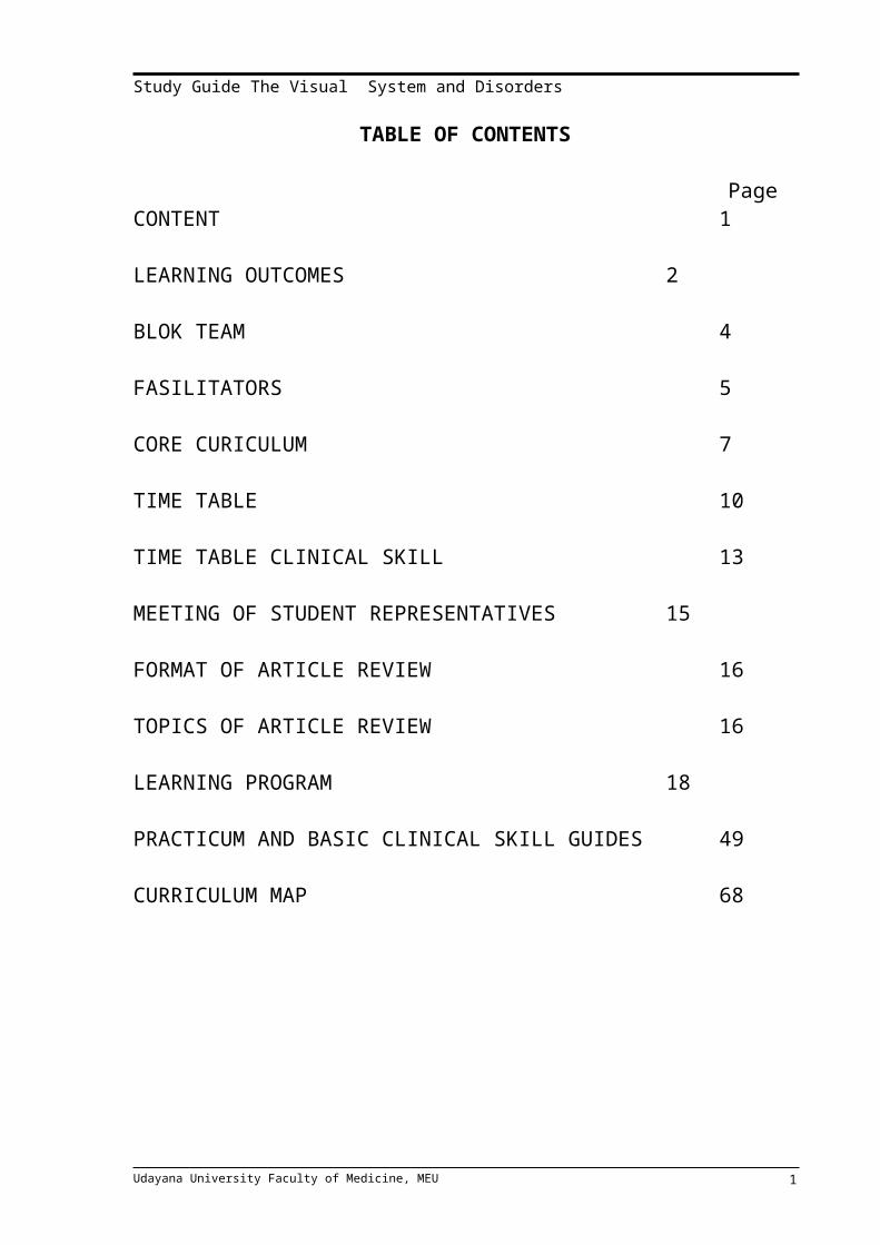
TABLE OF CONTENTS
PageCONTENT 1
LEARNING OUTCOMES 2
BLOK TEAM 4
FASILITATORS 5
CORE CURICULUM 7
TIME TABLE 10
TIME TABLE CLINICAL SKILL 13
MEETING OF STUDENT REPRESENTATIVES 15
FORMAT OF ARTICLE REVIEW 16
TOPICS OF ARTICLE REVIEW 16
LEARNING PROGRAM 18
PRACTICUM AND BASIC CLINICAL SKILL GUIDES 49
CURRICULUM MAP 68
Udayana University Faculty of Medicine, MEU 1

Study Guide The Visual System and Disorders
LEARNING OUTCOMES
THE AIMS Comprehend the underlying normal structure and function of the visual system and its
practical or clinical implications.
Understanding the pharmacology and pharmacokinetic of the ocular medicines.
Able to manage common eye and visual disorders and refer of high risks patient with visual
disorders for further investigation and management.
Awareness and responsiveness to the community aspects of health care, needs, education
and promotion.
LEARNING OUTCOMES
Able to know and understand anatomy of the eye structures
Able to know and understand Histology of the eye structures
Able to know and understand Physiology of the eye, and Physiology of vision
Able to know and understand the pharmacology and pharmacokinetic of the ocular
medicines
Able to establish diagnosis and management patient with Refraction disorders such as
Mild Hipermetropia, mild myopia, mild astigmatism and presbyopia.
Able to initially diagnose, manage and later refer patient with Refraction disorders such as
anisometropia and contact lens problems.
Able to establish diagnosis and management patient with external eye diseases such us
conjunctivitis, Dry eye Syndrome, Blepharitis, Hordeolum, and Episcleritis
Able to initially diagnose, manage and later refer patient with external eye diseases such
as chalazion, scleritis, keratitis, corneal ulcer, and uveitis
Able to initially diagnose and later refer patient with external eye diseases such as
keratokonjungtivitis sicca and endophthalmitis.
Able to initially diagnose, manage and later refer patient with glaucoma disorders.Able to establish diagnosis and management patient with ocular injuries such as foreign
bodies in the conjunctiva and subconjunctival bleeding
Able to manage and initially diagnose and later refer patient with ocular injuries such as
hyphema and eyelids lacerations.
Able to initially diagnose and later refer patient with ocular injuries such as corneal
erosion, foreign bodies on the cornea, thermal corneal burn and lacrimal duct lacerations.
Udayana University Faculty of Medicine, MEU 2

Study Guide The Visual System and Disorders
Able to establish diagnosis and management patient with eyelids and lacrimal system disorders such us trichiasis.
Able to initially diagnose, manage and later refer patient with eyelids and lacrimal systems disorders such as dacrioadenitis and dacriocystitis
Able to initially diagnose and later refer patient with eyelids and lacrimal sytems disorders such as entropion, lagophthalmus, epichantus, ptosis, eyelids retraction and
xanthelasma
Able to initially diagnose and later refer patient with cataract, lens dislocation and corneal disorders such as pterygium and keratoconus.Able to initially diagnose and later refer patient with retinal disorders.
Able to initially diagnose and later refer patient with neuro ophthalmology & strabismus disorders including amblyopia and binocular diplopia.
Able to establish diagnosis and management patient with community ophthalmology disorders such as night blindness
Able to initially diagnose, manage and later refer patient with community ophthalmology disorders such as Xerophthalmia
Able to initially diagnose and later refer patient with community ophthalmology disorders such as blindness due to 5 most common eye disorders (Cataract, Glaucoma, refractive
errors, infection and immunology eye diseases and retina disorders)
CURRICULUM CONTENT
Anatomy, histology and physiology of the eye.Pharmacology of the eye medicines.Refractive Errors.Infection & Immunologic Eye Diseases.Glaucoma disorders Eyelids and Lacrimal systems disorders Ocular InjuriesCataract, corneal and lens disorders.Vitreous and Retinal disorders Neuro Ophthalmology & strabismus disorders.
Udayana University Faculty of Medicine, MEU 3

Study Guide The Visual System and Disorders
BLOCK VISUAL SYSTEM AND DISORDERS
COORDINATOR : Dr. Putu Budhiastra, SpM(K) SECRETARY : Dr. Ariesanti Tri Handayani SpM(K)TIME : 19 June 2015 – 13 July 2015
Block Team
No Name
1 dr. Putu Budhiastra, Sp.M (K)2 Prof.dr.NK Niti Susila, SpM (K)3 dr. Nyoman Sunerti, Sp.M4 dr. AAA Sukartini Djelantik, Sp.M (K)5 dr. Made Agus Kusumadjaja, Sp.M (K)6 dr. W.G Jayanegara, Sp.M (K)7 Dr. dr. AAA Mas Putrawati T, Sp.M(K)8 dr. Putu Yuliawati, Sp.M(K)9 dr. Ariesanti Tri Handayani, Sp.M(K)10 dr. Yuliana,MBiomed11 Prof.dr.I Dewa Putu Sutjana,PFK,M.Erg12 dr. I.G.A.Dewi Ratnayanti13 Dr. dr. Made Jawi14 dr. Cok Istri Dewiyani Pemayun,SpM(K)15 dr. Ni Kompyang rahayu,SpM(K)16 dr. IGAM Juliari,SpM17 dr. Wayan Eka Sutyawan,SpM18 dr. Ari Andayani,SpM19 dr. Krisna Dinata
Lectures
No Name Dept No Telp
1 dr. Putu Budhiastra, Sp.M (K) Ophtalmology 0852382389992 Prof.dr.NK Niti Susila,SpM (K) Ophtalmology 081236438163 dr. Nyoman Sunerti, Sp.M Ophtalmology 081239826244 dr. AAA Sukartini Djelantik, Sp.M (K) Ophtalmology 0813373149115 dr. Made Agus Kusumadjaja, Sp.M (K) Ophtalmology 081239813496 dr. W.G Jayanegara, Sp.M (K) Ophtalmology 0818909147
7 Dr. dr. AAA Mas Putrawati T, Sp.M(K) Ophtalmology 081238469958 dr. Putu Yuliawati, Sp.M(K) Ophtalmology 0813386017249 dr. Ariesanti Tri Handayani, Sp.M(K) Ophtalmology 0818375611
10 dr. Yuliana,M.Biomed Anatomy 08579265236311 Prof.dr.I Dewa Putu Sutjana,PFK,M.Erg Physiology 0812392447712 dr. I.G.A.Dewi Ratnayanti, m.Biomed Histology 0361855034413 Dr. dr. Made Jawi, M.Kes Pharmacology 08179787972
Udayana University Faculty of Medicine, MEU 4

Study Guide The Visual System and Disorders
14 dr. Cok Istri Dewiyani Pemayun,Sp.M(K) Ophthalmology 0856375030415 dr. Ni kompyang Rahayu,SpM(K) Ophthalmology 08523909564516 dr. IGAM Juliari,Sp.M Ophthalmology 0812361562517 dr. Wayan Eka Sutyawan,Sp.M Ophthalmology 08133853849918 dr. Ari Andayani,Sp.M Ophthalmology 0811380366619 dr. I Made Krisna Dinata, M.Erg Physiology 08174742566
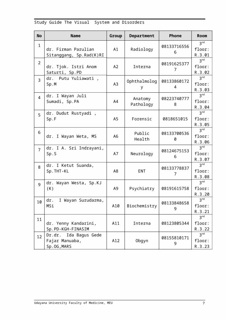
FACILITATORS
Regular Class (Class A)
No Name Group Department Phone Room
1 dr. Firman Parulian Sitanggang, Sp.Rad(K)RI A1 Radiology 081337165566 3nd floor:
R.3.01
2 dr. Tjok. Istri Anom Saturti, Sp.PD A2 Interna 081916253777 3nd floor:
R.3.02
3 dr. Putu Yuliawati , Sp.M A3 Ophthalmology 081338601724 3nd floor: R.3.03
4 dr. I Wayan Juli Sumadi, Sp.PAA4
Anatomy Pathology 082237407778 3nd floor:
R.3.04
5 dr. Dudut Rustyadi , Sp.F A5 Forensic 0818651015 3nd floor: R.3.05
6 dr. I Wayan Weta, MS A6 Public Health 081337005360 3nd floor: R.3.06
7 dr. I A. Sri Indrayani, Sp.S A7 Neurology 081246751536 3nd floor: R.3.07
8 dr. I Ketut Suanda, Sp.THT-KL A8 ENT 081337788377 3nd floor: R.3.08
9 dr. Wayan Westa, Sp.KJ (K) A9 Psychiatry 08191615758 3nd floor: R.3.20
10 dr. I Wayan Surudarma, MSi A10 Biochemistry 081338486589 3nd floor: R.3.21
11 dr. Yenny Kandarini, Sp.PD-KGH-FINASIM A11 Interna 08123805344 3nd floor:
R.3.22
12 Dr.dr. Ida Bagus Gede Fajar Manuaba, Sp.OG,MARS A12 Obgyn 081558101719 3nd floor:
R.3.23
Udayana University Faculty of Medicine, MEU 5

Study Guide The Visual System and Disorders
English Class (Class B)
No Name Group Department Phone Room
1 Dr.dr. Cokorda Bagus Jaya Lesmana, Sp.KJ B1 Psychiatry 0816295779 3nd floor:
R.3.01
2 dr. Desak Made Wihandani, M.Kes B2 Biochemistry 081338776244 3nd floor:
R.3.02
3 dr. I Wayan Gede Sutadarma, M Gizi B3 Biochemistry 082144071268 3nd floor:
R.3.03
4 Dr.dr. Ni Nyoman Sri Budayanti, Sp.MK(K) B4 Microbiology 08553711398 3nd floor:
R.3.04
5 Dr.dr. Gde Ngurah Indraguna Pinatih, M.Sc, Akp.,Sp.GK B5 Public Health 08123816424 3nd floor:
R.3.05
6 dr. I Ketut Wibawa Nada, Sp.An B6 Anasthesi 08123650164 3nd floor: R.3.06
7 dr. Elysanti Dwi Martadiani, Sp.Rad B7 Radiology 081805673099 3nd floor:
R.3.07
8 Dr.dr. Tjokorda Gde Bagus Mahadewa, M.Kes,Sp.BS B8 Surgery 0818484654 3nd floor:
R.3.08
9 Dr.rer.Nat. dr. Ni Nyoman Ayu Dewi, M.Si B9 Biochemistry 081337141506 3nd floor:
R.3.20
10 dr. Tjokorda Gde Oka, MS, Sp.PK B10 Pathology 081338454245 3nd floor:
R.3.21
11 dr. I Gusti Nyoman Sri Wiryawan , M.Repro
B11 Histology 08123925104 3nd floor: R.3.22
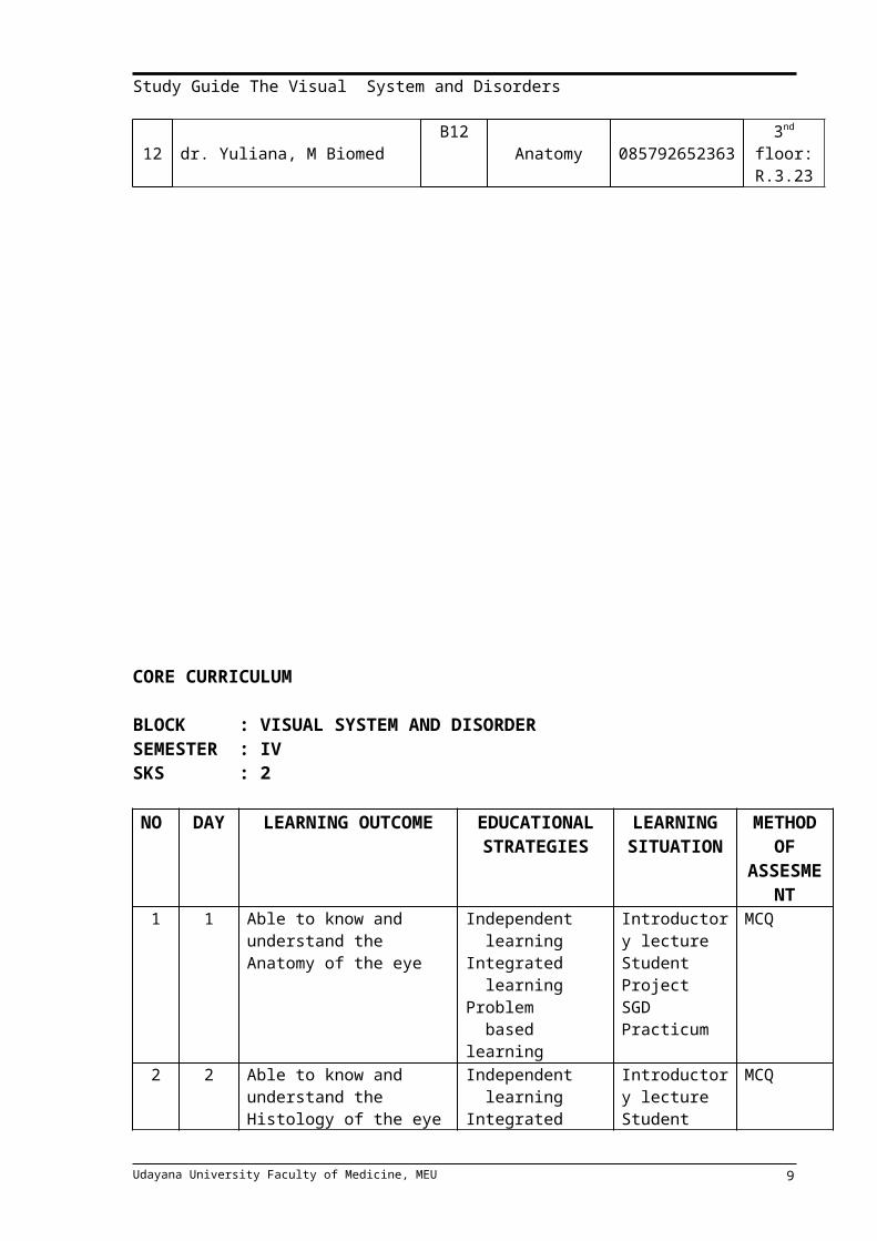
12 dr. Yuliana, M Biomed B12 Anatomy 085792652363 3nd floor: R.3.23
Udayana University Faculty of Medicine, MEU 6
Study Guide The Visual System and Disorders
CORE CURRICULUM
BLOCK : VISUAL SYSTEM AND DISORDERSEMESTER : IVSKS : 2
NO DAY LEARNING OUTCOME EDUCATIONAL STRATEGIES
LEARNING SITUATION
METHOD OF
ASSESMENT
1 1 Able to know and understand the Anatomy of the eye
Independent learningIntegrated learningProblem based learning
Introductory lectureStudent ProjectSGDPracticum
MCQ
2 2 Able to know and understand the Histology of the eye
Independent learningIntegrated learningProblem based learning
Introductory lectureStudent ProjectSGDPracticum
MCQ
3 3 Able to know and understand the Physiology of the eye
Independent learningIntegrated learningProblem based learning
Introductory lectureStudent ProjectSGDPracticum
MCQ
4 4 Able to know and understand the Pharmacology and Pharmacokinetic of the eye medicines
Independent learningIntegrated learningProblem based learning
Introductory lectureStudent ProjectSGDPracticum
MCQ
5 5 Able to establish diagnosis and manage patient with INFECTION & IMMUNOLOGIC EYE DISEASES such as : - Conjunctivitis, blepharitis, hordeoulum, chalazion and dry eye syndrome- Scleritis, episcleritis- Keratitis,uveitis- Kerato-conjunctivi. sicca- Iridocyclitis, Iritis- Endopthalmitis, corneal ulcer
Independent learningIntegrated learningProblem/case based learning
Introductory lectureStudent ProjectSGDBCS
MCQOSCE
6 6 Able to establish diagnosis and manage patient with OCULAR INJURY such as:
- Conjunctiva & cornea
Independent learningIntegrated learning
Introductory lectureStudent Project
MCQOSCE
Udayana University Faculty of Medicine, MEU 7

Study Guide The Visual System and Disorders
foreign body,corneal erosion, thermal /burn injury
- Sub conjunctiva Haemorrhage, hyphema
- eyelid laceration, lacrimal duct laceration
Problem/case based learning
SGDBCS
7 7 Able to establish diagnosis and manage patient with GLAUCOMA DISORDERS such as : - Acute glaucoma- Chronic glaucoma- Secondary glaucoma- Congenital glaucoma
Independent learningIntegrated learningProblem/case based learning
Introductory lectureStudent ProjectSGDBCS
MCQOSCE
8 8 Able to establish diagnosis and manage patient with CORNEA & LENS DISORDERS such as : - Cataract, pseudophakia- Pterygium- Lens dislocation- Korneal dystrophy- Keratokonus
Independent learningIntegrated learningProblem/case based learning
Introductory lectureSGDBCS
MCQ
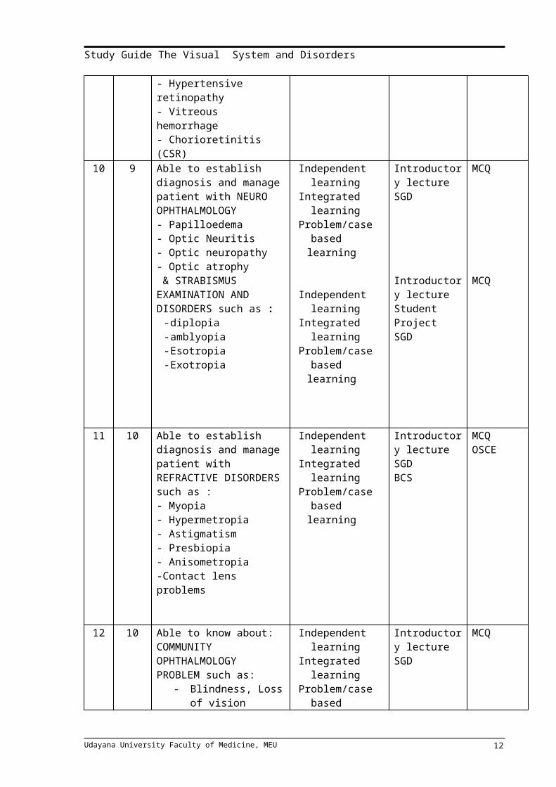
9 8 Able to establish diagnosis and manage patient with VITREO RETINAL DISORDERS such as : - Retinal detachment- Retinal vessel occlusion- Degeneration macula- Diabetic retinopathy- Hypertensive retinopathy- Vitreous hemorrhage- Chorioretinitis (CSR)
Independent learningIntegrated learningProblem/case based learning
Introductory lectureStudent ProjectSGDBCS
MCQOSCE
10 9 Able to establish diagnosis and manage patient with NEURO OPHTHALMOLOGY- Papilloedema- Optic Neuritis- Optic neuropathy- Optic atrophy & STRABISMUS EXAMINATION AND DISORDERS such as :
- diplopia- amblyopia - Esotropia- Exotropia
Independent learningIntegrated learningProblem/case based learning
Independent learningIntegrated learningProblem/case based learning
Introductory lectureSGD
Introductory lectureStudent ProjectSGD
MCQ
MCQ
Udayana University Faculty of Medicine, MEU 8
Study Guide The Visual System and Disorders
11 10 Able to establish diagnosis and manage patient with REFRACTIVE DISORDERS such as : - Myopia- Hypermetropia- Astigmatism- Presbiopia- Anisometropia -Contact lens problems
Independent learningIntegrated learningProblem/case based learning
Introductory lectureSGDBCS
MCQOSCE
12 10 Able to know about:COMMUNITY OPHTHALMOLOGY PROBLEM such as:
- Blindness, Loss of vision
- Community ophthalmology
Independent learningIntegrated learningProblem/case based learning
Introductory lectureSGD
MCQ
Udayana University Faculty of Medicine, MEU 9

Study Guide The Visual System and Disorders
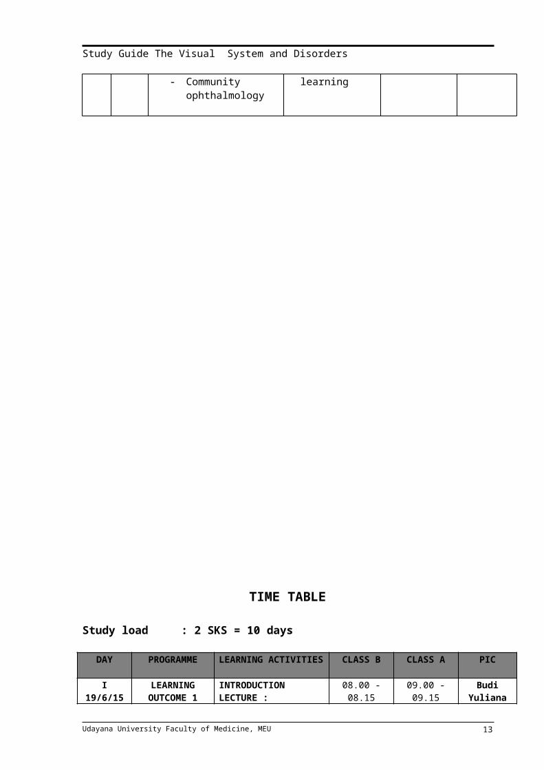
TIME TABLE
Study load : 2 SKS = 10 days
DAY PROGRAMME LEARNING ACTIVITIES CLASS B CLASS A PIC
I19/6/15
LEARNING OUTCOME 1
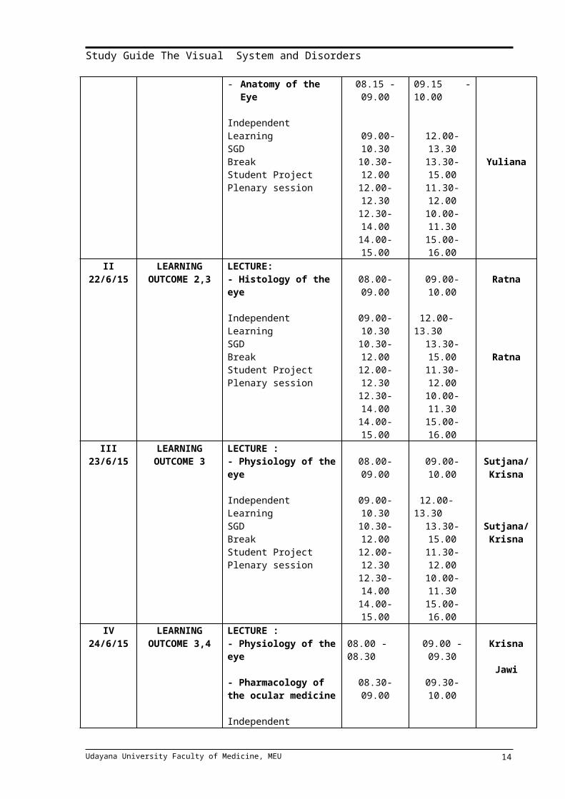
INTRODUCTIONLECTURE :- Anatomy of the Eye
Independent LearningSGDBreakStudent ProjectPlenary session
08.00 - 08.1508.15 - 09.00
09.00-10.3010.30-12.0012.00-12.3012.30-14.0014.00-15.00
09.00 - 09.1509.15 - 10.00
12.00-13.3013.30-15.0011.30-12.0010.00-11.3015.00-16.00
BudiYuliana
YulianaII
22/6/15LEARNING
OUTCOME 2,3LECTURE:- Histology of the eye
Independent LearningSGDBreakStudent ProjectPlenary session
08.00-09.00
09.00-10.3010.30-12.0012.00-12.3012.30-14.0014.00-15.00
09.00-10.00
12.00-13.3013.30-15.0011.30-12.0010.00-11.3015.00-16.00
Ratna
Ratna III
23/6/15LEARNING
OUTCOME 3LECTURE :- Physiology of the eye
Independent LearningSGDBreakStudent ProjectPlenary session
08.00-09.00
09.00-10.3010.30-12.0012.00-12.3012.30-14.0014.00-15.00
09.00-10.00
12.00-13.3013.30-15.0011.30-12.0010.00-11.3015.00-16.00
Sutjana/Krisna
Sutjana/Krisna
IV24/6/15
LEARNING OUTCOME 3,4
LECTURE :- Physiology of the eye
- Pharmacology of the ocular medicine
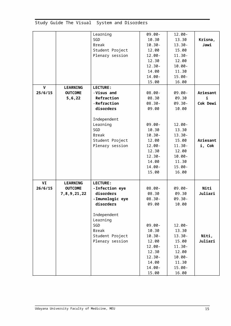
Independent LearningSGDBreakStudent ProjectPlenary session
08.00 - 08.30
08.30-09.00
09.00-10.3010.30-12.0012.00-12.3012.30-14.0014.00-15.00
09.00 - 09.30
09.30-10.00
12.00-13.3013.30-15.0011.30-12.0010.00-11.3015.00-16.00
Krisna
Jawi
Krisna, Jawi
V25/6/15
LEARNING OUTCOME
5,6,22
LECTURE:- Visus and Refraction- Refraction disorders
Independent LearningSGDBreakStudent ProjectPlenary session
08.00-08.3008.30-09.00
09.00-10.3010.30-12.0012.00-12.3012.30-14.0014.00-15.00
09.00-09.3009.30-10.00
12.00-13.3013.30-15.0011.30-12.0010.00-11.3015.00-16.00
AriesantiCok Dewi
Ariesanti, Cok
Udayana University Faculty of Medicine, MEU 10

Study Guide The Visual System and Disorders
VI26/6/15
LEARNING OUTCOME 7,8,9,21,22
LECTURE:- Infection eye
disorders- Imunologic eye
disorders
Independent LearningSGDBreakStudent ProjectPlenary session
08.00-08.3008.30-09.00
09.00-10.3010.30-12.0012.00-12.3012.30-14.0014.00-15.00
09.00-09.3009.30-10.00
12.00-13.3013.30-15.0011.30-12.0010.00-11.3015.00-16.00
Niti Juliari
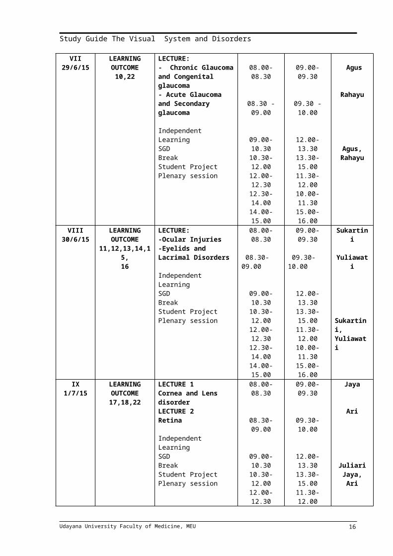
Niti, JuliariVII
29/6/15LEARNING OUTCOME
10,22
LECTURE:- Chronic Glaucoma and Congenital glaucoma- Acute Glaucoma and Secondary glaucoma
Independent LearningSGDBreakStudent ProjectPlenary session
08.00-08.30
08.30 -09.00
09.00-10.3010.30-12.0012.00-12.3012.30-14.0014.00-15.00
09.00-09.30
09.30 -10.00
12.00-13.3013.30-15.0011.30-12.0010.00-11.3015.00-16.00
Agus
Rahayu
Agus, Rahayu
VIII30/6/15
LEARNING OUTCOME
11,12,13,14,15,16
LECTURE:-Ocular Injuries-Eyelids and Lacrimal Disorders
Independent LearningSGDBreakStudent ProjectPlenary session
08.00-08.30
08.30-09.00
09.00-10.3010.30-12.0012.00-12.3012.30-14.0014.00-15.00
09.00-09.30
09.30-10.00
12.00-13.3013.30-15.0011.30-12.0010.00-11.3015.00-16.00
Sukartini
Yuliawati
Sukartini, Yuliawati
IX1/7/15
LEARNING OUTCOME
17,18,22
LECTURE 1Cornea and Lens disorderLECTURE 2Retina
Independent LearningSGDBreakStudent ProjectPlenary session
08.00-08.30
08.30-09.00
09.00-10.3010.30-12.0012.00-12.3012.30-14.0014.00-15.00
09.00-09.30
09.30-10.00
12.00-13.3013.30-15.0011.30-12.0010.00-11.3015.00-16.00
Jaya
Ari
JuliariJaya, Ari
Udayana University Faculty of Medicine, MEU 11
Study Guide The Visual System and Disorders
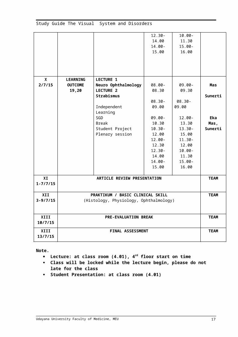
X2/7/15
LEARNING OUTCOME
19,20
LECTURE 1Neuro OphthalmologyLECTURE 2Strabismus
Independent LearningSGDBreakStudent ProjectPlenary session
08.00-08.30
08.30-09.00
09.00-10.3010.30-12.0012.00-12.3012.30-14.0014.00-15.00
09.00-09.30
08.30-09.00
12.00-13.3013.30-15.0011.30-12.0010.00-11.3015.00-16.00
Mas
Sunerti
EkaMas,
Sunerti
XI1-7/7/15
ARTICLE REVIEW PRESENTATION TEAM
XII3-9/7/15
PRAKTIKUM / BASIC CLINICAL SKILL (Histology, Physiology, Ophthalmology)
TEAM
XIII10/7/15
PRE-EVALUATION BREAK TEAM
XIII13/7/15
FINAL ASSESSMENT TEAM
Note. Lecture: at class room (4.01), 4rd floor start on time Class will be locked while the lecture begin, please do not late for the class Student Presentation: at class room (4.01)
Udayana University Faculty of Medicine, MEU 12
Study Guide The Visual System and Disorders
TIME TABLE BASIC CLINICAL SKILL Study load : 1 SKS = 5 daysDATE : 3 July 2015 – 9 July 2015
DAY LEARNING ACTIVITY CLASS B CLASS A PIC
I3/7/15
LECTURE:VISUAL ACUITY EXAMINATIONBCS Visual Acuity Exam
BreakStudent Project Presentation
08.00 – 09.00
09.30-11.30
11.30-12.0012.00-14.00
09.00 – 10.00
13.00-15.00
12.30-13.0010.30-12.30
Eka
TEAM
Agus,Rahayu
II6/7/15
LECTURE :- TONOMETRY- VISUAL FIELD
BCS Tonometry
BreakStudent Project Presentation
08.00-09.00
09.30-11.30
11.30-12.0012.00-14.00
09.00-10.00
13.00-15.00
12.30-13.0010.30-12.30
Mas
TEAM
Budi/Ari
III7/7/15
LECTURE :- POST SEGMENT
EXAMINATION- AMSLER GRID
EXAMINATION
BCS Post Segment Exam
BreakStudent Project presentation
08.00-09.00
09.30-11.30
11.30-12.0012.00-14.00
09.00-10.00
13.00-15.00
12.30-13.0010.30-12.30
Ari A
TEAM
Yuliawati
IV8/7/15
LECTURE :- Anterior Segment
Examination- Therapeutic
Instillation & Drug Prescription
BCS Ant Segment ExamBreakBCS Therapeutic inst
08.00-08.30
08.30-09.00
09.30-11.3011.30-12.0012.00-14.00
09.00-09.30
09.30-10.00
13.00-15.0012.30-13.0010.30-12.30
Juliari
Yuliawati
TEAM
TEAM
Udayana University Faculty of Medicine, MEU 13
Study Guide The Visual System and Disorders
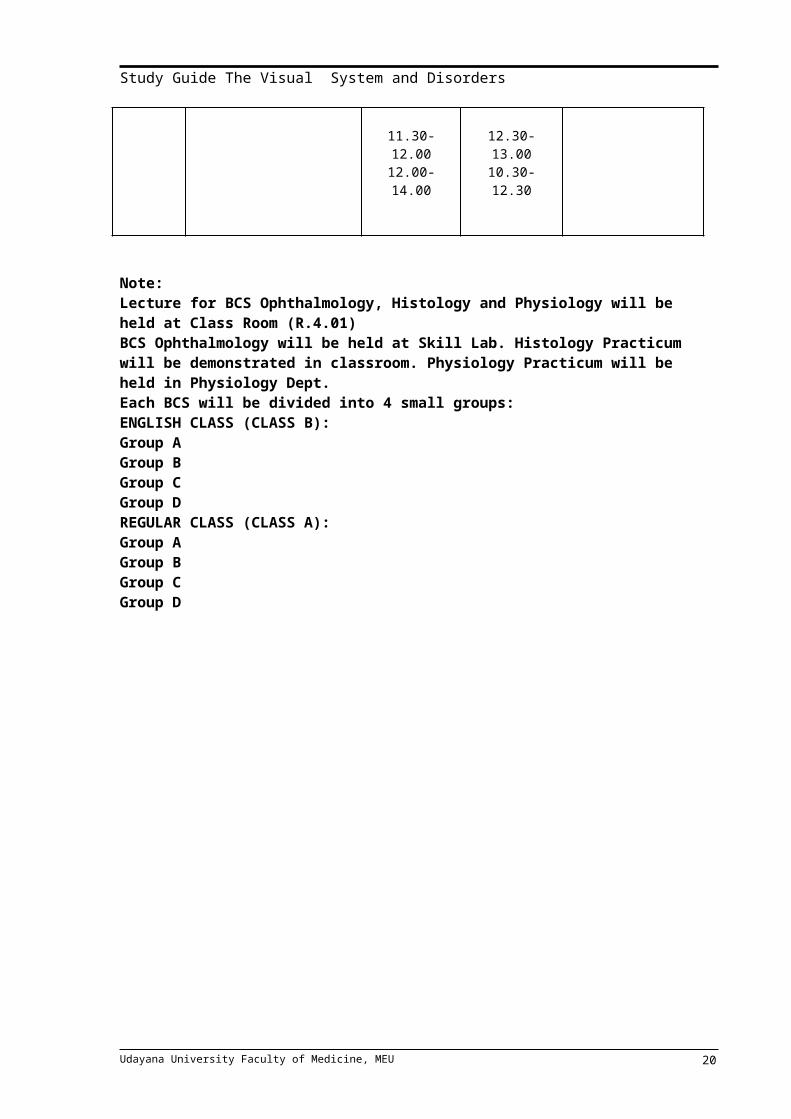
V9/7/15
LECTURE :- Histology
Examination- Physiology Practice
Lecture
Practicum Histology
BreakPracticum Physiology
08.00-08.30
08.30-09.00
09.30-11.30
11.30-12.0012.00-14.00
09.00-09.30
09.30 -10.00
13.00-15.00
12.30-13.0010.30-12.30
Ratna
Krisna
Histology team
PHYSIOLOGY TEAM
Note:Lecture for BCS Ophthalmology, Histology and Physiology will be held at Class Room (R.4.01)BCS Ophthalmology will be held at Skill Lab. Histology Practicum will be demonstrated in classroom. Physiology Practicum will be held in Physiology Dept.Each BCS will be divided into 4 small groups:ENGLISH CLASS (CLASS B):Group AGroup BGroup CGroup DREGULAR CLASS (CLASS A):Group AGroup BGroup CGroup D
Udayana University Faculty of Medicine, MEU 14
Study Guide The Visual System and Disorders
MID BLOCK MEETING
The meeting between block planner team, facilitators and the student group representatives will be held on 30 June 2015 at classroom (4.01) if necessary. In this meeting all the facilitator and student group representative are expected to give suggestion and input as an evaluation to improve the study guide and educational process of visual system and disorder. Because of the important of this meeting, all the facilitators and student group representative are strongly expected to attend the meeting. All of student group representatives (approximately 10 students) are expected to give suggestion and input or complain to the team planner for improvement. For this purpose, every student group must choice one student as their representative to attend the meeting.
PLENARY SESSION
For each learning task, the student is requested to prepare a group report. The report will be presented in plenary session. Lecturer in charge will choose the group randomly. The aim of this presentation is to make similar perception about the topic that has been given.
ASSESSMENT METHOD
Assessment will be held on Monday, 13 July 2015. The Final examination will be held with the format of Computer Based Test. There are 100 questions for the examination that consist out Multiple Choice Questions (MCQ). The time provision is 100 minutes. The number of MCQ is 100. The minimal passing score for the assessment is 70. The proportions of examination score are: Small Group Discussion : 5%Article Review (Student Project) : 15%Final assessment (MCQ) : 80%
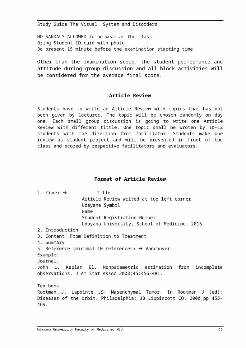
The Prerequisites of Final Examination:Attend 75% of total student activitiesUniform for Examination: white shirt, black trouser/ skirt, shoes. NO SANDALS ALLOWED to be wear at the classBring Student ID card with photoBe present 15 minute before the examination starting time
Other than the examination score, the student performance and attitude during group discussion and all block activities will be considered for the average final score.
Article Review
Students have to write an Article Review with topics that has not been given by lecturer. The topic will be chosen randomly on day one. Each small group discussion is going to write one Article Review with different tittle. One topic shall be wroten by 10-12 students with the direction from facilitator. Students make one review as student project and will be presented in front of the class and scored by respective facilitators and evaluators.
Udayana University Faculty of Medicine, MEU 15
Study Guide The Visual System and Disorders
Format of Article Review
1. Cover: Title Article Review writed at top left cornerUdayana SymbolNameStudent Registration NumberUdayana University, School of Medicine, 2015
2. Introduction3. Content: From Definition to Treatment 4. Summary5. Reference (minimal 10 references) VancouverExample: Journal.John L, Kaplan El. Nonparametric estimation from incomplete observations. J Am Stat Assoc 2008;45:456-481.
Tex bookRootman J, Lapointe JS: Masenchymal Tumor. In Rootman J (ed): Diseases of the orbit. Philadelphia: JB Lippincott CO, 2000,pp 455-469.
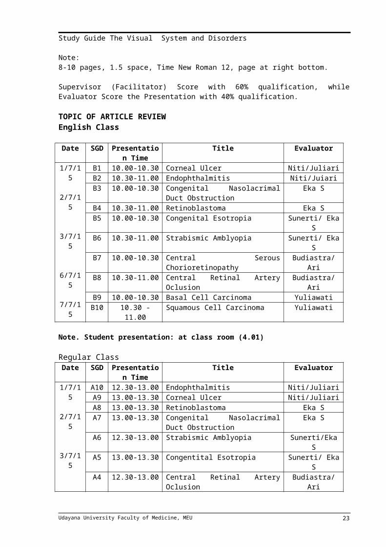
Note:8-10 pages, 1.5 space, Time New Roman 12, page at right bottom.
Supervisor (Facilitator) Score with 60% qualification, while Evaluator Score the Presentation with 40% qualification.
TOPIC OF ARTICLE REVIEWEnglish Class
Date SGD Presentation Time
Title Evaluator
1/7/15
2/7/15
3/7/15
6/7/15
7/7/15
B1 10.00-10.30 Corneal Ulcer Niti/JuliariB2 10.30-11.00 Endophthalmitis Niti/JuiariB3 10.00-10.30 Congenital Nasolacrimal Duct
ObstructionEka S
B4 10.30-11.00 Retinoblastoma Eka SB5 10.00-10.30 Congenital Esotropia Sunerti/ Eka SB6 10.30-11.00 Strabismic Amblyopia Sunerti/ Eka SB7 10.00-10.30 Central Serous Chorioretinopathy Budiastra/AriB8 10.30-11.00 Central Retinal Artery Oclusion Budiastra/Ari B9 10.00-10.30 Basal Cell Carcinoma YuliawatiB10 10.30 -11.00 Squamous Cell Carcinoma Yuliawati
Note. Student presentation: at class room (4.01)
Regular ClassDate SGD Presentation
TimeTitle Evaluator
1/7/15
2/7/15
A10 12.30-13.00 Endophthalmitis Niti/JuliariA9 13.00-13.30 Corneal Ulcer Niti/JuliariA8 13.00-13.30 Retinoblastoma Eka SA7 13.00-13.30 Congenital Nasolacrimal Duct Eka S
Udayana University Faculty of Medicine, MEU 16
Study Guide The Visual System and Disorders
3/7/15
6/7/15
7/7/15
ObstructionA6 12.30-13.00 Strabismic Amblyopia Sunerti/Eka SA5 13.00-13.30 Congentital Esotropia Sunerti/ Eka SA4 12.30-13.00 Central Retinal Artery Oclusion Budiastra/ Ari A3 13.00-13.30 Central Serous Chorioretinopathy Budiastra/Ari A2 12.30-13.00 Squamous Cell Carcinoma YuliawatiA1 13.00-13.30 Basal Cell Carcinoma Yuliawati
Note. Student presentation: at class room (4.01)
Article Review Assessments FormBlock of Visual system and disorders
Name :…………………………………………..Student Reg. Number :…………………………………………..Facilitator :…………………………………………..Title :…………………………………………..
Time Table of Consultation
No Point of Discussion Date Supervisor Sign1 Outline of Paper2 Final Discussion
No Item Assessment Range Score (%) Score1 Ability to find the literature 0-202 Communication/attitude/presentation 0-303 Quality of material (SOAP) 0-404 Student interest and motivation 0-10
TOTAL 100Facilitator,
(………………………………………)NIP
No Item Assessment Range Score (%) Score1 Quality of material 0-602 Capability of Information Searching 0-103 Critical Thinking 0-30
TOTAL 100Evaluator/Supervisor,
(………………………………………)NIP
Udayana University Faculty of Medicine, MEU 17

Study Guide The Visual System and Disorders
LEARNING PROGRAMDay 1
MODULE 1ANATOMY OF THE EYEBy dr. Yuliana, M.Biomed
SUMMARY
The eyes lie within two bony orbits, located on either side of the root of the nose. The medial walls of the orbits are almost parallel. They border the nasal cavity anteriorly and the ethmoidal air cell and the sphenoid sinus posteriorly. The lateral walls border the middle cranial, temporal, and pterygopalatine fossae. Superior to the orbit is the anterior cranial fossa and the frontal and supraorbital sinus. The maxillary sinus and the palatine air cell are located inferiorly. Seven bones make up the bony orbit such as frontal, zygomatic, maxillary, ethmoidal, sphenoid, lacrimal, and palatine. The four rectus muscles insert anteriorly on the globe along the Spiral of Tillaux. Two oblique muscles are superior and inferior oblique muscle.Six of the twelve cranial nerves (CV II-VII) directly innervates the eye and periocular tissue. The principal arterial supply of the orbit and its structures derives from the ophthalmic artery, the first major branch of the intracranial portion of the internal carotid artery.
LEARNING TASK:
1. Describe four walls of orbit and the bones that construct the walls.
2. Describe blood supply of the eye
3. Describe about lacrimal apparatus components4. Describe about orbit muscles 5. Describe about layers of eyeball6. Describe about nerve supply of the eyes
SELF ASSESSMENT:
1. Describe about the position of bones that construct the walls 2. Describe about the position of lacrimal apparatus
LEARNING RESOURSES:
1. Moore.K.L, Agar A.M.R : Essensial Clinical Anatomy, Second Edition, Lippincott Williams & Wilkins. 1995, USA
Udayana University Faculty of Medicine, MEU 18

Study Guide The Visual System and Disorders
Day 2MODULE 2
Title of LectureHISTOLOGY OF THE EYE
DR. RATNAYANTI
Abstract
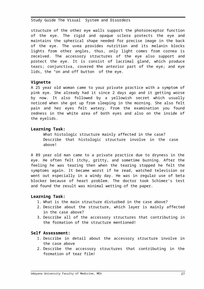
Like any other senses, eye is our window to the external world. Eye captures image continuously and transmits it to the brain for next processing and understanding. The structure of the eye is perfectly designed to perform its course. The light reflected by object enters the eye through cornea and pupil. The amount of light permitted is regulated by iris by dilating or constricting the pupil. Then, the refractive structures, lens and vitreous body, directing the light to the retina where it translated to a signal the brain can understand and perceived as an image. The shape and structure of the other eye walls support the photoreceptor function of the eye. The rigid and opaque sclera protects the eye and maintains the spherical shape needed for precise image in the back of the eye. The uvea provides nutrition and its melanin blocks lights from other angles, thus, only light comes from cornea is received. The accessory structures of the eye also support and protect the eye. It is consist of lacrimal gland, which produce tears; conjunctiva, covered the anterior part of the eye; and eye lids, the “on and off button” of the eye.
VignetteA 25 year old woman came to your private practice with a symptom of pink eye. She already had it since 2 days ago and it getting worse by now. It also followed by a yellowish secret which usually noticed when she got up from sleeping in the morning. She also felt pain and her eyes felt watery. From the examination you found redness in the white area of both eyes and also on the inside of the eyelids. Learning Task:
What histologic structure mainly affected in the case?Describe that histologic structure involve in the case above!
A 89 year old man came to a private practice due to dryness in the eye. He often felt itchy, gritty, and sometime burning. After the feeling he was tearing then when the tearing stopped he felt the symptoms again. It became worst if he read, watched television or went out especially in a windy day. He was in regular use of beta blocker because of heart problem. The doctor took Schimer’s test and found the result was minimal wetting of the paper.
Learning Task:1. What is the main structure disturbed in the case above?2. Describe about the structure, which layer is mainly affected in the case above?3. Describe all of the accessory structures that contributing in the formation of the
structure mentioned!
Self Assessment:1. Describe in detail about the accessory structure involve in the case above 2. Describe the accessory structures that contributing in the formation of tear film!3. Describe about the circulation and function of aqueous humor and tears!1. Describe about the refractive media of the eye:
1. Cornea2. Aqueous humor
Udayana University Faculty of Medicine, MEU 19
Study Guide The Visual System and Disorders
3. Lens4. Vitreous humor
2. Describe in detail about the histologic structure of the eye’s wall:1. Sclera2. Choroid3. Retina
3. Describe about the structure that holds the lens, including the choroid body!4. What fluid drains from the eye to the canal of Schlemm?5. What structures absorb the light that enters the eye?6. The area in the retina that contains no photoreceptor cells is called….7. What is the sclera made of?8. What cells in the conjunctiva which produce substance that constitute the tear film?
References: 1. Gartner, L. P. & Hiatt, J. L. 2011. Color Textbook of Histology. 3rd Ed. Philadelphia:
Saunders Elsevier. Pp. 514-526.2. Fawcett, D. W. & Jensh, R. P. 2002. Concise Histology. 2nd Ed. London: Arnold.
Pp.301-313.
DAY 3Module 3
Title of LecturePHYSIOLOGY OF THE EYE
By Prof.dr.I Dewa Putu Sutjana, PFK,M.Erg
INTRODUCTIONThe human know the environment because they had special senses, such as visual
system, auditory, smell, taste, tactile which are call five senses. The visual system was control around 90% of the daily activity. The visual system detects and interprets light stimulations. The light stimulations are in form of electromagnetic waves of lengths between 400 to 700 nm, which make up visible light. The human can be known the environment because they had:
The optic system, for reflected the light enters the eye and focuses it on the retinaThe retina as photoreceptors transducer light energy into an nerves impulsesThe optical neural, Neural pathways from the retina to the visual cortex at occipital lob of brain.The visual cortex of the brain process nerve impulses into visual images.
I.OPTIC SYSTEM OF THE EYEThe optics system of the eye is equivalent to the usually photographic camera. It has
a lens system, aperture system (call pupil) and retina that correspond to the film. The lens system of the eye is composed of four refractive interfaces. If all the refractive surfaces of the eye are added together and considered to be one single lens known as reduced eye. In reduced eye total refractive power 59 diopters when the eye accommodated for distant vision.
Udayana University Faculty of Medicine, MEU 20

Study Guide The Visual System and Disorders
FORMATION OF AN IMAGE ON THE RETINAThe light from the objects refracted and focused by the optics system of the eye on
the retina. The image is inverted and reversed with respect to the object. However, the mind perceives objects in the upright position.
PUPILLARY DIAMETERLike in the camera the eye had the pupil which is formed by the iris. The major function of the iris is to control the diameter of the pupil. The diameter of the pupil is control the amount of light enters the eye. The stimulation of the parasympathetic nerve increases the diameter of pupil. The change of pupil diameter followed the accommodation ACCOMODATION
The eye can be adjusted automatically the vision changes from the near to par objects, by changes the refractive power of the eye which is known as accommodation. In human eye the accommodation is due to change the refractive power of the lens (from 20 diopters to about 34 diopters), especially in children or the young person. The ability of eye accommodation (lens refractive power) is reduced with increased of age, and than followed by presbyopia.
NORMAL VISION AND ERROR OF REFACTIONNormal vision known as an emmetropic eye, if parallel light rays from distant objects are in sharp focus on the retina, when ciliary muscle completely relaxed. If the parallel light rays are focus behind the retina is known as hyperopia, if parallel light rays focus in the front of retina known as myopia, but if the different of the curvature of the cornea is known as astigmatism. The correction of refractive error is use of lens, concave spherical lens for myopia, convex lens for hyperopia and cylindrical lens for astigmatism.
VISUAL ACUITYVisual acuity is the ability of the person to distinguished two bright pin point spots of
light 10 meters away can barely distinguished the spots as separate entities when they are 1.5 to 2 millimeters apart. Clinical method to tested the visual acuity use the Snellen’s chart, consist of letter of different sizes placed 20 feet away from the person being tested, with the formula:
V = d/D V= visual acuity, d = distant of the person can read the letter, and D=distant of normal vision can read that letter.
FLUID SYSTEM OF THE EYEFor normal vision, the eye filled with intraocular fluid, which maintains sufficient
pressure in the eyeball to keep it distended. The fluid can be divided into two portions: aqueous humor, which lies in front of the lens and vitreous humor, which is between the posterior surface of the lens and the retina. The aqueous humor is a freely flowing fluid, whereas the vitreous humor sometimes called the vitreous body, is gelatinous mass. Both water and dissolved substances can diffuse slowly in the vitreous humor but there is little flow of fluid. Aqueous humor is continually being formed and reabsorbed. The balance between formation and reabsorption of aqueous humor regulates the total volume and pressure of intra ocular fluid.
RECEPTOR AND NEURAL FUNCTION OF THE RETINARetina is the light sensitive portion of the eye, contain 9 layers from outside to inside
as follows (1) pigmented layer, (2) layer of rods and cones, (3) outer nuclear layer, (4) outer plexiform layer, (5) inner nuclear layer, (6) inner plexiform layer, (7) ganglionic layer, (8) layer of optic nerve fiber, (9) inner limiting membrane. After the light passes through the lens system of the eye and then through the vitreous humor, it enters the retina from the inside layer and then finally reaches the layer of rods and cones. Cones located at the central fovea, a minute area in the center of retina occupying a total area around 1 square
Udayana University Faculty of Medicine, MEU 21
Study Guide The Visual System and Disorders
millimeter. The central fovea only 0.3 millimeters in diameter is composed almost entirely of cones. Outer of the center fovea located by rods.
PHOTORECEPTOR RHODOPSIN-RETINAL VISUAL CYCLEAND EXCITATION OF THE RODS
When light energy is absorbed by rhodopsin the rhodopsin begins to decompose within a very small fraction, cause photoactivation of electron, and then re-formation of rhodopsin again, which is call rhodopsin-retinal cycle. In that cycle vitamin A more important for normal vision, but if vitamin A is not enough, the night blindness occurs.
PHOTOCHEMISTRY OF COLOR VISION BY CONESPhoto chemicals in the cones have almost same to the rhodopsin at the rods. The
difference is the protein portions, call the opsins (photopsins). According to the Young-Helmholtz theory, there are three type of color pigment in the cones cells (blue, green, red): blue sensitive pigment, green sensitive pigment, and red sensitive pigment. So different cones (three type of cone) are sensitive to different colors of light.Color blindness. When a single group of color-receptive cones is missing from the eye, the person is unable to distinguish some colors from others, call color blindness. If the person loss of red cones it is called a protanope, if lacks of green called deuteranope. Red green color blindness is a genetic disorders, that at the female X chromosome. So that the color blindness almost in males.
NEURAL FUNCTION OF THE RETINANeural organization at the retina is more complex. The function of the neural
pathways is to transmit the neural impulse from the retina (rods and cone) through the optic nerve to the brain. It is different at the peripheral retina and in the center foveal retina. That is way the center foveal retina for detail and color vision
LEARNING TASKDescribe the equivalent and the different optic system of the human eye to the photographic camera.Describe the optic system of the eyeDescribe the different of emmetropia, hyperopia and myopia.What is accommodation, and in the human eyes which part of the optic system most important.Describe the circulation of the eye fluidDescribe the different of rods and cones in the vision perceptionDescribe the rhodopsin-retinal cycle, during light exposure to the rodDescribe the role of vitamin A in rhodopsin retinal cycleDescribe the Young-Helmholtz theory of the color visionDescribe many kind of color blindness
SELF ASSESSMENTCan you mention the part of optic systemWhat is mean by presbyopia and why it is occur in the human?Can you explain the role of cilliary muscle during accommodation?Can you explain why color blindness almost occur in males.Can you explain the neural function of the retinaCan you explain why the niktalopia person difficult to see in the afternoon but not problem at the morning?
Udayana University Faculty of Medicine, MEU 22
Study Guide The Visual System and Disorders
RESOURCES:Guyton, A. The Textbook of Medical PhysiologySilverthorn, D.U. 2010. Human physiology. An Integrated Approach.Fifth Ed. Pearson. San Fransisco.
DAY 4Module 3
Title of LecturePHYSIOLOGY OF THE EYE – Visual Pathways
dr. I Made Krisna Dinata, M.Erg
AbstractThe Visual Pathways
The visualnerve signals leave the retinas through the opticnerves. At the optic chiasm, the optic nerve fibersfrom the nasal halves of the retinas cross to theopposite sides, where they join the fibers from theopposite temporal retinas to form the optic tracts. The fibers of each optic tractthen synapse in the dorsal lateral geniculate nucleus of the thalamus, and fromthere, geniculocalcarine fibers pass by way of the optic radiation (also called thegeniculocalcarine tract) to the primary visual cortex in the calcarine fissure areaof the medial occipital lobe. Visual fibers also pass to several older areas of the brain:
From the optic tracts to the suprachiasmatic nucleus of the hypothalamus, presumably tocontrol circadian rhythms that synchronize various physiologic changes of the body with night and dayInto the pretectal nuclei in the midbrain, to elicit reflex movements of the eyes to focus on objects of importance and to activate the pupillary light reflexInto the superior colliculus, to control rapid directional movements of the two eyes Into the ventral lateral geniculate nucleus of the thalamus and surrounding basal regions of the brain, presumably to help control some of the body’s behavioral functions.
The visual pathways can be divided roughly into an old systemto the midbrain and base of the forebrain and a new system for direct transmissionof visual signals into the visual cortex located in the occipital lobes. Inhuman beings, the new system is responsible for perception of virtually allaspects of visual form, colors, and other conscious vision.
Function of the Dorsal Lateral Geniculate Nucleusof the ThalamusThe optic nerve fibers of the new visual system terminate in the dorsal
lateralgeniculate nucleus, located at the dorsal end of the thalamus and also calledsimply the lateral geniculate body, as shown in Figure 51–1. The dorsal lateralgeniculate nucleus serves two principal functions: First, it relays visual informationfrom the optic tract to the visual cortex by way of the optic radiation(also called the geniculocalcarine tract). This relay function is so accurate thatthere is exact point-to-point transmission with a high degree of spatial fidelityall the way from the retina to the visual cortex.
Visual FieldThe field of vision is the visual area seen by an eye at agiven instant. The area seen
to the nasal side is calledthe nasal field of vision, and the area seen to the lateralside is called the temporal field of vision.Each called monocular visual field . The area which are seen by two eyes is called binocular visual field
Udayana University Faculty of Medicine, MEU 23

Study Guide The Visual System and Disorders
Fusion of the Visual Images From the Two EyeTo make the visual perceptions more meaningful, thevisual images in the two eyes
normally fuse with eachother on “corresponding points” of the two retinas.The visual cortex plays an important role in fusion. Itwas pointed out earlier in the chapter that correspondingpoints of the two retinas transmit visual signals todifferent neuronal layers of the lateral geniculatebody, and these signals in turn are relayed to parallelneurons in the visual cortex. Interactions occurbetween these cortical neurons to cause interferenceexcitation in specific neurons when the two visualimages are not “in register”—that is, are not precisely“fused.”
Case A woman, 65 yo, came to physician with complainthat hervisual doesn’t work properly. The examination found that she had bipolar hemianopia.
Learning TaskWhich area in visual pathways had been damage in that patient?Describe the transmit of visual impulse from retina to the brainWhat is called the monocular and binocular visual fieldDifferentiate the nasal and temporal visual field, and what tool for evaluation of the visual fieldDescribe how the visual image can be fusion
Self AssessmentExplain the different of visual field from left and right visual fieldWhat is called the monocular and binocular visual fieldDiscribe the neural pathways to control pupillary light reflexDescribe the neural pathways to the optic nerve to form the consensual reflex
RefferencesMedical Physiology eleventh edition, Guyton & Hall.Physiology fifth edition, Linda S. Costanzo.Silvertharn, D.U. 2010. Human Physiology. An Integrated Approach. Fifth Ed. Pearson. San Fransisco
DAY 4MODULE
4Title of lecture
OCULAR PHARMACOLOGYBy Dr. dr. I Made Jawi, M.Kes
Abstract
Various pharmacologic agents are used for the treatment of eye disorders. The major challenge faced by today’s pharmacologist and formulation scientist is ocular drug delivery. Topical eye drop is the most convenient and patient compliant route of drug administration, especially for the treatment of anterior segment diseases. Delivery of drugs to the targeted ocular tissues is restricted by various precorneal, dynamic and static ocular barriers. Also, therapeutic drug levels are not maintained for longer duration in target tissues. In the past two decades, ocular drug delivery research acceleratedly advanced towards developing a novel, safe and patient compliant formulation and drug delivery devices/techniques, which may surpass these barriers and maintain drug levels in tissues.
Udayana University Faculty of Medicine, MEU 24

Study Guide The Visual System and Disorders
Anterior segment drug delivery advances are witnessed by modulation of conventional topical solutions with permeation and viscosity enhancers. Also, it includes development of conventional topical formulations such as suspensions, emulsions and ointments. Various nanoformulations have also been introduced for anterior segment ocular drug delivery. On the other hand, for posterior ocular delivery, research has been immensely focused towards development of drug releasing devices and nanoformulations for treating chronic vitreoretinal diseases. These novel devices and/or formulations may help to surpass ocular barriers and associated side effects with conventional topical drops. Also, these novel devices and/or formulations are easy to formulate, no/negligibly irritating, possess high precorneal residence time, sustain the drug release, and enhance ocular bioavailability of therapeutics. An update of current research advancement in ocular drug delivery necessitates and helps drug delivery scientists to modulate their think process and develop novel and safe drug delivery strategies. Systemic administration of a drug to treat eye disease would require a high concentration of circulating drug in the plasma to achieve therapeutic quantities in the aqueous humours, with the increased risk of side effect. Three important factors have to be considered when attempting drug delivery to the eye: (1) how the blood-eye barrier (systemic to ocular) or cornea (external to ocular) is crossed to reach the site of action; (2) how to localize the pharmacodynamic action at the eye and minimize drug action on other tissues; (3) how to prolong the duration of drug action so that the frequency of drug administration can be reduced. Many of the pharmacological agents commonly used for the treatment of eye disorders have been discussed in other blocks, such as anti bacterial, anti viral, anti allergy and anti inflammation agents. A number of antibacterial antibiotics have been formulated for topical ocular use. Natamycin is the only ophthalmic antifungal in use. Autonomic agents have several uses in ophthalmology, including diagnostic evaluation of anisocoria, as adjunctive therapy in laser and incisional surgeries, and in the treatment of glaucoma. Glaucoma is a condition of the eye in which there is an increase in the intraocular pressure (IOP), causing progressive atrophy of the optic nerve with deterioration of vision. Drugs reduce IOP either by increasing outflow of aqueous humour, by decreasing aqueous production (beta-adrenergic blocking drugs, alpha-adrenergic agonists, and carbonic anhydrase inhibitors), or transiently reducing the volume of intraocular fluids (osmotic agents).
SELF-DIRECTED LEARNING1. Basic principles of drug that affect outonomic nervus system 2. Pharmacokinetic and pharmacodynamic of some drugs that use in visual system3. Important side effects of drugs that use in visual system4. Important drug for glaucoma treatment, cycloplegic and antibiotic.
SCENARIO
VignetteNyoman Dewi,70 y.o, is diagnosed by an ophthalmologist as having glaucoma. Your initial assessment reveals that she has high blood pressure and appears to have difficulty in following instructions. Please discuss the following issues.
Learning Tasks1. Which anti-glaucoma will you give to the patient according to the patient condition?
Explain your answer.2. What is the mechanism of action on the muscle of the iris and cilia?3. What receptor mediates the action?4. List parts of the eyes that are innervated by the autonomic nervous system and
explain the effects of sympathetic and parasympathetic drugs to those parts.
Udayana University Faculty of Medicine, MEU 25

Study Guide The Visual System and Disorders
5. List the drugs that can be used to treat glaucoma and explain their mechanism of action to reduce the intraocular pressure
6. Please explain, why topical application sometimes has systemic effect?7. List the important side effects of anti-glaucoma agents.8. Which drug can produce cycloplegia? Explain the mechanism of action of this drug.9. Please compare the advantages and disadvantages between eye drops and eye
ointments.
Self Assessments1. Please explain the factors that determine the rate and the extent of absorption of the
drug after topical application to the eye.2. Please explain the possible absorption pathways of an ophthalmic drug following
topical application to the eye. Which routes are desired to localize ocular drug effects?
3. Please explain, why topical eye medication can cause systemic side effects?4. Please explain how to apply eye drop to the eye to get optimal effect.5. Please explain the characteristics of cholinoceptors in the peripheral nervous
system.6. Please explain the characteristic of adrenoceptors in the ANS.7. Please explain the effects of sympathetic and parasympathetic drugs on the eye.8. Please list some drugs used in glaucoma and explain their mechanisms of action.9. Please list important side effects of drugs used in glaucoma.10. Please list some drugs used in ophthalmology diagnostic.
RESOURCESStandard textbook
1. Trevor AJ, Katzung BG, and Masters SB. Katzung & Trevor’s Pharmacology Examination & Board Review. Seventh Ed. Singapore: McGraw Hill 2005.
Additional Readings1. Moroi SE, Lichter PR. Ocular Pharmacology in Goodman & Gillman’s: The
Pharmacology Basis of Therapeutics. 10th Ed. New York: McGraw Hill. 2001.
DAY 5TH
MODULE5
Title of lectureREFRACTION DISORDERS
By Ariesanti TH, MD and Cok Dewiyani Pemayun, MD
AIMS:Describe the Signs, Symptoms, Patophysiology and Management of refractive errors
LEARNING OUTCOMES:Can describe the Signs, Symptoms, Patophysiology and Management of:
1. Myopia2. Hyperopia3. Astigmatism4. Presbyopia5. Anisometropia
Udayana University Faculty of Medicine, MEU 26
Study Guide The Visual System and Disorders
CURRICULUM CONTENTS1. Optics and Refraction2. Refraction examination3. Emetropia and Ametropia / Refractive Errors4. Contact lens
ABSTRACT / SUMMARY OF LECTURE
The eye change refractive power to focus on near object by a process call it accommodation. Emetropia is absence of refractive errors and ammetropia is the present of refractive errors.
Light rays are focused on the retina to create sharp image. The light has to pass trough refractive media of the eye such as cornea, lens and vitreous body to reach retina. Some equipment are needed to examine refraction as follows Snellen chart, Trial lenses, trial frame, pupil distance ruler, lensometer, astigmatism chart, streak retinoscopy and autorefractor. The technique used in subjective refraction is trial and errors technique, while Astigmatic Clock Dial technique and Jackson Cross Cylinder technique were used to find astigmatism of the patient. Emmetropia is term used for eye with parallel light from distant object focused on retisna without accommodation of the eye. Ametropia is condition where parallel light is not focused on retina without eye accommodation. Classification of ammetropia is myopia, hypermetropia and astigmatism.
Miopia is a refractive error in which focus for light rays from a distant object is anterior to the retina. Hypermetropia is a refractive error in which the focus of light rays from a distant object is behind the retina. Astigmatism is a refractive error that prevents the light rays from coming to a point focus on the retina because of different degrees of refraction in the various meridians of the cornea or crystalline lens. Presbyopia (“old sight”) is a physiologically blurred near vision, commonly evident soon after age 40, due to reduction in the power of accommodation.
The management of refractive errors could be glasses, contact lens and refractive surgery. Contact lens could be used to treat refractive errors, theraphy for some ocular pathologies as well as cosmetic or prosthetic use. Some complication could be developed regarding the inappropriate handle and use of contact lens, such as allergic, Keratitis and corneal ulcer.
SELF DIRECTING LEARNING (in depth learning of above lecture)1. Physiology of Optics and refraction2. Definition of Emetropia and Ametropia 3. Refraction examination4. Refractive error: Myopia5. Refractive Error: Hyperopia6. Refractive Error: Astigmatism7. Refractive Error: Presbyopia8. Anisometropia9. Managements of refractive errors10. Benefits and complications of Contact lens use
SCENARIO
1. A 8 years old boy complain about intermittent blurred vision and headache at the frontal region. Everyday, the patient spent his time in front of his gadget all day. From physical examination found that he able to read all the letters from the Snellen chart. After given mydriatic drops and done the refraction, he can read all the letters with correction of S+2.00 D in both eyes. a. What is the diagnosis of this case?b. Mention about the types of hypermetropia based on the accomodation
Udayana University Faculty of Medicine, MEU 27

Study Guide The Visual System and Disorders
c. What kind of glasses should be prescripse for hypermetropia patientd. What are the complications of hypermetropia and how it can happened?e. What is the differences between hypermetropia and presbyopia
2. A 25 years old woman complained of blurred vision and headache especially in frontal region. From the examination there are C-1.50 A 180° found in the right eyes and S-2.00 C-0.50 A0° in the left eye.
a. What is the diagnosis of the both eye?b. Mention about the types of regular astigmatismc. Mention about the symptoms of astigmatism
3. A 30 years old man come to the ophthalmologist and has checked his visual acuity. From the right eye there is S-6.00 and his BCVA is 6/10 and from his left eye there is 6/6 with S-0.50
a. What is the diagnosis of this patient?b. What is the complication of myopia?c. What happen if the patient is given maximum correction of glasses?d. So what is the best treatment in this case to make better visual acuity and
avoid the complication?
SELF ASSESSMENTS
1. Mentions and explain about types of myopia2. Methods of objective refraction:
a. Trial and errors techniquesb. Streak retinoscopyc. Astigmatic clock diald. Fogging techniquee. Jackson cross cylinder
LEARNING RESOURSES:
1. Vaughan: General Ophthalmology2. Ilyas S: Ilmu Penyakit Mata. FK UI3. Deborah PL: Manual Diagnostic &Ocular Treatmentt.4. PERDAMI: Panduan Ketrampiilan dan Klinis Penyakit Mata, Jakarta, 2006
Udayana University Faculty of Medicine, MEU 28

Study Guide The Visual System and Disorders
DAY 6MODULE
6Title of lecture
INFECTION & IMMUNOLOGIC EYE DISEASESBy NK.Niti Susila, MD/IGAM Juliari, MD
AIMS:Describe the Signs, Symptom, Patophysiology and management OF INFECTION AND IMMUNOLOGIC EYE DISEASES
LEARNING OUTCOMES:Can describe the Signs, Symptoms, Patophysiology and Management of:
1. Blepharitis2. Hordeolum & Chalazia3. Conjunctivitis4. Scleritis & Episcleritis5. Keratitis6. Anterior Uveitis (Iritis & Iridosiklitis)7. Dry Eye Syndrome & Keratoconjunctivitis sicca
SELF DIRECTING LEARNING ( in depth learning of above lecture)1. Blepharitis
a. Pathogenesis of blepharitisb. Definition of chronic anterior blepharitisc. Definition of chronic posterior blepharitisd. Definition of Angular blepharitise. Aetiology of blepharitisf. Signs and symptoms of chronic anterior and posterior blepharitis and angular
blepharitis g. Diagnosis of chronic anterior and posterior blepharitis and angular blepharitish. Management of blepharitisi. Associations of chronic blepharitis
2. Hordeoluma. Pathogenesis of hordeolumb. Definition of external and internal hordeolumc. Aetiology of external and internal hordeolumd. Signs and symptoms of external and internal hordeolume. Diagnosis of external and internal hordeolumf. Management of external and internal hordeolumg. Complication of external and internal hordeolum
3. Chalaziona. Pathogenesis of chalazionb. Definition of chalazionc. Aetiology of chalaziond. Signs and symptoms of chalazione. Diagnosis of chalazionf. Management of chalaziong. Complication of chalazion
Udayana University Faculty of Medicine, MEU 29

Study Guide The Visual System and Disorders
4. Conjunctivitis a. Pathogenesis of conjunctivitis (Allergy, viral, bacterial)b. Definition of conjunctivitisc. Aetiology of conjunctivitisd. Signs and symptoms of conjunctivtse. Diagnosis of conjunctivitisf. Management of conjunctivitisg. Prognosis of conjunctivitish. Differential diagnosis conjunctivitis due to aetiologyi. Complication of conjunctivitis
5. Scleritis a. Pathogenesis of infection scleritis non infection scleritisb. Definition of infection or non infection scleritisc. Aetiology of infection or non infection scleritisd. Signs and symptoms of infection or non infection scleritise. Diagnosis of infection or non infection scleritisf. Prognosis of infection or non infection scleritis g. Management of infection or non infection scleritish. Different diagnosis of infection or non infection scleritisi. Complication of scleritis
6. Episcleritis a. Pathogenesis of episcleritisb. Definition of episcleritisc. Aetiology of episcleritisd. Signs and symptoms of episcleritise. Diagnosis of episcleritisf. Prognosis of episcleritis g. Management of episcleritish. Different diagnosis of episcleritis
7. Keratitis a. Pathogenesis of keratitisb. Definition of keratitisc. Aetiology of keratitisd. Signs and symptoms of keratitise. Diagnosis of keratitisf. Prognosis of keratitis g. Management of keratitish. Different diagnosis of keratitisi. Prognosis of kearatitisj. Complication of keratitis
8. Uveitis anteriorPathogenesis of uveitis anteriorDefinition of iritis and iridocyclitisAetiology of iritis and iridocyclitisSigns and symptoms of iritis and iridocyclitisDiagnosis of iritis and iridocyclitis
Udayana University Faculty of Medicine, MEU 30
Study Guide The Visual System and Disorders
Prognosis of iritis and iridocyclitis Management of iritis and iridocyclitisDifferent diagnosis of iritis and iridocyclitisPrognosis of iritis and iridocyclitis Complication of iritis and iridocyclitis
9. Dry eye disordersPhysiology of tear filmPathogenesis of dry eye syndromeDefinition of dry eye Aetiology of dry eye Signs and symptoms of dry eye Diagnosis of dry eye Prognosis of dry eye Management of dry eye Different diagnosis of dry eye Prognosis of dry eye Complication of dry eye
10.Keratoconjunctivitis siccaPhysiology of tear filmPathogenesis of Keratoconjunctivitis siccaDefinition of Keratoconjunctivitis sicca Aetiology of Keratoconjunctivitis sicca Classification of Keratoconjunctivitis sicca Signs and symptoms of Keratoconjunctivitis sicca Diagnosis of Keratoconjunctivitis sicca Prognosis of Keratoconjunctivitis sicca Management of Keratoconjunctivitis sicca Different diagnosis of Keratoconjunctivitis sicca Prognosis of Keratoconjunctivitis sicca Complication of Keratoconjunctivitis sicca
ABSTRACT / SUMMARY
The external eye is the most crucial part of the body exposed to outside word. The normal structure and function of the healthy eye rely on homeostasis of the entire body for protection against an adverse environment. Genetic and nutrition determine the embryogenesis and growth of the eye. Intact vascular and nervous systems stable metabolism, and immune system maintains surveillance.
The cushioning effect of the periocular tissues and local barriers such as the orbital rim are needed to safeguard the globe. The eyebrows and eyelashes catch small particles, and cilia also work as sensors to stimulate reflex eyelid closure. Blinking augments the lacrimal pump to rinse tears over the eye and flush off foreign material. The tear film also dilute toxins and allergens and contains proteins that control the normal flora. Mucin stabilizes that tear film and demarcates the living cells of the ocular surface from surrounding environment.
The epidermis and epithelium of healthy eyelids, conjunctiva, and cornea adhere tightly to their basement membranes. Regulation of cellular growth and metabolism are critical to maintenance of an intact ocular surface and transparent cornea. The underlying extracellular matrix of the eye's mucous membrane is rich in blood vessels and conjunctiva-associated lymphoid tissue (CALT). The anterior segment of the eye provides a clear, protected entrance for light that is to be processed by the visual pathways through the central nervous system.
Udayana University Faculty of Medicine, MEU 31
Study Guide The Visual System and Disorders
Understanding the eye's innate defenses requires study of ocular histology and biochemistry and observation of many people, both healthy and ill. The practice of corneal and external eye disease builds on this understanding and extends from clinical examination to clinic-pathologic problem solving, molecular medicine, and microsurgery. The student should become familiar with ocular embryology, anatomy, physiology and biochemistry, ocular immunology, and ophthalmic pathology.
Although the protections of the eye are very strong but the eye still can also be infected by bacteria, virus, fungi, and parasites.
A detailed history and physical examination are essential to proper diagnosis of external eye infections or inflammatory etiology. The patient's chief complaint and a complete systemic and ocular history, including the presence of risk factors for infections of the external eye, should be noted. A complete eye examination should included special attention to skin of the face and eyelids, the preauricular lymph node, the globe-orbit relationship, ocular discharge, and conjunctiva and corneal morphology. Diagnostic tests are chose to differentiate between likely diagnostic entities and to assist in therapy. (eg. Antimicrobial sensitivity testing in microbial keratitis.)
BlepharitisBlepharitis is inflammation of the eyelidThe chronic anterior blepharitis is very common cause of ocular discomfort and irritation. Involvement is usually bilateral and symmetrical. Blepharitis may be subdivided into anterior and posterior although there is overlap and both are often present. The poor correlation between symptoms and signs, the uncertain etiology and mechanism of the disease process all conspire to make management difficult.
Hordeolum and ChalaziaHordeolum is infection of the glands of the eyelid. It could involve meibomian gland (internal hordeolum) or Zeis's or Moll's glands (external hordeolum or stye). Pain, redness, and swelling are the principal symptoms. Most hordeola are caused by staphylococcal infections, usually Staphylococcal aureus. Treatment consists of warm compresses three or four times a day for 10-15 minutes, antibiotic ointment applied to the conjunctival sac every 3 hours. If the process does not begin to resolve within 48 hours, incision and drainage is indicated. A chalazion is an idiopathic sterile chronic granulomatous inflammation of a meibomian gland, usually characterized by painless localized swelling. Surgical excision is performed via a vertical incision into the tarsal gland from the conjunctival surface followed by curettement. Intralesional steroid injection maybe usefull for small lesions.
ConjunctivitisConjunctivitis is inflammation of the conjunctiva. It is the most common eye disease worldwide. There were several causes of conjunctivitis, such as bacterial, chlamydial, viral, rickettsial, fungal, parasitic, immunologic (allergic), chemical (irritative), systemic disease, secondary to dacryocystitis or canaliculitis, or unknown etiology. The important symptoms of conjunctivitis are foreign body sensation, a scratching or burning sensation, a sensation of fullness around the eye, itching, and photofobia. The signs of conjunctivitis are hyperemia conjunctiva, tearing, exudation, pseudoptosis, papillary hypertrophy, chemosis, follicles, pseudomembranes and membranes. Specific therapy for conjunctivitis depends on the causes.
Scleritis & EpiscleritisEpiscleritis is a relatively common localized inflammation of the vascularized connective tissue overlying the sclera. It tends to affect young people, third or fourth decade, affects woman three times as frequently as men. Symptoms of episcleritis include redness and mild irritation or discomfort. The condition is benign, and the course is generally self-limited in 1-2 weeks. Others therapy was needed in special causes.
Udayana University Faculty of Medicine, MEU 32
Study Guide The Visual System and Disorders
Scleritis is an uncommon disorder characterized by cellular infiltration, destruction of collagen, and vascular remodeling. These changes may be immunologically mediated or less commonly, the result of infection. Laboratory studies are often helpful in identifying associated systemic disease. There were 2 types of scleritis, anterior and posterior. Initial treatment of scleritis is with systemic nonsteroidal anti-inflammatory agents. If there is no response in 1-2 weeks, or if vascular closure becomes apparent, oral prednisone, 0.5-1.5 mg/kg/d, should be started.
KeratitisKeratitis is an inflammation of cornea. The specific symptoms are pain and photophobia. Examination is often fascilitated by instillation of a local anesthetic. Fluorescein staining can outline a superficial epithelial lesion that might otherwise be impossible to see. A patient's history is important in corneal disease. A history of trauma, corneal disease, and local medications should be investigated.
Anterior Uveitis (Iritis & Iridocyclitis)Anterior uveitis is most common and is usually unilateral and acute in onset. Typical symptoms include pain, photophobia, and blurred vision. Examination usually revealed circumcorneal redness with minimal palpebral conjunctival injection or discharge. The pupil may be miosis or irregular due to the formation of posterior synechiae. Inflammation limited to the anterior chamber is called "iritis"; inflammation involving both anterior chamber and the anterior vitreous is called "iridocyclitis".
Dry Eye Syndrome & Keratoconjunctivitis siccaDryness of the eye may result from any disease associated with deficiency of the tear film components (aqueous, mucin, or lipid), lid surface abnormalities, or epithelial abnormalities. Patients with dry eyes complain most frequently of a scratchy or sandy (foreign body) sensation. Other common symptoms are itching, excessive mucus secretion, inability to produce tears, a burning sensation, photosensitivity, redness, pain, and difficulty in moving the eyelid. The most characteristic feature on slitlamp examination is the interrupted or absent tear meniscus at the lower lid margin. Tenacious yellowish mucus strands are sometimes seen in the lower conjunctival fornix. The bulbar conjunctival loses its normal luster and may be thickened, edematous, and hyperemic. Diagnosis and grading of the dry eye conditions can be achieved with good accuracy using the following diagnostic methods, such as schirmer test, tear film break-up time, ocular ferning test, impression cytology, fluorescein staining, rose Bengal staining, tear lysozyme assay, tear osmolality, and lactoferrin. The treatment is according to the gradation of the dry eye.
SCENARIOA 79-year-old woman complains of red eyes that constantly tear and burn. She also
feels foreign-body sensation and reports that her vision is not clear as before. The vision varies with tear blink. She has noticed this condition over past several years. On exam find a poor tear film filled with debris.What is the diagnosis?What is the definition of dry eye?What are the components of the tear film?What are the most common signs of dry eye?What are the treatments for dry eye patients?
A 25-year-old man states that his eyes have been dripping with discharge over the past 8 hours. You notice significant purulent discharge, a preauricular node, and marked chemosis.
What is the diagnosis?What is the next step?
Udayana University Faculty of Medicine, MEU 33

Study Guide The Visual System and Disorders
What are you looking for on the Gram stain?How should the patient be treated?What is the complication?
A 35-year-old complained of pain in his left eye for several days, watery discharge, and blurred vision. He thinks he has the same symptoms before. He admits to stress on the job as well as a recent cold sore.
What is the diagnosis?What are you looking for on fluorescein staining?What are the signs of the herpes simplex keratitis?What is the complication of herpes simplex keratitis?How should the patient be treated?
A 30-year-old man presents with severe phtophobia, pain, tearing, and decreased vision in his right eye for two days. This condition has occurred several times before. He says that it was better by using drops. On exam, his vision 20/50 in the right eye and 20/20 in the left eye. His pupil is poorly reactive on the right and miotic. The right eye is diffuse injected, especially the limbus. The anterior chamber is deep, but cell and flare are present with few fine keratic-precipitates.
What is the diagnosis?What is the etiology of anterior uveitis?What are differences between nongranulomatous anterior uveitis and
granulomatous anterior uveitis?What is the complication of anterior uveitis?How should the patient be treated?
A 27-year-old man present with foreign-body sensation and photophobia in both eyes after sleeping with soft contact lenses during his call night.
What is the diagnosis?What is the etiology?What is the complication that could be happened?How should the patient be treated?
SELF ASSESMENTSMention about pathophysiology of blepharitisMention and explain about types of conjunctivitisMention and explain about types of keratitisMention about symptom and sign of anterior uveitisMention about tear film components and dry eye examination
LEARNING RESOURCES:1. Vaughan: General Ophthalmology2. Ilyas S: Ilmu Penyakit Mata. FK UI3. Deborah PL: Manual Diagnostic &Ocular Treatment.4. PERDAMI: Panduan Ketrampiilan dan Klinis Penyakit Mata, Jakarta, 2006
DAY 7MODULE
7Title of lectureGLAUCOMA
By Md Agus Kusumadjaja,MD, Ni Kompyang Rahayu,MD
Udayana University Faculty of Medicine, MEU 34
Study Guide The Visual System and Disorders
AIMS:Describe the signs, symptoms, patophysiology and inital management of glaucoma disorders
LEARNING OUTCOMES:Can describe the signs, symptoms, pathophysiology and inital management of:
1. Acute angle closure2. Primary Acute angle closure glaucoma3. Secondary glaucoma 4. Primary open angle glaucoma5. Congenital glaucoma
CURRICULUM CONTENT:1. Acute angle closure 2. Primary Acute angle closure glaucoma3. Secondary glaucoma due to lens opacity and trauma (hyphema)4. Primary open angle glaucoma5. Congenital glaucoma
ABSTRACT / SUMMARY
Glaucoma is a group of eye disorders characterized by progressive optic nerve damage causing typical visual field defects, and increase of intraocular pressure (IOP) as one of risk factors. Glaucoma can be classified into primary, secondary, congenital and absolute glaucoma. Glaucoma also can de differentiated into acute and chronic glaucoma based on their onset. Visual acuity examination, IOP measurement, Gonioscopy, Ophthalmoscopy and Visual field examination are required to establish glaucoma diagnosis. Primary acute angle closure glaucoma (PACG) occurs when sufficient iris bombe develops to cause occlusion of the anterior chamber angle by the peripheral iris. This blocks aqueous outflow and the intraocular pressure rises rapidly, causing severe pain, redness, and blurring of vision. Angle closure is likely to develop only in eyes with preexisting anatomic narrowing of the anterior chamber angle usually when it is exacerbated by enlargement of the crystalline lens associated with aging. The acute attack is often precipitated by papillary dilatation. This occurs spontaneously in the evenings when the level of illumination is reduced.
Primary open angle glaucoma (POAG) / chronic glaucoma is the most common form in blacks and whites. The chief pathologic feature of POAG is a degenerative process in the trabecular meshwork, including deposition of extra cellular material within the meshwork and beneath the endothelial lining of Schlemm’s canal. The consequence is a reduction in adequate drainage leading to a rise in intraocular pressure. The major problem in detection of (POAG) is the absent of symptoms until relatively late in the disease.
SELF DIRECTING LEARNING (In depth learning of above lecture)1. Basic knowledge of aquous humor production and outflow2. Mechanism of primary and secondary acute angle closure3. Signs, symptoms and diagnosis of acute angle closure and acute angle closure
glaucoma4. Initial management of acute angle closure angle and acute angle closure
glaucoma
Udayana University Faculty of Medicine, MEU 35

Study Guide The Visual System and Disorders
5. Mechanism of primary and secondary open angle glaucoma6. Signs, symptoms and diagnosis of open angle glaucoma7. Initial management of open angle glaucoma8. Mechanism of congenital glaucoma9. Signs, symptoms and diagnosis of congenital glaucoma
SCENARIOCase 1.
A woman, 45 yo complained her eye suddenly painful, nausea and vomiting when she worked in her office. On the examination doctors found there is visual acuity decreasing and hyperemia of conjunctiva and pericorneal, corneal edema, and high intra ocular pressure.
1. Mention things you should elaborate from the patient during anamnesis2. Mention physical examination you should do to this patient3. Explain about the result from the examination4. Mention differential diagnosis of the case5. Which diagnosis is the most appropriate?6. Explain about the initial management of this case
Case 2.A man, 65 yo complained about blur vision and narrowing visual field since three months ago. There was no redness and pain on his eye, but the blur vision getting worse every time.
1. Mention thing you should elaborate from the patient during anamnesis2. Mention physical examination you should do to this patient3. Explain about the result from the examination 4. Mention differential diagnosis of the case5. Which diagnosis is the most appropriate?6. Explain about the management of this case.
Case 3.A man 65 years old complained about blur vision since six months ago and the blur vision getting worse every time. Ophthalmology examination found increasing intra ocular pressure on right eye, decreasing visual acuity both eye caused by lens opacity. There was no redness and pain on his eye.
1. Mention thing you should elaborate from the patient during anamnesis2. Mention physical examination you should do to this patient3. Explain about the result from the examination 4. Mention differential diagnosis of the case5. Which diagnosis is the most appropriate? 6. Explain about the management of this case.7. What is the suggestion for the patient?
SELF ASSESSMENT:
1. Describe definition of acute glaucoma & chronic glaucoma.2. Explain about pathogenesis of acute glaucoma & chronic glaucoma.3. Mention classification of glaucoma based on onset and pathogenesis.4. Mention symptoms could be found in acute glaucoma & chronic glaucoma.
Udayana University Faculty of Medicine, MEU 36
Study Guide The Visual System and Disorders
5. Mention the examination needed to make diagnosis of acute glaucoma & chronic glaucoma.
6. Explain how you can establish diagnosis of acute glaucoma & chronic glaucoma.7. What should you do if you have a patient with acute glaucoma & chronic glaucoma?8. Mention what you should tell to the patient who has history of acute glaucoma &
chronic glaucoma.9. Mention about the pathogenesis, signs and symptoms of congenital glaucoma
LEARNING RESOURCES:
1. Vaughan: General Ophthalmology2. Ilyas S: Ilmu Penyakit Mata. FK UI3. Deborah PL: Manual Diagnostic &Ocular Treatment.4. PERDAMI: Panduan Ketrampiilan dan Klinis Penyakit Mata, Jakarta, 2006
DAY 8MODULE
8Title of Lecture
RECONSTRUCTION, OCULOPLASTY & ONCOLOGYBy Sukartini AAA D, Yuliawati Putu, MD
AIMS:Describe the sign, symptoms, patophysiology and management of reconstruction, oculoplasty & oncology case
LEARNING OUTCOMES:Can Describe the signs, symptoms, pathophysiology and management of:
1. Eyelid & lacrimal apparatus (lacrimal punctum & duct) laceration2. Subconjunctival bleeding and hyphema3. Conjunctival, corneal, and intraocular foreign body4. Corneal erosion, burn & chemical injury5. Entropion & Ectropion6. Trichiasis7. Lagophthalmos & eyelid retraction8. Epichantus9. Ptosis10. Xanthelasma11. Dacryoadenitis12. Dacryocystitis13. Dacryostenosis14. Palpebral tumor (basal cell carcinoma & squamous cell carcinoma)15. Intraocular tumor (retinoblastoma, melanoma)16. Lacrimal apparatus tumor (benign mixed tumor)
CURRICULUM CONTENTS:1. Ocular injury
a. Mechanical injury (Sub Conjunctiva Bleeding & Hypema)b. Sharp injury (Laceration of the eyelid & Lacrymal apparatus)
2. Eyelid disorders3. Lacrymal apparatus disorders4. Ocular tumor
Udayana University Faculty of Medicine, MEU 37

Study Guide The Visual System and Disorders
ABSTRACT / SUMMARY OF LECTURE:Laceration of the eyelid can be partial-thickness or full-thickness. The wound should
be cleaned thoroughly and antibiotics administered. Laceration must be repaired carefully to prevent lid margin notching and trichiasis.
Subconjuntival bleeding may occur spontaneously, usually in only one eye, in any age group. The hemorrhage is caused by rupture of a small conjunctival vessel, sometimes preceded by a bout of severe coughing or sneezing.
Contusive forces will frequently tear the iris vessels and damage the anterior chamber angle. Blood in the aqueous may settle out in a visible layer as hyphema.
A pattern of vertical scratch marks on cornea indicates foreign bodies embedded on tarsal conjunctiva surface of the upper eyelid. Corneal foreign bodies and abrasion cause pain and irritation.
A complaint of discomfort or blurred vision in an eye with history of striking metal upon metal, explosion, or high-velocity projectile injury should arouse a strong suspicion of intraocular foreign body.
Entropion is turning inward of the eyelid, and may be involutional (spastic, senile), cicatrical, or congenital. The most common entropion is involutional which occurs as a result of aging.
Ectropion (sagging and eversion of the lower eyelid) is usually bilateral and is frequent findings in older persons. It may be caused by relaxation of orbicularis oculi muscle, either as part of the aging process or following seventh nerve palsy. The symptoms are tearing and irritation, and exposure keratitis may occur.
Trichiasis is impingement of eyelashes on the cornea and may be due to entropion, epibhlepharon, or simply misdirected growth. It causes corneal irritation and encourages ulceration.
Epichantus characterized by vertical folds of skin over the medial canthi. It is typical of Asian and is present to some degree in most children of all races. The skinfolds is often too large to cover the part of nasal sclera and cause “pseudoesotropia”.
Normally, the upper eyelid rests approximately midway between the superior limbus and the papillary margin. Ptosis or bhlepharoptosis is the condition in which one or both upper eyelids assume an abnormally low position. Ptosis may be congenital or acquired and can be hereditary in either case.
Xanthelasma is a common disorder that occurs on the anterior surface of the eyelid, usually bilaterally near the inner angle of the eye. Xanthelasma represents lipid deposits in histiocytes in the dermis of the eyelid.
Dacryoadenitis is the inflammation of lacrimal gland, which can be acute or chronic inflammation. It is a rare condition, which is most often seen in children as a complication of mumps, measles, or influenza and in adults in association with gonorrhea.
Dacryocystitis is the infection of the lacrimal sac. It is moft often unilateral and always secondary to obstruction of nasolacrimal duct. Usually occurs in infants or postmenopausal women.
Excessive tearing (epiphora) is occasionally due to canalicular stenosis or obstruction of the common canaliculus and lacrimal sac. Most cases of the stenosis are acquired, due to the result of viral infections.
Basal cell & squamous cell carcinoma are the most common malignant ocular tumors, which occur most frequently in fair-complexioned individuals who have had chronic exposure to the sun. Both of these types usually grow slowly and painlessly as a nodule. Biopsy is usually required to establish the correct diagnosis.
Retinoblastoma is a rare but life-endangering tumor of childhood. Two-thirds of cases appear before the end of the third year. Retinoblastoma may exhibit outward (exophytic) or inward (endophytic) growth-either or both.
Melanoma of the eyelids is similar to that elsewhere in the skin and includes three varieties: superficial spreading melanoma, lentigo malignant melanoma, and nodular melanoma.
Udayana University Faculty of Medicine, MEU 38
Study Guide The Visual System and Disorders
Benign mixed tumor is the most common benign epithelial tumor of the lacrimal gland. It can occur in any age (mean 39 years old).
SELF DIRECTING LEARNING (In depth learning of above lecture) 1. Anatomy of eyelid and lachrymal apparatus (punctum, lachrymal sac and duct)2. Fisiology of lachrymal apparatus3. Patophysiology of mechanical injury4. Patophysiology of sharp and blunt injury5. Patophysiology of base and acid ocular injury6. Sign & symptom of eyelid disorders7. Sign & symptom of lachrymal apparatus disorders8. Sign & symptom of ocular tumors
SCENARIO:1. A boy 5 years old came to the eye clinic with chief complained pain on left eye and
suddenly blur after hit by mango while he played since 3 hours ago. Blur vision become worse while he lying down and better when he stand up. Ophthalmology examination found right eye within normal limit, and left eye visual acuity 1/60, unclear fundus reflex. According case above:a. Could you explain how this happened? (Why the visual acuity decreased and
there was unclear fundus reflex?)b. Describe things you should elaborate from the patient to make diagnosis?c. Explain about diagnosis of the patient!d. What is the first aid for this case?e. What is the complication?
2. A woman 30 years old came to the eye clinic with chief complained left eye bleeding after felt down in the bathroom since 5 hours ago. Ophthalmology examination on left eye found there are eyelid ruptures. The visual acuity both eyes were good. According case above:a. Describe things you should elaborate from the patient to make diagnosis?b. Explain about diagnosis of the patient!c. What is the first aid for this case?d. What we should do after the first aid?
3. A child 2 years old came to the eye clinic with her parent and her mother complained about her child’s eyes seems like cat’s eyes since 6 month ago.1. Describe things you should elaborate from the patient to make diagnosis!2. Mention sign and symptom could be seen in the patient!3. Explain about diagnosis of the patient!4. What should you do after making the diagnosis?
4. A Women 65 years old, complained about black mass on her lower eyelid since one year ago. The mass became more big, itchy and growth hair since a few month ago. Since a month ago the mass produce purulent liquid and became more pain. 1. Describe things you should elaborate from the patient to make diagnosis!2. Mention sign and symptom could be seen in the patient!3. Explain about diagnosis of the patient!4. Explained about the different diagnosis?5. What should you do after making the diagnosis?
SELF ASSESMENT:1. Explain about conjunctiva, corneal & intraocular foreign bodies2. What is corneal erosion?
Udayana University Faculty of Medicine, MEU 39
Study Guide The Visual System and Disorders
3. Explain about the types of corneal burn injury and how to manage it4. Explain about sign & symptoms of basal cell carcinoma5. Explain about sign & symptoms of squamous cell carcinoma
LEARNING RESOURCES:1. Vaughan: General Ophthalmology2. Ilyas S: Ilmu Penyakit Mata. FK UI3. Deborah PL: Manual Diagnostic &Ocular Treatment.4. PERDAMI: Panduan Ketrampiilan dan Klinis Penyakit Mata, Jakarta, 2006
DAY 9MODULE
9Title of lecture
LENS AND CATARACTBy IWG Jayanegara, MD
AIMS:Describe the signs, symptoms, patophysiology and management of lens disorders
LEARNING OUTCOMES:Can describe the signs, symptoms, patophysiology and management of:
1. Cataract (senile, juvenile, congenital cataract2. Ectopia lentis (subluxated and luxated lens )3. Pterygium & pinguecula4. Keratokonus5. Microcornea & macrocornea
CURRICULUM CONTENTS1. Lens & lens disorders2. Corneal disorders
ABSTRACT / SUMMARY OF LECTUREA cataract is a clouding of the lens inside the eye that leads to a decrease in vision. It is the most common cause of blindness and is conventionally treated with surgery. Visual loss occurs because opacification of the lens obstructs light from passing and being focused on to the retina at the back of the eye.It is most commonly due to biological aging but there are a wide variety of other causes. Over time, yellow-brown pigment is deposited within the lens and this, together with disruption of the normal architecture of the lens fibers, leads to reduced transmission of light, which in turn leads to visual problems.Those with cataract commonly experience difficulty appreciating colors and changes in contrast, driving, reading, recognizing faces, and experience problems coping with glare from bright lights. Ectopia lentis is a displacement or malposition of the eye's crystalline lens from its normal location. A partial dislocation of a lens is termed lens subluxation or subluxated lens; a complete dislocation of a lens is termed lens luxation or luxated lens.
Udayana University Faculty of Medicine, MEU 40
Study Guide The Visual System and Disorders
A pinguecula is a yellowish, slightly raised thickening of the conjunctiva on the white part of the eye (sclera), close to the edge of the cornea. Pingueculae typically occur on the part of the sclera that is between your eyelids and therefore exposed to the sun. A pterygium is an elevated, wedged-shaped growth of the scleral conjunctiva that invades the cornea. Pterygia are benign (non-cancerous) growths, but contain blood vessels and form scar tissue on the eye. Because a pterygium resembles tissue or film growing over the eye, a person who has one may become concerned about personal appearance. As with pingueculae, prolonged exposure to ultraviolet light from the sun may play a role in the formation of pterygia. Keratoconus is a degenerative disorder of the eye in which structural changes within the cornea cause it to thin and change to a more conical shape than its normal gradual curve. Keratoconus can cause substantial distortion of vision, with multiple images, streaking and sensitivity to light.Macrocornea is a non-progressive enlargement of the cornea (more than 13 mm in diameter) without significant change in corneal thickness and normal clarity and function. Microcornea is an abnormally small cornea with a horizontal diameter of less than 10 mm. The condition is usually inherited, either as an autosomal dominant or autosomal recessive trait.
SELF DIRECTING LEARNING (in depth learning of above lecture)1. Physiology of cormea and lens2. Definition of Cataract (senile,juvenile, congenital cataract) ,Ectopia lentis
( subluxated and luxated lens ), Pterygium & pinguecula, Keratokonus, Microcornea & macrocornea
3. Management of Lens & lens disorders and Corneal disorders
SCENARIO1. A 70 years old man complains of difficulty driving because of reduced vision. His
best corrected visual acuity is 20/80 OD and 20/40 OS. Progressive nearsightedness. Problems with glare during the day.
a. What need to be asking to the patient to make the diagnosis?b. Make the physical examination of this patient with imagination in correlation with the
story above.c. What is the diagnosis of this case?d. How to diagnose this kind of cases?e. How to treated this case?
2. A 35 years old man came to the ophthalmologist and has checked his eyes. He complained of on both eye usually be seen as a fleshy, pink growth on the white of the eye. They occur between the eyelids ,on the right eye, in the corner of the eye, close to the nose, extend onto the cornea.
a. What need to be asking to the patient to make the diagnosis?b. Make the physical examination of this patient with imagination in correlation with
the story above.c. What is the diagnosis of this case?d. How to diagnosed this case?e. What is the best treatment in this case?
Udayana University Faculty of Medicine, MEU 41
Study Guide The Visual System and Disorders
3. A young woman with her baby come to the ophthalmologist to checked her baby’s eye. She found Gray or white cloudiness of the pupil on her babys eye are present at birth.a. What need to be asking to the patient to make the diagnosis?b. Make the physical examination of this patient with imagination in correlation with
the story above.c. What is the diagnosis of this case?d. How to diagnosed this case?e. What is the best treatment in this case?
SELF ASSESSMENTS1. Mention and explain about types of cataract2. Mention and explain about types of ectopia lentis3. Mention and explain about corneal disorders4. Referal procedure
LEARNING RESOURSES:1. Vaughan: General Ophthalmology2 Ilyas S: Ilmu Penyakit Mata. FK UI3 Deborah PL: Manual Diagnostic &Ocular Treatment.4. PERDAMI: Panduan Ketrampiilan dan Klinis Penyakit Mata, Jakarta, 2006
Day 9MODULE 10
Title of LectureVITREORETINAL DISORDERS
By : Budhiastra MD and Ari Andayani MDAIMS : Describe the Signs, Symptoms, Patophysiology and Management of Vitreo Retina disorders.
LEARNING OUTCOMESCan describe the Signs, Symptoms, Patophysiology and Management of:
1. Diabetic Retinopathy2. Hypertensiv Retinopathy3. Retinal Occlusion Retinal Disease4. Retinal Detachment5. Age Related Macular Degeneration
CURRICULUM CONTENT1. Fundus Examination by Funduscopy Direct, Slit lamp with lens , Fundus Camera
and OCT2. Laser Photocoagulation3. Anti VEGF Injection Intra Vitreal4. Sceral Buckling dan Vitrectomy
ABSTRACT / SUMMARY OF LECTUREDiabetic Retinopathy is progressive microangiopathy characteristized by small
vessel damage and occlusion. The earliest pathologic changes are thickening of the
Udayana University Faculty of Medicine, MEU 42
Study Guide The Visual System and Disorders
capillary endothelial basement membrane and reduction of the number of pericytes. In term both prognosis and treatment, it is useful to divide DR into Nonproliferative (NPDR) and Proliferative Diabetic Retinopathy (PDR). Diabetic Macular Edema (DME) can occur in both conditions.
Hypertensive retinopathy is the retinal change in hypertension is secondary to focal ischemia. Hypertension causes increases arterioral light reflex called copper and silver wiring. Classification of hypertensive retinopathy according to Keith-Wagener-Baker (KW): 1.KW 1: sclerosis, mild stenosis of arterial vessel, 2.KW 2: a-v crossing, hard exudates, 3 KW 3:hemorrhagic,soft exudates,4.KW 4:KW 3+papillaedema (optic disc edema).
Retinal detachment is separation of the sensory retina (photoreceptor and inner tissue layers) from underlying retinal pigment epithelium (RPE) layer. RD can be divided into:
1. Rhegmatogenous retinal detachment (RRD) with retinal tear (due to myopia, aphakia, trauma, degeneration or can be spontaneous).
2. Exudative (serous) retinal detachment with serous fluid under retina without tear, found in hypertensive retinopathy, eclampsia.
3. Tractional retinal detachment, due to traction on vitreous body by fibrotic tissue, found vitreous bleeding at diabetic retinopathy or trauma. The main treatment of Rhegmatogenous detachment are Drainage, Air (gas) injection, Cryo and Explants (DACE), but Tractional RD commonly need vitrectomy surgery.
Vascular disorders retinal occusion includes central retinal vein occlusion (CRVO), branch retinal vein occlusion (BRVO), and central retinal arterial occlussion (CRAO), branch (BRAO). Usually present with sudden loss of visual field and with painless visual loss. CRVO is a common and easily diagnose retinal vascular disorders with potentially blind complication. Retinal artery occlusion can be caused by embolism, arteriosclerosis, infection, and phlebitis. We distinguish between ischemic and non - ischemic vein occlusion. The causes of retinal vein occlusion include compression of the veins, arteriosclerosis, infection, phlebitis cardiovascular diseases, hypertension and diabetes mellitus. The main treatment CRVO is anti-VEGF intra vitreal and laser photocoagulation.
Age-Related Macular degeneration is a atrophy and degeneration of outer retina, retinal pigment epithelium, Bruch’s membrane and choriocapillaris. The disease includes a broad spectrum of clinical and pathologic finding that can classify into two groups: non exudative (“dry”) and exudative (“wet”). The exat cause is unknown, but the incidence increased with each decade over age 50, race usually Caucasian, sex slight female predominan, family history and a history of cigarette smoking. The main treatment of wet is injection anti-VEGF intravitral.
SELF DIRECTING LEARNING (in depth learning of above lecture)1. History of Vitreo Retinal Diseases2. Vitreo Retina Examination3. Sign and Symptoms Vitreo Retinal Diseases.4. Complications of Vitreo Retinal Diseases5. Medication of Retinal Deseases6. Treatment Vitreo Retinal Diseases by Laser Photocoagulation7. Surgery of Vitreo Retinal Diseases by Surgery such as Screla Buckling, Cryo and
Vitrctomy.8. Prognosis of Vitreo- Retinal Diseases.
SCENARIO
Udayana University Faculty of Medicine, MEU 43
Study Guide The Visual System and Disorders
A 50 years old male patient complained blurry vision on both of his eyes since 2 months prior. He has a history of hypertension, and diabetes mellitus. Redness and history of accident was denied. From the examination, the visual acuity was 6/30, hard to be corrected, anterior segment was normal, intraocular pressure was normal. From funduscopy examination there were flame shaped hemorrhages on all quadrants, exudates, microaneurysma, NVD on posterior pole. What kind of anamnesis that would be needed on this patient?Laboratory examination that would be needed on this patient?Opthalmology examination that would be needed on this patient?Explain retinal hemorrhages patophisiology on this patientWhat would be the Predisposision factors that influence retinal hemorhages on this caseWhat would be the differential diagnosis for this case?What would be the therapy for this case?How would be the follow up for this patient? A 55 years old man complained blurry vision on his left eye since 3 days before. He was diagnosed with hypertension and diabetes mellitus 3 years ago. From examination, visual acuity was 3/60, IOP was normal, anterior segment was normal. From funduscopy examination there were flame shaped hemorrhages on all quadrants, exudates, cotton woll spots, the optic nerve head was not well demarcated, the vessel were bending, and hemorrhaging. What kind of anamnesis that would be needed on this patient?Laboratory examination that would be needed on this patient?Opthalmology examination that would be needed on this patient?Explain retinal hemorrhages patophisiology on this patientWhat would be the Predisposision factors that influence retinal hemorhages on this caseWhat would be the differential diagnosis for this case?What would be the therapy for this case?How would be the follow up for this patient? A 41 years old woman complained a sudden blurry vision on her left eye since 5 days before. She has been wearing glasses since high school. From the examination, the visual acuity was 1/60 and was hard to be corrected, IOP was 10 mmhg, The anterior segment was normal, from funduscopy examination vitreous was clear and the retina was elevated on the inferior.What kind of anamnesis that would be needed on this patient?Laboratory examination that would be needed on this patient?Opthalmology examination that would be needed on this patient?Explain the patophisiology of this caseWhat would be the Predisposision factors on this caseWhat would be the differential diagnosis for this case?What would be the therapy for this case?How would be the follow up for this patient?
SELF ASSESSMENTS
1. Describe definition of retinal detachment, central artery & vein occlusion vitreous hemorrhagic, hypertensive retinophaty, and diabetic retinophaty.
Udayana University Faculty of Medicine, MEU 44
Study Guide The Visual System and Disorders
2. Explain about pathogenesis of retinal detachment, central artery & vein occlusion vitreous hemorrhagic, hypertensive retinophaty, and diabetic retinophaty.
3. Mention classification of retinal detachment, central artery & vein occlusion vitreous hemorrhagic, hypertensive retinophaty, and diabetic retinophaty.
4. Mention symptoms could be found in retinal detachment, central artery & vein occlusion vitreous hemorrhagic, hypertensive retinophaty, and diabetic retinophaty.
5. Mention the examination needed to make diagnosis of retinal detachment, central artery & vein occlusion vitreous hemorrhagic, hypertensive retinophaty, and diabetic retinophaty.
6. Explain how you can establish diagnosis of retinal detachment, central artery & vein occlusion vitreous hemorrhagic, hypertensive retinophaty, and diabetic retinophaty.
7. What should you do if you have a patient with retinal detachment, central artery & vein occlusion vitreous hemorrhagic, hypertensive retinophaty, and diabetic retinophaty?
8. Mention what you should tell to the patient who has history of retinal detachment, central artery & vein occlusion vitreous hemorrhagic, hypertensive retinophaty, and diabetic retinophaty.
LEARNING RESOURSES :1. Vaughan, Asburhy : General Ophthalmology2. Deborah P Langstone : Manual Diagnosis and Ocular Tretment.3. Perdami : Panduan Ketrampilan dan Klinis Penyakit Mata, Jakarta, 2006.4. American Academy of Ophthalmology : 2012
Day 10MODULE
11Title of lecture
NEURO-OPHTHLMOLOGY DISORDERSBy AA Mas Putrawati Triningrat, MD
AimsDescribe: the sign, symptoms, patophysiology and management of neuro ophthalmology disorder
Lerning outcomes:Can describe the:
1. Optic disc cupping2. Edema papil3. Atrophy optic4. Neuropathy optic5. Neuritis optic
Curriculum contents. 1. Optic disc evaluation 2. Visual pathway
Udayana University Faculty of Medicine, MEU 45
Study Guide The Visual System and Disorders
3. Optic disc cupping4. Edema papil5. Atrophy optic6. Neuropathy optic7. Neuritis optic
Abstract / summary of lectureIt is important to recognize that any disturbance in afferent function may result in the
same complaints of visual loss seen with pathology affecting the retina, optic nerve and visual pathways. The optic nerve begins anatomically at optic disc but physiology and functionally within the ganglion cell layer that covers the entire retina.
Optic neuropathies typically are associated with visual field loss, most demonstrate an afferent pupilary defect, although in case with very mild optic nerve dysfunction or with bilateral symmetric dysfunction, the defect may not detectable. The optic disc may be abnormal or normal in apprearance; abnormal disc maybe edematous or may show other abnormalities, such aas excavation, or atrophy. Certain causes of optic neuropathy may present in more than one way; for example, orbital compressive or infiltrate lesions may initially demonstrate either normal, edematous or atrophic.
Edema papil is an optic disk swelling due to raised intracranial pressure of which the most common causes are cerebral tumors, absesces, subdural haematom, arteriovenous malformations, subarachnoid hemorrhage acquired hydrocephalus, meningitis and encephalitis.
Optic atrophy is a nonspecific response to optic nerve damage from any causes since optic nerve consist of retinal ganglion cell axons, optic atrophy may be the consequences of primary retinal disorder, such as retinitis pigmentosa, CRAO.
Optic neuritis is a disk swelling caused by inflammation at the nerve head (intraocular optic nerve). Loss of vision is the cardinal symptom of optic neuritis and is particulary useful in differentiating papilitis from papiledema which it may resemble on ophthalmoscopic examination. Optic neuritis may be due to a variety of causes, but the most common is demyelinating dss, including multiple sclerosis.
Self directing learning1. Anatomy of optic nerve2. Visual pathways3. Pupillary reactivity4. Relative afferent pupillary defect5. Optic disc cupping6. Edema papil7. Atrophy optic8. Neuropathy optic9. Neuritis optic
Scenario
Case 1Women 30 years old, complained about sudden blur vision, and visual filed disturbance on her left eye since two days before. He also complained about pain when moving her eye. On ophthalmology examination, doctor found decreasing visual acuity (VOS 3/60).
1. Mention things you should elaborate from the patien during anamnesis2. Mention physical examination you should do for this patient3. Explain about the result from the examination4. Mention differential diagnosis of the case5. Which diagnosis is the most appropriate?6. Explain about the management of this case
Udayana University Faculty of Medicine, MEU 46
Study Guide The Visual System and Disorders
Case 2Men 45 years old, complained about headache, nausea, vomit and disturbance visual field on both eyes since 3 days before. On the ophthalmology examination, doctor found no disturbance of vision, but there was edema optic nerve both eyes.
1. Mention things you should elaborate from the patien during anamnesis2. Mention physical examination you should do for this patient3. Explain about the result from the examination4. Mention differential diagnosis of the case5. Which diagnosis is the most appropriate?6. Explain about the management of this case
Self assessment NO1. Describe definition of edema papil, atrophy optic, neuropathy optic, neuritis
optic2. Explain about pathogenesis of edema papil, atrophy optic, neuropathy optic,
neuritis optic3. Mention symptom could be found in edema papil, atrophy optic, neuropathy
optic, neuritis optic4. Mention the examination needed to make diagnosis of edema papil, atrophy
optic, neuropathy optic, neuritis optic5. Explain how you can establish diagnosis of edema papil, atrophy optic,
neuropathy optic, neuritis optic
LEARNING RESOURSES:1. Vaughan: General Ophthalmology2. Ilyas S: Ilmu Penyakit Mata. FK UI3. Deborah PL: Manual Diagnostic &Ocular Treatmentt.4. PERDAMI: Panduan Ketrampiilan dan Klinis Penyakit Mata, Jakarta, 20
DAY 10MODULE
12Title of LectureSTRABISMUS
By Eka Sutyawan MD, Sunerti MD
AIMS:Describe the determination of angle strabismus (angle of deviation), diplopia, amblyopia
LEARNING OUTCOMES:Can describe determination strabismus, diplopia, amblyopia
CURRICULUM CONTENTS:Determination of angle strabismusDiplopiaAmbliopiaABSTRACT / SUMMARY OF LECTURE:
Udayana University Faculty of Medicine, MEU 47

Study Guide The Visual System and Disorders
STRABISMUS EXAMINATIONA. Prism and cover test
Cover tes consist of four parts: (1) the cover test, (2) The uncover test, (3) alternate cover test and (4) the prism cover test. 1. Cover tests: a cover is placed in front of the other eye to block its view of
target while the examiner observes one eye. If the observe eye move to take up fixation, it was not previously fixating the target, and a manifest deviation (strabismus) is present
2. Uncover tests: As the cover is removed from the eye following the cover test, the eye emerging from undercover is observed. If the position of the eye changes, interruption of binocular vision has allowed it to deviate and heterophoria is present.
3. Alternate cover test: the cover is placed alternately in front of one eye and then the other. This test reveals the total deviation (heterotropia + heterophoria if also present)
4. Prism plus cover tests: to quantitatively measure the deviation and increasing strength of the prism is placed in front of one or both eyes until there is neutralization of eye movement on alternate cover testing.
B. Objective TestHirschberg Method – the patient fixates a light at a distance about 33 cm. decentering of the light reflection is noted in the deviating eye.
DIPLOPIAUp to age 7 or 8 years the brain usually developed responses to abnormal binocular vision that may not occure if the onset of strabismus is later. These changes include diplopia, subpression, anomallous retinal correspondence.If strabismus is present each fovea receiver a different image. The object image on the two fovea are same in the same direction in space the object visual by one of the fovea is image on a peripheral retinal area in the other eye.The foveal image is localized straight a head, while the peripheral image of the object in the other eye is localized in similar other direction. Thus the same object is seen in two places (Diplopia). Diplopia must be determined weather double vision is monocular or binocular (which is disappears of one eye is covered). Monocular diplopia is often split shadow, causes uncorrected refractive errors, cataract, corneal irregularities (Scar, keratoconus) Binocular can be vertical, horizontal, torsional.
AMBLYOPIAAmblyopia is decreased vision in one or both eyes due to abnormal development of vision in infancy or childhood. Prolonged abnormal visual experience in child under the age of 7 years may lead to amblyopia (reduced visual acuity in the absence of detectable organic disease in one eye). There are several different types and causes of amblyopia: strabismic amblyopia, deprivation amblyopia (eg congenital cataract) and refractive amblyopia (anisometropia). The end result of all forms of amblyopia is reduced vision in the affected eye
SELF DIRECTING LEARNING (In depth learning of above lecture) 1. Definition of binocular single vision.2. Definition of Orthophoria, Heterotropia, Heterophoria3. Anatomy of Extra ocular muscle 4. Management of Strabismus5. Management of Diplopia6. Management of amblyopia
SCENARIO
Udayana University Faculty of Medicine, MEU 48

Study Guide The Visual System and Disorders
A 6 years old girl complain blurred vision and eye discomport. From the visual acuity examination of the right eye is 6/30 with correction ∫ -2.25 visual acuity improve 6/12 and the left eye the visual acuity 6/6. Hirschberg test result that the corneal light reflex on the right eye fall in the lateral side at limbus
a. What is the diagnosis of this case?b. What is the choice of medical treatment of this case?c. How about Hirschberg test?d. What is the complication of uncorrected refractive error?e. What is the treatment of amblyopia?
SELF ASSESSMENT:1. Mention and explain about type of strabismus 2. Management of strabismus, Diplopia, and Ambliopia
LEARNING RESOURSES:1. Vaughan: General Ophthalmology2. Ilyas S: Ilmu Penyakit Mata. FK UI3. Deborah PL: Manual Diagnostic &Ocular Treatmentt.4. PERDAMI: Panduan Ketrampiilan dan Klinis Penyakit Mata, Jakarta, 2006
PRACTICUM DAN BASIC CLINICAL SKILL STUDY GUIDE
I. BASIC CLINICAL SKILL VISUAL PHYSIOLOGY STUDY GUIDE
A. GOALThe goals of the study are to enchand the knowledge and the student skill for examination of:1. Visual acuity (visus)2. Visual field3. Pupil reflex, consensual reflex and pupil reflex during accomodation.4. Organic and functional color blindness5. Cornea arch
B. EQUIPMENT USE:1. Optotype snellen2. Perimeter3. flashlight4. Ishihara chart and wall thread5. Keratoskope placido
C. WORK METHOD:
1. VISUAL ACUITY, (Snellen chart)Hang the Snellen chart at the wall at eye height during sitting Ask the subject sit on the chair which 6 meter distance from the Snellen chartFor examination of the right eye, close the left eye with the left hand.Ask the subject to read the letter on the chart, begin with the upper to the lower raw, without any mistakeRepeat this test to the other eye, with same method.Visual acuity (visus) is calculatefrom the formula: V = d/D, where V= visual acuity, d= the distance of subject from Snellen chart, and D= the distance that normal person can be read correctly (this is write at the Snellen chart).
Udayana University Faculty of Medicine, MEU 49

Study Guide The Visual System and Disorders
Must be remember that the value from d/D, can’t be to simplified.
Learning task: Comprehend the influence of aging process to the visual acuity, what is the cause that problem? 2. VISUAL FIELD (use perimeter)The equipment for visual field test is perimeter. Place the perimeter on the laboratory table Ask the subject sitting face to the perimeter, and close one eye which is note test. If the right eye would be tested, close the feft eye and visepersa.Palace the subject chic on the chic support and ajust the height so the lower margin of their eye and the eye level equal to the center of perimeter.Fit he perimeter fist so the perimeter arc on the plane surfaceAsk the subject to focus their sight to the centre of perimeter during the examination.Take the sign tool (white) and move it along the perimeter arc from side to the center , and stop when the subject begin sea it, and read the number at the perimeter arc, and than write on the form.Repeated again that method after move the perimeter arc 30 degree clock wise directionDo it to the other eye with the same method. Learning task: comprehend the difference of visual field of right and left eye. What is the mono and binocular vision?. 3. PUPIL REFLEX, CONSENSUIL REFLEX and PUPIL REFLEX DURING ACCOMMODATION (use flashlight)Ask the subject sitting on the chair and relaxBeam of light to the right eye of the subject with flashlight, and look at the change of pupil diameter of the right eye (if the pupil diameter is smaller, it mean pupil reflex positive).Repeat it again but now look the change of pupil diameter of the left eye (consensual reflex, If the diameter of left eye is smaller, it mean consensual reflex positive)Ask the subject to watch the examiner’s fingers from distance around 50 cm and than moving it approach the subject (focusing the eyes or accommodation), and the examiner watching the changes of pupil diameter .During accommodation the pupil diameter should be smaller. Learning task: describe the mechanism of pupil reflex and consensual reflex. Why the pupil diameter change to smaller during accommodation? 4. ORGANIC AND FUNCTIONAL COLOR BLINDNESS (use Ishihara chart and wall thread).4.1. Organic visual blindness:Ask the subject to select many color of wall thread and collect it with same color. The examiner write the incorrect color.Ask the subject to read the Ishihara chart, and write their mistake, use the guide of the Ishihara chart.
4.2. Functional color blindness.Ask the subject to watch the white sky through the green or red glass for view minute, and than ask them to watch the wall thread or read the Ishihara chart.The subject difficult to choose the wall thread or read the Ishihara chart. It is functional color blindness.
Udayana University Faculty of Medicine, MEU 50

Study Guide The Visual System and Disorders
Learning task: Describe the mechanism of organic or functional color blindness. 5. CORNEA ARCH (use keratoskope placido)Ask the subject to sit down back to the light, and place the keratoskope placido 20 cm front of the subject.Ask the subject always watch the keratoskope placido during examination, and the examiner watch the cornea of the subject through the hole at the center of keratoskope palcido. The image of the keratoskope placido can be watch at front surface of the cornea in form of black circle.If the image form is full and concentric circle, it mean the surface of cornea is normalIf the image form is crooked on one side and difference to the other side, it mean the front surface of the cornea is abnormal.Learning task: what is call astigmatism? what is to correct it?
STUDY GUIDE BASIC CLINICAL SKILL OPHTHALMOLOGY
CONTENTS:
1. Subjective visual examination2. External inspection:
Eyelid Conjunctiva Sclera Lachrymal apparatus Pupil Light reflex and convergence Cornea Camera Oculi Anterior Iris Lens
3. Intra Ocular Pressure Palpation Tonometry Schiotz
4. Therapeutic Skill Eyelid eversion Eye drops installation Eye ointment installation Applying eye dressing Removal conjunctiva foreign body Removal corneal foreign body (with cotton bud)
I. SUBJECTIVE VISUAL EXAMINATION
Eye Examination:• Visual Acuity
• Visual field examination with confrontation test, perimetry (kinetic and static)
• Dark adaptation – measurement of least luminance required to produce a visual sensation
Udayana University Faculty of Medicine, MEU 51

Study Guide The Visual System and Disorders
• Contrast sensitivity – is measurement of the smallest distinguishable contrast, it is assessment of quality of vision
• Colour vision –with lantern test (Edridge green lantern) and Isochromatic charts
Subjective examination of the function of eye Definition-It is defined as the measurement of the smallest retinal image which can be appreciated with reference to its shape and size. it is actually measure of form sense.
• Central or direct vision
• Distance vision with Snellen test type
• Near vision with Snellen test type or Jaeger’s test type
• The principal of assessment is measurement of spatial resolution of the eye i.e. an estimation of ability of eye to discriminate between two points.
DISTANCE VISION Two distant points can be visible as separate only when they subtend an angle of 1 minute at the nodal point of eye.
Principle • Each individual letter subtends an angle of 5 minutes and each component of letter
subtends an angle of 1 minute at the nodal point of eye from the distance in meters written as numerical.
• Snellen chart is having different number of letters in different rows and the letter at top line should be read clearly at distance of 60 m. similarly the letters at subsequent lines as are read at 36, 24,18,12,9,6,5mts respectively
• Numerical convention is used for recording visual acuity. In fraction, the numerator is the distance at which the patient is sitting from chart and the denominator is the distance at which person (with normal vision) should be able to read the last line that person is able to read.
Udayana University Faculty of Medicine, MEU 52

Study Guide The Visual System and Disorders
Procedure of testing• Patient is seated at the distance of 6 meters from Snellen’s chart (distance of 6 mts
is taken as at this distance it is assumed that the rays are almost parallel and patient exert minimum accommodation)
• The chart should be properly illuminated at minimum of 20 feet candles. Patient is asked to wear trial frame. It is adjusted according to patient inter pupillary distance.
• Ask the patient to read with one eye from the top letter while the contra lateral eye is closed gently with the patient palm or with occulder in the trial frame.
• Now patient is asked to reads the Snellen’s chart from top letter to smaller letter, and depending upon the smallest line that the patient can read from distance of 6mt. His vision is recorded as 6/6, 6/9, 6/12,6/18, 6/24, 6/36, 6/60.
• But if patient is not able to see the top line from 6mts he is asked to count the examiner finger at 5,4, 3, 2, 1 mts (or reverse, from 1 to 5 mts) and noted as 5/60,4/60,3/60,2/60,1/60 respectively (or CF=counting finger 1m, CF 2m, CF 3m,CF 4m, CF 5m)
• If patient not able to count examiner finger close to face then examiner waves or moves his hand in about 25 cm from the patient eye and asks patient whether he is able to see hand movement or not. Visual acuity then recorded as HM+ or 1/300
• When patient cannot distinguish hand movements, the examiner use penlight in front of the patient eye (± 20 – 25cm) and notes whether the patient can perceive light or not. If he perceive light it is noted as LP (light perception) +ve otherwise as LP-ve. The examiner then reflect the penlight from four directions (nasal, superior, temporal, inferior) and asked the patient to mention the direction of the light.
• Record accordingly if present patient perceive light from all directions it is marked as PR (Projection of rays ) present or else mark as absent or defective. The test is repeated for the other eye in similar fashion
Pin hole test Method
Udayana University Faculty of Medicine, MEU 53

Study Guide The Visual System and Disorders
• Place the pin hole occluder in front of the eye with reduced vision
• Ask the patient to move their eye and head until some letters can be read on the letter chart
• Ask the patient to read the lowest line of letters he can see looking through the pinhol
Interpretation • If patient vision is improved with pin hole it means the poor acuity is due to refractive
errors. (eg. 6/12 PH 6/6 means visual acuity 6/12 can be improved with pinhole until 6/6)
• If static acuity means may be due to structural or organic cause.(eg.6/12 NI PH = non improved PH means the visual acuity still 6/12 with non improvement with pin hole)
• If reduced the poor visual acuity may be due to corneal opacity or lenticular opacity occupying papillary area or macular pathology.
Charts for testing near vision are :1) Snellen near vision chart 2) Jaeger chart3) Roman test type
Method of recording near vision• Ask the patient to sit with his back to the light
• If the patient is using glasses for distance the same number will be put on the trial frame. Occlude one eye with an occulder
• Ask the patient to hold the near vision by his right hand at a distance of 25 to 33 cms.
• Note the near vision as per the letter read
• Repeat the test for the other eye.
II. EXTERNAL INSPECTION
1. EyelidsEyelids conditions
Diffuse swollen or edema, usually found in nephritic syndrome, heart disease, anemia, dacryoadenitis and hyperthyroid..
Eyelid swollen with sharp edge in chalazion, tumor. Blepharospasm, happened on corneal erosin, anterior uveitis, acute
glaucoma. Essential Blepharospasm did not result from organic disorders and usually happened billateraly. Blepharospasm could also be found at psychiatric patient with hysteria.
Udayana University Faculty of Medicine, MEU 54
Study Guide The Visual System and Disorders
Echymosis, the color of the eyelid changes as a result of blood extravatation after trauma.
Ectropion, is turning outward of the eyelids, could be found in elderly, paralise of the muscles, cikatriks and other.
Entropion, turning inward of the eyelids.In Trachomas patient the entropion usually happen in upper eyelids. Entropion could also happen due to parese of the muscles, cikatriks and senile condition.
Lagoftalmos: inabillity to close the eyelids completely. Redness, inflamation, squama, tumour Merah, radang, keropeng (skuama),
tumor. Pseudoptosis, difficulties to open the eyelids as if it is drooping. Happen on
enophthalmos, phtisis bulbi, chalazion or the other eyelids tumour, eyelids edema and blepharochalazis.
Ptosis, drooping eyelids. Usually happen in elderly with history of intraocular surgery, Myasthenia gravis, Horner Syndrome, N III palsy, botulinum toxin injection
Cikatriks, scar on the eyelids Trikiasis, silia atau bulu mata tumbuh salah arah sehingga dapat merusak
kornea. Trikiasis dapat disebabkan blefaritis dan entropion. Xantelasma, penimbunan deposit berwarna kekuning-kuningan pada
kelopak, terutama nasal atas dan bawah. Xantelasma biasanya dihubungkan dengan hiperlipidemia dan dapat tanpa hiperlipidemia seperti pada histiosis dan retikulohistositoma.
Abnormality of lower eyelids: Similar with upper eyelids Swollen of lacrimal sac, redness and sometimes pus came out when it be
pushed. Madarosis.
Inerpalpebral Fissure Normal Narrow or small, if there is eyelids edema, blepharitis, ptosis, pseudoptosis,
blepharophymosis Wide or bigger, happened in hyperthyroid or intraocular tumour.
Eyelids margin Complete cilia Trichiasis Meibomian gland punctum secretion Redness, pain and ulceration
2. Conjunctiva
Upper Tarsal Conjunctiva Can be checked by eversion the upper eyelids using finger or cotton tip applicator. Abnormality of the conjunctiva:
Cobble stone follicles, deposite of macrophages and lymphoid cells under the conjunctiva. Dome shaped appearance, about 1 mm. Most follicles can be seen in forniks area because in this area consist of a lot of lymphoid tissues.
Membrane, inflammatory cells beyond the conjunctiva that will be bleed iwhen excised. This membrane usually appears like mass surrounding tarsal
Udayana University Faculty of Medicine, MEU 55

Study Guide The Visual System and Disorders
or bulbar conjunctiva. It is consists of necrotic tissues, penetrated to the deeper layer and greyish, usually can be found in patient with bacteria conjunctivitis or rarely happened on viral conjunctivitis.
Pseudomembrane, membranes that not get bleed if excised. Happened in ocular pemphygoid and Steven Johnson Syndrome.
Papillae, tiny dome shaped nodule that consist of hyperemic central core blood vessels of the conjunctiva that protrude up and perpendicular to the tarsal plate surrounded by edema and inflammatory cells
Giant Papillae, polygonal shaped, flat and coukd be found in vernal conjunctivitis, superior limbic keratitis, and iatrogenic conjunctivitis.
Cicatriks, in trachoma usually the direction of the cicatriks is parallel with eyelids margin.
Simblepharon, stickyness of tarsal and bulbar conjunctiva and kornea. Can be found in chemical trauma or Steven Johnson Syndromes
Hordeolum or Stye Chalazion.
Inferior tarsal conjunctiva Konjungtiva tarsal inferiorThe abnormalities could be:
Cobble stone follicles. Papil. cicatriks Hordeolum or Stye. Chalazion.
Bulbar conjunctiva The abnormalities could be:
Discharge Conjunctival Injection, vasodilatation of superior conjunctival arteries Cilliary Injection, vasodilatation of pericorneal arteries or anterior cilliaris
arteries. Episcleral injection, vasodilatation of episcleral vessels Subconjunctival bleeding. Flickten, inflammatory surrounded with neovascularization on conjunctiva Simblepharon. Degeneration plaque Pinguecula, conjunctival degeneration plaque in palpebral fissure area,
triangular shape in nasal and temporal cornea Pterygium, proliferation of fibrous tissues process with neovascularization on
conjunctiva, triangular shape with the apeks toward the cornea Pseudopterygium. Flickten, inflammatory cells and neovascularization on the cornea.
3. ScleraAbnormalities that coulb be found in sclera:
Local or diffuse episcleral injection. Nodulle.
4. Lacrimal Apparatus The abnormalities includes:
Udayana University Faculty of Medicine, MEU 56

Study Guide The Visual System and Disorders
Epifora. Stenosis or obstruction of lacrimal punctum Lacrmal sac inflammation Peradangan di sakus lakrimal. Yellowish discharge or pus in lacrimal punctum
5. Pupil Pupil abnormalities:
Isokoria, similarities of shape and size of pupil in both eyes Anisokoria, the size of both pupil is different, found in monocular granuloma
uveitis and Afferent pupillary defect Midriasis, happened as a results of parasympatolitik drugs (atropine,
skopolamine) atau sympatomimetik (adrenaline and cocaine). Miosis, happened in miotic spastics (meningitis, ensephalitis dan ventrikel
haemorraghe), morfine and antikolinesterase intoxication. In miotic paralitic or simpatic parese as Horner syndrome, miosis, ptosis dan anhidrosis were the Trias.
Hippus, also known as pupillary athetosis, is spasmodic, rhythmic, but irregular dilating and contracting pupillary movements between the sphincter and dilator muscles
Pupil occlusion, pupil covered by inflammatory tissues in front of the lens Seklusi pupil, the whole pupil is attached to anterior lens Leukokoria, white pupil or whitish reflex of the pupil. Can be found in
cataract, retrolental fibroplasia, endophthalmitis, vitreous hyperplasia, high myopia, retinal detachment andretinal tumour as retinoblastoma
No pupil reaction, can be found in intoxication of mydriatics and miotic drug, sphincter pupil rupture, posterior sinekia, blind
Light reflex and convergence Positive light reflex, miotic effect when pupil get exposed to the light Negative light reflex, happened on sphincter pupillae rupture, no Light
perception patient, parasympatic abnormalities, drug induced angd posterior synekia
Convergence reflex consist of accommodation, miosis and convergence if there is a changes focus from far point to near point.
KorneaAbnormalities of the cornea:
Normal corneal diameter is 12 mm Macrocornea: diameter of the corena is larger than normal Microcornea: diameter of the cornea is smaller than normal Arkus senilis, whitish or grey ring in outer segement of cornea Corneal edema, the cornea is unclear and thickened, happened in congenital
glaucoma as well as acute glaucoma, after intra ocular surgeries, endothelial decompensation, trauma and corneal infection
Erosion, corneal epithelial detachment, give rise to positive fluourescein test Infiltrat, deposit of inflammatory cells on the cornea that makes cornea
unclear and positive placido test
Udayana University Faculty of Medicine, MEU 57
Study Guide The Visual System and Disorders
Pannus, inflammatory cells with neovascularization, usually at the superior limbal area of the cornea, happened in trachoma, contact lens warpage, flicten, superior limbic keratoconjunctivitis and corneal burn
Corneal Ulcer, loss half of corneal layer due to necrosis in infection or allergic condition
Corneal Xerosis, the dryness of the corneal surface and unclear cornea. Keratomalasia, softened and protruded cornea Cicatriks, scar on the cornea, consist of nebula, macula and lecoma Leukoma adheren, cornea is attached to the iris Corneal Staphyloma, protrusion of the cornea due to corneal ulcer or cornea
become thin with exposed uvea in the back of the cornea Keratik presipitat, inflammatory cell in the corneal endothel
6. Anterior ChamberAbnormalities:
Shallow anterior chamber in lens dislocation, iris tumour, anterior synekia, iris bombe due to pupillary block and acute glaucoma
Deep anterior chamber, in aphakia, myopia, congenital glaucoma and angle resession
Flare, deposit of inflammatory cells and fibrin in anterior chamber Hipopion, the deposit of inflammatory cells in lower part of anterior chamber.
7. IrisAbnormalities:
Coloboma Aniridia Iris atrophy Rubeosis iridis, neovascularization in the iris Anterior Synekia, attachment of the iris with cornea Posterior Synekia, attachment of the iris to the lens
8. LensAbnormalities:
Cataract, clouding of the lens Lens dislocation, changes in lens position from its normal position, can be
subluxated or luxated to anterior or posterior
II. TONOMETRI
1.PALPATION TONOMETRI
Basic: intra ocular pressure measurement by examiner finger
Instrument: examiner finger tekanan bola mata dengan jari pemeriksa.
Technique
The patient close his eyes Penderita memejamkan mata with down gaze
One of the pointing finger pushed the eyeball while the other finger constantly hold
the eyeball, and the other finger hold the forehead and patient chin.
Udayana University Faculty of Medicine, MEU 58

Study Guide The Visual System and Disorders
Interpretation
The eyeball soft enough when it is pushed by finger (N = normal palpation)
N+1, N+2, N+3 atau N-1, N-2, N-3 is the notation that show the higher or lower
intraocular pressure
If the intraocular pressure is higher than normal the glaucoma is suspicious
Notes
This methods can be used if tonometry is not available or the tonometry could not be used
in some eye condition such as cicatriks on the cornea, irregular cornea and corneal
infection. This methods need more practical skill because of subjective interpretation.
Beware of oculo cardiac reflex if the eyeball being pushed innapropiatelly.
2. SCHIOTZ TONOMETRI
The Schiotz tonometer was once the most widely used tonometer. It is used much less often in today’s office setting, but still serves as an accurate, inexpensive, portable, autoclavable instrument for use in the operating room or in nonophthalmologic or nonoptometric settings. The instrument is a simple mechanical device that employs a weight to press a freely moving plunger against the cornea, indenting it. The amount of indentation produced by this weight is read from a scale with a needle indicator moved by the plunger. The plunger must move freely within the cylinder of the tonometer, so the instrument must be kept scrupulously clean. Any debris or oils from the hands can accumulate in the cylinder and affect the movement of the plunger.
Udayana University Faculty of Medicine, MEU 59

Study Guide The Visual System and Disorders
The Schiotz tonometer. (Courtesy of Sklar Instruments.)
Cleaning the TonometerThe entire instrument must be carefully cleaned between each patient to avoid the possible spread of infection. The instrument is made entirely of metal, so it can withstand steam sterilization and noncorrosive chemical disinfection. However, it is more common to clean the tonometer with isopropyl alcohol.
Calibrating the SchiotzA metal test block is provided with every instrument. It is in the corner of the storage box and should be wiped clean before use. The calibration is checked by resting the tonometer perpendicularly on the test block. The needle should indicate zero on the left end of the scale. If it does not, a small screw at the base of the needle can be loosened to rezero the needle. The needle itself should be perfectly straight because bending it will give a false reading.
Udayana University Faculty of Medicine, MEU 60
Study Guide The Visual System and Disorders
The instrument is placed on the test block to test calibration. (Photo by Mark Arrigoni.) (Reprinted from HerrinMP. Ophthalmic Examination and Basic Skills. Thorofare, NJ: SLACK Incorporated; 1990.)
Performing Schiotz TonometryThe patient must be reclining so that the eyes are looking straight up at the ceiling. A drop of anesthetic is instilled in each eye. The patient is instructed to relax and keep both eyes open and positioned straight ahead; a target placed on the ceiling is helpful. The tonometer should already have been cleaned and tested.A right-handed assistant should hold the tonometer in the right hand using the 2 curved arms attached to the side of the cylinder. The scale mount rotates easily and should be turned so that the scale is facing the assistant. The left hand is used to gently hold the lids of the patient’s right eye apart, anchoring them against the orbital rim so that no pressure is applied to the globe. An alternative hand position is to hold the upper lid with the left hand and the lowerLids with the small finger of the right hand. In either case, the hand holding the tonometer can rest on the patient’s cheek or forehead for stability. The tonometer is gently lowered onto the eye so that the footplate rests on the central cornea and the instrument is perpendicular. The cylinder is then lowered slightly, so that the tonometer is resting on the eye and the assistant is providing only lateral support. The cylinder should neither lift up nor press down on the footplate at this point. Looking straight at the scale, the needle position is noted. If the scale is not directly facing the assistant, an erroneous reading will be made. The tonometer is quickly lifted straight up off. Because the plunger indents the cornea, any movement of the tonometer while it is on the eye may cause an abrasion. The scale readings do not indicate IOP in mm Hg but must be converted according to a table provided with the instrument. The scale readings are inversely proportional to the IOP; lower numbers indicate higher pressures. The standard weight on the plunger is 5.5 gm, and 2 additional weights (2.0 and 4.5 gm) may be added to this for a total of 7.5 and 10.0 gm, respectively. If the scale reading is less than 3 (ie, the IOP is more than 25), the next weight is added, and the measurement is taken again. If the 7.5 weight still gives a reading of less than 3, then the third weight is added, and the measurement is retaken. Along with the IOP, the assistant should also document the weight used.
Udayana University Faculty of Medicine, MEU 61
Study Guide The Visual System and Disorders
Manual technique for Schiotz tonometry. (Courtesy of Sklar Instruments.)
IV. THERAPEUTIC SKILL
1. EYELID EVERSION2. EYE DROP INSTILATION3. EYE OINTMENT INSTILLATION4. APPLY EYE DRESSING5. REMOVAL CONJUNCTIVAL FOREIGN BODY6. REMOVAL CORNEAL FOREIGN BODY (WITH COTTON BUD)
1. EYELID EVERSION
INFERIOR CONJUNCTIVA AND FORNIKS EXAMINATION 1. Patient look down, pushed the skin in the inferior part of inferior eyelids with
thumb or point finger then pulled toward the maxilar bone. 2. Patient asked to see upgaze so the inferior forniks can be exposed and inferior
SUPERIOR PALPEBRAL CONJUNCTIVA EXAMINATION (2 HANDS TECHNIQUE) 1. Using thumb or point finger, pull several eyelashes, and pull away the edge of
superior eyelids against the eyeball 2. Put cotton tip applicator (cotton bud) in the sulcus of superior eyelids, along
theegde of tarsus and pushed the applicator to the temporal part of the eye. 3. Pull the edge of superior eyelids outward and upward to see the superior bulbar
conjunctiva, through the applicator. Pulled the applicator and hold the edge of the eyelids to the skin of superior orbital rim with thumb to see the superior palpebral conjunctiva.
Udayana University Faculty of Medicine, MEU 62

Study Guide The Visual System and Disorders
SUPERIOR PALPEBRAL CONJUNCTIVA EXAMINATION (1 HAND TECHNIQUE) 1. Ask the patient to look up, Pasien diminta untuk melihat ke atas, use the hand
that temporal to the checked eye, and placed the thumb in inferior eyelids, then pull up.
2. Placed the edge of point finger to press superior eyelids up. Patient then asked to look down, hold for a couple of second. Pinch the superior eyelids using thumb and point finger
3. Rotate with the finger and wrist so the superior eyelids could be seen. The point finger holds the resistance in the lid crease. Thumb holds the edge of superior eyelids to the superior orbits.
2.EYE DROP INSTALLATION1. Patient look up gaze 2. Hold the inferior eyelids under the eyelashes and pull the eyelids out from the
eyeball. 3. Give 1 drop into cul –de –sac, do not touch the eyelashes or eyelids to avoid
contamination. 4. Gently pull the inferior eyelids upward until touching the superior eyelid while the eye
looking downgaze. 5. Close the eyelids for about 3 minutes and try not to blink, so the drug can be pump
into the nose and increase systemic absorption. 6. The excessive drug in medial canthal should be wipe out before open the eyelids.
Another drug that should be instilled may be given in 10 minutes after the first drug instilled..
3.EYE OINTMENT INSTALLATION1. Ointment used if the eye need longer duration of drug. Diperlukan bila
menginginkan kontak dengan mata yang lebih lama.2. Similar with application of eyedrops, but the amount that given to the patient
usually as much as needed. 3. Patient asked to extend his head or lay in bed in supine position 4. Give the ointment into the cul – de – sac and keep away the tube from the
eyelashes or eyelids. 5. Gently pull the inferior eyelids upward until touching the superior eyelid while the
eye looking downgaze 6. For about 2 minutes press with thumb and point finger with closed eyelids.
Udayana University Faculty of Medicine, MEU 63
Study Guide The Visual System and Disorders
4.APPLICATION OF A PRESSURE PATCH 1. The patient closes both eyes; the examiner must ensure good closure of the eyelids. 2. The examiner applies an eye pad to the closed eyelids, either lengthwise or folded in
half 3. The examiner places a second patch lengthwise over the first patch.4. Finally, the examiner secures the patches with tape placed from the center of the
forehead to the angle of the jaw across the patched eye. Usually, an antibiotic ointment is applied before patching, and the patient’s progress should be monitored daily
A B
C D
(Taken from Pallay DA, Krachmer JH, M.D. Primary Care Ophthalmology, Mosby,2005)
5.REMOVAL CONJUNCTIVAL FOREIGN BODY1. Patient could be seat or lay in supine position. Extend the head while the patient sit. 2. Separate the eyelids with thumb and point finger in 1 hand or use the eye spreader /
speculum.3. Wet the cotton bud with topical anesthesia, or normal saline or artifisial tears. Do not
use the dry cotton bud because the cotton could leave its part in the eye.4. Swab and rotate the mucous so it can be bond to the cotton bud. Usually swaping is
done to inner canthal or inferior forniks.5. Swab cotton bud through the superior and inferior forniks to clean the debris..6. Conjunctival sac can be filled with sterile isotonic liquid.
6.REMOVAL CORNEAL FOREIGN BODY (WITH COTTON BUD)1. Instill 1 drop of topical anesthesia 2. Open the eyelids with thumb and point finger, remove the foreign body with wet
cotton bud. 3. Rmove the foreign body that embedded in the cornea using 1 cc spuit with slitlamp.4. If there is rust still stay in cornea, do the kuretase with needle or ring rust removal.
Ring rust should not be remove all if unnecessary to avoid cicatriks of the cornea.
Udayana University Faculty of Medicine, MEU 64

Study Guide The Visual System and Disorders
5. Do the eye patching. Evealuate within 24 hours. (as the corneal erosion protocol))
V. DIRECT FUNDUSCOPY
Purpose:Test to know the posterior segment condition, including optic nerve, retina, blood vessel, vitreous and macula
The funduscopy or direct ophthalmoscopy (taken from Funduscopy made easy, Wong Yee Ming, 2009)
Basic :The light that come into the posterior segment will give fundal reflex. Orange reflex is seen in clear media and called positive fundal reflex. Negative fundal reflex if there is no orange reflex or dark reflex (black)
Instruments:
1. Ophtalmoscopy2. Midriatic drug
1. tropicamide 0.5%-1% (mydriacyl)2. fenilefrin hidroklorida 2.5% (kerja lebih cepat)
Notes:To dilated the pupil, at first we should measure the intraocular pressure. Beware of these condition and avoid the dilatation of the pupil:- Shallow anterior chamber- Head trauma - Iris fixated IOL- The patient drive the vehicles alone - Narrow angle glaucoma
Udayana University Faculty of Medicine, MEU 65
Study Guide The Visual System and Disorders
Technique Funduscopy should be done on the same side for patient andthe examiner. This
being said, while examining the right eye, hold the funduscope with your right hand, and examining with your right eye on the right side of the patient
Setting the illuminated lens disc at positive (Green) usually from 4-10, stand from approximately 1 arm’s length from the patient while illuminating the patient’s both eyes using the large aperture. This enable you to examine the red reflex of the patient
Select “0” on the illuminated lens disc and start with the small aperture as you approach the patient while fixing the “red reflex” pupil as your target. Remember to ask the patient to look straight at the distance to maintain pupil dilation
Tilting slightly at 15-25° lateral to the patient, move forward as you direct the light beam into the pupil. The optic disc should be within view as you are about 1-2 inches from the patient’seye. Remember that the optic disc is slightly towards the nasal aspect of the fundus.
Do not panic if you do not see the optic disc initially. Look for anearby retinal blood vessels. You’ll most likely find the optic disc by tracing at either one “end” or the other of the vessel. This is due to the developmental fact that retinal vessels branch from optic disc to the peripheral retina
The optic disc may not be focused as you see it, as hypermetropic patients require more “plus” (green numbers) lenses for clear focus of the fundus while myopia patients require more “minus” (red numbers).
Examine the optic disc for (the 4C’s):
Color (pink, pale, hyperemia, etc) Contour (margin, shape, elevation, etc) Cup-disc ratio (compare the vertical diameter)Caliber of vessels (normal AV ratio around 2:3)
the AV ratio mentioned is measured from the width of the vessels before 3rd bifurcation from the origin of optic disc
Follow each vessel as far to the periphery as you can look for any abnormalities such as venous dilatation, AV nipping,etc
To examine the periphery, ask the patient to:o Look up for examination of the superior retinao Look down for inferior retinao Look temporally for temporal retinao Look nasally for nasal retina
Lastly, locate the macula which is approximately 2 disc diameters temporally from the optic disc, between the superotemporal and inferotemporal vessels. Or you can ask the patient to look at the light of the ophthalmoscope, which would put the macula in good view. Look for abnormalities. Red filter facilitates the view of the macula
For the examination of the left eye, the same procedure can be repeated, but with left hand and left eye on the left side
Udayana University Faculty of Medicine, MEU 66

Study Guide The Visual System and Disorders
LEARNING TASK BASIC CLINICAL SKILL OPHTHALMOLOGY
A. Please demonstrate the subjective visual acuity examinationB. Please demonstrate how to external inspection of anterior segment C. Please demonstrate how to measure intraocular pressure using Palpation and
Schiotz tonometry D. Please demonstrate how to apply eye drops E. Please demonstrate how to apply eye ointment F. Please demonstrate how to remove foreign body from the conjunctiva and corneaG. Please demonstrate how to examine the posterior segment using direk
ophthalmoscope
Udayana University Faculty of Medicine, MEU 67

Study Guide The Visual System and Disorders
~ CURRICULUM MAP ~Smstr Program or curriculum blocks
10 Senior Clerkship
9 Senior Clerkship
8 Senior clerkship
7
Medical Emergency(3 weeks)
BCS (1 weeks)
Special Topic:-Travel medicine(2 weeks)
Elective Study III(6 weeks)
Clinic Orientation (Clerkship)(6 weeks)
6
The Respiratory System and Disorders(4 weeks)
BCS (1 weeks)
The Cardiovascular System and Disorders(4 weeks)
BCS (1 weeks)
The Urinary System and Disorders(3 weeks)
BCS (1 weeks)
The Reproductive System and Disorders (3 weeks)
BCS (1 weeks)
5
Elective Study II(1 weeks)
Alimentary & hepato-biliary systems& disorders(4 Weeks)
BCS (1 weeks)
The Endocrine System, Metabolism and Disorders(4 weeks)
BCS (1 weeks)
Clinical Nutrition and Disorders(2 weeks)
BCS (1 weeks)
Special Topic : - Palliative medicine-Complementary & Alternative Medicine - Forensic(3 weeks)
Elective Study II(1 weeks)
4
Musculoskeletal system &connectivetissue disorders(4 weeks)
BCS (1 weeks)
Neuroscienceandneurologicaldisorders(4 weeks)
BCS (1 weeks)
Behavior Changeand disorders(4 weeks)
BCS(1 weeks)
The Visualsystem &disorders(2 weeks)
BCS(1 weeks)
3
Hematologicsystem & disor-ders & clinical oncology(4 weeks)
BCS (1 weeks)
Immune system &disorders(2 weeks)
BCS(1 weeks)
Infection & infectiousdiseases(5 weeks)
BCS (1 weeks)
The skin & hearing system& disorders(3 weeks)
BCS(1 weeks)
2
Medical Professionalism(2 weeks)
BCS (1 weeks)
Evidence-based Medical Practice(2 weeks)
Health System-based Practice(3 weeks)
BCS (1 weeks)
Community-based practice(4 weeks)
Special Topic- Ergonomi- Geriatri(2 weeks)
Elective Study I(2 weeks)
1
StudiumGenerale and Humaniora(3 weeks)
Medicalcommunication(3 weeks)
BCS (1 weeks)
The cellas bioche-mical machinery(3 weeks)
BCS(1 weeks)
Growth&development(4 weeks)
BCS: (1 weeks)
Pendidikan Pancasila & Kewarganegaraan (3 weeks)
Udayana University Faculty of Medicine, MEU 68