cyclosporin a promotes in vivo myogenic response in ... · francesca gattazzo 1,2, sibilla molon...
TRANSCRIPT

AGING NEUROSCIENCEORIGINAL RESEARCH ARTICLE
published: 15 September 2014doi: 10.3389/fnagi.2014.00244
Cyclosporin A promotes in vivo myogenic responsein collagen VI-deficient myopathic miceFrancesca Gattazzo1,2, Sibilla Molon1,Valeria Morbidoni 1, Paola Braghetta1, Bert Blaauw 3,Anna Urciuolo1*† and Paolo Bonaldo1*1 Department of Molecular Medicine, University of Padova, Padova, Italy2 Interdepartmental Research Center E. Piaggio, University of Pisa, Pisa, Italy3 Department of Biomedical Sciences, University of Padova, Padova, Italy
Edited by:Emanuele Marzetti, CatholicUniversity of the Sacred Heart, Italy
Reviewed by:Urs Ruegg, University of Geneva,SwitzerlandAntonio Musarò, Sapienza Universityof Rome, Italy
*Correspondence:Anna Urciuolo and Paolo Bonaldo,Department of Molecular Medicine,University of Padova, Via U. Bassi58/B, Padova I-35131, Italye-mail: [email protected];[email protected]†Present address:Anna Urciuolo, Stem Cells andRegenerative Medicine, UniversityCollege London Institute of ChildHealth, London, UK
Mutations of genes encoding for collagen VI cause various muscle diseases in humans,including Bethlem myopathy and Ullrich congenital muscular dystrophy. Collagen VI null(Col6a1−/−) mice are affected by a myopathic phenotype with mitochondrial dysfunction,spontaneous apoptosis of muscle fibers, and defective autophagy. Moreover, Col6a1−/−
mice display impaired muscle regeneration and defective self-renewal of satellite cellsafter injury. Treatment with cyclosporin A (CsA) is effective in normalizing the mitochondr-ial, apoptotic, and autophagic defects of myofibers in Col6a1−/− mice. A pilot clinical trialwith CsA in Ullrich patients suggested that CsA may increase the number of regenerat-ing myofibers. Here, we report the effects of CsA administration at 5 mg/kg body weightevery 12 h in Col6a1−/− mice on muscle regeneration under physiological conditions andafter cardiotoxin (CdTx)-induced muscle injury. Our findings indicate that CsA influencessatellite cell activity and triggers the formation of regenerating fibers in Col6a1−/− mice.Data obtained on injured muscles show that under appropriate administration, regimensCsA is able to stimulate myogenesis in Col6a1−/− mice by significantly increasing the num-ber of myogenin (MyoG)-positive cells and of regenerating myofibers at the early stagesof muscle regeneration. CsA is also able to ameliorate muscle regeneration of Col6a1−/−
mice subjected to multiple CdTx injuries, with a concurrent maintenance of the satellitecell pool. Our data show that CsA is beneficial for muscle regeneration in Col6a1−/− mice.
Keywords: collagen VI, skeletal muscle, congenital muscular dystrophy, animal model, cyclosporin A, muscleregeneration
INTRODUCTIONCollagen VI is an extracellular matrix protein that forms amicrofibrillar network in the endomysium of skeletal muscle.The critical role played by this protein in muscle is clearlyshown by collagen VI null (Col6a1−/−) mice, which display anearly onset myopathic phenotype characterized by mitochondr-ial dysfunction, defective autophagy, and spontaneous apopto-sis of muscle fibers (Bonaldo et al., 1998; Irwin et al., 2003;Grumati et al., 2010). Cyclosporin A (CsA) is a well-knownimmunosuppressant drug that was found to have multiple ben-eficial effects on the myopathic phenotype of Col6a1-/- mice,including (i) decreased opening of the mitochondrial perme-ability transition pore; (ii) rescue of myofiber apoptosis; (iii)stimulation of autophagy in muscle fibers; and (iv) recovery ofmuscle strength (Irwin et al., 2003; Grumati et al., 2010). Muta-tions of COL6 genes in humans cause several muscle disorders,including Bethlem myopathy and Ullrich congenital musculardystrophy (Lampe and Bushby, 2005). A pilot clinical trial inUllrich and Bethlem patients showed that CsA favorably affectsmitochondrial function and dramatically decreases the incidenceof apoptosis in muscle fibers. Notably, a significant increase inthe number of regenerating myofibers was observed in youngerpatients undergoing CsA treatment, suggesting that CsA may also
increase the overall efficiency of muscle regeneration in patients(Merlini et al., 2008, 2011).
Muscle regeneration relies on the presence of satellite cells,which are quiescent under physiological conditions but becomeactivated upon damage, thus undergoing proliferation and ter-minal differentiation. At the same time, a subset of activatedsatellite cells returns to the quiescent state in their original nicheunder the basal lamina, through a self-renewal process (Tedescoet al., 2010). The differentiation of satellite cells is regulated bya number of transcription factors, where Pax7 is required forsatellite cell specification and survival, whereas MyoD, myogenin(MyoG), and MRF4 are essential for satellite cell proliferation anddifferentiation (Buckingham and Rigby, 2014). Terminal differ-entiation coincides with the abundant synthesis of myosin heavychain (MHC). The cardiotoxin (CdTx) injury model is widelyused to investigate skeletal muscle regeneration (Chargé and Rud-nicki, 2004; Shi and Garry, 2006). We recently demonstrated thatcollagen VI is a critical component of satellite cell niche andthat ablation of collagen VI leads to impaired muscle regenera-tion and reduced satellite cell self-renewal after injury (Urciuoloet al., 2013). Studies performed in tibialis anterior (TA) mus-cle showed that Col6a1−/− mice undergo a marked depletion ofthe satellite cell pool 7 days after CdTx injection, and this defect
Frontiers in Aging Neuroscience www.frontiersin.org September 2014 | Volume 6 | Article 244 | 1

Gattazzo et al. Cyclosporin in Col6a1 null muscle
becomes much more dramatic after multiple rounds of CdTxinjury (Urciuolo et al., 2013). Given the effects displayed by CsAin collagen VI-deficient mice and Ullrich/Bethlem patients (Irwinet al., 2003; Merlini et al., 2008), here we analyzed in detail the out-comes of CsA administration on muscle regeneration and satellitecells in Col6a1−/− mice under physiological conditions and afterCdTx-induced injury.
MATERIALS AND METHODSMICEWe performed experiments in wild-type mice of the inbredC57BL/6NCrl strain and in Col6a1−/−mice that were backcrossedin the C57BL/6NCrl strain for eight generations (Irwin et al.,2003). All data were obtained from 6-month-old mice. Mice werehoused in individual cages in an environmentally controlled room(23°C, 12 h light/12 h dark cycle) and provided food and waterad libitum. Mouse procedures were approved by the Ethics Com-mittee of the University of Padova and authorized by the ItalianMinistry of Health.
IN VIVO TREATMENTSCyclosporin A (Sandimmun 50 mg/ml, Novartis) was dissolvedin olive oil and a stock solution at a concentration of 10 mg/mlwas prepared. For CsA administration under physiological con-ditions, mice were subjected to intraperitoneal (i.p.) injectionof vehicle (olive oil) or CsA at 5 mg/kg body weight every 12 hfor 10 days. In experiments with higher dosage CsA, mice weresubjected to i.p. injection of vehicle or CsA at 25 mg/kg bodyweight every 24 h for 10 days. Animals were sacrificed 12 h afterthe last administration of CsA or vehicle. For single CdTx injury(Couteaux et al., 1988), mice were treated by i.p. injection withvehicle or CsA at 5 mg/kg body weight every 12 h for 10 days. Atday 4 from the first administration of vehicle or CsA, mice wereanesthetized with isoflurane (Merial) and TA muscles injected with30 µl CdTx (Naja mossambica mossambica, 10 µM; Sigma). Anal-gesia (Rimadyl) was administered subcutaneously for 3 days andmice were sacrificed 7 days after muscle damage (i.e., 10 days afterthe first injection of vehicle or CsA). For multiple injury experi-ments, TA muscles were subjected to three distinct injections ofCdTx, each one every 30 days. Four days before the third CdTxinjection, mice were treated by i.p. injection with vehicle or CsAat 5 mg/kg body weight every 12 h for 10 days. Mice were sacri-ficed 30 days after the third CdTx injury (i.e., 24 days after the lastinjection of vehicle or CsA).
HISTOLOGICAL ANALYSISTibialis anterior muscles were isolated from mice, frozen in liq-uid nitrogen, weighted on a precision balance, and kept at −80°Cuntil use. Cross-sections (10 µm thick) were used and processedfor hematoxylin–eosin or Azan-Mallory staining following stan-dard protocols. Samples were analyzed with a Zeiss Axioplan lightmicroscope equipped with Leica DC500 digital camera. Myofibercross-sectional area and the area of fibrosis were evaluated withthe IM1000 software (Leica).
ISOLATION OF EXTENSOR DIGITORUM LONGUS SINGLE MYOFIBERSWe carefully dissected extensor digitorum longus (EDL) mus-cles from 6-month-old mice and subjected them to enzymatic
digestion with collagenase I (2 mg/ml, Gibco) for 80 min at37°C. We blocked the digestion with Dulbecco’s ModifiedEagle Medium (DMEM, Sigma), supplemented with 0.2 M l-glutamine (Invitrogen), 1:100 penicillin-streptomycin (Invitro-gen), 1:100 fungizone (Invitrogen), and 10% horse serum (Gibco),and gently released single myofibers from muscles. Every 15–25 min, undamaged and non-contracted fibers were transferredin a new dish containing fresh medium, and this procedurewas repeated five times in order to remove debris and inter-stitial cells. Freshly isolated fibers were finally fixed in 4%paraformaldehyde in PBS for 15 min and maintained at 4°C inPBS until use.
IMMUNOFLUORESCENCEFor immunofluorescence on muscle sections, frozen TA sections(7 µm) were fixed for 20 min with 4% paraformaldehyde inPBS and permeabilized for 6 min with cold methanol. For theunmasking of Pax7 and MyoG, slides were treated twice with0.01 M citric acid (pH 6) at 90°C for 5 min. For mouse anti-bodies staining, samples were first incubated for 2.5 h with 4%bovine serum albumin (BSA IgG-Free, Jackson Immunoresearch)in PBS and then treated for 30 min with a blocking solutioncontaining 0.05 mg/ml Fab fragment anti-mouse IgG (JacksonImmunoresearch). When mouse antibodies were not used, sam-ples were only incubated for 1 h at room temperature with 4%bovine serum albumin in PBS. After the blocking step, sam-ples were incubated with primary antibodies at 4°C overnight.The following primary antibodies were used: mouse anti-Pax7(1:20; Developmental Studies Hybridoma Bank); mouse anti-MyoG (F5D, 1:15; Developmental Studies Hybridoma Bank);mouse anti-embryonic MHC (eMHC) (F1.652, 1:20; Develop-mental Studies Hybridoma Bank); rabbit anti-laminin (L9393,1:800; Sigma). After washing, samples were incubated with theappropriate secondary antibody for 1 h at room temperature.Secondary antibodies used were biotinylated anti-mouse (115-007-003, 1:1000), Cy2 or Cy3 anti-mouse (115-226-062, 1:500,or 115-165-006, 1:1000), Cy2 or Cy3 anti-rabbit (111-225-144,1:500, or 115-165-006, 1:1000) (all Jackson Immunoresearch). Toreveal the biotinylated antibody, Cy2 or Cy3 streptavidin (016-220-084, 1:1500, or 016-160-084, 1:2500; Jackson Immunoresearch)was used. For immunofluorescence of EDL single myofibers,cells were permeabilized with 0.5% Triton X-100 in PBS, treatedwith 20% goat serum (Invitrogen) in PBS for 1 h, and incu-bated at 37°C for 1 h or at 4°C overnight with mouse anti-Pax7antibody (1:20; Developmental Studies Hybridoma Bank). Afterwashing, samples were incubated with the appropriate secondaryantibody as described above. Nuclei were stained with Hoechst33258 (Sigma). Samples were analyzed with a Zeiss Axioplan LeicaDC500 epifluorescence microscope or with a Leica SP5 confocalmicroscope.
TUNELFor apoptosis analysis on TA cryosections, the DeadEnd™ Fluo-rometric TUNEL assay (Promega) was used. Samples were fixedfor 15 min with 4% paraformaldehyde, permeabilized for 5 minwith 0.5% Triton X-100, and processed following manufacturerinstructions.
Frontiers in Aging Neuroscience www.frontiersin.org September 2014 | Volume 6 | Article 244 | 2

Gattazzo et al. Cyclosporin in Col6a1 null muscle
FIGURE 1 | Cyclosporin A induces muscle regeneration and increasessatellite cell number in Col6a1−/− mice. (A) Schematic diagram of CsAtreatment. Wild-type and Col6a1−/− mice were treated with vehicle or withCsA (5 mg/kg body weight) every 12 h for 10 days. Animals were sacrificed12 h after the last administration of CsA or vehicle. (B) Meancross-sectional area of regenerating centrally nucleated myofibers in TAmuscles derived from wild-type and Col6a1−/− mice treated with vehicle orCsA. Fibers were divided into four size ranges, and at least 150 centrallynucleated myofibers were analyzed for each group. Error bars indicateSEM (*P < 0.05 for Col6a1−/− CsA vs. Col6a1−/− vehicle; n=3–4, each
group). (C) Left panel, representative images of immunofluorescencelabeling for laminin (green) and Pax7 (red) in TA cross-sections ofCsA-treated Col6a1−/− mice. The arrowhead points at one satellite cell,shown at higher magnification in the inset. Nuclei were stained withHoechst (blue). Scale bar, 25 µm. Right panel, quantification of totalPax7-positive cells and of satellite cells, calculated as the number on 100myofibers in TA muscles derived from wild-type and Col6a1−/− micetreated with vehicle or CsA. Error bars indicate SEM (*P < 0.05; n=5–7,each group). CNF, centrally nucleated fibers; CSA, cross-sectional area;SCs, satellite cells; WT, wild-type.
STATISTICAL ANALYSESData are expressed as means± SEM. We determined statisticalsignificance by unequal variance Student’s t -test, and a P value of<0.05 was considered statistically significant.
RESULTSCsA INDUCES MUSCLE REGENERATION IN Col6a1−/− MICE UNDERPHYSIOLOGICAL CONDITIONSTo investigate the effects of CsA administration in Col6a1−/−miceunder physiological conditions, we subjected animals to i.p. injec-tion of vehicle or CsA at 5 mg/kg body weight every 12 h andanalyzed muscles after 10 days of treatment (Figure 1A). Thisdosage of CsA was previously found to trigger a marked ameliora-tion of the myopathic phenotype of Col6a1−/− mice, with rescuefrom mitochondrial depolarization and apoptosis and reactiva-tion of the autophagic flux in muscle fibers (Irwin et al., 2003;Grumati et al., 2010). To evaluate whether this CsA treatmenttriggered de novo formation of myofibers in Col6a1−/− mice, wefirst analyzed the cross-sectional area of regenerating, centrallynucleated fibers in TA muscle, by dividing regenerating myofibers
into four different size ranges. Unlike wild-type animals, CsAtreatment led to a significant increase of the percentage of regen-erating myofibers with small area (<500 µm2) in Col6a1−/− micewhen compared to vehicle-treated Col6a1−/− animals (Figure 1B).These data were also confirmed by immunofluorescence analysisfor eMHC, an established marker of newly forming fibers (Ciciliotand Schiaffino, 2010). Immature myofibers expressing eMHC werepresent in TA muscles of Col6a1−/−mice treated with CsA, but notin those treated with vehicle (Figure S1A in Supplementary Mater-ial). Based on these results, we evaluated the number of myogeniccells by performing immunostaining for Pax7. CsA administra-tion increased the total number of Pax7-positive cells and also thenumber of satellite cells (i.e., Pax7-positive cells located under-neath the basal lamina) in Col6a1−/− TA but not in wild-typeTA (Figure 1C). These data were confirmed by analyzing freshlyisolated EDL myofibers, which showed a significant increase inthe number of Pax7-positive cells on myofibers derived from CsA-treated Col6a1−/−mice when compared to vehicle-treated animals(Figure S1B in Supplementary Material). To assess whether theincreased number of Pax7-positive cells in Col6a1−/− animals was
Frontiers in Aging Neuroscience www.frontiersin.org September 2014 | Volume 6 | Article 244 | 3
Gattazzo et al. Cyclosporin in Col6a1 null muscle
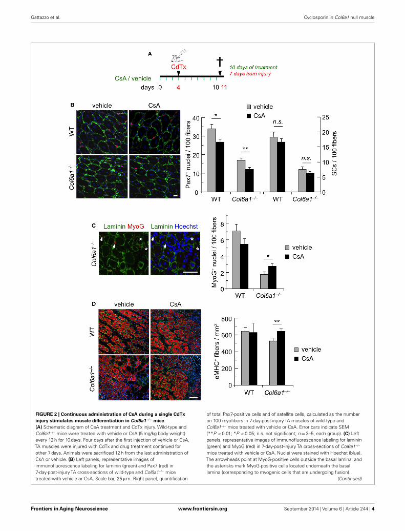
FIGURE 2 | Continuous administration of CsA during a single CdTxinjury stimulates muscle differentiation in Col6a1−/− mice.(A) Schematic diagram of CsA treatment and CdTx injury. Wild-type andCol6a1−/− mice were treated with vehicle or CsA (5 mg/kg body weight)every 12 h for 10 days. Four days after the first injection of vehicle or CsA,TA muscles were injured with CdTx and drug treatment continued forother 7 days. Animals were sacrificed 12 h from the last administration ofCsA or vehicle. (B) Left panels, representative images ofimmunofluorescence labeling for laminin (green) and Pax7 (red) in7-day-post-injury TA cross-sections of wild-type and Col6a1−/− micetreated with vehicle or CsA. Scale bar, 25 µm. Right panel, quantification
of total Pax7-positive cells and of satellite cells, calculated as the numberon 100 myofibers in 7-day-post-injury TA muscles of wild-type andCol6a1−/− mice treated with vehicle or CsA. Error bars indicate SEM(**P < 0.01; *P < 0.05; n.s. not significant; n=3–5, each group). (C) Leftpanels, representative images of immunofluorescence labeling for laminin(green) and MyoG (red) in 7-day-post-injury TA cross-sections of Col6a1−/−
mice treated with vehicle or CsA. Nuclei were stained with Hoechst (blue).The arrowheads point at MyoG-positive cells outside the basal lamina, andthe asterisks mark MyoG-positive cells located underneath the basallamina (corresponding to myogenic cells that are undergoing fusion).
(Continued)
Frontiers in Aging Neuroscience www.frontiersin.org September 2014 | Volume 6 | Article 244 | 4

Gattazzo et al. Cyclosporin in Col6a1 null muscle
FIGURE 2 | ContinuedScale bar, 50 µm. Right panel, quantification of total MyoG-positive cells,calculated as the number on 100 myofibers in 7-day-post-injury TA muscles ofwild-type and Col6a1−/− mice treated with vehicle or CsA. Error bars indicateSEM (*P < 0.05; n=3–5, each group). (D) Left panels, representative imagesof immunofluorescence for laminin (green) and eMHC (red) in
7-day-post-injury TA cross-sections of wild-type and Col6a1−/− mice treatedwith vehicle or CsA. Nuclei were stained with Hoechst (blue). Scale bar,50 µm. Right panel, quantification of the number of eMHC-positive myofibersper regenerating area in 7-day-post-injury TA cross-sections of wild-type andCol6a1−/− mice treated with vehicle or CsA. Error bars indicate SEM(**P < 0.01; n=3–5, each group). SCs, satellite cells; WT, wild-type.
only provided by this drug dosage or could be also elicited byhigher CsA concentrations known to cause strong immunosup-pressive effects (Homan et al., 1980), we subjected mice to i.p.injection of CsA at 25 mg/kg body weight every 24 h for 10 days.Interestingly, at this higher dosage, CsA led to a dramatic decreasein the number of total Pax7-positive cells and of satellite cells bothin wild-type and in Col6a1−/− mice when compared to vehicle-treated animals (Figure S1C in Supplementary Material). Theseresults highlight the relevance of CsA dosage in inducing beneficialeffects in Col6a1−/− muscles and accordingly with previous stud-ies carried out with the immunosuppressive drug FK506 (Irwinet al., 2003), they suggest that immunosuppression exacerbatesthe phenotype of Col6a1−/− mice.
CONTINUOUS ADMINISTRATION OF CsA DURING CdTx INJURYSTIMULATES THE EARLY PHASES OF MUSCLE DIFFERENTIATION INCol6a1−/− MICETo further assess the capability of CsA to ameliorate the muscleregenerative defects of Col6a1−/− mice, we carried out CsA treat-ment under experimentally induced muscle injury. Toward thisaim, wild-type and Col6a1−/− mice were treated for 10 days withvehicle or CsA at 5 mg/kg body weight every 12 h; 4 days after thestart of treatment, TA muscles were subjected to CdTx damage andmice were sacrificed 7 days after injury (Figure 2A). TUNEL assayshowed that the incidence of apoptotic nuclei 7 days after CdTxinjection was very low in wild-type TA, in agreement with the con-cept that myofiber demise is almost completed at this stage frominjury (Hawke et al., 2003). Conversely, Col6a1−/− TA showed ahigher number of TUNEL-positive myonuclei 7 days after injury,and CsA administration was able to significantly decrease theincidence of apoptotic myofibers triggered by CdTx injury inCol6a1−/− TA muscles (Figure S2 in Supplementary Material).We next evaluated the number of myogenic cells by immunoflu-orescence for the Pax7 and MyoG markers. CsA administrationled to a significant decrease in the number of total Pax7-positivecells in both wild-type and Col6a1−/− injured TA, without anysignificant change in the number of satellite cells (i.e., Pax7-positive cells located underneath basal lamina) (Figure 2B). Thisresponse was paralleled by a significant increase of the total num-ber of MyoG-positive cells in injured TA muscles of CsA-treatedCol6a1−/− mice when compared to vehicle-treated Col6a1−/−
mice, whereas wild-type injured TA muscles did not show anysignificant difference in MyoG positivity between vehicle andCsA-treated animals (Figure 2C). Additionally, Col6a1−/− micetreated with CsA showed an increased number of regeneratingmyofibers expressing eMHC, whereas no differences in eMHC-positive regenerating myofibers were found in wild-type animals(Figure 2D). As the defective satellite self-renewal of Col6a1−/−
mice is strictly dependent on the lack of extracellular collagen VIand on the lower muscle stiffness (Urciuolo et al., 2013), it was
not surprising to observe that in this experimental condition CsAdoes not display any overt effect on satellite cell maintenance. Onthe other hand, the remarkable increase in the number of differ-entiated (i.e., MyoG-positive) myogenic cells, together with thehigher number of newly forming (i.e., eMHC-positive) myofibers,indicates that CsA administration is able to improve muscle dif-ferentiation upon damage in the Col6a1−/− myopathic mousemodel.
ADMINISTRATION OF CsA DURING REPEATED MUSCLE INJURYCOUNTERACTS MUSCLE LOSS AND FIBROSIS AND PRESERVES THESATELLITE CELL POOL IN Col6a1−/− MICEAlthough muscles of Col6a1−/− animals display a delayed regen-eration after injury, we have previously shown that at 30 daysafter CdTx injury they are still able to complete the regenerationprocess (Urciuolo et al., 2013). However, and at difference fromwild-type mice, the capability of Col6a1−/− animals to undergomuscle regeneration and preserve the satellite cell pool is lost aftermultiple muscle injuries, leading to a severe loss of muscle mass(Urciuolo et al., 2013). Therefore, we investigated whether CsAis able to counteract the defective muscle regeneration and thedepletion of the satellite cell pool triggered by multiple injuriesin Col6a1−/− mice. Toward this aim, we subjected TA muscles ofwild-type and Col6a1−/− mice to three rounds of CdTx injury.Animals were treated for 10 days with vehicle or CsA at 5 mg/kgbody weight every 12 h during the third injury and sacrificed30 days after the last injury (Figure 3A). Interestingly, CsA admin-istration was highly effective in reducing the extensive musclefibrosis triggered by triple injury in Col6a1−/− mice (Figure 3B).The beneficial effects of CsA in Col6a1−/− muscles undergoingmultiple injuries were also confirmed by the increased myofibercross-sectional area and by the improvement of the muscle massin CsA-treated Col6a1−/− mice when compared to vehicle-treatedCol6a1−/− mice (Figure S3 in Supplementary Material). Notably,CsA administration led to marked increase of both total Pax7-positive cell number and satellite cell number in Col6a1−/− TAmuscles subjected to multiple injuries (Figures 3C,D). Altogether,these results show that at this regimen CsA is capable to preservenot only muscle fibers but also the satellite cell pool of collagenVI-deficient mice.
DISCUSSIONIn the present study, we evaluated the potential beneficial effectsexerted by CsA on skeletal muscle regeneration in Col6a1−/−
mice, both under physiological condition and after muscle dam-age. The rationale for this study was based on previous findingsin patients affected by collagen VI myopathies, suggesting thatbesides counteracting myofiber apoptosis and mitochondrial dys-function, CsA treatment may also increase muscle regeneration(Merlini et al., 2008, 2011). In addition, our recent findings
Frontiers in Aging Neuroscience www.frontiersin.org September 2014 | Volume 6 | Article 244 | 5

Gattazzo et al. Cyclosporin in Col6a1 null muscle
FIGURE 3 | Administration of CsA during a triple muscle injurycounteracts muscle loss and fibrosis and preserves the satellite cellpool in Col6a1−/− mice. (A) Schematic diagram of CsA treatment andCdTx injuries. TA muscles of wild-type and Col6a1−/− mice were given threerepeated injections of CdTx every 30 days. Four days before the last injury,Col6a1−/− mice were treated with vehicle or CsA (5 mg/kg body weight)every 12 h for 10 days. Mice were sacrificed 30 days after the third injury(i.e., 24 days from the last administration of vehicle or CsA). (B) Left panels,Azan-Mallory staining of triple injured TA cross-sections from wild-type andCol6a1−/− mice treated with vehicle or CsA. Scale bar, 50 µm. Right panel,quantification of the fibrotic area in triple-injured TA cross-sections from
wild-type and Col6a1−/− mice treated with vehicle or CsA. Error barsindicate SEM (**P < 0.01; *P < 0.05; n=4–8, each group).(C) Representative images of immunofluorescence labeling for laminin(red) and Pax7 (green) in triple-injured TA cross-sections from wild-type andCol6a1−/− treated with vehicle or CsA. Arrows point at Pax7-positive nuclei.Nuclei were stained with Hoechst (blue). Scale bar, 100 µm.(D) Quantification of total Pax7-positive cells and of satellite cells,calculated as the number on 100 myofibers in triple-injured TAcross-sections from wild-type and Col6a1−/− mice treated with vehicle orCsA. Error bars indicate SEM (**P < 0.01; n=4–8, each group). SC,satellite cells; WT, wild-type.
showed impaired regeneration and defective satellite cell self-renewal in collagen VI-deficient muscles (Urciuolo et al., 2013).Therefore, we investigated how CsA treatment impacts on the
regeneration of collagen VI-deficient mice. Under physiologicalconditions, CsA was capable to amplify the pool of total Pax7-positive cells and of satellite cells and increased the amount of
Frontiers in Aging Neuroscience www.frontiersin.org September 2014 | Volume 6 | Article 244 | 6
Gattazzo et al. Cyclosporin in Col6a1 null muscle
newly formed centrally nucleated myofibers, thus suggesting anew role for CsA in stimulating myogenesis in Col6a1−/− mus-cles. Notably, these beneficial effects were dose-dependent, as theywere observed at 10 mg/kg/day but not at a higher immunosup-pressant dose (25 mg/kg/day), which conversely had a negativeimpact on the satellite cell pool. These data are in agreementwith previous results in which the same protocol of CsA admin-istration was found to desensitize the mitochondrial permeabil-ity transition pore and reduce myofiber apoptosis in Col6a1−/−
mice (Irwin et al., 2003). Although we previously demonstratedthat CsA stimulates autophagy in skeletal myofibers (Grumatiet al., 2010), the increased number of satellite cells in musclesof CsA-treated Col6a1−/− animals is not a direct consequenceof a stimulatory effect on autophagy. Indeed, reactivation of theautophagic flux in Col6a1−/− mice by different pharmacologi-cal or dietary treatments does not exert any significant effect onsatellite cells (Urciuolo et al., 2013). Our present findings are con-sistent with the strong amelioration of the myophatic phenotypein Col6a1−/−mice following CsA administration and indicate thatbesides decreasing mitochondrial dysfunction and apoptosis andreactivating autophagy in muscle fibers (Irwin et al., 2003; Grumatiet al., 2010), the drug is also able to increase the pool of functionalPax7-positive cells and stimulate the formation of newly formedfibers.
The beneficial effects exerted by CsA on the regeneration capa-bilities of Col6a1−/− mice become very evident under experi-mentally induced single and multiple muscle injuries. Our dataindicate that CsA is capable to induce myogenesis in Col6a1−/−
mice after muscle damage. In fact, when TA muscles were dam-aged during a continuous CsA administration, analysis at 7 dayspost-injury showed that CsA elicits a significant increase in thenumber of MyoG-positive cells and of regenerating myofibers inCol6a1−/− muscles. Interestingly, this response is not associatedwith an improvement of the number of satellite cells, suggestingthat under these conditions CsA is unable to ameliorate satellitecell self-renewal. The effect of CsA on muscle regeneration waseven more remarkable when we exacerbated the muscle pheno-type of Col6a1−/−mice through triple CdTx damage. Our findingsindicate that CsA is protective against fibrotic tissue formation,maybe exerting this effect through an indirect regulation of theinflammatory state that occurs during muscle regeneration (Ser-rano et al., 2011). A similar beneficial effect of CsA in reducingmuscle fibrosis was reported for mdx mice undergoing exercise(De Luca et al., 2005). Furthermore, CsA administration was ableto counteract the loss of satellite cells elicited by repeated muscleinjuries in Col6a1−/− animals, concurrently guaranteeing myo-genic differentiation, as confirmed by the increase of myofibercross-sectional area and muscle mass. Although it was beyondthe scope of this study to dissect the mechanism(s) through whichCsA leads to increased satellite cell number in Col6a1−/−mice afterrepeated injuries, it can be hypothesized that CsA administrationmay not directly influence the self-renewal capability of satellitecells and that the preservation of satellite cell pool may be medi-ated by an increase of their survival. This assumption is supportedby the fact that the defective satellite cell self-renewal of Col6a1−/−
mice is strictly dependent on the lack of collagen VI itself and itsconsequences on muscle stiffness (Urciuolo et al., 2013) and that
CsA treatment is able to reduce apoptosis in Col6a1−/− muscles(Irwin et al., 2003). To our knowledge, no literature work hasinvestigated in detail the effects of in vivo CsA administration onstem cell homeostasis in skeletal muscles. A recent study reportedsome beneficial effects of CsA on neuronal stem cells, showing thatin vivo CsA administration increases the number of neurospheresdue to enhanced neuronal stem cell survival, rather than increasedproliferation (Hunt et al., 2010).
The pharmacology of CsA is complex, and the drug binds afamily of cellular peptidyl-prolyl cis–trans isomerases known ascyclophilins. Binding of CsA with the abundant cyclophilin Aleads to inhibition of calcineurin, a cytosolic phosphatase foundin many cell types, thus preventing dephosphorylation of its sub-strates (Liu et al., 1991). A number of studies have shown thatcalcineurin signals are involved in the control of myofiber size,myofiber type, and skeletal muscle regeneration (Schiaffino andSerrano, 2002; Sakuma and Yamaguchi, 2010; Hudson and Price,2013). Although inhibition of calcineurin was shown to delaymuscle regeneration (Sakuma et al., 2003, 2005), literature stud-ies investigating the outcomes of calcineurin inhibition by geneticapproaches or by CsA administration in animal models of musclediseases have produced contrasting results (Stupka et al., 2004; DeLuca et al., 2005; Parsons et al., 2007). The reasons for these dis-crepancies rely upon multiple factors, including the genetic modelstudied, the dose of the drug, the type of muscle, the durationof treatment, and the route of treatment. For instance, the effi-cacy of CsA in the mdx mice, an animal model of Duchennemuscular dystrophy, was reported to be dependent on the dosageand length of the treatment (Stupka et al., 2004; De Luca et al.,2005). Notably, the protective effects of CsA in Col6a1−/− micedo not rely upon calcineurin inhibition, as the same beneficialeffects are also displayed by non-immunosuppressive CsA analogsthat do not bind calcineurin, such as Debio 025 and NIM811(Angelin et al., 2007; Zulian et al., 2014), whereas they cannot bemimicked by the calcineurin inhibitor FK506 (Irwin et al., 2003).Although our interest was far from the study of calcineurin activ-ity, in this work we used a definite CsA dosage (5 mg/kg every12 h, i.e., the same dose shown to be effective in rescuing differ-ent aspects of the muscle pathology of Col6a1−/− mice), and thisdosage is known to only partially reduce the activity of calcineurin(Dunn et al., 2002; Michel et al., 2004).
In conclusion, our results indicate that besides the alreadyknown beneficial effects of CsA administration in amelioratingthe myophatic phenotype of Col6a1−/− mice through the rescuefrom mitochondrial and autophagic dysfunction of muscle fibers,CsA is also capable to stimulate muscle regeneration and preservethe satellite cell pool in this disease model. These findings sup-port and strengthen the increased muscle regeneration observedin Ullrich patients undergoing clinical trial with CsA, pointing atCsA and its non-immunosuppressive derivatives as a promisingtherapeutic route for this group of inherited muscle diseases.
AUTHOR CONTRIBUTIONSFrancesca Gattazzo planned and performed in vivo and ex vivoexperiments and wrote the paper. Sibilla Molon performedcardiotoxin damage, immunofluorescence, and histology. ValeriaMorbidoni performed in vivo satellite cell quantification. Bert
Frontiers in Aging Neuroscience www.frontiersin.org September 2014 | Volume 6 | Article 244 | 7

Gattazzo et al. Cyclosporin in Col6a1 null muscle
Blaauw carried out part of the in vivo studies. Paola Braghetta wasinvolved in CsA administration. Anna Urciuolo oversaw the resultsand interpreted the data. Paolo Bonaldo oversaw the results andwrote the paper. All the authors discussed the results, revised thework, commented on the manuscript, and agreed on the final draft.
ACKNOWLEDGMENTSThis work was supported by grants from Telethon-Italy(GGP10225 and GGP11082), the Italian Ministry of Edu-cation, University and Research (FIRB Strategic ProjectRBAP11Z3YA_003), and the University of Padova.
SUPPLEMENTARY MATERIALThe Supplementary Material for this article can be found onlineat http://www.frontiersin.org/Journal/10.3389/fnagi.2014.00244/abstract
REFERENCESAngelin, A., Tiepolo, T., Sabatelli, P., Grumati, P., Bergamin, N., Golfieri, C., et al.
(2007). Mitochondrial dysfunction in the pathogenesis of Ullrich congenitalmuscular dystrophy and prospective therapy with cyclosporins. Proc. Natl. Acad.Sci. U.S.A. 104, 991–996. doi:10.1073/pnas.0610270104
Bonaldo, P., Braghetta, P., Zanetti, M., Piccolo, S., Volpin, D., and Bressan, G. M.(1998). Collagen VI deficiency induces early onset myopathy in the mouse:an animal model for Bethlem myopathy. Hum. Mol. Genet. 7, 2135–2140.doi:10.1093/hmg/7.13.2135
Buckingham, M., and Rigby, P. W. J. (2014). Gene regulatory networks andtranscriptional mechanisms that control myogenesis. Dev. Cell 28, 225–238.doi:10.1016/j.devcel.2013.12.020
Chargé, S. B. P., and Rudnicki, M. (2004). Cellular and molecular regulation ofmuscle regeneration. Physiol. Rev. 84, 209–238. doi:10.1152/physrev.00019.2003
Ciciliot, S., and Schiaffino, S. (2010). Regeneration of mammalian skeletal mus-cle: basic mechanisms and clinical implications. Curr. Pharm. Des. 16, 906–914.doi:10.2174/138161210790883453
Couteaux, R., Mira, J. C., and D’Albis, A. (1988). Regeneration of muscles aftercardiotoxin injury. I. Cytological aspects. Biol. Cell 62, 171–182. doi:10.1111/j.1768-322X.1988.tb00719.x
De Luca, A., Nico, B., Liantonio, A., Didonna, M. P., Fraysse, B., Pierno, S., et al.(2005). A multidisciplinary evaluation of the effectiveness of cyclosporine a indystrophic mdx mice. Am. J. Pathol. 166, 477–489. doi:10.1016/S0002-9440(10)62270-5
Dunn, S. E., Simard,A. R., Prud’homme, R. A., and Michel, R. N. (2002). Calcineurinand skeletal muscle growth. Nat. Cell Biol. 4, E46. doi:10.1038/ncb0302-e46aauthor reply E46–47,
Grumati, P., Coletto, L., Sabatelli, P., Cescon, M., Angelin, A., Bertaggia, E., et al.(2010). Autophagy is defective in collagen VI muscular dystrophies, and its reac-tivation rescues myofiber degeneration. Nat. Med. 16, 1313–1320. doi:10.1038/nm.2247
Hawke, T. J., Meeson, A. P., Jiang, N., Graham, S., Hutcheson, K., DiMaio, J. M.,et al. (2003). p21 is essential for normal myogenic progenitor cell function inregenerating skeletal muscle. Am. J. Physiol. Cell Physiol. 285, C1019–C1027.doi:10.1152/ajpcell.00055.2003
Homan, W. P., Fabre, J. W., Williams, K. A., Millard, P. R., and Morris, P. J.(1980). Studies on the immunosuppressive properties of cyclosporin A in ratsreceiving renal allografts. Transplantation 29, 361–366. doi:10.1097/00007890-198005000-00003
Hudson, M. B., and Price, S. R. (2013). Calcineurin: a poorly understood regulatorof muscle mass. Int. J. Biochem. Cell Biol. 45, 2173–2178. doi:10.1016/j.biocel.2013.06.029
Hunt, J., Cheng, A., Hoyles, A., Jervis, E., and Morshead, C. M. (2010). CyclosporinA has direct effects on adult neural precursor cells. J. Neurosci. 30, 2888–2896.doi:10.1523/JNEUROSCI.5991-09.2010
Irwin, W. A., Bergamin, N., Sabatelli, P., Reggiani, C., Megighian, A., Merlini, L.,et al. (2003). Mitochondrial dysfunction and apoptosis in myopathic mice withcollagen VI deficiency. Nat. Genet. 35, 367–371. doi:10.1038/ng1270
Lampe, A. K., and Bushby, K. M. D. (2005). Collagen VI related muscle disorders. J.Med. Genet. 42, 673–685. doi:10.1136/jmg.2002.002311
Liu, J., Farmer, J. D., Lane, W. S., Friedman, J., Weissman, I., and Schreiber, S. L.(1991). Calcineurin is a common target of cyclophilin-cyclosporin A and FKBP-FK506 complexes. Cell 66, 807–815. doi:10.1016/0092-8674(91)90124-H
Merlini, L., Angelin, A., Tiepolo, T., Braghetta, P., Sabatelli, P., Zamparelli, A., et al.(2008). Cyclosporin A corrects mitochondrial dysfunction and muscle apopto-sis in patients with collagen VI myopathies. Proc. Natl. Acad. Sci. U.S.A. 105,5225–5229. doi:10.1073/pnas.0800962105
Merlini, L., Sabatelli, P.,Armaroli,A., Gnudi, S.,Angelin,A., Grumati, P., et al. (2011).Cyclosporine A in Ullrich congenital muscular dystrophy: long-term results.Oxid. Med. Cell. Longev. 2011, 139194. doi:10.1155/2011/139194
Michel, R. N., Dunn, S. E., and Chin, E. R. (2004). Calcineurin and skeletal musclegrowth. Proc. Nutr. Soc. 63, 341–349. doi:10.1079/PNS2004362
Parsons, S. A., Millay, D. P., Sargent, M. A., Naya, F. J., McNally, E. M., Sweeney, H. L.,et al. (2007). Genetic disruption of calcineurin improves skeletal muscle pathol-ogy and cardiac disease in a mouse model of limb-girdle muscular dystrophy. J.Biol. Chem. 282, 10068–10078. doi:10.1074/jbc.M609368200
Sakuma, K., Nakao, R., Aoi, W., Inashima, S., Fujikawa, T., Hirata, M., et al. (2005).Cyclosporin A treatment upregulates Id1 and Smad3 expression and delays skele-tal muscle regeneration. Acta Neuropathol. 110, 269–280. doi:10.1007/s00401-005-1049-x
Sakuma, K., Nishikawa, J., Nakao, R., Watanabe, K., Totsuka, T., Nakano, H.,et al. (2003). Calcineurin is a potent regulator for skeletal muscle regenera-tion by association with NFATc1 and GATA-2. Acta Neuropathol. 105, 271–280.doi:10.1007/s00401-002-0647-0
Sakuma, K., and Yamaguchi, A. (2010). The functional role of calcineurin in hyper-trophy, regeneration, and disorders of skeletal muscle. J. Biomed. Biotechnol.2010, 721219. doi:10.1155/2010/721219
Schiaffino, S., and Serrano, A. L. (2002). Calcineurin signaling and neural controlof skeletal muscle fiber type and size. Trends Pharmacol. Sci. 6147, 569–575.doi:10.1016/S0165-6147(02)02111-9
Serrano, A. L., Mann, C. J., Vidal, B., Ardite, E., Perdiguero, E., and Muñoz-Cánoves,P. (2011). Cellular and molecular mechanisms regulating fibrosis in skeletal mus-cle repair and disease. Curr. Top. Dev. Biol. 96, 167–201. doi:10.1016/B978-0-12-385940-2.00007-3
Shi, X., and Garry, D. J. (2006). Muscle stem cells in development, regeneration, anddisease. Genes Dev. 20, 1692–1708. doi:10.1101/gad.1419406
Stupka, N., Gregorevic, P., Plant, D. R., and Lynch, G. S. (2004). The calcineurinsignal transduction pathway is essential for successful muscle regeneration inmdx dystrophic mice. Acta Neuropathol. 107, 299–310. doi:10.1007/s00401-003-0807-x
Tedesco, F. S., Dellavalle, A., Diaz-Manera, J., Messina, G., and Cossu, G. (2010).Repairing skeletal muscle: regenerative potential of skeletal muscle stem cells. J.Clin. Invest. 120, 11–19. doi:10.1172/JCI40373
Urciuolo, A., Quarta, M., Morbidoni, V., Gattazzo, F., Molon, S., Grumati, P., et al.(2013). Collagen VI regulates satellite cell self-renewal and muscle regeneration.Nat. Commun. 4, 1964. doi:10.1038/ncomms2964
Zulian, A., Rizzo, E., Schiavone, M., Palma, E., Tagliavini, F., Blaauw, B., et al. (2014).NIM811, a cyclophilin inhibitor without immunosuppressive activity, is bene-ficial in collagen VI congenital muscular dystrophy models. Hum. Mol. Genet.doi:10.1093/hmg/ddu254
Conflict of Interest Statement: The authors declare that the research was conductedin the absence of any commercial or financial relationships that could be construedas a potential conflict of interest.
Received: 27 June 2014; accepted: 29 August 2014; published online: 15 September 2014.Citation: Gattazzo F, Molon S, Morbidoni V, Braghetta P, Blaauw B, Urciuolo A andBonaldo P (2014) Cyclosporin A promotes in vivo myogenic response in collagen VI-deficient myopathic mice. Front. Aging Neurosci. 6:244. doi: 10.3389/fnagi.2014.00244This article was submitted to the journal Frontiers in Aging Neuroscience.Copyright © 2014 Gattazzo, Molon, Morbidoni, Braghetta, Blaauw, Urciuolo andBonaldo. This is an open-access article distributed under the terms of the CreativeCommons Attribution License (CC BY). The use, distribution or reproduction in otherforums is permitted, provided the original author(s) or licensor are credited and thatthe original publication in this journal is cited, in accordance with accepted academicpractice. No use, distribution or reproduction is permitted which does not comply withthese terms.
Frontiers in Aging Neuroscience www.frontiersin.org September 2014 | Volume 6 | Article 244 | 8