cytochemical studies on the mouse mammary tumor virus
TRANSCRIPT
(CANCER RESEARCH 27 Part 1, 2179-2196,November 1967]
Cytochemical Studies on the Mouse Mammary Tumor Virus
GILBERT H. SMITH1
Viral Biology Branch, National Cancer Institute, Bethesda, Maryland 20014
SUMMARY
The chemical nature of the ultrastructure of the mouse mammary tumor virus has been investigated in situ. Small pieces ofspontaneous mammary tumors from C3H mice were fixed inphosphate-buffered glutaraldehyde, infiltrated, and embedded inglycol methacrylate, a water-miscible plastic introduced in 1963by Leduc and her collaborators as a medium suitable for cyto-chemical studies at the ultrastructural level. Electron microscopicexamination of the tissue revealed the presence of numerouscytoplasmic A particles and extracellular H particles. Considerable evidence has accumulated demonstrating that the latteris the active, mature mammary tumor virus. The cytoplasmic Aparticle is considered to be formed in the cytoplasm as a precursor of the B particle; however, there is little evidence fromimmunologie, biochemical, and biophysical parameters to supportthis hypothesis. Ultrathin sections of the tumor containing bothparticles were subjected to digestion with RNase, DNase, pepsin,papain, and trypsin and combinations of these enzymes. Consecutive sections were incubated in the appropriate controlsolutions. The results reported (a) provide evidence for thepresence of RNA in the cytoplasmic A particle, (6) indicate thatthe stereochemical nature of the A particle is synonymous withthe nucleoid of the B particle, and (c) present further support ofBernhard's hypothesis that the cytoplasmic A particle is the
intracellular precursor of the mammary tumor virus.
INTRODUCTION
The discovery of the mammary tumor virus (MTV) in 1936by Bittner (8) has led over the years to intensive studies concerning its biologic, morphologic, biophysical, and immunologiecharacteristics. Two types of particles visualized with the electron microscope and originally described by Bernhard areconsidered to represent MTV: (a) intracytoplasmic or type Aparticles, and (6) extracellular or type B particles. Bernhard (5)postulated that the A particles are formed in the cytoplasm asprecursors of mature MTV and evolve into the latter by movingto the cell membrane to produce buds which are pinched off andsubsequently develop into mature B particles.
Considerable evidence has accumulated since Bernhard's ob
servations that support the hypothesis that the B particlerepresents the active MTV. Electron microscopic studies byDmochowski (14, 15), Bang et al. (3, 4), Bernhard et al. (5),Suzuki (38), and Pitelka et al. (33); Pitelka et al. (34), Goldfederet al. (18), Feldman (17), Haguenau et al. (19), and others have
»NIH,USPHS, HEW.Received May 8, 1907; accepted July 11, 1967.
indicated the presence of B particles in spontaneous mammarytumors and in preneoplastic aveolar nodules in a variety ofinbred mouse strains infected with MTV. The biophysical studiesof Moore et al. (30) and Lasfargues et al. (22) provided strongphysical and biologic evidence that the B particle and its nucleoid represent the active MTV. Recent immunologie studies byBlair and Pitelka (9) have demonstrated that the specific anti-genicity related to MTV, as measured by the immunodiffusiontechnic, is carried by the B particle. Despite these observations,there still exists a discrepancy between the biologic and morphologic results with reference to the presence of the B particle(34). Although at this time one cannot say that every B particlecarried the classic MTV tumorigenicity, the fact that MTVactivity is carried by a B particle is not in dispute.
The A particle has been observed in other murine neoplasticcells, e.g., Swiss lymphoma cells (13), spontaneous plasma celltumors (10), and spontaneous leukemic cells (12). In these tumorsthey apparently are not transformed into B particles. It has alsobeen suggested that the B particle may develop from buds at thecellular surface without the migration of the A particle to themembrane. (1, 18, 21, 30). These observations were challengedby Imai et al. (20), who examined serial sections of spontaneousC3H mammary cancers in three dimensions. He demonstratedthat most of the buds were associated with complete A particles.He suggested that the earlier results were due to the observationsmade on thin nonserial sections. Still, no intermediate stagesrepresenting the alteration of the A particle to the eccentricnucleoid of the B particle following its release from the cell havebeen reported.
It is the purpose of the experiments to be reported here (a) toestablish the chemical nature of the ultrastructural morphologyof the B particle, and (6) to correlate the cytochemical nature ofthe A particle with the nucleoid of the B particle within theframework of Bernhard's hypothesis concerning the maturation
of the mammary tumor virus.
MATERIALS AND METHODS
Tissue Preparation
Small pieces of a spontaneous mammary tumor taken from aC3H/AnWi female known to harbor the MTV were fixed invarious concentrations of glutaraldehyde (2.0, 3.5, 5.0, 8.0%) in0.1 M Sörensenphosphate buffer at pH 7.4. The blocks wereinfiltrated and dehydrated in glycol methacrylate (Rohm andHaas Company, Philadelphia) according to the method of Leducet al. (27). The samples were embedded in a mixture of 70%glycol methacrylate and 30% 85/15 butyl/methyl methacrylatewhich had been prepolymerized in an H2O bath at 90°C.Poly
merization was completed in the cold under ultraviolet light.
NOVEMBER 1967 2179
Research. on February 13, 2018. © 1967 American Association for Cancercancerres.aacrjournals.org Downloaded from
Gilbert H. Smith
Triple Digestion
Double DigestionLoop
Single Digestion Loop
Loop
ITwo rinsesDist. H20
Samples token
for E.M.
DiluentII
Two rinsesDist. H20
Samples taken for E.M.
Dluent111
i One rinseDist. H20
Samples tokenfor E.M.
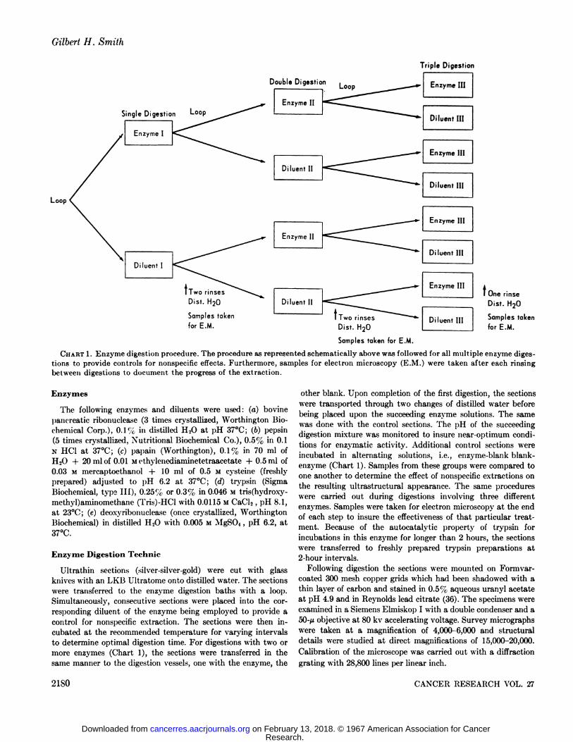
CHART1. Enzyme digestion procedure. The procedure as represented schematically above was followed for all multiple enzyme digestions to provide controls for nonspecific effects. Furthermore, samples for electron microscopy (E.M.) were taken after each rinsingbetween digestions to document the progress of the extraction.
Enzymes
The following enzymes and diluents were used: (a) bovinepancreatic ribonuclea.se (3 times crystallized, Worthington Biochemical Corp.), 0.1 % in distilled H,0 at pH 37°C;(6) pepsin
(5 times crystallized, Nutritional Biochemical Co.), 0.5% in 0.1N HC1 at 37°C;(c) papain (Worthington), 0.1% in 70 ml of
H20 + 20 ml of 0.01 Methylenediaminetetraacetate + 0.5ml of0.03 M mercaptoethanol + 10 ml of 0.5 M cysteine (freshlyprepared) adjusted to pH 6.2 at 37°C;(d) trypsin (SigmaBiochemical, type III), 0.25% or 0.3% in 0.046 M tris(hydroxy-methyl)aminomethane (Tris)-HCl with 0.0115 MCaCl«,pH 8.1,at 23°C;(e) deoxyribonuclease (once crystallized, Worthington
Biochemical) in distilled H20 with 0.005 M MgS04, pH 6.2, at37°C.
Enzyme Digestion Technic
Ultrathin sections (silver-silver-gold) were cut with glassknives with an LKB Ultratome onto distilled water. The sectionswere transferred to the enzyme digestion baths with a loop.Simultaneously, consecutive sections were placed into the corresponding diluent of the enzyme being employed to provide acontrol for nonspecific extraction. The sections were then incubated at the recommended temperature for varying intervalsto determine optimal digestion time. For digestions with two ormore enzymes (Chart 1), the sections were transferred in thesame manner to the digestion vessels, one with the enzyme, the
other blank. Upon completion of the first digestion, the sectionswere transported through two changes of distilled water beforebeing placed upon the succeeding enzyme solutions. The samewas done with the control sections. The pH of the succeedingdigestion mixture was monitored to insure near-optimum conditions for enzymatic activity. Additional control sections wereincubated in alternating solutions, i.e., enzyme-blank blank-enzyme (Chart 1). Samples from these groups were compared toone another to determine the effect of nonspecific extractions onthe resulting ultrastructural appearance. The same procedureswere carried out during digestions involving three differentenzymes. Samples were taken for electron microscopy at the endof each step to insure the effectiveness of that particular treatment. Because of the autocatalytic property of trypsin forincubations in this enzyme for longer than 2 hours, the sectionswere transferred to freshly prepared trypsin preparations at2-hour intervals.
Following digestion the sections were mounted on Formvar-coated 300 mesh copper grids which had been shadowed with athin layer of carbon and stained in 0.5%; aqueous uranyl acetateat pH 4.9 and in Reynolds lead citrate (36). The specimens wereexamined in a Siemens Elmiskop I with a double condenser and a50-Mobjective at 80 kv accelerating voltage. Survey micrographswere taken at a magnification of 4,000-6,000 and structuraldetails were studied at direct magnifications of 15,000-20,000.
Calibration of the microscope was carried out with a diffractiongrating with 28,800 lines per linear inch.
2180 CANCER RESEARCH VOL. 27
Research. on February 13, 2018. © 1967 American Association for Cancercancerres.aacrjournals.org Downloaded from
Mouse Mammary Tumor Virus
RESULTS
Preliminary Observations
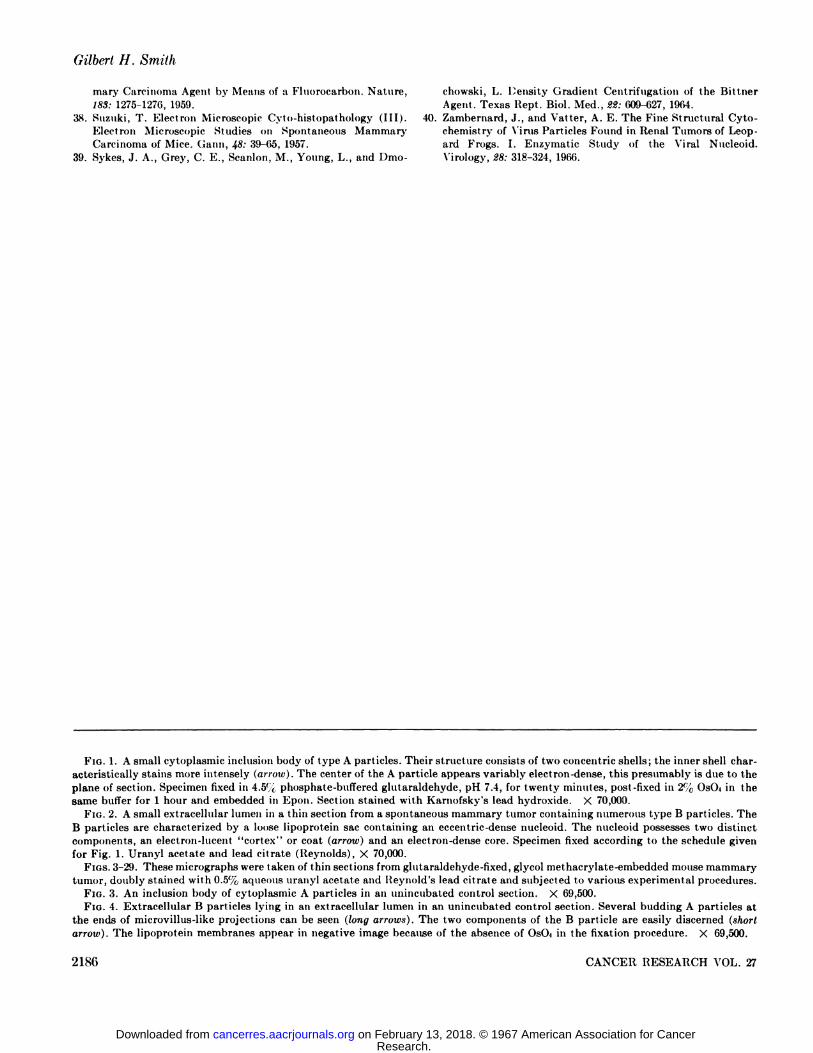
Observations on thin sections from glutaraldehyde-fixed andpostosmicated mammary tumor cells revealed the characteristictype A and B particles of Bernhard (5). The A particles werefound singly or in large inclusions in the cytoplasm. In sectionthey appeared as doughnuts consisting of two concentric ringsthe outer diameter of which measured 700-750 A (Fig. 1). Theinnermost shell of the doughnut exhibited a strong affinity forelectron staining with lead salts. This characteristic staining hasbeen interpreted by several authors as an indication of thepresence of nucleic acid, presumably RNA. The 15 particle,primarily extracellular, was characterized by possessing a lipo-protein sac measuring 1000-1400 A, within which was containedan eccentrically located nucleoid measuring 400-500 A (Fig. 2).The nucleoid possessed two distinct zones, an electron-dense coreand an electron-lucent shell or cortex.
Following glutaraldehyde fixation, infiltration, and embedmentin glycol methacrylate, the ultrastructural appearance of boththe A and B particle is quite comparable to that observed afterconventional treatment. The diameters of both the A particleand the nucleoid of the B particle are, however, greater; 900-1000A (Fig. 3) and 600-690 A (Fig. 4), respectively. These measure
ments were consistent throughout all the blocks used during thecourse of the experiment and following all enzymatic treatmentsand their controls except those in which certain proteolyticenzymes were used.
The cellular ultrastructure closely paralleled the descriptiongiven by Leduc el al. (25) in regard to the appearance of thenuclear chromatin, mitochondria, ribosomes, cellular membranes,and nucleolus for glutaraldehyde-fixed, glycol methacrylate-embedded tissues. No appreciable swelling or shrinkage wasobserved in any of the sections. In all instances, the contrastattained with double staining with aqueous 0.5% uranyl acetateand lead citrate was satisfactory.
Effect of Single Enzyme DigestionsRNase. After 20-30 minutes of incubation with 0.1 <"cRNase,
the densely staining ribosomes of the cytoplasm were lost (Fig.5). The cytoplasm was homogeneous and moderately dense. Thenuclear chromatin became more electron dense. With longerdigestions, the granular component of the nucleolus was nolonger present. No effect on the other components of the nucleusor cytoplasm was observed even with much longer digestions (5hours). The A particle during these treatments reflected noconsistent change, although in some cases the inner shell showeda decreased affinity for the lead stain (Fig. 6). In most sections,the inclusion bodies consisting of A particles were obscured by adiffuse electron-dense material. The B particle also remainedunchanged in appearance. After 30 minutes of digestion withRNase, occasional B particles contained nucleoids with electron-lucent cores (Fig. 7). Although this phenomenon was not observedin the controls or in sections singly digested with other enzymes,no increase in the number of particles with electron-lucent coreswas observed upon longer digestion with RNase.
DXase. The only cellular component that was affected duringdigestion with 0.1% DNase alone was the nuclear chromatin,
which became more dense. Deoxyribonuclease digestion had noeffect on either Particle A or B, even following incubation forperiods up to seven hours.
Pepsin. An overall reduction of the density of the cell waseffected with 1-hour digestion with 0.5% pepsin. The mitochon-drial matrix, the agranular component of the nucleolus, and thenuclear chromatin were the organelles most obviously affected.The outer diameter of the A particles was reduced by 140-170 Ain comparison to those in control sections (Fig. 8), which hadbeen incubated in 0.1 N HC1 (Fig. 9). No change in the outerdiameter of the B particle was observed. However, the outer coatof the eccentric nucleoid was degraded. The outer diameter of thenucleoid was reduced by 140 A (Figs. 10, 11). The core of thenucleoid became more dense in appearance due to the loss of itsouter coat. No further degradation of the outer coat of thenucleoid or the inner shell of the A particle could be obtainedwith longer digestion times (up to 5 hours).
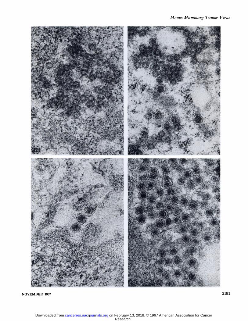
Trypsin. The same overall reduction in the density of pro-teinaceous cellular components that was observed after pepsindigestion followed incubation in 0.3% trypsin. However, theeffect was not as rapid. There was no alteration of the diametersof the A or B particle. The outer shell of the A particle wasroughened in appearance after 4 hours incubation (Fig. 12) whencompared with the controls incubated in 0.045 M Tris chloridebuffer (Fig. 13). The nucleoid of the B particle was unaffected(Figs. 14, 15). No further change was observed for digestions upto seven hours.
Papain. The cellular components affected were the cytoplasmand the matrix of mitochondria. The nuclear chromatin remaineddense. The ribosomes were found to clump into dense masses.The structure of the A particle and B particle was altered inmuch the same manner as with pepsin digestion. Difficulty inkeeping sections adherent to the Formvar-eoated grids was notedfollowing digestions in 0.1 % papain of 60 minutes or more. Overtreduction in the density of the matrix of mitochondria and cytoplasm was observed abruptly after 90 minutes digestion. Thisphenomenon corresponded directly to the digestion period required for the alterations of the outer shell of the A particle andthe outer coat of the B particle nucleoid.
Effects of Double Enzyme Digestion
Pepsin-Nuclease. Sections were incubated for two hours in0.5% pepsin at 37°Cto degrade the outer components of the A
particle and the B particle nucleoid. The sections were thenwashed and transferred to solutions containing either 0.1%RNase, 0.1% DNase, or distilled water and incubated for 30 or60 minutes at 37°C.Some sections were routinely sampled for
electron microscopy after preliminary incubation in order todetermine the effect of the proteolytic treatment. Additionalcontrols were performed by incubating sections in 0.1 N HC1 fortwo hours followed by 0.1% RNase, 0.1% DNase, and distilledH20 . Digestion with pepsin followed by RNase resulted in thedecreased density of the agranular component of the nucleolus,matrix of mitochondria and nuclear chromatin, and the disappearance of the densely staining ribosomes and granular component of the nucleolus. The combination of pepsin and DNasehydrolysis brought about a striking loss of density in the nuclearchromatin, but ribosomal staining was unaffected. In sections
NOVEMBER 1907 21S1
Research. on February 13, 2018. © 1967 American Association for Cancercancerres.aacrjournals.org Downloaded from
Gilbert H. Smith
placed in distilled water after pepsin pretreatment, the ultra-structural changes were identical with those obtained with pepsindigestion alone. In the other controls, i.e., 0.1 NHC1 pretreatmentfollowed by RNase, DNase, or distilled H20, the results werecomparable to those observed when sections were incubated inthe nuclea.se preparations alone or in 0. l N HC1 alone.
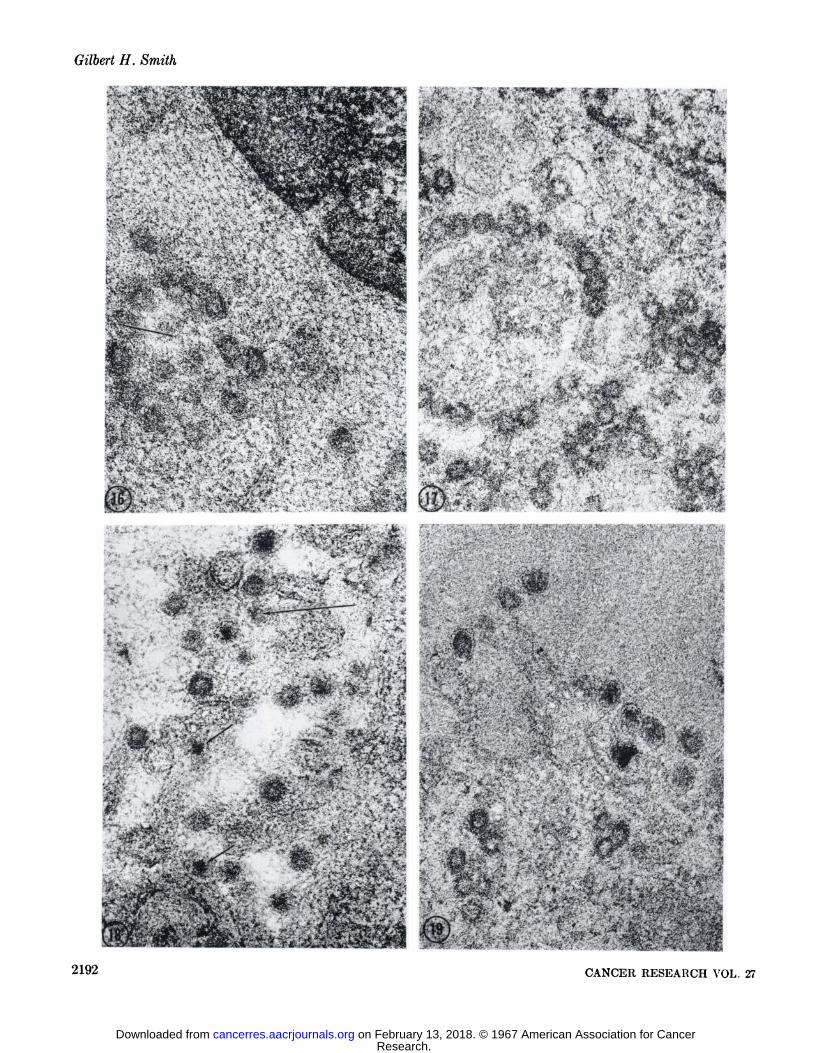
Some of the A particles were seen as open circles followingdigestion with pepsin and RXase (Fig. 16). When this occurred,the inner shell had little or no affinity for lead salt staining.However, the inner shell of the vast majority of the A particlesremained unaffected by RNase hydrolysis. Deoxyribonucleasehad no effect on any of the denuded A particles (Fig. 17). In allcases the inner shell was stained densely. Although the "cortex"
of the nucleoid had been degraded by the action of pepsin inmost of the B particles, the core remained densely stained andapparently resistant to the action of either nuclease (Figs. 18, 19).
Trypsin-Nuclease. The same ultrastructural alterations incellular components that were observed for pepsin-nucleasedigestion apply for sections incubated in 0.25% trypsin followedby either 0.1% RNase, 0.1% DNase, or distilled water for 30 or60 minutes. The sections were exposed to tryptic proteolysis forfour hours before transfer to succeeding solutions. The combination of trypsin and DNase had a more pronounced effect on thenuclear chromatin than did pepsin succeeded by DNase. Controlsections were incubated in 0.045 M Tris chloride buffer for fourhours and then transferred to RNase, DNase, or distilled water.None of the above procedures had any appreciable net effect onthe ultrastructure of either the A or B particle when comparedwith the result observed in the controls or when either enzymehad been used alone. This was not true for cellular morphology.For example, trypsin followed by RNase produced no furtherchanges in the A particle than had RNase digestion alone. Therefore, tryptic digestion alone provided no enhancement of theeffect of ribonuclease on either the inner shell of the A particleor the core of the B particle nucleoid. On the other hand, trypsinpretreatment facilitated a much more rapid removal of ribosomalstaining by ribonuclease action (go minutes).
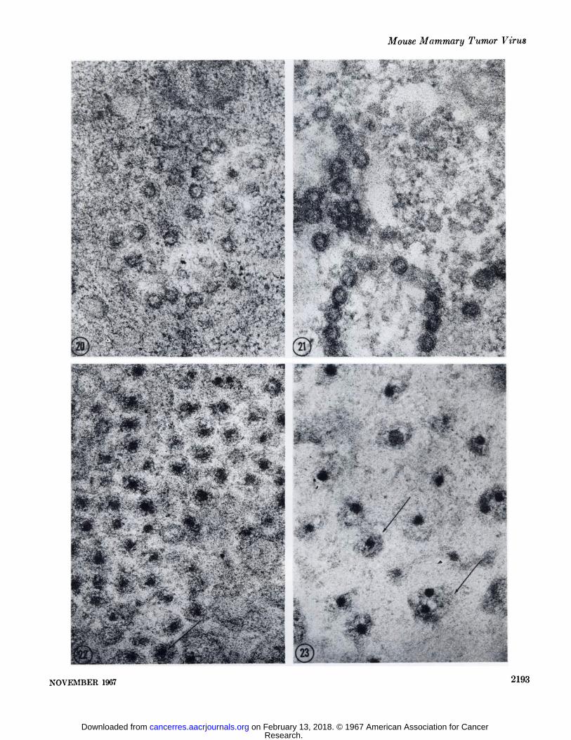
Pepsin-Trypsin. Two hours of digestion with 0.5% pepsinsucceeded by two hours of incubation in 0.25% trypsin resultedin extreme overall decrease in the density of the cytoplasm,matrix of the mitochondria, the granular component of thenucleolus, and the nuclear chromatin. The ribosomes were notstained intensely after this treatment and could not be readilyobserved. The same applied to the granular component of thenucleolus in most sections. This situation was most likely due tothe fact that commercial trypsin preparations are contaminatedwith low levels of ribonuclease activity. If the order was reversed,i.e., trypsin digestion preceding pepsin, the ribosomes remainedintensely stained. Apparently, the proteolytic action of pepsinrenders the ribonucleic acid component of the ribosomes moresensitive to the level of ribonuolease activity contaminating thetrypsin preparation. The controls incubated in 0.1 N HC1 andthen trypsin or pepsin and then 0.045 MTris chloride buffer gaveresults very similar to those observed in sections incubated withtrypsin or pepsin singly. The A and B particles following doubledigestion with pepsin-trypsin or trypsin-pepsin were affected aswith pepsin digestion alone, leaving the A particle with a muchreduced outer diameter and an intact inner shell (Figs. 20, 21).The outer coat of the B particle nucleoid was completely re
moved, leaving only the dense core within the remnants of thelipoprotein sac (Figs. 22, 23).
Effect of Triple Enzyme Digestion
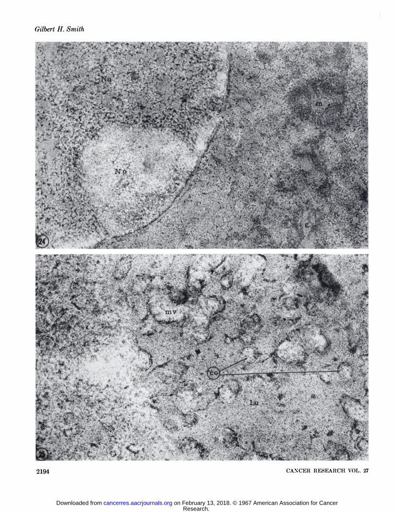
Pepsin-Trypsin-RNase. For each multiple digestion involving three different enzymes, there were seven controls (see Chart1). The effect of this procedure on the cellular organelles and theMTV are represented in Table 1 in terms of the change in densityin comparison with identical structures in unincubated controlsections. Sections were incubated with 0.5% pepsin for 2 hours,0.25% trypsin for 2 hours, and then placed on 0.1% ribonuclea«efor 30 minutes. The controls were done simultaneously. Theultrastructure changes noted for triple digestion with pepsintrypsin and RNase were similar to those observed when RNasefollowed protein hydrolysis. The reduction of the density innucleolar structure was so extreme that the nucleolus appeared innegative contrast to the nucleoplasm (Fig. 24). No trace of the Aparticles could be found except in the thicker portions of somesections and then only a faint outline of the inner shell wasdiscernible. The juxtanuclear areas where remnants of inclusionbodies could be recognized were more electron lucent in appea:-ance than the surrounding cytoplasm (Fig. 26). The B particleswere detected in the extracellular lumina, but only by thepresence of the lipoprotein sac. None of the nucleoid structurewas present, in no instance were the dense cores of the nucleoidsvisible (Fig. 25).
The alterations observed in the fine structure of the A andB particle in the control sections which had been placed in (a) 0.1N HC1 followed by trypsin and RNase; (6) in pepsin followed by0.045 M Tris chloride buffer and RNase; or (c) in 0.1 x HCIfollowed by 0.045 MTris chloride and RNase were identical withthose observed in the earlier experiments when RNase had beenused separately or in combination with pepsin or trypsin. Thusextinction of the structure of the A particle and the B particlenucleoid could only be attained if pepsin, trypsin, and RNasedigestion was carried out consecutively. If trypsin hydrolysis wasperformed first for two hours and followed by pepsin and RNase,the A and B particles were degraded to the same extent as whenpepsin and RNase were used together.
In order to be certain that the results obtained with consecutivehydrolysis with pepsin, trypsin, and RNase were due to thespecific activity of ribonuclease, an identical experiment wascarried out in which DNase digestion was substituted for RNase.In this experiment the sections were incubated for 40 minutes in0.1% DNase after pepsin and trypsin proteolysis. In sectionsprocessed in this manner, the nucleus was rendered completelyelectron lucent and was seen in negative contrast surrounded bythe more dense cytoplasm (Fig. 27). Deoxyribonuclease hydrolysis had no further effect on the A or B particle beyond thatproduced by the preceding digestions with pepsin and trypsin.The inner shell of the A particle and core of the B particlenucleoid were left intact and densely stained (Figs. 28, 29).
DISCUSSION
Extensive studies on the purification and analysis of MTVfrom milk and from mammary tumors of mouse strains withhigh indices of mammary cancer have been carried out by Mooreand his collaborators (30, 37). As a result of these studies, the
2182 CANCER RESEARCH VOL. 27
Research. on February 13, 2018. © 1967 American Association for Cancercancerres.aacrjournals.org Downloaded from
Mouse Mammary Tumor Virus
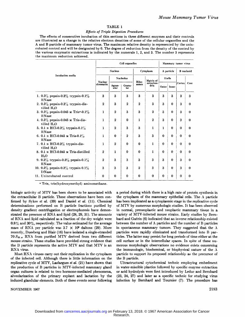
TABLE 1
Effects of Triple Digestion Procedures
The effects of consecutive incubation of thin sections in three different enzymes and their controlsare illustrated as a change in the relative electron densities of some of the cellular organelles and theA and B particle of mammary tumor virus. The maximum relative density is represented by the unin-cubated control and will be designated by 0. The degree of reduction from the density of the control bythe various enzymatic extractions is indicated by the numerals 1, 2, and 3. The number 3 representsthe maximum reduction achieved.
Incubationmedia1.
0.5% pepsin-0.3%trypsin-0.1%RNase2.
0.5% pepsiu-0.3%trypsin-dis-tiliedH,O3.
0.5% pepsin-0.045 MTris"-0.1%RNase4.
0.5% pepsin-0.045 MTris-dis-tilledH2O5.
0.1 N HC1-0.3%trypsin-0.1%RNase6.
0.1 N HC1-0.045 MTris-0.1%RNase7.
0.1 x HC1-0.3%trypsin-dis-tilledH2O8.
0.1 N HC1-0.045 MTris-distilledH209.
0.3% trypsin-0.5%pepsin-0.1%RNase10.
0.5% pepsin-0.3%trypsin-0.1%DNase11.
Unincubated controlCell
organellesNucleusXuclear
chromatin32111112230N'ucleolusAgranular33222021330Granular32303200330CytoplasmRibo-
somes32313300320Matrixof
mitochondria33221311330Mammary
tumorvirusA
particleShellsOuter3333100033Inner3000000BnucleoidCortex33330000
;I)000
0330Core30000000000
" Tris, tris(hydroxymethyl) aminomethane.
biologic activity of MTV has been shown to be associated withthe extracellular B particle. These observations have been confirmed by Sykes et al. (39) and Daniel el al. (11). Chemicaldeterminations performed on B particle fractions purified bydensity gradient centrifugation or electrophoresis have demonstrated the presence of RNA and lipid (28, 29, 31). The amountsof RXA and lipid calculated as a fraction of the dry weight were0.8% and 30%, respectively. The value estimated for the averagemass of RNA per particle was 3.7 X IO6 daltons (29). More
recently, Duesberg and Blair (16) have isolated a single-stranded
70 iS2o.»RNA from purified MTV derived from two differentmouse strains. These studies have provided strong evidence thatthe B particle represents the active MTV and that MTV is anRNA virus.
Most RNA viruses carry out their replication in the cytoplasmof the infected cell. Although there is little information on thereplicative cycle of MTV, Lasfargues el al. (21) have shown thatthe production of B particles in MTV-infected mammary glandorgan cultures is related to two hormone-mediated phenomena,
alveolarization of the primary expiant and lactation by theinduced glandular elements. Both of these events occur following
a period during which there is a high rate of protein synthesis inthe cytoplasm of the mammary epithelial cells. The A particlehas been implicated as a cytoplasmic stage in the replicative cycleof MTV by numerous morphologic studies. It has been observedin normal, preneoplastic and neoplastic mammary tissue in avariety of MTV-infected mouse strains. Early studies by Bern-
hard and Guérin(6) indicated that an inverse relationship existedbetween the number of A particles and the number of B particlesin spontaneous mammary tumors. They suggested that the Aparticles were rapidly eliminated and transformed into B particles. The latter may persist for long periods of time either at thecell surface or in the intercellular spaces. In spite of these numerous morphologic observations no evidence exists concerningthe immunologie, biochemical, or biophysical nature of the Aparticle to support its proposed relationship as the precursor ofthe B particle.
Ultrastructural cytochemical technic employing embedmentin water-miscible plastics followed by specific enzyme extraction
or acid hydrolysis were first introduced by Leduc and Bernhard(23, 24, 27) and later as a specific technic for studying virusinfection by Bernhard and Tournier (7). The procedure has
NOVEMBER 1907 2183
Research. on February 13, 2018. © 1967 American Association for Cancercancerres.aacrjournals.org Downloaded from
Gilbert H. Smith
advanced over recent years to provide good ultrastructuralpreservation combined with a minimum of denaturatici! ofcellular proteins and nucleic acids so that specific enzymehydrolysis is possible within sections of embedded tissues (25,26, 40).
In the present study, the chemical nature of the intracellularA particle and the extracellular B particle, with special referenceto the nucleoid of the latter, has been investigated with respectto alterations in their ultrastructural appearance and stainingproperties in thin sections subjected to a variety of specific en-zymic digestions. Adequate controls were carried out to document the specificity of enzyme hydrolysis and to provide information on the nature and extent of nonspecific extraction due tothe chemical and physical character of the enzyme diluents. Inall experiments high concentrations of enzymes were used toreduce the necessary reaction time and, therefore, nonspecificeffects, and also because of the limited mobility of the enzymemolecules within the plastic polymer.
The results obtained with single enzyme digestions were indicative of good specificity with regard to target components withinthe sections. For example, RNase digestion resulted only in a lossof staining of the ribosomes and the granular component of thenucleolus. However, the structures within the A particle and Bparticle that were presumed to contain RNA were inconsistentlyaltered. A point that bears directly on this observation is theconsideration of the plane of section. In order for the nucleic acidpresumably present within the inner shell of the A particle andcore of the B particle nucleoid to be in contact with the enzymesolution, two requirements must be met: (a) the plane of sectionmust pass through the outer shell exposing the inner surface;(b) the cut or open face must be oriented within the sectiontoward the enzyme solution to facilitate the penetrance of andcontact with the enzyme. If the outer diameters of the A particleand 15particle nucloid, 1000 A and 600-690 A, respectively, arecompared with the average thickness of section selected, 600 A,it is readily apparent that all the A particles observed in any onethin section have been cut. On the other hand, in only a few Bparticles will the plane of section pass through the core of thenucleoid. If both requirements are satisfied, then one of thenucleates, DNase or RNase, should be effective in removing thedensity associated with one or both of these structures. The innershells of some of the A particles and occasional B particle nucleoid cores were rendered electron lucent by ribonuclease action.This phenomenon was never observed following incubation withDNase or distilled water.
Because of the problem encountered due to orientation andplane of section, an attempt was made to remove the outer shellof the A particle and the B particle nucleoid within the thinsections with protease. Pepsin and papain hydrolysis wereeffective in this regard, pepsin reducing the outer diameter of theA particle and the nucleoid of the B particle by 140-170 A.Trypsin had only a limited effect on these structures. Thisprobably indicates that only a small number of lysine or arginineresidues are associated with the protein composing the outercomponent of these structures. Pretreatment of thin sections witheither pepsin or trypsin promoted no significant enhancement ofthe effect of ribonuclease on either particle. However, preliminaryproteolysis with either enzyme brought about a more rapid andcomplete hydrolysis of other cell constituents with succeeding
nuclease digestion, e.g., nuclear chromatin by protease-DNase orribosomes by protease-RNase.
When pepsin and trypsin hydrolysis were combined, the innershell of the A particle and the core of the B particle were unaffected. However, this treatment rendered those structures susceptible to RNase activity but not to DNase. The importance oftryptic digestion on the inner shell of the A and core of thenucleoid of the B particle was clarified by the fact that RNasewas ineffective against either structure when trypsin incubationpreceded pepsin digestion. The same was true if Tris chloridebuffer was substituted for trypsin after pepsin incubation andbefore RNase. The activity of all the enzymes employed was welldocumented throughout the course of the experiment. Thenecessity of tryptic lysis prior to ribonuclease hydrolysis suggeststhat the inner shell of the A particle and core of the B particlenucleoid are basic protein-nucleic acid conjugates. Trypsin actionis quite narrowly restricted to "basic" bonds, i.e., such bonds
which link the carboxyl group of a basic amino acid (arginine andlysine) to the amino group of another. Basic proteins such asprotamines and polyamines are often associated with the nucleicacid components of the bacterial viruses (2).
In conclusion, the results reported support the hypothesis thatthe A particle is the precursor of the nucleoid of the B particle,since the chemical organization at the ultrastructural level is thesame in both. This is further evidence in support of Bernhard'»
postulate regarding the maturation of MTV. In terms of theirrelative susceptibility to enzymic degradation, the outer shell ofthe intracellular A particle and the outer coat of the extracellularB particle nucleoid appear to have the same stereochemicalstructure. Both are attacked by pepsin and papain but not bytrypsin. The diameter of both structures are reduced by approximately the same amount following pepsin digestion. Theinner shell of the A particle and core of the B particle nucleoidare subject to RNase hydrolysis but only after pepsin and trypsinproteolysis. This observation is compatible with the concept thattheir chemical construction is similar, and furthermore that theyare basic protein-ribonucleic acid conjugates.
The final proof that the A particle is truly an intracellular,immature form of MTV rests upon (a) the demonstration ofMTV virion-associated antigenicity with the A particle and (6)the incorj¡orationof isotopically labeled A particles into matureactive MTV virions. The work presented here provides the firstconvincing evidence for the presence of RNA and protein in theultrastructure of the A particle. The observations furnish a modelthat can be tested with the use of mammary tumor tissue isotopically labeled with protein and RNA precursors in conjunctionwith the water-miscible cytochemical technic. This is possiblebecause the A and B particles apparently possess structures thatare differentially susceptible to enzymatic hydrolysis in thinsections when compared to the cellular organelles. The newdiscovery of a soluble antigen MTVsi by Ninowski el al. (32),which apparently is an internal component of the MTV virion,opens the possibility of correlating this antigen to the intracellular A particle in si'u cytochemically, with the use of specificferritin-labeled antibody.
ACKNOWLEDGMENTS
The author sincerely appreciates the helpful criticisms andcomments of Drs. A. J. Dalton ¡meiE. II. Leduc during the prep-
2184 CANCER RESEARCH VOL. 27
Research. on February 13, 2018. © 1967 American Association for Cancercancerres.aacrjournals.org Downloaded from
Mouse Mammary Tumor Virus
«ration of the manuscript and the valuable technical assistance ofMr. David Longfellow.
REFERENCES
1. Amano, >S.The Peculiarity of the Proliferating Mode of CancerViruses. Symp. Cell. Chem., 10: 203-289, 1960.
2. Ames, B. N., and Dubin, D. T. The Role of Polyamines in theNeutralization of Bacteriophage Deoxyribonucleic Acid. J.Biol. Chem., 235: 769-774, 1960.
3. Bang, F. B., Andervont, H. B., and Vellisto, I. An ElectronMicroscope Study of Spontaneous and Induced MammaryTumors of Mice. Bull. Johns Hopkins Hosp., 98: 287-308, 1956.
4. Bang, F. B., Vellisto, I., and Libert, R. Electron MicroscopicEvidence Concerning the Mammary Tumor Inciter. I: AStudy of Normal and Malignant Cells from Mammary (¡landsof Mice. Bull. Johns Hopkins Hosp., 98: 255-285, 1956.
5. Bernhard, W., Bauer, A., Guérin,M., and Oberling, C. Étudeau Microscope Electronique de Corpuscles d'Aspect Viriisal
dans des Epitheliomas Mammaires de la Souris. Bull. Cancer,42: 163-178, 1955.
6. Bernhard, W., and Guérin,M. Evaluation Quantitative duVirus dans les Tumeurs Mammaires Spontanéesau GrefféesdeDifférentesSouches de Souris et Étudede des Rapports avecl'Apparteil de (îolgi.In: L. Severi (ed.), Division of Cancer
Research, Intern. Symp. Mammary Cancer, Perugia, Italy, pp.627-639, 1958.
7. Bernhard, W., and Tournier, P. Ultrastructural Cytochemistry Applied to the Study of Virus Infection. Cold SpringHarbor Symp. Quant. Biol., 27: 67-82, 1962.
8. Bit t ner, J. J. Some Possible Effects of Nursing on the Mammary Gland Tumor Incidence in Mice. Science, 84: 162, 1936.
9. Blair, P. B., and Pitelka, D. K. Immunology of the MouseMammary Tumor Virus. Correlation of the InimunodiffusionPrecipitate Line with Type-B Virus Particles. J. Nati. CancerInst., 37: 261-278, 1966.
10. Dalton, A. J., Potter, M., and Merwin, R. M. Some Ultra-structural Characteristics of a Series of Primary and Transplanted Plasma-cell Tumors of the Mouse. J. Nati. CancerInst., 26: 1221-1267, 1961.
11. Daniel, C. W., De Orne, K. B., Pitelka, D. R., and Sekhri,K. K. The Infectivity of Density Gradient Purified B Particlesfrom C3H Mammary Tumor. Proc. Am. Assoc. Cancer Res.,6: 12, 1965.
12. de Harven, E., and Friend, C. Electron Microscope Study of aCell-free Induced Leukemia of the Mouse: A PreliminaryReport. J. Biophys. Biochem. Cytol., 4: 151-156, 1958.
13. de Harven, E., and Friend, C. Electron Microscope Studies onMouse Lymphomas. In: S. S. Bréese(ed.), Fifth InternationalCongress for Electron Microscopy, Vol. 2, Sect. MM, p. 5.New York and London: Academic Press, Inc., 1962.
14. Dmochowski, L., Haagensen, C. 1)., and Moore, D. H. Studiesof Sections from Normal and Malignant Cells of High- andLow-Cancer Strain Mice by Means of Electron Microscope.Acta Unió Intern. Contra Cancrum, //: 640-645, 1955.
15. Dmochowski, L., and Moore, D. H. Discussion. J. Nati. CancerInst., 15: 785-787, 1954.
16. Duesberg, P. H., and Blair, P. B. Isolation of the Nucleic Acidof Mouse Mammary Tumor Virus (MTV). Proc. Nati. Acad.Sei. U. S., 55: 1490-1497, 1966.
17. Feldman, D. G. Origin and Distribution of Virus-like ParticlesAssociated with Mammary Tumors in DBA Strain Mice. I.Virus-like Particles in Mammary Gland Tissue. J. Nati.Cancer Inst,, 30: 477-501, 1963.
18. Goldfeder, A., Gelber, D., and Moore, D. H. An ElectronMicroscope Study of Spontaneous Mammary Carcinoma in a
Subline of Strain DBA Mice. J. Nati. Cancer Inst., 29: 827-845,1960.
19. Haguenau, F., Hollman, K. H., Mouriquand, J., and Mouri-quand, C. ÉtudeUltrastructurale de Tumeurs Mammaires etdes Organes Leucemiquies des Souris d'une Souche Nouvellement Isolée(Souche PS). J. Microscop., 4: 253-264, 1965.
20. Imai, T., Hiromitsu, P., Matsumoto, A., and Horie, A. TheMode of Virus Elaboration in C3H Mouse Mammary Carcinoma as Observed by Electron Microscopy in Serial ThinSections. Cancer Res., 26: 443-453, 1966.
21. Lasfargues, E. Y., and Feldman, D. G. Hormonal and Physiological Background in the Production of B Particles by MouseMammary Epithelium in Organ Cultures. J. Nati. CancerInst., 23: 191-196, 1963.
22. Lasfargues, E. Y., Moore, D. H., Murray, M. R., Haagensen,C. D., and Pollard, E. C. Production of Milk Agent in Cultureof Mouse Mammary Carcinoma. J. Biophys. Biochem. Cytol.,5: 93-96, 1959.
23. Leduc, E. H., and Bernhard, W. Enzyme and Acid Hydrolysisof Nucleic Acids and Protein. J. Biophys. Biochem. Cytol.,10: 437-455, 1961.
24. Leduc, E. H., and Bernhard, W. Water Soluble EmbeddingMedia for Ultrastructural Cytochemistry. In: S. C. Harris(ed.), The Interpretation of Ultrastructure. Symposia of theInternational Society for Cell Biology, Vol. 1, pp. 21-45. NewYork: Academic Press, 1962.
25. Leduc, li. H., Bernhard, W., and Tournier, P. Cyclic Appearance of Intramitochoiidrial DNA Fibers in Cultures of anAdenovirus 12-induced Hamster Tumor. Exptl. Cell Res., 42:597-616, 1966.
26. Leduc, E. H., and Holt, S. S. Hydroxypropyl Methacrylate, aNew Water-miscible Embedding Medium for Electron Microscopy. J. Cell Biol., 26: 137-155, 1965.
27. Leduc, E. H., Marinozzi, V., and Bernhard, W. The Use ofWater-soluble Glycol Methacrylate in Ultrastructural Cytochemistry. J. Roy. Microscop. Soc., 81: 119-130, 1963.
28. Lyons, M. J., and Moore, D. II. Purification of the MouseMammary Tumor Virus. Nature, 194: 1141-1142, 1962.
29. Lyons, M. J., and Moore, D. H. Isolation of the Mouse Mammary Tumor Virus: Chemical and Morphological Studies. J.Nati. Cancer Inst., 35: 549-565, 1965.
30. Moore, D. H., Lasfargues, K. Y., Murray, M. li., Haagensen,C. D., and Pollard, E. C. Correlation of Pln-sical and Bi
ological Properties of Mouse Mammary Tumor Agent. J.Biophys. Biochem. Cytol., 5: 85-92, 1959.
31. Moore, D. H., and Lyons, M. J. Electrophoretic Separation ofthe Mouse Mammary Tumor Virus. J. Nati. Cancer Inst., 31:1255-1273, 1963.
32. Ninowski, R. C., Old., L. J., Moore, D. H., Geering, G., andBoyse, E. A. A Soluble Antigen of the Mammary Tumor Virus.Virology, SI: 1-14, 1967.
33. Pitelka, D. R., Bern, H. A., De Orne, K. B., Schooley, C. N.,and Wellings, S. R. Virus-like Particles in HyperphisticAveolar Nodules of the Mammary Gland of the C3H/He Crgl.Mouse. J. Nati. Cancer Inst., W: 541-553, 1958.
34. Pitelka, D. R., Bern, II. A., Nandi, S., and De Orne, K. B.On the Significance of Virus-like Particles in MammaryTissues of C3Hf Mice. J. Nati. Cancer Inst., S3: £67-885,1964.
35. Pitelka, D. R., De Orne, K. B., and Bern, H. Virus-like Particles in Precancerous Hyperplastic Mammary Tissues ofC3H and C3Hf Mice. J. Nati. Cancer Inst., 25: 753-778, 1900.
36. Reynolds, E. W. The Use of Lead Citrate at High pH as anElectro-opaque Stain in Electron Microscopy. J. Cell Biol.,17: 208-212, 1963.
37. Stone, R. S., and Moore, D. H. Purification of Mouse Mam-
NOVEMBER 1967 2isr>
Research. on February 13, 2018. © 1967 American Association for Cancercancerres.aacrjournals.org Downloaded from
Gilbert H. Smith
mary Carcinoma Agent by Means of a Flnorocarbon. Nature, chowski, L. density Gradient Centrifngation of the Bittner183: 1275-1270,1959. Agent. Texas Kept. Biol. Med., 2«:609-627, 1964.
38. Suzuki, T. Electron Microscopic Cyto-histopathology (III). 40. Zambernard, J., and Vatter, A. E. The Fine Structural Cyto-Electron Microscopic Studies on Spontaneous Mammary chemistry of Virus Particles Found in Renal Tumors of Leop-Carcinoma of Mice. Uann, 4&'39-65, 1957. ard Frogs. I. Enzymatic Study of the Viral Nucleoid.
39. Sykes, J. A., Grey, C. E., Scanlon, M., Young, L., and Dmo- Virology, 88: 318-324, 1966.
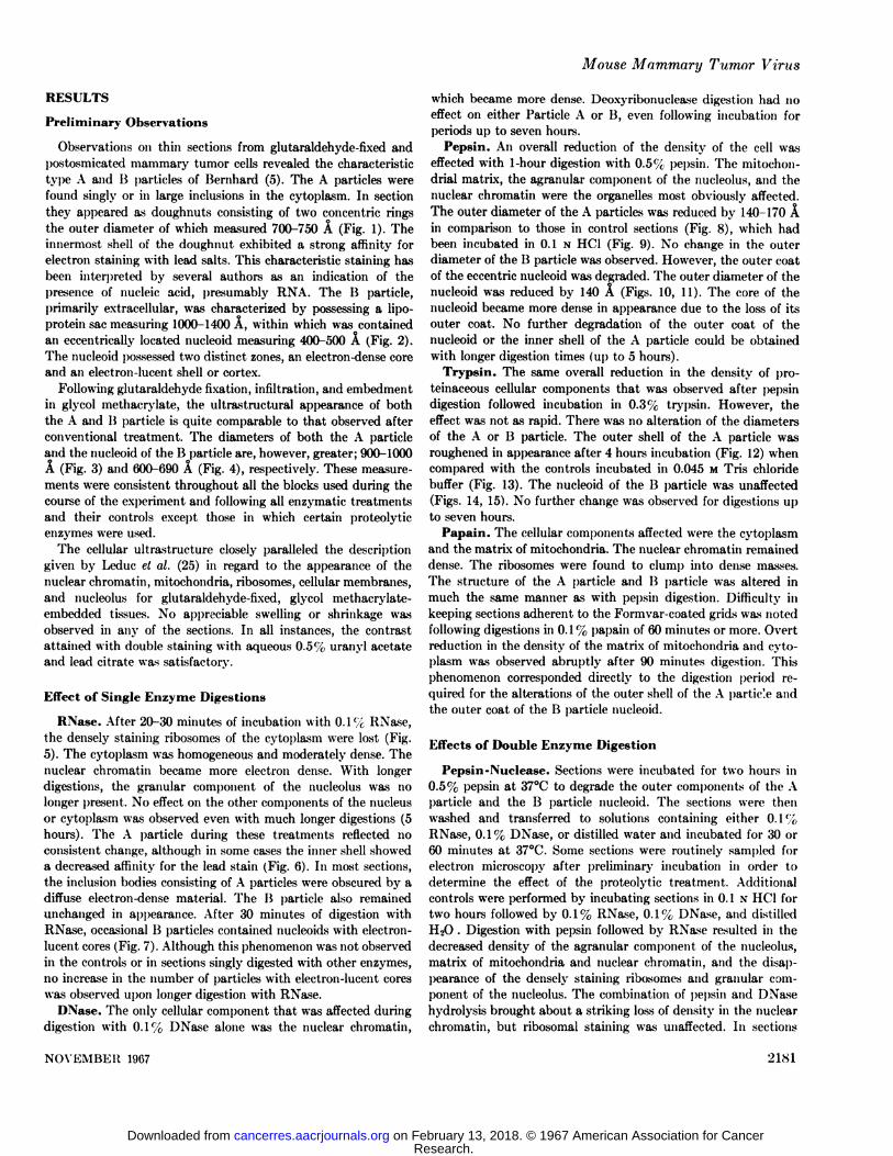
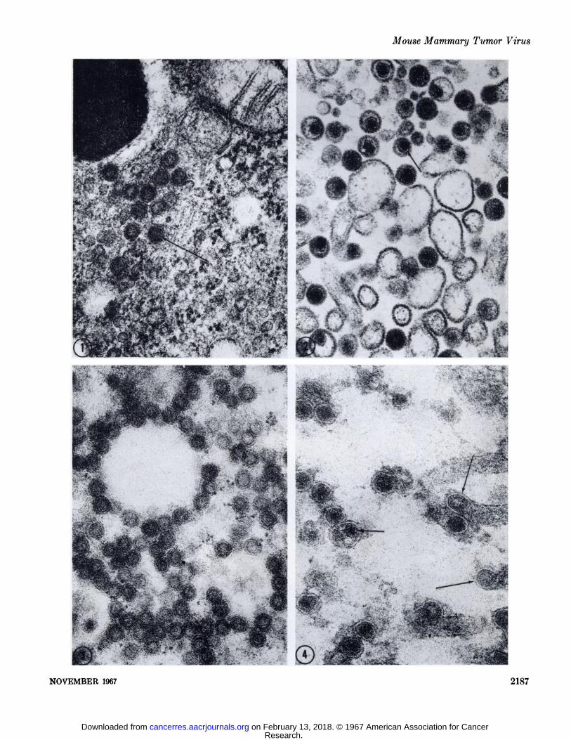
FIG. 1. A small cytoplasmic inclusion body of type A particles. Their structure consists of two concentric shells; the inner shell characteristically stains more intensely (arrow). The center of the A particle appears variably electron-dense, this presumably is due to theplane of section. Specimen fixed in 4.5' ( phosphate-buffered glutaraldehyde, pH 7.4, for twenty minutes, post-fixed in 2% OsOi in thesame buffer for 1 hour and embedded in Epon. Section stained with Karnofsky's lead hydroxide. X 70,000.
FIG. 2. A small extracellular lumen in a thin section from a spontaneous mammary tumor containing numerous type B particles. TheB particles are characterized by a loose lipoprotein sac containing an eccentric-dense nucleoid. The nucleoid possesses two distinctcomponents, an electron-lucent "cortex" or coat (arrow) and an electron-dense core. Specimen fixed according to the schedule given
for Fig. 1. Uranyl acetate and lead citrate (Reynolds), X 70,000.FIGS.3-29. These micrographs were taken of thin sections from glutaraldehyde-fixed, glycol methacrylate-embedded mouse mammary
tumor, doubly stained with 0.5% aqueous uranyl acetate and Reynold's lead citrate and subjected to various experimental procedures.
FIG. 3. An inclusion body of cytoplasmic A particles in an unincubated control section. X 69,500.FIG. 4. Extracellular B particles lying in an extracellular lumen in an unincubated control section. Several budding A particles at
the ends of microvillus-like projections can be seen (long arrows). The two components of the B particle are easily discerned (shortarrow). The lipoprotein membranes appear in negative image because of the absence of OsOi in the fixation procedure. X 69,500.
2186 CANCER RESEARCH VOL. 27
Research. on February 13, 2018. © 1967 American Association for Cancercancerres.aacrjournals.org Downloaded from
Mouse Mammary Tumor Virus
•
.- •-#-
•»-
SËA-^
NOVEMBER 1967 2187
Research. on February 13, 2018. © 1967 American Association for Cancercancerres.aacrjournals.org Downloaded from
(Hlbert H. Smith
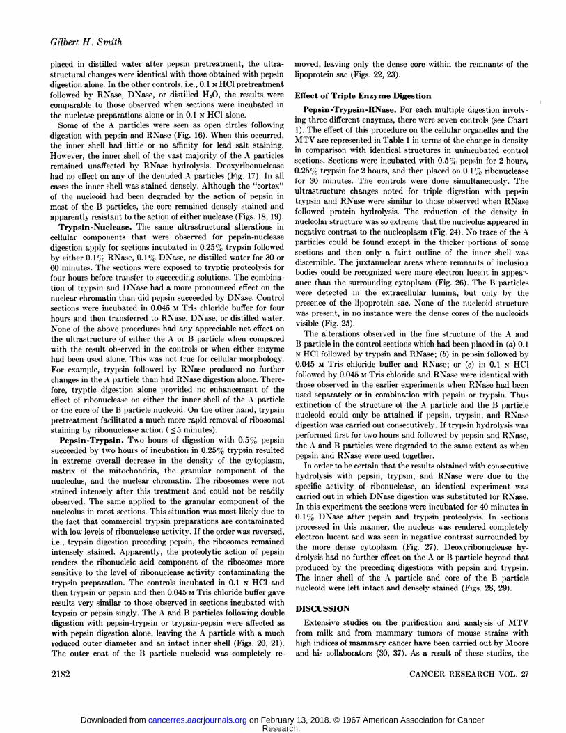
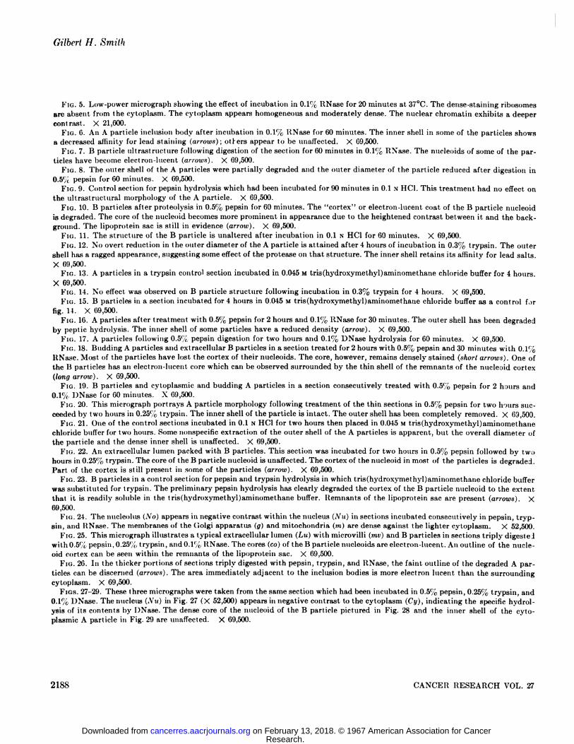
FIG. 5. Low-power micrograph showing the effect of incubation in 0.1% RNase for 20 minutes at 37°C.The dense-staining ribosomes
are absent from the cytoplasm. The cytoplasm appears homogeneous and moderately dense. The nuclear chromatin exhibits a deepercontrast. X 21,600.
Fio. 6. An A particle inclusion body after incubation in 0.1% RNase for 60 minutes. The inner shell in some of the particles showsa decreased affinity for lead staining (arrows); others appear to be unaffected. X 69,500.
FIG. 7. B particle ultrastructure following digestion of the section for 60 minutes in 0.1% RNase. The nucleoids of some of the particles have become electron-lucent (arrows). X 69,500.
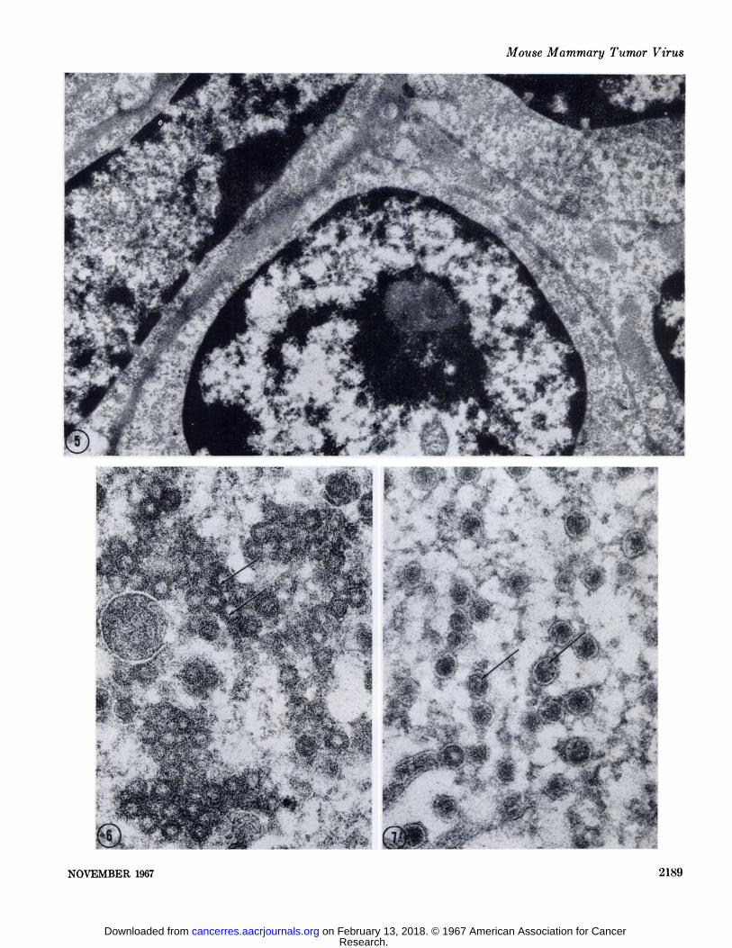
FIG. 8. The outer shell of the A particles were partially degraded and the outer diameter of the particle reduced after digestion in0.5% pepsin for 60 minutes. X 69,500.
FIG. 9. Control section for pepsin hydrolysis which had been incubated for 90 minutes in 0.1 N*HC1. This treatment had no effect on
the ultrastructural morphology of the A particle. X 69,500.FIG. 10. B particles after proteolysis in 0.5% pepsin for 60 minutes. The "cortex" or electron-lucent coat of the B particle nucleoid
is degraded. The core of the nucleoid becomes more prominent in appearance due to the heightened contrast between it and the background. The lipoprotein sac is still in evidence (arrow). X 69,500.
FIG. 11. The structure of the B particle is unaltered after incubation in 0.1 N HC1 for GOminutes. X 69,500.FIG. 12. No overt reduction in the outer diameter of the A particle is attained after 4 hours of incubation in 0.3% trypsin. The outer
shell has a ragged appearance, suggesting some effect of the protease on that structure. The inner shell retains its affinity for lead salts.X 69,500.
FIG. 13. A particles in a trypsin control section incubated in 0.045 M tris(hydroxymethyl)aminomethane chloride buffer for 4 hours.X 69,500.
FIG. 14. No effect was observed on B particle structure following incubation in 0.3% trypsin for 4 hours. X 69,500.FIG. 15. B particles in a section incubated for 4 hours in 0.045 M tris(hydroxymethyl)aminomethane chloride buffer as a control for
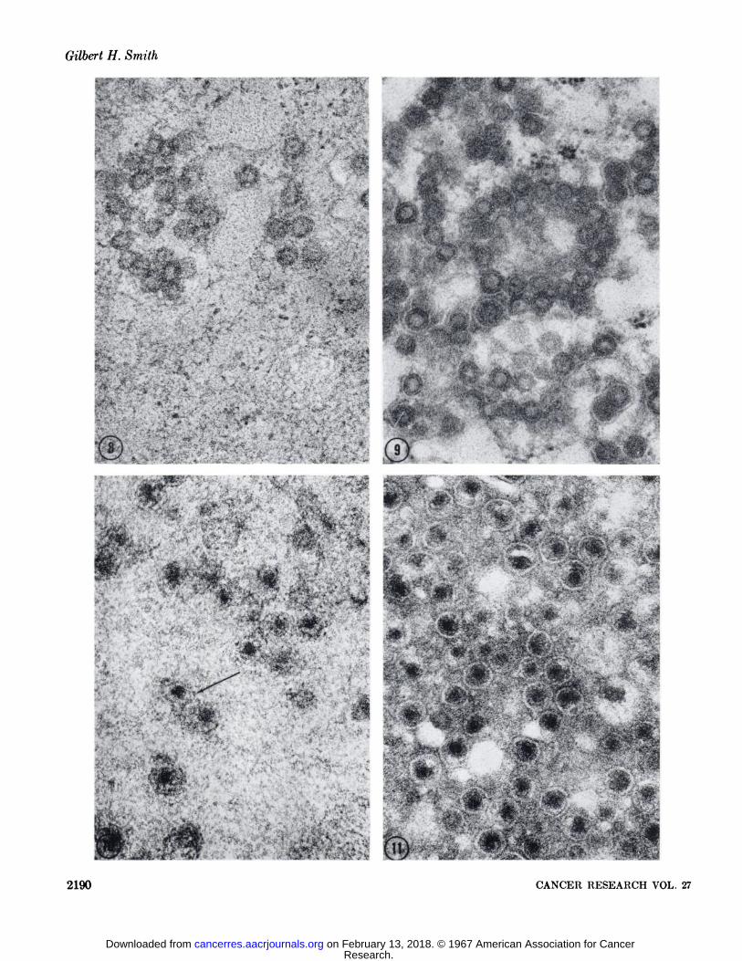
fig. 14. X 69,500.FIG. 16. A particles after treatment with 0.5% pepsin for 2 hours and 0.1% RNase for 30 minutes. The outer shell has been degraded
by peptic hydrolysis. The inner shell of some particles have a reduced density (arrow). X 69,500.FIG. 17. A particles following 0.5% pepsin digestion for two hours and 0.190 DNase hydrolysis for GOminutes. X 69,500.FIG. 18. Budding A particles and extracellular B particles in a section treated for 2 hours with 0.5% pepsin and 30 minutes with 0.1%
RNase. Most of the particles have lost the cortex of their nucleoids. The core, however, remains densely stained (short arrows). One ofthe B particles has an electron-lucent core which can be observed surrounded by the thin shell of the remnants of the nucleoid cortex(long arrow). X 69,500.
FIG. 19. B particles and cytoplasmic and budding A particles in a section consecutively treated with 0.5% pepsin for 2 h;>urs and0.1%, DNase for 60 minutes. X 69,500.
FIG. 20. This micrograph portrays A particle morphology following treatment of the thin sections in 0.5% pepsin for two hours succeeded by two hours in 0.25% trypsin. The inner shell of the particle is intact. The outer shell has been completely removed. X 69,500.
FIG. 21. One of the control sections incubated in 0.1 N HC1 for two hours then placed in 0.045 M tris(hydroxymethyl)aminomethanechloride buffer for two hours. Some nonspecific extraction of the outer shell of the A particles is apparent, but the overall diameter ofthe particle and the dense inner shell is unaffected. X 69,500.
FIG. 22. An extracellular lumen packed with B particles. This section was incubated for two hours in 0.5% pepsin followed by twohours in 0.25% trypsin. The core of the B particle nucleoid is unaffected. The cortex of the nucleoid in most of the particles is degraded.Part of the cortex is still present in some of the particles (arrow). X 69,500.
FIG. 23. B particles in a control section for pepsin and trypsin hydrolysis in which tris(hydroxymethyl)aminomethane chloride bufferwas substituted for trypsin. The preliminary pepsin hydrolysis has clearly degraded the cortex of the B particle nucleoid to the extentthat it is readily soluble in the tris(hydroxymethyl)aminomethane buffer. Remnants of the lipoprotein sac are present (arrows). X69,500.
FIG. 24. The nucleolus (A'o) appears in negative contrast within the nucleus (A'u) in sections incubated consecutively in pepsin, tryp
sin, and RNase. The membranes of the Golgi apparatus (g) and mitochondria (TO)are dense against the lighter cytoplasm. X 52,500.FIG. 25. This micrograph illustrates a typical extracellular lumen (Lu) with microvilli (mv) and B particles in sections triply digested
wit h 0.5% pepsin, 0.25%, trypsin, and 0.1% RNase. The cores (co) of the B particle nucleoids are electron-lucent. An outline of the nucleoid cortex can be seen within the remnants of the lipoprotein sac. X 69,500.
FIG. 26. In the thicker portions of sections triply digested with pepsin, trypsin, and RNase, the faint outline of the degraded A particles can be discerned (arrows). The area immediately adjacent to the inclusion bodies is more electron lucent than the surroundingcytoplasm. X 69,500.
FIGS. 27-29. These three micrographs were taken from the same section which had been incubated in 0.5% pepsin, 0.25% trypsin, and0.1% DNase. The nucleus (\u) in Fig. 27 (X 52,500) appears in negative contrast to the cytoplasm (Cy), indicating the specific hydrolysis of its contents by DNase. The dense core of the nucleoid of the B particle pictured in Fig. 28 and the inner shell of the cytoplasmic A particle in Fig. 29 are unaffected. X 69,500.
2188 CANCER RESEARCH VOL. 27
Research. on February 13, 2018. © 1967 American Association for Cancercancerres.aacrjournals.org Downloaded from
Mouse Mammary Tumor Virus
«•»Ja*?*"'-".•^»dSSœrr...•a
m ^-fF ^, '
NOVEMBER 1967 2189
Research. on February 13, 2018. © 1967 American Association for Cancercancerres.aacrjournals.org Downloaded from
Gilbert H. Smith
•-,. '•
.;..>>, #•:
f-ts*
«'*'•,.-
•kl
CWM
m '•»'
«i- - -- "^--
.''.
'&>*.
":' ''•' 'Aw
*
'<&
'•'•-&)
-A?av•? - -
¿J.' •n >-.#iy£j
t-*iï_;**-
2190 CANCER RESEARCH VOL. 27
Research. on February 13, 2018. © 1967 American Association for Cancercancerres.aacrjournals.org Downloaded from
Mouse Mammary Tumor Virus
•••*»
$*••
ÌÌy&%~: '
- 'V i» • *-TT*•v "Jrt ;, ' -¿f'¡-'' •J,"¿- •"j. i- '-i. ' •'- ' • "•»' -•-•^~^=*H-•v*£Ä^'v;f ^J^*^^^;^i'v^;.; ;:" " "*.•r--,---t A-'^f'i*'x*. ^: ^;;---v;vt**4 ^-V1 ^-tr »•-'>•';"v-iii'^if •^^•tea^ltó-"; ^fe-vV" v';¿^i'-;: ^-a^v^-^Äf-?' -- 'Jk ;'^L«>" - -,-., -,
; i-f-'^V-'r'.../ '.--,,;':.^P
•»•;•, ^•.'-<'»Vf- •'"i^V ' ••. •; ' •^'•*•'- '••'V '":-.%i^r -° »C,-'- " ' ' V'; V •-••'
£>•" ••<»^f4->"."•'•a'ü»Ä!.i,"i.-
"*"^»*V ' •--> f* -*-..-• •¿*''*t^1* " ' '-"V- 'v*i'<- <** ' '•••^^^^v^^-...Mf^*'•"•.';' •* -nc«A,»**„. ,. ' f^*>", - • •' ( r : -J .•••;.
Ä?ÄÄSSÄ-.:SÄ- -Ã-
NOVEMBER 1967 2191
Research. on February 13, 2018. © 1967 American Association for Cancercancerres.aacrjournals.org Downloaded from
Gilbert H. Smith
¿v
111 lä :ï$ : ;•,'.'^^Jr-S^v^,;*;/ ;V^f
IÛSP •'?/•""1-5- ^jf u |
. ••"-,- •'¿M - •••¿&£-';',,,:.--\*%; •j ;-^'
P'ir^v* ' ••'
K^&^Ä
:«d
- ^
M*SÕ":^;-n.-/. -• .. . -
,. v. ,g-, -flp" " '•'^táAi; :•,.'•: - .
-m¿m^ •; ., -Ä%- Ä LÃe•^'- ^id^?' -;, . *?»i •'"••;Vvk':V-»Ãvr':,.'.¿"'Ã.•T% ' '"':>$"'l'-4&- :'- ' '** *•:"-•>':i'/.-"^
rjg&x..'' ; v.^.^,..,•'-.-%;"¿v^i
i'.;'-\___:.,
:^^WÕ ^:^s|•$ ' ;. P- ' •^Sa%¿'^^A^' ' ,¿&/"•*-*^^;;^-^@
É>*" .. '•.'". . -' - . ••*'_.Õ^K V.V:'..'».* ••"*' ' • 'U''.. ^ I V
•"•>••>,y:-; .', -~jrf:~~"- SS«i ' "•-'»"^4?
«¡wippi m
$ ''^'i'-'Õ.
^'•-'^"•v•-""•'-'' '? •;>-•-'.- ••* -.-<•#v ;-^«^'-- -',.•"•-"-- - ' -•'"'••>*;1
:. ^-v'-" ' •:...' * •'••-;'; •- . . : , ••;.',v\Ã.
2192 CANCER RESEARCH VOL. 27
Research. on February 13, 2018. © 1967 American Association for Cancercancerres.aacrjournals.org Downloaded from
Mouse Mammary Tumor Virus
«*vi &£. v 3|;;"--v->\
&&:. .:•;^;*"%^^.^S,,ï?tyjfèÉ ',
-c.S; ;¿?Ã-'•^'*s**I
E-fö«li.'Ä-:l^^ 1 ^
'- :>' '„^C"s>¿;;'* v:."\- r" --•.-;. ^
K^vi^, ':;, •.r-">:>. ¿Jc!'J> :'i'j>**¥^''' %?'*>?
^'>*'« , ;••i' «...... •.. •• ,.; v •'<-••-'î^tsçiù'•'v"'1^' '•'';; '
".
-•: 4:^^;?^ipï r J'-;^ '-4ft^ ^^' ' "
-'?m;'. ^f ;;:Ti.
NOVEMBER 1907 2193
Research. on February 13, 2018. © 1967 American Association for Cancercancerres.aacrjournals.org Downloaded from
Gilbert H. Smith
•'r
-
'••ri1
- . MYWV: :/,**«•¿ff- ,-•.-'.;«*'•:
.'-'., -
> -A- ,**».?••'.'
' ••>'
>. - •
•:-•:•"'"**.*:" '*> ' ¿*- -•**
i¿M-^?r*
^ **!•k*">Ã;>?;v''- <»:W-'<4':; £$<s'--. i'ai"« -r"/-"" ;J; -•' •"¿i- . " "^V^^'Ã^Ipi^f ä.^yy ^--^'X^^t^**t' ' .'-''.i- * '»»t-^'"' .' 'ä'-lf;-"^^^ ^ •.-' -fi 'jja^ ^'¿¿'jÃ&*'& '-v '- • '.*'•"•'-'V^.' ! d
»^NÄi^ai•*' '''^Jift'"'^""ff' i^"'1 •" '- •-* •"* - ••»ÕS''*"'-^Ir^"'"'•*':>*:^-tà ""-''^ :'ÃÃs•"••^^Ã^r'^iSipfe?- ^v:ÕA.^f 'Ã'^|'*J»?»Ã:'.n^'»ÕÕ*Ã;/V.-<Ã'Ã";*|^.^: ^.' >!'/;' f^-< ' ••- va^
•*t.'-'/';'T"" r^^.". ,"';':'!' ••''%(•.-v^-" '•'^***'*-''•'*''• <,v.s';'
£"'^~^< - '
A • <;,ï;-,' ["*•!•*•:•' •
^lÉji ' cÃ^^^ -V;^M'^•'/M .;';^'î-:
';.- ' ' . . >"•>.-'-^'.'k'-^,'
IftilS^**! ">•> i^ "/;. ' ^ '
";-^ Ãw;. 4* 'ti -
\-" ^:'' •••"' '•-'. -': '•'•;- "-.'•"; -";&•••>.*-.»
2194 CANCER RESEARCH VOL. 27
Research. on February 13, 2018. © 1967 American Association for Cancercancerres.aacrjournals.org Downloaded from
Mouse Mammary Tumor Virus
f' • X" - . —*
.
-••—I
•-. •7 .•»•»*•-i; ¡V•- * » "•r "i -- .;-•>••-.f y* :••• '•
. i '••'..•'.*>••- . -S i "'• ; .;•' V.. - V''-'- • „ *' . --'.'. • •"• •;. ).•-et v. ..-v-r-:vi--r••.--,.-••:•--. \..i
mNOVEMBER 1967 2190
Research. on February 13, 2018. © 1967 American Association for Cancercancerres.aacrjournals.org Downloaded from
Gilbert H. Smith
. -' -- -: * .* •'v- '--r -•,' ••--••-••:.•TV--* jr•*-"* - '-'/ > '. -•r-w**?^
.l»'•
2196
I ?•. •I •.*.:«£y¡-; -
CANCER RESEARCH \OL. 27
Research. on February 13, 2018. © 1967 American Association for Cancercancerres.aacrjournals.org Downloaded from
1967;27:2179-2196. Cancer Res Gilbert H. Smith Cytochemical Studies on the Mouse Mammary Tumor Virus
Updated version
http://cancerres.aacrjournals.org/content/27/11_Part_1/2179
Access the most recent version of this article at:
E-mail alerts related to this article or journal.Sign up to receive free email-alerts
Subscriptions
Reprints and
To order reprints of this article or to subscribe to the journal, contact the AACR Publications
Permissions
Rightslink site. Click on "Request Permissions" which will take you to the Copyright Clearance Center's (CCC)
.http://cancerres.aacrjournals.org/content/27/11_Part_1/2179To request permission to re-use all or part of this article, use this link
Research. on February 13, 2018. © 1967 American Association for Cancercancerres.aacrjournals.org Downloaded from