developmental and cytochemical studies of the …
TRANSCRIPT
*e-mail: [email protected]
DEVELOPMENTAL AND CYTOCHEMICAL STUDIES OF THEENDOSPERM CHALAZAL HAUSTORIUM OF RHINANTHUS SEROTINUS
(SCROPHULARIACEAE)
JOANNA ŚWIERCZYŃSKA*, MAŁGORZATA KOZIERADZKA-KISZKURNO, AND JERZY BOHDANOWICZ
Department of Plant Cytology and Embryology, University of Gdańsk, Wita Stwosza 59, 80-308 Gdańsk, Poland
Received February 4, 2013; revision accepted May 10, 2013
We examined the development of the endosperm chalazal haustorium of Rhinanthus serotinus, using histo-chemical assays and light and electron microscopy. The chalazal haustorium is a huge single cell containing twoenlarged nuclei. The nuclei are located in the middle of the haustorium cell. At the chalazal end of the haustori-um cell structure, ultrastructural study revealed the presence of a transfer wall forming wall ingrowths. At allexamined stages of haustorium cell development we identified insoluble polysaccharides, proteins, nucleic acidsand lipid droplets. Macromolecules were especially abundant in the fully differentiated haustorium cell. Ourresults suggest that the endosperm chalazal haustorium is a site of intense metabolic activity.
KKeeyy wwoorrddss:: Rhinanthus serotinus, endosperm, chalazal haustorium, differentiation, histochemistry.
ACTA BIOLOGICA CRACOVIENSIA Series Botanica 55/1: 99–106, 2013DOI: 10.2478/abcsb-2013-00012
PL ISSN 0001-5296 © Polish Academy of Sciences and Jagiellonian University, Cracow 2013
INTRODUCTION
The endosperm of numerous angiosperms producesspecial types of structures which function as haus-toria. They most often develop at the chalazal andmicropylar end or only at the chalazal end of theendosperm. It is very rare for the endosperm to pro-duce a haustorium over its entire surface.Production of endosperm haustoria is commonamong angiosperms. Their presence has beenreported in Cucurbitaceae (Chopra and Seth, 1977),Plantaginaceae (Mikesell, 1990), Poaceae (Mausethet al., 1985), Scrophulariaceae (Schmid, 1906;Arekal, 1963; Tiagi, 1966; Johri and Ambegaokar,1984), Ericaceae (Olson, 1993), Fabaceae (Dute andPeterson, 1992) and Lentibulariaceae (Płachno andŚwiątek, 2011; Płachno et al. 2011, 2012). The cyto-plasm of endosperm haustoria usually is densealthough it contains a number of small vacuoles.These cells are often deformed and filled withhydrolyzable content. It is believed that endospermhaustoria take nutrients from the mother plant tis-sue, transporting them to the endosperm, wheresooner or later these abundant nutrients are taken upby the developing embryo (Johri and Ambegaokar,1984). Some studies on the cytochemistry, ultra-
structure, cytoskeleton and synthetic activity ofhaustoria indicate that haustorial cells may beinvolved in the absorption, synthesis and transportof nutrients to the endosperm (Bhatnagar andKallarackal, 1980; Torosian, 1971; Pacini et al.,1975; Nagl, 1992; Brrison and Peterson, 1975; Duteand Peterson, 1992; Olson, 1993; Świerczyńska andBohdanowicz, 2003; Świerczyńska et al, 2005,2006; Płachno and Świątek, 2011; Płachno et al.2011, 2012). Here we report findings on the devel-opment and cytochemical characteristics of theendosperm chalazal haustorium of Rhinanthusserotinus (Scrophulariaceae).
MATERIALS AND METHODS
Seeds of Rhinanthus serotinus (Scrophulariaceae)were obtained from several plants growing at natu-ral stations in the towns of Rumia and Puck innorthern Poland. Flowers at various developmentalstages were collected in June and July. The ovuleswere fixed in 5% glutaraldehyde in 0.1 M cacodylatebuffer (pH 7.0) for 4 h at room temperature. Thenthe plant material was washed in the same buffer,dehydrated in an acetone series, and embedded in
Spurr's resin (Spurr, 1969). Sections 1–2 μm thickwere cut with glass knives using a SORVALL MT 2Bultramicrotome and placed on glass slides. For LMobservations, sections were stained with 0.05%Toluidine Blue O (TBO). For TEM observations theovules were fixed in 2.5% formaldehyde and 2.5%glutaraldehyde for 4 h, post-fixed in 1% OsO4overnight, treated with 1% uranyl acetate for 1 h andembedded in Spurr's resin. After contrasting withuranyl acetate and lead citrate the ultrathin sectionswere examined in a Philips CM 100 transmissionelectron microscope at 80 kV. Sections for cyto-chemical studies were stained with Periodic Acid-Schiff (PAS) for insoluble polysaccharides (Jensen,1962), with Azure B bromide (Flax and Himes,1952) for nucleic acids, with 1% Aniline Blue Blackin 7% acetic acid for proteins (Jensen, 1962), andwith Sudan Black B (in 70% ethanol) for lipids(Bronner, 1975). The preparations were analyzedwith a Nikon Eclipse E 800 microscope fitted with aNikon CCD camera.
RESULTS
The endosperm of Rhinanthus serotinus developsafter double fertilization from a central cell of theembryo sac according to the cellular type. Theendosperm mother cell is divided into two unequalcells: the smaller micropylar and the larger chalazalone. The micropylar cell enlarges and divides longi-tudinally and then transversely, forming theendosperm micropylar haustorium along with initialcells of the endosperm proper. The large chalazalcell increases as the embryo grows, and forms a uni-cellular endosperm chalazal haustorium. Duringdevelopment, the nucleus of the chalazal haustori-um cell undergoes only one division. The binuclearchalazal haustorium enlarges and damages adjacenttissues (Fig. 1).
In R. serotinus the following stages can be dis-tinguished during the development of theendosperm chalazal haustorium cell: (i) differentia-tion of the chalazal haustorium (at the several-cellstage of the endosperm proper); (ii) full developmentof the chalazal haustorium (when the endosperm
proper has a few dozen cells); and (iii) degenerationof the chalazal haustorium (when the endospermproper has several hundred cells) (Fig. 2).
CHALAZAL HAUSTORIUM CELL DURING DIFFERENTIATION
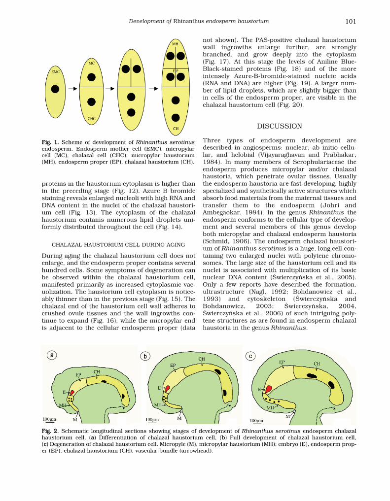
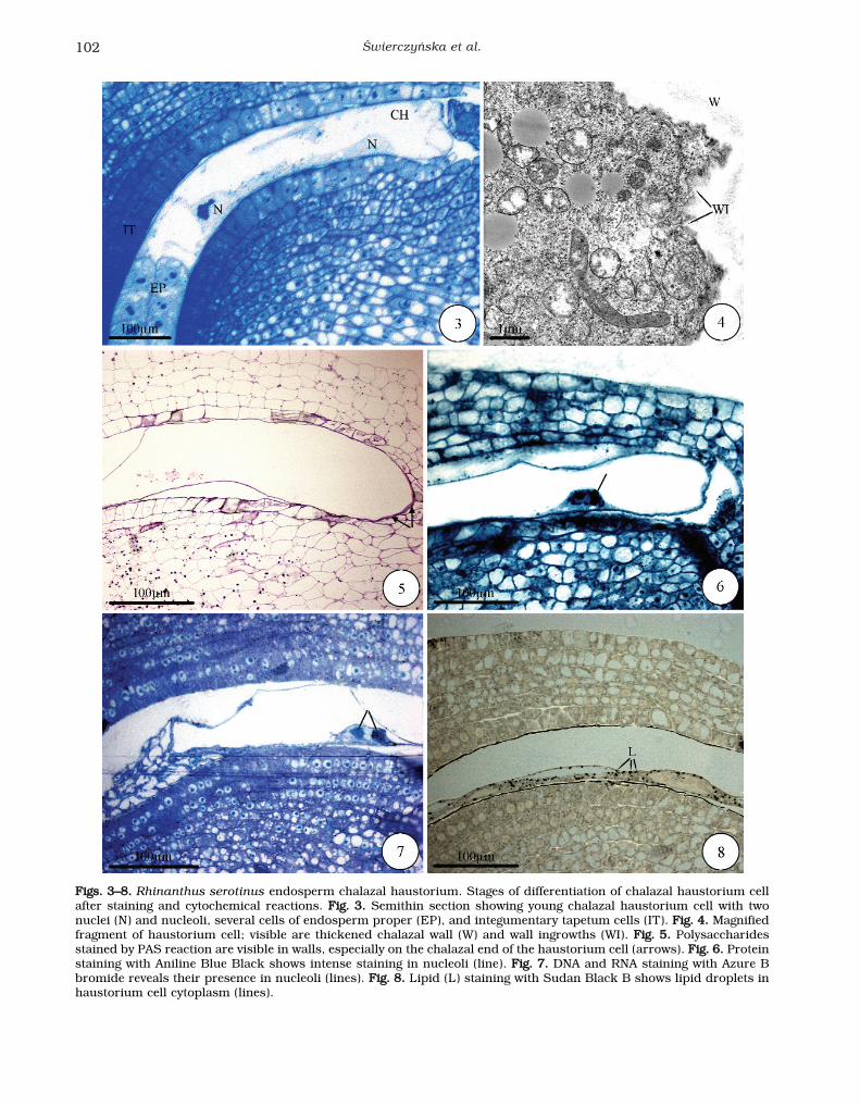
This is the stage in which the chalazal haustoriumcell of R. serotinus gradually differentiates. Thehaustorium cell greatly elongates to 600 μm and itsdiameter reaches 150 μm. The haustorium has twoenlarged nuclei. Its micropylar end adheres to cellsof the endosperm proper (Fig. 3). Its chalazal endadheres to the several-cell pedestal residues and thecell wall is distinctly thickened (Figs. 3, 4). Table 1gives the cytochemistry results for the differentdevelopmental stages of the chalazal endospermhaustorium. The haustorium walls are PAS-positive.A PAS-positive thickened wall is present at the cha-lazal end of the haustorium cell (Fig. 5). Proteinstaining in the cytoplasm of the haustorium cell andin nucleolus proteins is more intense than in thecells of the endosperm proper (Fig. 6). Similarly,staining with Azure B bromide (Fig. 7) shows thepresence of RNA and DNA in the enlarged chalazalhaustorium cell nucleoli. There are many evenly dis-tributed lipid droplets in the haustorium cell cyto-plasm (Fig. 8); they also occur in the cells of theendosperm proper (data not shown).
CHALAZAL HAUSTORIUM CELL DURING FULL DEVELOPMENT
A completely developed chalazal haustorium cell iskidney-shaped. Haustorium length reaches 800 μmand its diameter 250 μm. In the middle of its lengththe cell has two enormous polytene nuclei. At themicropylar end the haustorium adheres to cells ofthe endosperm proper. The chalazal wall of thehaustorium grows into the ovule tissues in the direc-tion of the vascular bundle (Fig. 9). It is equippedwith a labyrinth of wall ingrowths reaching deepinside the cytoplasm (Fig. 10). PAS-positive wallingrowths are formed in the chalazal part of thehaustorium wall, which are longer and wider than inthe previous stage (Fig. 11). The concentration of
Świerczyńska et al.100
TABLE 1. Results of cytochemical tests at the different stages of development of the chalazal endosperm haustorium inRhinanthus serotinus. + positive staining; ++ intense staining
proteins in the haustorium cytoplasm is higher thanin the preceding stage (Fig. 12). Azure B bromidestaining reveals enlarged nucleoli with high RNA andDNA content in the nuclei of the chalazal haustori-um cell (Fig. 13). The cytoplasm of the chalazalhaustorium contains numerous lipid droplets uni-formly distributed throughout the cell (Fig. 14).
CHALAZAL HAUSTORIUM CELL DURING AGING
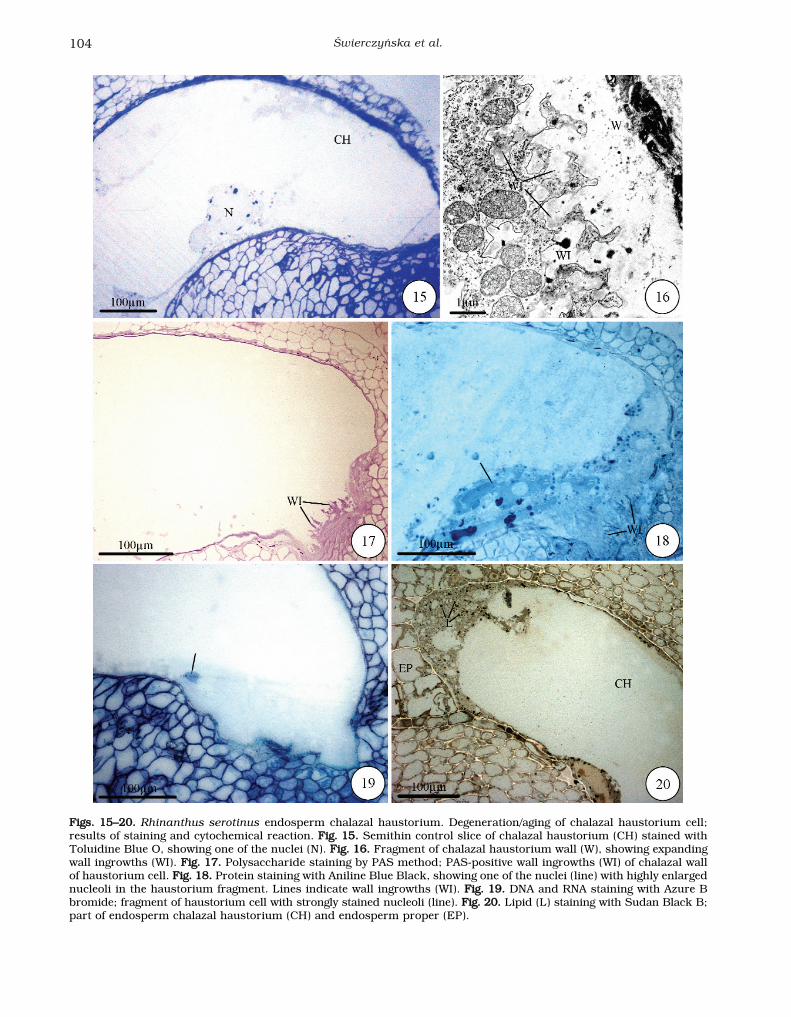
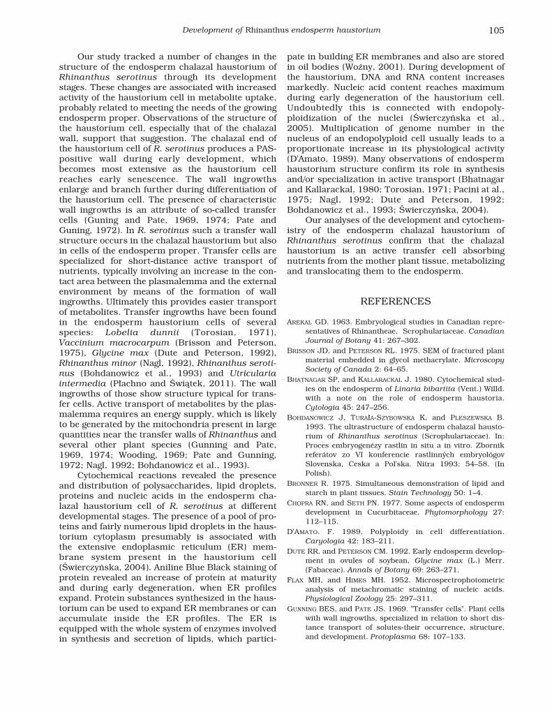
During aging the chalazal haustorium cell does notenlarge, and the endosperm proper contains severalhundred cells. Some symptoms of degeneration canbe observed within the chalazal haustorium cell,manifested primarily as increased cytoplasmic vac-uolization. The haustorium cell cytoplasm is notice-ably thinner than in the previous stage (Fig. 15). Thechalazal end of the haustorium cell wall adheres tocrushed ovule tissues and the wall ingrowths con-tinue to expand (Fig. 16), while the micropylar endis adjacent to the cellular endosperm proper (data
not shown). The PAS-positive chalazal haustoriumwall ingrowths enlarge further, are stronglybranched, and grow deeply into the cytoplasm (Fig. 17). At this stage the levels of Aniline Blue-Black-stained proteins (Fig. 18) and of the moreintensely Azure-B-bromide-stained nucleic acids(RNA and DNA) are higher (Fig. 19). A larger num-ber of lipid droplets, which are slightly bigger thanin cells of the endosperm proper, are visible in thechalazal haustorium cell (Fig. 20).
DISCUSSION
Three types of endosperm development aredescribed in angiosperms: nuclear, ab initio cellu-lar, and helobial (Vijayaraghavan and Prabhakar,1984). In many members of Scrophulariaceae theendosperm produces micropylar and/or chalazalhaustoria, which penetrate ovular tissues. Usuallythe endosperm haustoria are fast-developing, highlyspecialized and synthetically active structures whichabsorb food materials from the maternal tissues andtransfer them to the endosperm (Johri andAmbegaokar, 1984). In the genus Rhinanthus theendosperm conforms to the cellular type of develop-ment and several members of this genus developboth micropylar and chalazal endosperm haustoria(Schmid, 1906). The endosperm chalazal haustori-um of Rhinanthus serotinus is a huge, long cell con-taining two enlarged nuclei with polytene chromo-somes. The large size of the haustorium cell and itsnuclei is associated with multiplication of its basicnuclear DNA content (Świerczyńska et al., 2005).Only a few reports have described the formation,ultrastructure (Nagl, 1992; Bohdanowicz et al.,1993) and cytoskeleton (Świerczyńska andBohdanowicz, 2003; Świerczyńska, 2004,Świerczyńska et al., 2006) of such intriguing poly-tene structures as are found in endosperm chalazalhaustoria in the genus Rhinanthus.
Development of Rhinanthus endosperm haustorium 101
FFiigg.. 22. Schematic longitudinal sections showing stages of development of Rhinanthus serotinus endosperm chalazalhaustorium cell. (aa) Differentiation of chalazal haustorium cell, (bb) Full development of chalazal haustorium cell, (cc) Degeneration of chalazal haustorium cell. Micropyle (M), micropylar haustorium (MH); embryo (E), endosperm prop-er (EP), chalazal haustorium (CH), vascular bundle (arrowhead).
FFiigg.. 11.. Scheme of development of Rhinanthus serotinusendosperm. Endosperm mother cell (EMC), micropylarcell (MC), chalazal cell (CHC), micropylar haustorium(MH), endosperm proper (EP), chalazal haustorium (CH).
Świerczyńska et al.102
FFiiggss.. 33––88.. Rhinanthus serotinus endosperm chalazal haustorium. Stages of differentiation of chalazal haustorium cellafter staining and cytochemical reactions. FFiigg.. 33.. Semithin section showing young chalazal haustorium cell with twonuclei (N) and nucleoli, several cells of endosperm proper (EP), and integumentary tapetum cells (IT). FFiigg.. 44.. Magnifiedfragment of haustorium cell; visible are thickened chalazal wall (W) and wall ingrowths (WI). FFiigg.. 55.. Polysaccharidesstained by PAS reaction are visible in walls, especially on the chalazal end of the haustorium cell (arrows). FFiigg.. 66.. Proteinstaining with Aniline Blue Black shows intense staining in nucleoli (line). FFiigg.. 77.. DNA and RNA staining with Azure Bbromide reveals their presence in nucleoli (lines). FFiigg.. 88.. Lipid (L) staining with Sudan Black B shows lipid droplets inhaustorium cell cytoplasm (lines).

Development of Rhinanthus endosperm haustorium 103
iiggss.. 99––1144.. Rhinanthus serotinus endosperm chalazal haustorium. Stage of complete development of chalazal haustori-um cell; staining and cytochemical reactions. FFiigg.. 99.. Semithin control slice stained with Toluidine Blue O shows strong-ly vacuolized cell of chalazal haustorium (CH) and one of the nuclei (N). FFiigg.. 1100.. Highly magnified fragment of chalazalhaustorium wall (W), showing wall ingrowths (WI). FFiigg.. 1111.. Polysaccharide staining by PAS reaction; lines point to PAS-positive wall ingrowths (WI) of chalazal wall of haustorium cell. FFiigg.. 1122.. Protein staining with Aniline Blue Black; one ofthe nuclei with strongly stained nucleoli (lines) visible in haustorium fragment. FFiigg.. 1133.. DNA and RNA staining withAzure B bromide; fragment of haustorium with strongly stained nucleoli (lines). FFiigg.. 1144.. Lipid (L) staining with SudanBlack B; spherical lipid droplets present in haustorium cytoplasm.
Świerczyńska et al.104
FFiiggss.. 1155––2200.. Rhinanthus serotinus endosperm chalazal haustorium. Degeneration/aging of chalazal haustorium cell;results of staining and cytochemical reaction. FFiigg.. 1155.. Semithin control slice of chalazal haustorium (CH) stained withToluidine Blue O, showing one of the nuclei (N). FFiigg.. 1166.. Fragment of chalazal haustorium wall (W), showing expandingwall ingrowths (WI). FFiigg.. 1177.. Polysaccharide staining by PAS method; PAS-positive wall ingrowths (WI) of chalazal wallof haustorium cell. FFiigg.. 1188.. Protein staining with Aniline Blue Black, showing one of the nuclei (line) with highly enlargednucleoli in the haustorium fragment. Lines indicate wall ingrowths (WI). FFiigg.. 1199.. DNA and RNA staining with Azure Bbromide; fragment of haustorium cell with strongly stained nucleoli (line). FFiigg.. 2200.. Lipid (L) staining with Sudan Black B;part of endosperm chalazal haustorium (CH) and endosperm proper (EP).
Our study tracked a number of changes in thestructure of the endosperm chalazal haustorium ofRhinanthus serotinus through its developmentstages. These changes are associated with increasedactivity of the haustorium cell in metabolite uptake,probably related to meeting the needs of the growingendosperm proper. Observations of the structure ofthe haustorium cell, especially that of the chalazalwall, support that suggestion. The chalazal end ofthe haustorium cell of R. serotinus produces a PAS-positive wall during early development, whichbecomes most extensive as the haustorium cellreaches early senescence. The wall ingrowthsenlarge and branch further during differentiation ofthe haustorium cell. The presence of characteristicwall ingrowths is an attribute of so-called transfercells (Guning and Pate, 1969, 1974; Pate andGuning, 1972). In R. serotinus such a transfer wallstructure occurs in the chalazal haustorium but alsoin cells of the endosperm proper. Transfer cells arespecialized for short-distance active transport ofnutrients, typically involving an increase in the con-tact area between the plasmalemma and the externalenvironment by means of the formation of wallingrowths. Ultimately this provides easier transportof metabolites. Transfer ingrowths have been foundin the endosperm haustorium cells of severalspecies: Lobelia dunnii (Torosian, 1971),Vaccinium macrocarpum (Brisson and Peterson,1975), Glycine max (Dute and Peterson, 1992),Rhinanthus minor (Nagl, 1992), Rhinanthus seroti-nus (Bohdanowicz et al., 1993) and Utriculariaintermedia (Płachno and Świątek, 2011). The wallingrowths of those show structure typical for trans-fer cells. Active transport of metabolites by the plas-malemma requires an energy supply, which is likelyto be generated by the mitochondria present in largequantities near the transfer walls of Rhinanthus andseveral other plant species (Gunning and Pate,1969, 1974; Wooding, 1969; Pate and Gunning,1972; Nagl, 1992; Bohdanowicz et al., 1993).
Cytochemical reactions revealed the presenceand distribution of polysaccharides, lipid droplets,proteins and nucleic acids in the endosperm cha-lazal haustorium cell of R. serotinus at differentdevelopmental stages. The presence of a pool of pro-teins and fairly numerous lipid droplets in the haus-torium cytoplasm presumably is associated with the extensive endoplasmic reticulum (ER) mem-brane system present in the haustorium cell(Świerczyńska, 2004). Aniline Blue Black staining ofprotein revealed an increase of protein at maturityand during early degeneration, when ER profilesexpand. Protein substances synthesized in the haus-torium can be used to expand ER membranes or canaccumulate inside the ER profiles. The ER isequipped with the whole system of enzymes involvedin synthesis and secretion of lipids, which partici-
pate in building ER membranes and also are storedin oil bodies (Woźny, 2001). During development ofthe haustorium, DNA and RNA content increasesmarkedly. Nucleic acid content reaches maximumduring early degeneration of the haustorium cell.Undoubtedly this is connected with endopoly-ploidization of the nuclei (Świerczyńska et al.,2005). Multiplication of genome number in thenucleus of an endopolyploid cell usually leads to aproportionate increase in its physiological activity(D'Amato, 1989). Many observations of endospermhaustorium structure confirm its role in synthesisand/or specialization in active transport (Bhatnagarand Kallarackal, 1980; Torosian, 1971; Pacini at al.,1975; Nagl, 1992; Dute and Peterson, 1992;Bohdanowicz et al., 1993; Świerczyńska, 2004).
Our analyses of the development and cytochem-istry of the endosperm chalazal haustorium ofRhinanthus serotinus confirm that the chalazalhaustorium is an active transfer cell absorbingnutrients from the mother plant tissue, metabolizingand translocating them to the endosperm.
REFERENCES
AREKAL GD. 1963. Embryological studies in Canadian repre-sentatives of Rhinantheae, Scrophulariaceae. CanadianJournal of Botany 41: 267–302.
BRISSON JD, and PETERSON RL. 1975. SEM of fractured plantmaterial embedded in glycol methacrylate. MicroscopySociety of Canada 2: 64–65.
BHATNAGAR SP, and KALLARACKAL J. 1980. Cytochemical stud-ies on the endosperm of Linaria bibartita (Vent.) Willd.with a note on the role of endosperm haustoria.Cytologia 45: 247–256.
BOHDANOWICZ J, TURAłA-SZYBOWSKA K, and PLESZEWSKA B.1993. The ultrastructure of endosperm chalazal hausto-rium of Rhinanthus serotinus (Scrophulariaceae). In:Proces embryogenézy rastlín in situ a in vitro. Zbornikreferátov zo VI konferencie rastlinných embryológovSlovenska, Ceska a Pol'ska. Nitra 1993: 54–58. (InPolish).
BRONNER R. 1975. Simultaneous demonstration of lipid andstarch in plant tissues. Stain Technology 50: 1–4.
CHOPRA RN, and SETH PN. 1977. Some aspects of endospermdevelopment in Cucurbitaceae. Phytomorphology 27:112–115.
D'AMATO. F. 1989. Polyploidy in cell differentiation.Caryologia 42: 183–211.
DUTE RR, and PETERSON CM. 1992. Early endosperm develop-ment in ovules of soybean, Glycine max (L.) Merr.(Fabaceae). Annals of Botany 69: 263–271.
FLAX MH, and HIMES MH. 1952. Microspectrophotometricanalysis of metachromatic staining of nucleic acids.Physiological Zoology 25: 297–311.
GUNNING BES, and PATE JS. 1969. "Transfer cells". Plant cellswith wall ingrowths, specialized in relation to short dis-tance transport of solutes-their occurrence, structure,and development. Protoplasma 68: 107–133.
Development of Rhinanthus endosperm haustorium 105
GUNNING BES, and PATE JS. 1974. Transfer cells. In: RobardsAW. [ed.], Dynamic Aspects of Plant Ultrastructure,441–480. McGraw-Hill, London.
JENSEN WA. 1962. Botanical Histochemistry. W. H. Freemanand Co. San Francisco.
JOHRI BM, and AMBEGAOKAR KB. 1984. Embryology: Then andnow. In: Johri BM [ed.], Embryology of Angiosperms,1–52. Springer-Verlag, Berlin, Heidelberg, New York,Tokyo.
MIKESELL J. 1990. Anatomy of terminal haustoria in the ovuleof Plantain (Plantago major L.) with taxonomic compar-ison to other angiosperm taxa. Botanical Gazette 151(4): 452–464.
MAUSETH JD, MONTENEGRO G, and WALCKOWIAK AM. 1985.Host infection and flower formation by the parasiteTristerix aphyllus (Loranthaceae). Canadian Journal ofBotany 63: 567–581.
NAGL W. 1992. The polytenic endosperm haustorium ofRhinanthus minor (Scrophulariaceae): functional ultra-structure. Canadian Journal of Botany 70: 1997–2004.
OLSON AR. 1993. Patterns of embryo and endosperm forma-tion in Monotropa hypopitys (Monotropaceae) fromNorth America and Western Sweden. American Journalof Botany 80(7): 839–846.
PACINI E, SIMONCIOLI C, and CRESTI M. 1975. Ultrastructure ofnucellus and endosperm of Diplotaxis erucoides duringembryogenesis. Caryologia 28: 525–538.
PATE JS, and GUNNING BES. 1972. Transfer cells. AnnualReview of Plant Physiology 23: 173–196.
PłACHNO BJ, and ŚWIąTEK P. 2011. Syncytia in plants: cellfusion in endosperm – placental syncytium formation inUtricularia (Lentibulariaceae). Protoplasma 248: 425–435.DOI:10.1007/s00709-010-0173-1.
PłACHNO BJ, ŚWIąTEK P, and KOZIERADZKA-KISZKURNO M. 2011.The F-actin cytoskeleton in syncytia from non-clonal pro-genitor cells. Protoplasma 248: 623–629. DOI:10.1007/s00709-010-0209-6.
PłACHNO BJ, ŚWIąTEK P, SAS-NOWOSIELSKA H, and KOZIERADZKA-KISZKURNO M. 2012. Organisation of the endosperm andendosperm-placenta syncytia in bladderworts(Utricularia, Lentibulariaceae) with emphasis on themicrotubule arrangement. Protoplasma, in press DOI:10.1007/s00709-012-0468-5.
SCHMID E. 1906. Beitraege zur Entwicklungsgeschichte derScrophulariaceae. Beihefte Botanische Zentral-bibliothek 20: 175–299.
SPURR AR. 1969. A low-viscosity epoxy resin embedding medi-um for electron microscopy. Journal of UltrastructuralResearch 26: 31–43.
ŚWIERCZYŃSKA J, and BOHDANOWICZ J. 2003. Microfilamentcytoskeleton of endosperm chalazal haustorium ofRhinanthus serotinus (Scrophulariaceae). ActaBiologica Cracoviensia Series Botanica 45(1): 143–148.
ŚWIERCZYŃSKA J. 2004. The cytoskeleton of endosperm cha-lazal haustorium of Rhinanthus serotinus (Schönheit)Oborny (Scrophulariaceae). Ph. D. dissertation,University of Gdańsk, Gdańsk. [In Polish].
ŚWIERCZYŃSKA J, KOZIERADZKA-KISZKURNO M, and BOHDANOWICZ J.2005. Polyploidization of endosperm chalazal haustori-um of Rhinanthus serotinus (Scrophulariaceae). ActaBiologica Cracoviensia Series Botanica 47 (1):123–128.
ŚWIERCZYŃSKA J, BEDNARA J, and BOHDANOWICZ J. 2006. Thecytoskeleton of endosperm chalazal haustorium ofRhinanthus serotinus. Acta Biologica CracoviensiaSeries Botanica 48 (suppl. 1): 35.
TIAGI B. 1966. Development of the seed and fruit inRhinanthus major and R. serotinus. American Journalof Botany 53(7): 645–651.
TOROSIAN CD. 1971. Ultrastructural study of endosperm haus-torial cells of Lobelia dunnii Greene (Campanulaceae,Lobelioideae). American Journal of Botany 58:456–457.
VIJAYARAGHAVAN MR, and PRABHAKAR K. 1984. The endosperm.In: Johri BM [ed.], Embryology of Angiosperms,319–376. Springer Verlag, Berlin, Heidelberg, New York,Tokyo.
WOODING FBP. 1969. Absorptive cells in protoxylem: associa-tion between mitochondria and the plasmalemma.Planta 84: 235–238.
WOźNY A. 2001. System błon wewnętrznych. In: Woźny A,Michejda J, Ratajczak L. [ed.], Podstawy BiologiiKomórki Roślinnej, 93–158. Wydawnictwo NaukoweUAM, Poznań.
Świerczyńska et al.106