cytochemical localization of activity in yeast peroxisomesjb.asm.org/content/104/1/581.full.pdf ·...
TRANSCRIPT

JOURNAL OF BACTERIOLOGY, Oct. 1970, p. 581-584Copyright 0 1970 American Society for Microbiology
Vol. 104, No. IPrinted in U.S.A.
Cytochemical Localization of Catalase Activityin Yeast Peroxisomes
H.-P. HOFFMANN, A. SZABO, AND C. J. AVERS
Departmenit of Biological Sciences, Douglass Campus, Rutgers University, New Brunswick, New Jersey 08903
Received for publication 4 May 1970
Diaminobenzidine oxidation product occurred in peroxisomes and in the intra-cristate spaces of mitochondria. The reaction was inhibited only in peroxisomeswhen 3-amino-I , 2, 4-triazole was present, but cyanide and azide inhibited deposi-tion in both kinds of organelles.
Peroxisomes have been identified in Sac-charomyces cerevisiae (2, 16) by the criteria ofultrastructure and enzyme activities (5). Becauseof problems in obtaining, by centrifugation,peroxisome and mitochondrial fractions whichare free of cross-contamination, we were unableto provide clear evidence showing the uniquelocation of catalase in peroxisomes of bakers'yeast (16). In the present cytochemical study,however, we found that catalase peroxidaticactivity (6) was restricted to peroxisomes, whereasenzymes other than catalase probably are respon-sible for reduction of H202 in mitochondria ofthese same cells. We used modifications of theGraham-Karnovsky (8) diaminobenzidine (DAB)method, which has proved successful for bothplant (7) and animal (4, 6, 14) materials.
Wild-type strain iso-N of S. cerevisiae (3)was grown at 25 C in a liquid semisyntheticmedium (12) supplemented with 1 or 10%glucose. Cells were harvested in the late expo-nential phase (3) and were fixed immediately ina cacodylate-buffered glutaraldehyde-paraformal-dehyde mixture (9) at 0 to 4 C for 4 hr (or aslong as 16 hr for some materials). After threewashes in water and two washes in the samebuffer solution as was to be used in the stainingmedium, about 0.5 g (wet weight) of cells wasincubated in stoppered test tubes at 37 C for1 to 2 hr in a freshly prepared incubation mediumcontaining 10 mg of 3,3'-diaminobenzidinetetrahydrochloride (Sigma Chemical Co., St.Louis, Mo.) dissolved in 5 ml of an alkalinebuffer [50 mm Veronal acetate, pH 9.0; 50 mM2-amino-2-methyl-1-propanol, pH 9.1; or 100mM tris(hydroxymethyl)aminomethane-chloride,pH 8.6 to 8.9], and 0.007% H202. The tubes were
put in ice to stop the reaction, after which thecells were rinsed once with buffer and then eitherwere postfixed for 1 hr in 5% NaMnO4 in 0.1 Mcacodylate buffer (pH 7.2) or first were convertedto spheroplasts (1, 13) and then were postfixedfor 12 hr at 4 C in 2%5OS04 in Millonig's buffer(11). The fixed cells were rinsed in water, em-bedded in 2% warmed agar, and stained for 1 hrat room temperature in a mixture of equal partsof saturated aqueous uranyl acetate and 100%acetone; they were then dehydrated rapidly in agraded acetone series and embedded in Epon 812epoxy resin (10). Sections of 40- to 90-nm thick-ness were cut with diamond knives and sometimesstained with lead citrate (15) before scanning andphotography, for which we used an RCA 3Gelectron microscope at an accelerating voltageof 50 kv.The DAB oxidation product occurred in
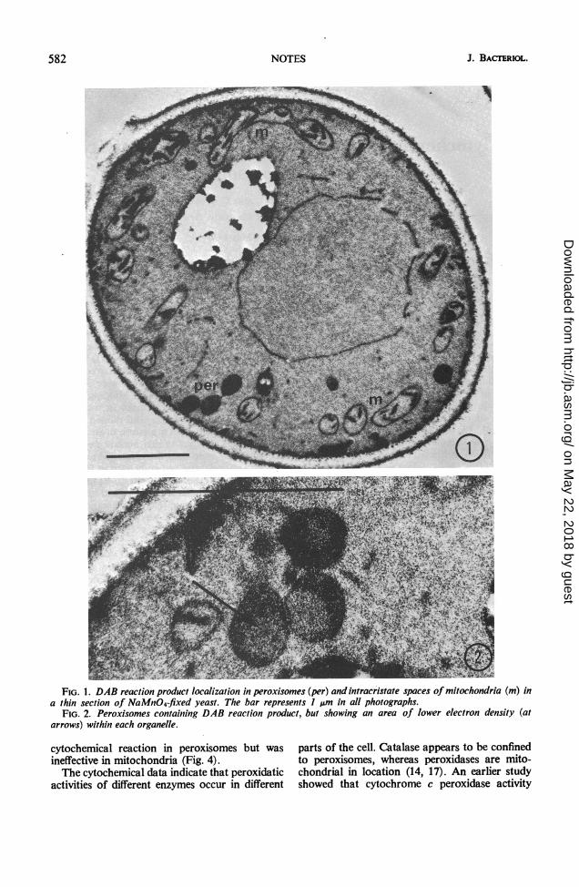
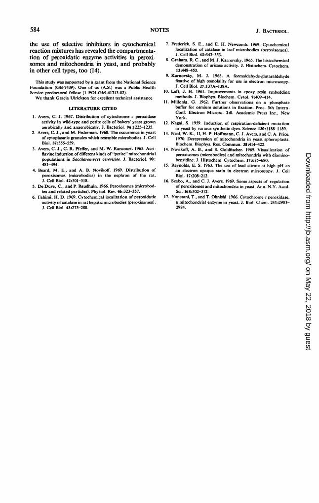
peroxisomes and in the intracristate spaces ofmitochondria (Fig. 1). No crystalloid or tubularinclusions were present in peroxisomes fixed ineither NaMnO4 or OS04, but there was a lesselectron-dense region in the matrix (Fig. 2). Theorganelles usually were round or dumbbell-shapedand often were in clusters near the cell periphery.Sometimes peroxisomes occurred in very closeassociation, separated only by a thin electron-transparent line (Fig. 2).There was no DAB oxidation product in
either organelle type when the incubation medialacked H202 or contained 10 to 50 mm KCNor 0.1 M sodium azide in a complete reactionmixture (Fig. 3). On the other hand, 20 mm3-amino-1 ,2,4-triazole (Aldrich Chemical Co.,Milwaukee, Wis.) completely inhibited the
581
on May 22, 2018 by guest
http://jb.asm.org/
Dow
nloaded from
J. BACrERIOL.
S~~~~~~~'jt'}%
;fe , >.,P -5 ,-* z z
FIG. 1. DAB reaction product localization in peroxisomes (per) and intracristate spaces of mitochondria (m) ina thin section of NaMnO4-fixed yeast. The bar represents I pm in all photographs.
FIG. 2. Peroxisomes containing DAB reaction product, but showing an area of lower electron density (atarrows) within each organelle.
cytochemical reaction in peroxisomes but wasineffective in mitochondria (Fig. 4).The cytochemical data indicate that peroxidatic
activities of different enzymes occur in different
parts of the cell. Catalase appears to be confinedto peroxisomes, whereas peroxidases are mito-chondrial in location (14, 17). An earlier studyshowed that cytochrome c peroxidase activity
582 NOTES
on May 22, 2018 by guest
http://jb.asm.org/
Dow
nloaded from

VOL. 104, 1970
FIG. 3. KCN control showing absence of DAB reaction product from peroxisomes (per) and mito-chondria (m).
FIG. 4. Thin section of cell after DAB reaction in the presence of aminotriazole. Peroxisome lacks deposit(at arrow), whereas mitochondria contain electron-dense product within the cristae.
yielded a grossly particulate reaction productalong cytoplasmic and mitochondrial membranes(1). The intracristate pattern of DAB reactionproduct may be due to the activity of some
peroxidase which is different from cytochrome cperoxidase (1, 17), or to the difference betweenDAB and the diphenylamines used in the separatecytochemical test systems (1, 6). At the least,
583NOTES
on May 22, 2018 by guest
http://jb.asm.org/
Dow
nloaded from
584 NOTES
the use of selective inhibitors in cytochemicalreaction mixtures has revealed the compartmenta-tion of peroxidatic enzyme activities in peroxi-somes and mitochondria in yeast, and probablyin other cell types, too (14).
This study was supported by a grant from the National ScienceFoundation (GB-7439). One of us (A.S.) was a Public HealthService predoctoral fellow (I FOI-GM-41713-02).We thank Gracia Ulrickson for excellent technical assistance.
LITERATURE CITED
1. Avers, C. J. 1967. Distribution of cytochrome c peroxidaseactivity in wild-type and petite cells of bakers' yeast grownaerobically and anaerobically. J. Bacteriol. 94:1225-1235.
2. Avers, C. J., and M. Federman. 1968. The occurrence in yeastof cytoplasmic granules which resemble microbodies. J. CellBiol. 37:555-559.
3. Avers, C. J., C. R. Pfeffer, and M. W. Rancourt. 1965. Acri-flavine induction of different kinds of "petite" mitochondrialpopulations in Saccharomyces cerevisiae. J. Bacteriol. 90:
481-494.4. Beard, M. E., and A. B. Novikoff. 1969. Distribution of
peroxisomes (microbodies) in the nephron of the rat.J. Cell Biol. 42:501-518.
5. De Duve, C., and P. Baudhuin. 1966. Peroxisomes (microbod-ies and related particles). Physiol. Rev. 46:323-357.
6. Fahimi, H. D. 1969. Cytochemical localization of peroxidaticactivity of catalase in rat hepatic microbodies (peroxisomes).J. Cell Biol. 43:275-288.
J. BACrERIOL.
7. Frederick, S. E., and E. H. Newcomb. 1969. Cytochemicallocalization of catalase in leaf microbodies (peroxisomes).J. Cell Biol. 43:343-353.
8. Graham, R. C., and M. J. Karnovsky. 1965. The histochemicaldemonstration of uricase activity. J. Histochem. Cytochem.13:448-453.
9. Karnovsky, M. J. 1965. A formaldehyde-glutaraldehydefixative of high osmolality for use in electron microscopy.J. Cell Biol. 27:137A-138A.
10. Luft, J. H. 1961. Improvements in epoxy resin embeddingmethods. J. Biophys. Biochem. Cytol. 9:409-414.
11. Millonig, G. 1962. Further observations on a phosphatebuffer for osmium solutions in fixation. Proc. 5th Intern.Conf. Electron Microsc. 2:8. Academic Press Inc., NewYork.
12. Nagai, S. 1959. Induction of respiration-deficient mutationin yeast by various synthetic dyes. Science 130:1188-1189.
13. Neal, W. K., II, H.-P. Hoffmann, C. J. Avers, and C. A. Price.1970. Derepression of mitochondria in yeast spheroplasts.Biochem. Biophys. Res. Commun. 38:414-422.
14. Novikoff, A. B., and S. Goldfischer. 1969. Visualization ofperoxisomes (microbodies) and mitochondria with diamino-benzidine. J. Histochem. Cytochem. 17:675-680.
15. Reynolds, E. S. 1963. The use of lead citrate at high pH asan electron opaque stain in electron microscopy. J. CellBiol. 17:208-212.
16. Szabo, A., and C. J. Avers. 1969. Some aspects of regulationof peroxisomes and mitochondria in yeast. Ann. N.Y. Acad.Sci. 168:302-312.
17. Yonetani, T., and T. Ohnishi. 1966. Cytochrome c peroxidase,a mitochondrial enzyme in yeast. J. Biol. Chem. 241:2983-2984.
on May 22, 2018 by guest
http://jb.asm.org/
Dow
nloaded from