methods for the cytochemical/ histochemical localization
TRANSCRIPT

27
From: Methods in Plant Electron Microscopy and Cytochemistry
Edited by: W. V. Dashek © Humana Press Inc., Totowa, NJ
Methods for the Cytochemical/Histochemical Localizationof Plant Cell/Tissue Chemicals
2
William V. Dashek
CONTENTS
INTRODUCTION
PURPOSE OF THE CHAPTER
PROTOCOLS
CONCLUSIONS
REFERENCES
1. INTRODUCTION
1.1. Value of Cytochemistry and Histochemistry
Although many published volumes exist regarding the cytochemical/
histochemical localizations of cellular and tissue chemicals for animal
systems (1–10), there are only a few relatively recent monographs con-
cerning plant cell/tissue cytochemistry and histochemistry (11–15).
Perhaps the main reason for the rather numerous volumes centering
about animal systems stems from the obvious importance of localiz-
ing cellular/tissue chemicals for clinical histopathology. For example,
embryonic surface antigens appear during transformation of a healthy
cell to a malignant one. Although there are plant cancers, e.g., crown
galls, the development of cytochemical stains to reveal possible surface
antigens in plant neoplasms has not been extensively explored. Neverthe-
less, plant cytochemistry has yielded a wealth of information regarding
28 Dashek
the distribution of cellular and tissue chemicals in diverse systems. Fur-
thermore, cytochemistry/histochemistry has provided significant details
about the organization of the vascular system in monocot and dicot roots
and stems.
1.2. Continued Evolution of Cytochemistry
Plant cytochemistry/histochemistry continues to evolve as fluores-
cence microscopy (16–19), confocal fluorescence microscopy (20,21),
and microspectrophotometry (22) expand our quantitative knowledge
of the distributions of chemical constituents in plant cells and tissues.
With regard to microspectrophotometry, this is possible for single cells,
as the Arcturus Corporation (Mountain View, CA) has developed an instru-
ment capable of isolating single cells.
2. PURPOSE OF THE CHAPTER
Since the classic plant histochemistry volume of Jensen (23), a few
volumes regarding the topic (see opening paragraphs of the introduc-
tion) have appeared over the last four decades. Certain of these volumes
contain updated methods for fixation, dehydration, and embedding of
plant cells and tissues for the light microscopic localization of certain of
their chemical constituents. Much of the older botanical microtechnique
volumes, e.g., Sass (24) abound with paraffin embedding and sectioning
methods. These volumes remain very useful, as they contain highly rele-
vant information regarding microtomy and affixing sections to slides.
This chapter offers some select, recent developments regarding fixa-
tion, dehydration, and embedding. In addition, some tried-and-true proce-
dures are described for the localizations of cellular and tissue chemicals
in stems and roots of young Zea mays seedlings. Also provided are more
recently developed fluorochromes for DNA and RNA localizations (18,19).
Finally, the localizations of low-molecular-weight compounds requires
special specimen preparation techniques, as these compounds are often
diffusible, water- or organic-solvent soluble, and solubilized by conven-
tional fixation and dehydration procedures. The reader is referred to ref.
(12) for the processing of cells and tissues for the cytochemical and
histochemical localization of these compounds.
3. PROTOCOLS
3.1. Preparation of Plant Cells and Tissues
for Light Microscope Cytochemistry/Histochemistry
1. Cut tissue block, with at least one dimension a maximum of 5 mm, into
1.5% (w/v) formaldehyde, 2.5% (v/v) glutaraldehyde in 0.05 M phos-
Chapter 2 / Localization of Plant Cell/Tissue Chemicals 29
phate buffer, pH 7.0. Note that speed is important to prevent autolytic
changes. See Table 1 for other fixatives employed for light microscopy.
2. Fix overnight at 4ºC (volume of fixative >10X volume of sample).
3. Wash twice in phosphate buffer, 30 min each time.
4. Dehydrate through a graded alcohol series (10%, 25%, 40%, 60%,
75%, 95%) with two changes at each step (15 min each change) and three
15–30 min changes in 200-proof alcohol.
5. Embed in 1:1 ethanol:polyethylene glycol 1000 overnight at 40ºC.
6. Infiltrate with polyethylene glycol for 48–72 h at 56ºC with changes to
fresh PEG each morning and evening.
7. Place in prewarmed embedding molds with fresh polyethylene glycol
and cool on ice at 4ºC.
3.1.1. WAX EMBEDDING PROCEDURE FOR SECTIONING (11,12)
1. Wash with 2:1 ethanol: Histo-Clear for 2 h at room temperature.
2. Repeat with 1:1 and 2:1 ethanol: Histo-Clear and leave in Histo-Clear
overnight.
3. Infiltrate with Histo-Clear: wax (or paraplast) at 1:1 for 8 h at 56ºC.
4. Infiltrate with wax (or paraplast) for 96 h at 56ºC with changes every
24 h.
5. Place in prewarmed embedding molds with fresh wax (or paraplast) and
cool on ice at 4ºC. Adapted from ref. (13). The reader is referred to Jensen
(23) and Berlyn and Miksche (11) for older methods of clearing with
xylene and subsequent progressive embedding in graded mixtures of
paraplast and xylene or toluene with final embedding in pure paraplast.
These methods have endured and are still widely used today. The reader
is urged to examine the early papers of Rosen et al. (25) and Reynolds
and Dashek (26) for celloidin-embedding procedures.
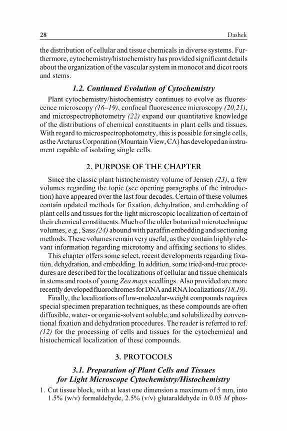
Table 1Fixatives Employed for Light Microscopy
Acetic acid 45%
Acetic acid—alcohol
Acetic acid—alcohol—chloroform
Chromium tetroxide
Chromium—formal
Ethanol 50-70% aqueous
Formalin—calcium
Formalin 10%
Formalin—alcohol—acetic acid (FAA)
Glutaraldehyde 20%a
aHarris et al. (13) suggest using a combination
of glutaraldehyde and paraformaldehyde.
30 Dashek
Table 2Summary of the Specificity of Cytochemical Stains Availablefor the Detection of Various Classes of Cellular Chemicals a,b
Compound Stain
Carbohydrates Periodic Acid—Schiff’s
Callose Aniline blue fluorescence
Cellulose Zinc chlor-iodide
Pectin Hydroxylamine—ferric chloride
Ruthenium red
Nucleic acids Calcofluro white M2R fluorescence
DNA/RNA Methyl green—pyronin
Azure B
DNA Feulgen
Acridine orange as a fluorochrome
Ethidium bromide as a fluorochrome
4', 6'- diamido-2-phenylindole
as a fluorochrome
Lignin Acidic phloroglucinol
Lipids Nile blue
Sudan black B
Sudan IV
Phospholipids Acid haematin
Bromine—Sudan black
Bromine—Acetone—Sudan black
Protein (total) Fast green pH2
Ninhydrin—Alloxan Schiff’s
Mercuric-bromphenol blue
Proteins
Containing tyrosine Million’s diazotization
Containing arginine Sakaguchi reaction
Containing tryptophan N-(1-Naphthyl)-ethylenediamine
Containing sulfhydrils or Rosindole
disulfide
Tannins Tetrazolium
Mercaptide formation
Ferric chloride—HC1
aAdapted from refs. (23), (11), (12), (18).
bSee ref. 26a.
3.2. Protocol
3.2.1. CYTOCHEMICAL/HISTOCHEMICAL LOCALIZATIONS OF CHEMICALS
IN STEMS AND ROOTS OF ZEA MAYS SEEDLINGS (SEE TABLE 2)
Chemicals Plant material
Adhesive such as Haupt’s Zea mays seedlings
dH2O Prepare in advance
Chapter 2 / Localization of Plant Cell/Tissue Chemicals 31
Ethanol Excise stem or roots
Glutaraldehyde (see Introductory Material)
Fast green Equipment
Paraformaldehyde Analytical balance and weighing paper
Paraplast Greenhouse or hood light banks (Grolux)
Periodic acid Incubator
Permount or Polymount Light microscope with or without camera
Phosphate buffer Microtome with blade
Polyethylene Glycol 1000 Ocular micrometer
Safranin Slide warmer
Schiff’s reagent Tissuetek or Paraplast dispenser
Sudan stains (optional—embedding can be
Vermiculite or perlite accomplished without them—see text)
Xylene (histological grade) Top-loading balance and weighing boats
or Histo-Clear, a recent Water bath
commercially available
clearing agent
Supplies
Aluminum foil
Camel’s hair brush
Coplin jars or staining dishes
Coverslips 22 × 50 mm
Embedding molds
Embedding rings
Flats for growing corn seedlings
Graduated cylinder
Ice bucket
Kimwipes
Microscope slides—frosted end
Pasteur pipets
Permount
Pipets 1,5, and 10 mL
Probes
Pro-pipets
Pyrex bottles
Single-edge razor blades
Vials for fixation
3.2.2. USE COPLIN JARS OR A RACK OF STAINING DISHES
Carbohydrates—periodic acid—
Schiff (use ref. 12 controls) Nucleic acids—azure bluea
Deparaffinize with two xylene Use freeze-dried or freeze-substituted
changes, 5 min each tissue; can also use chemically fixed
(continued)
Chemicals Plant material
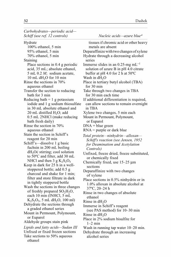
32 Dashek
Hydrate tissues if chromic acid or other heavy
100% ethanol, 5 min metals are absent
95% ethanol, 5 min Deparaffinize with two changes of xylene
70% ethanol, 5 min Hydrate through a decreasing alcohol
Staining series
Place sections in 0.4 g periodic Immerse slides in an 0.25-mg mL−1
acid, 35 mL; absolute ethanol, solution of azure B in pH 4.0 citrate
5 mL 0.2 M; sodium acetate, buffer at pH 4.0 for 2 h at 50ºC
10 mL dH2O for 10 min Wash in dH
2O
Rinse the sections in 70% Place in tertiary butyl alcohol (TBA)
aqueous ethanol for 30 min
Transfer the section to reducing Take through two changes in TBA
bath for 3 min for 30 min each time
Reducing bath = 1 g potassium If additional differentiation is required,
iodide and 1 g sodium thiosulfate can allow sections to remain overnight
in 30 mL absolute ethanol and in TBA
20 mL distilled H2O; add Xylene two changes, 5 min each
0.5 mL 2NHCl (make reducing Mount in Permount, Polymount,
bath fresh daily) or Euparol
Rinse the section in 70% DNA = blue green
aqueous ethanol RNA = purple or dark blue
Stain the section in Schiff’s
reagent for 20 min
Schiff’s—dissolve 1 g basic
fuchsin in 200 mL boiling
dH2Oc stirring; cool solution
to 50ºC and filter, add 30 mL
NHCl and then 3 g K2S
2O
5
Keep in dark for 25 h in a well-
stoppered bottle; add 0.5 g
charcoal and shake for 1 min;
filter and store filtrate in dark
in tightly stoppered bottle
Wash the sections in three changes
of freshly prepared SO2H
2O,
each 10 min (INHCl, 5 mL
K2S
2O
5, 5 mL dH
2O, 100 ml)
Dehydrate the sections through
a graded ethanol series
Mount in Permount, Polymount,
or Euparol
Aldehyde groups stain pink
Lipids and fatty acids—Sudan III
Unfixed or fixed frozen sections
Take sections to 50% aqueous
ethanol
Carbohydrates—periodic acid—
Schiff (use ref. 12 controls) Nucleic acids—azure bluea
Total protein—ninhydrin—alloxan—
Schiff's reaction (see Jensen, 1952
for Deamination and Acetylation
Controls)
Unfixed, freeze dried, freeze substituted,
or chemically fixed
Chemically fixed, use 15–25 µm
sections
Deparaffinize with two changes
of xylene
Place sections in 0.5% ninhydrin or in
1.0% alloxan in absolute alcohol at
37ºC, 20–24 h
Rinse in two changes of absolute
ethanol
Rinse in dH2O
Immerse in Schiff’s reagent
(see PAS method) for 10–30 min
Rinse in dH2O
Place in 2% sodium bisulfite for
1–2 min
Wash in running tap water 10–20 min.
Dehydrate through an increasing
alcohol series

Chapter 2 / Localization of Plant Cell/Tissue Chemicals 33
Stain in Sudan III in 70% ethanol for 30 min
Rinse sections in 50% aqueous ethanol
Mount in glycerine
Avoid the use of absolute ethanol, as lipids will be soluble
Mount in Permount, Polymount, or Euparol xylene two times, 5 min each time
Neutral fats and fatty acids stain red
aThe specificity of azure B for DNA and RNA must be verified in each system by
DNase and RNase treatments as well as other cytochemical tests, The Feulgen reaction
for DNA and acridine orange (DNA and RNA) coupled with fluorescence microscopy
are particularly useful. Similarly, the specificity of fast green at pH2 for total protein
must be verified by treating sections with proteases.
Table 3Summary of Wood-Decay Fungal H2O2 Investigations
Tests employed References
3' 3-Diaminobenzidine; Forney et al. (27); Highley and Murmanis (28);
horseradish peroxidase, Illman and Highley (30);
and ABTS or o-diansidine; Micales and Highley (31)
titanium reagent
3.3. Cytochemical/Histochemical Localizations
of Low-Molecular-Weight Compounds—H2O
2
Some of the most comprehensive investigations of H2O
2 localizations
in plant tissues have been those of Highley and his co-workers (Table 3).
These investigators were concerned with localizing H2O
2 in decaying
wood and wood decay fungi as H2O
2 is thought to function in proposed
Fenton chemistry-mediated wood decay (see Chapter 12 and refs. 32).
Highley and his co-workers both present and cite methods for localizing
H2O
2. With modification for systems differences, the tests cited in Table
3 should be applicable to a wide variety of plant systems.
4. CONCLUSIONS
The future of cytochemistry resides in its usefulness as an adjunct to
biochemistry. As mentioned, fluorescence (33–35) and confocal (36–40)
microscopies have provided new dimensions to cytochemistry.
Finally, photomicrography is the culmination of the preparation of
specimens for optical microscopy. This is a very technical area requiring
proper illumination (41–43), focusing, choice of films, as well as exposure
and appropriate film development. This critical area of microscopy should
see continued technological innovations as much of photomicrography
is being computerized (44, 45). This effort is witnessing the concomitant
Lipids and fatty acids—Sudan III
34 Dashek
improvement of basic measuring techniques for light microscopy and
image analysis (46–48).
REFERENCES
1. Galigher AE, Kosloff EN. Essentials of Practical Microtechnique, Lea and Febiger,
Philadelphia, PA, 1964.
2. Hayat MA. Stains and Cytochemical Methods, Plenum Press, New York, 1993.
3. Horubin RW. Understanding Histochemistry: Selection, Evaluation and Design of
Biological Stains, Hopwood, Chichester, UK, 1988.
4. Kiernan JA. Histological and Histochemical Methods: Theory and Practice,
Pergamon Press, Oxford, UK, 1990.
5. Lillie RD, Fuller HM. Histopathology Techniques and Practical Histochemistry,
McGraw-Hill, New York, 1976.
6. Pearse AGE. Histochemistry Theoretical and Applied, Little, Brown, Boston, MA,
1964.
7. Sannes PL. The Histochemical and Cytochemical Localization of Proteases, Deer-
field BEA, Stuttgart, Germany, 1988.
8. Sheehan DC. Histotechnology—Theory and Practice, The CV. Mosby Co., St.
Louis, MO, 1980.
9. Horobin RW. Histochemistry and the light microscope, in Light Microscopy in
Biology (Lacey AJ, ed.), IRL Press, Oxford, UK, 1989, pp. 137–162.
10. Sumner BEH. Basic Histochemistry, Wiley, Chichester, New York, 1988.
11. Berlyn GP, Miksche JP. Botanical Microtechnique and Cytochemistry, Iowa State
University Press, Ames, IA, 1976.
12. Gahan PB. Plant Histochemistry and Cytochemistry: An Introduction, Academic
Press, London, UK, 1984.
13. Harris N, Spence J, Oparka KJ. General and enzyme histochemistry, in Plant Cell
Biology (Harris N, Oparka KJ, eds.), IRL Press, Oxford University Press, Oxford,
UK, 1994, pp. 51–68.
14. Vaughn KC. Handbook of Plant Cytochemistry, CRC Press, Boca Raton, FL, 1987.
15. Vigil EL, Hawes CR. Cytochemical and Immunological Approaches to Plant Cell
Biology, Academic Press, London, UK, 1989.
16. Rost FWD. Quantitative Fluorescence Microscopy, Cambridge University Press,
New York, 1991.
17. Rost FWD. Fluorescence Microscopy, Vols. I and II, Cambridge University Press,
New York, 1992.
18. Oparka KJ, Read ND. The use of fluorescent probes for studies of living plant cells,
in Plant Cell Biology (Harris N, Oparka KJ, eds.), IRL Press, Oxford University
Press, Oxford, UK, 1994, pp. 27–50.
19. Oparka KJ, Roberts AG, Santa Cruz S, Boevnik P, Prior D, Smallcombe A. Using
GFP to study virus invasion and spread in plant tissues. Nature 1997; 388: 401–402.
20. Wilson T. Confocal Microscopy, Academic Press, New York, 1990.
21. Shuming N, Chiu DT. Probing molecules with confocal fluorescence microscopy.
Science 1994; 266: 1018.
22. Cherry RJ. New Techniques of Optical Microscopy and Microspectrophotometry,
CRC Press, Boca Raton, FL, 1990.
23. Jensen WA. Botanical Histochemistry, Freeman, San Francisco, CA, 1952.
24. Sass JE. Botanical Microtechnique, 3rd ed., Iowa State College Press, Ames, IA,
1958.
25. Rosen WG, Gawlik SR, Dashek WV, Siegesmund KA. Fine structure and cyto-
chemistry of Lilium pollen tubes. Am J Bot 1964; 51: 61–67.
Chapter 2 / Localization of Plant Cell/Tissue Chemicals 35
26. Reynolds JD, Dashek WV. Cytochemical analysis of callose distribution in Lilium
longiflorum pollen tubes. Ann Bot 1976; 40: 409–416.
26a.Prasad B. Staining Techniques in Botany, State Mutual Book and Periodical Ser-
vices, New York, 1986.
27. Forney LJ, Reddy CA, Pankvatz HS. Ultrastructural localization of hydrogen per-
oxide production in ligninolytic Phanerochaete chrysosporium cells. Appl Environ
Microbiol 1982; 44: 732–736.
28. Highley T, Murmanis LL. Determination of hydrogen peroxide production in
Coriolus versicolor and Poria placenta during wood degradation. Mater Org 1985;
29: 241–252.
29. Highley T. Effect of carbohydrate and nitrogen on hydrogen peroxide formation by
wood decay fungi in solid medium. FEMS Microbiol Lett 1987; 48: 373–378.
30. Illman B, Highley TL. Hydrogen peroxide formation by wood decay fungi in liquid
medium. Phytopathology 1988; 78: 1590.
31. Micales JA, Highley TL. In vitro production of hydrogen peroxide by degradative
and non-degradative isolates of brown-rot wood decay fungi. Phytopathology 1989;
77: 988.
32. Highley TL, Dashek WV. Biotechnology in the study of white-rot and brown-rot
decay, in Forest Products Biotechnology (Bruce A, Palfreyman JW, eds.), Taylor
and Francis, London, UK. 1998, pp. 15–36.
33. Slavik J. Fluorescence Microscopy and Fluorescent Probes, Proceedings of a Con-
ference Held in Digre, Czech Republic, Plenum Press, New York, 1996.
34. Tarke HJ. Fluorescence Microscopy, Bios Scientific UK, Cornett Books, Oxford,
UK, 1996.
35. Wang XF, Herman B. Fluorescence Imaging Spectroscopy and Microscopy, Wiley,
New York, 1996.
36. Van der Voort HTM, Valkenburg JAC, Van Spronsen EA, Woldringh CL, Braken-
hoff GJ. Confocal microscopy in comparison with electron and conventional micro-
scopy, in Correlative Microscopy in Biology. Instrumentation and Methods (Hayat
MA, ed.), Academic Press, Orlando, FL, 1987, pp. 60–81.
37. Pauley B. Handbook of Biological Confocal Microscopy, Plenum Press, New York,
1990.
38. Corle TR, Kino GS. Confocal Scanning Optical Microscopy and Related Imaging
Systems, Academic Press, New York, 1996.
39. Sheppard C, Shottan, D. Confocal Laser Scanning Microscopy, Springer-Verlag,
Berlin, Germany, 1997.
40. Paddock SW. Confocal Microscopy Methods and Protocols, Humana Press, Totowa,
NJ, 1998.
41. Smith RF. Microscopy and Photomicrography. A Working Manual, 2nd ed., CRC
Press, Boca Raton, FL, 1994.
42. Thompson DJ, Bradbury S. An Introduction to Photomicrography (Book 13),
Oxford University-Royal Microscopical Society, Oxford, UK, 1991.
43. Weiss DG, Marle W, Wick RA. Video microscopy, in Light Microscopy in Biology:
A Practical Approach (Lacey AJ, ed.), IRL Press, Oxford, UK, 1989, pp. 221–278.
44. Bradbury S. Basic Measurement Techniques for Light Microscopy, Oxford Univer-
sity- Royal Microscopical Society, Oxford, UK, 1991.
45. Swatland HJ. Computer Operation for Microscopy Photometry, CRC Press, Boca
Raton, FL, 1997.
46. Hader DP. Image Analysis in Biology, CRC Press, Boca Raton, FL, 1992.
47. Russ JC. The Image Processing Handbook, CRC Press, Boca Raton, FL, 1995.
48. Jahne B. Practical Handbook on Image Processing for Scientific Applications,
CRC Press, Boca Raton, FL, 1997.