cytogenetics ii structural chromosomal aberrations rndr z.polívková lecture no 432 - course :...
TRANSCRIPT
Cytogenetics IICytogenetics IIStructural chromosomal Structural chromosomal
aberrationsaberrations
RNDr Z.PolívkováRNDr Z.Polívková
Lecture No 432Lecture No 432 - - coursecourse:: Heredity Heredity
Causes of structural aberrations:
external mutagens (except Robertsonian translocations)
origin in: G1,S, G2, mitosis, meiosis
Structural CHA:
• unbalanced – loss or gain of chromosomal material
• balanced – abnormal rearrangement without loss or gain of chromosomal material
Unbalanced• Deletion (del) = partial monosomy – terminal
- interstitial - break and loss of chromosomal segment in G1
- unequal crossing over in meiosis
- segregation of balanced aberration in meiosis → unbalanced product
• Duplication (dup) = partial trisomy
- duplication in S phase,
- insertion of a segment of sister chromatid
- unequal crossing over in meiosis,
- segregation of balanced aberration in meiosis
• Ring chromosome (r) – partial monosomy of segments distal to
breaks on short and long arms - reunion of broken chromosome to a ring formation
• Dicentric chromosome (dic) – abnormal chromosome with 2 centromeres - 2 breaks of 2 chromosomes or 2 chromatids and
reunion of broken ends, in G1, G2
• Isochromosome – partial monosomy of one arm and partial trisomy of other arm
- misdivision (transverse splitting) of centromere in MI, MII, mitosis
• Additional marker chromosome (+mar) small chromosome of unknown origin – supernumerrary - if heterochromatic – mostly without clinical consequences
Detection of origin of marker chromosome – FISH method
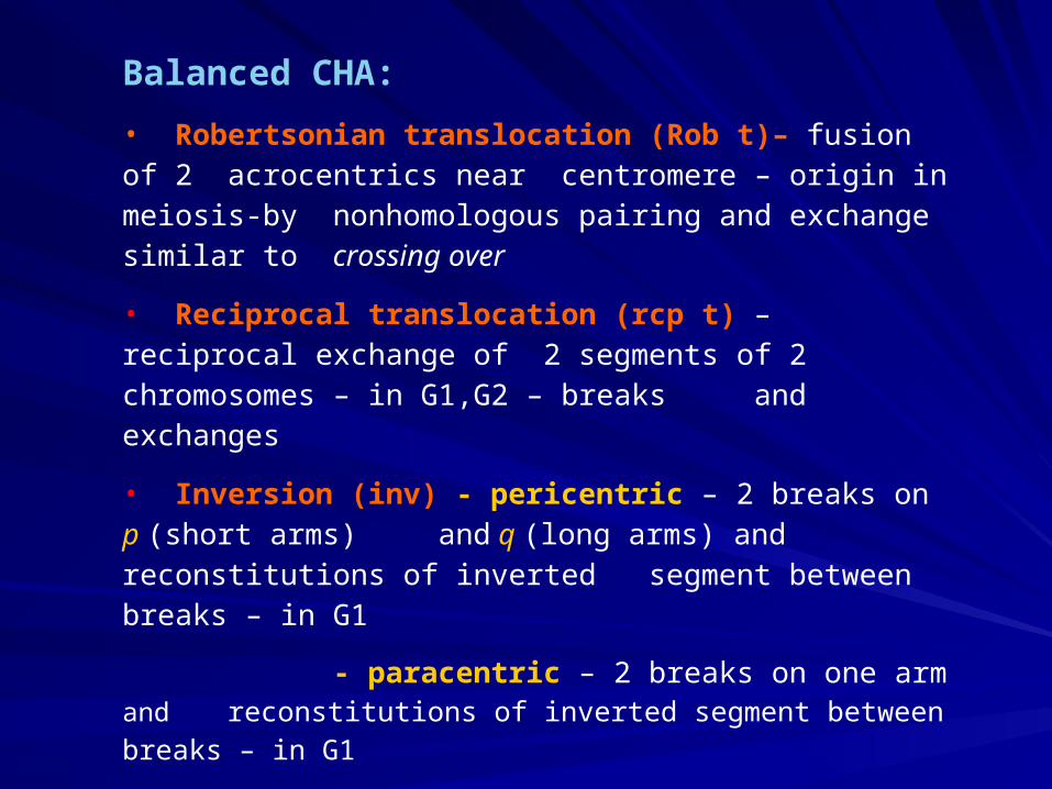
Balanced CHA:
• Robertsonian translocation (Rob t)– fusion of 2 acrocentrics near centromere – origin in meiosis-by nonhomologous pairing and exchange similar to crossing over
• Reciprocal translocation (rcp t) – reciprocal exchange of 2 segments of 2 chromosomes – in G1,G2 – breaks and exchanges
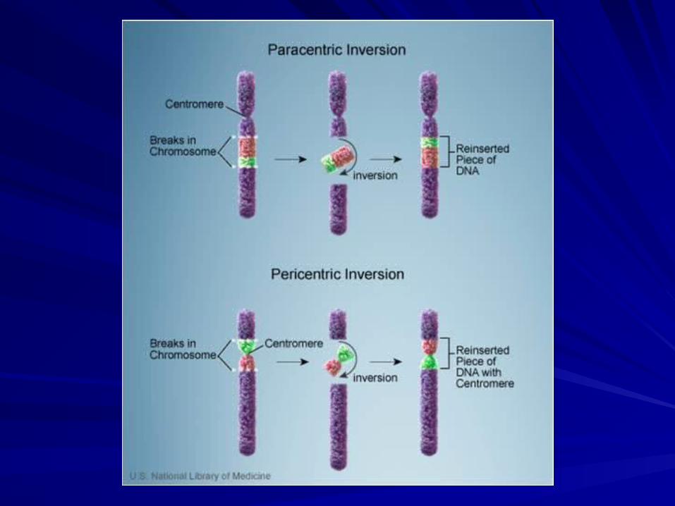
• Inversion (inv) - pericentric – 2 breaks on p (short arms) and q (long arms) and reconstitutions of inverted segment between breaks – in G1
- paracentric – 2 breaks on one arm and
reconstitutions of inverted segment between breaks – in G1
• Insertion – segment removed from 1 chromosome is inserted into another chromosome – 3 breaks rearrangement
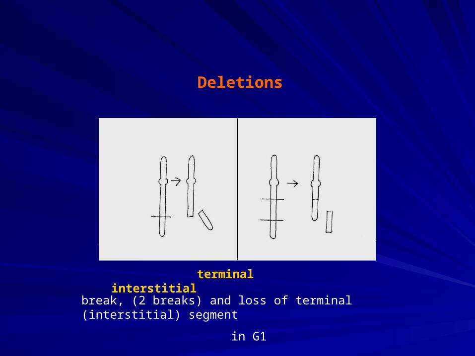
Deletions
terminal interstitial
break, (2 breaks) and loss of terminal (interstitial) segment
in G1
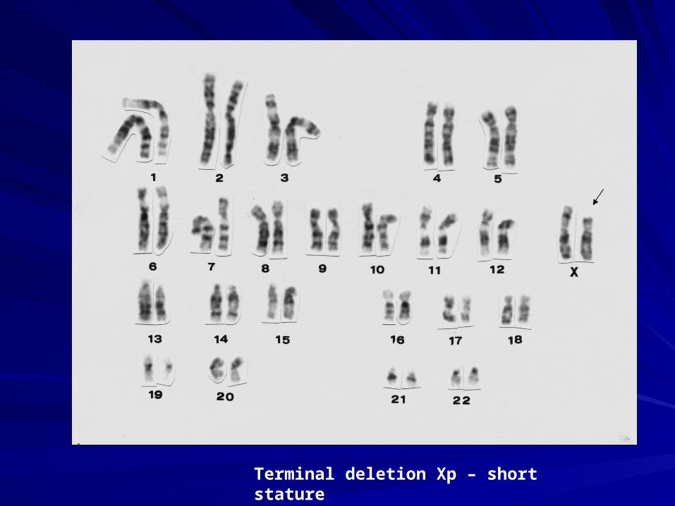
Terminal deletion Xp – short stature
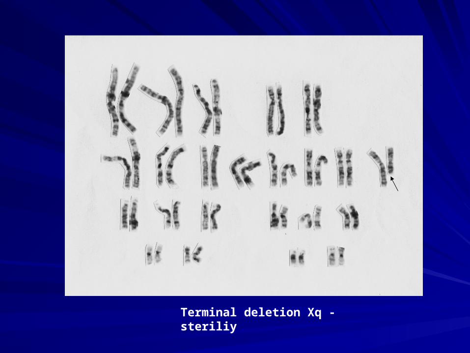
Terminal deletion Xq -steriliy
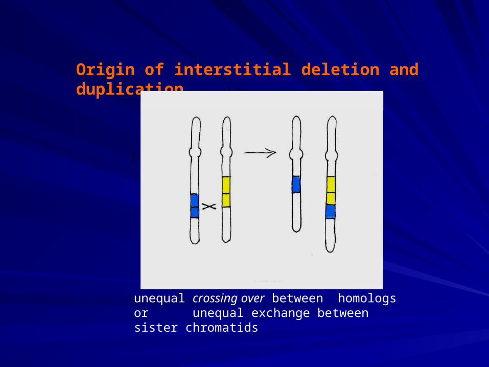
Origin of interstitial deletion and duplication
unequal crossing over between homologs or unequal exchange between sister chromatids
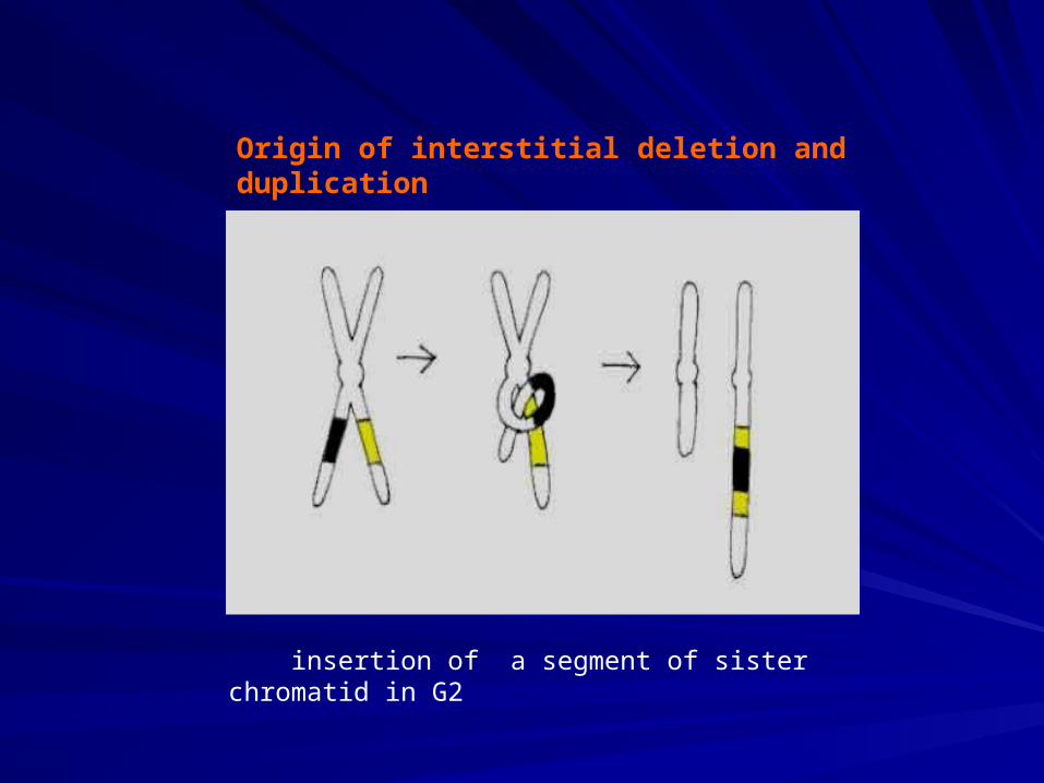
Origin of interstitial deletion and duplication
insertion of a segment of sister chromatid in G2
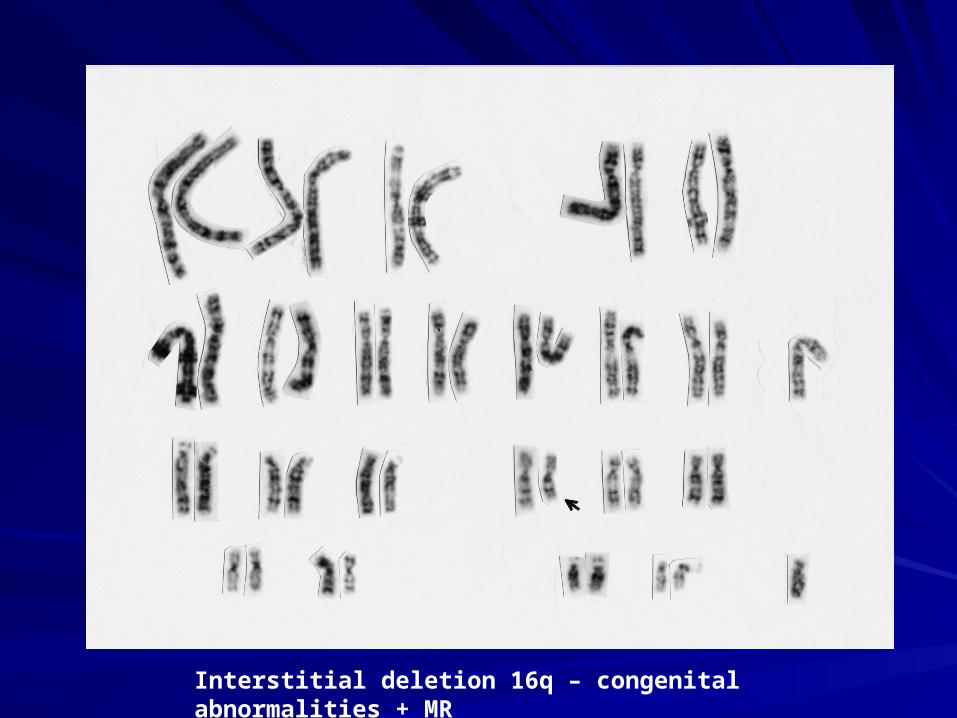
Interstitial deletion 16q – congenital abnormalities + MR
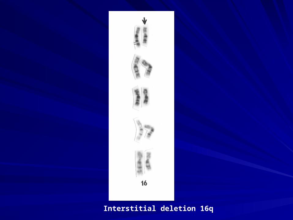
Interstitial deletion 16q
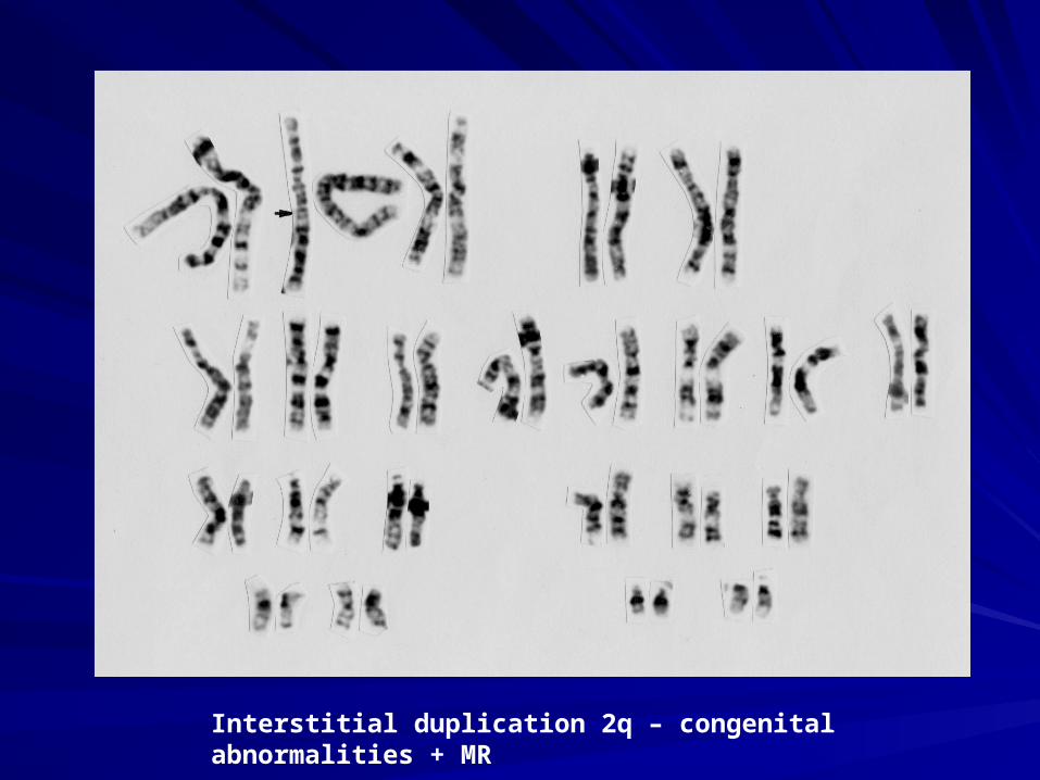
Interstitial duplication 2q – congenital abnormalities + MR
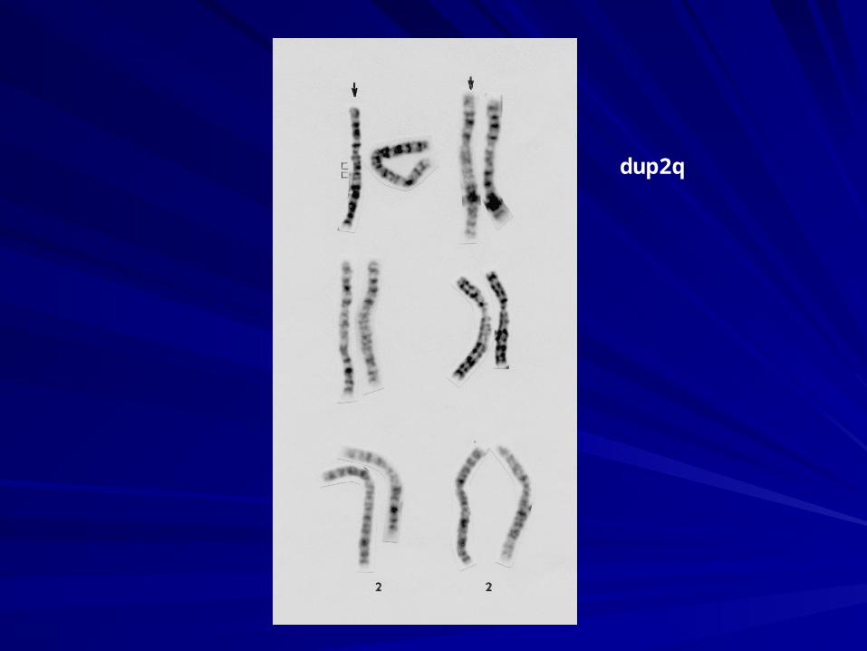
dup2q
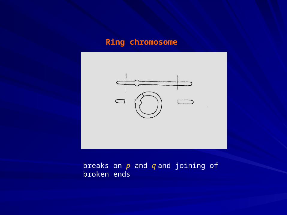
Ring chromosome
breaks on p and q and joining of broken ends
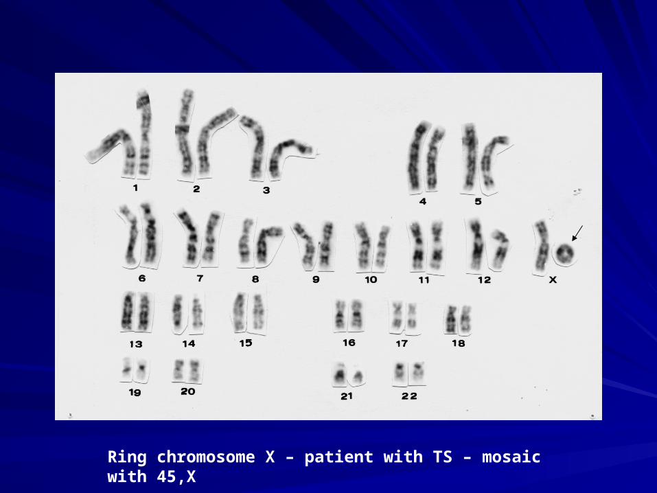
Ring chromosome X – patient with TS – mosaic with 45,X
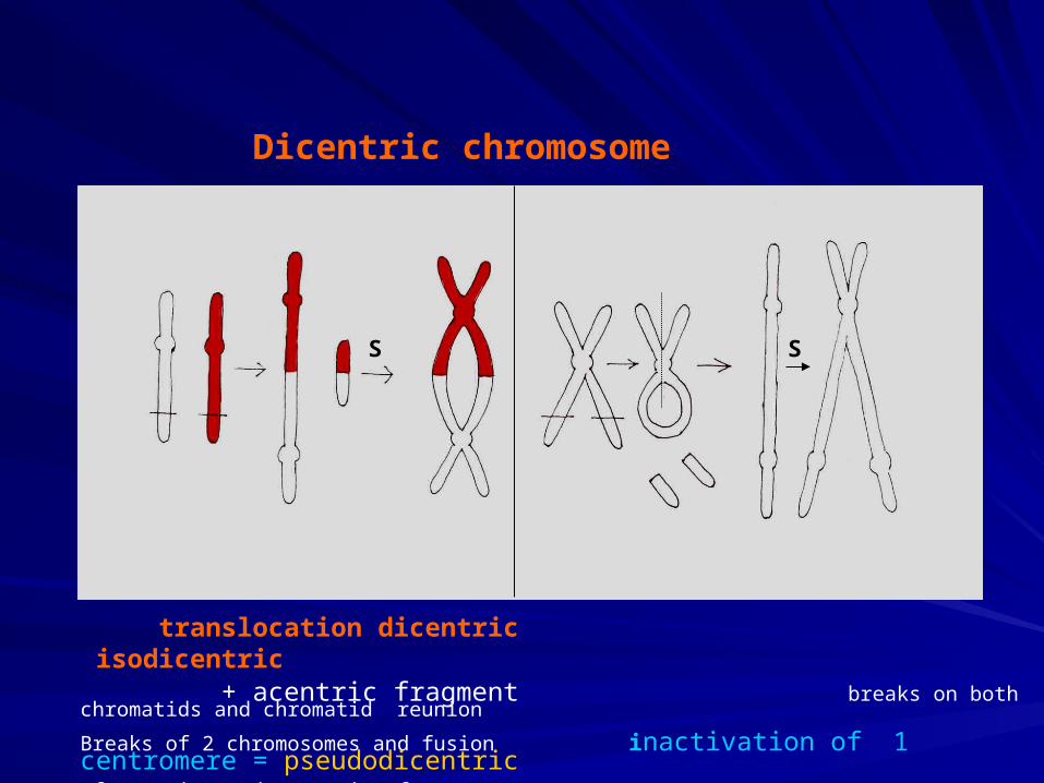
Dicentric chromosome
translocation dicentric isodicentric + acentric fragment breaks on both chromatids and chromatid reunion
Breaks of 2 chromosomes and fusion inactivation of 1 centromere = pseudodicentricof centric (and acentric) fragmens
SS
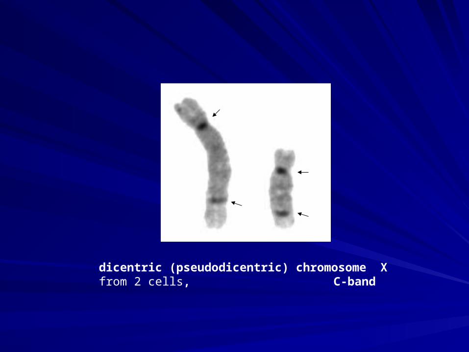
dicentric (pseudodicentric) chromosome X from 2 cells, C-band
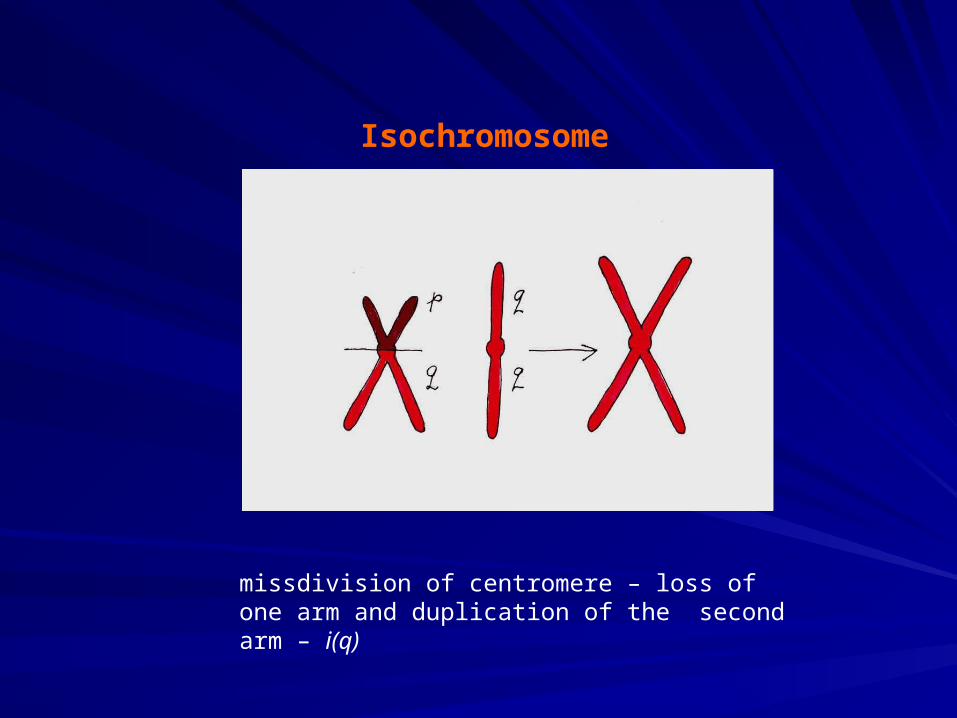
Isochromosome
missdivision of centromere – loss of one arm and duplication of the second arm – i(q)
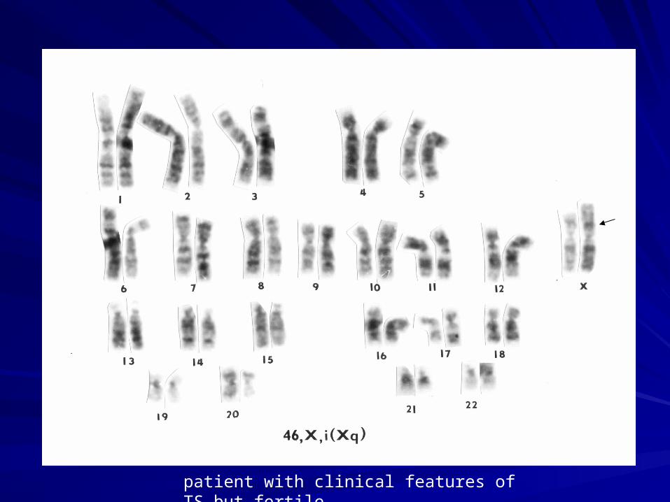
patient with clinical features of TS but fertile
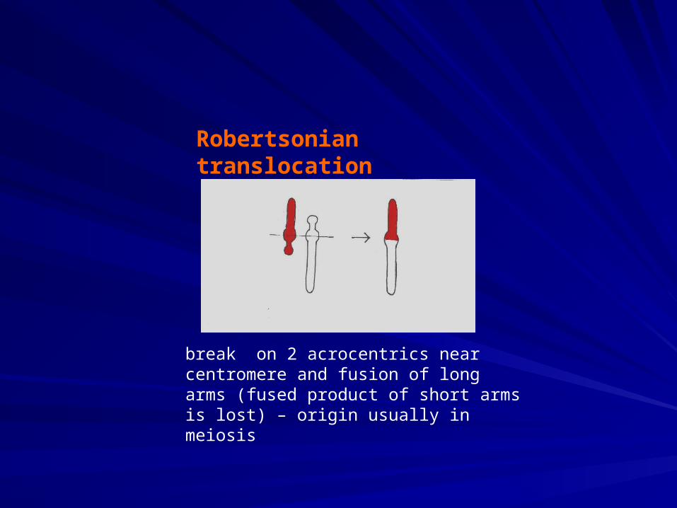
Robertsonian translocation
break on 2 acrocentrics near centromere and fusion of long arms (fused product of short arms is lost) – origin usually in meiosis
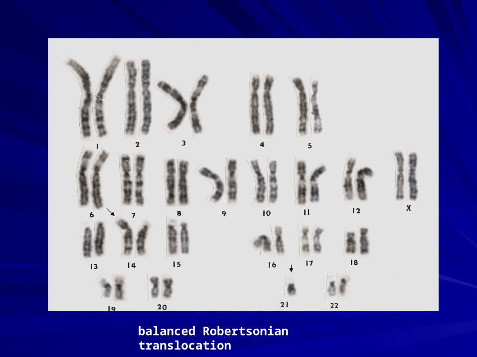
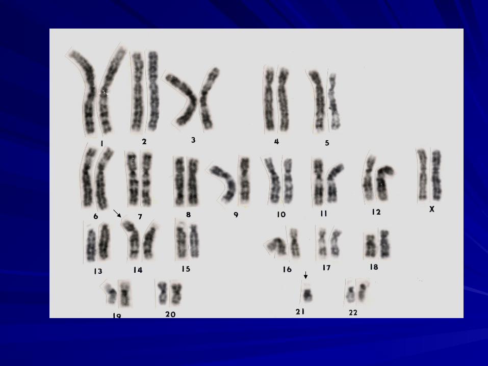
balanced Robertsonian translocation
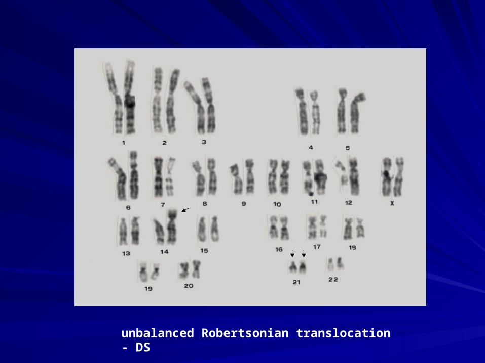
unbalanced Robertsonian translocation - DS
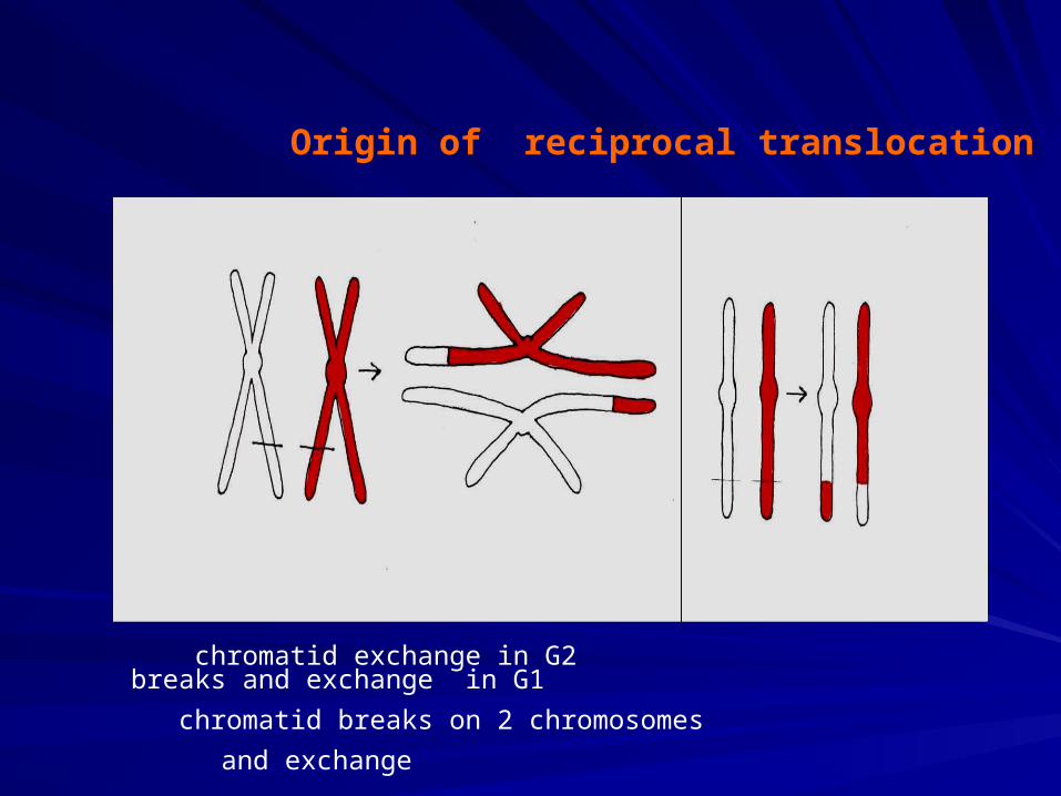
Origin of reciprocal translocation
chromatid exchange in G2 breaks and exchange in G1
chromatid breaks on 2 chromosomes
and exchange
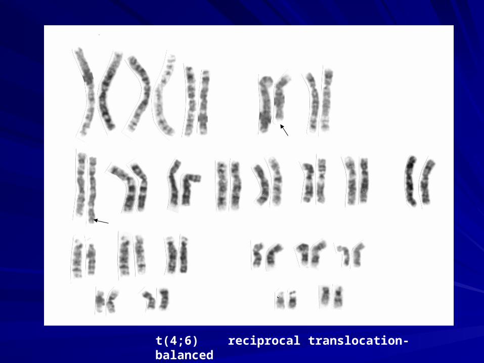
t(4;6) reciprocal translocation-balanced
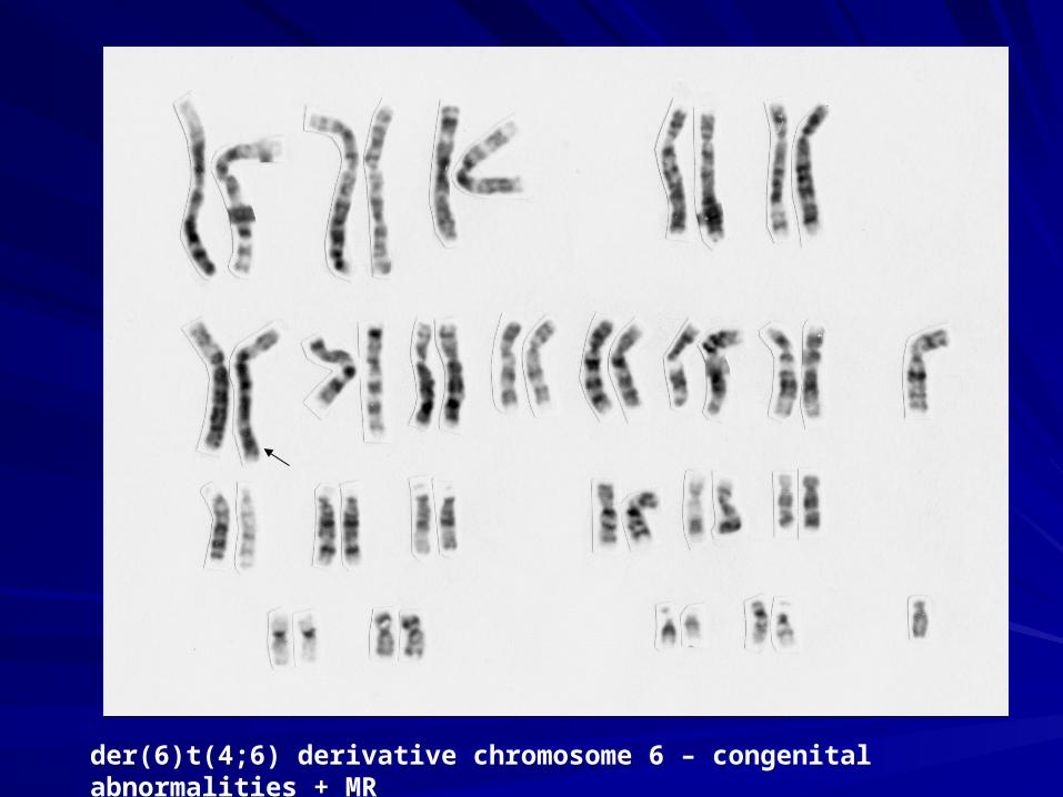
der(6)t(4;6) derivative chromosome 6 – congenital abnormalities + MR
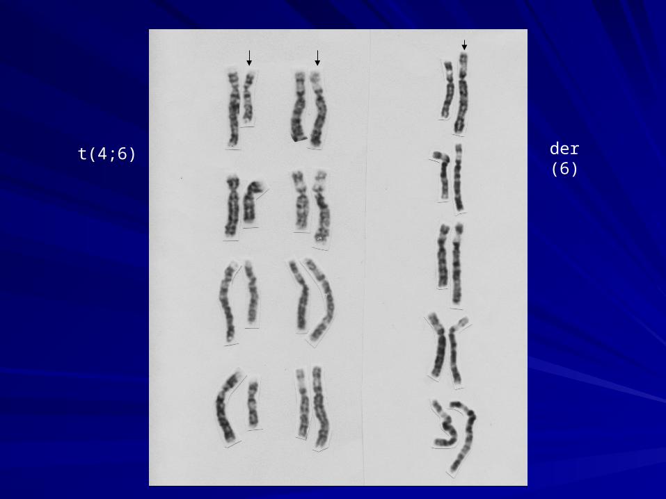
t(4;6) der (6)
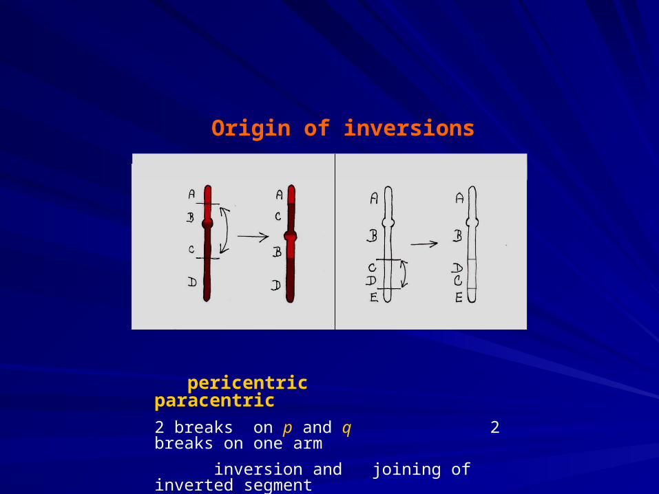
Origin of inversions
pericentric paracentric
2 breaks on p and q 2 breaks on one arm
inversion and joining of inverted segment
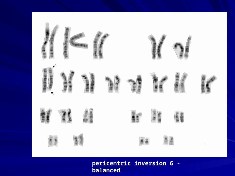
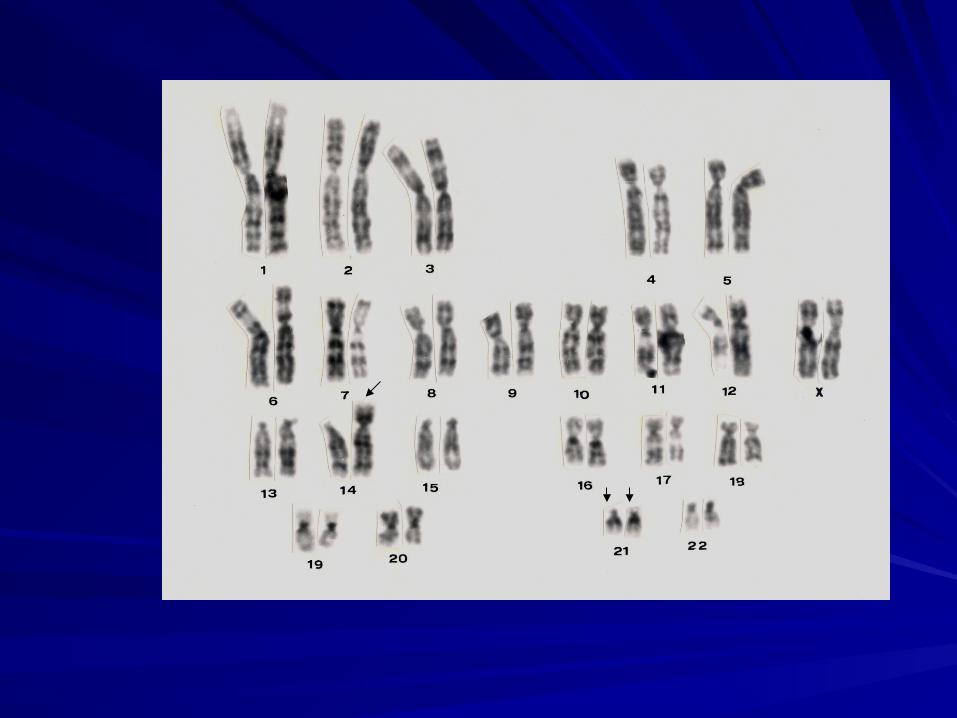
pericentric inversion 6 - balanced
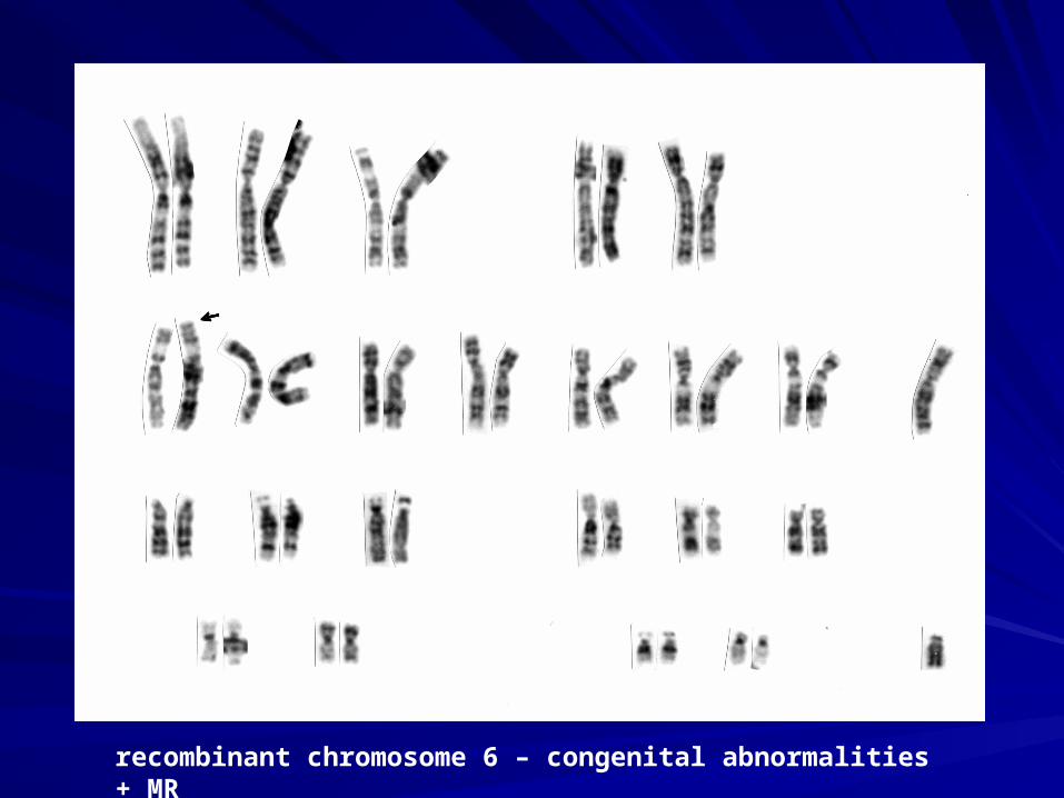
recombinant chromosome 6 – congenital abnormalities + MR
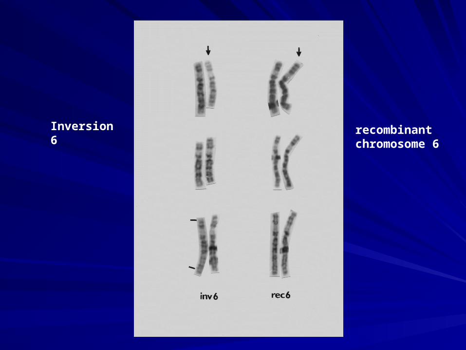
Inversion 6 recombinant chromosome 6
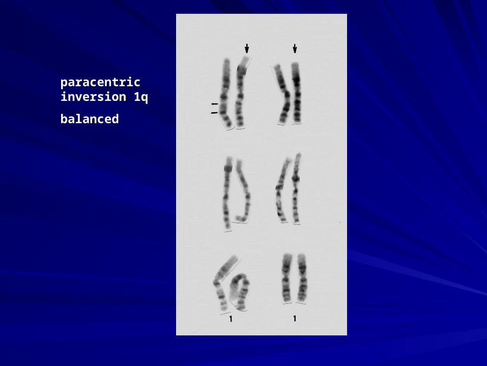
paracentric inversion 1q
balanced
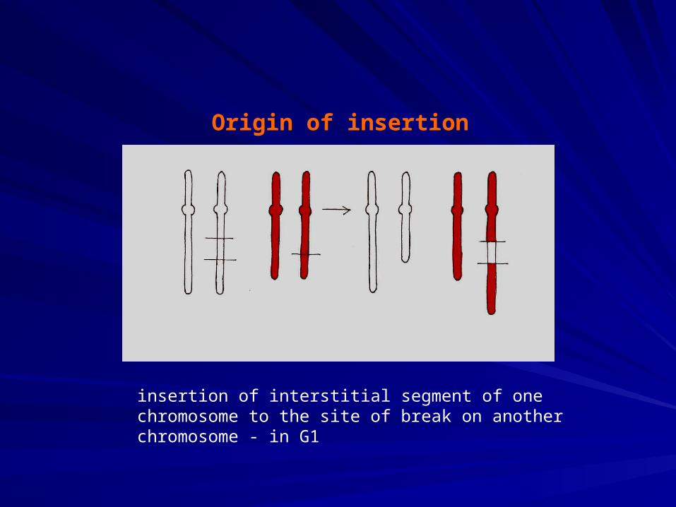
Origin of insertion
insertion of interstitial segment of one chromosome to the site of break on another chromosome - in G1
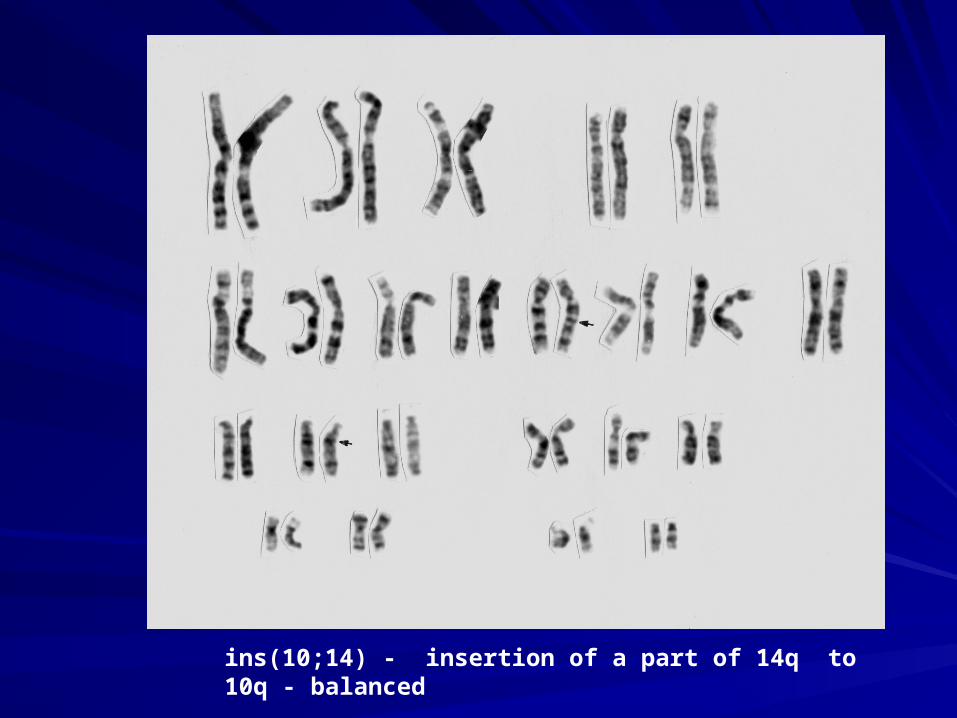
ins(10;14) - insertion of a part of 14q to 10q - balanced
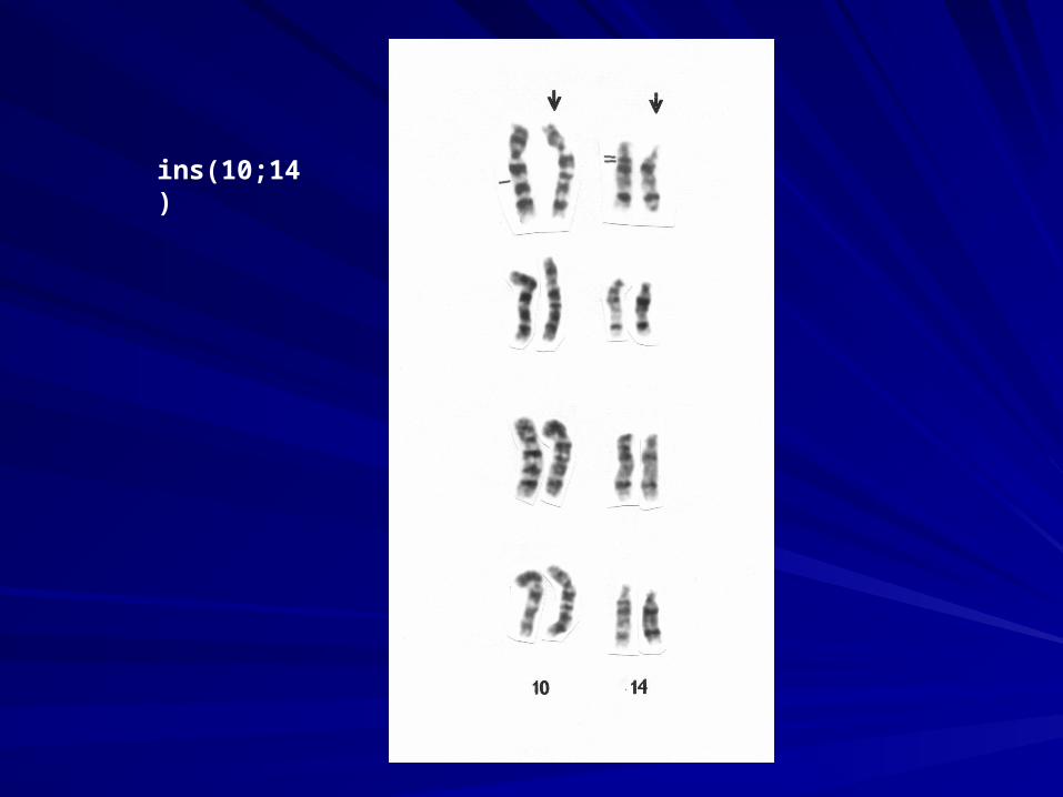
ins(10;14)
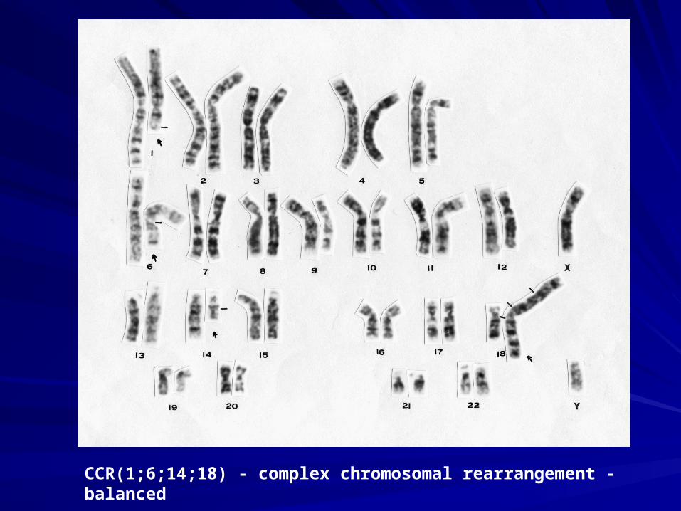
CCR(1;6;14;18) - complex chromosomal rearrangement - balanced

Risk of balanced structural aberration:
Carrier of balanced structural aberration
usually without clinical signs
risk of unbalanced aberration in progeny
+
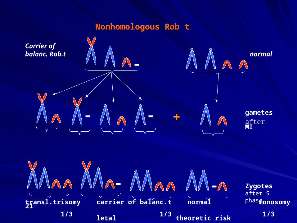
Carrier ofbalanc. Rob.t normal
Nonhomologous Rob t
gametes
after MI
Zygotes after S phase
transl.trisomy carrier of balanc.t normal monosomy 21 1/3 1/3 1/3 letal theoretic risk
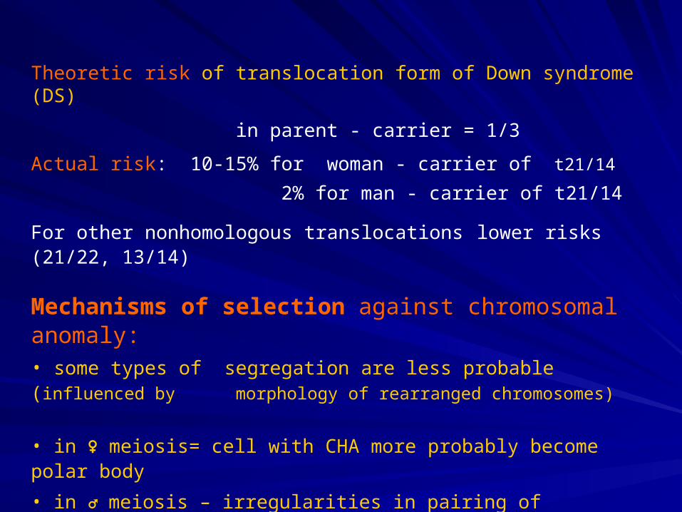
Theoretic risk of translocation form of Down syndrome (DS)
in parent - carrier = 1/3
Actual risk: 10-15% for woman - carrier of t21/14
2% for man - carrier of t21/14
For other nonhomologous translocations lower risks (21/22, 13/14)
Mechanisms of selection against chromosomal anomaly:• some types of segregation are less probable (influenced by morphology of rearranged chromosomes)
• in ♀ meiosis= cell with CHA more probably become polar body
• in ♂ meiosis – irregularities in pairing of rearranged chromosomes → poor sperm development → oligospermia, azoospermia
• gamete with CHA – selectional disadvantage in fertilisation (in sperm)
• selection against abnormal zygote = spontaneous abortion
46,XX
22,X-21
23,Xt21/21
23,X
45,XX
t21/21
23,Xt21/21
23,Xt21/21
23,Y
23,X
23,Xt21/21
22,X-21
46,XY
t21/21
46,XY
t21/21
+ +
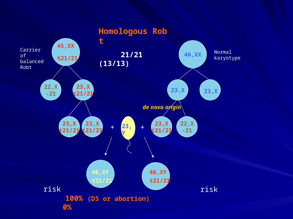
100% (DS or abortion) 0%
Homologous Rob t
21/21 (13/13)
de novo origin
risk risk
Carrier of balanced Robt
Normal karyotype
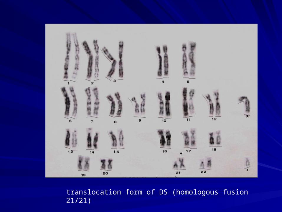
translocation form of DS (homologous fusion 21/21)
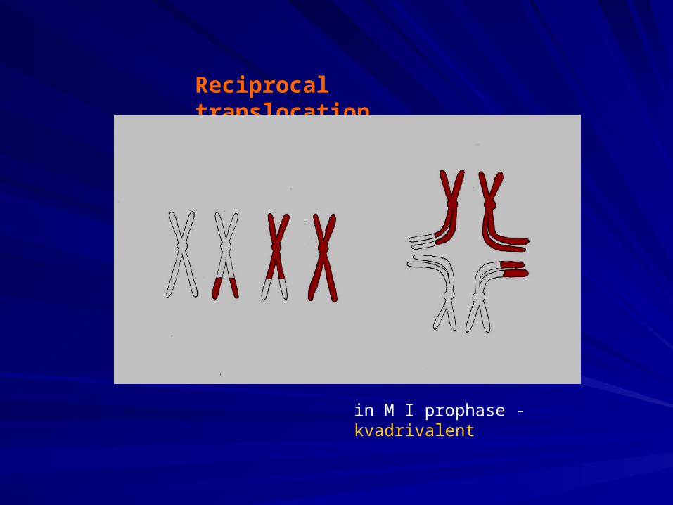
Reciprocal translocation
N1 T1 T2 N2
T2
N1
N2
T2
in M I prophase - kvadrivalent
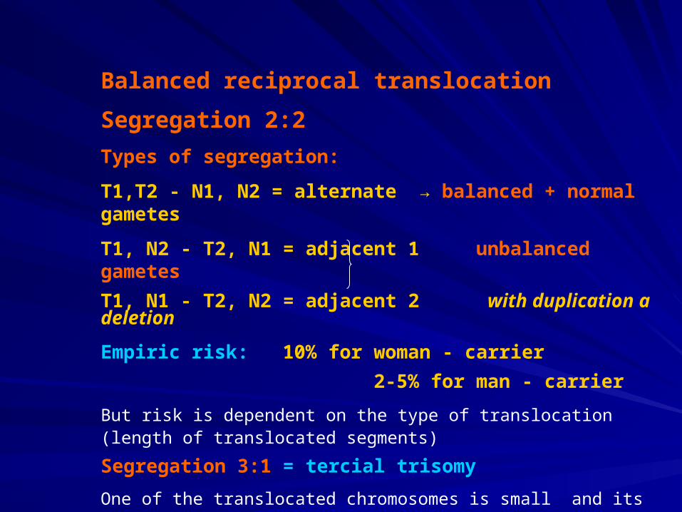
Balanced reciprocal translocation
Segregation 2:2
Types of segregation:
T1,T2 - N1, N2 = alternate → balanced + normal gametes
T1, N2 - T2, N1 = adjacent 1 unbalanced gametes
T1, N1 - T2, N2 = adjacent 2 with duplication a deletion
Empiric risk: 10% for woman - carrier
2-5% for man - carrier
But risk is dependent on the type of translocation (length of translocated segments)
Segregation 3:1 = tercial trisomy
One of the translocated chromosomes is small and its trisomy is compatible with life
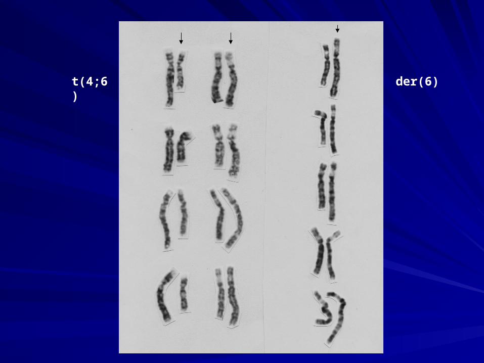
t(4;6) der(6)
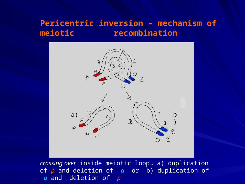
Pericentric inversion – mechanism of meiotic recombination
crossing over inside meiotic loop→ a) duplication of p and deletion of q or b) duplication of q and deletion of p
a) b)

Risk of meiotic recombination in a carrier of pericentric inversion
depends on the length of inverted segment
in average: for woman – carrier - 10%
for man - carrier - 5%
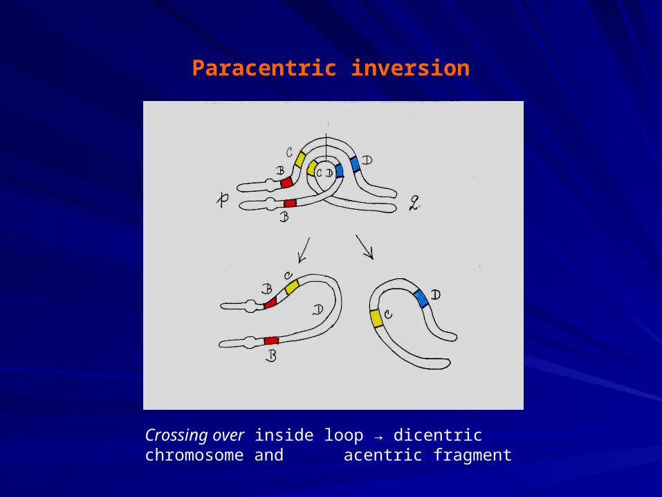
Paracentric inversion
Crossing over inside loop → dicentric chromosome and acentric fragment
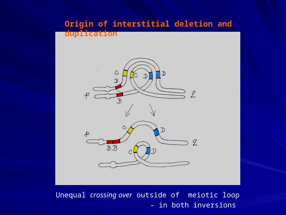
Origin of interstitial deletion and duplication
Unequal crossing over outside of meiotic loop
– in both inversions
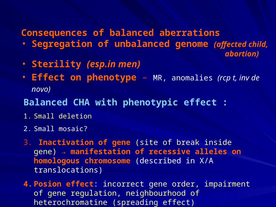
Consequences of balanced aberrations• Segregation of unbalanced genome (affected child,
abortion)• Sterility (esp.in men)• Effect on phenotype – MR, anomalies (rcp t, inv de novo)
Balanced CHA with phenotypic effect :
1. Small deletion
2. Small mosaic?
3. Inactivation of gene (site of break inside gene) → manifestation of recessive alleles on homologous chromosome (described in X/A translocations)
4. Posion effect: incorrect gene order, impairment of gene regulation, neighbourhood of heterochromatine (spreading effect)
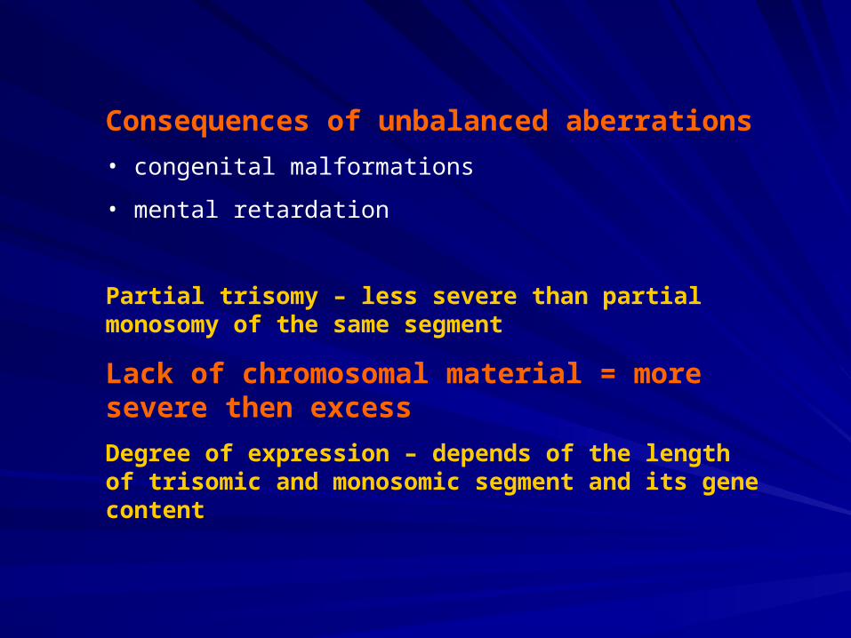
Consequences of unbalanced aberrations
• congenital malformations
• mental retardation
Partial trisomy – less severe than partial monosomy of the same segment
Lack of chromosomal material = more severe then excess
Degree of expression – depends of the length of trisomic and monosomic segment and its gene content
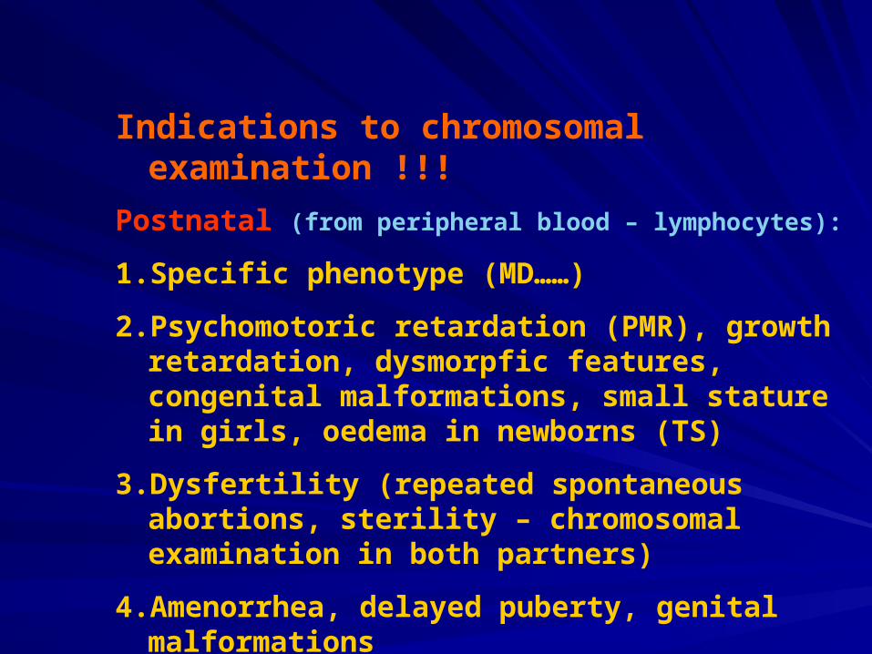
Indications to chromosomal examination !!!
Postnatal (from peripheral blood – lymphocytes):
1. Specific phenotype (MD……)
2. Psychomotoric retardation (PMR), growth retardation, dysmorpfic features, congenital malformations, small stature in girls, oedema in newborns (TS)
3. Dysfertility (repeated spontaneous abortions, sterility – chromosomal examination in both partners)
4. Amenorrhea, delayed puberty, genital malformations
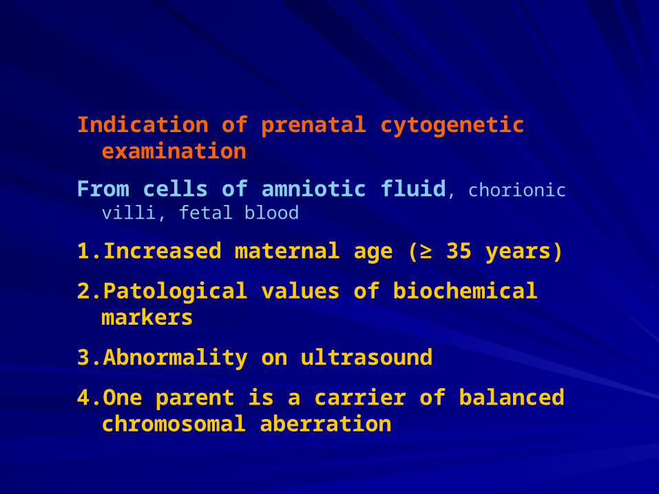
Indication of prenatal cytogenetic examination
From cells of amniotic fluid, chorionic villi, fetal blood
1. Increased maternal age (≥ 35 years)
2. Patological values of biochemical markers
3. Abnormality on ultrasound
4. One parent is a carrier of balanced chromosomal aberration

Indication of prenatal cytogenetic examination
From cells of amniotic fluid, chorionic villi, fetal blood
1. Increased maternal age (≥ 35 years)
2. Patological values of biochemical markers
3. Abnormality on ultrasound
4. One parent is a carrier of balanced chromosomal aberration
http://dl1.cuni.cz/course/view.php?id=324 presentation
http://dl1.cuni.cz/course/view.php?id=324 supplementary text to cytogenetics
Thompson &Thompson: Genetics in medicine, 7th ed.
Chapter 5: Principles of clinical cytogenetics
Chapter 6: Clinical cytogenetics: Disorders of the autosomes and the sex chromosomes
+ informations from presentation