deepak.p medical electronics mr. deepak p. associate professor ece department sngce 1
TRANSCRIPT
DEEPAK.P
MEDICAL ELECTRONICS
Mr. DEEPAK P.Associate ProfessorECE Department
SNGCE
1
DEEPAK.P
UNIT 1
2
Introduction to Human Physiological Systems and
Transducers

DEEPAK.P3
Objective At the end of this UnitYou will learn
Physiological Systems of human body
Bio medical Transducers and Electrodes

DEEPAK.P4
Introduction to Biomedical Engineering

Biomedical EngineeringBiomedical Engineering is the application of engineering principles and design concepts to medicine and biology
The biomedical engineering provides electrical, electronic, electro-optical, and computer engineering support to clinical and biomedical applications.
Biomedical Engineering improves the field of healthcare diagnosis, monitoring and therapy.
5 DEEPAK.P

DEEPAK.P6
Medical Instruments

Biomedical InstrumentsClassification of Biomedical Equipments
1. Diagnostic equipment
2. Therapeutic equipment
3. Clinical equipment
4. Laboratory equipment
7 DEEPAK.P
DEEPAK.P8
Man- Instrument System

Components in Man – Instrument system
9 DEEPAK.P
Control feedback
Recording , data processing and transmission of data
Signal conditioning equipment
DisplayTransducer
Transducer
Transducer
Sti
mul
us

Man – Instrument systemMeasurement in biomedical instrumentation can be divided in to two
1.VIVO
•Measurement is made on or within the human body
•Eg . Device inserted in to the blood stream to measure PH of blood
2.VITRO
•Measurement is performed outside of the body.
•Eg . Measurement of blood PH from blood samples.
10 DEEPAK.P

DEEPAK.P11
Bioelectric Potentials

Sources of Bioelectric potentialsThe systems in the human body generate their on
monitoring signals when they carry out their functions.
These signals provide useful information about their function.
Bioelectric potentials are actually ionic voltages produced as a result of electro chemical activity of certain cell.
Transducers are used to convert these ionic potentials in to electrical signals
12 DEEPAK.P

Resting and Action potentialsCertain types of cells within the body , such as nerve and
muscle cells are encased in a semi permeable membrane.
This membrane permits some substances to pass through while others are kept out.
Surrounding the cells of the body are the body fluids
These fluids are conductive solutions containing charged atoms known as ions
13 DEEPAK.P

Resting potentialsThe principle ions are sodium(Na+)
Potassium(K+) and chloride(C-)
The membrane of excitable cells permit entry of Potassium(K+) and chloride(C-) ions but blocks the entry of sodium(Na+) ions.
So inside the cell is more negative than outside cell
This membrane potentials is called Resting potentials
This potential is measured from inside the cell with respect to body fluids.
So resting potential of a cell is negative. 14 DEEPAK.P

Resting potentials/PolarizationThis resting potential ranging from -60mv to -100 mv.Cell in the resting state is called polarized cell.
15 DEEPAK.P
Ground
V
Cell Membrane
-70 mV

Depolarization of cellWhen a cell is exited, the membrane
change its characteristic.
The sodium ions are rushed in to the cell.
At the same time potassium ions try move from inside.
After a equilibrium state is reached, the sodium is moved back to outside
16 DEEPAK.P

Depolarization of cell
17 DEEPAK.P
Cell MembraneNa+
Na+
Na+
Na+
Na+
Na+
Na+
Na+
K+
K+
K+ K+
K+
K+
K+K+

Action potentials
18 DEEPAK.P
Cell Membrane
V
Ground
20 mV

Re PolarizationCell comes from de polarized state in to polarized state is
called Re polarization.
19 DEEPAK.P
Ground
V
Cell Membrane
-70 mV

Resting and Action potentials
20 DEEPAK.P

Propagation of Action potentialsWhen a cell is exited and generates an action potentials
ionic currents to flow.This process excite neighboring cells or adjacent area of
the same cell
21 DEEPAK.P

DEEPAK.P22
Physiological Systems in Human body

Physiological systems of human body
In simple terms "Human Physiology" is the study of the body and its functions in each of the different systems in any living body.
23 DEEPAK.P
SystemInput Output

Physiological Systems in the Human body
24 DEEPAK.P
Behavior
Liquid wastes
Solid wastes
Body movements
Expired air
Appearance
Speech
Food intake
Liquid intake
Vision
Smell
Taste
Hearing
Inspired air
Tactile sensation
INP
UT
S
OU
TP
UT
S

Physiological systems of human bodyThere are 11systems in the body: 1. The Skeletal System Bones & joints
2. Muscular System Skeletal muscle
3. Nervous System Brain, spinal cord & nerves
4. Endocrine System Hormone-producing cells & glands
5. Cardiovascular System Blood, heart & blood vessels
6. Respiratory System Lungs & airways
7. Digestive System Organs of the gastrointestinal tract
8. Urinary System Kidneys, bladder and ureters
9. Reproductive System Male & female reproductive organs
10.The Integumentary System The skin & derived structures
11.Lymphatic & Immune System Lymphatic vessels & fluid
25 DEEPAK.P

DEEPAK.P26
Physiology of Cardiovascular system

Physiology•Physiology can be classified in to
1.Cell Physiology
• Study of cells
2.Patho Physiology
• Pathological Functions
3.Circulatory Physiology
• Study of blood circulation
4.Respiratory Physiology
• Study of breathing organs
27 DEEPAK.P

Cardio Vascular systemCardio vascular system can be viewed as closed hydraulic system with 4 chamber pump.
Cardio Vascular system is mainly used for transportation of oxygen, Carbon dioxide, numerous chemical compounds and the blood cells.
•Pump-----Heart
•Flexible tubes---Blood vessels
28 DEEPAK.P

Cardio Vascular system•In some part of the system diameter of the arteries are changed to control pressure.
•Pump(heart) is a isolated two stage synchronized chamber
1.The first stage is to collect blood from the system and pump it in to 2nd stage.
2.The second stage then pump these blood to the system
29 DEEPAK.P

Heart & Valves
30 DEEPAK.P
Right Atrio-ventricular valve
Bicuspid/Left Atrio-ventricular valve

Heart LayersHeart wall consists of three layers
1.Pericardium
• Outer most layer, keeps outer surface moist, prevents friction
2.Myocardium
• Middle layer, Main muscle of heart, made up of short cylindrical fibres
3.Endocardium
• Inner layer of heart, Provides smooth lining for blood flow
31 DEEPAK.P

Heart ValvesHeart has 4 valves
1.Tricuspid/Right Atrio-Ventricular valve• Between Right A and V, Prevents blood flow from right V to A
2.Bicuspid/ left Atrio-Ventricular valve• Between left A and V, Prevents blood flow from left V to A
3.Pulmonary valve• At right ventricle, It has 3 cusps
4.Aortic Valve• Between left ventricle and aorta, It has 3 cusps
32 DEEPAK.P
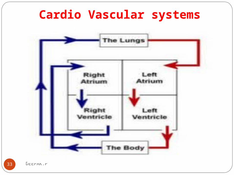
Cardio Vascular systems
33 DEEPAK.P

Cardio Vascular SystemOne of the two stage pump(Right side) collect fluid from the system and pump it through oxygenation system(Lungs).
Other side pump receives blood from oxygenation system(Lungs) and pump blood to main hydraulic system.
Blood act as communication and supply network for all parts of the body
34 DEEPAK.P

Cardio Vascular systemFluid contains fuel suppliers and waste particles are transported to destination.
Fluid contain mechanism for rejecting foreign elements and mechanism for repairing small system puncture.
Sensors are provided to detect the changes in the need of suppliers, the build of waste material and out-of- tolerance pressure in the system known as chemoreceptors, Pco2 sensors and baroreceptors respectively.
35 DEEPAK.P

Cardio Vascular CirculationThe blood is carried out to the various parts of the body through blood vessels.
There are three types of blood vessels
1)Arteries--- Thick, Carries oxygenated blood
2)Veins--- Thin, De-oxygenated blood
3)Capillaries---Smallest, Last level of blood vessels, 800000 km of capillaries
36 DEEPAK.P

Cardio Vascular CirculationHeart pumps blood through the pulmonary circulation to the lungs and through the systemic circulation to the other parts of the body.
1)Pulmonary circulation
2)Systemic circulation
In pulmonary circulation, venous blood(de-oxygenated) flows from right ventricle through pulmonary artery to lungs .
The arterial( oxygenated) blood flows to left atrium through pulmonary veins.
In systemic circulation blood flows from left auricle to left ventricle and it is pumped to aorta and its branches37 DEEPAK.P

Cardio Vascular Circulation
38 DEEPAK.P

DEEPAK.P39
Respiratory system

Respiratory systemsIt is the Pneumatic system.
A system that work with air pressure.
An air pump(diaphragm) which alternatively create negative and positive pressures in a sealed chamber(Thoracic cavity).
Thoracic cavity sucked air in to and forced out to two elastic bags(Lungs).
The lungs are connected to the external environment through a pass way (nasal cavities, pharynx, larynx, trachea, bronchi and bronchioles)
40 DEEPAK.P

Respiratory systems
41 DEEPAK.P

Respiratory systemsAt one point , this passage is common with the tube that carries liquid and solids to stomach.
A special valving arrangement interrupts the respiratory system whenever solid or liquid passes through the common region.
The passage divides to carry air in to each bag.
In each bag , it is sub divided many times to carry air in to and out of each of many tiny air spaces (pulmonary alveoli).
42 DEEPAK.P

Respiratory systemsIn case of nasal blockage , air input can be taken from mouth.
Oxygen is taken from the air and transferred in to blood.
Cabondioxide is transferred from blood to air.
The system has a number of fixed volumes and capacities.
43 DEEPAK.P

Respiratory systems
Tidal volume
The volume inspired and expired during each normal breath
Inspiratory reserve volume
Additional volume that can be inspired after a normal inspiration.
Expiratory reserve volume
Additional volume that can be expired after a normal expiration.
44 DEEPAK.P

Respiratory systemsResidual volume
Amount of air remaining in the lungs after all possible air has been forced out.
Vital capacity
Tidal volume+ Inspiratory reserve volume+ Expiratory reserve volume
45 DEEPAK.P

Respiratory systems
46 DEEPAK.P

Blood purification in the human bodyThe overall functioning of our body heavily depends on the
proper functioning of our blood.
We take toxins into our body daily and these toxins disrupts the functions of our internal organs.
Regular detoxification of our blood is important
Detoxifying the blood and body helps to remove harmful toxins from our body and improve the functioning of our vital organs such as the kidney and liver
47 DEEPAK.P

Blood purification in the human body
When our body has too much toxins, our vital organs start to get damage and under-perform and we start to develop symptoms of allergies, low immunity, headaches, fatigue and several other health related problems.
The lungs help remove carbon dioxide, the kidneys remove water-soluble waste and the liver removes fat soluble wastes and many other impurities from the blood.
48 DEEPAK.P

Blood purification by lungsThe un oxygenated (unpurified) blood comes
into right atrium of heart by superior and inferior vena-cava which then passes to right ventricle, then to lungs by pulmonary artery.
In lungs this blood gets oxygenated (purified) which then goes into left chamber of heart from which blood is passed to aorta and then circulated to whole body
49 DEEPAK.P

Blood purification by lungs
50 DEEPAK.P

Blood purification by lungs
51 DEEPAK.P
DEEPAK.P52
Muscular System

Muscular SystemThe muscular system is the biological system of humans that produces movement.
It permits movement of the body, maintains posture, and circulates blood throughout the body.
The muscular system is controlled through the nervous system.
Muscles provide strength, balance, Posture, movement and heat for the body to keep warm.
53 DEEPAK.P

Muscular System
• More than 50% of body weight is muscle.
• Muscle is made up of proteins and water
54 DEEPAK.P

Muscular SystemThere are three distinct types of muscles: skeletal muscles,
cardiac or heart muscles, and smooth muscles.
55 DEEPAK.P

Muscular SystemSmooth muscle or "involuntary muscle" consists of spindle
shaped muscle cells found within the walls of stomach, intestines, bronchi, uterus, ureters, bladder, and blood vessels.
Smooth muscle cells contain only one nucleus.
Cardiac muscle is also an "involuntary muscle" but it is striated in structure and appearance.
Like smooth muscle, cardiac muscle cells contain only one nucleus.
56 DEEPAK.P

Muscular SystemCardiac muscle is found only within the heart.
Skeletal muscle or "voluntary muscle" is anchored by tendons to the bone and is used to effect skeletal movement such as locomotion.
Skeletal muscle cells are multinucleated with the nuclei peripherally located.
Skeletal muscle is called 'striated' because of the longitudinally striped appearance under light microscopy.
57 DEEPAK.P

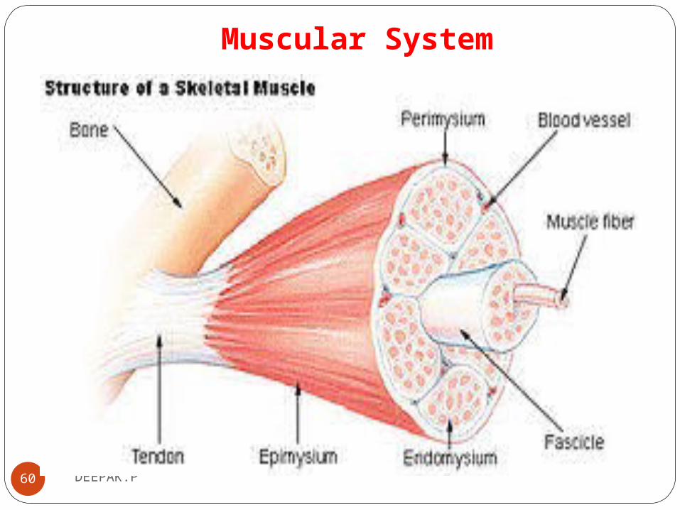
Muscular SystemMuscle is composed of muscle cells (sometimes known as
"muscle fibers").
Within the cells are myofibrils; myofibrils contain sarcomeres which are composed of actin and myosin.
Individual muscle cells are lined with endomysium.
Muscle cells are bound together by perimysium into bundles called fascicles.
58 DEEPAK.P

Muscular SystemThese bundles are then grouped together to form muscle, and
is lined by epimysium.
59 DEEPAK.P
Muscular System
60 DEEPAK.P

Muscular System
61 DEEPAK.P

DEEPAK.P62
Nervous systems

Nervous systemsThe task of controlling various functions of body and
coordinating them in to a integrated living organism(human body) is the function of Nervous system
It is the most complex system in the human body
It is the communication network in the human body.
It composed of Brain, Sensors, high speed communication links ,spinal cord.
63 DEEPAK.P
Nervous systemsIt provides regulation of body functions and sensory
perception.
Functions of Nervous systems
1. Control of the body
2. Integration
3. Communication
64 DEEPAK.P

Nervous systems
Its center is a self adapting processor(Brain).
Self adapting means --- If a certain section is damaged, other sections can adapt take over the function of damaged sections
This processor has memory, computational power, decision making capability.
65 DEEPAK.P

Nervous systemsWith the use of this processor , human can take decisions,
solve complex problems, create art, poetry and music, feel emotions and integrate input information from all parts of the body and produce output signals of meaningful information.
Central computer has millions of communication lines( afferent and efferent nerves) that bring sensory information and transfer control information from brain.
66 DEEPAK.P

Nervous systems
67 DEEPAK.P

Nervous systems
This lines are not single lines but complicated networks.
Information signal are normally coded by means of electro
chemical pulses that travel along the nerves.
The output control signals are channeled to specific motor
devices(motor units of muscles)
68 DEEPAK.P
Nervous systems
In addition to brain , a large number of simple decision making
g devices(Spinal reflexes) are present to control directly
certain motor devices from certain sensory inputs.
69 DEEPAK.P
Divisions of Nervous systemsThe human nervous system can be divided into three main parts:
1. Central nervous system (CNS) *is composed of brain and spinal cord...*
2. Peripheral nervous system (PNS)*is composed of all body nerves that lie outside of your
central nervous system...*
3. Autonomic nervous system (ANS) *Controls the involuntary actions of your body
organs...*
70 DEEPAK.P
1. Central Nervous systems
Central Nervous System (CNS)Structures of the CNS: [1] Brain[2] Spinal cord
The CNS coordinates and interprets information to determine the best response
71 DEEPAK.P

Anatomy of the Central Nervous systems
72DEEPAK.P
Spinal cord
CerebellumBrain stem
Cerebrum

2. Peripheral Nervous systems
The primary role of the PNS is to connect the CNS to the
organs, limbs and skin.
The nerves that make up the peripheral nervous system are
actually the axons or bundles of axons from neuron cells.
The peripheral nervous system is divided into two parts:
The somatic nervous system
The autonomic nervous system
73 DEEPAK.P

Somatic Nervous systems
The somatic system is the part of the peripheral nervous
system responsible for carrying sensory and motor
information to and from the central nervous system.
This system contains two major types of neurons:
Sensory neurons (or afferent neurons) that carry information
from the nerves to the central nervous system.
Motor neurons (or efferent neurons) that carry information from
the brain and spinal cord to muscle fibers throughout the body.
74 DEEPAK.P

3. Autonomic Nervous systemsThe autonomic system is the part of the peripheral nervous
system responsible for regulating involuntary (reflex/Un
intentional)body functions, such as blood flow, heartbeat,
digestion and breathing.
This system is further divided into two branches:
The sympathetic system regulates the flight-or-fight responses.
75 DEEPAK.P

Autonomic Nervous systems
The "fight or flight response" is our body's primitive, automatic, inborn response that prepares the body to "fight" or "flee" from perceived attack, harm or threat to our survival.
Parasympathetic system helps maintain normal body functions and conserves physical resources
76 DEEPAK.P
Anatomy of the Nervous systems
Basic unit of nervous system is the neuron.
Neurons are the basic building blocks of the nervous system.
Neuron is a single cell with a cell body.
It is sometimes called soma
77 DEEPAK.P

Anatomy of the Nervous systems
Neuron cells are the information-processing units of the brain
responsible for receiving and transmitting information.
One or more I/P fibers branches are called dendrites
Long transmitting fiber is called axon
Each part of the neuron plays a role in the communication of
information throughout the body.
78 DEEPAK.P

Anatomy of the Nervous systems
79 DEEPAK.P

Anatomy of the Nervous systemsNeurons Composed of: a. Cell Body
Part that contains the nucleusb. Dendrite(s)
Carries a nerve impulse towards the cell bodyc. Axon(s)
Carries a nerve impulse away from the cell body (and towards the dendrite of the next neuron)
Axons are also called nerve fibers.
80 DEEPAK.P

Types of Neurons
The three basic types of idealized neurons include;
Bipolar, (Pseudo) Uni polarMulti polar neurons,
Typically these neurons are found in different places around the body:
81 DEEPAK.P

Types of Neurons
82 DEEPAK.P

Types of NeuronsBipolar – Specialized sensory neurons for the transmission of
special senses.
As such, they are part of the sensory pathways for smell, sight, taste and hearing functions.
The most common example are the bipolar neurons found in the retina
83 DEEPAK.P

Types of Neurons
(Pseudo) Unipolar
Many types of primary sensory neurons are Unipolar.
Multi polar – Multi polar neurons constitute the majority of neurons in the brain and include motor neurons and interneuron's.
84 DEEPAK.P

DEEPAK.P85
Sources of Biomedical Signals

Sources of Biomedical Signals
86 DEEPAK.P

DEEPAK.P87
Bio- Potential Electrodes

Measurement of Bioelectric potentials
To measure bioelectric potentials , a transducer is required.
Electrical signals produced by various body activities are
used in monitoring / diagnosis
In order to measure and record potentials and, hence, currents
in the body, it is necessary to provide some interface
between the body and the electronic measuring apparatus.
.88 DEEPAK.P

Bio Potential Electrodes
Bio-potential electrodes carry out this interface function.
A transducer consists of two electrodes, which measure ionic
potential difference between two points.
The designation of the Bio potential waveform ends with
“Gram”.
The name of the instrument bio potential normally ends
with “Graph”
89 DEEPAK.P

Bio Potential Electrodes
Propagation of action potential through different body tissues
produces final waveform recorded by electrodes
Electrical activity is explained by differences in ion concentrations within the body (sodium, Na+; cloride, Cl–; potassium, K+)
A potential difference (voltage) occurs between 2 points with different ionic concentrations
90 DEEPAK.P
DEEPAK.P91
Electrodes Theory

Equivalent circuit for bio-potential electrode
92 DEEPAK.P
Bod
y E
lect
roly
tes
Vha=Electrode potential developed across interface
C=Charges at the Interface at the skin metal interface
Met
al E
lect
rode

Equivalent circuit for bio-potential with two electrode
94 DEEPAK.P

Bio Potential Electrodes
Bio-potential electrodes transduce ionic conduction to
electronic conduction so that bio-potential signals can be
obtained
They generally consist of metal contacts packaged so that they
can be easily attached to the skin or other body tissues
95 DEEPAK.P

DEEPAK.P96
Classification of Electrodes

Bio Potential Electrodes1. Micro Electrodes--- Bio electric potential near or within a
single cellMetal Type—Tip must be tungsten or stainless steelMicro pipette---It is a glass micropipet with size of 1
micron, It is filled with electrolyte
2. Skin surface electrode —Measure ECG,EEG,EMG
3. Needle electrode ---Penetrate the skin to record EEG
97 DEEPAK.P

MicroelectrodesUsed to measure bio-potential signals at
the cellular level
Due to small dimensions (mm), impedance levels are high
So amplifier needs very high input impedance

Microelectrodes

METAL MICROELECTRODES

MICROPIPETTE ELECTRODES

Surface electrodesThese are placed in contact with the skin
of the subject
Early stages immersion electrodes were used.
A bucket of saline water is used
An improvement of immersion electrode is the plate electrode.
Another old type electrode is suction type

Immersion electrodes

Surface electrodes
METAL-PLATE ELECTRODESHistorically, one of the most frequently used forms of
bio-potential sensing electrodes is the metal-plate electrode.
In its simplest form, it consists of a metallic conductor in contact with the skin.
An electrolyte soaked pad or gel is used to establish and maintain the contact.

METAL-PLATE ELECTRODES

Floating electrodesConductive paste reduces effect of
electrode slippage and resulting motion artifact

Needle electrodesUnipolar electrode---Single wire inside a
needle
Bipolar electrode---Two wires inside a needle
Mostly used for contacting with internal body tissues
(a) Insulated needle electrode .(b) Coaxial needle electrode .(c) Bipolar coaxial electrode .(d) Fine -wire electrode connected to
hypodermic needle, before being inserted .(e) Coiled fine -wire electrode in place

Needle electrodes

Implantable electrodes

ELECTRODE ARRAYS

ELECTRODE ARRAYS

DEEPAK.P113
Transducers

TransducersA transducer is necessary to convert one variable in to another
form
Used to measure physiological variables
Variable is a quantity that vary with time.
The term active and passive has different meaning when they are applied to Transducers
114 DEEPAK.P

TransducersTransducer can be classified in to twoActive Transducer
Known principles is used to convert variables in to electrical signal
Passive TransducerIt involves control of an excitation voltage or modulation
of a carrier signal
115 DEEPAK.P

Principles used in Active TransducersIt can convert electrical signal in to physical variables and
also in reverse direction.
1. Magnetic Induction
2. Piezoelectric effect
3. Thermoelectric effect
4. Photoelectric effect
116 DEEPAK.P

Passive TransducersUtilize the principles of controlling a DC excitation or an AC
carrier signal.
It consists of a passive circuit element which changes it value as a function of physical variables to be measured.
It cannot convert electrical signal in to physical variables
117 DEEPAK.P

Principles used in Passive TransducersUsing Resistive element
1. Ordinary Potentiometera. Linearb. Rotary
2. Strain gagea. Un bondedb. Bondedc. Semiconductor strain gage
Using Inductive element1. Variable reluctance Transducer
LVDT• Using Capacitive element
118 DEEPAK.P

Transducers for Biomedical field
Force transducer
Photoelectric displacement transducer
Pressure Transducers
Flow transducers
Transducers with digital O/P
119 DEEPAK.P
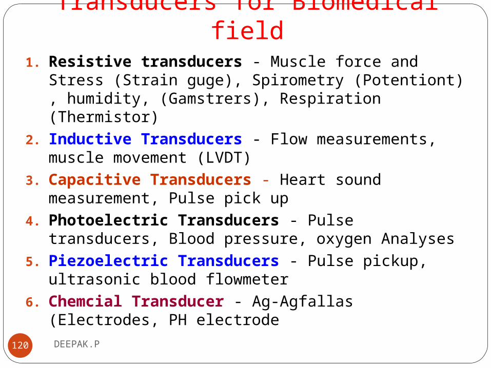
Transducers for Biomedical field1. Resistive transducers - Muscle force and Stress (Strain guge),
Spirometry (Potentiont) , humidity, (Gamstrers), Respiration (Thermistor)
2. Inductive Transducers - Flow measurements, muscle movement (LVDT)
3. Capacitive Transducers - Heart sound measurement, Pulse pick up
4. Photoelectric Transducers - Pulse transducers, Blood pressure, oxygen Analyses
5. Piezoelectric Transducers - Pulse pickup, ultrasonic blood flowmeter
6. Chemcial Transducer - Ag-Agfallas (Electrodes, PH electrode
120 DEEPAK.P

DEEPAK.P121
pH Electrode

pH ElectrodeThis is a device for measuring the concentration of hydrogen
ions and hence the degree of acidity of a solution.
pH is defined as the negative logarithm of the hydrogen ion
concentration.
pH=7 means a concentration of 1x10⁻⁷ moles per litre.
The most essential component of a pH electrode is a special,
sensitive glass membrane which permits the passage of
hydrogen ions, but no other ionic species.
122 DEEPAK.P

pH ElectrodeWhen the electrode is immersed in a test solution containing
hydrogen ions the external ions diffuse through the membrane
until an equilibrium is reached between the external and internal
concentrations.
Thus there is a build up of charge on the inside of the membrane
which is proportional to the number of hydrogen ions in the
external solution.The potential difference developed across the membrane is in
fact directly proportional to the Logarithm of the ionic concentration in the external solution.
123 DEEPAK.P

pH ElectrodeThe relationship between the ionic concentration
(activity) and the electrode potential is given by the
Nernst equation:E = E0 + (2.303RT/ nF) x Log(A)Where E = the total potential (in mV) developed between the sensing and reference electrodes.
E0 = is a constant which is characteristic of the particular ISE/reference pair.
(It is the sum of all the liquid junction potentials in the electrochemical cell, see later)
2.303 = the conversion factor from natural to base10 logarithm.
R = the Gas Constant (8.314 joules/degree/mole).
T = the Absolute Temperature.
n = the charge on the ion (with sign).
F = the Faraday Constant (96,500 coulombs).
Log(A) = the logarithm of the activity of the measured ion.
124 DEEPAK.P

pH Electrode
125 DEEPAK.P
DEEPAK.P126
Agcl Electrode

Ag-Agcl Electrode
127 DEEPAK.P

Ag-Agcl ElectrodeA silver chloride electrode is a type of reference electrode,
commonly used in electrochemical measurements.
The silver/silver chloride reference electrode is a widely used reference electrode because it is simple, inexpensive, very stable and non-toxic.
Typical laboratory electrodes use a silver wire that is coated with a thin layer of silver chloride either by electroplating or by dipping the wire in molten silver chloride.
128 DEEPAK.P

Ag-Agcl ElectrodeThe electrode functions as a redox electrode and
the reaction is between the silver metal (Ag) and its salt — silver chloride (AgCl, also called silver(I) chloride).
129 DEEPAK.P