degradation dna rna ribonuclease h anddna polymerases cellular
TRANSCRIPT

Proc. Nat. Acad. Sci. USAVol. 69, No. 11, pp. 3360-3364, November 1972
Degradation of DNA RNA Hybrids by Ribonuclease H and DNAPolymerases of Cellular and Viral Origin
(RNA tumor viruses/DNA synthesis)
WALTER KELLER* AND ROBERT CROUCHt* Cold Spring Harbor Laboratory, Cold Spring Harbor, Long Island, New York 11724; and t Laboratory of Molecular Genetics,National Institute of Child Health and Human Development, National Institutes of Health, Bethesda, Maryland 20014
Communicated by Barbara McClintock, September 7, 1972
ABSTRACT Ribonuclease H from human KB cells,chick embryos, calf thymus, avian myeloblastosis virus,and Rous associated virus specifically degrades the RNAof DNA RNA hybrids, producing mono- and oligoribo-nucleotides terminated in 5'-phosphates. The cellularRNase H is an endonuclease, whereas the viral enzymeappears to be an exonuclease. Viral DNA polymerase andRNase H copurify through all separation steps. Therefore,RNase H activity is an intrinsic part of the viral DNApolymerase. DNA RNA hybrids are also degraded by nu-cleases associated with cellular DNA polymerases and byexonuclease III. However, these nucleases differ fromRNase H in their ability to degrade both strands of DNARNA hybrids.
Ribonuclease H (RNase H), an enzyme originally discoveredin calf-thymus tissue, specifically degrades the RNA strandof DNA.RNA hybrids to acid-soluble products (1, 2). Re-cently, Mdlling et al. (3) demonstrated an RNase H activityin partially purified DNA polymerase preparations of avianmyeloblastosis virus (AMV), and suggested a possible in-volvement of this nuclease in the RNA-directed synthesis ofviral DNA. As we reported earlier (4), RNase H can remove5'-terminal RNA, covalently linked to DNA, that had beensynthesized in vitro. On this basis, we suggested a possiblerole of RNase H-type activities in RNA-primed DNA syn-thesis in vivo. In the model of M6lling et al. (3), RNase H wasconceived to degrade the template RNA after its transcrip-tion into RNA-DNA hybrid, releasing single-stranded DNA,which could then be converted into double-stranded productDNA.As a first step in testing this hypothesis, we determined the
specificity and mode of action of the RNase H found in avianRNA tumor viruses and compared these properties withthose of RNase H from uninfected cells. In addition, we at-tempted to decide whether the nuclease activity associatedwith the viral DNA polymerase constitutes an intrinsic partof this enzyme or whether it represents contaminating host-cell RNase H.
MATERIALS AND METHODS
Reagents. Unlabeled ribonucleoside triphosphates anddeoxyribonucleoside triphosphates came from Calbiochemand Schwartz/Mann BioResearch, respectively; [a-82P]ATPwas purchased from New England Nuclear Corp., and [a-32p]-dATP and [a-'2P]dTTP were from International Chemicaland Nuclear Corp. Poly(dA) oligo(dT) and poly(rA) oligo-
(dT) were prepared by incubation of poly(dA) or poly(rA)(both from Miles Laboratories) at a concentration of 0.2mg/ml with 0.05 mg/ml of oligo(dT) (chain length: 12-18nucleotides, Collaborative Research, Inc.) in 0.1 M NaCl-1 mM Na-EDTA (pH 7.6), for 30 min at 250.
Enzymes. DNA-Dependent RNA polymerase from Esche-richia coli was purified by the procedure of Berg et al. (5), andwas a gift from Dr. Michael Cashel. Exonuclease III fromE. coli (6, 7) was a gift from Dr. Malcolm Gefter. DNA poly-merase I from E. coli [fraction VII of Jovin et al. (8)] waspurchased from Boehringer Mannheim Corp. DNA poly-merase I, (9, 10), DNA polymerase II, (9, 11), and RNase Hfrom KB cells, as well as RNase H from 9-day-old chickembryos (Truslow Farms Inc., Chestertown, Md.), werepurified as described (4). Both preparations of RNase H wereabout 50% pure as judged by polyacrylamide gel electro-phoresis in the presence of 0.1% sodium dodecyl sulfate (12).DNA polymerase (RNA-dependent) from avian myeloblastosisvirus (kindly provided by Drs. Dorothy and J. Beard) wasisolated and purified, essentially as described by Kacian et al.(13), by chromatography on DEAE-cellulose, phosphocellu-lose, and Sephadex G-150. DNA polymerase (RNA-dependent)from Rous associated virus (RAV-1) (a gift from Dr. JohnBader) was extracted as described (13), and was purified bysucrose gradient sedimentation and chromatography onphosphocellulose. RNase H from calf thymus was a gift fromDr. Roy C. Haberkern.
Substrates. Poly(dT) .poly([32P]rA) was synthesized in areaction mixture containing in 0.5 ml: 0.05 M Tris-HCl (pH7.9), 5 mM MgCl2, 1 mM MnCl2, 1 mM dithiothreitol, 0.1mM [a-_2P]ATP (1.2 Ci/mmol), 50 ,ug of poly(dT), 50 ,ug ofDNA-dependent RNA polymerase (E. coli), and 5% of glyc-erol.
Poly( [32PIdT. (rA) was synthesized in a reaction mixturecontaining in 0.5 ml: 0.05 M Tris HCl (pH 7.9), 5 mM Mg-C12, 0.02 M KCl, 1 mM dithiothreitol, 0.04 mM [a-32P]dTTP(1 Ci/mmol), 5 ,ug of poly(rA) - oligo(dT), about 10 ,ug of RNA-dependent DNA polymerase (AMV), and 5% of glycerol.
Poly(['2P]dT. (dA) was synthesized in a reaction mixturecontaining in 0.5 ml: 0.05 M Tris HC1 (pH 7.9), 5mM MgC12,0.06 M KCl, 1 mM dithiothreitol, 0.04 mM [a-'2P]dTTP(1 Ci/mmol), 5 ,g of poly(dA) * oligo(dT), 20 ,g of KB DNApolymerase II, and 5% of glycerol.
After incubation for 30 min at 370, the reaction mixtureswere extracted with phenol (saturated with 1 M Tris*HCl,
3360
Abbreviations: AMV, avian myeloblastosis virus; RAV, Rousassociated virus.
Proc. Nat. Acad. Sci. USA 69 (1972)
pH 7.5) and chloroform-isoamylalcohol 24:1. The materialin the aqueous phase was precipitated with ethanol, and waspurified on a 1 X 25 cm column of Sephadex G-75 equili-brated with 0.05 .M NaCl-0.01 M Tris HCl (pH 7.5)-0.1 mMEDTA.
Covalently-closed, circular DNA of the colicinogenic factorE1 (Col El) of E. coli was a kind gift of Drs. Peter Williamsand Donald Helinski. The DNA was extracted from chloram-phenicol-treated cells. About 70% of the molecules containedone or more ribonucleotides inserted at a single site in oneof the two complementary strands (14). The Col El DNA waslabeled with 32p and had a specific activity of about 100,000cpm/lg.
RESULTS
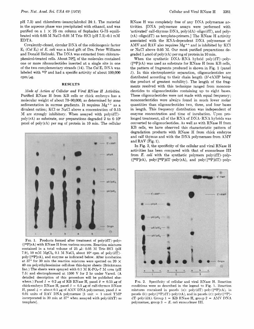
Mode of Action of Cellular and Viral RNase H Activities.Purified RNase H from KB cells or chick embroys has amolecular weight of about 70-90,000, as determined by zonesedimentation in sucrose gradients. It requires Mig++ as adivalent cation; KCl or NaCl above a concentration of 0.15M are strongly inhibitory. When assayed with poly(dT).poly(rA) as substrate, our preparations degraded 2 to 6 106pmol of poly(rA) per mg of protein in 10 min. The cellular
FIG. 1. Products formed after treatment of poly(dT)-poly-([32P]rA) with RNase H from various sources. Reaction mixturescontained in a total volume of 25 Al: 0.05 'AI Tris HCl (pH7.9), 10 mM AgCl2, 0.1 M NaCl, about 104 cpm of poly(dT)-poly( [32P]rA), and enzyme as indicated below. After incubationat 370 for 30 min the reaction mixtures were spotted on 20 X40 cm polyethyleneimine cellulose thin-layer sheets (BrinkmannInc.) The sheets were sprayed with 0.1 M K-PO4-7 MI urea (pH7.5) and electrophoresed at 1500 V for 2 hr under Varsol. (Adetailed description of this procedure will be published else-where.) Panel 1 = 0.2 jig of KB RNase H, panel 2 = 0.35 lig ofchick-embryo RNase H, panel 3 = 0.5 s4g of calf-thymus RNaseH, panel 4 about 0.1 jig of AYIV DNA polymerase, panel 5 =0.01 units of IRAY' DNA polymerase [1 unit = 1 nmol TMPincorporated in 30 min at 370 when assayed with poly(dAT) astemplate].
RNase H was completely free of any DNA polymerase ac-tivities. [DNA polymerase assays were performed with'activated' calf-thymus DNA, poly(dA) oligo(dT), and poly-(rA) oligo(dT) as template-primers.] The RNase H activityassociated with the RNA-dependent DNA polymerase ofAMV and RAV also requires Mg++ and is inhibited by KClor NaCl above 0.05 MI. Our most purified preparations de-graded 1 ,mol of poly(rA) per mg of protein in 10 min.When the synthetic DNA RNA hybrid poly(dT) poly-
([32P]rA) was used as substrate for RNase H from KB cells,the pattern of fragments produced is shown in Fig. 1 (panel1). In this electrophoretic separation, oligonucleotides aredistributed according to their chain length (5'-rA.MIP beingthe product of greatest mobility). The length of the frag-ments resolved with this technique ranged from mononu-cleotides to oligonucleotides containing up to eight bases.These oligonucleotides were not made with equal frequency;mononucleotides were always found in much lower molarquantities than oligonucleotides two, three, and four basesin length. This frequency distribution was independent ofenzyme concentration and time of incubation. Upon pro-longed treatment, all of the RNA of DNA RNA hybrids wasconverted to oligonucleotides. As well as with RNase H fromKB cells, we have observed this characteristic pattern ofdegradation products with RNase H from chick embryosand calf thymus and with the DNA polymerases from AMVand RAV (Fig. 1).
In Fig. 2, the specificity of the cellular and viral RNase Hactivities has been compared with that of exonuclease IIIfrom E. coli with the synthetic polymers poly(dT) poly-([32p rA), poly( [32P]dT * poly(rA), and poly( [32P]dT) * poly-
FTG. 2. Specificity of cellular and viral RNase H. Reactionconditions were as described in the legend to Fig. 1. Reactionmixtures contained in panels (a): poly(dT) poly( [32P] rA), inpanels (b): poly([32P]dT).poly(rA), and in panels (c): poly([32p]-dT poly(dA). Group 1 = KB RNase H, group 2 = A-\VI' DNApolymerase, group 3 = E. coli exonuclease III.
* i---- 5~rAMP!
* ~~* - pApA
0~~~~~~~0pAXpA)4
w | ~~~~PA (PA),
- Origin
2 3 4 5
5'TMP
5'rAMP 5'rAMP |
* ------pApA-- *
*9 -pA(pA)3-
pA(pA43 A
******|* *-O Origin
a b c a b c a b cI~~~~~~ 3S
Cellular and Viral RNase H 3361

3362 Biochemistry: Keller and Crouch
23S 17SA I I
It
I I.
10 20 30 10 20 34
Fraction number0 10 20 30
FIG. 3. Sedimentation analysis of the DNA of the plasmid Col E, after treatment with cellular and viral RNase H. Incubationmixtures contained in 0.1 ml: 0.05 M Tris -HCl (pH 7.9), 10 mM MgC12, 20 mM NaCl, about 3000 cpm [32P]DNA of Col E1 from E.coli grown in the presence of chloramphenicol (14), about 600 cpm of SV40 [3H]DNA, and enzyme. After incubation for 30 min at 370,0.1 ml of 0.1 M Na-EDTA (pH 7.5) was added and the mixtures were layered on 5-20% sucrose gradients containing 0.05 Tris HCl(pH 7.9)-0.55 M NaCl-55 mM EDTA. The gradients were centrifuged for 3 hr at 45,000 rpm and 15° in a Spinco SW56 rotor. Fractionswere collected from the bottom of the centrifuge tube and counted in 'Aquasol'. Panel A: no enzyme, panel B: incubation with 0.1 jug ofchick-embryo iRNase H, panel C: incubation with 8 /Ag of AMV DNA polymerase.
(dA) as substrates. The RNase H from KB cells and AMVexclusively degraded the RNA strand of synthetic hybrids.Exonuclease III, in contrast, could attack both the DNA andthe RNA of these hybrids. In addition, exonuclease III pro-
duced mainly mononucleotides as products.To determine whether the cleavage by RNase H activities
occurs at the 5' or the 3' side of the phosphodiester bond, we
subjected the oligonucleotides formed after incubation ofpoly(dT) - poly(rA) to alkaline hydrolysis and subsequentchromatography. As illustrated in Table 1, the oligonucleo-tides generated by both cellular and viral RNase H were
terminated with a 5'-phosphate, as revealed by the formationof pAp and Ap upon alkaline hydrolysis. The production of5'-phosphate-terminated oligonucleotides by RNase Hactivities has also been demonstrated by D. Baltimore andD. F. Smoler for AMV DNA polymerase (21a) and by R. C.
8-~~~~~S
10 20 6 8 1II 14FrmCtol.numbrFIG. 4. Cosedimentation of viral DNA polymerase and RNase
H. About 0.2 mg of AMV DNA polymerase (phosphocellulosefraction) was layered on a gradient -containing 5-20% sucrose,
0.2 M KPO4 (pH 7.5), 2 mM dithiothreitol, 0.1 mM EDTA, and10% glycerol and centrifuged for 19 hr at 55,000 rpm in a SpincoSW56 rotor at 4°. 0.2-ml Fractions were collected from the bot-tom of the tube. DNA-polymerase activity was assayed (4) withpoly(rA)-oligo(dT) as template. RNase H was detected in 2-/Alaliquots as described in the legend to Fig. 1. A 0.2-ml aliquot ofthe combined peak fractions was subjected to polyacrylamide(5%) gel electrophoresis in the presence of 0.1% sodium dodecylsulfate (12). The gel pattern obtained is shown in the insert ofthe left panel. BSA indicates the position of bovine-serum albu-min, centrifuged in a separate tube.
Haberkern and G. L. Cantoni for the calf-thymus enzyme
(personal communications). Using instead of the homopolymerhybrid poly(dT) poly(rA), DNA RNA hybrid made withDNA of phage 4X174 as template, we found that RNase Hcould cleave phosphodiester bonds next to any of the fourribonucleotides (unpublished results).The formation of oligoribonucleotides as degradation
products by cellular and viral RNase H suggested that bothactivities operate in an endonucleolytic manner. To test thismechanism more directly, we took advantage of the closed-circular superhelical DNA of the plasmid Col El as a sub-strate for RNase H. When this plasmid replicates in thepresence of chloramphemicol, its DNA contains one or more
ribonucleotides covalently inserted in one of the two DNAstrands, as shown by Blair et al. (14). Fig. 3 shows a sedi-mentation analysis of Col El DNA that had been treated withchick-embryo RNase H (panel B) or the RNase H associatedwith AMV DNA polymerase (panel C). The intact, co-
valently-closed Cot El DNA (4.2 X 10⁶ daltons) sediments at23 S, slightly faster than superhelical SV40 DNA (3 X 10⁶daltons), used as a marker (Fig. 3, panel A). Upon treatmentwith chick-embryo RNase H, about 70% of the Col E1 DNAwas converted into a form that sedimented at 17 S, which ischaracteristic of open circles. Incubation with RNase H fromKB cells resulted in the same sedimentation pattern (notshown).
(Alkali treatment of the same Col El DNA preparationgave the identical result, indicating that 70% of the DNAmolecules contained ribonucleotides.) In contrast to thecellular RNase H, the nuclease associated with viral DNApolymerase did not convert any of the Col El DNA into theopen-circular form even when used at very high concentra-tions. On this basis we conclude that the viral RNase Hrequires RNA with free ends to initiate its nucleolytic actionand appears, therefore, to be an exonuclease. Leis et al.(28) using a different approach, came to the same conclusion.The results illustrated in Fig. 3 also indicate that both cellu-lar and viral enzymes are completely free of DNase (endo-nuclease) activity that would convert superhelical SV40 DNAinto a slower-sedimenting open-circular form.
I4
02
E
9
E
0
B
I'tI'
2 ~~~~~~~~~~It2 ~~~~~~~~~~~~~~~~~~~~~~~~~~~~~~~~~~~~~II
I'
I~~~~~~~I
I1
3
2
Proc. Nat. Acad. Sci. USA 69 (1972)

Cellular and Viral RNase H 3363
TABLE 1. The presence of 5' phosphate in oligoriboncleotidesproduced by treatment of poly(dT) -poly([32P]rA) with
RNase H from various sources
Source ofRNase H
Radioactivitymoving fromorigin afterdigestion ofpoly(dT) -
poly(rA)(%)
IFractionof 32p
in pAp (%o)
* *I 4 I ~ -------- 5'rAMP
SKB cells 97 37Calf thymus 99 33Chick embryos 99 29AMV 51 8RAV 46 4None 0 0
The reaction conditions were as described in Fig. 1, except thatthe concentration of enzyme from KB cells, calf thymus, andchick embryos was tripled. After 30 min at 370, 5 ,Al of 4 N KOHwas added to the reaction mixtures and the samples were in-cubated at 370 for 18 hr. After neutralization of the mixturewith 4 N HCl04, Ap was separated from pAp by two-dimensionalchromatography on polyethyleneimine thin layers by the LiCl-NaCOOH method (27). The compounds were localized by auto-radiography and their radioactivity was measured in a liquidscintillation counter.
Relationship of Viral RNase H to the Host Enzyme. Asshown above, the cellular RNase H differs from the viralenzyme by its ability to cleave superhelical Col El DNAcontaining inserted ribonucleotides. To demonstrate furtherthat the viral RNase H activity was not simply due to con-tamination by cellular enzyme, we purified the virus-asso-ciated nuclease by chromatography on DEAE-cellulose andphosphocellulose, and by sucrose gradient sedimentation,essentially following the procedure of Kacian et al. (13) forthe purification of A'MV DNA polymerase. Throughout thesesteps, the profile of RNase H activity followed closely that ofthe DNA polymerase (Fig. 4). Upon analysis by polyacryl-amide electrophoresis in the presence of 0.1% sodium dodecylsulfate, our purified enzyme preparation revealed two poly-peptides with molecular weights of about 100,000 and 70,000,in accord with the report of Kacian et al. (13) (Fig. 4). Theseauthors presented strong evidence that the two polypeptidesrepresent subunits of the viral DNA polymerase. Therefore,it is very likely that the viral RNase H activity also resideswithin one of these two subunits. The cellular and viral RNaseH activities could also be clearly distinguished by their dif-ferent sedimentation rates in sucrose gradients. The viralDNA polymerase-RNase H complex sediments at 7 S,whereas the host RNase H sediments at 5 S (results notshown). We can thus conclude that the RNase H activityconstitutes a specific component of viral DNA polymerase.
Degradation of DNA -RNA Hybrids by Cellular DNAPolymerases. The discovery that AMV DNA polymerase isassociated with RNase H (3), and our finding that the hostRNase H and the DNA polymerase-associated nuclease fromAMIV produce similar products, led us to investigate otherDNA polymerases for RNase H activity. Fig. 5 shows thatDNA polymerase I of E. coli and DNA polymerases I and IIfrom KB cells were all capable of degrading the RNA strand
I
; ..l~~~~~~~~~~~~~~~~~~~~~~~~~~~~~~~~~~~~~~~~~~~~~~~.f
*------- Origin
3<12 3 4
FIG. 5. Degradation of poly(dT) poly( [32P] rA) by variousDNA polymerases and exonuclease III. Reaction mixtures wereas described in the legend to Fig. 1. Panel 1 = 2 /Ag of E. coliDNA polyrmerase I, panel 2 = 5 jig of KB DNA polyrmerase I,panel 3 = 5 lig of KB DNA polymerase II, panel 4 = 5 jug ofE. coli exonuclease III.
of poly(dT) poly([32P]rA). It is interesting to note that E.coli DNA polymerase I and KB DNA polymerase I generated,in addition to mononucleotides as main products, oligoribonu-cleotides up to four bases long. The ability of E. coli DNApolymerase I to degrade DNA. RNA hybrids has also beenobserved by L. Bertsch and A. Kornberg (personal communi-cation). The degradative processes observed with cellularDNAlpolymerases differed from those observed with cellularand viral RNXase H in two important aspects: (i) these en-zymes were not specific for the RNA strand of D.NA. RNAhybrids, but degraded the DNA strand as well (results notshown) and (ii) the products formed were mainly mononu-cleotides. The latter finding suggests an exonucleolytic modeof action, similar to that shown for exonuclease III of E. coli(Figs. 2 and 5).
DISCUSSION
Mdolling et al. (3) found that when an extract of AMYIV waschromatographed on DEAE-sephadex or subjected to zone
3
_ON (I)
3 i f fi1~ ll lll llllll (2)
3' i~ (4)31
_' 3' (5)
3' 1'';'''; ll'"l'l'! H 7
3' (3)
FIG. 6. Hypothetical involvement of RNase H activities inthe synthesis of double-stranded DNA on viral IRNA as template.See text for explanations. Thick-strokc lines represent RNA,thi n-stroke lines represent, DNA.
Proc. Nat. Acad. Sci. USA 69 (1972)

3364 Biochemistry: Keller and Crouch
sedimentation in sucrose gradients, a nuclease capable ofdegrading the RNA of DNA-RNA hybrids accompanied thepeaks of viral DNA polymerase. These activities however,were not more extensively purified.Our results presented here show that DNA polymerase from
AMV, when purified to apparent homogeneity, still retainsRNase H activity. The viral nuclease can be distinguishedfrom the cellular enzyme by its different mode of action andits faster sedimentation rate. These observations support thesuggestion of a specific association of RNase H-type activitywith viral DNA polymerase, as proposed by M6lling et al.The inability of the viral RNase H to cleave the internal
ribonucleotides of Col El DNA suggests that its mode ofaction must be exonucleolytic i.e., the enzyme seems to re-quire RNA with free ends to initiate its degradative action.The finding that the products are not mononucleotides, butare oligonucleotides of various chain lengths, seems at firstsight difficult to reconcile with an exonucleolytic process.However, as has recently been shown for the coli-phage T5-induced nuclease (15) and for the rec BC nuclease of E. coli(16), oligonucleotides can be the main degradation productsof exonucleases. As for the cellular RNase H, our resultsclearly show that this enzyme can act endonucleolytically,but we cannot exclude the possibility that it also has anexonucleolytic activity. It is conceivable that the enzymemakes endonucleolytic cleavages and then proceeds alongits substrate as an exonuclease.
It is not clear where the RNase H that is associated withthe viral DNA polymerase originates. It is conceivable that itis derived from the host RNase H, possibly in a manneranalogous to Qf3-replicase, where only one of the four sub-units of the enzyme is specified by the phage, the other threesubunits being host proteins [see Blumenthal et al. (17) forreferences]. If this mechanism is correct, we must assumethat the host RNase H loses its endonuclease function as aresult of association with the viral DNA polymerase. On theother hand, it is equally possible that the viral RNase H is anew activity, unrelated to the host enzyme. We are currentlydoing reconstitution experiments on the subunits of the viraland cellular enzyme in order to obtain a more accurate correla-tion.The mechanism of double-stranded DNA synthesis in
virus-infected cells is still not understood. How could RNaseH-type activities be involved in this process? Experimentswith detergent-treated virus have shown that the reactionproceeds in two stages. First, the viral RNA is transcribedinto a RNA DNA hybrid (18-20). Low molecular weightRNA, complementary to the 70S viral RNA, seems to serveas a primer in this reaction (21b, 22). In the second stage, thenewly synthesized DNA strand is copied, resulting in double-stranded DNA as final product. It is at this stage of the reac-tion that we would suggest the participation of both cellularand viral RNase H activities. As shown in the diagram of Fig.6, cellular RNase H could introduce a gap (gaps) near the5'-end of the template RNA after it has been copied into anRNA *DNA hybrid. This would create new primers withfree 3'-hydroxyl groups for the viral DNA polymerase. As theviral DNA polymerase proceeds to copy the template DNA,it could in a simultaneous reaction remove any remainingviral RNA via its RNase H activity, resulting in double-stranded DNA. This is formally analogous to the 'nick-trans-lation' reaction of E. coliDNA polymerase I (23, 24).
We made no attempt to further characterize the nucleasesassociated with E. coli DNA polymerase I and KB DNApolymerases I and II that are responsible for the degradationof poly(dT) - poly(rA) (Fig. 5), but it seems likely that theseare exonuclease activities that have been described (24-26).The absence of these exonucleases in viral DNA polymerasesprovides an additional criterion for the distinction of these'reverse transcriptase' activities from ordinary DNA poly-merases.
We thank Dr. J. D. Watson for his support and help in thepreparation of the manuscript and Drs. D. Baltimore and J.Hurwitz for helpful discussions. We are very grateful to Drs.John Bader, Dorothy and Joseph Beard, Michael Cashel,Donald Helinski, and Peter Williams for providing us with virus,enzymes, and DNA preparations used in our experiments. Thiswork was supported by Grant 1 P01 CA 13106-01 from theNational Cancer Institute.1. Stein, H. & Hausen, P. (1969) Science 166, 393-395.2. Hausen, P. & Stein, H. (1970) Eur. J. Biochem. 14, 278-283.3. Molling, K., Bolognesi, D. P., Bauer, H., Busen, W., Plass-
mann, H. W. & Hausen, P. (1971) Nature New Biol. 234,240-243.
4. Keller, W. (1972) Proc. Nat. Acad. Sci. USA 69, 1560-1564.5. Berg, D., Barrett, K. & Chamberlin, M. (1971) in Methods in
Enzymology, eds. Grossmann, L. & Moldave, K. (AcademicPress, New York), Vol. XXI, pp. 506-519.
6. Richardson, C. C. & Kornberg, A. (1964) J. Biol. Chem. 239,242-250.
7. Richardson, C. C., Lehman, I. R. & Kornberg, A. (1964) J.Biol. Chem. 239, 251-258.
8. Jovin, T. M., Englund, P. T. & Bertsch, L. L. (1969) J. Biol.Chem. 244, 2996-3008.
9. Weissbach, A., Schlabach, A., Fridlender, B. & Bolden, A.(1971) Nature New Biol. 231, 167-170.
10. Chang, L. M. S. & Bollum, F. J. (1971) J. Biol. Chem. 246,5835-5837.
11. Yoneda, M. & Bollum, F. J. (1965) J. Biol. Chem. 240,3385-3391.
12. Maizel, J. V. (1969) in Fundamental Techniques of Virology,eds. Habel, K. & Salzman, N. P. (Academic Press, NewYork), pp. 334-362.
13. Kacian, D. L., Watson, K. F., Burny, A. & Spiegelman, S.(1971) Biochem. Biophys. Acta 246, 365-383.
14. Blair, D. G., Sherratt, D. J., Clewell, D. B. & Helinski, D.R. (1972) Proc. Nat. Acad. Sci. USA 69, 2518-2522.
15. Frenkel, G. D. & Richardson, C. C. (1971), J. Biol. Chem.246, 4839-4847.
16. Goldmark, P. J. & Linn, S. (1972) J. Biol. Chem. 247, 1849-1860.
17. Blumenthal, T., Landers, T. A. & Weber, K. (1972) Proc.Nat. Acad. Sci. USA 69, 1313-1317.
18. Fanshier, L., Garapin, A. C., McDonnell, J., Faras, A., Lev-inson, W. & Bishop, J. M. (1971) J. Virol. 7, 77-86.
19. Verma, I. M., Meuth, N., Bromfeld, E., Manly, K. F. &Baltimore, D. (1971) Nature New Biol. 233, 131-134.
20. Taylor, J. M., Faras, A. J., Varmus, H. E., Levinson, W. E.& Bishop, J. M. (1972) Biochemistry 11, 2343-2351.
21a. Baltimore, D. & Smoler, D. F. (1972) J. Biol. Chem., in press.21b. Manly, K. F., Smoler, D. F., Bromfeld, E. & Baltimore, D.
(1971) J. Virol. 7, 106-111.22. Canaani, E. & Duesberg, P. (1972) J. Virol. 10, 23-31.23. Kelly, R. B., Cozzarelli, H. R., Deutscher, M. P., Lehman, I.
R. & Kornberg, A. (1970) J. Biol. Chem. 245, 39-45.24. Kornberg, A. (1969) Science 163, 1410-1418.25. Roychoudhury, R. & Block, D. P. (1969) J. Biol. Chem. 244,
3359-3368.26. Greene, R. & Korn, D. (1970) J. Biol. Chem. 245, 254-261.27. Randerath, K. & Randerath, E. (1967) in Methods in Enzy-
mology, eds. Grossman, L. & Moldave, K. (Academic Press,New York), Vol. XII A, pp. 323-347.
28. Leis, J., Berkower, I. & Hurwitz, J. (1972) in 2nd AnnualSteenbock Symp., "DNA synthesis in vitro," eds. Wells, R.& Inman, R., University Park Press, Baltimore, Md.
Proc. Nat. Acad. Sci. USA 69 (1972)