demonstrated by richard hatchett, deputy head of school
TRANSCRIPT
Newborn (resting)Infant (resting)Child 2-6 yearsChild 6-12 yearsAdolescent-adult
100-180 80-150 75-120 70-110 60-90
Page 1 of 5
ObservationsAdults
Assessing the pulseDemonstrated by Richard Hatchett, Deputy Head of School, The Royal Marsden School
©2015 Clinical Skills Limited. All rights reserved
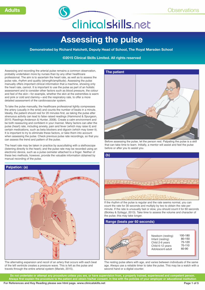
Assessing and recording the arterial pulse remains a common observation, probably undertaken more by nurses than by any other healthcare professional. The aim is to ascertain the heart rate, as well as to assess the pulse rate, rhythm and quality (strength/amplitude). Assessing the pulse manually offers important clinical information that a machine, showing only the heart rate, cannot. It is important to use the pulse as part of an holistic assessment and to consider other factors such as blood pressure, the colour and feel of the skin—for example, whether the skin at the extremities is warm and pink or cold and clammy—and the respiratory rate, to offer a more detailed assessment of the cardiovascular system.
To take the pulse manually, the healthcare professional lightly compresses the artery (usually in the wrist) and counts the number of beats in a minute. Ideally, the patient should rest for 20 minutes first, as taking the pulse after strenuous activity can lead to false raised readings (Hammond & Spurgeon, 2015; Rawlings-Anderson & Hunter, 2008). Create a calm environment and be both reassuring and confident in your manner. Many factors can alter the pulse (heart) rate, including anxiety, pain and fever (which may raise it) and certain medications, such as beta blockers and digoxin (which may lower it). It is important to try to eliminate these factors, or take them into account when assessing the pulse. Check previous pulse rate recordings, so that you can assess the trend and pattern of the pulse.
The heart rate may be taken in practice by auscultating with a stethoscope (listening directly to the heart); and the pulse rate may be recorded using an electronic device, such as a pulse oximeter attached to a finger. Neither of these two methods, however, provide the valuable information obtained by manual recording of the pulse.
Palpation: (a)
(b)
Do not undertake or attempt any procedure unless you are, or have supervision from, a properly trained, experienced and competent person.Always first explain the procedure to the patient and obtain his/her consent, in line with the policies of your employer or educational institution
Before assessing the pulse, let the person rest. Palpating the pulse is a skill that can take time to learn. Initially, a mentor will assist and feel the pulse before or after you to assist you.
The resting pulse alters with age, and varies between individuals of the same age. Always use a reliable timer to take the pulse. This may be a watch with a second hand or a digital counter.
The patient
Range (beats per 60 seconds)
The alternating expansion and recoil of an artery that occurs with each beat of the left ventricle creates a pressure wave. This is felt as the pulse and travels through the entire arterial system (Marieb, 2015).
If the rhythm of the pulse is regular and the rate seems normal, you can count the rate for 30 seconds and multiply by two to obtain the rate per minute. If the rate is unusually fast or slow, you should count it for 60 seconds (Bickley & Szilagyi, 2013). Take time to assess the volume and character of the pulse; this may take longer.
For References and Key Reading please see html page. www.clinicalskills.net
Observations
Adults
Assessing the pulse Page 2
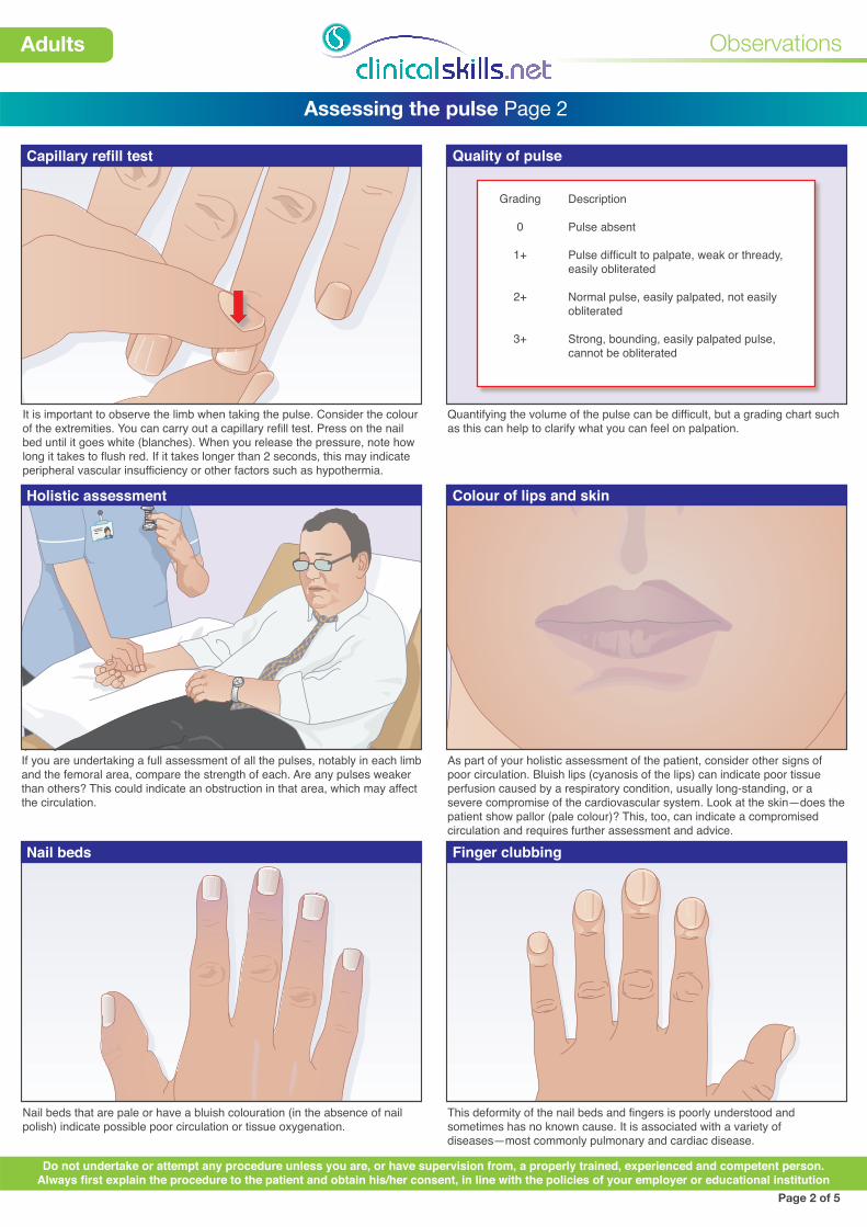
Capillary refill test Quality of pulse
It is important to observe the limb when taking the pulse. Consider the colour of the extremities. You can carry out a capillary refill test. Press on the nail bed until it goes white (blanches). When you release the pressure, note how long it takes to flush red. If it takes longer than 2 seconds, this may indicate peripheral vascular insufficiency or other factors such as hypothermia.
Quantifying the volume of the pulse can be difficult, but a grading chart such as this can help to clarify what you can feel on palpation.
Holistic assessment Colour of lips and skin
As part of your holistic assessment of the patient, consider other signs of poor circulation. Bluish lips (cyanosis of the lips) can indicate poor tissue perfusion caused by a respiratory condition, usually long-standing, or a severe compromise of the cardiovascular system. Look at the skin—does the patient show pallor (pale colour)? This, too, can indicate a compromised circulation and requires further assessment and advice.
If you are undertaking a full assessment of all the pulses, notably in each limb and the femoral area, compare the strength of each. Are any pulses weaker than others? This could indicate an obstruction in that area, which may affect the circulation.
Nail beds Finger clubbing
Nail beds that are pale or have a bluish colouration (in the absence of nail polish) indicate possible poor circulation or tissue oxygenation.
This deformity of the nail beds and fingers is poorly understood and sometimes has no known cause. It is associated with a variety of diseases—most commonly pulmonary and cardiac disease.
Page 2 of 5
Do not undertake or attempt any procedure unless you are, or have supervision from, a properly trained, experienced and competent person.Always first explain the procedure to the patient and obtain his/her consent, in line with the policies of your employer or educational institution
Description
Pulse absent
Pulse difficult to palpate, weak or thready, easily obliterated
Normal pulse, easily palpated, not easily obliterated
Strong, bounding, easily palpated pulse, cannot be obliterated
Grading
0
1+
2+
3+

Observations
Adults
Assessing the pulse Page 3
Carotid pulse
Common peripheral pulse points Radial pulse
Ulnar pulse
Listening directly to the heart (auscultation) can offer far more detail than the pulse alone. Concern over the rate, volume or character of the pulse may be an indication to auscultate the heart directly. (See also clinicalskills.net procedure on “Cardiovascular examination”.)
The carotid pulse is one of the last pulses to disappear in the event of cardiac arrest. It is therefore useful when it is unclear if there is a cardiac output in an unconscious individual. It is a notoriously difficult pulse to palpate. Current resuscitation guidelines suggest that the public, and healthcare professionals not used to palpating this pulse, should use other signs to assess whether there is a circulation, such as an unresponsive patient and the absence of breathing (Resuscitation Council, 2010).
Page 3 of 5
Do not undertake or attempt any procedure unless you are, or have supervision from, a properly trained, experienced and competent person.Always first explain the procedure to the patient and obtain his/her consent, in line with the policies of your employer or educational institution
To assess the pulse by palpating the radial artery in the wrist, use your fingers as shown. Do not use your thumb, as this has a pulse of its own (Marieb, 2015). The pulse is easily accessible here, offering a good guide to the heart and its regularity. As the radial pulse is some distance from the heart, its use to assess the character of the pulse, and its volume, can be limited.
The ulnar artery is occasionally used if it is difficult to assess the pulse using the radial artery.
There are many points in the body where it is possible to palpate the pulse. The radial artery is the most commonly used, but other pulses may be utilised to assess the circulation in various parts of the body.
Temporal artery
Facial artery
Carotid artery
Brachial artery
Radial artery
Ulnar artery
Femoral artery
Popliteal artery
Posterior tibial artery
Dorsalis pedis artery
Apical pulse
Observations
Adults
Assessing the pulse Page 4
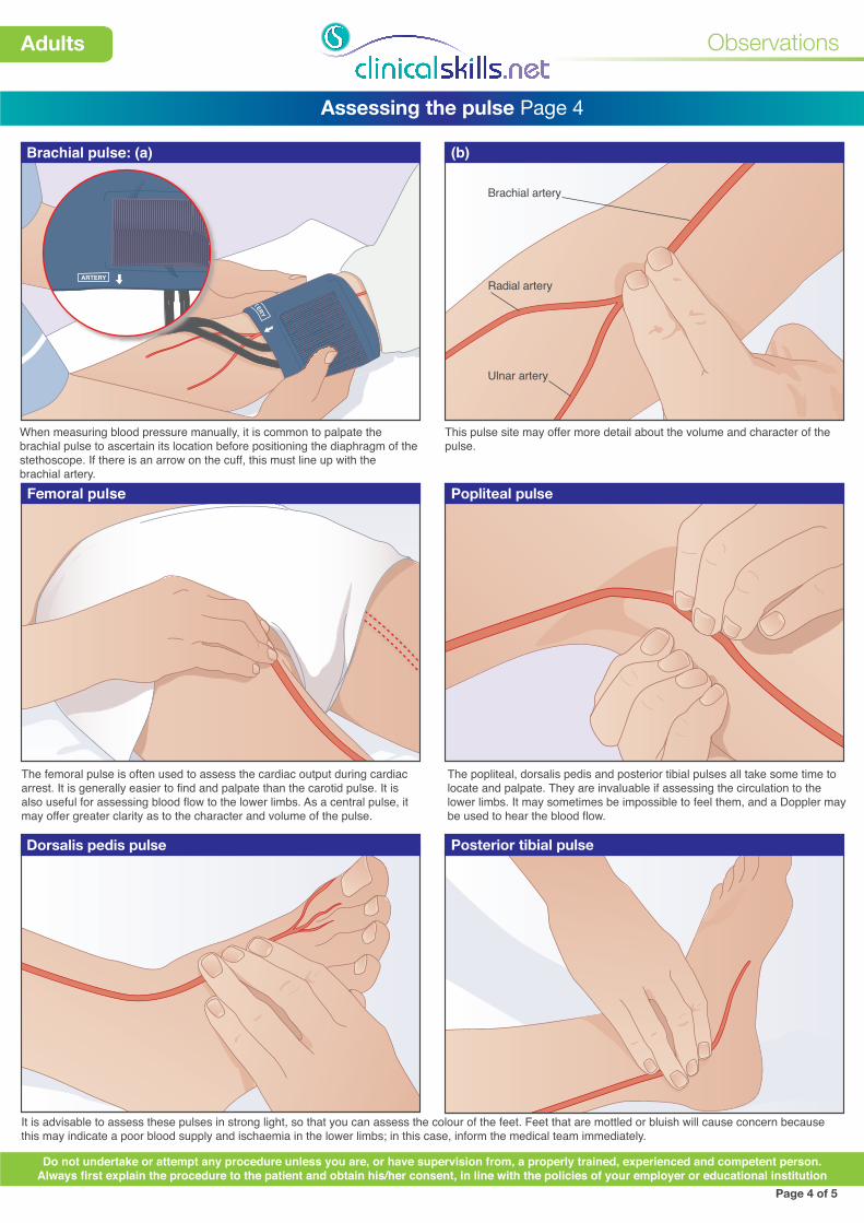
Dorsalis pedis pulse Posterior tibial pulse
Brachial artery
Radial artery
Ulnar artery
(b)
Popliteal pulse
It is advisable to assess these pulses in strong light, so that you can assess the colour of the feet. Feet that are mottled or bluish will cause concern because this may indicate a poor blood supply and ischaemia in the lower limbs; in this case, inform the medical team immediately.
Page 4 of 5
Do not undertake or attempt any procedure unless you are, or have supervision from, a properly trained, experienced and competent person.Always first explain the procedure to the patient and obtain his/her consent, in line with the policies of your employer or educational institution
This pulse site may offer more detail about the volume and character of the pulse.
ARTERY
Femoral pulse
When measuring blood pressure manually, it is common to palpate the brachial pulse to ascertain its location before positioning the diaphragm of the stethoscope. If there is an arrow on the cuff, this must line up with the brachial artery.
The popliteal, dorsalis pedis and posterior tibial pulses all take some time to locate and palpate. They are invaluable if assessing the circulation to the lower limbs. It may sometimes be impossible to feel them, and a Doppler may be used to hear the blood flow.
The femoral pulse is often used to assess the cardiac output during cardiac arrest. It is generally easier to find and palpate than the carotid pulse. It is also useful for assessing blood flow to the lower limbs. As a central pulse, it may offer greater clarity as to the character and volume of the pulse.
Brachial pulse: (a)
ARTERY
IND
EX L
INE

Observations
Adults
Assessing the pulse Page 5
Rhythm
Sinus arrhythmia
Tachycardia
The illustrations here help you consider the character of the pulse. Take the pulse for 60 seconds to assess the rhythm. If the patient is in sinus rhythm, the pulse should be regular and steady. This diagram, and the ones below, provide a visual representation of the pattern of the pulse, shown as the red line.
A slight elevation and decrease in pulse rate, coinciding with respiration, is normal in younger people. This phenomenon is called sinus arrhythmia.
Bradycardia
Irregular pulse
Fibrillation (ventricular)
For some, a slow heart rate may be normal and healthy, such as in a young, athletic person. Generally, bradycardia is defined as a heart rate slower than 60 beats/minute (Bickley & Szilagy, 2013; Resuscitation Council (UK), 2010; Riley, 2007), although some texts use a figure of 50 beats/minute. Importantly, assess the patient to ascertain the effect of a slow pulse, such as feeling unwell or dizzy, a drop in blood pressure or chest pain. A pulse below 40 beats/minute would be cause for concern in most patients. (Normal pulse shown as a beige line.)
The most common cause of an irregular pulse is atrial fibrillation. This is more common in the older patient. It is caused by continuous disorganised electrical activity in the atria. Electrical impulses pass intermittently to the ventricles, creating an irregular pulse. It may also occur when normal sinus rhythm is interrupted by ventricular extrasystoles (ectopics). These are premature beats occurring from within the ventricles.
Fibrillation refers to the chambers of the heart in electrical disarray. The result is uncoordinated contraction, affecting the atria or the ventricles. In atrial fibrillation (AF), the pulse rate is characteristically irregular. This is because the electrical activity in the atria varies; consequently the atrioventricular node conducts in an intermittent fashion, producing irregular ventricular contractions. Atrial fibrillation is a common long-term arrhythmia in the elderly. In ventricular fibrillation (VF), shown above, the ventricles are not conducting in a strong, steady fashion, so there is no pulse. The result is cardiac arrest: immediate defibrillation is required.
Page 5 of 5
Do not undertake or attempt any procedure unless you are, or have supervision from, a properly trained, experienced and competent person.Always first explain the procedure to the patient and obtain his/her consent, in line with the policies of your employer or educational institution
S =systole
D = diastole
mm
Hg
100
A rapid heart rate through the normal conduction pathway is known as sinus tachycardia. However, a fast pulse rate can be initiated by an electrical impulse outside of the sinoatrial node, and the tachycardia is denoted by the origin of the impulse, e.g. ventricular tachycardia. Sinus tachycardia is said to occur with pulse rates above approximately 100 beats/minute (Bickley & Szilagyi, 2013). (Normal pulse shown as a beige line.) Assess the patient for possible complications of tachycardia, such as shortness of breath or drop in blood pressure.
Time sec 0 3 4 5 6 7 8 9 10D
S S S S S S S S S S S S
D D D D D D D D D D D21