dengue outbreak is a global recurrent crisis: review of
TRANSCRIPT
Dengue Outbreak is a Global Recurrent Crisis: Review of the Literature
Yusha Araf1, Md. Asad Ullah2*, Nairita Ahsan Faruqui3, Sadrina Afrin Mowna3, Durdana
Hossain Prium3, Bishajit Sarkar2
1Department of Genetic Engineering and Biotechnology, School of Life Sciences, Shahjalal
University of Science and Technology, Sylhet, Bangladesh.
2Department of Biotechnology and Genetic Engineering, Faculty of Biological Sciences,
Jahangirnagar University, Dhaka, Bangladesh.
3Biotechnology Program, Department of Mathematics and Natural Sciences, BRAC
University, Dhaka, Bangladesh
Corresponding Author: Md. Asad Ullah*
Email of the corresponding author: [email protected]
Preprints (www.preprints.org) | NOT PEER-REVIEWED | Posted: 14 May 2020 doi:10.20944/preprints202004.0246.v2
© 2020 by the author(s). Distributed under a Creative Commons CC BY license.
Abstract
Purpose
This review features a generalized overview of dengue outbreaks, dengue pathogenesis,
symptoms, immune response, diagnosis methods and preventive measures which facilitates the
better understanding of the global expansion and concerns relating to the disease.
Recent Findings
A recent study showed that natural killer cells of the infected person become activated soon
after the infection which may help in treatment and vaccine development. A research team has
also produced synthetically engineered mosquitoes that can prevent the transmission and
dissemination of the dengue virus by the activation of an antibody. Furthermore, a mutation in
the protein envelope of the dengue virus leads to variation in shapes, developing resistance
towards the vaccine.
Summary
The increasing number of reported cases indicated the worldwide distribution of the mosquito
vectors, which was further facilitated by the growth in the shipping and commerce industries.
The immune system, through activation of the innate and adaptive immune responses,
facilitates the recruitment of an array of leukocytes which help neutralize the virus. However,
the 4 different viral serotypes increases the risk of a life-threatening secondary infection due to
the varying serotypes. Apart from the laboratory standard PRNT method, several other dengue
detection methods such as ELISA, RT-LAMP and several optical, microfluidic and
electrochemical methods have been developed. Since Dengvaxia® (CYD-TDV) has its own
set of drawbacks and limitations, several companies have been investing for the production of
more potential vaccines that are currently in trial.
Preprints (www.preprints.org) | NOT PEER-REVIEWED | Posted: 14 May 2020 doi:10.20944/preprints202004.0246.v2
Keywords: Dengue Hemorrhagic Fever (DHF); Aedes Aegypti; Epidemic; Pathogenesis;
identification; vaccine
1. Introduction
Dengue is one of the most rapidly spreading mosquito borne viral infections in humans leading
to about 10,000 deaths annually across over 125 countries in the world [1]. The spherical
enveloped dengue virus is a positive sense single stranded RNA virus. It genome consists of
about 10,200 nucleotides which codes for structural proteins (capsid (C), envelope (E), and
membrane (M) proteins) and nonstructural proteins (NS1, NS2A, NS2B, NS3, NS4A, NS4B,
and NS5) [2]. Dengue virus which is a member of the genus Flavivirus (family Flaviviridae)
are classified into four serotypes (DEN-1, DEN-2, DEN-3 and DEN-4) [3][4]. Mosquitoes of
the genus Aedes aid to transmit dengue virus from one human to another [5][6]. These
mosquitoes are ubiquitous in the tropical and subtropical regions from 30̊ north to 20̊ south
latitude. Escalation of dengue infection is associated with climatic changes which include an
increase in temperature, high levels of precipitation, humidity and the pressure of vapour. [7].
All these factors along with degree of globalization, trade and travel are coherent which
facilitates the transmission of dengue vectors. Initially the person experiences mild flu
syndrome known as dengue fever or break bone fever accompanied with skin rash which may
later proceed to dengue hemorrhagic fever (DHF) or dengue shock syndrome (DSS) eventually
leading to death in very critical cases [8]-[11]. With no treatment available, this virus presents
a life-threatening health concern for people in many countries. Understanding of symptoms
and prognostic factors, timely diagnosis, conventional guidelines of patient management,
supportive treatments are the possible options to combat this viral disease. This article holds
Preprints (www.preprints.org) | NOT PEER-REVIEWED | Posted: 14 May 2020 doi:10.20944/preprints202004.0246.v2
an integrated knowledge of dengue pathogenesis, preventive measures, epidemiology and
treatment options which should augment further progresses in dengue research and help
preventing dengue outbreak.
2. Origin of Dengue Viruses
The first emergence of this virus seems to be recorded during the 17th century, when there were
many cases which had clinical symptoms similar to that of dengue. However, the first
confirmed dengue outbreaks were reported simultaneously in Asia, Africa and North America
in 1779-1780 [12]. In 1951, an American physician, Benjamin Rush mentioned about the
probable dengue fever which occurred in Philadelphia, United States in 1780 [13]. He named
this fever as “break bone fever”- term which is synonymous to the pain the patients described
they endured back then [14]. Immediately after World War 2, global dengue pandemic emerged
in Southeast Asia. This was due to the destruction of water systems during the war, creating
many stagnant water containers which provided a suitable environment for the mosquitoes to
breed. The movement of the war equipment allowed the vectors to bypass the geographic
barrier very easily [15]. Due to the dispersal of the dengue virus throughout, pacific regions
and the Americans experienced DHF outbreaks. Followed by World War 2, in 1975, Southeast
Asia experienced DHF endemicity due to the increased degree of urbanization and the
ecological setting which was just perfect for the vectors. The countries soon became
hyperendemic- all 4 serotypes were present among the human- vector cycle. The second DHF
epidemic began in 1980 in Asia which expanded towards India, Pakistan, Sri Lanka, Maldives
as well as the People’s Republic of China [16].
Dengue virus has been proved to be a major threat to the human population in the last decade
and thus transmission of viruses must be considered wisely in order to prevent the reemergence
of the infection.
Preprints (www.preprints.org) | NOT PEER-REVIEWED | Posted: 14 May 2020 doi:10.20944/preprints202004.0246.v2
3. Vectors and Hosts of Dengue Viruses
Dengue, an arthropod-borne viral infection, is transmitted from one person to another via
mosquito vectors of the genus Aedes. Aedes aegypti being the principal vector and Aedes
albopictus, also known as “Asian tiger mosquito”-the competent vector has a limited ability to
transmit dengue virus [17]. They are well adapted to the urban environment and are found both
indoors and outdoors close to human abode which makes them suitable vectors for horizontal
transmission of dengue virus (mosquito-human). The primary vector of dengue virus, Aedes
aegypti from the tropical and subtropical regions of the world are mostly associated with the
space occupied by humans for living and feeds on human blood [18]. Female Aedes aegypti
requires blood before laying eggs. Moreover, Aedes albopictus which is an exophilic mosquito
even prefers feeding on human blood. Therefore, their strong preference for human blood in
order to support their survival makes them an ideal vector for DENV virus transmission [19].
The transmission of dengue virus by a vector begins as a mosquito bites an infected person,
carrying dengue virus in his or her blood. The mosquito eventually gets infected and hence
becomes a dengue vector which can prove to be a threat to the population since it can
horizontally transmit this virus for the rest of its lifespan (two weeks to one month). Although
early studies have shown no clear evidence of vertical transmission of DENV virus in Aedes
mosquitoes, recent studies prove that vertical transmission of DENV virus is attainable both
experimentally as well as in nature. In addition to that, some evidence affirms that Aedes
albopictus are much more efficient than Aedes aegypti in terms of vertical transmission [20].
A case of vertical transmission of dengue virus was reported in Guangzhou, China. A 25 years
old woman, 39 weeks pregnant was admitted in a general hospital. She experienced fever for
5 days and then gradually developed skin rash. She went into labour and delivered a girl.
Results from ELISA dengue virus NS1 antigen test (Wantai, Beijing, China) and dengue virus
fluorogenic quantitative PCR test (Liferiver, Shanghai, China) confirmed that both the mother
Preprints (www.preprints.org) | NOT PEER-REVIEWED | Posted: 14 May 2020 doi:10.20944/preprints202004.0246.v2
and the baby suffered from dengue [21]. Another study with 54 pregnant women was carried
out in French Guiana between 2012 and 2014. Peripheral blood samples from the mothers ,
blood specimens from the newborns as well as the placenta was tested. The samples collected
at each stage of the study was then used to carry out serological examinations i.e., reverse
transcription-polymerase chain reaction (RT-PCR), identification of nonstructural 1 antigen
(NS1 Ag) using Platelia™ Dengue NS1 Ag capture enzyme-linked immunosorbent assay
(ELISA) (Bio-Rad laboratories, Marnes-La-Coquette, France), tests for specific dengue
immunoglobulin using in-house antibody capture MAC-ELISA and also tests for dengue-
specific IgM/IgG antibodies using Panbio® capture ELISA (Panbio, Brisbane, Australia).
Analysis of the serological and the molecular results suggested that vertical transmission is
much more persistent if the mothers are infected at a later stage of pregnancy. Valid and
appropriate tests must be carried out in order to detect vertical transmission of dengue virus
[22].
This indicates that along with horizontal transmission, vertical transmission also plays an
effective role in transmission of dengue virus and imposes additional challenge to control the
viral transmission.
4. Pathogenesis of Dengue Virus Infection
4.1. Entry of Virus inside Cell and Replication
Replication along with initiation of pathogenesis of the dengue virus begins immediately after
it enters into the body. Once the mosquito feeds on the blood of an infected person, the DENV
develops an infection in the midgut from where it spreads and replicates in other tissues of the
mosquito. It takes about 8 to 10 days and a temperature of about 25 ̊C for the virus to multiply,
mature, migrate and infect the salivary glands and eventually shed in the saliva [23]. While the
female dengue vector bites and feeds on the blood of a healthy individual, it injects its saliva
Preprints (www.preprints.org) | NOT PEER-REVIEWED | Posted: 14 May 2020 doi:10.20944/preprints202004.0246.v2
in order to prevent blood clotting of the host and facilitates feeding. Thus, infecting the
individual by inoculating the virus into the dermis and epidermis as well as injecting some
viruses directly into the bloodstream. It is then followed by infection in the most common cell
type in the skin- keratinocyte [24]. It infects and replicates inside a specialized immune cell in
the skin- Langerhans cell (a type of dendritic cell) [25]. The infection spreads to the lymph
node thus activating the recruitment of leukocytes.
The replication of a flavivirus commences as it attaches to the extracellular surface receptor on
the host cell (Figure 01). The virus is then taken up into the cell by endocytosis and is confined
within an endosomal vacuole. Acidification of the endosome causes a change in the E-protein
thus facilitating the release of the viral genome into the cytoplasm [26]. The viral genome can
engage in any of the two fates-either the released genome can be transported to the ER in order
to be translated in a polyprotein which undergoes post translational modification to produce
structural and nonstructural protein for virus assembly and maturation. After making multiple
copies of virions inside infected cells, they lyse the cells, get outside by endocytosis and
continue to infect other healthy cells [27].
Preprints (www.preprints.org) | NOT PEER-REVIEWED | Posted: 14 May 2020 doi:10.20944/preprints202004.0246.v2
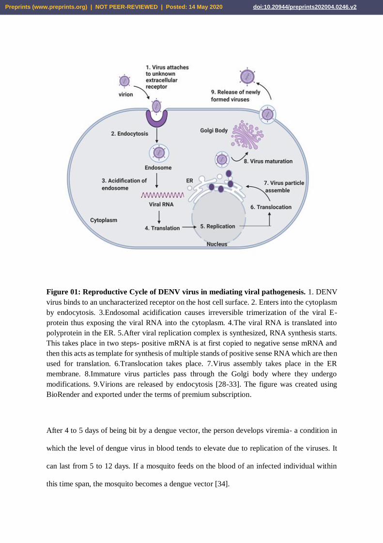
Figure 01: Reproductive Cycle of DENV virus in mediating viral pathogenesis. 1. DENV
virus binds to an uncharacterized receptor on the host cell surface. 2. Enters into the cytoplasm
by endocytosis. 3.Endosomal acidification causes irreversible trimerization of the viral E-
protein thus exposing the viral RNA into the cytoplasm. 4.The viral RNA is translated into
polyprotein in the ER. 5.After viral replication complex is synthesized, RNA synthesis starts.
This takes place in two steps- positive mRNA is at first copied to negative sense mRNA and
then this acts as template for synthesis of multiple stands of positive sense RNA which are then
used for translation. 6.Translocation takes place. 7.Virus assembly takes place in the ER
membrane. 8.Immature virus particles pass through the Golgi body where they undergo
modifications. 9.Virions are released by endocytosis [28-33]. The figure was created using
BioRender and exported under the terms of premium subscription.
After 4 to 5 days of being bit by a dengue vector, the person develops viremia- a condition in
which the level of dengue virus in blood tends to elevate due to replication of the viruses. It
can last from 5 to 12 days. If a mosquito feeds on the blood of an infected individual within
this time span, the mosquito becomes a dengue vector [34].
Preprints (www.preprints.org) | NOT PEER-REVIEWED | Posted: 14 May 2020 doi:10.20944/preprints202004.0246.v2
Some epidemiological studies show that the development and severity of dengue infection in
humans is triggered by many factors. Age is one of them [35]. Other factors include genetic
background of the host [36][37], vector, serotype of virus [38], gender, genotype [39][40],
environmental condition, immune condition of the infected people, socioeconomic level of the
population and secondary infection by heterologous serotype [41].
4.2. Immune Response of Human Body Against Dengue Virus
The immune system is the primary defense system of the body against dengue virus and
consists of mainly two parts: The innate immune system and the adaptive immune system.
While the innate immune response facilitates the immediate recognition and protection towards
any invading pathogen, the adaptive immune response produces cells that specifically and
efficiently target the pathogen or infected cell providing a long term immunity, unlike the
innate immune system [42]. The cells produced by the adaptive immune system include the
antibody-secreting B cells, which are capable of recognizing and binding to foreign cells with
high specificity and the cytotoxic T cells which are known to attack the infected cells.
Since the keratinocytes and Langerhan cells are infected through viral replication, the
Langerhans, through proper detection, display the antigens from the invading pathogens on
their surfaces [43] - [46]. The display of the viral antigens triggers the innate immune response,
summoning the white blood cells, monocytes and macrophages for the ingestion and
destruction of the dengue virus. The virus infects these cells instead and spreads throughout the
entire body as they travel through the lymphatic system [46] [47]. As the virus spreads
throughout, infecting cells of the bone marrow, lymph nodes, spleen, liver or blood, it facilitates
the emergence of viremia.
While some of the infected Langerhans travel to the lymph nodes (small glands present
throughout the body that are known to fight infections) to trigger the immune response, the
remaining secrete proteins called interferons which disrupt the replication process of viruses
Preprints (www.preprints.org) | NOT PEER-REVIEWED | Posted: 14 May 2020 doi:10.20944/preprints202004.0246.v2
through the activation of innate and adaptive immune system defenses (Figure 02) [24] [48].
They help in the recognition of the infected cells and protection of the uninfected cells. The
individuals experience dengue fever (DF) while their body’s immune system fights the virus.
Antibodies from B cells, immunoglobulin M (IgM) and IgG are secreted into the bloodstream
and the lymph fluid in order to neutralize the virus while on the other hand, cytotoxic T cells
and killer T cells are used in order to specifically recognize and destroy the virus through
adaptive immune mechanisms. The innate immune response further activates the complement
system in order to destroy the virus with the help of antibodies and leukocytes, clearly
indicating the contribution of both the immune systems, in order to neutralize the dengue
infection [49].
The individual remains protected from the other three serotypes for about 2 to 3 months after
recovery from the first dengue infection. However, this provides short term protection only and
the usual observation was that a second dengue infection was much worse in individuals
infected beforehand than those who were not infected earlier [47] [50]. Even though the
memory B cells and memory T cells are normally known to provide protection through adaptive
immune response when the virus strikes again, the mechanism fails in case of a second dengue
attack. These observations were explained by Halstead through the ‘‘antibody-dependent
enhancement of infection’’ phenomenon which supports the idea that the antibodies present
from the first dengue infection or the newly produced memory B cells facilitate the efficient
viral attack of host cells instead of providing protection against the other three serotypes
[50][51] - [55]. This happens since the antibodies from the first dengue infection form a
complex with the newly attacking serotype which further binds to the Fcγ receptors (FcγR)
present on circulating monocytes [56] - [60]. This results in severe dengue fever, also known
as dengue hemorrhagic fever (DHF), since more cells are infected with the help of the pre-
existing antibodies. This phenomenon is also active among children who received antibodies
Preprints (www.preprints.org) | NOT PEER-REVIEWED | Posted: 14 May 2020 doi:10.20944/preprints202004.0246.v2
for dengue from their mothers while in the womb, putting them at a greater risk of developing
severe dengue [61] [62]. Furthermore, it was also seen that the cytotoxic T cells secreted greater
amounts of cytokines during the second dengue infection which are known to cause serious
inflammations or tissue damage.
As dengue virus attacks our body, our body's innate and adaptive immune systems defend us,
through the recruitment of several cells which eventually help to neutralize the virus. A clearer
understanding of the pathogenesis of DHF facilitates early detection of severe diseases as well
as accurate management.
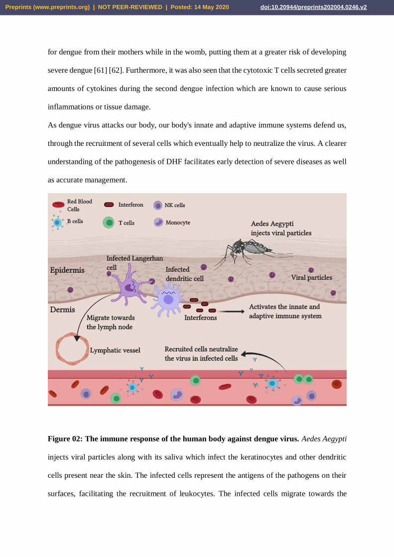
Figure 02: The immune response of the human body against dengue virus. Aedes Aegypti
injects viral particles along with its saliva which infect the keratinocytes and other dendritic
cells present near the skin. The infected cells represent the antigens of the pathogens on their
surfaces, facilitating the recruitment of leukocytes. The infected cells migrate towards the
Preprints (www.preprints.org) | NOT PEER-REVIEWED | Posted: 14 May 2020 doi:10.20944/preprints202004.0246.v2
lymph node to trigger the immune response. Soon after, more and more cells get infected and
lead to viremia. The infected cells release interferons which aid the recruitment of B cells that
release the antibodies, IgG and IgM and the cytotoxic T cells which target the infected cells.
The complement system and NK cells are also recruited to neutralize the virus. The B cells also
produce memory B cells in case the patient is infected again however, these memory cells will
only be able to help if the patient is infected with the same serotype again [63][64]. The figure
was created using BioRender and exported under the terms of premium subscription.
5. Symptoms and Clinical Presentation
5.1. Signs and Symptoms
The signs and symptoms of dengue virus infection depends on the severity of the case, and it
can even be asymptomatic. However, if they do make an appearance, they begin 3 to 14 days
(incubation period) after the initial infection [65]. It can be severe in about 5% of the cases and
in less than 1%, it may be life-threatening and eventually lead to death, despite intensive care.
Dengue fever (DF) results in a high-grade fever further leading to a number of other symptoms
which include headache, retro-orbital pain, muscle pain, vomiting and rash. The symptoms
may become worse and life-threatening if the disease is moderate or severe [66][67]. The
symptomatic cases are classified into undifferentiated febrile illness (UF), dengue fever (DF),
dengue hemorrhagic fever (DHF), dengue shock syndrome (DSS) and unusual dengue (UD) or
expanded dengue syndrome (EDS) [68]. Due to damaged blood vessels, blood plasma leakage
and a decreased platelet count, the risk of acquiring dengue hemorrhagic fever (DHF) increases.
The symptoms may also progress to massive bleeding, shock, and death; this is called dengue
shock syndrome (DSS). Dengue infections and DHF severity can be classified into several
groups according to the visibility of certain signs and symptoms, and also with the help of
laboratory evaluations.
Preprints (www.preprints.org) | NOT PEER-REVIEWED | Posted: 14 May 2020 doi:10.20944/preprints202004.0246.v2
They are as follows: [69]
● Dengue Fever (DF): Patients may experience fever along with two other symptoms
which may include rash, myalgia, headache, hemorrhagic manifestations but, no signs
of plasma leakage. These patients will also experience leukopenia (white blood cell
count ≤5000 cells/mm3), thrombocytopenia (platelet count <150 000 cells/mm3) and a
rising hematocrit (HCT) level of about 5% – 10%.
DHF severity can be further classified into 4 grades:
● DHF - I: The patients will experience fever and hemorrhagic manifestations followed
by plasma leakage. Among others, they will have thrombocytopenia with WBC <100
000 cells/mm3 and a HCT rise ≥ 20%
● DHF - II: Symptoms along with thrombocytopenia level and HCT rise follow that in
DHF I, with additional bleeding (spontaneous)
● DHF - III: Symptoms along with thrombocytopenia level and HCT rise is similar to
Grade I or II, and also leads to circulatory failure
● DHF - IV: Profound shock with undetectable BP and pulse along with Grade III
characteristics
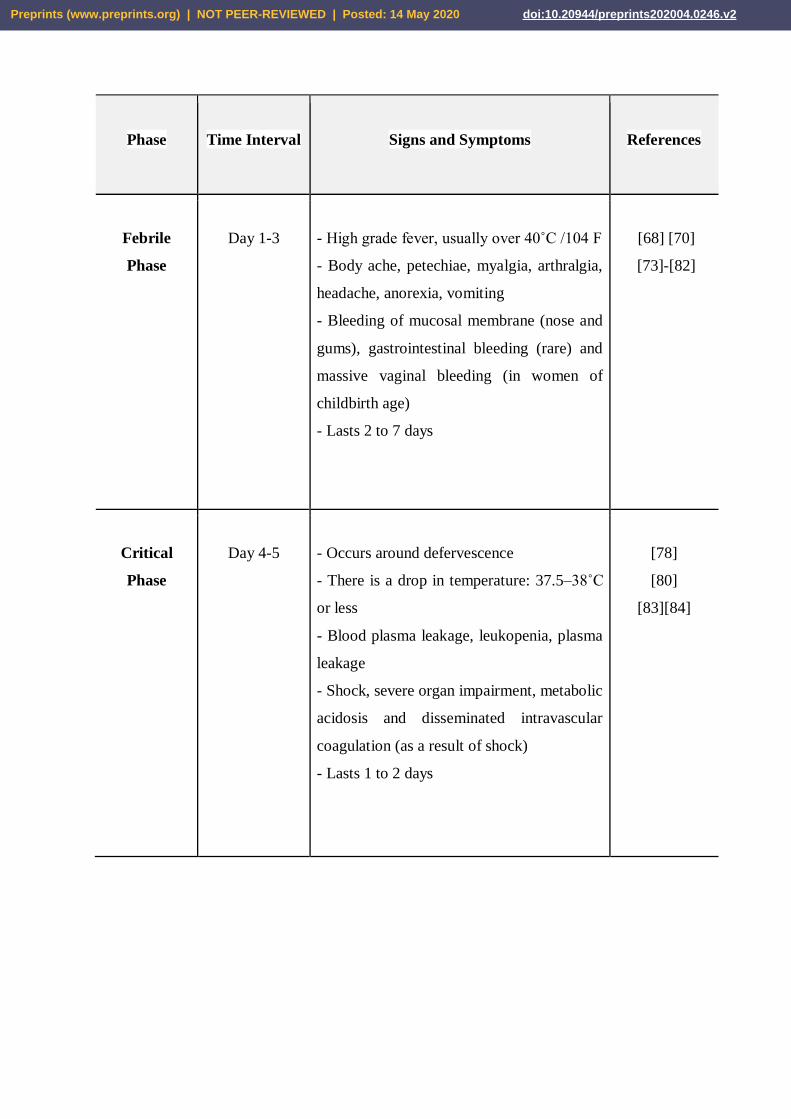
The course of dengue infection takes off after the incubation period and is divided into 3 main
phases: Febrile phase, critical phase (which may include hemorrhagic manifestations and/or
dengue shock syndrome), recovery or convalescence phase (Table 01) [70] - [72].
Preprints (www.preprints.org) | NOT PEER-REVIEWED | Posted: 14 May 2020 doi:10.20944/preprints202004.0246.v2
Phase
Time Interval
Signs and Symptoms
References
Febrile
Phase
Day 1-3
- High grade fever, usually over 40˚C /104 F
- Body ache, petechiae, myalgia, arthralgia,
headache, anorexia, vomiting
- Bleeding of mucosal membrane (nose and
gums), gastrointestinal bleeding (rare) and
massive vaginal bleeding (in women of
childbirth age)
- Lasts 2 to 7 days
[68] [70]
[73]-[82]
Critical
Phase
Day 4-5
- Occurs around defervescence
- There is a drop in temperature: 37.5–38˚C
or less
- Blood plasma leakage, leukopenia, plasma
leakage
- Shock, severe organ impairment, metabolic
acidosis and disseminated intravascular
coagulation (as a result of shock)
- Lasts 1 to 2 days
[78]
[80]
[83][84]
Preprints (www.preprints.org) | NOT PEER-REVIEWED | Posted: 14 May 2020 doi:10.20944/preprints202004.0246.v2
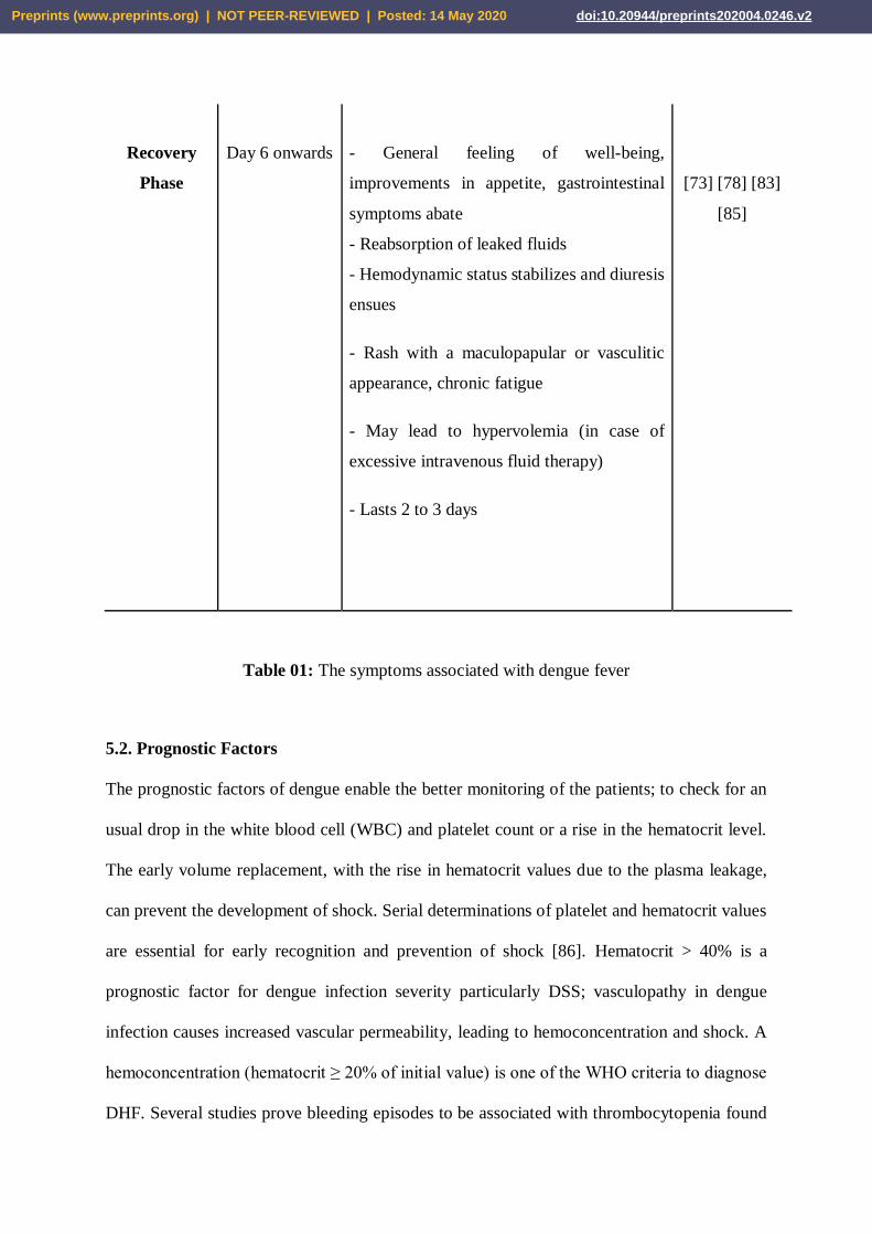
Recovery
Phase
Day 6 onwards
- General feeling of well-being,
improvements in appetite, gastrointestinal
symptoms abate
- Reabsorption of leaked fluids
- Hemodynamic status stabilizes and diuresis
ensues
- Rash with a maculopapular or vasculitic
appearance, chronic fatigue
- May lead to hypervolemia (in case of
excessive intravenous fluid therapy)
- Lasts 2 to 3 days
[73] [78] [83]
[85]
Table 01: The symptoms associated with dengue fever
5.2. Prognostic Factors
The prognostic factors of dengue enable the better monitoring of the patients; to check for an
usual drop in the white blood cell (WBC) and platelet count or a rise in the hematocrit level.
The early volume replacement, with the rise in hematocrit values due to the plasma leakage,
can prevent the development of shock. Serial determinations of platelet and hematocrit values
are essential for early recognition and prevention of shock [86]. Hematocrit > 40% is a
prognostic factor for dengue infection severity particularly DSS; vasculopathy in dengue
infection causes increased vascular permeability, leading to hemoconcentration and shock. A
hemoconcentration (hematocrit ≥ 20% of initial value) is one of the WHO criteria to diagnose
DHF. Several studies prove bleeding episodes to be associated with thrombocytopenia found
Preprints (www.preprints.org) | NOT PEER-REVIEWED | Posted: 14 May 2020 doi:10.20944/preprints202004.0246.v2
in severe dengue infection which can be a prognostic factor along with a systolic blood pressure
< 90 mmHg and pulse pressure ≤ 20 mmHg, which are considered to be the prognostic factors
for DSS [87]. An elevated level of aminotransferase (aspartate aminotransferase and alanine
aminotransferase) from the liver is also known to be associated with thrombocytopenia and
leukopenia.
Similarly, it was observed that a decrease in the platelet count led the patients to a state of
shock. WHO provided an overview of the platelet count to categorize dengue infection into
DHF grade I-IV [88]. Furthermore, elevated liver enzymes (SGOT and SGPT) are also known
as prognostic factors for DSS however, they are not routinely investigated in many hospitals
and are therefore known to be less frequently used as prognostic factors.
Similarly, the prognostic factors can also help to keep a track on the patient's improvement and
recovery; this may include a normalized WBC count, platelet count, hematocrit level, etc.
Monitoring the prognostic factors can provide an idea about both the improvement and the
deteriorating condition of a patient and, further help in the diagnosis and management of
patients.
5.3. Management of Patients
The management of dengue patients depends on the phase of illness, i.e. the febrile phase, the
critical/leakage phase and the recovery or convalescence phase.
For the febrile phase, patients should only be given supportive and symptomatic treatment
which may include the use of paracetamol or tepid sponge for fever reduction, promoting oral
feeding with the help of fruit juice, milk or oral rehydration therapy (ORS) and avoiding IV
fluids if the patient is not vomiting or dehydrated. Patients are advised to follow up CBC
everyday and to return to the hospital if no improvement is seen [68][89].
Preprints (www.preprints.org) | NOT PEER-REVIEWED | Posted: 14 May 2020 doi:10.20944/preprints202004.0246.v2
For the critical phase, isotonic salt solution is used except in the very young infants (<6 months
of age), in whom 0.45% sodium chloride may be used. The total amount of fluid needed during
this period of 24 - 48 hours is estimated to be maintenance + 5% deficit (according to guidelines
stated by WHO), including oral and IV fluids [90]. In DSS patients the duration of IV fluid
may be 24–36 hours and in non-shock DHF patients, 48–60 hours . The rate of IV fluid should
be adjusted according to the clinical vital signs such as blood pressure (BP), pulse, respiratory
rate, temperature or the hematocrit and urine output, and also according to the grade of severity
of dengue. Hyper-oncotic colloid solutions (osmolarity of >300 mOsm/l) such as dextran 40 or
starch solutions can be used in patients with massive plasma leakage [91]. In case of a lack of
a positive clinical response, patients showing unstable vital signs or other negative signs are
further investigated and treated for acidosis (corrected with NaHCO3, if pH is <7.35 and serum
bicarbonate is <15 mEq/L), bleeding, iCa and other electrolytes (Na and K), and blood sugar .
Furthermore, in cases of significant bleeding, blood transfusion is recommended as soon as
possible [92]. Platelets are indicated in cases with significant bleeding. However, it may cause
fluid overload and possible acute pulmonary edema in patients who already show signs of fluid
overload. Plasma and steroids do not play a role in the management of acute DHF and DSS,
respectively. Patients with obesity or diabetes mellitus are advised to get blood glucose levels
examined and those experiencing prolonged/profound shocks should take additional tests in
order to monitor them accordingly [93].
During the convalescence/recovery phase, IV fluids should be stopped upon signs of recovery
(increase in appetite, convalescence rash, greater than 30 hours before shock, etc). Patients who
fail to regain their appetite as a result of potassium loss through urine or diuresis, are
recommended a potassium rich diet. This is the period of reabsorption and therefore, patients
with massive ascites and pleural effusion may require diuretics.
Preprints (www.preprints.org) | NOT PEER-REVIEWED | Posted: 14 May 2020 doi:10.20944/preprints202004.0246.v2
As mentioned earlier, the rate of IV fluid also depends on the severity of dengue i.e., according
to the different grades of DHF. DHF I and II which involves non-shock patients, the fluid
allowance (oral + IV) is about maintenance (for one day) + 5% deficit (oral and IV fluid
together), to be administered over 48 hours [94]. The rate is different between children and
adults and often follows ideal weight measures. The rate of IV replacement should be adjusted
according to the rate of plasma loss, following the clinical condition, vital signs, urine output
and hematocrit levels.
DHF III, which includes shock or DSS, is caused due to plasma leakage that leads to an
increased systemic vascular resistance and narrowed pulse pressure [95]. Patients must be
checked for bleeding or gastrointestinal bleeding along with the plasma leakage in cases of
hypotension . Fluid resuscitation in DSS is different than other types of shocks and patients
usually respond to the 10 ml/kg in children or 300–500 ml in adults over one hour or by bolus
procedure or, follow a particular rate of infusion chart/graph and also, before reducing the IV
replacement rate, the patient must be checked for improvements.
For DHF IV (prolonged/profound shock), in order to restore the blood pressure, the initial fluid
resuscitation is vigorous and also, other laboratory tests must be done and treated for. 10 ml/kg
of bolus fluid should be given within 10 to 15 minutes. After BP restoration, IV fluids can be
given however, if shock is not reversible after the first 10 ml/kg, a repeat bolus of 10 ml/kg
should be given and the laboratory tests must be continued to check for correction and progress
[96]. Patients should be closely monitored, given blood transfusions in case of need and
managed accordingly in case of organ impairments.
Preprints (www.preprints.org) | NOT PEER-REVIEWED | Posted: 14 May 2020 doi:10.20944/preprints202004.0246.v2
Patients with severe hemorrhage may take blood transfusions in order for the HCT levels to
drop to normal. Apart from this, several other measures can be taken. Patients who are at high
risks such as, obese patients, infants, pregnant women, patients with diabetes mellitus or heart
diseases along with those experiencing a fluid overload or encephalopathy (damage or disease
affecting the brain), must be cared for and managed accordingly in order to prevent any further
health complications [97].
A better understanding of the signs and symptoms of dengue along with the frequent
monitoring of the prognostic factors and the application of adequate measures for the better
management of patients, can contribute greatly for a quick recovery.
6. Epidemiology of Dengue Virus Infection
Dengue is an important arthropod borne viral disease that is known to affect an estimated 2.5
billion people around the globe; approximately 975 million are habitants of the urban areas in
tropical and subtropical countries in Southeast Asia, the Pacific and America [4] [98] - [101].
The transmission range also covers the African, the Eastern Mediterranean and the rural
communities. The number of cases reported each year includes more than 500 million infected
and about 500,000 individuals hospitalized for dengue hemorrhagic fever [6] [102] [103].
The first epidemic of dengue dates back to 1635 in the French West Indies and similarly, a
disease outbreak identical to dengue was reported in China during 992 AD [104] [105]. The
1779-1780 dengue outbreaks in Asia, Africa, and North America along with simultaneous
outbreaks in three other continents indicated the world-wide distribution of the virus and their
mosquito vectors. According to the World Health Organization (WHO), the average annual
number of reported cases for DF or DHF includes 925,896 people during the 2000-2004 period
which is double that of 1990-1999, indicating an overwhelming increase in confirmed cases.
Again, from 2000 to 2008, the average annual number of cases reported was 1,656,870 which
Preprints (www.preprints.org) | NOT PEER-REVIEWED | Posted: 14 May 2020 doi:10.20944/preprints202004.0246.v2
is almost three and a half times that noted previously. While there were little to no cases in the
African or Eastern Mediterranean regions in the 2005-2006 period, countries such as Pakistan,
Saudi Arabia, Yemen, Sudan and Madagascar had suspected outbreaks [106]. There was an
expansion of the dengue transmission regions worldwide, that included the four existing
serotypes of dengue and the leading factors could have been the travelling of individuals from
endemic regions acting as carriers, uncontrolled vectors, unprecedented population growth and
uncontrolled urbanization [103] [107] - [110].
6.1 Dengue in Asia
After World War II, the global pandemic of dengue emerged in Southeast Asia and almost 75%
of the world’s dengue burden involved countries such as the Philippines, Indonesia and
Thailand [111]. Also, following the years of World War II, the unprecedented urbanization led
to inadequate housing, deterioration of water, sewer and waste management systems in
Southeast Asia. This ecological setting facilitated the increase in transmission and frequency
of epidemics. Apart from the economic expansion, the migration of people to different cities
and countries also led to the cocirculation of the different serotypes resulting in the
hyperendemicity [112]. Furthermore, apart from an adaptable climate for the vectors,
population growth and global travel has also led to an increase in the spread of the virus. Even
though DHF had appeared first as an epidemic in the 1950s, it had become one the leading
causes of hospitalizations and death by 1975 [16] [113]. DHF had expanded into Asia during
the 1980s and China and Taiwan also reported cases of epidemic DF after an absence for 35
years. After a successful control program, Singapore was able to prevent transmission for over
20 years [114]. Even though only sporadic cases of dengue were reported before 2000, it raised
a serious public health concern during 2000 with 5,551 reported cases and 93 deaths [99] [115]
[116]. Countries like Timor-Leste, Bhutan and Nepal had also reported their first dengue
Preprints (www.preprints.org) | NOT PEER-REVIEWED | Posted: 14 May 2020 doi:10.20944/preprints202004.0246.v2
outbreaks in the year 2004. Soon after dengue had become endemic in several regions
throughout the years, by the end of 2016, countries such as China, Malaysia and Singapore had
reported about 2,91,964 cases [117]. Also, the cases of dengue had spiked ever since its first
outbreak with DEN- 3 during the year 2000, in Bangladesh [118] [119].
6.1.1. Recent Dengue outbreaks in Asia (2019-2020)
Recently, dengue outbreak was reported to be a terrifying epidemic in many countries of Asia.
By the end of 2019, a total of 101,354 cases and 179 deaths were reported in Bangladesh.
Again, this country recorded about 263 cases till 16th of March in 2020 [120] [121]. While
Malaysia reported a cumulative number of 127,407 cases and 176 deaths till December 21,
2019, the Philippines reported 420,453 cases including 1,565 deaths till December 14 of the
same year and Singapore reported about 15,622 cases on week 51 of 2019 [122]. Dengue
transmission was also recorded in Afghanistan for the first time, in 2019. China reported 268
cases in January 2020, Malaysia reported 32,951 cases including 48 deaths from December 29,
2019 till March 21, 2020, Philippines reported 37,058 cases including 112 deaths as of
February 2020 and Viet Nam reported 20,673 cases including 4 deaths till March 20, 2020
[123].
6.2. Dengue in America
Dengue has been a major public health problem in America. A campaign by the Pan American
Health Organization (PAHO) played a role in the eradication during the 1960s from the central
and south American countries [124]. The discontinuation of the campaign in the 1970s further
led to the reinfestation of the species. In the 1970s, DEN-2 and DEN-3 were present in America
however, the major epidemic in 1977 was due to the DEN-1 virus which had an epidemic
period of 16 years. In 1981, DEN-4 was introduced along with another strain of DEN-2 from
Southeast Asia which led to a major DHF epidemic and this new strain caused outbreaks in
Preprints (www.preprints.org) | NOT PEER-REVIEWED | Posted: 14 May 2020 doi:10.20944/preprints202004.0246.v2
Venezuela, Puerto Rico, Columbia, Brazil, French Guiana, the French Antilles and Suriname
[125]. A total of 14 countries in America had endemic DHF after its emergence in 1995. The
failed eradication program led to an increase in infected cases from 2000 to 2010 and therefore,
over 1.7 million cases were reported including 50,235 severe cases and 1,185 deaths.
According to a recent report by WHO, the number of cases increased by 15 fold over the past
two decades with an increase from 50,5430 cases in 2000 to 2,400,138 cases in 2010 [126]. In
November 2018, PAHO had alerted the countries to increase their preparedness and response
efforts in order to prepare for a much intense dengue outbreak. Following that, according to a
report from PAHO, America recorded the largest outbreak of dengue with about 3 million cases
in the region till 2019 and the number of cases reported was 320,000 till February 9, 2020
[127]. Reports from the 2019 dengue cases also suggested additional findings such as an
increase in severe dengue cases, shifts in epidemic season and a higher risk of death for the
younger age groups (5-9 years). The early and appropriate diagnosis and clinical management
of patients along with effective ways of breeding site destruction, and tackling the social and
environmental determinants that are linked to dengue can help slow down and eventually stop
the disease transmission, preventing further deaths.
6.3. Dengue in Australia
The first outbreaks in Australia date back to 1879 in Queensland at Townsville and 1885 in
Rockhampton [128]. In 1898, the first cases were reported in New South Wales and during
1925-26 it extended to the south. In the Northern Territory, it was prevalent during 1955 [129]
- [131]. By the end of the 19th century, dengue was widely distributed and it further spread
through various mediums which included the presence of major breeding sites provided by
rainwater tanks and water-holding domestic containers. The species had soon disappeared from
the northern and western parts. There was a reduction in the population during the 1960s in
Preprints (www.preprints.org) | NOT PEER-REVIEWED | Posted: 14 May 2020 doi:10.20944/preprints202004.0246.v2
Queensland however, after it's disappearance for 25 years, it had reappeared during the 1980s
[132] - [134]. Several factors had influenced its decline; the leading factor being the conversion
of urban water supplies from household rainwater tanks to a reticulated supply, change from
steel to diesel locomotives, the use of domestic insecticides along with public education and
increased awareness had all contributed greatly. In four years, from 2005 to 2008, nine
outbreaks of dengue were reported in Queensland, Australia [135]. On December 1, 2008, a
dengue outbreak was declared in the tropical north region of Queensland, Cairns and several
confirmed cases were reported soon after in 2009 affecting towns and cities like Townsville,
Port Douglas, Yarrabah, Injinoo, Innis fail and Rockhampton . Also, from the period 2005-
2006 to 2009-2010, the number of dengue cases had increased from 156 to 581. Australia
reported a total of 1,419 till December 18, 2019, since the start of the year and about 78 cases
till February 26, 2020 [123] [136]. In Australia, dengue occurs seasonally in northern
Queensland, with a peak transmission from January to April. Even though dengue is not
endemic in Australia, the presence of the vectors, Aedes Aegypti and Aedes Albopictus
mosquitoes raises the risk of a possible outbreak.
6.4. Dengue in Europe
Dengue was endemic in regions of southern Europe until the 1930s due to the presence of
Aedes Aegypti. In 1927 and 1928, several outbreaks infected millions and led to a death toll of
around 2100 in Greece and Turkey [137]. Disappearance of the disease was noted however,
several locally transmitted cases in Croatia, France and Portugal were also reported; Ae.
Albopictus and Ae. Aegypti being the responsible vectors. From the period 2012-2013, a large
outbreak in Madeira and Portugal was reported which not only included more than 2100 cases
but also involved the spread of the disease to 14 other European countries [138]. The leading
causes included the introduction of the efficient mosquito vector species and the arrival of
Preprints (www.preprints.org) | NOT PEER-REVIEWED | Posted: 14 May 2020 doi:10.20944/preprints202004.0246.v2
infected people. According to a report from WHO, Ae. Albopictus had been spreading and was
known to have established from Spain to Greece and also to the Eastern countries and the Black
sea coast. In the year 2019, over 15,000 autochthonous cases of dengue were confirmed till
June 11, including at least 9 deaths by the end of the year [139]. Several surveillance and control
programs for dengue vectors are available in the European countries but only a few selected
programs include the disease apart from the vector as well . The dengue transmission models
that represent Europe, suggest that Europe is at a lower risk but they rarely include the
transmission of the other vector, Aedes Albopictus [106][140]. The unavailability of any
specific treatment makes integrated vector management the only sustainable control option.
Several factors contribute to the facilitation of the dengue vectors for their survival and
transmission along with the presence of the different serotypes. The adaptation of better
surveillance systems and control programs alongside individual preventive measures by the
countries can help reduce the cases of dengue, and the following deaths.
7. Diagnosis Methods
The accurate and efficient diagnosis of dengue is significant in order to differentiate dengue
from other diseases such as leptospirosis, rubella, and other infections caused by flavivirus to
provide clinical care, surveillance support as well as to carry out pathogenesis studies and
vaccine research along with the case confirmation of DHF/DSS. During the first 5 days of
illness, dengue virus and antigen detection are the most accurate diagnostic tools, as IgG and
IgM antibodies are not produced until 5–7 days after the onset of symptoms in primary
infections [141][142]. Rapid Diagnostic Tests (RTDs) are a simple and quick way of screening
for dengue virus especially in countries that do not have the facilities to perform tests such as
ELISA or PRNT tests. However, RTD has its own share of disadvantages as they cannot detect
Preprints (www.preprints.org) | NOT PEER-REVIEWED | Posted: 14 May 2020 doi:10.20944/preprints202004.0246.v2
past DENV infections in the patient and might lack sensitivity and specificity in order to ensure
safe vaccination [143].
Another method of directly testing for the DENV virus is the Dengue non - structural protein
NS1 antigen test which is a serotype - specific identification test and is used for early detection,
that is, less than five days since the onset of the fever. NS1 test has been the most popular as it
is rapid and less costly than other methods such as viral isolation or viral reverse transcriptase-
polymerase chain reaction [144]. When mammalian cells are infected, during the acute phase
of the infection, they secrete the NS1 antigen which is a glycoprotein synthesised by all
flaviviruses [145]. Although NS1 tests are the most effective dengue detection tests so far, NS1
tests alone cannot determine the serotype of the DENV infection and is mainly an indicative
test [146].
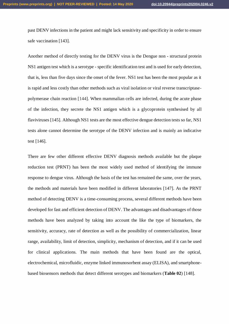
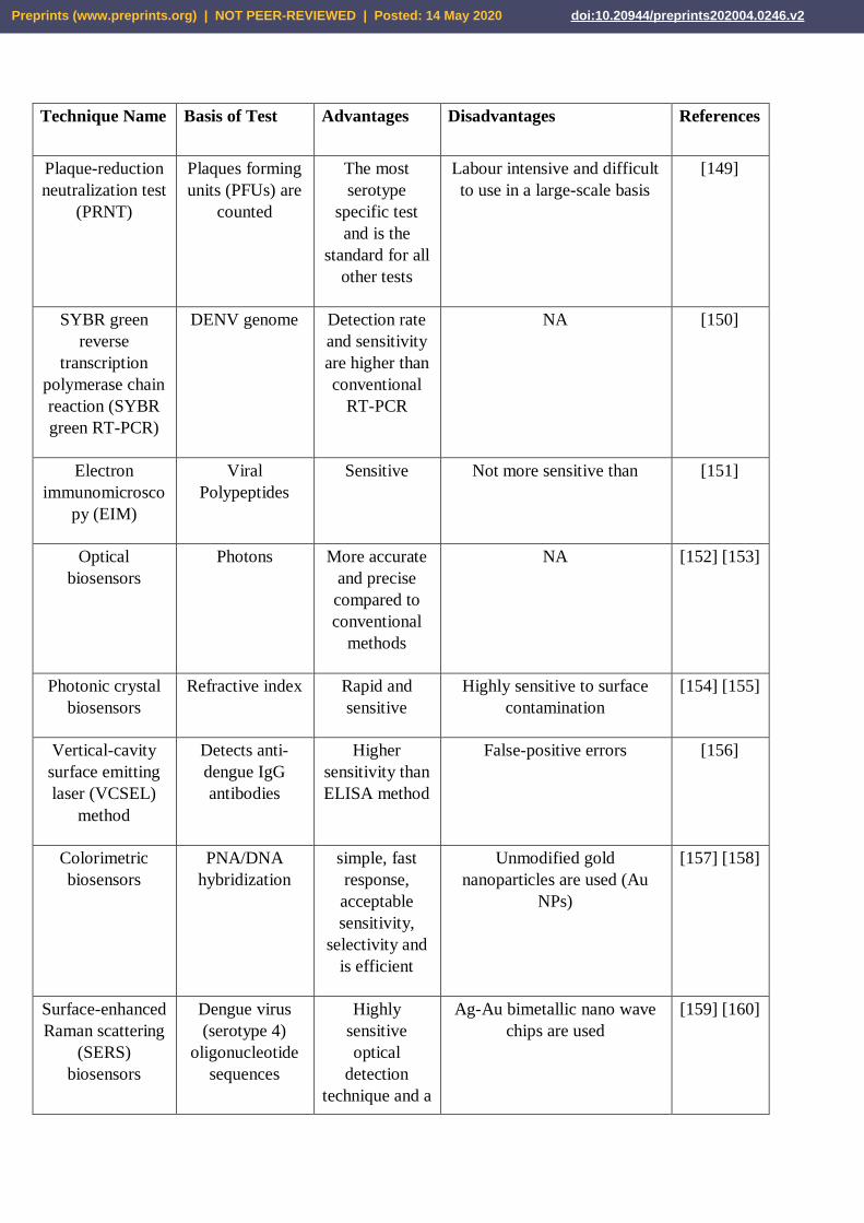
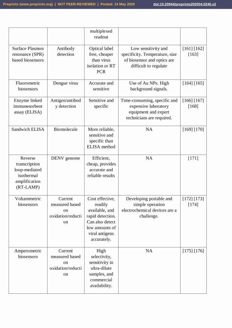
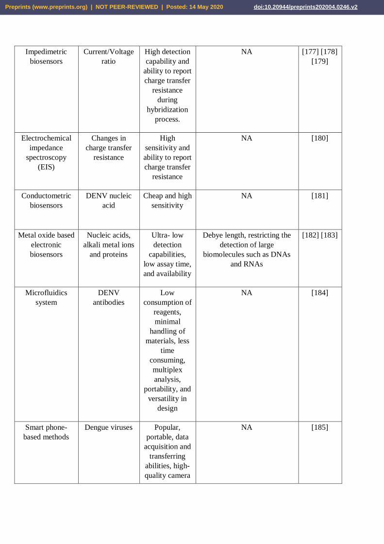
There are few other different effective DENV diagnosis methods available but the plaque
reduction test (PRNT) has been the most widely used method of identifying the immune
response to dengue virus. Although the basis of the test has remained the same, over the years,
the methods and materials have been modified in different laboratories [147]. As the PRNT
method of detecting DENV is a time-consuming process, several different methods have been
developed for fast and efficient detection of DENV. The advantages and disadvantages of those
methods have been analyzed by taking into account the like the type of biomarkers, the
sensitivity, accuracy, rate of detection as well as the possibility of commercialization, linear
range, availability, limit of detection, simplicity, mechanism of detection, and if it can be used
for clinical applications. The main methods that have been found are the optical,
electrochemical, microfluidic, enzyme linked immunosorbent assay (ELISA), and smartphone-
based biosensors methods that detect different serotypes and biomarkers (Table 02) [148].
Preprints (www.preprints.org) | NOT PEER-REVIEWED | Posted: 14 May 2020 doi:10.20944/preprints202004.0246.v2
Technique Name Basis of Test Advantages Disadvantages References
Plaque-reduction
neutralization test
(PRNT)
Plaques forming
units (PFUs) are
counted
The most
serotype
specific test
and is the
standard for all
other tests
Labour intensive and difficult
to use in a large-scale basis
[149]
SYBR green
reverse
transcription
polymerase chain
reaction (SYBR
green RT-PCR)
DENV genome Detection rate
and sensitivity
are higher than
conventional
RT-PCR
NA [150]
Electron
immunomicrosco
py (EIM)
Viral
Polypeptides
Sensitive Not more sensitive than [151]
Optical
biosensors
Photons More accurate
and precise
compared to
conventional
methods
NA [152] [153]
Photonic crystal
biosensors
Refractive index Rapid and
sensitive
Highly sensitive to surface
contamination
[154] [155]
Vertical-cavity
surface emitting
laser (VCSEL)
method
Detects anti-
dengue IgG
antibodies
Higher
sensitivity than
ELISA method
False-positive errors [156]
Colorimetric
biosensors
PNA/DNA
hybridization
simple, fast
response,
acceptable
sensitivity,
selectivity and
is efficient
Unmodified gold
nanoparticles are used (Au
NPs)
[157] [158]
Surface-enhanced
Raman scattering
(SERS)
biosensors
Dengue virus
(serotype 4)
oligonucleotide
sequences
Highly
sensitive
optical
detection
technique and a
Ag-Au bimetallic nano wave
chips are used
[159] [160]
Preprints (www.preprints.org) | NOT PEER-REVIEWED | Posted: 14 May 2020 doi:10.20944/preprints202004.0246.v2
multiplexed
readout
Surface Plasmon
resonance (SPR)
based biosensors
Antibody
detection
Optical label
free, cheaper
than virus
isolation or RT
PCR
Low sensitivity and
specificity. Temperature, size
of biosensor and optics are
difficult to regulate
[161] [162]
[163]
Fluorometric
biosensors
Dengue virus Accurate and
sensitive
Use of Au NPs. High
background signals.
[164] [165]
Enzyme linked
immunesorbent
assay (ELISA)
Antigen/antibod
y detection
Sensitive and
specific
Time-consuming, specific and
expensive laboratory
equipment and expert
technicians are required.
[166] [167]
[168]
Sandwich ELISA Biomolecule More reliable,
sensitive and
specific than
ELISA method
NA [169] [170]
Reverse
transcription
loop-mediated
isothermal
amplification
(RT-LAMP)
DENV genome Efficient,
cheap, provides
accurate and
reliable results
NA [171]
Voltammetric
biosensors
Current
measured based
on
oxidation/reducti
on
Cost effective,
readily
available, and
rapid detection.
Can also detect
low amounts of
viral antigens
accurately.
Developing portable and
simple operation
electrochemical devices are a
challenge.
[172] [173]
[174]
Amperometric
biosensors
Current
measured based
on
oxidation/reducti
on
High
selectivity,
sensitivity in
ultra-dilute
samples, and
commercial
availability.
NA [175] [176]
Preprints (www.preprints.org) | NOT PEER-REVIEWED | Posted: 14 May 2020 doi:10.20944/preprints202004.0246.v2
Impedimetric
biosensors
Current/Voltage
ratio
High detection
capability and
ability to report
charge transfer
resistance
during
hybridization
process.
NA [177] [178]
[179]
Electrochemical
impedance
spectroscopy
(EIS)
Changes in
charge transfer
resistance
High
sensitivity and
ability to report
charge transfer
resistance
NA [180]
Conductometric
biosensors
DENV nucleic
acid
Cheap and high
sensitivity
NA [181]
Metal oxide based
electronic
biosensors
Nucleic acids,
alkali metal ions
and proteins
Ultra- low
detection
capabilities,
low assay time,
and availability
Debye length, restricting the
detection of large
biomolecules such as DNAs
and RNAs
[182] [183]
Microfluidics
system
DENV
antibodies
Low
consumption of
reagents,
minimal
handling of
materials, less
time
consuming,
multiplex
analysis,
portability, and
versatility in
design
NA [184]
Smart phone-
based methods
Dengue viruses Popular,
portable, data
acquisition and
transferring
abilities, high-
quality camera
NA [185]
Preprints (www.preprints.org) | NOT PEER-REVIEWED | Posted: 14 May 2020 doi:10.20944/preprints202004.0246.v2
lenses and cost-
effectiveness
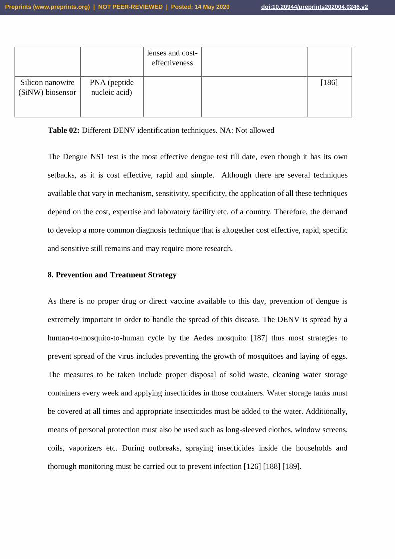
Silicon nanowire
(SiNW) biosensor
PNA (peptide
nucleic acid)
[186]
Table 02: Different DENV identification techniques. NA: Not allowed
The Dengue NS1 test is the most effective dengue test till date, even though it has its own
setbacks, as it is cost effective, rapid and simple. Although there are several techniques
available that vary in mechanism, sensitivity, specificity, the application of all these techniques
depend on the cost, expertise and laboratory facility etc. of a country. Therefore, the demand
to develop a more common diagnosis technique that is altogether cost effective, rapid, specific
and sensitive still remains and may require more research.
8. Prevention and Treatment Strategy
As there is no proper drug or direct vaccine available to this day, prevention of dengue is
extremely important in order to handle the spread of this disease. The DENV is spread by a
human-to-mosquito-to-human cycle by the Aedes mosquito [187] thus most strategies to
prevent spread of the virus includes preventing the growth of mosquitoes and laying of eggs.
The measures to be taken include proper disposal of solid waste, cleaning water storage
containers every week and applying insecticides in those containers. Water storage tanks must
be covered at all times and appropriate insecticides must be added to the water. Additionally,
means of personal protection must also be used such as long-sleeved clothes, window screens,
coils, vaporizers etc. During outbreaks, spraying insecticides inside the households and
thorough monitoring must be carried out to prevent infection [126] [188] [189].
Preprints (www.preprints.org) | NOT PEER-REVIEWED | Posted: 14 May 2020 doi:10.20944/preprints202004.0246.v2
The life cycle of dengue virus involves the entry of the virus into our body, membrane fusion,
RNA genome replication, assembly, and ultimate release from the infected cell. Thus, the
treatment of dengue is not specific and remains to be supportive [190]. Supportive treatment
includes oral fluid administration and antipyretic treatment with paracetamol along with daily
full blood counts. If there is excessive vomiting or diarrhea observed, admission to hospital is
necessary to avoid dehydration. The patient must be administered oral fluid as much as possible
[191]. The symptoms of dengue are flu-like and so there is no specific treatment but an early
diagnosis and clinical management can reduce the severity of the effects of dengue on the body
[126] [192].
Currently, there is no licensed antiviral agent available against dengue as the drug needs to
address all four serotypes of the dengue virus. Researchers have been trying to develop drugs
by targeting several different steps in the disease pathway in order to interfere with the
replication of dengue virus. For example, nucleoside analogue blocks a dengue infection by
preventing synthesis of the viral RNA genome so that the dengue virus cannot replicate. Viral
replication can be difficult to prevent because there is a short timeline for treatment which is
why it is important to target other steps in the disease pathway to prevent dengue at later stages
[193]. Therefore, steps other than viral replication such as protein synthesis and viral assembly
may be potential targets to produce a vaccine or drug for dengue and thus require further
research.
9. Vaccine for Dengue Virus
A vaccine can boost the immune system in our body against a particular pathogen which is
paramount in order to fight against viral diseases such one caused by DENV. But unfortunately,
no effective direct vaccine has been discovered yet that can successfully combat the dengue
infection. There is a major array of concerns that needs to be addressed in order to produce a
Preprints (www.preprints.org) | NOT PEER-REVIEWED | Posted: 14 May 2020 doi:10.20944/preprints202004.0246.v2
direct vaccine which is why producing one has been a major challenge. The vaccines produced
must work against all four of the DENV serotypes and must provide lifelong protection. This
may be done by incorporating an antigen that is common for all four serotypes. As vaccines
with live, attenuated or nonliving viruses usually produce less antibody than an infection with
wild-type virus, it is most likely that two doses of the virus will be required. Several other
factors need to be taken into account such as the level of symptoms seen after the vaccination,
if the vaccine can be used by people of all ages and the cost of the vaccine etc. [187] [194].
Providing lifelong protection against all four serotypes by neutralizing antibodies is difficult
due to antibody-dependent enhancement (ADE) or immune enhancement. ADE observed
during dengue infection due to heterologous non-neutralizing antibodies may cause dengue
hemorrhagic fever (DHF) or dengue shock syndrome (DSS) during secondary infection by a
different serotype. Additionally, over time, antibody responses below protective levels may
increase the possibility of immune enhancement by a natural infection due to wild type DENV.
Another major challenge of producing a dengue vaccine is the lack of an appropriate human
like animal model for the trials of the vaccines which will allow us to observe the pathogenesis,
immune response and clinical course of dengue infection in humans. Inoculation of mouse-
adapted DENV strain has caused paralysis or death of that animal [195].
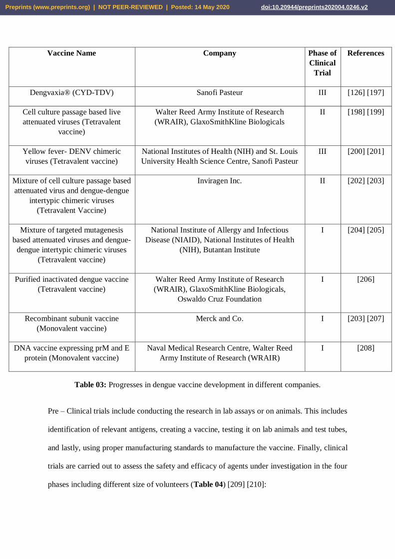
So far only one such vaccine for dengue has been approved, Dengvaxia® (CYD-TDV).
Dengvaxia® (CYD-TDV) has been developed by Sanofi Pasteur and it is for people who have
already been infected by DENV once. This works on people ages 9 to 45 and must be given
three doses in intervals (126, 188, 196). However, a large number of vaccines are now in
different phases of clinical trial in different companies (Table 03).
Preprints (www.preprints.org) | NOT PEER-REVIEWED | Posted: 14 May 2020 doi:10.20944/preprints202004.0246.v2
Vaccine Name Company Phase of
Clinical
Trial
References
Dengvaxia® (CYD-TDV) Sanofi Pasteur III [126] [197]
Cell culture passage based live
attenuated viruses (Tetravalent
vaccine)
Walter Reed Army Institute of Research
(WRAIR), GlaxoSmithKline Biologicals
II [198] [199]
Yellow fever- DENV chimeric
viruses (Tetravalent vaccine)
National Institutes of Health (NIH) and St. Louis
University Health Science Centre, Sanofi Pasteur
III [200] [201]
Mixture of cell culture passage based
attenuated virus and dengue-dengue
intertypic chimeric viruses
(Tetravalent Vaccine)
Inviragen Inc. II [202] [203]
Mixture of targeted mutagenesis
based attenuated viruses and dengue-
dengue intertypic chimeric viruses
(Tetravalent vaccine)
National Institute of Allergy and Infectious
Disease (NIAID), National Institutes of Health
(NIH), Butantan Institute
I [204] [205]
Purified inactivated dengue vaccine
(Tetravalent vaccine)
Walter Reed Army Institute of Research
(WRAIR), GlaxoSmithKline Biologicals,
Oswaldo Cruz Foundation
I [206]
Recombinant subunit vaccine
(Monovalent vaccine)
Merck and Co. I [203] [207]
DNA vaccine expressing prM and E
protein (Monovalent vaccine)
Naval Medical Research Centre, Walter Reed
Army Institute of Research (WRAIR)
I [208]
Table 03: Progresses in dengue vaccine development in different companies.
Pre – Clinical trials include conducting the research in lab assays or on animals. This includes
identification of relevant antigens, creating a vaccine, testing it on lab animals and test tubes,
and lastly, using proper manufacturing standards to manufacture the vaccine. Finally, clinical
trials are carried out to assess the safety and efficacy of agents under investigation in the four
phases including different size of volunteers (Table 04) [209] [210]:
Preprints (www.preprints.org) | NOT PEER-REVIEWED | Posted: 14 May 2020 doi:10.20944/preprints202004.0246.v2
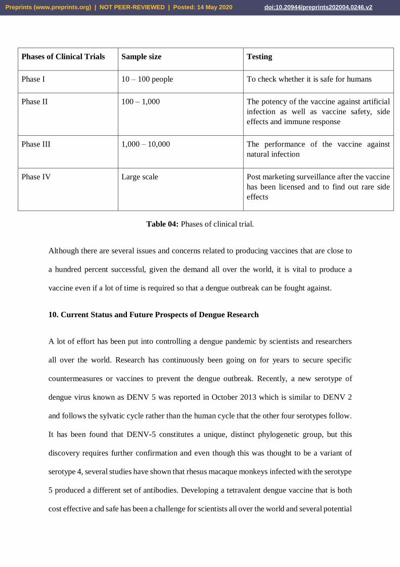
Phases of Clinical Trials Sample size Testing
Phase I 10 – 100 people To check whether it is safe for humans
Phase II 100 – 1,000
The potency of the vaccine against artificial
infection as well as vaccine safety, side
effects and immune response
Phase III 1,000 – 10,000 The performance of the vaccine against
natural infection
Phase IV Large scale Post marketing surveillance after the vaccine
has been licensed and to find out rare side
effects
Table 04: Phases of clinical trial.
Although there are several issues and concerns related to producing vaccines that are close to
a hundred percent successful, given the demand all over the world, it is vital to produce a
vaccine even if a lot of time is required so that a dengue outbreak can be fought against.
10. Current Status and Future Prospects of Dengue Research
A lot of effort has been put into controlling a dengue pandemic by scientists and researchers
all over the world. Research has continuously been going on for years to secure specific
countermeasures or vaccines to prevent the dengue outbreak. Recently, a new serotype of
dengue virus known as DENV 5 was reported in October 2013 which is similar to DENV 2
and follows the sylvatic cycle rather than the human cycle that the other four serotypes follow.
It has been found that DENV-5 constitutes a unique, distinct phylogenetic group, but this
discovery requires further confirmation and even though this was thought to be a variant of
serotype 4, several studies have shown that rhesus macaque monkeys infected with the serotype
5 produced a different set of antibodies. Developing a tetravalent dengue vaccine that is both
cost effective and safe has been a challenge for scientists all over the world and several potential
Preprints (www.preprints.org) | NOT PEER-REVIEWED | Posted: 14 May 2020 doi:10.20944/preprints202004.0246.v2
vaccines are being tested after Mahidol University, Thailand and Walter Reed Army Institute
of Research (USA) initiated the research [211] [212]. Several other organizations such as
Takeda Pharmaceuticals Company Limited, Bill & Melinda Gates Foundation, The Global
Health Innovative Technology (GHIT) Fund and many more have invested millions towards
dengue research [213 - 216]. A study in Singapore confirmed that right after the infection, the
immune cell called natural killer (NK) cells were activated in the blood and researchers hope
that this knowledge may be beneficial while developing vaccines and drugs [217]. Moreover,
another research team at University of California San Diego has synthetically engineered
mosquitoes that will prevent the transmission of dengue virus by the activation of an antibody
that prevents the replication of the virus and its dissemination [218]. Additionally, a study has
shown that mutations in the protein envelope cause the dengue virus to change its shape and
become resistant to vaccines and therapeutics [219].
Taking climate change into account for dengue research is important for the government and
public health officials to take actions to protect the public from future dengue outbreaks. As
the global climate changes, the global surface temperature will change and so will the patterns
of rainfall around the world, affecting the environmental suitability for the survival and growth
of dengue viruses and mosquitoes, which will eventually lead to the change in patterns of
dengue globally, nationally, and locally. Having said that, several issues need to be taken into
account during the research on DENV such as the sociodemographic factors e.g., travel and
demographic change, and other climatic factors in some areas where temperature may not be
the most vital aspect influencing the spread of dengue. Moreover, the non-climatic factors
affecting the spread of A. aegypti and A. albopictus must also be explored [220]. Scientists
have been investigating dengue pathogenesis in order to gain a better understanding of dengue
infection. Improving surveillance of dengue cases by databases such as DengueNet that
continuously updates and shares current and historical data on dengue cases by providing early
Preprints (www.preprints.org) | NOT PEER-REVIEWED | Posted: 14 May 2020 doi:10.20944/preprints202004.0246.v2
warnings prior to epidemics can help improve the preparedness of public health officials, and
help reduce fatality rates [221]. Many experts have said that dengue may increase in the future
due to viral evolution, climate change, globalization, travel and trade. Settlement factors and
socioeconomic factors also play a role in the spread of dengue. In light of this, it is important
to follow the guidelines in WHO Global Strategy for Dengue Prevention and Control, 2012–
2020 in order to prevent a huge outbreak as neither a perfect remedy nor a vaccine has been
found yet [222].
11. Conclusion
Even though dengue is the most emerging global infection in the 21st century, the adaptation
of a successfully planned control program can prove as a preventative measure. Apart from
this, our body's immune system also plays a major role in the process of viral neutralization.
Growth in population, frequent travels as well as urbanization has shown to affect the re-
emergence of the dengue epidemic. The increasing dengue outbreak along with no specific
existing treatment for the disease makes prevention our first priority. Some methods of
prevention may include the use of insecticides, removal of mosquito vectors or the use of
vaccines, mostly in the high burden epidemiologic areas. Rapid and more efficient dengue
detection methods are required for an immediate approach towards treatment. As the factors
contributing to the spreading of dengue virus are increasing with the rise in globalization and
climate change, the research for dengue vaccine and drug development requires more attention
in order to prevent and prepare for further devastating outbreaks.
Conflict of Interest
Authors declare no conflict of interest regarding the publication of the manuscript.
Preprints (www.preprints.org) | NOT PEER-REVIEWED | Posted: 14 May 2020 doi:10.20944/preprints202004.0246.v2
Acknowledgements
Authors acknowledge the members of Swift Integrity Computational Lab, Dhaka, Bangladesh,
a virtual platform of young researchers for their support during the preparation of the
manuscript.
Funding Information
Authors received no specific funding from any external sources.
References
1. Messina, J., Brady, O., Golding, N., Kraemer, M., Wint, G., Ray, S., Pigott, D., Shearer, F.,
Johnson, K., Earl, L., Marczak, L., Shirude, S., Davis Weaver, N., Gilbert, M., Velayudhan,
R., Jones, P., Jaenisch, T., Scott, T., Reiner, R. and Hay, S., 2019. The current and future global
distribution and population at risk of dengue. Nature Microbiology, 4(9), pp.1508-1515.
2. Chawla, P., Yadav, A. and Chawla, V., 2014. Clinical implications and treatment of dengue.
Asian Pacific Journal of Tropical Medicine, 7(3), pp.169-178.
3. Ebi, K. and Nealon, J., 2020. Dengue In A Changing Climate
4. Guzman, M., Halstead, S., Artsob, H., Buchy, P., Farrar, J., Gubler, D., Hunsperger, E.,
Kroeger, A., Margolis, H., Martínez, E., Nathan, M., Pelegrino, J., Simmons, C., Yoksan, S.
and Peeling, R., 2010. Dengue: a continuing global threat. Nature Reviews Microbiology,
8(S12), pp.S7-S16.
5. Nonyong, P., Pientong, C., Overgaard, H., Thaewnongiew, K., Aromseree, S., Phanitchat,
T., Phanthanawiboon, S. and Ekalaksananan, T., 2019. Correlation between dengue virus
serotypes in dengue patients and in mosquitoes at patients’ houses and surrounding in
Northeastern Thailand. International Journal of Infectious Diseases, 79, p.149
Preprints (www.preprints.org) | NOT PEER-REVIEWED | Posted: 14 May 2020 doi:10.20944/preprints202004.0246.v2
6. Bhatt, S., Gething, P., Brady, O., Messina, J., Farlow, A., Moyes, C., Drake, J., Brownstein,
J., Hoen, A., Sankoh, O., Myers, M., George, D., Jaenisch, T., Wint, G., Simmons, C., Scott,
T., Farrar, J. and Hay, S., 2013. The global distribution and burden of dengue. Nature,
496(7446), pp.504-507.
7. Cunha, B., Johnson, D. and McDermott, B., 2009. Atypical Dengue Fever Mimicking
Typhoid Fever in a College Student Traveler. The American Journal of Medicine, 122(4),
pp.e1-e3.
8. Günther J, Martínez-Muñoz J, Pérez-Ishiwara D, Salas-Benito J. Evidence of Vertical
Transmission of Dengue Virus in Two Endemic Localities in the State of Oaxaca, Mexico.
Intervirology. 2007;50(5):347-352
9. Guha-Sapir D, Schimmer B. Emerging Themes in Epidemiology. 2005;2(1):1.
10. Tuiskunen Bäck A, Lundkvist Å. Dengue viruses – an overview. 2020.
11. Waterman S, Gubler D. Dengue fever. Clinics in Dermatology. 1989;7(1):117-122
12. Dengue: Dengue: Practice Essentials, Background, Pathophysiology [Internet].
Emedicine.medscape.com. 2020 [cited 9 April 2020]. Available from:
https://emedicine.medscape.com/article/215840-overview#a2
13. Rush B. An account of the bilious remitting fever. The American Journal of Medicine.
1951;11(5):546-550.
14. Nelson E, Bierman H. Dengue Fever: A Thrombocytopenic Disease?. JAMA. 1964;190(2).
15. Gubler D. Epidemic dengue/dengue hemorrhagic fever as a public health, social and
economic problem in the 21st century. Trends in Microbiology. 2002;10(2):100-103.
Preprints (www.preprints.org) | NOT PEER-REVIEWED | Posted: 14 May 2020 doi:10.20944/preprints202004.0246.v2
16. Gubler D, Clark G. Dengue/Dengue Hemorrhagic Fever: The Emergence of a Global
Health Problem. Emerging Infectious Diseases. 1995;1(2):55-57.
17. Wilson M, Chen L. Dengue: Update on Epidemiology. Current Infectious Disease Reports.
2014;17(1).
18. Higa Y. Dengue Vectors and their Spatial Distribution. Tropical Medicine and Health.
2011;39(4SUPPLEMENT):S17-S27.
19. Carrington L, Simmons C. Human to Mosquito Transmission of Dengue Viruses. Frontiers
in Immunology. 2014;5.
20. Kyle J, Harris E. Global Spread and Persistence of Dengue. Annual Review of
Microbiology. 2008;62(1):71-92.
21. Yin X, Zhong X, Pan S. Vertical transmission of dengue infection: the first putative case
reported in China. 2020.
22. Basurko C, Matheus S, Hildéral H, Everhard S, Restrepo M, Cuadro-Alvarez E et al.
Estimating the Risk of Vertical Transmission of Dengue: A Prospective Study. The American
Journal of Tropical Medicine and Hygiene. 2018;98(6):1826-1832.
23. Guzman M, Gubler D, Izquierdo A, Martinez E, Halstead S. Dengue infection. Nature
Reviews Disease Primers. 2016;2(1).
24. Martina B, Koraka P, Osterhaus A. Dengue Virus Pathogenesis: an Integrated View.
Clinical Microbiology Reviews. 2009;22(4):564-581.
25. Diamond M. Evasion of innate and adaptive immunity by flaviviruses. Immunology and
Cell Biology. 2003;81(3):196-206.
Preprints (www.preprints.org) | NOT PEER-REVIEWED | Posted: 14 May 2020 doi:10.20944/preprints202004.0246.v2
26. Dengue virus replication | Learn Science at Scitable [Internet]. Nature.com. 2020 [cited 9
April 2020]. Available from: https://www.nature.com/scitable/content/dengue-virus-
replication-22401525/
27. Daep C, Muñoz-Jordán J, Eugenin E. Flaviviruses, an expanding threat in public health:
focus on dengue, West Nile, and Japanese encephalitis virus. Journal of NeuroVirology.
2014;20(6):539-560.
28. Mukhopadhyay S, Kuhn R, Rossmann M. A structural perspective of the flavivirus life
cycle. Nature Reviews Microbiology. 2005;3(1):13-22.
29. Clyde K, Kyle J, Harris E. Recent Advances in Deciphering Viral and Host Determinants
of Dengue Virus Replication and Pathogenesis. Journal of Virology. 2006;80(23):11418-
11431.
30. Takahashi H, Suzuki Y. Cellular Control of Dengue Virus Replication: Role of Interferon-
Inducible Genes. Dengue - Immunopathology and Control Strategies. 2017;.
31. Pokhrel P. Replication of Dengue Virus - Microbiology Notes [Internet]. Microbiology
Notes. 2020 [cited 11 April 2020]. Available from: https://microbiologynotes.com/replication-
of-dengue-virus/
32. Byk L, Gamarnik A. Properties and Functions of the Dengue Virus Capsid Protein. Annual
Review of Virology. 2016;3(1):263-281.
33. Alcaraz-Estrada S, Yocupicio-Monroy M, del Angel R. Insights into dengue virus genome
replication. Future Virology. 2010;5(5):575-592.
Preprints (www.preprints.org) | NOT PEER-REVIEWED | Posted: 14 May 2020 doi:10.20944/preprints202004.0246.v2
34. Dengue Transmission | Learn Science at Scitable [Internet]. Nature.com. 2020 [cited 9
April 2020]. Available from: https://www.nature.com/scitable/topicpage/dengue-transmission-
22399758/
35. Guzmán M, Kouri G, Bravo J, Valdes L, Susana V, Halstead S. Effect of age on outcome
of secondary dengue 2 infections. International Journal of Infectious Diseases. 2002;6(2):118-
124.
36. Guzmán M, Kourí G, Valdés L, Bravo J, Vázquez S, Halstead S. Enhanced severity of
secondary dengue-2 infections: death rates in 1981 and 1997 Cuban outbreaks. Revista
Panamericana de Salud Pública. 2002;11(4):223-227.
37. Halstead S, Sun W, Kanesa-Thasan N, Russell K, Putvatana R, Streit T et al. Haiti: absence
of dengue hemorrhagic fever despite hyperendemic dengue virus transmission. The American
Journal of Tropical Medicine and Hygiene. 2001;65(3):180-183.
38. BALMASEDA A, SILVA S, CUADRA R, PÉREZ M, MERCADO J, HAMMOND S et
al. SEROTYPE-SPECIFIC DIFFERENCES IN CLINICAL MANIFESTATIONS OF
DENGUE. The American Journal of Tropical Medicine and Hygiene. 2006;74(3):449-456.
39. Messer W, Gubler D, Harris E, Sivananthan K, de Silva A. Emergence and Global Spread
of a Dengue Serotype 3, Subtype III Virus. Emerging Infectious Diseases. 2003;9(7):800-809.
40. Rico-Hesse R, Harrison L, Salas R, Tovar D, Nisalak A, Ramos C et al. Origins of Dengue
Type 2 Viruses Associated with Increased Pathogenicity in the Americas. Virology.
1997;230(2):244-251.
41. Burke D, Scott R, Johnson D, Nisalak A. A Prospective Study of Dengue Infections in
Bangkok. The American Journal of Tropical Medicine and Hygiene. 1988;38(1):172-180.
Preprints (www.preprints.org) | NOT PEER-REVIEWED | Posted: 14 May 2020 doi:10.20944/preprints202004.0246.v2
42. Navarro-Sánchez E, Desprès P, Cedillo-Barrón L. Innate Immune Responses to Dengue
Virus. Archives of Medical Research. 2005;36(5):425-435
43. Costa V, Fagundes C, Souza D, Teixeira M. Inflammatory and Innate Immune Responses
in Dengue Infection. The American Journal of Pathology. 2013;182(6):1950-1961
44. St. John A, Rathore A. Adaptive immune responses to primary and secondary dengue virus
infections. Nature Reviews Immunology. 2019;19(4):218-230.
45. Taweechaisupapong S, Sriurairatana S, Angsubhakorn S, Yoksan S, Bhamarapravati N. In
vivo and in vitro studies on the morphological change in the monkey epidermal Langerhans
cells following exposure to dengue 2 (16681) virus. Southeast Asian J Trop Med Public Health
1996;27:664–672.
46. Taweechaisupapong S, Sriurairatana S, Angsubhakorn S, Yoksan S, Khin MM, Sahaphong
S, Bhamarapravati N. Langerhans cell density and serological changes following intradermal
immunisation of mice with dengue 2 virus. J Med Microbiol 1996; 45:138–145.
47. Moi M, Takasaki T, Kurane I. Human antibody response to dengue virus: implications for
dengue vaccine design. Tropical Medicine and Health. 2016;44(1).
48. Schmid M, Diamond M, Harris E. Dendritic Cells in Dengue Virus Infection: Targets of
Virus Replication and Mediators of Immunity. Frontiers in Immunology. 2014;5.
49. Yam-Puc J, Cedillo-Barrón L, Aguilar-Medina E, Ramos-Payán R, Escobar-Gutiérrez A,
Flores-Romo L. The Cellular Bases of Antibody Responses during Dengue Virus Infection.
Frontiers in Immunology. 2016;7.
Preprints (www.preprints.org) | NOT PEER-REVIEWED | Posted: 14 May 2020 doi:10.20944/preprints202004.0246.v2
50. Halstead, S. B. Dengue hemorrhagic fever: Two infections and antibody dependent
enhancement, a brief history and personal memoir. Revista Cubana de Medicina Tropical 54,
171–179 (2002)
51. Halstead SB. Pathogenesis of dengue: challenges to molecular biology. Science 1988;
239:476–478.
52. Halstead SB. Dengue hemorrhagic fever. Trans R Soc Trop Med Hyg 1983; 77:739.
53. Halstead SB, O’Rourke EJ. Dengue viruses and mononuclear phago-cytes. I. Infection
enhancement by non-neutralizing antibodies. J Exp Med 1977; 146:201–217
54. Halstead SB, O’Rourke EJ. Antibody-enhanced dengue virus infection in primate
leukocytes. Nature 1977; 265:739–741.
55. Wahala W, de Silva A. The Human Antibody Response to Dengue Virus Infection. Viruses.
2011;3(12):2374-2395.
56. Halstead, S.B. (2003). Neutralization and antibody-dependent enhancement of dengue
viruses. Adv. Virus Res. 60, 421–467.
57. Beltramello M, Williams K, Simmons C, Macagno A, Simonelli L, Quyen N et al. The
Human Immune Response to Dengue Virus Is Dominated by Highly Cross-Reactive
Antibodies Endowed with Neutralizing and Enhancing Activity. Cell Host & Microbe.
2010;8(3):271-283.
58. Whitehead S, Blaney J, Durbin A, Murphy B. Prospects for a dengue virus vaccine. Nature
Reviews Microbiology. 2007;5(7):518-528.
Preprints (www.preprints.org) | NOT PEER-REVIEWED | Posted: 14 May 2020 doi:10.20944/preprints202004.0246.v2
59. Ho T, Wang S, Anderson R, Liu C. Antibodies in dengue immunopathogenesis. Journal of
the Formosan Medical Association. 2013;112(1):1-2.
60. Wan S, Lin C, Yeh T, Liu C, Liu H, Wang S et al. Autoimmunity in dengue pathogenesis.
Journal of the Formosan Medical Association. 2013;112(1):3-11.
61. Kliks SC, Nisalak A, Brandt WE, Wahl L, Burke DS. Antibody-dependent enhancement
of dengue virus growth in human monocytes as a risk factor for dengue hemorrhagic fever. Am
J Trop Med Hyg. 1989;40:444–51.
62. Kliks SC, Nimmanitya S, Nisalak A, Burke DS. Evidence that maternal dengue antibodies
are important in the development of dengue hemorrhagic fever in infants. Am J Trop Med Hyg.
1988;38:411–9.
63. Rathore A, St. John A. Immune responses to dengue virus in the skin. Open Biology.
2018;8(8):180087.
64. Sanyaolu A. Global Epidemiology of Dengue Hemorrhagic Fever: An Update. Journal of
Human Virology & Retrovirology. 2017;5(6).
65. Kularatne S. Dengue fever. BMJ. 2015; h4661.
66. Whitehorn J, Farrar J. Dengue. British Medical Bulletin. 2010;95(1):161-173.
67. Gubler D. The changing epidemiology of yellow fever and dengue, 1900 to 2003: full
circle? Comparative Immunology, Microbiology and Infectious Diseases. 2004
Sep;27(5):319–30.
68. Kalayanarooj S. Clinical Manifestations and Management of Dengue/DHF/DSS. Tropical
Medicine and Health. 2011;39(4SUPPLEMENT): S83-S87.
Preprints (www.preprints.org) | NOT PEER-REVIEWED | Posted: 14 May 2020 doi:10.20944/preprints202004.0246.v2
69. Comprehensive guidelines for prevention and control of dengue and dengue haemorrhagic
fever. New Delhi, India: World Health Organization Regional Office for South-East Asia;
2011.
70. Heilman JM, De Wolff J, Beards GM, Basden BJ. Dengue fever: A Wikipedia clinical
review. Open Med. 2014;8(4): e105–e115. Published 2014 Oct 2.
71. Muller D, Depelsenaire A, Young P. Clinical and Laboratory Diagnosis of Dengue Virus
Infection. The Journal of Infectious Diseases. 2017;215(suppl_2): S89-S95.
72. S M Rathnasiri Bandara., et al. “Management of Dengue and Post Dengue Complication
Syndrome: A Review”. Acta Scientific Microbiology 2.4 (2019): 22-29.
73. Simmons C, Farrar J, van Vinh Chau N, Wills B. Dengue. New England Journal of
Medicine. 2012;366(15):1423-1432.
74. Rigau-Pérez JG, Clark GG, Gubler DJ, Reiter P, Sanders EJ, Vorndam AV. Dengue and
dengue haemorrhagic fever. Lancet 1998; 352:971–7.
75. Kalayanarooj S, Vaughn DW, Nimmannitya S, et al. Early clinical and laboratory indicators
of acute dengue illness. J Infect Dis 1997; 176:313–21.
76. Chen L, Wilson M. Dengue and chikungunya infections in travelers. Current Opinion in
Infectious Diseases. 2010;23(5):438-444.
77. Dengue Guidelines for Diagnosis Treatment Prevention and Control. World Health
Organization; 2009.
Preprints (www.preprints.org) | NOT PEER-REVIEWED | Posted: 14 May 2020 doi:10.20944/preprints202004.0246.v2
78. Wolff K, Johnson R, Fitzpatrick T. Fitzpatrick's color atlas and synopsis of clinical
dermatology. New York: McGraw-Hill Medical; 2009.
79. Kalayanarooj S, Vaughn D, Nimmannitya S, Green S, Suntayakorn S, Kunentrasai N et al.
Early Clinical and Laboratory Indicators of Acute Dengue Illness. The Journal of Infectious
Diseases. 1997;176(2):313-321.
80. Ranjit S, Kissoon N. Dengue hemorrhagic fever and shock syndromes*. Pediatric Critical
Care Medicine. 2011;12(1):90-100.
81. Sun P, Kochel T. The Battle between Infection and Host Immune Responses of Dengue
Virus and Its Implication in Dengue Disease Pathogenesis. The Scientific World Journal. 2013;
2013:1-11.
82. Halstead, S.B. (1970). Observations related to pathogenesis of dengue hemorrhagic fever.
VI. Hypotheses and discussion. Yale J. Biol. Med. 42, 350–362.
83. Lum LCS, Ng CJ, Khoo EM. Managing dengue fever in primary care: A practical approach.
Malays Fam Physician 2014;9(2):2-10.
84. Kuo H, Lee I, Liu J. Analyses of clinical and laboratory characteristics of dengue adults at
their hospital presentations based on the World Health Organization clinical-phase framework:
Emphasizing risk of severe dengue in the elderly. Journal of Microbiology, Immunology and
Infection. 2018;51(6):740-748.
85. Jayadas T, Kumanan T, Arasaratnam V, Gajapathy K, Surendran S. The clinical profile,
hematological parameters and liver transaminases of dengue NS1 Ag positive patients admitted
to Jaffna Teaching Hospital, Sri Lanka. BMC Research Notes. 2019;12(1).
Preprints (www.preprints.org) | NOT PEER-REVIEWED | Posted: 14 May 2020 doi:10.20944/preprints202004.0246.v2
86. World Health Organization. Prevention and control of dengue and dengue haemorrhagic
fever. WHO Regional Office for South-East Asia; 1999.
87. Pongpan S, Wisitwong A, Tawichasri C, Patumanond J. Prognostic indicators for dengue
infection severity. International Journal of Clinical Pediatrics. 2013 Jun 16;2(1):12-8.
88. World Health Organization. Dengue hemorrhagic fever: diagnosis, treatment, prevention
and control. Geneva: WHO, 1997.
89. Kalayanarooj S, Rothman A, Srikiatkhachorn A. Case Management of Dengue: Lessons
Learned. The Journal of Infectious Diseases. 2017;215(suppl_2):S79-S88.
90. Kalayanarooj S. Standardized clinical management: evidence of reduction of dengue
hemorrhagic fever case- fatality rate in Thailand. Dengue Bulletin 1999; 23: 10–16.
91. Hung N. Fluid management for dengue in children. Paediatrics and International Child
Health. 2012;32(sup1):39-42.
92. Kaur P, Kaur G. Transfusion support in patients with dengue fever. International Journal
of Applied and Basic Medical Research. 2014;4(3):8.
93. World Health Organization, UNICEF. Handbook for clinical management of dengue.
Available from
https://scholar.google.com/scholar?hl=en&as_sdt=0%2C5&q=medical+officers+Handbook+f
or+clinical+management+of+dengue.&btnG=#d=gs_qabs&u=%23p%3DitdRkRrm4fsJ
94. Guidelines on Management of Dengue Fever & Dengue Haemorrhagic Fever In Adults .
Ministry of Health - Sri Lanka. 2012
Preprints (www.preprints.org) | NOT PEER-REVIEWED | Posted: 14 May 2020 doi:10.20944/preprints202004.0246.v2
95. Nhan N, Phuong C, Kneen R, Wills B, Van My N, Phuong N et al. Acute Management of
Dengue Shock Syndrome: A Randomized Double-Blind Comparison of 4 Intravenous Fluid
Regimens in the First Hour. Clinical Infectious Diseases. 2001;32(2):204-213.
96. World Health Organization, Regional Office for South-East Asia. (1999). Guidelines for
treatment of dengue fever/dengue haemorrhagic fever in small hospitals. WHO Regional
Office for South-East Asia. https://apps.who.int/iris/handle/10665/205177
97. Rajapakse S, Rodrigo C, Rajapakse. Treatment of dengue fever. Infection and Drug
Resistance. 2012;:103.
98. Prasert Thongcharoen. Monograph on dengue/dengue haemorrhagic fever. New Delhi:
World Health Organization, Regional Office for South-East Asia; 1993.
99. Guzman MG, Kouri G (2002) Dengue: an update. Lancet Infect Dis 2:33–42
100. Mutsuddy P, Tahmina Jhora S, Shamsuzzaman A, Kaisar S, Khan M. Dengue Situation
in Bangladesh: An Epidemiological Shift in terms of Morbidity and Mortality. Canadian
Journal of Infectious Diseases and Medical Microbiology. 2019;2019:1-12.
101. D. S. Shepard, E. A. Undurraga, and Y. A. Halasa, “Economic and disease burden of
dengue in Southeast Asia,” PLOS: Neglected Tropical Diseases, vol. 7, no. 2, article e2055,
2013.
102. WHO. Scientific Working Group Report on Dengue
[online]<http://apps.who.int/tdr/publications/tdr‐research‐publications/swg‐
reportdengue/pdf/swg_dengue_2.pdf> (WHO, Geneva, Switzerland, 2007).
103. TDR/WHO. Dengue: Guidelines for Diagnosis, Treatment, Prevention and Control
(TDR/WHO, Geneva, Switzerland, 2009).
Preprints (www.preprints.org) | NOT PEER-REVIEWED | Posted: 14 May 2020 doi:10.20944/preprints202004.0246.v2
104. Howe GM. A World geography of human diseases. New York: Academic Press, 1977.
p. 302–17.
105. Gubler DJ. Dengue and dengue haemorrhagic fever: its history and resurgence as a global
public health problem. In: Gubler DJ, Kuno G. Eds. Dengue and dengue haemorrhagic fever.
Wallingford, Oxon: CAB international, 1997. p. 1–22.
106. Schaffner F, Mathis A. Dengue and dengue vectors in the WHO European region: past,
present, and scenarios for the future. Lancet Infect Dis. 2014;14(12):1271–1280.
107. Gubler, D. J. The changing epidemiology of yellow fever and dengue, 1900 to 2003: full
circle? Comp. Immunol. Microbiol. Infect. Dis. 27, 319–330 (2004).
108. Wilder‑Smith, A. Dengue in travelers. New Engl. J. Med. 353, 924–932 (2005).
109. Freedman, D. O. et al. Spectrum of disease and relation to place of exposure among ill
returned travelers. New Engl. J. Med. 354, 119–130 (2006).
110. Wichmann, O. Dengue antibody prevalence in German travelers. Emerg. Infect. Dis. 11,
762–765 (2005).9. Jelinek, T. Dengue fever in international travelers. Clin. Infect. Dis. 31,
144–147 (2000)
111. Swain, S., Bhatt, M., Pati, S. et al. Distribution of and associated factors for dengue
burden in the state of Odisha, India during 2010–2016. Infect Dis Poverty 8, 31 (2019).
112. Wilder-Smith A, Murray, Quam M. Epidemiology of dengue: past, present and future
prospects. 2013.
113. Ooi E, Gubler Dengue in Southeast Asia: epidemiological characteristics and strategic
challenges in disease prevention. Cadernos de Saúde Pública. 2009;25(suppl 1):S115-S124.
Preprints (www.preprints.org) | NOT PEER-REVIEWED | Posted: 14 May 2020 doi:10.20944/preprints202004.0246.v2
114. The dengue situation in Singapore. Epidemiol Bull 1994; 20:31-3.
115. P. K. Russel, E. L. Busescher, J. M. McCown, and J. Ordonez,“Recovery of dengue
viruses from patients during epidemics in Puerto Rico and East Pakistan,” American Journal
of Tropical Medicine and Hygiene, vol. 15, no. 4, pp. 573–579,1966.
116. M. M. M. Amin, A. M. Z. Hussain, K. Nahar, I. A. Chowdhury, M. Murshed, and S. A.
Chowdhury, “Sero-diagnosis of dengue infections in four metropolitan cities of Bangladesh,”
Dengue Bulletin, vol. 24, pp. 29–33, 2000.
117. Guo C, Zhou Z, Wen Z, Liu Y, Zeng C, Xiao D et al. Global Epidemiology of Dengue
Outbreaks in 1990–2015: A Systematic Review and Meta-Analysis. Frontiers in Cellular and
Infection Microbiology. 2017;7.
118. Sharmin S, Viennet E, Glass K, Harley D. The emergence of dengue in Bangladesh:
epidemiology, challenges and future disease risk. Transactions of The Royal Society of
Tropical Medicine and Hygiene. 2015;109(10):619-627.
119. Hsan K, Hossain M, Sarwar M, Wilder-Smith A, Gozal D. Unprecedented rise in dengue
outbreaks in Bangladesh. The Lancet Infectious Diseases. 2019;19(12):1287.
120. 2019 dengue outbreak in Bangladesh [Internet]. En.m.wikipedia.org. 2020
Available from: https://en.m.wikipedia.org/wiki/2019_dengue_outbreak_in_Bangladesh
121. Page F, Alam H. Rise in dengue cases rings alarm [Internet]. The Daily Star. 2020
Available from: https://www.thedailystar.net/frontpage/news/rise-dengue-cases-rings-alarm-
1881676
122. Iris.wpro.who.int. 2020
Preprints (www.preprints.org) | NOT PEER-REVIEWED | Posted: 14 May 2020 doi:10.20944/preprints202004.0246.v2
Available from: https://iris.wpro.who.int/bitstream/handle/10665.1/14461/Dengue-
20200102.pdf
123. Who.int. 2020. Available from: https://www.who.int/docs/default-source/wpro---
documents/emergency/surveillance/dengue/dengue-20200326.pdf?sfvrsn=5160e027_24
124. Brathwaite Dick O, San Martín J, del Diego J, Montoya R, Dayan G, Zambrano B. The
History of Dengue Outbreaks in the Americas. The American Journal of Tropical Medicine
and Hygiene. 2012;87(4):584-593.
125. Introduction to International Disaster Management. Google Books. 2020 Available from:
https://books.google.com.bd/books?id=m2KP3qbY1aUC&pg=PA230&lpg=PA230&dq=in+
1981+den+4+and+another+strai
126. Dengue and severe dengue [Internet]. Who.int. 2020 [cited 10 April 2020]. Available
from: https://www.who.int/news-room/fact-sheets/detail/dengue-and-severe-dengue
127. Who.int. 2020 [cited 11 April 2020]. Available from: https://www.who.int/docs/default-
source/wpro---documents/emergency/surveillance/dengue/dengue-
20200227.pdf?sfvrsn=fc80101d_30
128. Lumley GF. Dengue. Part 1. Medical. In: Lumley GF, Taylor FH (eds) Dengue. Service
Publication (School of Public Health and Tropical Medicine) No. 3. Sydney, University of
Sydney and Commonwealth Department of Health. 1942: 9-142.
Preprints (www.preprints.org) | NOT PEER-REVIEWED | Posted: 14 May 2020 doi:10.20944/preprints202004.0246.v2
129. Russell RC, Lee DJ, Stanislas Y. Aedes aegypti (L.) (Diptera: Culicidae) in New South
Wales. Gen ApplEntomol 1984; 16:
130. Whelan PI. The Northern Territory remains full of dengue fever vectors. Bull Mosq Cont
Assoc Aust 1991; 3: 1-6.
131. McLean DM, Magrath WJ. Dengue in the Northern Territory. Med J Australia 1959; 46:
719-721.
132. Kay BH, Marks EN, Barker-Hudson P. Dengue in Queensland, Australia 198 1-83.
Proceedings of the International Conference on Dengue and Dengue Haemorrhagic Fever,
Kuala Lumpur, Malaysia. Sept. 1983, 110-123.
133. Sinclair DP. The distribution of Aedes aegypti in Queensland, 1990 to 30 June 1992.
Comm Dis Intell (Aust) 1992; 16: 400-403.
134. Lee DJ, Hicks MM, Griffiths M et al. The Culicidae of the Australasian Region, vol. 4.
Canberra, Australian Government Publishing Service. 1987
135. Hanna JN, Ritchie SA, Richards AR, Humphreys JL, Montgomery BL, Ehlers GJ, Pyke
AT, Taylor CT. Dengue in north Queensland, 2005-2008. Communicable diseases intelligence
quarterly report. 2009 Jun;33(2):198.
136. Mitchell C. PAHO/WHO | Cases of dengue in the Americas exceeded 3 million in 2019.
Pan American Health Organization / World Health Organization. 2020 [cited 11 April
Preprints (www.preprints.org) | NOT PEER-REVIEWED | Posted: 14 May 2020 doi:10.20944/preprints202004.0246.v2
2020].Available from:
https://www.paho.org/hq/index.php?option=com_content&view=article&id=15722:cases-of-
dengue-in-the-americas-exceeded-3-million-in-2019&Itemid=1926&lang=en
137. Louis, C. (2012). Daily Newspaper View of Dengue Fever Epidemic, Athens, Greece,
1927–1931. Emerging Infectious Diseases, 18(1), 78-82
138. Dengue in the WHO European Region – World Health Organization. Available from
http://www.euro.who.int/__data/assets/pdf_file/0009/234198/Dengue-in-the-WHO-
European-Region.pdf
139. Rapid risk assessment: Dengue outbreak in Réunion, France, and associated risk of
autochthonous outbreak in the EU/EEA. European Centre for Disease Prevention and
Control. 2020. Available from: https://www.ecdc.europa.eu/en/publications-data/rapid-risk-
assessment-dengue-outbreak-reunion-france-and-associated-risk
140. Rogers DJ, Hay SI, Suk J, Semenza J, Zeller H. The climatic suitability for dengue
transmission in continental Europe. European Centre for Disease Prevention and Control; 2012.
141. WHO T. Dengue: guidelines for diagnosis, treatment, prevention and control. Geneva:
WHO Library. 2009 Sep:10-2.
142. Centers for Disease Control and Prevention. Laboratory guidance and diagnostic testing.
Centers for Disease Control and Prevention, Atlanta, GA: http://www. cdc.
gov/dengue/clinicalLab/laboratory. html.[Google Scholar]. 2012
Preprints (www.preprints.org) | NOT PEER-REVIEWED | Posted: 14 May 2020 doi:10.20944/preprints202004.0246.v2
143. Luo R, Fongwen N, Kelly-Cirino C, Harris E, Wilder-Smith A, Peeling R. Rapid
diagnostic tests for determining dengue serostatus: a systematic review and key informant
interviews. Clinical Microbiology and Infection. 2019;25(6):659-666.
144. Wai MN. Revisiting the approach to dengue: the primary care perspective. The Singapore
Family Physician. 2015 Jul 1;41(2):65-73.
145. Peeling R, Artsob H, Pelegrino J, Buchy P, Cardosa M, Devi S et al. Evaluation of
diagnostic tests: dengue. Nature Reviews Microbiology. 2010;8(S12):S30-S37.
146. Dengue Virus Antigen Detection | Dengue | CDC [Internet]. Cdc.gov. 2019 [cited 11 May
2020]. Available from: https://www.cdc.gov/dengue/healthcare-providers/testing/antigen-
detection.html
147. Rainwater-Lovett K, Rodriguez-Barraquer I, Cummings D, Lessler J. Variation in dengue
virus plaque reduction neutralization testing: systematic review and pooled analysis. BMC
Infectious Diseases [Internet]. 2012 [cited 8 April 2020];12(1). Available from:
https://bmcinfectdis.biomedcentral.com/articles/10.1186/1471-2334-12-233
148. Eivazzadeh-Keihan R, Pashazadeh-Panahi P, Mahmoudi T, Chenab K, Baradaran B,
Hashemzadeh M et al. Dengue virus: a review on advances in detection and trends – from
conventional methods to novel biosensors. Microchimica Acta. 2019;186(6).).
149. Roehrig JT, Hombach J, Barrett AD. Guidelines for plaque-reduction neutralization
testing of human antibodies to dengue viruses. Viral immunology. 2008 Jun 1;21(2):123-32.
150. Sasmono RT, Aryati A, Wardhani P, Yohan B, Trimarsanto H, Fahri S et al (2014)
Performance of Simplexa dengue molecular assay compared to conventional and SYBR green
RT-PCR for detection of dengue infection in Indonesia. PLoS One 9:e103815
Preprints (www.preprints.org) | NOT PEER-REVIEWED | Posted: 14 May 2020 doi:10.20944/preprints202004.0246.v2
151. Limonta D, Falcón V, Torres G, Capó V, Menéndez I, Rosario D et al. Dengue virus
identification by transmission electron microscopy and molecular methods in fatal dengue
hemorrhagic fever. Infection. 2012;40(6):689-694.
152. Eivazzadeh-Keihan R, Pashazadeh-Panahi P, Baradaran B, de la Guardia M, Hejazi M,
Sohrabi H et al (2018) Recent progress in optical and electrochemical biosensors for sensing
of Clostridium botulinum neurotoxin. Trends Anal Chem 103:184–197
153. Kamil YM, Bakar MA, Mustapa M, Yaacob M, Abidin N, Syahir A et al (2018) Label-
free dengue E protein detection using a functionalized tapered optical fiber sensor. Sensors
Actuators B Chem 257:820–828
154. Santos A, Bueno PR, Davis JJ (2018) A dual marker label free electrochemical assay for
Flavivirus dengue diagnosis. Biosens Bioelectron 100:519–525
155. Goddard JM, Mandal S, Nugen SR, Baeumner AJ, Erickson D (2010) Biopatterning for
label-free detection. Colloids Surf B Biointerfaces 76:375–380
156. Huang MC, Mateus CF, Foley JE, Beatty R, Cunningham BT, Chang-Hasnain CJ (2008)
VCSEL optoelectronic biosensor for detection of infectious diseases. IEEE Photon Technol
Lett 20:443–445
157. Carrillo C, Werbajh S, Malnero C, Stolowicz F, Larocca L, Malirat V et al (2018)
Development of a colorimetric RT-LAMP amplification assay adapted to an early and easy
detection of dengue virus. Int J Infect Dis 73:171
158. Rahman SA, Saadun R, Azmi NE, Ariffin N, Abdullah J, Yusof NA et al (2014) Label-
free dengue detection utilizing PNA/DNA hybridization based on the aggregation process of
unmodified gold nanoparticles. J Nanomater 2014:106
Preprints (www.preprints.org) | NOT PEER-REVIEWED | Posted: 14 May 2020 doi:10.20944/preprints202004.0246.v2
159. Fu C, Gu Y, Wu Z, Wang Y, Xu S, Xu W (2014) Surface Enhanced Raman scattering
(SERS) biosensing based on nanoporous dielectric waveguide resonance. Sensors Actuators B
Chem 201:173–176
160. Ngo HT, Wang H-N, Fales AM, Nicholson BP, Woods CW, VoDinh T (2014) DNA
bioassay-on-chip using SERS detection for dengue diagnosis. Analyst 139:5655–5659
161. Moran K, Lemass D, O'Kennedy R (2018) Surface Plasmon resonance–based
immunoassays: approaches, performance, and applications. Elsevier, Handbook of
Immunoassay Technologies, pp 129–156
162. Mohammadzadeh-Asl S, Keshtkar A, Dolatabadi JEN, de la Guardia M (2018)
Nanomaterials and phase sensitive based signal Enhancement in surface Plasmon resonance.
Biosens Bioelectron 110:118–131
163. Hage DS (2018) Development of Immunochromatographic assays for the selective
detection of Zika virus or dengue virus serotypes in serum. Clin Chem 64:991–993
164. Fletcher SJ, Phillips LW, Milligan AS, Rodda SJ (2010) Toward specific detection of
dengue virus serotypes using a novel modular biosensor. Biosens Bioelectron 26:1696–1700
165. Xie B-P, Qiu G-H, Hu P-P, Liang Z, Liang Y-M, Sun B et al (2018) Simultaneous
detection of dengue and Zika virus RNA sequences with a three-dimensional cu-based
zwitterionic metal–organic framework, comparison of single and synchronous fluorescence
analysis. Sensors Actuators B Chem 254:1133–1140
166. Alamdari DH, Kostidou E, Paletas K, Sarigianni M, Konstas AG, Karapiperidou A et al
(2005) High sensitivity enzyme-linked immunosorbent assay (ELISA) method for measuring
protein carbonyl in samples with low amounts of protein. Free Radic Biol Med 39:1362–1367
Preprints (www.preprints.org) | NOT PEER-REVIEWED | Posted: 14 May 2020 doi:10.20944/preprints202004.0246.v2
167. Kim A, Li C-R, Jin C-F, Lee KW, Lee S-H, Shon K-J et al (2007) A sensitive and reliable
quantification method for bisphenol based on modified competitive ELISA method.
Chemosphere 68:1204–1209
168. Thiha A, Ibrahim F (2015) A colorimetric enzyme-linked immunosorbent assay (ELISA)
detection platform for a point-of-care dengue detection system on a lab-on-compact disc.
Sensors 15:11431–11441
169. Hosseini S, Ibrahim F, Djordjevic I, Rothan HA, Yusof R, van der Marel C et al (2014)
Synthesis and characterization of methacrylic microspheres for biomolecular recognition:
ultrasensitive biosensor for dengue virus detection. Eur Polym J 60:14–21
170. Hosseini S, Azari P, Farahmand E, Gan SN, Rothan HA, Yusof R et al (2015)
Polymethacrylate coated electrospun PHB fibers: an exquisite outlook for fabrication of paper-
based biosensors. Biosens Bioelectron 69:257–264
171. Lopez-Jimena B, Bekaert M, Bakheit M, Frischmann S, Patel P, Simon-Loriere E et al
(2018) Development and validation of four one-step real-time RT-LAMP assays for specific
detection of each dengue virus serotype. PLoS Negl Trop Dis 12:e0006381
172. Hasanzadeh M, Karimzadeh A, Sadeghi S, Mokhtarzadeh A, Shadjou N, Jouyban A
(2016) Graphene quantum dot as an electrically conductive material toward low potential
detection: a new platform for interface science. J Mater Sci-Mater El 27:6488–6495
173. Hasanzadeh M, Baghban HN, Shadjou N, Mokhtarzadeh A (2018) Ultrasensitive
electrochemical immunosensing of tumor suppressor protein p53 in unprocessed human
plasma and cell lysates using a novel nanocomposite based on poly-cysteine/ graphene
quantum dots/gold nanoparticle. Int J Biol Macromol 107:1348–1363
Preprints (www.preprints.org) | NOT PEER-REVIEWED | Posted: 14 May 2020 doi:10.20944/preprints202004.0246.v2
174. Hassanpour S, Baradaran B, Hejazi M, Hasanzadeh M, Mokhtarzadeh A, de la Guardia
M (2018) Recent trends in rapid detection of influenza infections by bio and nanobiosensor.
Trends Anal Chem 98:201–215
175. Oliveira MD, Nogueira ML, Correia MT, Coelho LC, Andrade CA (2011) Detection of
dengue virus serotypes on the surface of gold electrodes based on Cratylia mollis lectin affinity.
Sensors Actuators B Chem 155:789–795
176. Dias ACM, Gomes-Filho SL, Silva MM, Dutra RF (2013) A sensor tip based on carbon
nanotube-ink printed electrode for the dengue virus NS1 protein. Biosens Bioelectron 44:216–
221
177. Daniels JS, Pourmand N (2007) Label-free impedance biosensors: opportunities and
challenges, Electroanalysis (N.Y.N.Y.), 19:1239-57
178. Bao N, Wang J, Lu C (2008) Recent advances in electric analysis of cells in microfluidic
systems. Anal Bioanal Chem 391:933–942
179. Luna DM, Avelino KY, Cordeiro MT, Andrade CA, Oliveira MD (2015) Electrochemical
immunosensor for dengue virus serotypes based on 4-mercaptobenzoic acid modified gold
nanoparticles on self-assembled cysteine monolayers. Sensors Actuators B Chem 220:565–572
180. Tung Y-T, Wu M-F, Wang G-J, Hsieh S-L (2014) Nanostructured electrochemical
biosensor for th0065 detection of the weak binding between the dengue virus and the CLEC5A
receptor. Nanomedicine 10:1335–1341
181. Senapati S, Slouka Z, Shah SS, Behura SK, Shi Z, Stack MS et al (2014) An ion-exchange
nanomembrane sensor for detection of nucleic acids using a surface charge inversion
phenomenon. Biosens Bioelectron 60:92–100
Preprints (www.preprints.org) | NOT PEER-REVIEWED | Posted: 14 May 2020 doi:10.20944/preprints202004.0246.v2
182. Chartuprayoon N, Zhang M, Bosze W, Choa Y-H, Myung NV (2015) One-dimensional
nanostructures based bio-detection. Biosens Bioelectron 63:432–443
183. Wasik D, Mulchandani A, Yates MV (2017) A heparin functionalized carbon nanotube-
based affinity biosensor for dengue virus. Biosens Bioelectron 91:811–816
184. Sri S, Dhand C, Rathee J, Ramakrishna S, Solanki PR (2018) Microfluidic based
biosensors as point of care devices for infectious diseases management. Sensor Lett 16:1–13
185. Roda A, Michelini E, Zangheri M, Di Fusco M, Calabria D, Simoni P (2016) Smartphone-
based biosensors: a critical review and perspectives. Trends Anal Chem 79:317–325
186. Zhang G, Zhang L, Huang M, Luo Z, Tay G, Lim E et al. Silicon nanowire biosensor for
highly sensitive and rapid detection of Dengue virus. Sensors and Actuators B: Chemical.
2010;146(1):138-144.
187. Murphy B, Whitehead S. Immune Response to Dengue Virus and Prospects for a Vaccine.
Annual Review of Immunology. 2011;29(1):587-619.
188. Questions and Answers on Dengue Vaccines [Internet]. World Health Organization. [cited
8 April 2020]. Available from:
https://www.who.int/immunization/research/development/dengue_q_and_a/en/
189. Environmental management [Internet]. World Health Organization. [cited 8 April 2020].
Available from:
https://www.who.int/denguecontrol/control_strategies/environmental_management/en/
Preprints (www.preprints.org) | NOT PEER-REVIEWED | Posted: 14 May 2020 doi:10.20944/preprints202004.0246.v2
190. Wilder-Smith A, Ooi E, Vasudevan S, Gubler D. Update on Dengue: Epidemiology, Virus
Evolution, Antiviral Drugs, and Vaccine Development. Current Infectious Disease Reports.
2010;12(3):157-164
191. Rajapakse S, Rodrigo C, Rajapakse A. Treatment of dengue fever. Infection and Drug
Resistance. 2012;:103
192.What is dengue and how is it treated? [Internet]. World Health Organization. 2017 [cited
8 April 2020]. Available from: https://www.who.int/features/qa/54/en/
193. Future Dengue Fever Treatments | Learn Science at Scitable [Internet]. Nature.com. [cited
13 April 2020]. Available from: https://www.nature.com/scitable/topicpage/future-dengue-
fever-treatments-22404960/
194. Wilder-Smith A. Dengue vaccine development: status and future. Bundesgesundheitsblatt
- Gesundheitsforschung - Gesundheitsschutz. 2019;63(1):40-44.
195. Dar L, Ghosh A. Dengue vaccines: Challenges, development, current status and prospects.
Indian Journal of Medical Microbiology. 2015;33(1):3
196. Thomas S, Yoon I. A review of Dengvaxia®: development to deployment. Human
Vaccines & Immunotherapeutics. 2019;15(10):2295-2314.
197. Dorigatti I, Donnelly C, Laydon D, Small R, Jackson N, Coudeville L et al. Refined
efficacy estimates of the Sanofi Pasteur dengue vaccine CYD-TDV using machine learning.
Nature Communications. 2018;9(1).
198. Sun W, Edelman R, Kanesa-Thasan N, Eckels KH, Putnak JR, King AD, et al. Vaccination
of human volunteers with monovalent and tetravalent live-attenuated dengue vaccine
candidates. Am J Trop Med Hyg 2003;69:24-31.
Preprints (www.preprints.org) | NOT PEER-REVIEWED | Posted: 14 May 2020 doi:10.20944/preprints202004.0246.v2
199. Kanesa-Thasan N, Edelman R, Tacket CO, Wasserman SS, Vaughn DW, Coster TS, et al.
Phase 1 studies of Walter Reed Army Institute of Research candidate attenuated dengue
vaccines: Selection of safe and immunogenic monovalent vaccines. Am J Trop Med Hyg
2003;69 Suppl 6:17-23.
200. Bray M, Lai CJ. Construction of intertypic chimeric dengue viruses by substitution of
structural protein genes. Proc Natl Acad Sci U S A 1991;88:10342-6.
201. Guirakhoo F, Weltzin R, Chambers TJ, Zhang ZX, Soike K, Ratterree M, et al.
Recombinant chimeric yellow fever-dengue type 2 virus is immunogenic and protective in
nonhuman primates. J Virol 2000;74:5477-85
202. Osorio JE, Huang CY, Kinney RM, Stinchcomb DT. Development of DENVax: A
chimeric dengue-2 PDK-53-based tetravalent vaccine for protection against dengue fever.
Vaccine 2011;29:7251-60.
203. World Health Organization, Geneva, 2013. Dengue vaccine candidates in clinical
development. Available from: http:// www.who.int/immunization/sage/meetings/2013/april/1_
Dengue_SAGE_Apr2013_ Vaccine_Candidates.pdf [Last accessed on 2013 Dec 10].
204. Whitehead SS, Falgout B, Hanley KA, Blaney Jr JE Jr, Markoff L, Murphy BR. A live,
attenuated dengue virus type 1 vaccine candidate with a 30-nucleotide deletion in the 3′
untranslated region is highly attenuated and immunogenic in monkeys. J Virol 2003;77:1653-
7.
205. Men R, Bray M, Clark D, Chanock RM, Lai CJ. Dengue type 4 virus mutants containing
deletions in the 3′ noncoding region of the RNA genome: Analysis of growth restriction in cell
culture and altered viremia pattern and immunogenicity in rhesus monkeys. J Virol
1996;70:3930-7
Preprints (www.preprints.org) | NOT PEER-REVIEWED | Posted: 14 May 2020 doi:10.20944/preprints202004.0246.v2
206. Putnak R, Barvir DA, Burrous JM, Dubois DR, D’Andrea VM, Hoke CH, et al.
Development of a purified, inactivated, dengue-2 virus vaccine prototype in Vero cells:
Immunogenicity and protection in mice and rhesus monkeys. J Infect Dis 1996;174:1176-84.
207. Clements DE, Coller BA, Lieberman MM, Ogata S, Wang G, Harada KE, et al.
Development of a recombinant tetravalent dengue virus vaccine: Immunogenicity and efficacy
studies in mice and monkeys. Vaccine 2010;28:2705-15.
208. Danko JR, Beckett CG, Porter KR. Development of dengue DNA vaccines. Vaccine
2011;29:7261-6.
209. Stages of vaccine development | European Vaccine Initiative [Internet]. Eu Vaccine.eu.
[cited 11 April 2020]. Available from:
http://www.euvaccine.eu/vaccines-diseases/vaccines/stages-development
210. MODULE 1 – Pre-licensure vaccine safety - WHO Vaccine Safety Basics [Internet].
Vaccine-safety-training.org. [cited 11 April 2020]. Available from: https://vaccine-safety-
training.org/pre-licensure-vaccine-safety.html
211. Cegolon L, Heymann W, Lange J. Comment on: Definitive tests for dengue fever: when
and which should I use?. Singapore Medical Journal. 2018;59(3):165-165.
212. Mustafa M, Rastogi V, Jain S, Gupta V. Discovery of fifth serotype of dengue virus
(DENV-5): A new public health dilemma in dengue control. Medical Journal Armed Forces
India. 2015;71(1):67-70
213. Takeda to invest more than 100 Million Euros in Dengue Vaccine Manufacturing plant
in Germany [Internet]. Takeda.com. 2016 [cited 13 April 2020]. Available from:
https://www.takeda.com/newsroom/newsreleases/2016/Takeda-to-invest-more-than-100-
Preprints (www.preprints.org) | NOT PEER-REVIEWED | Posted: 14 May 2020 doi:10.20944/preprints202004.0246.v2
Million-Euros-in-Dengue/?fbclid=IwAR2FZslYM9ifdLNkgK-
X0DhDP3sQABToyrcdlFvw4HoaSzZvt8rJwzuTBfg
214. The WMP welcomes AUD$50m funding from the Gates Foundation and Wellcome Trust
[Internet]. Eliminate Dengue.com. 2018 [cited 13 April 2020]. Available from:
http://www.eliminatedengue.com/progress/index/view/news/1088?fbclid=IwAR02TUU0Dy
QxtC9gGLs9O6BQcgXYAARXeKL-EL4IkYFyBwoLZGye1oowRNI
215. GHIT Fund Announces New Investments in Vaccines for Dengue and Leishmaniasis, and
Drug Screening for Malaria and Tuberculosis [Internet]. Prnewswire.com. 2018 [cited 13 April
2020]. Available from: https://www.prnewswire.com/news-releases/ghit-fund-announces-
new-investments-in-vaccines-for-dengue-and-leishmaniasis-and-drug-screening-for-malaria-
and-tuberculosis-300764602.html?fbclid=IwAR2yPukXhCDXyi8Hq3gehlMVAIWQBf-
UFL2mnF0gWXA4VziaXNU2G1Eh-P0
216. BNDES approves R$ 97.2 million investment in dengue vaccine [Internet]. Brazil. 2017
[cited 13 April 2020]. Available from: http://www.brazil.gov.br/about-
brazil/news/2017/01/bndes-approves-r-97-2-million-investment-in-dengue-
vaccine?fbclid=IwAR1gcpErblNR1Pha8uV35HRwrIthwnYnFHAV6DEBTaMaRenYJyuJzw
jli5M
217. People's initial immune response to dengue fever analyzed [Internet]. ScienceDaily. 2019
[cited 13 April 2020]. Available from:
https://www.sciencedaily.com/releases/2019/08/190829081359.htm?fbclid=IwAR2URohGky
6kzKo0dX4l9xAEQ_rlMZ_HDP04awypYNv_iFgpeTpp8f4WdRA
218. Mosquitoes engineered to repel dengue virus [Internet]. ScienceDaily. 2020 [cited 13
April 2020]. Available from:
Preprints (www.preprints.org) | NOT PEER-REVIEWED | Posted: 14 May 2020 doi:10.20944/preprints202004.0246.v2
https://www.sciencedaily.com/releases/2020/01/200116141710.htm?fbclid=IwAR0ecqZaFfF
zR4Gw2anZgHEiIkCXOGDxDk6o1k7jNJ4IYISdUh8Rv_FyaVc
219. . Dengue virus becoming resistant to vaccines and therapeutics due to mutations in specific
protein [Internet]. ScienceDaily. 2019 [cited 13 April 2020]. Available from:
https://www.sciencedaily.com/releases/2019/09/190920102844.htm?fbclid=IwAR1o_AUmH
1Ez1zDelurvBKUzUiyEkzgIemafbcpvKoeIiWXfkadOyTwZVP0
220. Xu Z, Bambrick H, Frentiu F, Devine G, Yakob L, Williams G et al. Projecting the future
of dengue under climate change scenarios: Progress, uncertainties and research needs. PLOS
Neglected Tropical Diseases. 2020;14(3):e0008118
221. Current Dengue Fever Research | Learn Science at Scitable [Internet]. Nature.com. [cited
13 April 2020]. Available from: https://www.nature.com/scitable/topicpage/current-dengue-
fever-research-
22404441/?fbclid=IwAR0O1C8bv0d8ksocMTN4q0WzMIAZoFxJF7qoQi62OQ7pUE1O24j
93vP3GnU
222. Murray N, Quam M, Wilder-Smith A. Epidemiology of dengue: past, present and future
prospects. Clinical Epidemiology. 2013;:299.
Preprints (www.preprints.org) | NOT PEER-REVIEWED | Posted: 14 May 2020 doi:10.20944/preprints202004.0246.v2