determination of the relationship between epiphytes and...
TRANSCRIPT
Determination of the relationship between epiphytes
and selected filamentous bacteria in activated sludge
Submitted in fulfillment for the Degree of Masters of Applied Sciences
(Biotechnology) in the Department of Biotechnology and Food Technology,
Durban University of Technology, Durban, South Africa
Thobela Conco
Masters in Applied Sciences: Biotechnology
SEPTEMBER 2016
i
REFERENCE DECLARATION
I, Ms Thobela Precious Conco – 20823074, Prof. Faizal Bux (supervisor) and Prof.
Thor Stenström and Dr Sheena Kumari (co-supervisors) hereby declare that in
respect of the following dissertation:
Title: Determination of the relationship between epiphytes and filamentous
bacteria in activated sludge
1. As far as we ascertain no other similar dissertation exists.
2. All references as detailed in the dissertation are complete in terms of all published
works consulted.
ii
APPROVAL
I hereby approve the final submission of the following dissertation.
________
Supervisor
Doctoral Degree in Technology: Biotechnology
Durban Institute of Technology (DUT)
_
Co-Supervisor
PhD: Microbiology
Gothenburg University, Sweden
_
D
Co-Supervisor
PhD: Biosciences
Mangalore University, India
This 22 day of August 2016, at the Durban University of Technology.

iii
ACKNOWLEDGEMENTS
My Lord and Savior, Christ Jesus who has held me together through the ups and downs of
research and carried me in His loving arms for the duration of this work. Without whom
none of this would have been possible.
Mondisa Conco for the encouragement and willingness to walk this path full of laughter,
sweat and tears with me and being my biggest supporter.
My dear family for their patience, love and encouragement throughout my research career.
To an awesome trio, my supervisors Prof Faizal Bux, Prof Thor Stenström and Dr Sheena
Kumari. Words fail to express the gratitude in my heart for all you have done, your
mentorship, contributions, advice, guidance and most importantly the time (precious time
away from your very own families) invested in my career and molding the individual I am
today. A million thanks for recognizing potential in me and steering it in the right direction.
Dr Abimbola Enitan, Mr Yemi Awolusi, Johnson Zininga, Andile Mcoyi, Sihle Mchunu
your friendship and advice over the years has found a place in my heart, thank you dearly
for such priceless deposits.
Kriveshan Pillay, Nashia Deepnarain thank you for your assistance over the years. A
special thanks to the IWWT family each day with you all has been special in its own
unique way. Thank you for laughter and craziness that kept me going.
Great appreciation to Durban University of Technology and South African Research
Initiative (SARChI) for funding this project and providing me with a Scholarship.
iv
Table of Contents
REFERENCE DECLARATION ..................................................................................................... i
APPROVAL ................................................................................................................................... ii
ACKNOWLEDGEMENTS ........................................................................................................... iii
TABLE OF CONTENTS ............................................................................................................... iv
LIST OF FIGURES ...................................................................................................................... vii
LIST OF TABLES ....................................................................................................................... viii
LIST OF ABBREVIATIONS ......................................................................................................... x
OUTPUTS (PUBLICATIONS AND CONFERENCE PRESENTATIONS) ............................... xi
ABSTRACT ................................................................................................................................. xiii
1. Chapter One: Introduction ......................................................................................................... 1
1.1 Research objectives ............................................................................................................... 3
1.2 Outline of thesis .................................................................................................................... 4
2. Chapter Two: Literature review .................................................................................................. 6
2.1 Activated sludge process. ...................................................................................................... 6
2.2 Activated sludge flocculation ................................................................................................ 7
2.3 Floc structure and its microbial composition ........................................................................ 9
2.4 Filamentous bacteria ........................................................................................................... 11
2.5 Epiphytic growth and its occurrence in activated sludge .................................................... 12
2.5.1 Epiphytic growth compared to branching of filamentous bacteria in activated sludge 14
2.6 Bacterial adhesion and structures that mediate epiphytic attachment ................................. 16
2.6.1 Bacterial pili ................................................................................................................. 17
2.6.2 Amyloid like structures ................................................................................................ 18
2.7 Filamentous identification ................................................................................................... 21
2.7.1 Conventional identification .......................................................................................... 21
2.7.2 Molecular characterization of filamentous bacteria in wastewater .............................. 23
3. Chapter Three: Identification and characterization of filaments and epiphytic bacteria in
activated sludge ............................................................................................................................. 27
3.1 Introduction ......................................................................................................................... 27
3.2 Materials and Methods ........................................................................................................ 29
3.2.1 Sample collection ......................................................................................................... 29
3.2.2 Preliminary characterization and identification of filamentous bacteria with epiphytic
attachment using conventional staining techniques ............................................................... 29
v
3.3 Molecular characterization of filamentous bacteria with epiphytic attachment ................. 30
3.3.1 Oligonucleotide Probe Selection .................................................................................. 30
3.3.2 Fluorescent in-situ hybridization .................................................................................. 31
3.3.2.1 Sample fixation and dehydration ...................................................................................... 31
3.3.2.2 Whole cell hybridization ................................................................................................... 31
3.4 Results ................................................................................................................................. 33
3.4.1 Primary identification and characterization of filamentous bacteria with epiphytic
attachment .............................................................................................................................. 33
3.4.2 Characterization of epiphytic bacteria using fluorescent in situ hybridization ............ 35
3.5 Discussion ........................................................................................................................... 37
3.6 Conclusion ........................................................................................................................... 40
4. Chapter Four: Evaluation of the morphology between epiphytes and filamentous bacteria .... 41
4.1 Introduction ......................................................................................................................... 41
4.2 Materials and methods ........................................................................................................ 43
4.2.1 Sample preparation for scanning electron microscopy: concentration of filaments with
epiphytes. ............................................................................................................................... 43
4.2.2 Scanning electron microscopy ..................................................................................... 44
4.2.3 Sample preparation for transmission electron microscope: fixation and infiltration ... 44
4.2.3.1 Ultrastructure sectioning and image analysis ................................................................... 44
4.3 Results ................................................................................................................................. 45
4.3.1 Surface morphology of filaments and epiphytes using SEM: ...................................... 45
4.3.1.1 Assessment of branching .................................................................................................. 45
4.3.1.2 Assessment of attachment ................................................................................................. 46
4.3.2 Ultra structure analysis using Transmission electrom microscope .............................. 47
4.3.2.1 Assessment of attachment at interface .............................................................................. 47
4.3.2.2 Investigation of adhesion mechanism ............................................................................... 47
4.3.2.3 Intracellular inclusions ...................................................................................................... 48
4.4 Discussion ........................................................................................................................... 50
4.5 Conclusion ........................................................................................................................... 53
5. Chapter Five: Evaluation of the interaction between epiphytes and filamentous bacteria under
nutritional stress conditions .......................................................................................................... 54
5.1 Introduction ......................................................................................................................... 54
5.2 Material and Methods.......................................................................................................... 57
vi
5.2.1 Sample collection and concentration of filaments with epiphytic growth ................... 57
5.2.2 Polyhydroxybutyrate staining ....................................................................................... 57
5.2.3 Live/Dead viability staining of filaments with epiphytic growth ................................. 58
5.2.4 Elemental analysis ........................................................................................................ 58
5.3 Results ................................................................................................................................. 60
5.3.1 Concentration of filamentous bacteria with epiphytic attachment ............................... 60
5.3.2 Overall assessment of floc viability .............................................................................. 61
5.3.4 Viability assessment of filamentous bacteria ............................................................... 62
5.3.5 Intracellular inclusions staining .................................................................................... 63
5.3.6 Elemental analysis of intracellular inclusions .............................................................. 64
5.4 Discussion ........................................................................................................................... 65
5.5 Conclusions ......................................................................................................................... 69
6. Chapter Six: General summary and conclusions ...................................................................... 70
6.1 Significant Findings ............................................................................................................ 72
6.2 Future Recommendations .................................................................................................... 73
References ..................................................................................................................................... 74
Appendix 1: Gram Staining .......................................................................................................... 90
Appendix 2: Neisser staining ........................................................................................................ 92
Appendix 3: PHB staining ............................................................................................................ 94
Appendix 4: Live/Dead staining ................................................................................................... 95
Appendix 5: FISH ........................................................................................................................ 96
Appendix 6: SEM ....................................................................................................................... 103
Appendix 7: TEM ....................................................................................................................... 104
vii
LIST OF FIGURES
Figure 2.1 Basic structural composition of sludge floc. ................................................................ 8
Figure 2.2 Activated sludge floc representation and depiction of epiphytic growth colonizing
selected filamentous bacteria. ......................................................................................................... 9
Figure 2.3 Illustration of filamentous organisms (a) and pinpoint flocs. ..................................... 10
Figure 2.4 a) Bright field image depicts densely colonized filamentous morphotype found in AS
sample. b) Depicts the same densely colonized filament fluorescently labelled for FISH analysis.
....................................................................................................................................................... 13
Figure 2.5. Illustration of true branching of Nocardia spp and epiphytic attachment of morphotype
0041. a and b depicts the random positions of true branches while, c and d shows the uniform 90°
angle in the attachment of bacterial rods on filament trichomes. ................................................. 15
Figure 2.6 Typical structure of amyloids fiber structure. a) Depicts the characteristic cross b-sheet
amyloid structure. b) A transmission electron micrograph of negatively stained amyloid fibers. c)
An X-ray fiber-diffraction pattern from partially aligned amyloid fibers. ................................... 19
Figure 2.7 Typical hybridization step of Fluorescent in-situ hybridization procedure. ............... 25
Fig 3.1. Conventional staining of filamentous morphotypes prone to attached growth in activated
sludge. ........................................................................................................................................... 34
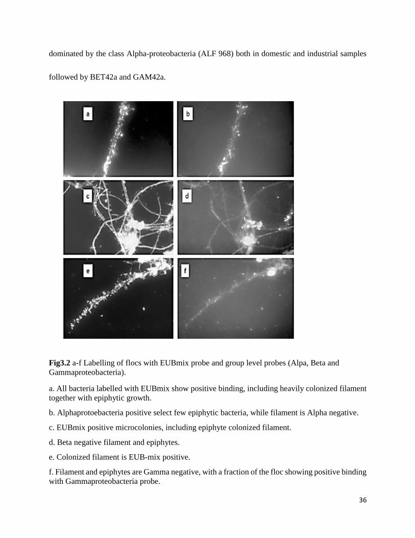
Fig3.2 Labelling of flocs with EUBmix probe and group level probes (Alpa, Beta and
Gammaproteobacteria). ................................................................................................................. 36
Figure 4.1 Irregular positioning of true branches of Nocardia spp., where cells of the filament
protrude at random points along the length of the filament. ......................................................... 46
Fig 4.2 TEM micrographs depicting the presence of fibrillar structures emanating from within
filamentous bacterial cells being targeted by bacterial rods. ........................................................ 49
Figure 5.1 Filamentous bacteria observed under phase contrast at 1000x after final filtration with
75µm pore sized sieve. a, b and c exhibit less compact and diminished floc .d) depicts complete
exposed heavily colonized filament. ............................................................................................. 60
Figure 5.2 Non-viable microflora within compact flocs, organisms towards center of flocs
fluoresce red, while certain organisms towards periphery fluoresced green signaling viability. d)
depicts viability of a filamentous bacteria as it protrudes out of the floc structure. ..................... 61
Figure 5.3 Micrograph illustrating the existence of filamentous bacteria beyond floc structure. 62
viii
Figure 5.4 Micrograph a, b and c depicting the presence of PHB granules within cells of heavily
colonized filamentous bacteria. .................................................................................................... 63
Figure 5.5 Scanning electron microscope micrograph depicting the elements present at surface
level. .............................................................................................................................................. 64
Figure 5.6 Transmission electron microscope micrograph of elements detected at ultrastructural
level. .............................................................................................................................................. 65
ix
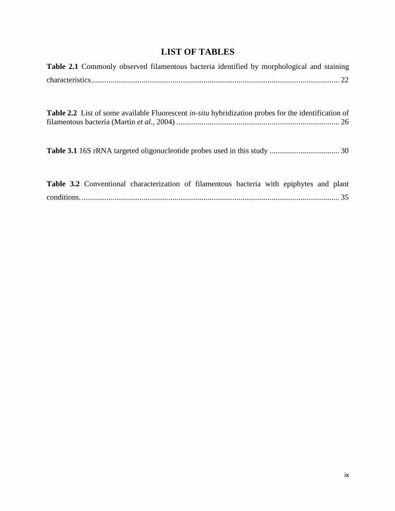
LIST OF TABLES
Table 2.1 Commonly observed filamentous bacteria identified by morphological and staining
characteristics ................................................................................................................................ 22
Table 2.2 List of some available Fluorescent in-situ hybridization probes for the identification of
filamentous bacteria (Martin et al., 2004) .................................................................................... 26
Table 3.1 16S rRNA targeted oligonucleotide probes used in this study .................................... 30
Table 3.2 Conventional characterization of filamentous bacteria with epiphytes and plant
conditions. ..................................................................................................................................... 35
x
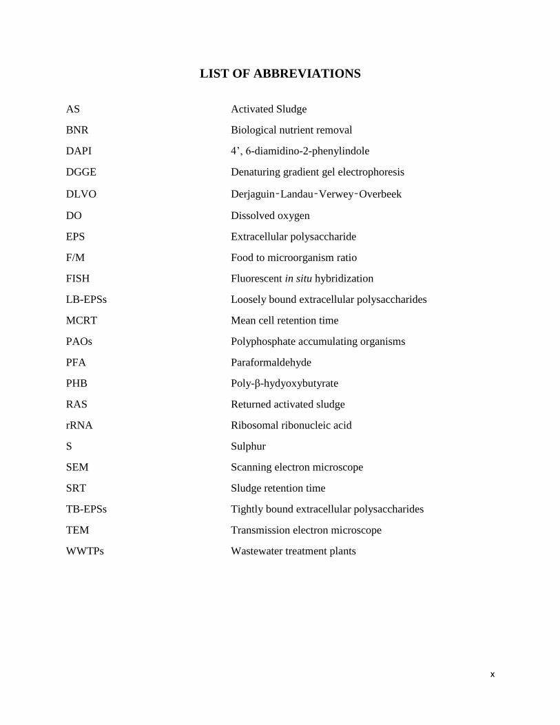
LIST OF ABBREVIATIONS
AS Activated Sludge
BNR Biological nutrient removal
DAPI 4’, 6-diamidino-2-phenylindole
DGGE Denaturing gradient gel electrophoresis
DLVO Derjaguin–Landau–Verwey–Overbeek
DO Dissolved oxygen
EPS Extracellular polysaccharide
F/M Food to microorganism ratio
FISH Fluorescent in situ hybridization
LB-EPSs Loosely bound extracellular polysaccharides
MCRT Mean cell retention time
PAOs Polyphosphate accumulating organisms
PFA Paraformaldehyde
PHB Poly-β-hydyoxybutyrate
RAS Returned activated sludge
rRNA Ribosomal ribonucleic acid
S Sulphur
SEM Scanning electron microscope
SRT Sludge retention time
TB-EPSs Tightly bound extracellular polysaccharides
TEM Transmission electron microscope
WWTPs Wastewater treatment plants
xi
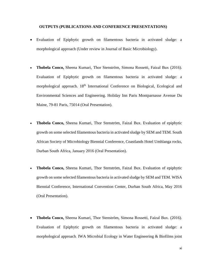
OUTPUTS (PUBLICATIONS AND CONFERENCE PRESENTATIONS)
Evaluation of Epiphytic growth on filamentous bacteria in activated sludge: a
morphological approach (Under review in Journal of Basic Microbiology).
Thobela Conco, Sheena Kumari, Thor Stenström, Simona Rossetti, Faizal Bux (2016).
Evaluation of Epiphytic growth on filamentous bacteria in activated sludge: a
morphological approach. 18th International Conference on Biological, Ecological and
Environmental Sciences and Engineering. Holiday Inn Paris Montparnasse Avenue Du
Maine, 79-81 Paris, 75014 (Oral Presentation).
Thobela Conco, Sheena Kumari, Thor Stenström, Faizal Bux. Evaluation of epiphytic
growth on some selected filamentous bacteria in activated sludge by SEM and TEM. South
African Society of Microbiology Biennial Conference, Coastlands Hotel Umhlanga rocks,
Durban South Africa, January 2016 (Oral Presentation).
Thobela Conco, Sheena Kumari, Thor Stenström, Faizal Bux. Evaluation of epiphytic
growth on some selected filamentous bacteria in activated sludge by SEM and TEM. WISA
Biennial Conference, International Convention Center, Durban South Africa, May 2016
(Oral Presentation).
Thobela Conco, Sheena Kumari, Thor Stenström, Simona Rossetti, Faizal Bux. (2016).
Evaluation of Epiphytic growth on filamentous bacteria in activated sludge: a
morphological approach. IWA Microbial Ecology in Water Engineering & Biofilms joint

xii
specialist conference: Environmental biotechnology: discovering and applying recently
discovered microbial physiologies. The National Museum of Denmark, Copenhagen,
Denmark, September 2016 (Poster presentation).
xiii
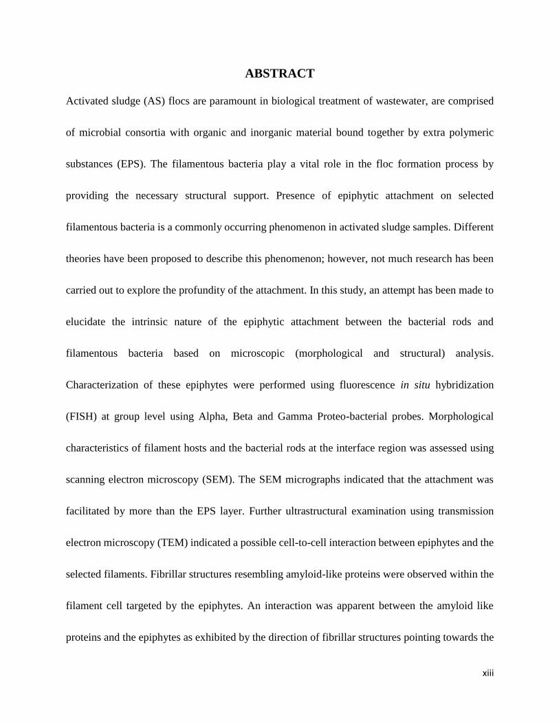
ABSTRACT
Activated sludge (AS) flocs are paramount in biological treatment of wastewater, are comprised
of microbial consortia with organic and inorganic material bound together by extra polymeric
substances (EPS). The filamentous bacteria play a vital role in the floc formation process by
providing the necessary structural support. Presence of epiphytic attachment on selected
filamentous bacteria is a commonly occurring phenomenon in activated sludge samples. Different
theories have been proposed to describe this phenomenon; however, not much research has been
carried out to explore the profundity of the attachment. In this study, an attempt has been made to
elucidate the intrinsic nature of the epiphytic attachment between the bacterial rods and
filamentous bacteria based on microscopic (morphological and structural) analysis.
Characterization of these epiphytes were performed using fluorescence in situ hybridization
(FISH) at group level using Alpha, Beta and Gamma Proteo-bacterial probes. Morphological
characteristics of filament hosts and the bacterial rods at the interface region was assessed using
scanning electron microscopy (SEM). The SEM micrographs indicated that the attachment was
facilitated by more than the EPS layer. Further ultrastructural examination using transmission
electron microscopy (TEM) indicated a possible cell-to-cell interaction between epiphytes and the
selected filaments. Fibrillar structures resembling amyloid-like proteins were observed within the
filament cell targeted by the epiphytes. An interaction was apparent between the amyloid like
proteins and the epiphytes as exhibited by the direction of fibrillar structures pointing towards the
xiv
approaching epiphytes. Common bacterial appendages such as pili and fimbria were absent at the
interface and further noted was the presence of cell membrane extensions on the epiphytic bacteria
protruding towards the targeted filamentous cell. The sheath of host filaments however, remained
intact and unpenetrated, during colonization. Amyloid-like fibrils at interface may potentially play
the role of attachment sites for the attaching epiphytes, as attachment facilitating appendages were
not visualized.
1
1. Chapter One: Introduction
Activated sludge (AS) wastewater treatment plants (WWTPs) are biologically engineered systems
in which nutrients and other compounds are removed by microorganisms. The microbial
population of AS are described from a functional point of view as aerobic heterotrophs, nitrifiers,
denitrifiers, sulphate reducers, iron reducers and phosphate-accumulating organisms (PAOs)
(Thomsen et al., 2004). The AS technology is based on the ability of these functional
microorganisms to form microbial units known as flocs. These bio-aggregated bacterial units
strongly adhere to the filamentous network “backbone” by means of extracellular polymeric
substances (EPS) secreted by bacteria. Within these, certain bacterial rods grow attached to other
microorganisms (Xia et al., 2008). The occurrence of such attached growth is a common
phenomenon in different ecosystems, as in soils or on plant surfaces. In activated sludge, the
colonization of certain filament trichomes by epiphytic bacteria has been observed as a common
occurrence (Xia et al,. 2008).
Presence of epiflora on the trichome has been used as an important criterion for a morphological
classification/identification of filamentous morphotypes in AS (Xia et al., 2008). Filamentous
morphotypes with attached growth that are identified from AS includes Eikelboom types 0041,
1701, and 1851. These filaments are often regarded as unwanted/nuisance filaments that are
involved in bulking and foaming incidence during biological wastewater treatment. In addition,
the operational conditions that favor these filamentous bacteria are well documented as low
dissolved oxygen (DO) levels, low organic loading rates, low substrate concentration gradients
2
and nutrient limitation (Guo et al., 2014). However, not many studies have been carried out to date
to understand the functional role (if any) of epiflora in AS plants. The role of EPS layer in epiphytic
attachment have been proposed by earlier workers (Jenkins et al., 2004). However, not all the
filamentous bacteria in the same floc bearing a sheath have these epiphytes attached to their
surfaces. Filaments such as Thiothrix spp, Type 021N, Gordonia spp are among those without
such attachment. These observations highlights the need for further research in this area to
understand this epiphytic growth beyond the surface attachment, as well as the level of interaction
between filamentous bacteria and their epiphytic counterparts. This also includes an understanding
in the further conditions inducing such attachment to selected filament morphotypes. The literature
currently give limited information on the identity and ecophysiology of microorganisms colonizing
filamentous bacteria in AS (Thomsen et al., 2002; Xia et al., 2008).
The aim of this study was therefore to evaluate the mechanisms used by epiphytic bacteria in
facilitating attachment on to filamentous hosts in AS. Furthermore the aim was to enhance the
understanding of the level of interaction between the filament host and epiphytic bacteria in AS
using microscopic observations. This includes evaluating surface and ultra-structural morphology
of host filament and that of colonizing epiphytic cell by means of SEM and TEM respectively, and
assessing the potential relationship that could exist between the two organisms using a cell viability
test (LIVE/DEAD staining technique).
3
1.1 RESEARCH OBJECTIVES
To achieve the overall aim of this study, specific objectives were established and detailed as
follows:
I. To identify and characterize filamentous bacteria with epiphytic attachment from
different wastewater treatment plants.
The objective focused on analyzing activated sludge samples from different wastewater
treatment plants to identify and characterize the filamentous bacteria with epiphytic attachment
using conventional microscopy and further confirmation using molecular techniques (FISH) in
relation to plan operational conditions.
II. Identification of the structural morphology and possible interaction between
filamentous bacteria and epiphytes.
This objective focused on understanding the structural morphology of filaments and epiphytes
using SEM and further observations at the interface between filaments and epiphytes, using TEM
to assess the level of interaction. .
III. Evaluation of the nature of the relationship between filamentous bacteria.
The objective focused on evaluating the nature of the relationship between filament and its
counterpart based on the availability of storage compounds on the targeted cells and the overall
viability of colonized filamentous bacteria and epiphytes using LIVE/DEAD staining technique.
4
1.2 OUTLINE OF THESIS
Thesis organization is as follows:
Chapter 2: Background study of current and available literature of wastewater treatment,
AS process and AS microbiology. The formation of the microbial floc structure, floc
morphology and its direct influence of the overall treatment process. Identification of
resident filamentous bacteria and their functional role in AS flocculation. Epiphytic growth
and mechanism that facilitates the attachment of epiphytic bacteria to biotic surfaces.
Chapter 3: Identification and characterization of filamentous morphotypes, focusing on
morphological characteristics of filamentous bacteria in activated sludge. Evaluation of
epiphytic growth on morphotypes prone to attached growth. Characterization of epiphytes
by molecular technique (FISH).
Chapter 4: Focuses on evaluating the morphology of filamentous host and epiphytic
counterparts by use of SEM and TEM at surface and ultrastructural levels, respectively.
Investigation of mechanism involved in attachment of epiphytes at interface. Assessing the
selectivity of filament cells colonized by epiphytes all length of filament.
5
Chapter 5: Evaluation of the interaction between epiphytes and filamentous bacteria under
nutritional stress conditions. The determination of the nature of relationship between host
and epiphyte. Elemental analysis of surface of epiphytes, interface and colonized filament
cells.
Chapter 6: Summary of the general conclusions made in the current study and
recommendations for future continuation in further investigation of the present work.
6
2. Chapter Two: Literature Review
2.1 Activated Sludge Process.
The emergence of wastewater treatment processes to solve water quality issues led to the
development of AS systems, as the main biological process to treat wastewater (Mesquita et al.,
2013). The principal of the AS system is based on the action of a highly complex mixture of
microbial populations that are crucial for biodegradation and nutrient removal. Bacteria represent
around 95% of the bio-volume of AS and play a key role in biological removal of organic carbon,
ammonium and phosphate from wastewater. In an activated sludge process, the biodegradation is
facilitated by microbial aggregates (flocs) formed by bio-flocculation of suspended
microorganisms during biological treatment. This is followed by subsequent settling of the
microbial flocs in the secondary clarifiers where the treated effluent is separated from the biomass
(Mesquita et al., 2013).
7
2.2 Activated Sludge Flocculation
The AS resident microorganisms aggregate to form microbial units, which are known as activated
sludge flocs (Perez et al., 2006). The process in which bacteria aggregate and adhere to one another
forming flocs is termed bioflocculation, which is of primary importance for the AS systems (Van
Dierdonck et al., 2012). The AS flocs are considered to be a collection of several particles, e.g.,
primary particles and microcolonies, held together by bridging by EPS and cations- and
hydrophobic and Derjaguin–Landau–Verwey–Overbeek (DLVO)-type of interactions (Van
Dierdonck et al., 2012). The EPS, a complex high-molecular-weight mixture of polymers, forms
an adhesive matrix for the micro-colonies and bacteria to attach each other to and are found both
on exterior and interior surfaces of sludge flocs (Sheng et al, 2010). The presence of EPS is
resourceful as it binds with cells through complex interactions to form a vast net-like structure
protecting against toxic substances as well serving as energy sources during nutrient shortage
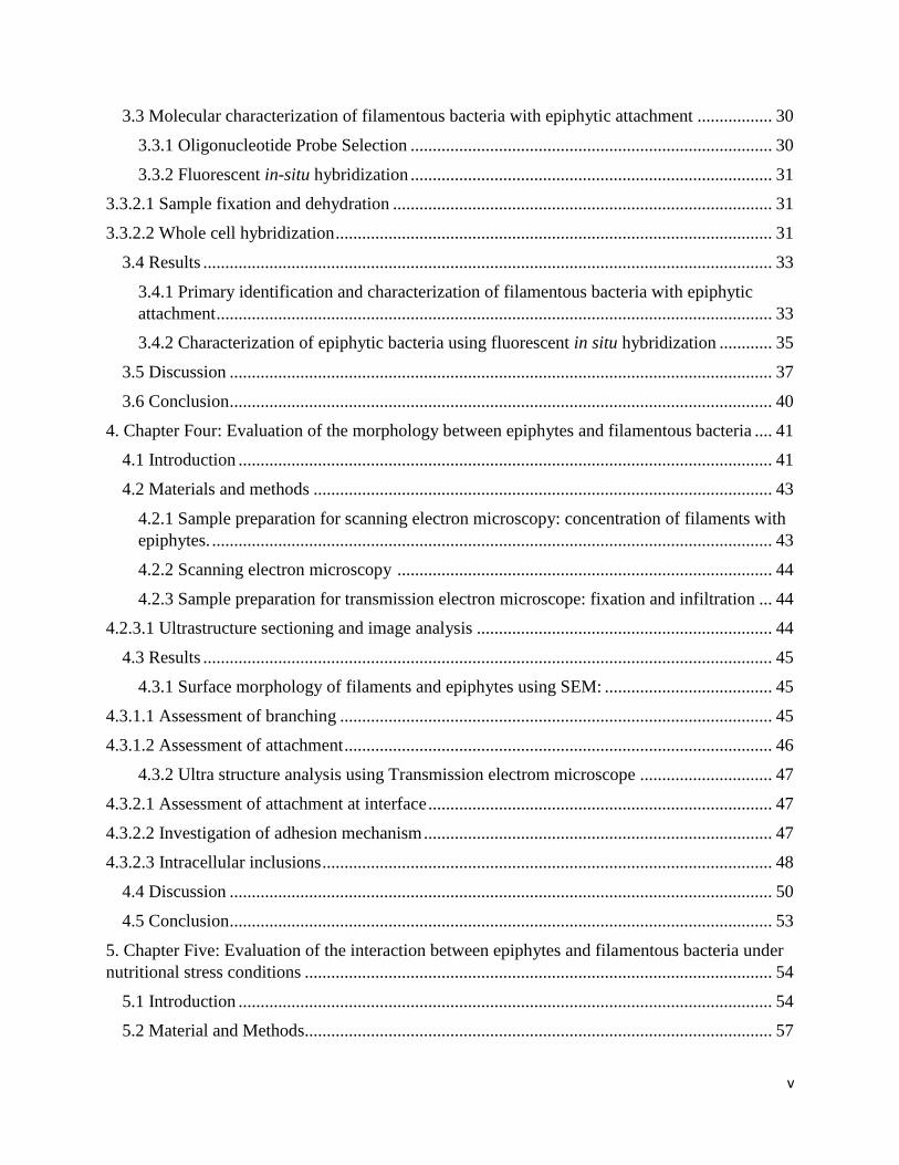
(Sheng et al., 2010). The significant contribution of the EPS towards the formation of strong
microbial flocs, is due to its dynamic double layered structure, where tightly bound EPSs (TB-
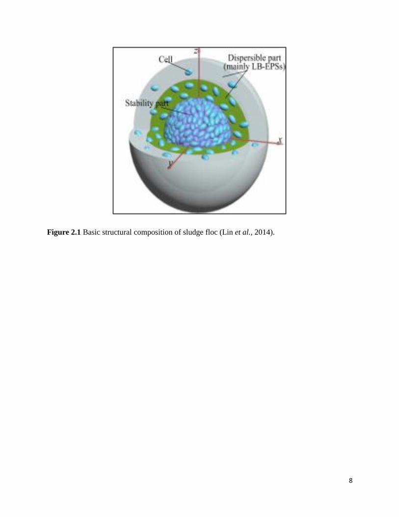
EPSs) forms the inner layer and loosely bound EPSs (LB-EPSs) diffuses into the outer layer (Lin
et al., 2014) (Fig 2.1). This contribution is of key importance for an efficient solid-liquid separation
of AS treatment systems (Thomsen et al., 2004).
8
Figure 2.1 Basic structural composition of sludge floc (Lin et al., 2014).
9
2.3 Floc structure and its microbial composition
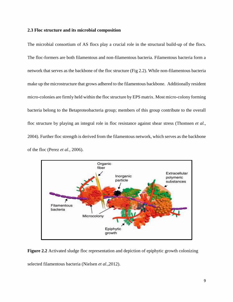
The microbial consortium of AS flocs play a crucial role in the structural build-up of the flocs.
The floc-formers are both filamentous and non-filamentous bacteria. Filamentous bacteria form a
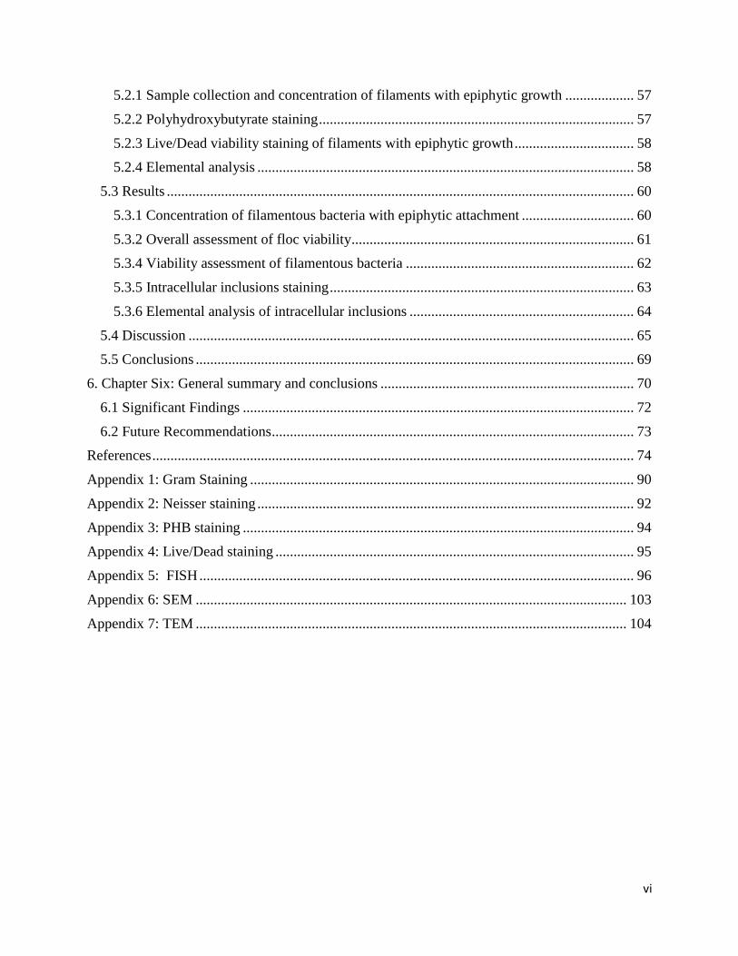
network that serves as the backbone of the floc structure (Fig 2.2). While non-filamentous bacteria
make up the microstructure that grows adhered to the filamentous backbone. Additionally resident
micro-colonies are firmly held within the floc structure by EPS matrix. Most micro-colony forming
bacteria belong to the Betaproteobacteria group; members of this group contribute to the overall
floc structure by playing an integral role in floc resistance against shear stress (Thomsen et al.,
2004). Further floc strength is derived from the filamentous network, which serves as the backbone
of the floc (Perez et al., 2006).
Figure 2.2 Activated sludge floc representation and depiction of epiphytic growth colonizing
selected filamentous bacteria (Nielsen et al.,2012).
10
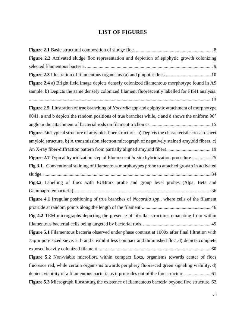
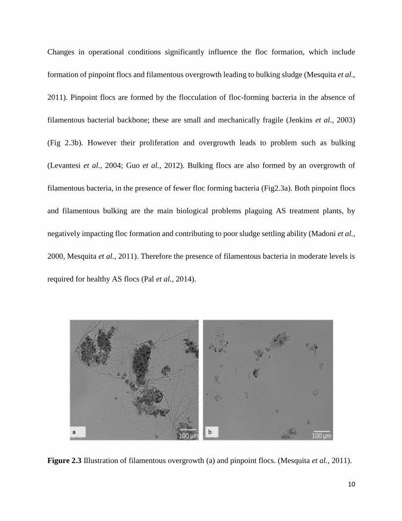
Changes in operational conditions significantly influence the floc formation, which include
formation of pinpoint flocs and filamentous overgrowth leading to bulking sludge (Mesquita et al.,
2011). Pinpoint flocs are formed by the flocculation of floc-forming bacteria in the absence of
filamentous bacterial backbone; these are small and mechanically fragile (Jenkins et al., 2003)
(Fig 2.3b). However their proliferation and overgrowth leads to problem such as bulking
(Levantesi et al., 2004; Guo et al., 2012). Bulking flocs are also formed by an overgrowth of
filamentous bacteria, in the presence of fewer floc forming bacteria (Fig2.3a). Both pinpoint flocs
and filamentous bulking are the main biological problems plaguing AS treatment plants, by
negatively impacting floc formation and contributing to poor sludge settling ability (Madoni et al.,
2000, Mesquita et al., 2011). Therefore the presence of filamentous bacteria in moderate levels is
required for healthy AS flocs (Pal et al., 2014).
Figure 2.3 Illustration of filamentous overgrowth (a) and pinpoint flocs. (Mesquita et al., 2011).
11
2.4 Filamentous bacteria
Filamentous bacteria are an important component of the AS process, maintaining the rigid
‘‘backbone’’ structure of AS flocs (Juang 2005; Pal et al., 2014). Their absence or presence in
excessive numbers significantly affects the AS floc structure and further leads to process problems
that affect the overall treatment process, these include bulking and foaming. A wide range of
filaments are identified as causative agents of bulking. Among these Microthrix parvicella, Types
0092, 0041 and 0675 are apparently the major morphotype filaments, mainly responsible for the
bulking events observed in biological nutrient removal (BNR) activated sludge systems (Martins
et al., 2004). The latter two morphotyes 0041 and 0675 are observed with epiphytic bacteria in AS
(Nielsen et al., 2009). This unique morphological trait is an important identification criterion for
these morphotypes. However, information surrounding the identity of the epiphytic bacteria
attaching to these morphotypes is still unclear (Xia et al., 2008). Therefore filament-type specific
control strategies require a complete understanding of each filamentous bacteria, based on proper
identification of the filamentous bacteria involved (Bradford et al., 1996; Martins et al., 2004).
12
2.5 Epiphytic growth and its occurrence in activated sludge
Epiphytic growth is a general and widespread phenomenon occurring in many regimes in the
biosphere. The growth on human tissues in relation to diseases and that on plant surfaces, where
interaction between the microorganism and plants are well described. Their presence in water
bodies where they play an important role in the degradation of organic substances has been
documented (Lalke-Porczyk and Donderski, 2003).
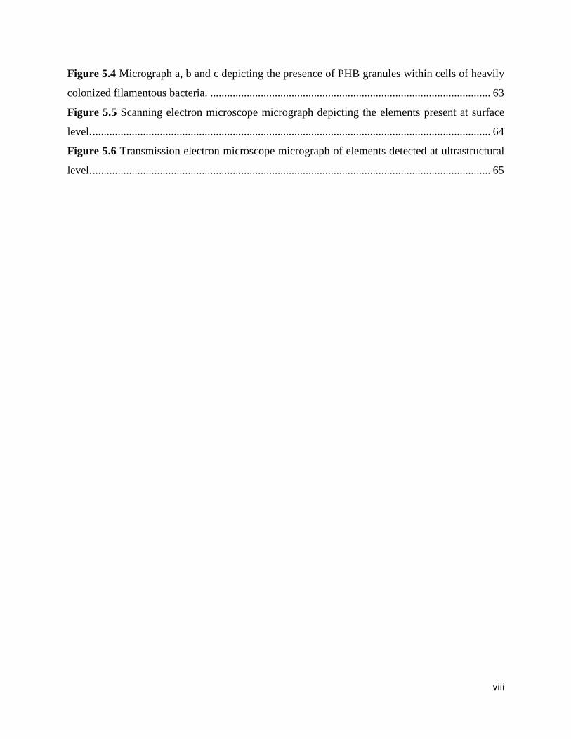
In AS, the presence and growth of rod-shaped bacteria colonizing selected filamentous
morphotypes has been observed over the years (Xia et al., 2008). The sheaths of large filamentous
bacteria (e.g. Beggiatoa and Thioploca) are often colonized by the epiphytes which are believed
to utilize the sheath material for their growth (Xia et al., 2008). Eikelboom morphotypes Type
0041, Type 1701 and Type 1851 implicated in sludge bulking have also been observed to be
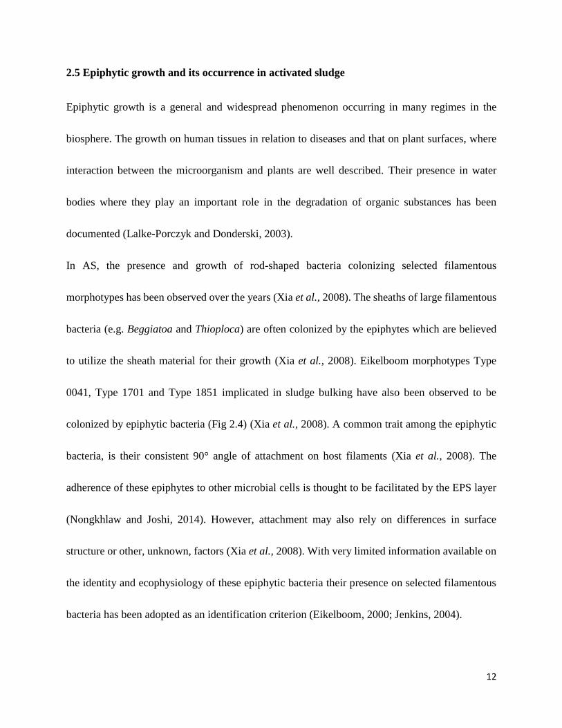
colonized by epiphytic bacteria (Fig 2.4) (Xia et al., 2008). A common trait among the epiphytic
bacteria, is their consistent 90° angle of attachment on host filaments (Xia et al., 2008). The
adherence of these epiphytes to other microbial cells is thought to be facilitated by the EPS layer
(Nongkhlaw and Joshi, 2014). However, attachment may also rely on differences in surface
structure or other, unknown, factors (Xia et al., 2008). With very limited information available on
the identity and ecophysiology of these epiphytic bacteria their presence on selected filamentous
bacteria has been adopted as an identification criterion (Eikelboom, 2000; Jenkins, 2004).
13
Figure 2.4 a) Bright field image depicts a densely colonized filamentous morphotype found in a
AS sample. b) Depicts the same densely colonized filament fluorescently labelled for FISH
analysis. (Xia et al., 2007).
14
2.5.1 Comparison of epiphytic growth with branching of filamentous bacteria in activated
sludge
In AS, species from the two genera Gordonia and Skermania are frequently encountered, often
associated with severe foaming episodes. Their morphotypes are commonly described,
respectively, as the right-angled branching Gordonia amarae-like organisms and the acute-angled
pine tree-like organism (Nielsen et al. 2009). The presence of branching is an important
characteristic used to ascertain the identity of these morphotypes. Due to the right angle appearance
of true branching, it often resembles epiphytic growth exhibited by bacterial rods on certain
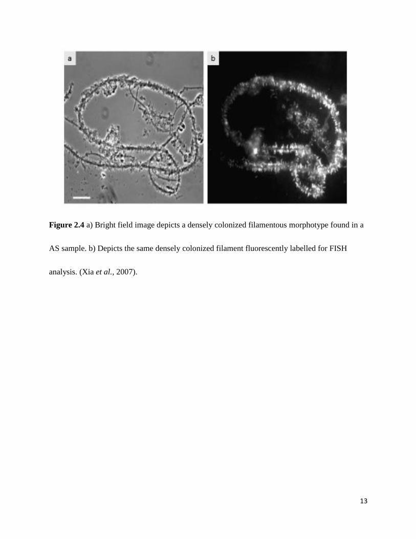
filamentous bacteria (Fig 2.5). Epiphytic attachment is consistently at a 90° angle, where bacterial
rods adhere to surfaces along the length of filamentous bacteria (Xia et al., 2008). True branching
however occurs at any random point on the filament surface.
15
Figure 2.5. Illustration of true branching of Nocardia spp and epiphytic attachment of morphotype
0041. a and b depicts the random positions of true branches while, c and d shows the uniform 90°
angle in the attachment of bacterial rods on filament trichomes (Jenkins, 2004).
16
2.6 Bacterial adhesion and structures that mediate epiphytic attachment
Bacteria generally exist in one of two types of population: planktonic, freely existing in bulk
solution, and sessile, as a unit attached to a surface (Garret et al., 2008). The immediate attachment
of bacteria to an inert or living surface is the initial step in microbial adhesion and colonization
(Hori and Matsumoto, 2010; Habimana et al., 2014). Microbial adhesion is beneficial in
bioreactors for wastewater treatment where the common growth pattern of bacteria in AS is on
available surfaces rather than in the surrounding aqueous phase (Katsikogianni and Missirli, 2004;
Hori and Matsumoto, 2010). Mechanisms by which bacteria are transported to a surface can
include Brownian motion, sedimentation due to differences in specific gravity between the bacteria
and the bulk liquid, or convective mass transport, by which cells are physically transported towards
the surface by the movement of the bulk fluids (Hori and Mastumoto, 2010; Habimana et al.,
2014).
Upon approaching a surface, bacteria must overcome an energy barrier to establish direct contact
with the surface (Habimana et al., 2014). Physiochemical variables further define the interaction
between the bacterial cell surface and the surface of interest (Dunne, 2002). The final outcome of
adhesion is, however determined by the attraction and repulsion forces existing between the
preferred surface and that of the bacterial cell (Dunne, 2002; Garrett et al., 2008). In instances of
repulsion, the repulsion charge between two negatively charged surfaces is minimized, when
bacteria approach a surface with the smaller face of one of their poles, initiate attachment, and
17
either remain attached only at the pole (Young, 2006). Furthermore the net repulsion between two
surfaces can be overcome by specific molecular interactions mediated by adhesins located on
structures extending from the cell surface, such as pili (Dunne, 2002). These cell surface
appendages, viz, bacterial pili are commonly used by bacteria as surface grabbing devices to
facilitate attachment (Young, 2006). Additionally the use of amyloid-like adhesions have been
utilized by bacteria in the formation of environmental biofilms (Jordal et al., 2009).
2.6.1 Bacterial pili
Pili are non-flagellar, proteinaceous, multi-subunit surface appendages, with lengths between
hundreds of nanometers to several micrometers that form long fibrous structures (Hori and
Matsumoto, 2010; Kline et al., 2010). Pili are used by bacteria both as common surface attachment
appendages and for specific attachment. When utilized by bacteria for adhesion purposes, pili are
capable of piercing energy barriers as well as altering cell adhesion behavior (Hori and Matsumoto,
2010). In addition, these pili are responsible for maintaining contact during the first stages of
bacterial colonization (Bullitt and Makowski, 1998). This is achieved by the successful recognition
and binding of these specialized surface structures to their host receptors (Bullitt and Makowski,
1998).
18
2.6.2 Amyloid like structures
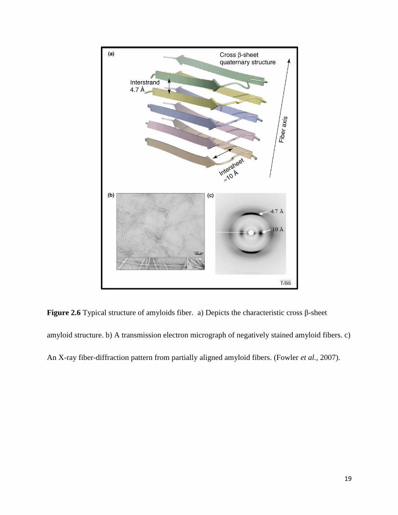
Amyloid can be defined as orderly repeats of protein molecules arranged in a cross-β structure, in
which the β strands are perpendicular to the fiber axis (Fig 2.6) (Otzen and Nielsen, 2007; Freire
et al., 2014). The outcome of the presence and formation of amyloid proteins has been associated
with diseases such as Alzheimers, Diabetes and Parkinsons in humans, where each disease is as a
result of specific protein or peptide aggregation (Fowler et al., 2007; Rambaran and Serpell, 2008).
From bacteria to humans, the characteristic cross-β-strand structure of amyloids, has been
observed to be common (Romero et al., 2010; Villar-Piqué and Ventura, 2012). The expression of
amyloid in their various kinds are believed to be important for aggregation and for making strong
attachment to the surfaces (Otzen and Nielsen, 2007). Curli which are amyloid fibers encoded by
Escherischia coli and other Enterobacteriaceae such as Salmonella spp. have been proposed as a
virulence factor in human disease (Hung et al., 2014).
19
Figure 2.6 Typical structure of amyloids fiber. a) Depicts the characteristic cross β-sheet
amyloid structure. b) A transmission electron micrograph of negatively stained amyloid fibers. c)
An X-ray fiber-diffraction pattern from partially aligned amyloid fibers. (Fowler et al., 2007).
20
Amyloid has been observed in bacteria from wastewater, where Otzen (2010) identified their
occurrence in 5-40% of assessed total bacterial species. In general functional amyloids often may
aid in adhesion to surfaces and tissues as well as changing surface properties (Blanco et al., 2012).
During bioremediation, amyloids play a pivotal role in forming aggregative clusters in the matrix
of AS-derived flocs (Blanco et al., 2012). Filamentous bacteria from the phylum Chloroflexi, have
been observed to express amyloids as part of the sheath or close to the septum between the
individual cells in the filaments (Otzen, 2010). A unique attribute of these Chloroflexi filaments is
the presence of epiphytic bacteria e.g. Candidatus Epiflobacter spp. (Saprospiraceae,
Bacteroidetes) that specialize in protein degradation, excreting high levels of proteases (Otzen
2010). Otzen (2010), speculated that the excreted proteases feasted on the accessible amyloid.
However an interesting observation made by Jordal et al. (2009) revealed that amyloids may be
deeply embedded in the cell envelope and not easily accessible (Jordal et al., 2009).
21
2.7 Filamentous identification
The conventional identification of AS filamentous bacteria has relied on the characterization of
morphological traits and response to different stains (Nielsen et al., 2009). These characteristics
have been incorporated into identification keys by Jenkins et al. (1993, 2004) and Ekeilboom
(2000). Most are still referred to as morphological types (e.g. Type 021N, Type 1863), using a
naming system based exclusively on their microscopic features (Beer et al., 2002).
2.7.1 Conventional Identification
Conventional staining techniques such as Gram and Neisser stains has been central in identifying
filamentous bacteria in AS (Jenkins, 1993) combined with microscopic direct visualization and
crude characterization (Moter and Göbel, 2000) where filament dimensions, the presence of a
sheath and attached particles (epiphytic bacteria) on the sheath are central features as outlined by
Sevior (2010) in Table 2.1.
Additionally, staining with special stains for the detection of intracellularly stored products aids in
further characterization. These include sulphur globules Sulphur oxidation test (S test) where the
globules appear bright yellow upon addition of sodium sulphide (Na2S.9H20) solution. The
presence of sulphur granules is a characteristic of Thiothrix spp. (Wagner et al., 1994). Further
poly-β-hydroxyl butyrate (PHB) staining is used, where polyhydroxybutyrate, appears as bluish
22
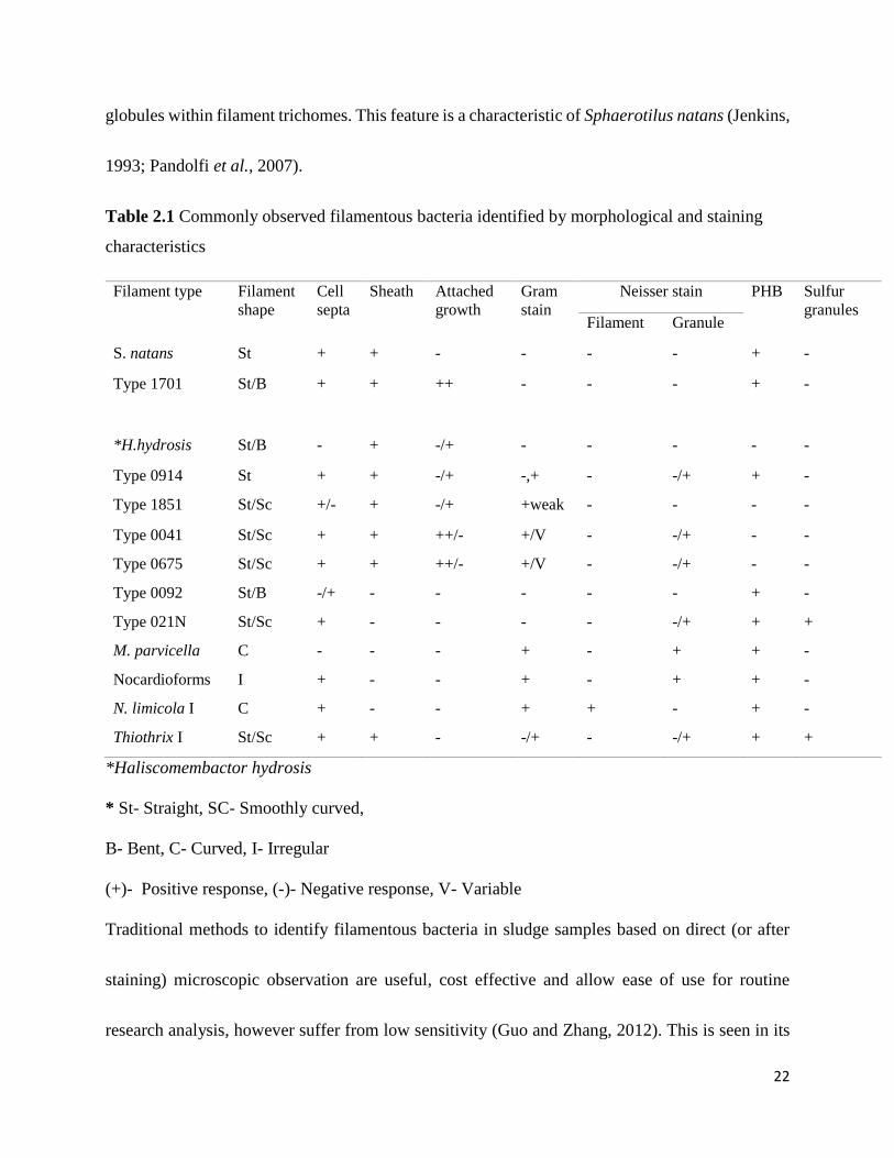
globules within filament trichomes. This feature is a characteristic of Sphaerotilus natans (Jenkins,
1993; Pandolfi et al., 2007).
Table 2.1 Commonly observed filamentous bacteria identified by morphological and staining
characteristics
Filament type Filament
shape
Cell
septa
Sheath Attached
growth
Gram
stain
Neisser stain PHB Sulfur
granules Filament Granule
S. natans St + + - - - - + -
Type 1701 St/B + + ++ - - - + -
*H.hydrosis St/B - + -/+ - - - - -
Type 0914 St + + -/+ -,+ - -/+ + -
Type 1851 St/Sc +/- + -/+ +weak - - - -
Type 0041 St/Sc + + ++/- +/V - -/+ - -
Type 0675 St/Sc + + ++/- +/V - -/+ - -
Type 0092 St/B -/+ - - - - - + -
Type 021N St/Sc + - - - - -/+ + +
M. parvicella C - - - + - + + -
Nocardioforms I + - - + - + + -
N. limicola I C + - - + + - + -
Thiothrix I St/Sc + + - -/+ - -/+ + +
*Haliscomembactor hydrosis
* St- Straight, SC- Smoothly curved,
B- Bent, C- Curved, I- Irregular
(+)- Positive response, (-)- Negative response, V- Variable
Traditional methods to identify filamentous bacteria in sludge samples based on direct (or after
staining) microscopic observation are useful, cost effective and allow ease of use for routine
research analysis, however suffer from low sensitivity (Guo and Zhang, 2012). This is seen in its
23
failure to differentiate between morphologically similar organisms with phylogenetic variance.
Further, these methods are limited in differentiating the same species whose morphology has been
altered by changes in environmental conditions (Martins et al., 2004). This morphological
approach for identification by use of staining alone, has therefore proved to be unreliable in the
identification of filamentous bacteria (Mielczarek et al., 2012).
24
2.7.2 Molecular Characterization of Filamentous Bacteria in Wastewater
The use of molecular techniques has complemented conventional identification techniques by a
non-destructive in situ analysis (Merkel et al., 1999). Further, this has provided the possibility of
identifying specific populations of microorganisms in their native habitat without the need to
isolate them (Sanz and Köchling, 2007). Molecular techniques such as FISH and Denaturing
gradient gel electrophoresis (DGGE) have been extensively applied to the study of microbial
consortia of flocs in activated sludge (Sanz and Köchling, 2007) (Martins et al., 2004). FISH is a
cytogenetic technique developed in the early 1980s, for the targeting of specific nucleic sequences
(Hu et al., 2014). FISH relies on the use of fluorescently labelled oligonucleotide probes, that
target and hybridize to highly conserved regions of microbial rRNA within intact cells (Moter and
Göbel, 2000; Schmidt et al., 2012). The 16 rRNAs sequence domains are the main target molecules
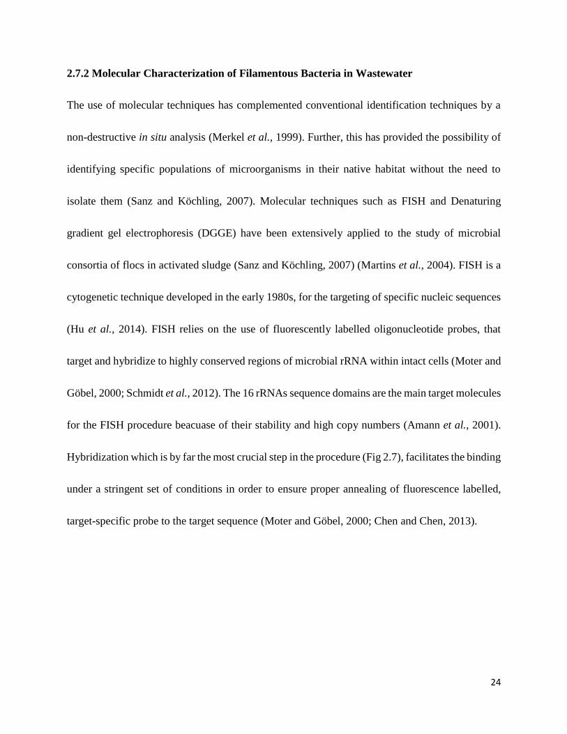
for the FISH procedure beacuase of their stability and high copy numbers (Amann et al., 2001).
Hybridization which is by far the most crucial step in the procedure (Fig 2.7), facilitates the binding
under a stringent set of conditions in order to ensure proper annealing of fluorescence labelled,
target-specific probe to the target sequence (Moter and Göbel, 2000; Chen and Chen, 2013).
25
Figure 2.7 Typical hybridization step of FISH procedure (Eickhorst and Tippkötter, 2008).
The specificity of both probe and target sequence has made FISH a method of choice in the reliable
identification and enumeration of individual bacterial cells, together with microscopic
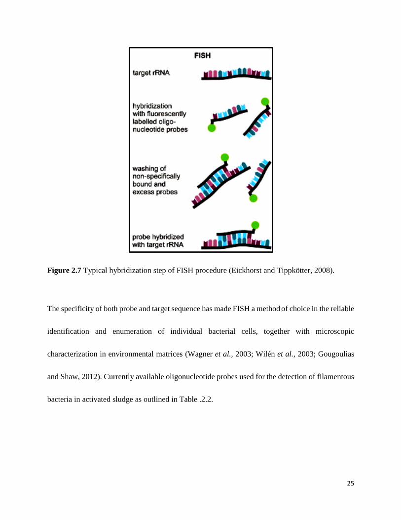
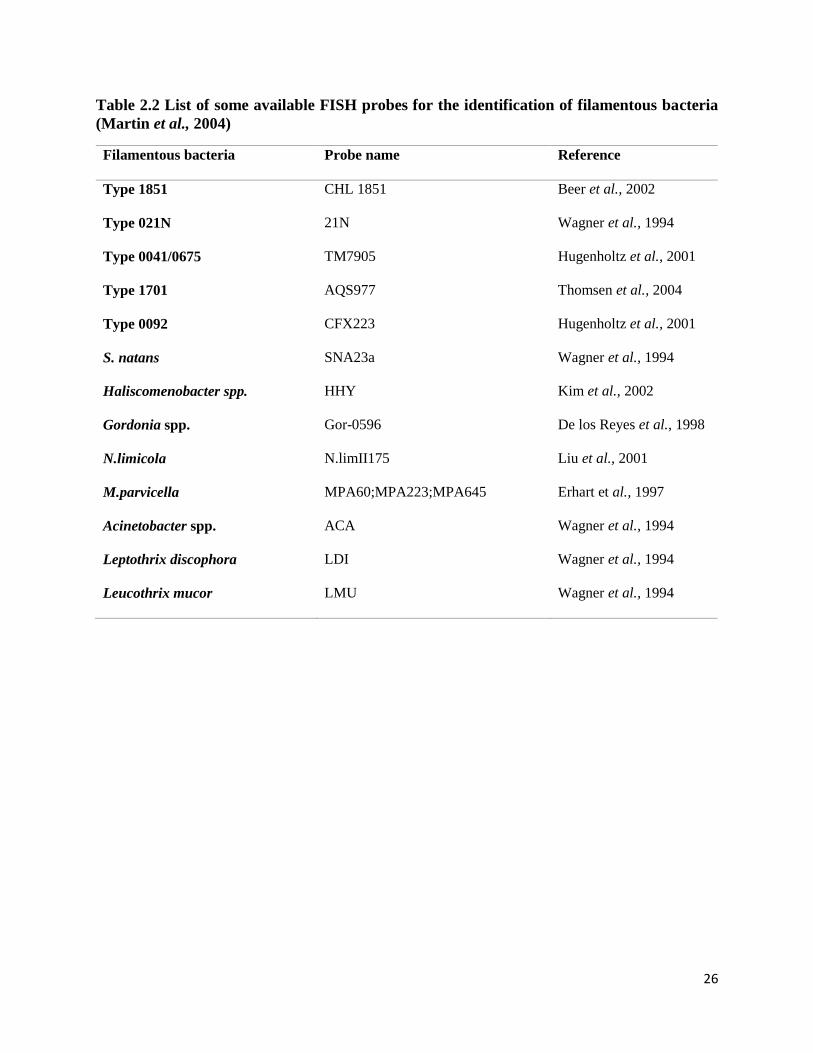
characterization in environmental matrices (Wagner et al., 2003; Wilén et al., 2003; Gougoulias
and Shaw, 2012). Currently available oligonucleotide probes used for the detection of filamentous
bacteria in activated sludge as outlined in Table .2.2.
26
Table 2.2 List of some available FISH probes for the identification of filamentous bacteria
(Martin et al., 2004)
Filamentous bacteria Probe name Reference
Type 1851 CHL 1851 Beer et al., 2002
Type 021N 21N Wagner et al., 1994
Type 0041/0675 TM7905 Hugenholtz et al., 2001
Type 1701 AQS977 Thomsen et al., 2004
Type 0092 CFX223 Hugenholtz et al., 2001
S. natans SNA23a Wagner et al., 1994
Haliscomenobacter spp. HHY Kim et al., 2002
Gordonia spp. Gor-0596 De los Reyes et al., 1998
N.limicola N.limII175 Liu et al., 2001
M.parvicella MPA60;MPA223;MPA645 Erhart et al., 1997
Acinetobacter spp. ACA Wagner et al., 1994
Leptothrix discophora LDI Wagner et al., 1994
Leucothrix mucor LMU Wagner et al., 1994
27
3. Chapter Three: Identification and characterization of filaments and
epiphytic bacteria in activated sludge
3.1 Introduction
The external colonization of Eikelboom morphotypes 0041, 0675 and 1851 commonly termed as
attached growth has for a long time been used in identification keys, for the characterization of
these morphotypes (Eikelboom, 2000; Jenkins, 2004). This has been helpfully in identifying
filamentous morphotypes in AS, which are believed to be causative agents of bulking (Kragelund
et al., 2007). Additionally, the existence of these morphotypes has been identified in conditions of
low dissolved oxygen, low food to microorganism and long mean cell retention time using their
unique appearance (Guo et al., 2014). However, very limited information is available in literature
on the identity and ecophysiology of microorganisms colonizing filamentous bacteria in activated
sludge (Xia et al., 2008).
Conventional identification based on staining (Gram and Neisser) and microscopy is limited in
ascertaining the species level identification of epiphytes on filamentous bacteria. This is further
complicated by the lack of pure culture representatives, as knowledge surrounding the exact
environments for their growth is currently unavailable (Stewart, 2012).
The use of molecular methods in ascertaining the identity of epiphytic bacteria, is crucial in
bridging gaps availed by traditional microscopic and culture dependent techniques (Martins et al.,
28
2004). In a detailed study by Xia et al. (2008) investigating the presence of epiphytic bacteria
colonizing filamentous bacteria it was found that most epi-flora bacteria hybridized with an
oligonucleotide probe designed to target most of the members of the family Saprospiraceae in the
phylum Bacteriodetes (Xia et al., 2008). The aim of this aspect of the study was to implement
culture independent FISH techniques for the group level characterization of filamentous
morphotypes and epiphytic bacteria attaching to their surfaces in activated sludge samples from in
and around Durban, South Africa.
29
3.2 Materials and Methods
3.2.1 Sample collection
AS samples were collected from the aeration tanks and returned activated sludge (RAS) streams
of different WWTP treating both domestic and industrial wastewaters in Kwa-Zulu Natal, South
Africa (Table 3.2). Grab samples of 750 mL were collected and stored at 4°C during transportation
for microscopic analysis within 24 h period of collection.
3.2.2 Preliminary characterization and identification of filamentous bacteria with epiphytic
attachment using conventional staining techniques
Preliminary identification and characterization of filamentous morphotypes prone to epiphytic
growth was achieved by the use of microscopy and staining techniques. Grams staining (Appendix
1) and Neisser staining (Appendix 2) was performed on air dried samples, to assess the presence
and abundance of epiphytic growth on filamentous bacteria in and around the floc. Morphological
assessment of the morphotypes taken into consideration in the identification exercise were
presence of sheath and filament shape, as outlined by Eikelboom (2000) and Jenkins et al. (2004)
(Table 2.1). Filamentous bacteria prone to epiphytic growth were characterized and identified
based on the morphological features and staining reactions to both stains in accordance with the
guidelines of Jenkins et al. (2004).
30
3.3 Molecular characterization of filamentous bacteria with epiphytic attachment
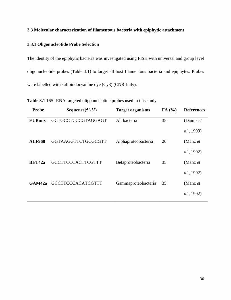
3.3.1 Oligonucleotide Probe Selection
The identity of the epiphytic bacteria was investigated using FISH with universal and group level
oligonucleotide probes (Table 3.1) to target all host filamentous bacteria and epiphytes. Probes
were labelled with sulfoindocyanine dye (Cy3) (CNR-Italy).
Table 3.1 16S rRNA targeted oligonucleotide probes used in this study
Probe Sequence(5’-3’) Target organisms FA (%) References
EUBmix GCTGCCTCCCGTAGGAGT All bacteria 35 (Daims et
al., 1999)
ALF968 GGTAAGGTTCTGCGCGTT Alphaproteobacteria 20 (Manz et
al., 1992)
BET42a GCCTTCCCACTTCGTTT Betaproteobacteria 35 (Manz et
al., 1992)
GAM42a GCCTTCCCACATCGTTT Gammaproteobacteria 35 (Manz et
al., 1992)
31
3.3.2 Fluorescence In-Situ Hybridization
3.3.2.1 Sample fixation and dehydration
Samples were fixed in 4% Paraformaldehyde (PFA) and stored at -20ºC for further analysis as per
the protocol by Amann (1995a). Previously fixed samples (10 µL) were spotted on wells of teflon
slides pre-coated with Poly-L- Lysine (Sigma Diagnostics, USA). Slides were thereafter air dried
and followed by dehydration in an ethanol series (50%, 80% and 100%) for 3 min incubation
period each, for the removal of excess water.
3.3.2.2 Whole cell hybridization
Hybridization was performed as previously described by Amann (1995). A range of formamide
concentrations was used for hybridization buffer preparation (Table 3.1) in 2 mL microcentrifuge
tubes. Using the freshly prepared buffers, all probes were diluted to obtain a working solution with
a concentration of 50 ng/µL. The buffer and probe mixture was then added to dehydrated samples
in wells. Remaining buffer was used to moisten tissue paper placed in 50 mL hybridization
chambers. Slides were introduced into hybridization chambers and incubation was carried out at
46°C overnight. Following hybridization, slides were rinsed with warm wash buffers and inserted
into wash buffer tubes which were then placed in water bath and incubated at 48°C for 10 min.
Afterwards the washed slides were rinsed with cold milliQ water. Following the wash step, slides
were air dried in a vertical position. Hybridized samples were counter-stained with 3µL of DAPI
32
(4’,6-diamidino-2-phenylindole) and allowed to stand at room temperature in the dark for 10 min
and thereafter rinsed with warm milliQ water. Dry Vector shield mounting agent drops were added
to wells and coverslips placed on; to these pressure was applied to ensure even distribution of
mounting agent. The slides were viewed using an Epifluorescence microscope (Olympus BX51).
33
3.4 Results
3.4.1 Primary identification and characterization of filamentous bacteria with epiphytic
attachment
Conventional staining techniques were employed to identify the specific filamentous bacteria
prone to epiphytic attachment as shown in Table 3.2. The common filamentous bacteria with
epiphytes viz. Type 0041, 0675, and 1851 were detected from all the samples analyzed and were
found to be more abundant in domestic wastewater as compared to the industrial samples. The
Eikelboom Type 0041 was found to be the dominant filamentous bacteria with epiphytes both in
domestic and industrial samples which was followed by Type 1851. Observations revealed a
variation in the abundance of epiphytes colonizing specific filaments in domestic and industrial
systems. It was interesting to note that the filamentous morphotype 0041 exhibited dense epiphytic
attachment in domestic wastewater samples compared to the industrial sample, while Type 1851
showed sparse attachment in both domestic and industrial samples (Fig 3.1a). The common plant
operational conditions that this phenomenon was observed to be prevalent under Low F/M and
DO (Table 3.2). Further, the presence of Type 1851 as a single bent filament was accompanied by
dense epiphytic attachment observed on the filament when in bundle formation (Fig 3.1 d).
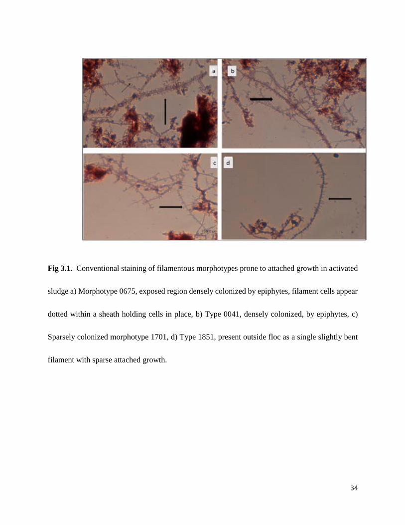
34
Fig 3.1. Conventional staining of filamentous morphotypes prone to attached growth in activated
sludge a) Morphotype 0675, exposed region densely colonized by epiphytes, filament cells appear
dotted within a sheath holding cells in place, b) Type 0041, densely colonized, by epiphytes, c)
Sparsely colonized morphotype 1701, d) Type 1851, present outside floc as a single slightly bent
filament with sparse attached growth.
35
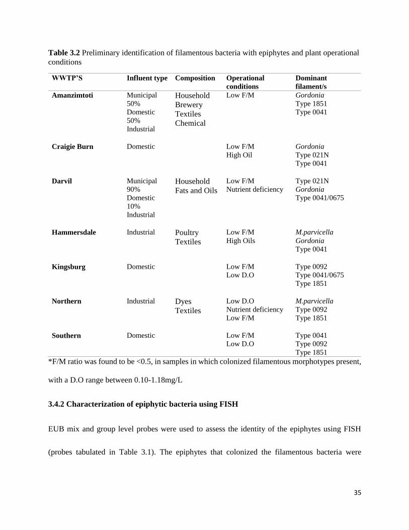
Table 3.2 Preliminary identification of filamentous bacteria with epiphytes and plant operational
conditions
WWTP’S Influent type Composition Operational
conditions Dominant
filament/s Amanzimtoti Municipal
50%
Domestic
50%
Industrial
Household
Brewery
Textiles
Chemical
Low F/M Gordonia
Type 1851
Type 0041
Craigie Burn Domestic Low F/M
High Oil Gordonia
Type 021N
Type 0041
Darvil Municipal
90%
Domestic
10%
Industrial
Household
Fats and Oils
Low F/M
Nutrient deficiency Type 021N
Gordonia
Type 0041/0675
Hammersdale Industrial Poultry
Textiles
Low F/M
High Oils M.parvicella
Gordonia
Type 0041
Kingsburg Domestic Low F/M
Low D.O Type 0092
Type 0041/0675
Type 1851
Northern Industrial Dyes
Textiles
Low D.O
Nutrient deficiency
Low F/M
M.parvicella
Type 0092
Type 1851
Southern Domestic Low F/M
Low D.O Type 0041
Type 0092
Type 1851
*F/M ratio was found to be <0.5, in samples in which colonized filamentous morphotypes present,
with a D.O range between 0.10-1.18mg/L
3.4.2 Characterization of epiphytic bacteria using FISH
EUB mix and group level probes were used to assess the identity of the epiphytes using FISH
(probes tabulated in Table 3.1). The epiphytes that colonized the filamentous bacteria were
36
dominated by the class Alpha-proteobacteria (ALF 968) both in domestic and industrial samples
followed by BET42a and GAM42a.
Fig3.2 a-f Labelling of flocs with EUBmix probe and group level probes (Alpa, Beta and
Gammaproteobacteria).
a. All bacteria labelled with EUBmix show positive binding, including heavily colonized filament
together with epiphytic growth.
b. Alphaprotoebacteria positive select few epiphytic bacteria, while filament is Alpha negative.
c. EUBmix positive microcolonies, including epiphyte colonized filament.
d. Beta negative filament and epiphytes.
e. Colonized filament is EUB-mix positive.
f. Filament and epiphytes are Gamma negative, with a fraction of the floc showing positive binding
with Gammaproteobacteria probe.
37
3.5 Discussion
The common filamentous bacteria prone to epiphytic attachment were present in all samples
analyzed from domestic, municipal and industrial plants. The most dominant filaments in all the
plants were morphotypes Type 0041, Type 0675 and Type 1851 (Table 3.2). A consistency in the
dominant morphotypes was apparent for all samples. This observation was in line with
observations of survey conducted by Lacko et al. (1999) of AS plants in Kwa-Zulu Natal,
additionally this observation was also in line with that of Eikelboom et al. (1998) and Madoni et
al (2000). Observations of epiphytic growth on morphotype 1851 showed a unique pattern, that
varied when the filament was seen as a single bent filament, and when it had formed bundles. The
epiphytic growth was sparse when the filament was in bundle and dense on single filaments (Fig.
3.1d). This can be attributed to the limited surface exposure for epiphytic attachment when the
filament was in bundles.
The epiphytes when visualized under light microscopy, often resembled branching which is a
characteristic feature of Nocardia spp. (Fig 2.5b). However the consistency of the 90° angle of
attachment exhibited by the epiphytes aided in ruling out branching which occur at random points
with no uniform pattern. This observation was found to be in line with that of Xia et al. (2008).
The identity and ecophysiology of microorganisms colonizing certain filamentous bacteria in AS
still remained unclear, based on conventional microscopic observations. This is due to the limited
38
information available on the epiphytes (Xia et al., 2008a). In a detailed study conducted eight
Danish WWTPs, one American WWTP and one Swedish WWTP by Xia et al. (2008), it was
observed that most epiflora colonizing filamentous bacteria hybridized with a probe designed for
members of Saprospiraceae belonging to phylum Bacteriodetes (Xia et al., 2008). However,
certain groups of epiphytes did not hybridize with this probe and group level probes were therefore
used in this study as the primary step of molecular characterization of the epiphytic bacteria. In
this study epiphytes that hybridized with the Alpha-proteobacteria probe showed a higher binding
affinity as compared to the beta and gamma groups. The FISH results also revealed that Alpha
positive cells were the heaviest colonizers of specific filamentous bacteria while Beta and
Gammaproteobacteria positive cells were the least colonizers prone to this epiphytic attachment.
A consistency in the binding affinity of the Alphaproteobacteria probe was apparent between the
samples.
Furthermore, FISH observation revealed binding of the universal probe EUBmix to morphotypes
with dense epiphytic growth (Fig 3.2e) and those prone to sparse epiphytic attachment varied (Fig
3.2c). Type 0041 which were densely colonized by epiphytes showed a negative binding with EUB
probes, while epiphytes colonizing this morphotype 0041 were positively bound with EUBmix
probe. The opposites was observed with Type 1851 which is sparsely colonized, both filament and
epiphytic counterparts were both bound by the EUBmix probe. In the case of Type 0041 the lack
of target sites for all EUBmix probes on Chloroflexi filaments can be taken into consideration

39
(Nielsen et al,. 2009). Additionally the possibility of inefficiency of probe to penetrate into the
filament cells due to the heavy attachment should also be considered.
40
3.6 Conclusion
This chapter focused on characterization and identification of epiphytic bacteria colonizing
Eikelboom morphotypes 0041/0675 and Type 1851 in domestic and industrial AS samples.
Morphotype 0041 was found to be heavily colonized by epiphytic bacteria in domestic samples
and less dense attached growth in industrial samples. Type 1851 was found to have more epiphytic
growth when present as a single bent filament in samples, however sparse growth was observed
on this morphotype when present as bundles. FISH aided in the identification of the epiphytes
based on their response to group level probes: Alphaproteobacteria, Betaproteobacteria and
Gammaproteobacteria. A consistency in the binding of Alphaproteobacteria probe was observed,
as majority of epiphytes show a high binding affinity with this group level probe. Additionally the
Alphaproteobacteria positive cells were seen to colonize all morphotypes prone to epiphytic
attachment, however more Alpha positive cells were visualized on morphotype 0041. The binding
of EUB mix probes was apparent of the epiphytes colonizing Type 0041, while the host filament
was however unbound by this universal probe. The response of this morphotype maybe attributed
to the lack of probe penetration due to the heavy epiphytic attachment on the surface of this
filament.
41
4. Chapter Four: Evaluation of the morphology between epiphytes and
filamentous bacteria
4.1 Introduction
The degree of association between filament hosts and epiphytic counterparts in AS treatment has
for a long time been unclear. Recent studies have shown that some of these epiphytes belong to a
protein-hydrolyzing group of bacteria suggesting a potential relationship between the filamentous
morphotypes and epiphytes (Xia et al., 2008; Otzen, 2010). The usefulness in assessing the interior
microstructure of flocs and that of individual cells with conventional light microscopy is limited
(Wilén et al., 2003). Therefore advanced microscopic techniques such as electron microscopy
ought to be incorporated in the detailed study of individual cell and floc morphology (Martins et
al., 2004).
SEM is a powerful surface visualization tool used in biological imaging. A high resolution 3D
image is created as a tightly focused beam of electrons scans over the specimen, and secondary
electrons are detected (Denk and Hortsmann, 2004). The use of SEM is pivotal in understanding
the surface morphology. However, for insight on the ultrastructural composition of the microbial
consortia the use of TEM is essential. TEM makes use of a broad beam of electrons which are
directed at a two-dimensional cross section of the sample, that is thin enough to allow a substantial
fraction of the electrons to pass through and thereby provide an accurate image of ultrastructure
(Denk and Hortsmann, 2004). In this study the morphology (relationship) of filamentous bacteria

42
with epiphytic bacteria was investigated at surface and intracellular levels using SEM and TEM
respectively.
43
4.2 Experimental Procedure
4.2.1 Sample preparation for scanning electron microscopy: Concentration of filaments
with epiphytes.
The AS samples (50 mL) were subjected to a series of washes with different concentrations of
Tween 80 (50 and 80 %), at exposure times between 1-5 mins. The determined optimal
concentration of Tween 80 was 50 % at exposure time of 5 min. Following this, the Tween 80
treated samples were transferred to 10 mL tubes and further sonicated at 2 watts for 30 sec to break
flocs and further release filamentous bacteria. The sludge samples were thereafter filtered using
sieves of different pore sizes (300 µm, 150 µm, 100 µm and 75 µm) rendering a less compact floc.
Following this, wet mounts were prepared and viewed using Nikon Eclipse 80i phase contrast
microscope with oil immersion.
44
4.2.2 Scanning electron microscopy
The filtered samples were spotted on glass coverslips, air dried and subsequently coated with gold
according to Li et al. (2005) (Appendix 6). The gold coated slides were viewed at low vacuum
under the Zeiss Ultra Plus field emission gun scanning electron microscope (FEGSEM).
4.2.3 Sample preparation for transmission electron microscope: Fixation and Infiltration
Two milliliter filtered sample was transferred to an Eppendorf tube and centrifuged at 7500 rpm
for 5 mins. The supernatant was discarded and the pellet was fixed in buffered 2.5 %
glutaraldehyde 0.1 M phosphate for 24 h. Furthermore, post fixation, infiltration and
polymerization was carried out in accordance with Williams and Unz. (1985) and Jang et al.(2014)
(Appendix 7).
4.2.3.1 Ultrastructure sectioning and image analysis
Ultra-thin sections were obtained using a Leica UC7 Ultra-microtome; the micro-sections were
cut to a thickness of 100 nm. These were stained with Toluidine blue and observed under light
microscopy and later mounted on grits and coated with gold to make them conductive and observed
under JEOL 1010 TEM at 60 keV.
45
4.3 Results
4.3.1 Surface morphology of filaments and epiphytes using scanning electron microscope:
SEM analysis was carried out on filtered sludge samples, to assess the morphology of the epiphytic
attachment to specific filamentous bacteria. At both lower and higher magnifications, it was noted
that the main fractions of organisms were embedded in the dense matrix formed by EPS. Epiphytic
attachment was observed on the exposed regions of filaments protruding out of the floc. Under
light microscopy attached growth resembled branching which is common in Nocardia spp. and
other Gram positive bacilli found in activated sludge (Fig 2.5 a and b).
4.3.1.1 Assessment of branching
Branching has been observed among some filamentous species such as Nocardia spp. (Figure
4.1a). Branching occurred at 2 major parts, along the length as well as at this tip of filaments of
the filamentous bacteria. A distinct difference between the true branching occuring when regions
of filament trichome protrude and extend in multiple directions, exhibited by e.g. Nocardia spp.
in AS samples) (Fig 4.1a) and attached growth (attachment of bacterial rods on surface of
filamentous bacteria) (Fig 4.1 b,c and d) were apparent under SEM.
46
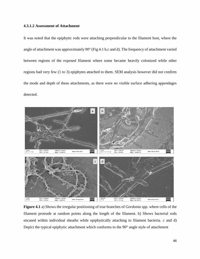
4.3.1.2 Assessment of Attachment
It was noted that the epiphytic rods were attaching perpendicular to the filament host, where the
angle of attachment was approximately 90° (Fig 4.1 b,c and d). The frequency of attachment varied
between regions of the exposed filament where some became heavily colonized while other
regions had very few (1 to 3) epiphytes attached to them. SEM analysis however did not confirm
the mode and depth of these attachments, as there were no visible surface adhering appendages
detected.
Figure 4.1 a) Shows the irregular positioning of true branches of Gordonia spp. where cells of the
filament protrude at random points along the length of the filament. b) Shows bacterial rods
encased within individual sheaths while epiphytically attaching to filament bacteria. c and d)
Depict the typical epiphytic attachment which conforms to the 90° angle style of attachment
47
4.3.2 Ultra structure analysis using TEM
4.3.2.1 Assessment of attachment at interface
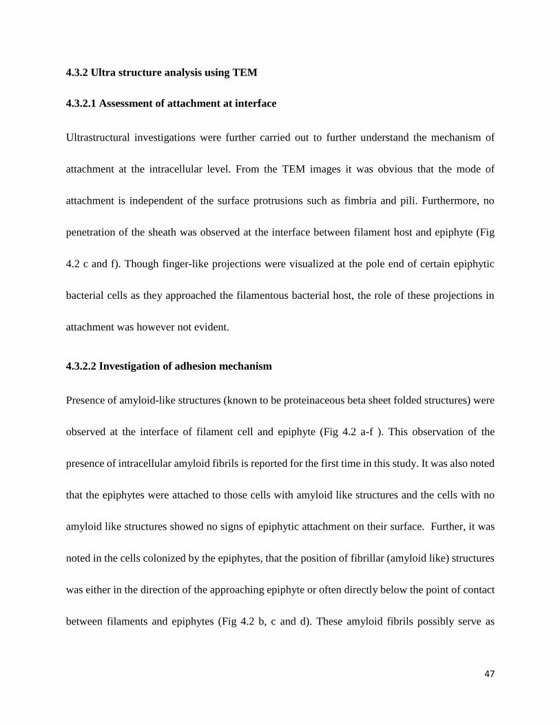
Ultrastructural investigations were further carried out to further understand the mechanism of
attachment at the intracellular level. From the TEM images it was obvious that the mode of
attachment is independent of the surface protrusions such as fimbria and pili. Furthermore, no
penetration of the sheath was observed at the interface between filament host and epiphyte (Fig
4.2 c and f). Though finger-like projections were visualized at the pole end of certain epiphytic
bacterial cells as they approached the filamentous bacterial host, the role of these projections in
attachment was however not evident.
4.3.2.2 Investigation of adhesion mechanism
Presence of amyloid-like structures (known to be proteinaceous beta sheet folded structures) were
observed at the interface of filament cell and epiphyte (Fig 4.2 a-f ). This observation of the
presence of intracellular amyloid fibrils is reported for the first time in this study. It was also noted
that the epiphytes were attached to those cells with amyloid like structures and the cells with no
amyloid like structures showed no signs of epiphytic attachment on their surface. Further, it was
noted in the cells colonized by the epiphytes, that the position of fibrillar (amyloid like) structures
was either in the direction of the approaching epiphyte or often directly below the point of contact
between filaments and epiphytes (Fig 4.2 b, c and d). These amyloid fibrils possibly serve as
48
potential preferred attachment sites for the epiphytes colonizing selected filaments (Fig 4.2 b, c
and d ).
4.3.2.3 Intracellular inclusions
The presence of intracellular storage compounds was noted in most of the filamentous bacterial
host cells colonized by epiphytic bacteria (Fig 4.2 b-f). In was interesting to note that the cells
observed with amyloid like structures also showed a high amount of intracellular storage
compounds. Rarely was intracellular storage observed with un-colonized filamentous bacterial
cells.
49
Fig 4.2 a-f TEM micrographs depicting the presence of fibrillar structures emanating from within
filamentous bacterial cells being targeted by bacterial rods, the direction of fibrils is towards the
approaching/attached bacterial rod.
a, b. Bacterial rods approaching filament compartment, have finger-like projections extending
from cell membrane of epiphytic cell as it approaches the targeted filamentous bacterial cell;
protrusion is in the direction of filament compartment.
c and f. TEM micrograph showing the attachment site between the filament and the epiphytic rod
bacteria. Intact filamentous sheath was observed.
b,c,d,e and f, depicts the presence of storage compounds within filament compartment being
approached and also attached to by epiphytic bacteria.
e. Upper adjacent compartment of filament is devoid of stored compounds.
50
4.4 Discussion
Apart from the basic morphological features, the growth of bacteria as branched or unbranched
filaments, living in sheathed or unsheathed chains, or aggregate in primitive or highly organized
multicellular composites has been displayed by organisms in AS (Young, 2006). Attached growth
is also a common mode of existence for certain bacteria in AS. This has been noted by the growth
of bacterial rods on the surface of filamentous bacteria (Xia et al., 2008).
Under light microscopy, the epiphytic attachment resembled branching. Owing to the high
magnification and resolution of SEM the distinction between branching which is a characteristic
feature of Nocardia and many other actinobacterial species (Fig 4.1a) and epiphytic attachment
were observed and diffrentiated.
Epiphytic attachment occurred as external entities (bacterial rods) colonizing and attaching at a
90° (Fig 4.1b,c and d ) angle to filamentous bacteria. This perpendicular attachment of epiphytic
bacteria to filaments in activated sludge were in line with findings by Xia et al. (2008). It was
interesting to note that the epiphytic bacteria colonizing filaments were individually enclosed in
sheaths (Fig 4.1b and d ).
Furthermore SEM observation at cell-to-cell level of exposed filament regions showed that the
attachment mechanism exhibited by the bacterial rods may have been facilitated by additional
factors than the presence of EPS, compared to that of the rest of the floc as these regions were not
51
completely embedded in the EPS matrix, suggesting a morphological mechanism of attachment.
This observation was in-line with the hypothesis made by Romero et al. (2010) of adhesion being
facilitated by a selective mechanism beyond the presence of the EPS layer (Romero et al., 2010).
TEM was used for ultra-structural level investigations of the mechanism of this attachment at the
interface between filament cells and epiphytes. Under TEM certain epiphytic cells exhibited
cellular membrane extensions and these protrusions were visualized on portions approaching the
filament host (Fig 4.2 a and b) and those resting on the surface of the filament cell being colonized
(Fig 4.2c). However, observations did not reveal common bacterial surface attachment appendages
such as fimbria or pili at the interface between filament host and epiphyte.
TEM observation further postulated the presence of amyloid like structures in the host filament
cells being targeted by the epiphyte. The presence of amyloid protein has been detected in AS in
relation to biofilm formation and floc aggregation (Larsen et al., 2007).
In addition amyloid fibrils play a functional role in facilitating adhesion, as well as invasion of
host cells and host-pathogen interactions (Zhou et al., 2012). Furthermore these proteins are known
to also function as attachment sites for the epiphytic bacteria (Nielsen et al., 2009b). The TEM
observation in this study suggested the possibility of a selective amyloid-like nature of interaction
between the host filamentous bacteria and epiphytes.
52
It was also interesting to note that the position of the intracellular amyloid-like structure were
inclined in the direction of the approaching epiphyte and observed often directly below the
interface point between filament and the epiphyte (Figure 4.2 a-f). The amyloid-like structures
were furthermore mostly visualized in cells containing an abundance of storage compounds
(Figure 4.2 b-f).
Epiphytes showed to selectively attach to those host cells with intracellular storage compound
(Figure 4.2 b-f ). These findings suggested a potential nutritional interaction between selected
filaments and epiphytes. Additionally the presence of small polyhydroxyalkanoates (PHA)
granules have been noted with the Curvibacter and Chloroflexi filaments, however TM7 filaments
have never been examined for PHA storage (Nielsen et al., 2009a). Xia et al. (2008) speculates
about a possible symbiotic relationship between the filamentous and epiflora bacteria which
however, was not verified. It has been noted by Otzen (2010) that the amyloid-protein producing
filamentous bacterium Chloroflexi is often covered by multiple copies of epiphytic Candidatus
Epiflobacter species (Saprospiracea Bacteriodetes) an epiphyte that specializes in protein
degradation (Otzen, 2010). According to a study conducted in Denmark, it was observed that
members of the Saprospiracea family were in abundance in AS, where they are involved in the
breakdown of complex organic compounds (McIlroy and Nielsen, 2014).
53
4.5 Conclusion
The focus of this chapter was on evaluation of the morphology between the filamentous bacteria
and their epiphytic counterparts and potentially understanding the mechanism used by the
epiphytic bacteria in facilitating attachment to filamentous hosts. The use of electron microscopy
(SEM and TEM) to elucidate ultrastructural details between the epiphytes and filamentous bacteria
at interface level was fundamental and it proved an important tool in understanding the interaction
between the two parties. The observations showed a level of selectivity between epiphytes and
filamentous morphotypes colonized, further a more intricate selectivity existed with the type of
filament cells that were colonized. Absence of bacterial attachment appendages was noted.
However amyloid-like structures were observed in such cells and were believed to be potential
attachment sites for the epiphytes. The preferred cells of filaments colonized were rich in storage
compound. The findings reported in this chapter suggest that a selective relationship exists
between filamentous morphotypes and epiphytes. Therefore, we could postulate that is a certain
degree specificity exists between the filament host and the colonizing epiphyte.
54
5. Chapter Five: Evaluation of the interaction between epiphytes and
filamentous bacteria under nutritional stress conditions
5.1 Introduction
The cellular morphology of filamentous bacteria serves a nutritional function in accessing nutrients
that would otherwise be completely out of reach (Young 2006). The added advantage of
proliferation, gives filamentous bacteria an edge over non-filamentous bacteria, that is not
protruding outside the main floc structure.
Production of storage compounds such as Polyhydroxybutyrate (PHB’s) by bacteria under feast-
famine conditions is a common phenomenon in AS (Fang et al., 2009). These are carbon and
energy reserve material stored intracellularly by a wide range of microorganisms under nutrient-
limiting and other environmental stress conditions (Liu et al., 2011).
The presence of EPS layer provide entrapment of organic material (Wilen et al., 2008) but
diffusion within EPS are lower than in water, which means that EPS strongly influences the
availability of nutrients and extrusion of metabolic products (Sheng et al., 2010). This significantly
negatively impacts microbial life within the floc. It is under such conditions of nutrient limitation
that filamentous bacteria proliferate in activated sludge (Guo et al., 2014).
Proliferation is a selective advantage for filamentous bacteria over non filamentous ones in the
sludge floc, which serves as an enhanced ability for substrate uptake under stress condition. In
55
activated sludge, the presence of bacterial rods colonizing filamentous bacteria with storage
globules has been observed. Sheaths of large filamentous bacteria (eg.Beggiatoa and Thioploca)
are often colonized by other bacteria belonging to the phylum Bacteroidetes, which are believed
to use sheath material for growth (Xia et al., 2008). Furthermore the presence of epiphytic bacterial
rods on filamentous morphotypes such as Type 0041, 0675, 1851 has been reported (Xia et al.,
2008; Nielsen et al., 2009).
Combination of operational conditions such as low F/M and extended sludge Retention time (SRT)
accelerates the rate and presence of inactive biomass. Live/dead staining methods have proved to
be a useful technique in assessing the viability of organisms by detecting active cells based on the
cell wall integrity (Sheng et al., 2010). Live/dead staining makes use of two individual fluorescent
dyes Propidium Iodide (PI) and Calcein AM, staining damaged and intact cells respectively. PI is
a widely used stain for detection of dead cells since the dye penetrate into cells with disrupted
membranes and intercalate with DNA, resulting in red fluorescence in the nucleus (Rieger et al.,
2010). Cells with intact membranes are stained with Calcein AM, a highly lipophilic vital dye that
rapidly enters viable cells, is converted by intracellular esterases to calcein that produces an intense
green signal (Bratosin et al., 2005). Since nutrient limiting conditions adversely affects all resident
microbes, with filamentous bacteria showing a resilience under stress conditions, the need to
evaluate the nutritional interaction between filament hosts and colonizing epiphytes was apparent.

56
The focus of this chapter is to evaluate the response of resident microbes to nutrient stress and to
determine the presence and nature of energy rich storage compounds.
57
5.2 Material and Methods
5.2.1 Sample collection and concentration of filaments with epiphytic growth
Grab samples of 750 mL were collected from returned activated sludge (RAS) streams and aeration
basins of different WWTP’s treating domestic, industrial and municipal wastewaters in Kwa-Zulu
Natal. RAS samples were diluted with equal parts of distilled water (1:1). The diluted samples
were thereafter sonicated at 2 watts for 30 seconds using a sonicator (Misonix Ultra Liquid
Processor, XL-2000 series, Trilab Support, South Africa). The sonicated samples were filtered in
descending order through wire mesh sieves (Labotec suppliers, South Africa) of different pore
sizes (300 µm; 150 µm; 100 µm and 75 µm) to remove compact flocs. Following this, wet mounts
were prepared and viewed using Nikon Eclipse 80i phase contrast microscope under oil
immersion.
5.2.2 Polyhydroxybutyrate (PHB) staining
PHB stain (Appendix 3) was used in the detection of polyhydroxybutyrate granules stored
intracellularly by filamentous bacteria in accordance with Jenkins et al. (1993).
58
5.2.3 Live/Dead viability staining of filaments with epiphytic growth
Filtered samples were subjected to viability staining (Appendix 4) using Live/Dead staining kit
(Sigma Aldrich). Working solutions of Calcein AM (1µM) and PI (1µM) in 100 µL Phosphate
saline buffer (PBS) were prepared. Filtered samples (98 µL) were mixed with 2µL Live/Dead stain
mixture (2µL Calcein AM + 2µL PI) and incubated at room temperature in the dark for 15 min.
Following the incubation, a drop of stained sample was spotted on glass slides and covered with
coverslips for viewing under Axiolab Zeiss fluorescent microscope. Viable cells were fluorescent
green and non-viable cells were fluorescent red.
5.2.4 Elemental Analysis
Elemental analysis was carried out with two microscopic methods as stated below.
a) SEM elemental analysis
AS samples were spotted on circular glass cover slips (Appendix 6) and gold sputter coated in
accordance with Li et al. (2005). Scanning electron microscopy was conducted on gold coated
samples using a Zeiss Ultra Plus field emission gun scanning electron microscope (FEGSEM)
fitted with an Oxford X-max 80mm SD detector for elemental analysis. Analysis was carried out
at 20 kV under high vacuum on surface of host filamentous bacterial cell and that of epiphytic at
the interface point.
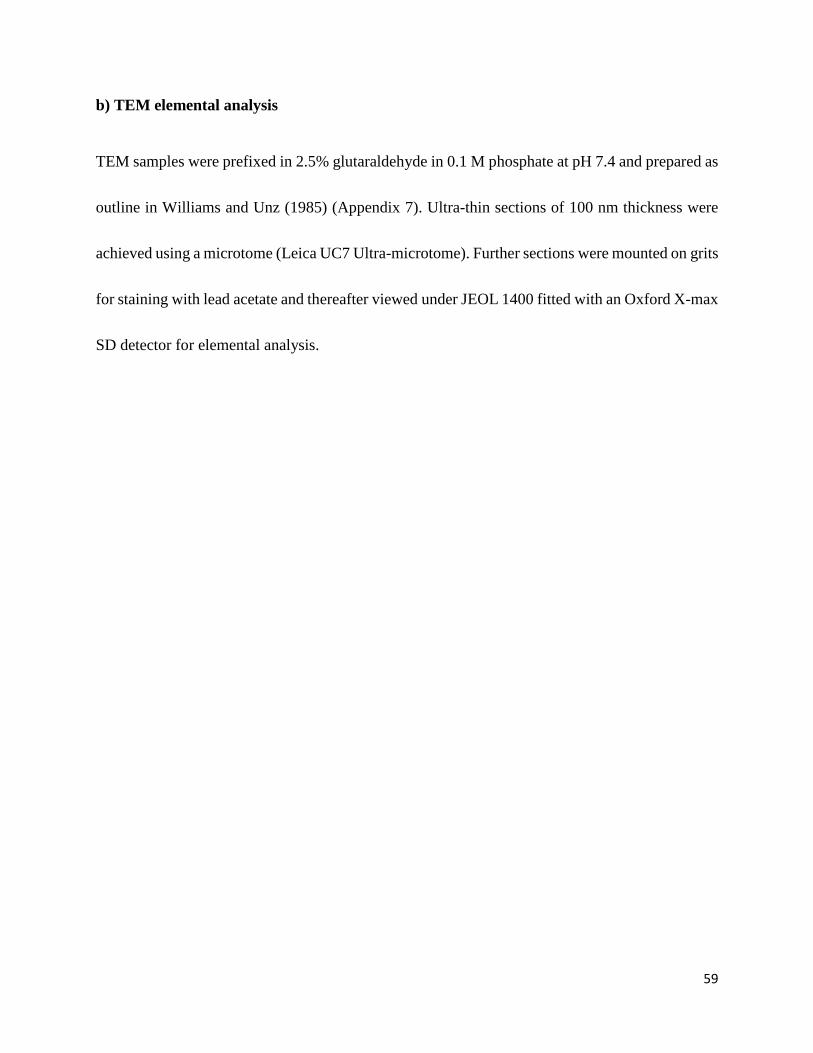
59
b) TEM elemental analysis
TEM samples were prefixed in 2.5% glutaraldehyde in 0.1 M phosphate at pH 7.4 and prepared as
outline in Williams and Unz (1985) (Appendix 7). Ultra-thin sections of 100 nm thickness were
achieved using a microtome (Leica UC7 Ultra-microtome). Further sections were mounted on grits
for staining with lead acetate and thereafter viewed under JEOL 1400 fitted with an Oxford X-max
SD detector for elemental analysis.
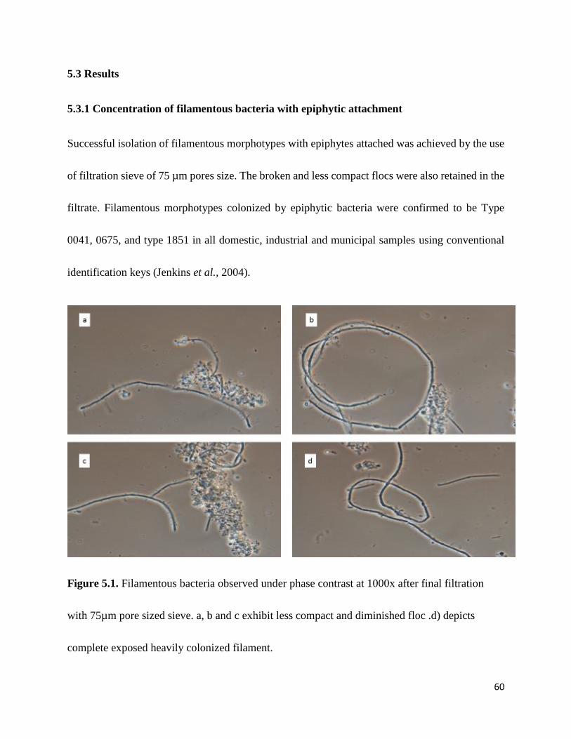
60
5.3 Results
5.3.1 Concentration of filamentous bacteria with epiphytic attachment
Successful isolation of filamentous morphotypes with epiphytes attached was achieved by the use
of filtration sieve of 75 µm pores size. The broken and less compact flocs were also retained in the
filtrate. Filamentous morphotypes colonized by epiphytic bacteria were confirmed to be Type
0041, 0675, and type 1851 in all domestic, industrial and municipal samples using conventional
identification keys (Jenkins et al., 2004).
Figure 5.1. Filamentous bacteria observed under phase contrast at 1000x after final filtration
with 75µm pore sized sieve. a, b and c exhibit less compact and diminished floc .d) depicts
complete exposed heavily colonized filament.
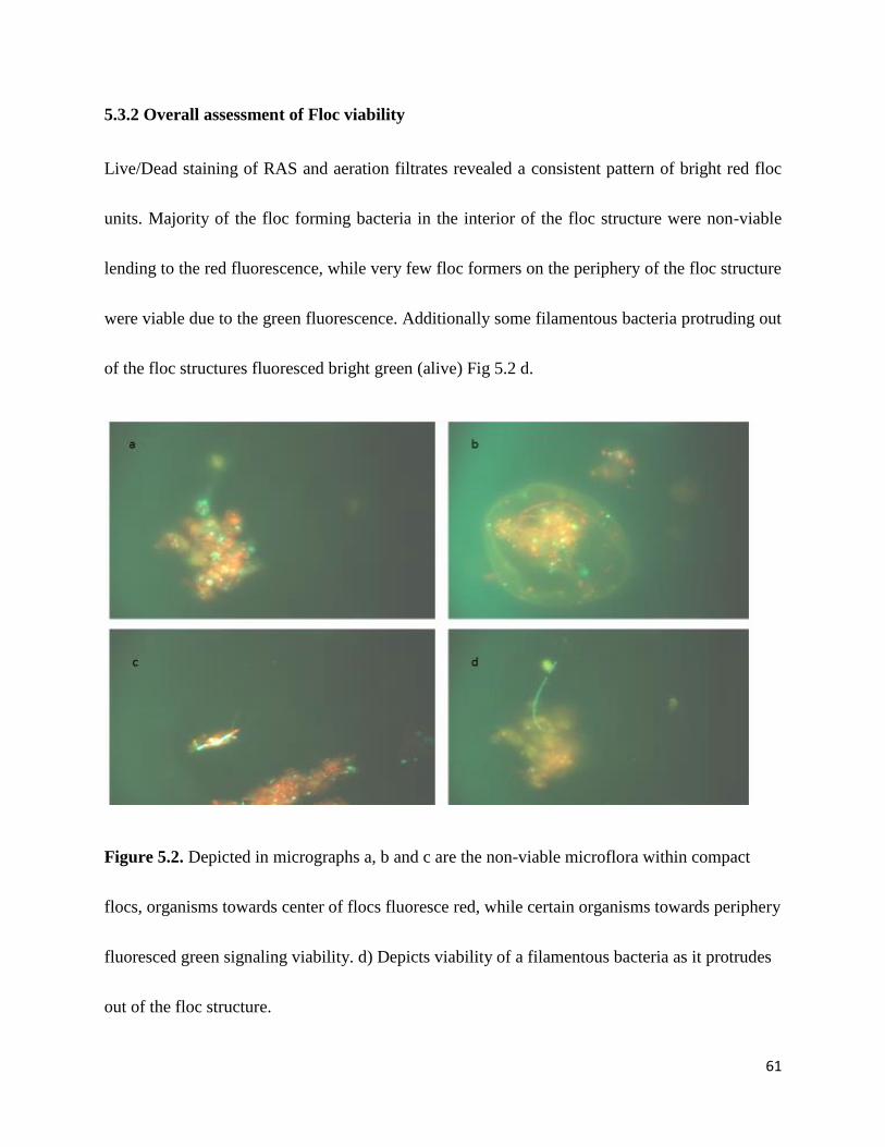
61
5.3.2 Overall assessment of Floc viability
Live/Dead staining of RAS and aeration filtrates revealed a consistent pattern of bright red floc
units. Majority of the floc forming bacteria in the interior of the floc structure were non-viable
lending to the red fluorescence, while very few floc formers on the periphery of the floc structure
were viable due to the green fluorescence. Additionally some filamentous bacteria protruding out
of the floc structures fluoresced bright green (alive) Fig 5.2 d.
Figure 5.2. Depicted in micrographs a, b and c are the non-viable microflora within compact
flocs, organisms towards center of flocs fluoresce red, while certain organisms towards periphery
fluoresced green signaling viability. d) Depicts viability of a filamentous bacteria as it protrudes
out of the floc structure.
62
5.3.4 Viability assessment of filamentous bacteria
Some filamentous bacteria isolated from the floc structure were observed to be viable while others
were seen to have damaged cells within their trichomes (Fig 5.2 a and b). Filamentous bacteria
colonized by epiphytic bacteria were observed to be viable together with their epiphytic
counterparts. Live/dead staining revealed that those viable cells within trichomes of filaments
showed the presence of epiphytic growth (Fig. 5.2b), while non-viable cells had no epiphytic
attachment.
Figure 5.3 Micrograph illustrates the existence of filamentous bacteria beyond floc structure. a)
depicts the presence of non-viable cells among viable cells in a filament trichome. b) shows
epiphytic bacteria attaching to only the viable cells of the trichome. c, d, e and f) viable epiphytic
growth attaching to the only viable filaments around non-viable flocs.

63
5.3.5 Intracellular inclusions staining
Polyhydroxybutyrate (PHB) staining was carried out on smeared and air dried samples for the
detection of intracellular polyhydroxybutyrate granules. Certain filament cells colonized by
epiphytic bacteria had a positive reaction as a result of the presence of PHB granules. These
appeared as dark blue/black globules within a pale pink background of the cell. Furthermore the
epiphytic cells did not have intracellular granules.
Figure 5.4 Micrograph a, b and c depicts the presence of PHB granules within cells of heavily
colonized filamentous bacteria. In micrograph a. only a select few cells are positive for the
intracellular storage of PHB.
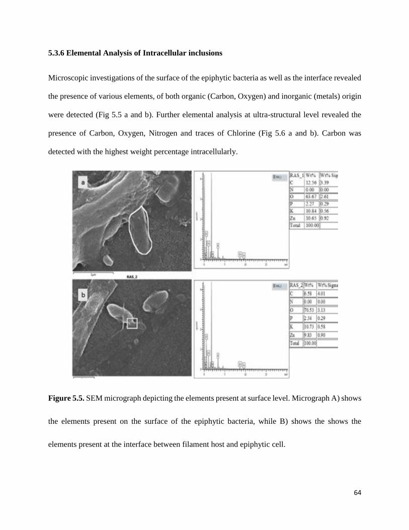
64
5.3.6 Elemental Analysis of Intracellular inclusions
Microscopic investigations of the surface of the epiphytic bacteria as well as the interface revealed
the presence of various elements, of both organic (Carbon, Oxygen) and inorganic (metals) origin
were detected (Fig 5.5 a and b). Further elemental analysis at ultra-structural level revealed the
presence of Carbon, Oxygen, Nitrogen and traces of Chlorine (Fig 5.6 a and b). Carbon was
detected with the highest weight percentage intracellularly.
Figure 5.5. SEM micrograph depicting the elements present at surface level. Micrograph A) shows
the elements present on the surface of the epiphytic bacteria, while B) shows the shows the
elements present at the interface between filament host and epiphytic cell.
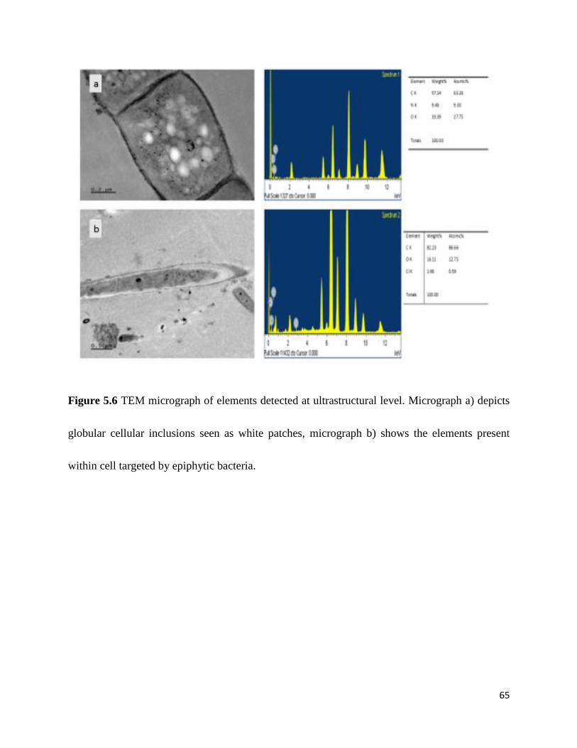
65
Figure 5.6 TEM micrograph of elements detected at ultrastructural level. Micrograph a) depicts
globular cellular inclusions seen as white patches, micrograph b) shows the elements present
within cell targeted by epiphytic bacteria.
66
5.4 Discussion
The EPS layer which is found within and on the surface of the floc (Sheng et al., 2010) is beneficial
for the compaction of the bacterial consortia. However, this can also serves as a hinderance in the
diffusion of nutrients. Filamentous organisms have an adavantage of protrusion into the bulk
solution, thereby having access to nutrients readily available in the bulk solution.
Among the protrduing filaments, morphotypes Type 0041, Type 0675 and Type 1851 were
observed to be proned to epiphytic attachment. The abundance of epiphytes colonizing these
filaments varied significantly between the samples from domestic and industrial wasterwater
analyzed. Live/dead cell stain test was used to assess the viability of the consortia within the floc,
where the organisms concentrated within the core of the floc fluoresced red indicating non-
viability (Fig 5.1 a, b and c). This observation is in line with an observation made by Sheng et al.
(2010), stating that strongly bound fraction of cells at the core of activated sludge flocs suffer the
fate of lowered diffusion co-efficients as a result of EPS, which may hinder the rapid import of
nutrients and expulsion of metabolic products (Sheng et al., 2010).
Furthermore, the bacteria in direct contact with the bulk solution, fluoresced green which made
apparent their status of viability ( Fig 5.1d). This observation was also in line with that of Liang et
al. (2010), stating loosely bound cells present on the surface of the floc have an advantage of ease
of access to nutrients present in bulk solution (Liang et al., 2010). The proliferation of filamentous
bacteria may be influenced by nutrient limitation within the floc structure, which is in line with
the observation of Young (2006).
It was further noted that individual filamentous bacteria in the bulk solution not forming part of
the floc often showed a positive reaction in the viability test (Fig 5.2 a,c and d ). Non-viable
67
filaments were also present in the bulk solution and yet others having just a few cells with their
trichome fluorescing red among a string of green cells (Fig 5.2 a and b). The protruding colonized
regions of filamentous morphotypes were found to be viable, together with their epiphytic
counterpart as they extented into the bulk solution (Fig 5.2 d, e and f). The attachment of epiphytes
to viable cells on filamentous trichomes was apparent and rarely was this attached growth
visualized on damages and non viable filament cells (Fig 5.2 d,e and f ).
PHB staining was conducted on aeration and RAS samples with a SRT of 25 days, to assess the
intracellular storage capacity of filaments prone to piphytic growth. Filament cells colonized by
attached cells exhibited the presence of PHB granules internalized within cell compartments (Fig
5.3a,b and c) . Trichomes of filaments together with their epiphytic counter parts stained pink in
line with findings by Liu et al. (2011).
SEM elemental analysis at both the interface and surface of epiphytic bacterial cell revealed the
presence of carbon, however at a lower weight percentage. Further, elemental analysis of filament
compartments using TEM made apparent a significantly higher carbon content within the filament
cell (Fig 5.6 a and b). This observation suggested more carbon is internalized within filament
compartments than that present at the interface. According to Sujatha et al. (2004) carbon is an
important building block in the synthesis of PHB’s (Sujatha et al., 2004). Therefore the presence
of higher volumes of carbon within filament cells may be an indication of the presence of energy
rich PHB reserves.
It was further interesting to note that phosphorus in low concentrations, while nitrogen was
completely absent. This observation was in line with that from a study conducted in China by Liu
et al. (2011), stating that in the absence of the nitrogen and or phosphorous sources PHB

68
production increases (Liu et al., 2011). These observation collectively point towards the possibility
a relationship founded on nutritional benefit for epiphytes under nutrient limiting condition in AS.
69
5.5 Conclusions
This chapter evaluated the interaction between the filamentous morphotypes and their epiphytic
bacterial counterparts under nutrient-limiting conditions. Techniques implored in evaluating this
interaction included sonication and filtration for the breaking up of compact sludge flocs and
concentration of filaments prone to epiphytic attachment. Filtration proved beneficial in this study
as it allowed less compact flocs to pass through series of sieve with descending pore size and
allowed for greater exposure of morphotypes with epiphytic growth. Further Live/dead viability
test revealed the status of resident floc formers at different levels of floc structure, with results
showing floc members densely concentrated at floc core fluorescing red (non-viable) and floc
memebers located on the surface of the flocs as well as filamentous bacteria protruding beyond the
floc surface fluorescing green (viable). Elemental analysis using SEM and TEM revealed the
presence of carbon at interface and intracellularly within filament compartments. This element was
however present in abundance within filament cells targeted by epiphytic bacteria. The presence
of globular structures within filament compartments analyzed for elements further confirmed the
findings of PHB staining which was positive for the presence of storage compounds under light
microscopy. The findings suggests the interaction may potential border on nutrional stress and
survival adaptation on the part of the epiphytes.
70
6. Chapter Six: General summary and conclusions
Occurrence of epiphytic growth on filamentous bacteria is a common phenomenon in AS plants
worldwide. Eikelboom morphotypes 0041, 0675, 1701 and 1851 are identified as the common
filament with their trichomes colonized by epiphytic bacteria. Traditionally the epiphytic growth
has been used as a morphological attribute for their characterization. These morphotypes are
present in all types of wastewater treatment plants including domestic, municipal and industrial
wastewater treatment plants where epiphytic colonization varies from dense to sparse respectively.
Conditions that favor the growth of these morphotypes are well documented in literature, including
their presence in bulking sludge which has earned these Eikelboom morphotypes the term of
“problem filaments”, however, relatively little is known thus far about this interaction. This study
was undertaken to investigate a potential existence of a relationship between colonized filamentous
morphotypes and epiphytes, further evaluate the level of interaction between filament host and
epiphytic counterpart.
Most filamentous bacteria in activated sludge are encapsulated by translucent hallow sheath where
some are prone to epiphytic attachment and others not. Those prone to and colonized by epiphytic
growth were consistently observed to Eikelboom Types 0041, 0675, 1851 filament morphotypes
and occasionally on S. natans and Type 1701. The exposed regions of these filament morphotypes
extending into the bulk region was colonized, while colonization of the portions embedded in the
floc structure could not be visualised. Dense colonization was particularly observed with Type
0041 in domestic sludge, while sparse growth was observed on this same morphotype in industrial
sludge. Additionally Type 1851 was observed with sparse growth in domestic sludge, however, in
71
industrial sludge epiphytic growth on this particular morphotype was fewer when present and
sometimes completely absent. The FISH observations revealed that Alpha positive epiphytes were
the majority epiphytic bacteria colonizing filamentous morphotypes prone to this attachment. Type
1851 showed a positive binding with EUB mix probe, while Type 0041 showed an opposite
response to the same probe. This may be due to lack of binding sites or resultant effects of
colonization. Therefore in depth investigation is crucial in understanding the outcome of both
morphotypes when hybridized with same probe.
SEM analysis proved to be successful in differentiating between branching (which was previously
visualized by use of light microscopy) and epiphytic attachment. The attachment consistently was
to a 90° angle. Further TEM revealed the presence of fibrillar structures in-vivo in the filaments
next to attachment sites of epiphytes, resembling amyloid fibrils. These fibrillar structures were
visible within filament cells with storage compounds at the interface between filament and
epiphytes, additionally in the direction of the approaching epiphyte. Additionally TEM made
apparent the absence of bacterial pili and other surface attachment commonly used by bacterial to
facilitate attachment.
In conclusion there seem to exist a level of selectivity in the filamentous morphotypes being
colonized by epiphytes in activated sludge, where the presence of amyloid fibrils was observed
particularly in the colonized cells at the interface. Absence of structural appendages that facilitate
attachment was evident, which suggested attachment being potentially facilitated by fibrillar
structures observed at interface. Further the association of the epiphyte to the specific filament
could be based on a symbiotic relationship.
72
It should be noted that the investigations in determining the relationship between filamentous
morphotypes and epiphytes in activated sludge was not detailed due to the non-availability of
facilities and lengthy time frame beyond that allocated for this qualification required to conduct a
more detailed investigation. However proposed future work in this area will be in-depth.
6.1 Significant Findings
Very limited information has been documented in current literature surrounding the interaction
between filamentous bacteria and epiphytes. The following findings can be considered as novel:
A clear relationship between epiphytic growth and viable filament cells. It was observed
that the presence of epiphytes was on viable filament cells and absent on non-viable
filament cell.
Consistent presence of amyloid-like structures at interface between certain filament cells
and epiphytes was apparent.
Only certain cells within filament trichome were specifically colonized, particularly those
containing storage compounds and exhibiting amyloid-like structures.
These findings can be considered as significant and making a novel contribution to the field of
study. Further these can serve as the basis for future research.
73
6.2 Future Recommendations
The presence of epiphytic growth on selected filamentous morphotypes in activated sludge is a
common occurrence, however, the exact identity of epiphytic bacteria and conditions surrounding
and promoting their growth is still unclear. The use of methods such as RT-PCR and Pyro-
sequencing would be beneficial for the characterization of epiphytic bacteria colonizing
filamentous bacteria which can only be done through micromanipulation. Further, their identity
would shed light on the preferred substrates utilized by the respective epiphytic groups.
Understanding the particular substrate uptake would further contribute to understanding the
conditions that promote dense epiphytic growth on some morphotypes and sparse epiphytic growth
on other morphotypes.
Additionally, the effects of such colonization on filamentous bacterial health are still in need of
in-depth investigation. This need may be addressed by use of bacterial Quorum Sensing. This
approach may be capable of bringing to our understanding the extent of interaction between the
filament hosts and epiphytic counterparts, furthermore revealing the nature of relationship between
these two groups of bacteria.
The presence of the amyloid-like structures were observed at interface, however confirmation of
these structures is in need and pivotal in understanding their role in attachment of epiphytic
bacteria. Detection and confirmatory studies using conformational antibody staining would be
advantageous for future studies of these fibrillary structures used as docking sites for epiphytic
bacteria.
74
References
Amann, R. 1995. In situ identification of micro-organisms by whole cell hybridization with rRNA-
targeted nucleic acid probes. In: Akkermans, A. L., Van Elsas, J. and De Bruijn, F. eds. Molecular
Microbial Ecology Manual. Springer Netherlands, 331-345.
Amann, R., Fuchs, B. M. and Behrens, S. 2001. The identification of microorganisms by
fluorescence in situ hybridisation. Current Opinion in Biotechnology, 12: 231-236.
Beer, M., Sevior, E.M., Kong, Y., Cunningham, M., Blackall, L.L., Sevior, R.J. 2002. Phylogeny
of the filamentous bacterium Eikelboom Type 1851, and design and application of a 16S rRNA
targeted oligonucleotide probe for its fluorescence in situ identification in activated sludge.
Federation of European Microbial Societies: Microbiology Letters, 207: 179-183.
Blanco, L.P., Evans, M.L., Smith, D.R., Badtke, M.P and Chapman, M. 2012. Diversity,
biogenesis and function of microbial amyloids. Trends in Microbiology, 20:66-73.
Bradford, D., Hugenholtz, P., Seviour, E. M., Cunningham, M. A., Stratton, H., Seviour, R. J. and
Blackall, L. L. 1996. 16S rRNA Analysis of Isolates Obtained from Gram-Negative, Filamentous
Bacteria Micromanipulated from Activated Sludge. Systematic and Applied Microbiology, 19:
334-343.
75
Bratosin, D., Mitrofan, L., Palii, C., Estaquier, J., Montreuil, J. 2005. Novel Fluorescence Assay
Using Calcein-AM for the Determination of Human Erythrocyte Viability and Aging. Cytometry,
66: 78-84.
Bullitt, E. and Makowski, L. 1998. Bacterial Adhesion Pili are Heterologous Assemblies of Similar
Subunits. Biophysical Journal, 74: 623-632.
Chen, A. Y. Y. and Chen, A. 2013. Fluorescence In Situ Hybridization. Journal of Investigative
Dermatology, 133:8.
Daims, H., Bruhl, A., Amann, R., Schleifer, K. and Wagner, M. 1999. The domain-specific probe
EUB338 is insufficient for the detection of all Bacteria: development and evaluation of a more
comprehensive probe set. Systematic and Applied Microbiology, 22: 434 - 444.
De los Reyes, F.L., Ritter, W., Raskin, L. 1997. Group-specific small-subunit rRNA hybridization
probes to characterize filamentous foaming in activated sludge systems. Applied and
Environmental Microbiology, 63:1107– 1117.
Denk, W. and Hortsmann, H. 2004. Serial block-face scanning electron microscopy to reconstruct
three-dimensional tissue nanostructure. PLOS Biology,11: 329- 332.
76
Dunne, W. M. 2002. Bacterial Adhesion: Seen Any Good Biofilms Lately? Clinical Microbiology
Reviews, 15: 155-166.
Eickhorst, T. and Tippkötter, R. 2008. Improved detection of soil microorganisms using
fluorescence in situ hybridization (FISH) and catalyzed reporter deposition (CARD-FISH). Soil
Biology and Biochemistry, 40: 1883-1891.
Eikelboom, D.H., Andreadakis, A., Andreasen, K.1998. Survey of filamentous populations in
nutrient removal plants in four European countries. Water Science and Technology, 37: 281-289.
Eikelboom, D.H. 2000. Process Control of Activated Sludge Plants by Microscopic Investigation,
IWA Publishing, London.
Erhart, R ., Bradford, D., Seviour, R ., Amann, R ., Blackall, L.L.1997. Development and use of
fluorescent in situ hybridization probes for the detection and identification of ‘‘Microthrix
parvicella’’ in activated sludge. Systematic and Applied Microbiology.20:310-318.
Fang, F., Liu, X.-W., Xu, J., Yu, H.-Q. and Li, Y.-M. 2009. Formation of aerobic granules and
their PHB production at various substrate and ammonium concentrations. Bioresource
Technology, 100: 59-63.
77
Fowler, D. M., Koulov, A. V., Balch, W. E. and Kelly, J. W. 2007. Functional amyloid – from
bacteria to humans. Trends in Biochemical Sciences, 32: 217-224.
Freire, S., de Araujo, M. H., Al-Soufi, W. and Novo, M. 2014. Photophysical study of Thioflavin
T as fluorescence marker of amyloid fibrils. Dyes and Pigments, 110: 97-105.
Garrett, T. R., Bhakoo, M. and Zhang, Z. 2008. Bacterial adhesion and biofilms on surfaces.
Progress in Natural Science, 18: 1049-1056.
Gougoulias, C. and Shaw, L. J. 2012. Evaluation of the environmental specificity of Fluorescence
In Situ Hybridization (FISH) using Fluorescence-Activated Cell Sorting (FACS) of probe
(PSE1284)-positive cells extracted from rhizosphere soil. Systematic and Applied Microbiology,
35: 533-540.
Guo, F. and Zhang, T. 2012. Profiling bulking and foaming bacteria in activated sludge by high
throughput sequencing. Water Research 46: 2772-2782.
Guo, J., Peng, Y., Wang, Z., Yuan, Z., Yang, X. and Wang, S. 2012. Control filamentous bulking
caused by chlorine-resistant Type 021N bacteria through adding a biocide CTAB. Water Research,
46: 6531-6542.
78
Guo, J., Peng, Y., Wang, S., Yang, X. and Yuan, Z. 2014. Filamentous and non-filamentous
bulking of activated sludge encountered under nutrients limitation or deficiency conditions.
Chemical Engineering Journal, 255: 453-461.
Habimana, O., Semião, A. J. C and Casey, E. 2014a. The role of cell-surface interactions in
bacterial initial adhesion and consequent biofilm formation on nanofiltration/reverse osmosis
membranes. Journal of Membrane Science. 454:82-96.
Han, Y., Liu, J., Guo, X. and Li, L. 2012. Micro-environment characteristics and microbial
communities in activated sludge flocs of different particle size. Bioresource Technology, 124: 252-
258.
Hori, K. and Matsumoto, S. 2010. Bacterial adhesion: From mechanism to control. Biochemical
Engineering Journal, 48: 424-434.
Hu, L., Ru, K., Zhang, L., Huang, Y., Zhu, X., Liu, H., Zetterberg, A., Cheng, T. and Miao, W.
2014. Fluorescence in situ hybridization (FISH): an increasingly demanded tool for biomarker
research and personalized medicine. Biomarker Research, 2: 1-13.
Hugenholtz, P., Tyson, G. W., Webb, R. I., Wagner, A. M. and Blackall, L. L. 2001. Investigation
of candidate division TM7, a recently recognized major lineage of the domain Bacteria with no
known pure-culture representatives. Applied and Environmental Microbiology, 67: 411-419.
79
Hung, C., Marschall, J., Burnham, C. A., Byun, A. S. and Henderson, J. P. 2014. The bacterial
amyloid curli is associated with urinary source bloodstream infection. PLoS One, 9: 86009.
Jang, H. J., Kim, J. M. and Choi, C. Y. 2014. Elemental analysis of sunflower cataract in Wilson's
disease: A study using scanning transmission electron microscopy and energy dispersive
spectroscopy. Experimental Eye Research, 121: 58-65.
Jassby, D., Xiao, Y. and Schuler, A. J. 2014. Biomass density and filament length synergistically
affect activated sludge settling: systematic quantification and modeling. Water Research, 48: 457-
465.
Jenkins, D., Richard, M. G. & Daigger, G. T. 1993. Manual on the Causes and Control of Activated
Sludge Bulkinf and Foaming., Michigan, Lewis Publishers.
Jenkins, D., Richard, M. G. & Daigger, G. T. 2004. Manual on the Causes and Control of Activated
Sludge Bulking. Foaming and Other Solids Seperation Problems., UK, IWA publishing.
Jordal, P. B., Dueholm, M. S., Larsen, P., Petersen, S. V., Enghild, J. J., Christiansen, G., Højrup,
P., Nielsen, P. H. and Otzen, D. E. 2009. Widespread Abundance of Functional Bacterial Amyloid
in Mycolata and Other Gram-Positive Bacteria. Applied and Environmental Microbiology, 75:
4101-4110.
80
Juang, D.F. 2005. Effects of synthetic polymer on the filamentous bacteria in activated sludge.
Bioresource Technology, 96: 31-40.
Kang, H. J. and Baker, E. N. 2012. Structure and assembly of Gram-positive bacterial pili: unique
covalent polymers. Current Opinion in Structural Biology, 22: 200-207.
Kim, S.B., Goodfellow, M., Kelly, J., Saddler, G.S., Ward, A.C. 2002. Application of
oligonucleotide probes for the detection of Thiothrix spp. in activated sludge plants treating paper
and board mill wastes. Water Science and Technology,46: 559–64.
Kline, K. A., Dodson, K. W., Caparon, M. G. and Hultgren, S. J. 2010. A tale of two pili: assembly
and function of pili in bacteria. Trends in Microbiology, 18: 224-232.
Kragelund, C., Remesova, Z., Nielsen, J. L., Thomsen, T. R., Eales, K., Seviour, R., Wanner, J.
and Nielsen, P. H. 2007. Ecophysiology of mycolic acid-containing Actinobacteria (Mycolata) in
activated sludge foams. Federation of European Microbiological Societies, 61:174-184.
Lacko, N., Bux, F., Kasan, H.C.1999. Survey of filamentous bacteria in activated sludge plants in
KwaZulu-Natal. Water SA, 25: 63-68.
81
Larsen, P., Nielsen, J. L., Dueholm, M. S., Wetzel, R., Otzen, D. and Nielsen, P. H. 2007. Amyloid
adhesins are abundant in natural biofilms. Environmental Microbiology, 9: 3077-3090.
Larsen, P., Nielsen, J. L., Otzen, D. and Nielsen, P. H. 2008. Amyloid-Like Adhesins Produced
by Floc-Forming and Filamentous Bacteria in Activated Sludge. Applied and Environmental
Microbiology, 74: 1517-1526.
Levantesi, C., Beimfohr, C., Geurkink, B., Rossetti, S., Thelen, K., Krooneman, J., Snaidr, J., van
der Waarde, J. and Tandoi, V. 2004. Filamentous Alphaproteobacteria associated with bulking in
industrial wastewater treatment plants. Systematic and Applied Microbiology, 27: 716-727.
Li, W.-J., Tuli, R., Huang, X., Laquerriere, P. and Tuan, R. S. 2005. Multilineage differentiation
of human mesenchymal stem cells in a three-dimensional nanofibrous scaffold. Biomaterials, 26:
5158-5166.
Liang, Z., Li, W., Yang, S. and Du, P. 2010. Extraction and structural characteristics of
extracellular polymeric substances (EPS), pellets in autotrophic nitrifying biofilm and activated
sludge. Chemosphere, 81: 626-632.
Lin, H., Zhang, M., Wang, F., Meng, F., Liao, B.-Q., Hong, H., Chen, J. and Gao, W. 2014. A
critical review of extracellular polymeric substances (EPSs) in membrane bioreactors:
82
Characteristics, roles in membrane fouling and control strategies. Journal of Membrane Science,
460: 110-125.
Liu, Z., Wang, Y., He, N., Huang, J., Zhu, K., Shao, W., Wang, H., Yuan, W. and Li, Q. 2011a.
Optimization of polyhydroxybutyrate (PHB) production by excess activated sludge and microbial
community analysis. Journal of Hazardous Materials, 185: 8-16.
Madoni, P., Davoli, D. and Gibin, G. 2000. Survey of filamentous microorganisms from bulking
and foaming activated-sludge plants in Italy. Water Research, 34: 1767-1772.
Majewsky, M., Gallé, T., Yargeau, V. and Fischer, K. 2011. Active heterotrophic biomass and
sludge retention time (SRT) as determining factors for biodegradation kinetics of pharmaceuticals
in activated sludge. Bioresource Technology, 102: 7415-7421.
Manz, W., Amann, R., Ludwig, W., Wagner, M. and Schleifer, K.-H. 1992a. Phylogenetic
Oligodeoxynucleotide Probes for the Major Subclasses of Proteobacteria: Problems and Solutions.
Systematic and Applied Microbiology, 15: 593-600.
Martins, A. M. P., Pagilla, K., Heijnen, J. J. and van Loosdrecht, M. C. M. 2004. Filamentous
bulking sludge—a critical review. Water Research, 38: 793-817.
83
McIlroy, S. and Nielsen, P. 2014. The Family Saprospiraceae. In: Rosenberg, E., DeLong, E.,
Lory, S., Stackebrandt, E. and Thompson, F. eds. The Prokaryotes. Springer Berlin Heidelberg,11:
863-889.
Merkel, W., Manz, W., Szewzyk, U. and Krauth, K. 1999. Population dynamics in anaerobic
wastewater reactors: modelling and in situ characterization. Water Research, 33: 2392-2402.
Mesquita, B., Lopes, P., Rodrigues, A., Pereira, D., Afonso, M., Leal, C., Henrique, R., Lind, G.
E., Jeronimo, C. and Lothe, R. A. 2013. Frequent copy number gains at 1q21 and 1q32 are
associated with overexpression of the ETS transcription factors ETV3 and ELF3 in breast cancer
irrespective of molecular subtypes. Breast Cancer Research and Treatment, 138: 37-45.
Mesquita, D. P., Amaral, A. L. and Ferreira, E. C. 2013. Activated sludge characterization through
microscopy: A review on quantitative image analysis and chemometric techniques. Analytica
Chimica Acta, 802: 14-28.
Mielczarek, A. T., Kragelund, C., Eriksen, P. S. and Nielsen, P. H. 2012. Population dynamics of
filamentous bacteria in Danish wastewater treatment plants with nutrient removal. Water
Research, 46: 3781-3795.
84
Moter, A. and Göbel, U. B. 2000. Fluorescence in situ hybridization (FISH) for direct visualization
of microorganisms. Journal of Microbiological Methods, 41: 85-112.
Nielsen, P. H., Kragelund, C., Seviour, R. J. and Nielsen, J. L. 2009. Identity and ecophysiology
of filamentous bacteria in activated sludge. Federation of European Microbiological Societies
Microbiology Review, 33: 969-998.
Nielsen, P. H., Saunders, A. M., Hansen, A. A., Larsen, P. and Nielsen, J. L. 2012. Microbial
communities involved in enhanced biological phosphorus removal from wastewater — a model
system in environmental biotechnology. Current Opinion in Biotechnology, 23: 452-459.
Nongkhlaw, F. M. W. and Joshi, S. R. 2014. Distribution pattern analysis of epiphytic bacteria on
ethnomedicinal plant surfaces: A micrographical and molecular approach. Journal of Microscopy
and Ultrastructure, 2: 34-40.
Otzen, D. 2010. Functional amyloid: Turning swords into plowshares. Prion, 4: 256-264.
Otzen, D. and Nielsen, P. H. 2007. We find them here, we find them there: Functional bacterial
amyloid. Cellular and Molecular Life Sciences, 65: 910-927.
Pal, P., Khairnar. K., Paunikari, W.N. 2014. Causes and remedies for filamentous foaming in
activated sludge treatment plant. Global NEST Journal, 16:762-772.
85
Pandolfi, D., Pons, M and da Motta, M. 2007. Characterization of PHB storage in activated sludge
extended filamentous bacteria by automated colour image analysis. Biotechnology Letters.
29:1263-1269.
Perez, Y. G., Leite, S. G. F. and Coelho, M. A. Z. 2006. Activated sludge morphology
characterization through an image analysis procedure. Brazilian Journal of Chemical Engineering,
23: 319-330.
Pizarro-Cerdá, J. and Cossart, P. 2006. Bacterial Adhesion and Entry into Host Cells. Cell, 124:
715-727.
Rambaran, R. N. and Serpell, L. C. 2008. Amyloid fibrils. Prion, 2: 112-117.
Rieger, A. M., Hall, B. E., Luong le, T., Schang, L. M. and Barreda, D. R. 2010. Conventional
apoptosis assays using propidium iodide generate a significant number of false positives that
prevent accurate assessment of cell death. Journal of Immunological Methods, 358: 81-92.
Romero, D., Aguilar, C., Losick, R. and Kolter, R. 2010. Amyloid fibers provide structural
integrity to Bacillus subtilis biofilms. Proceedings of the National Academy of Sciences of the
United States of America, 107: 2230-2234.
86
Sanz, J. L. and Köchling, T. 2007. Molecular biology techniques used in wastewater treatment:
An overview. Process Biochemistry, 42: 119-133.
Schmidt, H., Eickhorst, T. and Mußmann, M. 2012. Gold-FISH: A new approach for the in situ
detection of single microbial cells combining fluorescence and scanning electron microscopy.
Systematic and Applied Microbiology, 35: 518-525.
Seviour, R. J. 2010. Factors Affecting the Bulking and Foaming Filamentous Bacteria in Activated
Sludge. In: Seviour, R. J. & Nielsen, P. H. (eds.) Microbial Ecology of Activated Sludge. London:
IWA Publishing.
Sheng, G. P., Yu, H. Q. and Li, X. Y. 2010. Extracellular polymeric substances (EPS) of microbial
aggregates in biological wastewater treatment systems: a review. Biotechnology Advances, 28:
882-894.
Stewart, E. J. 2012. Growing unculturable bacteria. Journal of Bacteriology, 194: 4151-4160.
Sujatha, K., Mahalalakshmi A. and Shenbaragathai, R. 2003a. A study on accumulation of PHB
native Pseudomonas isolates LDC-5 and LDC-25. India Journal of Biotechnology, 4: 216-221.
87
Thomsen, T. R., Kjellerup, B. V., Nielsen, J. L., Hugenholtz, P. and Nielsen, P. H. 2002. In situ
studies of the phylogeny and physiology of filamentous bacteria with attached growth.
Environmental Microbiology, 4: 383-391.
Thomsen, T. R., Nielsen, J. L., Ramsing, N. B. and Nielsen, P. H. 2004. Micromanipulation and
further identification of FISH-labelled microcolonies of a dominant denitrifying bacterium in
activated sludge. Environmental Microbiology, 6: 470-479.
Van Dierdonck, J., Van den Broeck, R., Vervoort, E., D’haeninck, P., Springael, D., Van Impe, J.
and Smets, I. 2012. Does a change in reactor loading rate affect activated sludge bioflocculation?
Process Biochemistry, 47: 2227-2233.
Villar-Piqué, A. and Ventura, S. 2012. Modeling amyloids in bacteria. Microbial Cell Factories,
11: 166-166.
Wagner, M., Amann, R., Kampfer, P., Assmus, B., Hartmann, A., Hutzler, P., Springer, N.,
Schleifer, K.H. 1994. Identification and in situ detection of gram-negative filamentous bacteria in
activated sludge. Systematic and Applied Microbiology,17: 405–17.
Wagner, M., Horn, M. and Daims, H. 2003. Fluorescence in situ hybridisation for the identification
and characterisation of prokaryotes. Current Opinion in Microbiology, 6: 302-309.
88
Wang, J., Li, Q., Qi, R., Tandoi, V. and Yang, M. 2014. Sludge bulking impact on relevant
bacterial populations in a full-scale municipal wastewater treatment plant. Process Biochemistry,
49: 2258-2265.
Wilén, B.-M., Jin, B. and Lant, P. 2003. Impacts of structural characteristics on activated sludge
floc stability. Water Research, 37: 3632-3645.
Wilén, B.-M., Onuki, M., Hermansson, M., Lumley, D. and Mino, T. 2008. Microbial community
structure in activated sludge floc analysed by fluorescence in situ hybridization and its relation to
floc stability. Water Research, 42: 2300-2308.
Williams, T. M. and Unz, R. F. 1985. Filamentous sulfur bacteria of activated sludge:
characterization of Thiothrix, Beggiatoa, and Eikelboom type 021N strains. Applied and
Environmental Microbiology, 49: 887-898.
Xia, Y., Kong, Y., and Nielsen, P.H. 2007. In situ detection of protein-hydrolysing microorganisms
in activated sludge.Federation of European Microbiological Societies Microbiology Ecology, 70:
156-165.
89
Xia, Y., Kong, Y., Thomsen, T. R. and Halkjaer Nielsen, P. 2008. Identification and
ecophysiological characterization of epiphytic protein-hydrolyzing Saprospiraceae ("Candidatus
Epiflobacter" spp.) in activated sludge. Applied and Environmental Microbiology, 74: 2229-2238.
Young, K. D. 2006. The Selective Value of Bacterial Shape. Microbiology and Molecular Biology
Reviews, 70: 660-703.
Zhou, Y., Blanco, L., Smith, D. and Chapman, M. 2012. Bacterial Amyloids. In: Sigurdsson, E.
M., Calero, M. and Gasset, M. eds. Amyloid Proteins. Humana Press, 849: 303-320.
90
Appendix 1: Gram Staining
(Jenkins et al., 1993)
Reagents
Solution (1)
a) Crystal violet 2g
Distilled water 80mL
b) Ammonium oxalate 0.8 g
Ethanol, 95% 20 mL
Solution (2)
Iodine 1g
Potassium Iodide 2g
Distilled water 300mL
Solution (3)
Safranin O (2.5% w/v dissolved in 95% ethanol) 10mL
Distilled water 100mL
Method
Smear mixed liquor sample onto microscope slides and allow to air dry.
Flood sample smears with 1 ml solution (1) and allow to stand for 60 seconds; rinse with
H2O.

91
Add 1 ml solution (2) and allow to stand for 60 seconds: rinse well with H2O.
Decolorize with 95% ethanol onto the smeared sample for 30 seconds, by holding slide
at an angle and adding the ethanol in a drop-wise procedure. Blot dry.
Add solution (3) for 60 seconds, rinse well and blot dry.
92
Appendix 2: Neisser staining
(Jenkins et al., 1993)
Reagents
Solution (1):
a) Methylene Blue 0.1g
Ethanol, 95% 5 mL
Glacial acetic acid 5mL
Distilled water 100 mL
B: Crystal violet (10%w/v in 95% ethanol) 3.3 mL
Ethanol 95% 6.7 mL
Distilled water 100 mL
Two parts by volume of A are mixed with 1 part by volume of B. fresh stock to be prepared
monthly.
Solution (2):
Bismark Brown (1%w/v aqueous) 33.3 mL
Distilled water 66.7 mL
Method:
Prepare thin sample smears on microscope slides and allow to air dry.
Then stain slides for 30 seconds with solution 1 and then rinse with water for 1 second.

93
Stain this preparation with solution 2 for 1 minute, rinse well with water and then blot
dry.
94
Appendix 3: PHB staining
(Jenkins et al., 1993).
Reagents
Solution (1):
Sudan Black B (IV) 0.3% w/v in 60 % ethanol
Solution (2):
Safranin 0 0.5 %w/v aqueous
Method:
Prepare thin sample smears on microscope slides and allow to air dry.
Then satin slides for 10 minutes with solution 1 and the rinse well with water for 1
second. Stain this preparation with solution 2 for 10 seconds, rinse well with water, blot
dry and view under 1000x magnification.
95
Appendix 4: Live/Dead staining
Content
Solution A: 4 vials
Solution B: 1 vial
Methods
Add 10 μl Solution A and 5 μl Solution B to 5 mL PBS to prepare assay solution.a)
Prepare a cell suspension with a trypsin-EDTA treatment if cells are adhered to a culture
plate b
Centrifuge the cell suspension at 1,000 rpm for 3 min.
Wash the cell pellet with PBS several times to remove residual esterase activity.
Prepare a cell suspension with PBS in which the cell density is 1x10⁵ to 1x10⁶ cells/mL.
Mix 200 μl of cell suspension and 100 μl of assay solution and incubate the mixture at
37 ºC for 15 min.
Detect fluorescence using a fluorescence microscope with 490 nm excitation for
simultaneous monitoring of viable and dead cells. With 545 nm excitation, only dead
cells can be observed.
96
Appendix 5: FISH
Pretreatment of slides (8 welled Teflon coated slides (Merck, Germany) were used for FISH
technique)
1. Clean the slide surface by soaking in warm detergent solution for 1 hr.
2. Rinse with water and air dry.
3. Place clean slides in a 1: 10 diluted solution of Poly-P-Lysine (Sigma
Diagnostics, USA).
4. Allow the slides to soak for 5 minutes at room temperature.
5. Remove and allow the slides to air dry.
Cell Fixation
(Amann, 1995)
Solutions
1 x Phosphate Buffered saline (PBS) pH 7.2
10 mL 10X PBS
90 mL sterile deionized water
3 x Phosphate Buffered Saline (PBS) pH7.2
30 mL 10X PBS
70 mL sterile deionized water
4% Paraformaldehyde
98% Ethanol
99,99% Ethanol
97
Preparation of paraformaldehyde Fixative
Preparation of 4% paraformaldehyde:
heat 33 mL of deionized water to 60-65oC
add 2g paraformaldehyde while stirring
add 2M NaOH drop-wise until paraformaldehyde is dissolved completely
add 16.5mL 3 x PBS buffer adjust the pH to 7.2 - 7.4 at 20oC filter through 0.45µm
filter use within 24hrs, store in ice until use.
Fixation (Gram negative bacteria):
harvest cells by centrifugation(3500rpm, 4min) and discard the supernatant.
wash the pellet with 1 x PBS, centrifuge again and re-suspend pellet in an appropriate
volume of fresh 1 x PBS
add 3 volumes of fresh paraformaldehyde solution
incubate for 3hrs or overnight at 4oC
spin down the cells (5mins)
discard supernatant
wash the pellet with 1 x PBS, centrifuge again
resuspend the pellet in fresh 1 x PBS
add 1 volume of ice-cold EtOHabs store at -20oC
98
Ethanol Fixative (Gram positive bacteria):
Harvest cells by centrifugation.
Wash the pellet in 1 X PBS, and centrifuge.
Resuspend the pellet in an appropriate volume of fresh 1 X PBS.
Add one volume of ice-cold EtOHabs.
Store at -200C.
Immobilization of cells
(Amann 1995)
Prepare specimen on a Teflon coated slide: spot fixed cells (2-15 µL, 10 µl more
ideal).
Spread the sample evenly in the well and dry for about 10 mins at 460C.
Dehydrate the cells by successive passages through increasing alcohol series 50, 80
and 100% ethanol washes for 3 minutes each.
Allow to air dry. The slides can be stored at room temperature.
Whole Cell Hybridization
(Amann, 1995)
Materials
50 ml Polypropylene screw top tube
Whatman 3MM paper
Hybridisation buffer pH 7.2
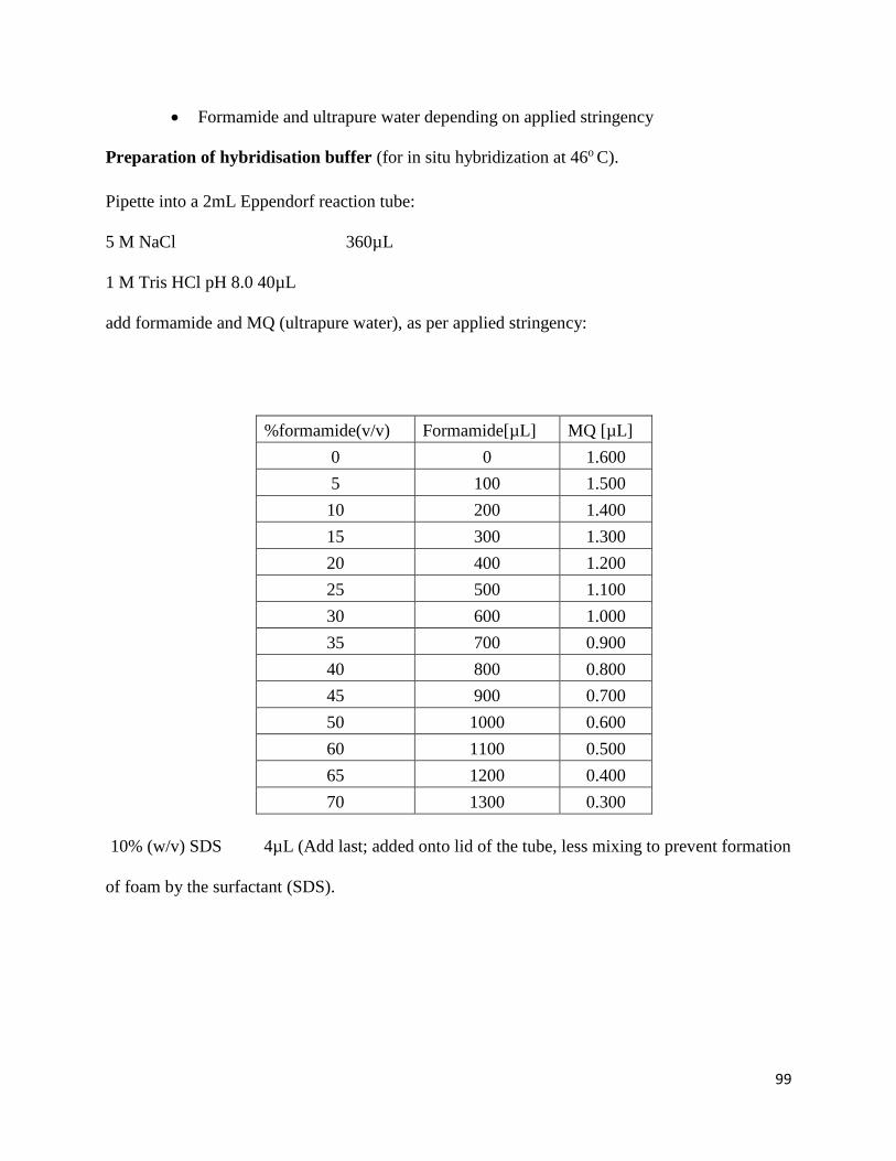
99
Formamide and ultrapure water depending on applied stringency
Preparation of hybridisation buffer (for in situ hybridization at 46o C).
Pipette into a 2mL Eppendorf reaction tube:
5 M NaCl 360µL
1 M Tris HCl pH 8.0 40µL
add formamide and MQ (ultrapure water), as per applied stringency:
%formamide(v/v) Formamide[µL] MQ [µL]
0 0 1.600
5 100 1.500
10 200 1.400
15 300 1.300
20 400 1.200
25 500 1.100
30 600 1.000
35 700 0.900
40 800 0.800
45 900 0.700
50 1000 0.600
60 1100 0.500
65 1200 0.400
70 1300 0.300
10% (w/v) SDS 4µL (Add last; added onto lid of the tube, less mixing to prevent formation
of foam by the surfactant (SDS).
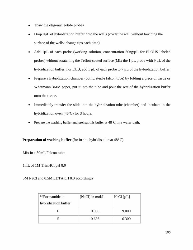
100
Thaw the oligonucleotide probes
Drop 9µL of hybridization buffer onto the wells (cover the well without touching the
surface of the wells; change tips each time)
Add 1µL of each probe (working solution, concentration 50ng/µL for FLOUS labeled
probes) without scratching the Teflon-coated surface (Mix the 1 µL probe with 9 µL of the
hybridization buffer. For EUB, add 1 µL of each probe to 7 µL of the hybridization buffer.
Prepare a hybridization chamber (50mL sterile falcon tube) by folding a piece of tissue or
Whatmann 3MM paper, put it into the tube and pour the rest of the hybridization buffer
onto the tissue.
Immediately transfer the slide into the hybridization tube (chamber) and incubate in the
hybridization oven (46oC) for 3 hours.
Prepare the washing buffer and preheat this buffer at 48oC in a water bath.
Preparation of washing buffer (for in situ hybridisation at 48o C)
Mix in a 50mL Falcon tube:
1mL of 1M Tris/HCl pH 8.0
5M NaCl and 0.5M EDTA pH 8.0 accordingly
%Formamide in
hybridization buffer
[NaCl] in mol/L NaCl [µL]
0 0.900 9.000
5 0.636 6.300
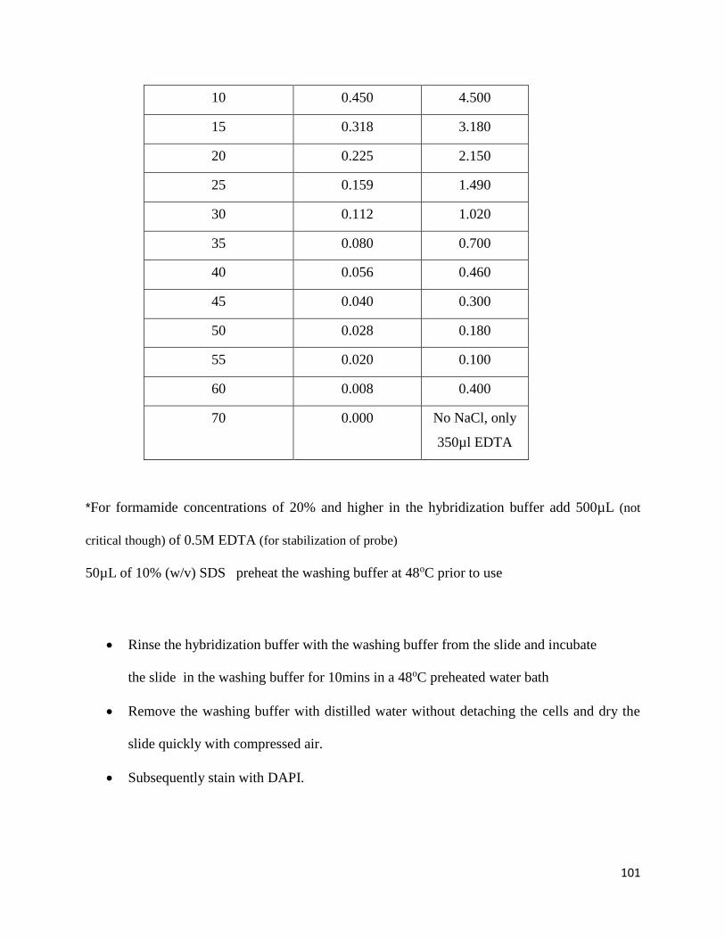
101
10 0.450 4.500
15 0.318 3.180
20 0.225 2.150
25 0.159 1.490
30 0.112 1.020
35 0.080 0.700
40 0.056 0.460
45 0.040 0.300
50 0.028 0.180
55 0.020 0.100
60 0.008 0.400
70 0.000 No NaCl, only
350µl EDTA
*For formamide concentrations of 20% and higher in the hybridization buffer add 500µL (not
critical though) of 0.5M EDTA (for stabilization of probe)
50µL of 10% (w/v) SDS preheat the washing buffer at 48oC prior to use
Rinse the hybridization buffer with the washing buffer from the slide and incubate
the slide in the washing buffer for 10mins in a 48oC preheated water bath
Remove the washing buffer with distilled water without detaching the cells and dry the
slide quickly with compressed air.
Subsequently stain with DAPI.
102
DAPI staining
(Hicks et al., 1992; modified)
Spread 10 µL of DAPI (0.25 µg/mL) on each well and allow to stain for 5 minutes.
Wash the slides with 1 X PBS and allow to air dry.
Mount the slide with Vectashield Mounting Media (Vector Laboratories,
Burlingame) and cover with a cover slide.
View immediately.
103
Appendix 6: SEM
Concentration of filaments:
Aeration and RAS samples were filtered using sieves of descending pore sizes (300µm;
150µm; 100µmand 75µm).
Spot filtrates on glass coverslips.
air dried at room temperature.
Sputter coating
(Li et al., 2005)
Mount glass coverslips with sample onto aluminum stubs
Sputter coat with gold.
104
Appendix 7: TEM
Harvesting of cells:
Two milliliter filtered sample was transferred to an Eppendorf tube.
centrifuged at 7500 rpm for 5 mins
The supernatant was discarded and the pellet was fixed in buffered 2.5 % Glutaraldehyde
for 24 h.
Post fixation
(William and Unz- 1985)
Osmium tetroxide fixation incubated at room temperature for 1 hour.
Phosphate buffer washes for 5 min repeated 3 times.
Acetone series dehydration :
(2x 5 min. 30% Acetone)
(2x 5 min. 50% Acetone)
(2x 5 min. 75% Acetone)
(2x 10 min. 100% Acetone).
105
Infiltration and Polymerization
(Jang et al.,2014)
Equal parts Resin and Acetone infiltration for 4 hours.
Whole resin incubation for 18-24 hours.
Orientation of specimen in whole mould resin
Place in oven for polymerization at 70°C for 8 hours.